Embed Size (px)
Citation preview
pubs.acs.org/jmcPublished on Web 08/09/2010r 2010 American Chemical Society
J. Med. Chem. 2010, 53, 6421–6431 6421
DOI: 10.1021/jm100621s
Structural and Functional Basis of Resistance to Neuraminidase Inhibitors of Influenza B Viruses†
Aaron J. Oakley,‡,§ Susan Barrett,‡ Thomas S. Peat,‡ Janet Newman,‡ Victor A. Streltsov,‡ Lynne Waddington,‡
Takehiko Saito, ),^ Masato Tashiro, ) and Jennifer L. McKimm-Breschkin‡,*
‡CSIRO Materials Science and Engineering, Parkville, 343 Royal Parade, Parkville, Victoria, 3052, Australia, §University of Wollongong,New South Wales, 2522, Australia, )WHO Collaborating Center for Reference and Research on Influenza, National Institute of InfectiousDiseases, Toyama 1-23-1 Shinjuku-ku, Tokyo 162-8640, Japan, and ^National Institute for Animal Health, Tsukuba City, Ibaraki, Japan
Received May 21, 2010
We have identified a virus, B/Perth/211/2001, with a spontaneous mutation, D197E in the neuramini-dase (NA), which confers cross-resistance to all NA inhibitors. We analyzed enzyme properties of theD197 and E197 NAs and compared these to a D197N NA, known to arise after oseltamivir treatment.Zanamivir and peramivir bound slowly to the wild type NA, but binding of oseltamivir was more rapid.The D197E/N mutations resulted in faster binding of all three inhibitors. Analysis of the crystalstructures of D197 and E197 NAs with and without inhibitors showed that the D197E mutationcompromised the interaction of neighboring R150 with the N-acetyl group, common to the substratesialic acid and all NA inhibitors. Although rotation of the E275 in the NA active site occurs uponbinding peramivir in both the D197 and E197 NAs, this does not occur upon binding oseltamivir in theE197 NA. Lack of the E275 rotation would also account for the loss of slow binding and the partialresistance of influenza B wild type NAs to oseltamivir.
Introduction
The influenza virus neuraminidase (NA,a EC 3.2.1.18)functions in virus infection to remove sialic acid from recep-tors present on the surface of host cells. In the absence of NAactivity, the ability of progeny virions to spread to uninfectedcells is compromised. The structure of the catalytic headgroupof influenzaANAhas been known since 1983,1,2 consisting ofindividual subunits of six-bladed β-propellers that form aboxlike tetramer with dimensions 100 A � 100 A � 60 A.Structures of the influenza B/Beijing/1/873,4 and B/Lee5 NAsshowed that the tetrameric head/β-propeller topology of theinfluenza-A NAs was conserved in influenza B NAs.
Analysis of NA crystal structures in complex with thesubstrate sialic acid resulted in the development of zanamivir(1)6 and oseltamivir (oseltamivir carboxylate is the activeingredient used here (2)).7 A further inhibitor, peramivir (3),is in clinical trials8,9 (Figure 1). These compounds are activeagainst all influenza A and B viruses. While previously therehave been reports of resistance from both influenza A and Bviruses isolated from both immunocompromised and immu-nocompetent patients after treatment with 2,10-14 more re-cently the global spread of seasonal influenza AH1N1 strainsresistant to 2
15,16 has been observed, although this appears tobe unrelated to the use of 2. Furthermore, resistance to 2 is
emerging in strains of the pandemic H1N1/09 viruses eitherwith or without treatment or prophylaxis.17,18 In contrast,resistance after 1 treatment has only been reported in animmunocompromised patient infected with an influenza Bstrain.19 Influenza B viruses with a D197N mutation (D198N2 numbering) have been isolated from an immunocompro-mised patient treated with 210 and arisen either spontaneouslyor by possible transmission from a treated patient.13 TheD197N mutant NA shows decreased binding to both 1 and2, thus demonstrating the importance ofD197 in the influenzaB NAs for tight binding of the NA inhibitors. Unlike otherresidues that confer resistance, D197 is not absolutely con-served across influenza A and B NAs, as analyses of NAsequences in the databases show thatwild type influenzaAN7and N9 subtype NAs have N197. Residue D197 does notinteract directly with substrate or inhibitor in the NA butengages in a salt bridge interaction with R150 (R152 N2numbering), which forms a hydrogen bond with the N-acetylgroup of sialic acid and the NA inhibitors. An influenza Bvirus with an R150K NA mutation was isolated after pro-longed treatment of an immunocompromised child with 119
with a significant impact on enzyme activity and cross-resis-tance to other NA inhibitors, clearly demonstrating the im-portance of interactions of the R150 with the substrate andinhibitors.
The B/Perth/211/2001 (B/Perth) virus was isolated from aninfant with no history of treatment with or contact with NAinhibitors. The sample contained both wild type and mutantviruses with a D197E mutation in the NA. The mutant NAhad reduced sensitivity to 1, 2,and 3.20 When expressed ininsect cells, recombinant B/Perth wild type and mutant NAshad properties similar to those of the virus associatedNAs.Aswe were unable to culture these viruses in eggs, we describehere the use of this recombinant NA for structural studies.
†Coordinates and structure factors have been deposited in the PDBunder accession numbers 3K36 (B/PerthD), 3K37 (B/PerthD peramivir3 complex), 3K38 (B/Perth E), 3K39 (B/Perth/E 3 complex), and 3K3A(B/Perth E oseltamivir 2 complex).
*To whom correspondence should be addressed. Address: CSIROMaterials Science and Engineering, 343 Royal Parade, Parkville, 3052,Australia. Telephone: 613 9662 7257. Fax: 613 9662 7101. E-mail:[email protected].
aAbbreviations: FU, fluorescent unit; MUNANA, 4-methylumbel-liferyl-N-acetylneuraminic acid; NA, neuraminidase; KDN, 2,3-di-fluoro-2-keto-3-deoxy-D-glycero-D-galactononulosonic acid.

6422 Journal of Medicinal Chemistry, 2010, Vol. 53, No. 17 Oakley et al.
The clinical effectiveness of 2 against influenza B infectionin children is reported to be less than against influenza A.21,22
However, until now, data on the structure of an influenza BNA with oseltamivir bound have not been available. Wepresent here structures of B/Perth/211/2001 NAs with D(B/Perth D) or E (B/Perth E) at position 197 in the apo formand in complex with 3 and provide an insight into themechanism of resistance of mutations at D197. Furthermore,the structure of theB/PerthE complexwith 2 is presented, andfrom thesedatawepropose themechanismof reducedbindingof 2 in wild type influenza B NAs.
Results
Protein Purification. We wished to determine the impactsthat themutations atD197 had on the structure and functionof the mutant proteins in order to understand the mecha-nisms of resistance to all NA inhibitors. We previouslyreported that the native B/Perth wild type D197 and mutantE197 NAs expressed in insect cells had similar resistanceprofiles as the influenza virus associated NAs.20 However,the recombinant NAs had no tags to facilitate purification.Hence, we developed conditions for cleavage of the mem-brane anchor and stalk regions and for purification of theNA from the insect cells. Acetone fixation of the cells enabledus to store them with no effect on the enzyme function.Digestionwith either pronase or trypsin showed that optimalcleavage was obtained with trypsin at 2 mg/mL, betweenresiduesK69 andG70 in the stalk, comparable to cleavage ofNA heads from other influenza B viruses.3,23
After separation of the digests by Superose-12 and lentillectin affinity chromatography, PAGE analysis showed asingle band corresponding to the NA (Figure 2). Yields ofpurified protein were approximately 100 μg/L.
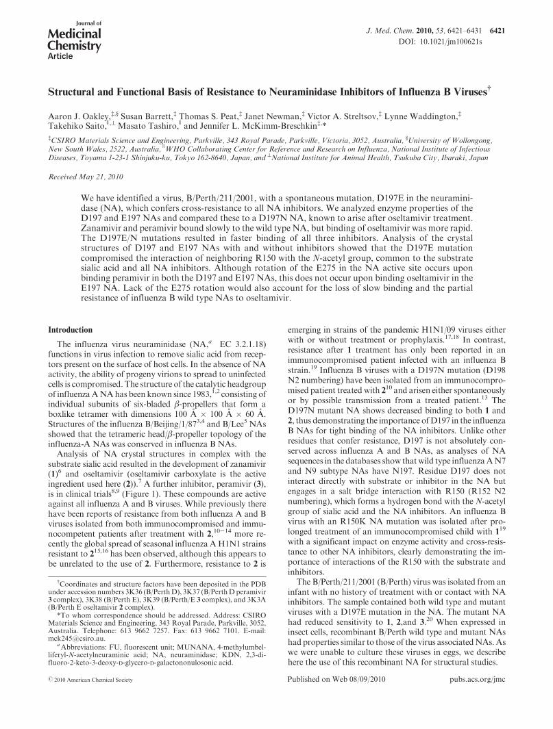
Electron Microscopy. Since this was the first time we hadattempted to use recombinant NA for structural studies,we used electron microscopy, as previously reported,24 toexamine the integrity of NA heads. The B/Perth D headswere clearly tetrameric but interestingly spontaneouslyformed two-dimensional arrays on the carbon substrate.Three different patterns were observed (Figure 3). The mostcommon was a simple square tessellation, and the secondform consisted also of orthogonal rows and columns butwith the square NA heads rotated through 45�, forming a“hound’s-tooth” pattern. The third and least common formhad NA heads oriented on their sides or edges, arranged inan open square lattice. These first two forms are a rareexample of actually seeing how the protein packs in crystal-line arrays, corresponding to crystallographic arrangementssubsequently detected by X-ray crystallography.
Enzyme Studies. We have previously shown that theB/Perth E197 NA and the B/Yamagata N197 NA demon-strate cross-resistance to the NA inhibitors 1, 2, and 3.20,25,26
Some influenza NAs demonstrate time dependent bindingof the NA inhibitors often called slow binding.27 Many NAswith amutation in the active site are reported to have lost thisproperty, binding the inhibitors rapidly.27-29 Since D197does not directly interact with substrate or inhibitor, wetherefore were interested in whether the D197E or D197Nmutations affected the enzyme function or the rate of bindingof the inhibitors.
Comparisons of the activity of the purified recombinantB/Perth D197 and E197 NA proteins demonstrated that thespecific activity of the mutant E197 NA was approximately70% of that of the wild type D197 enzyme.
For comparing values for the Km, Ki, and IC50, we useddetergent extracts of each of the four influenza viruses grown
Figure 1. Neuraminidase inhibitors used in the study.
Figure 2. Purification of recombinant B/PerthNAheads by trypsindigestion of SF21 cells expressing the full lengthB/PerthNA: (A) gelfiltration profile of crude extract; (B) silver stained PAGE of gelfiltration fractions; (C) samples after lentil lectin affinity chroma-tography.
Article Journal of Medicinal Chemistry, 2010, Vol. 53, No. 17 6423
in cell culture, since we did not have recombinant NA fromeither the B/Gifu or B/Yamagata viruses. The B/Perth wildtype D197, mutant E197, and the wild type D197 B/Gifu NAs had similar Km values for MUNANA of 12.4 (4.2, 12.1( 3.7, and 10.5( 1.4μM, respectively.Although theD197N mutation has less impact on IC50 than the E197(Table 1), it had a slightly higher Km of 18.8 ( 4.5 μM.
We carried out two different experiments for each virus/drug combination to study the rate of inhibitor binding. Thefirst experiment had no preincubation with the inhibitors,which enabled us to examine the rate of association ofthe drug with the NA, and the second assay had a 30 min
preincubation, which enabled us to examine whether anyfurther association or dissociation of the NA-inhibitorcomplex occurred upon addition of substrate. Both used a60 min reaction with substrate. Others have used preincuba-tion times from 10 min to 2 h,6,29,30 and it is not clear whatimpact these preincubation times, or the subsequent reactiontimes with substrate, may have on the calculated IC50. Sincethe IC50 is also known to vary with substrate concentration,others have calculatedKi values in addition to the IC50, sinceKi values aremeant to be amore invariantmeasure of affinityfor an inhibitor independent of substrate concentration. Wetherefore compared IC50 and Ki values at each 10 mininterval between 10 and 60 min either with no preincubationor after preincubation with the inhibitors.
When the curves for the total FU versus elapsed time werecompared, there were three types of curves. The first type ofcurve (Figure 4A) seen after preincubation with 1 or 3 in thewild type D197 NAs showed a gradual increase in the rate ofreaction, indicating slow dissociation of inhibitor. The sec-ond type of curve where there was no preincubation ofinhibitor, with 1 and 3 in the wild type NAs, showed agradually decreasing rate of reaction (Figure 4B), indicatingslow association of the inhibitor. The third type of curvereached a constant rate during the reaction (Figure 4C) andwas seen with 2 in the D197 NAs and with all inhibitors withthe mutant NAs, indicating that rapid equilibrium had beenreached.
The effects these changing rates have on the IC50 and Ki
values are shown in Figure 5. For both wild type D197NAswithout preincubation there was a gradual decrease in IC50
corresponding to a slow association of both 1 and 3. Afterpreincubation with these inhibitors there was a gradualincrease in IC50, indicating slow dissociation. In contrastthere was rapid association of 2without preincubation withlittle change in IC50 after the first 20 min. There was alsorapid dissociation of 2 after preincubation. Although theinitial IC50 values were not that much higher than for 1,there was more than a 10-fold change over the 60 minreaction time. Because of the slow association of 3 and 1
in the wild type NAs, the final 60 min IC50 values with nopreincubation were still 6- to 19-fold higher than withpreincubation (Table 1), demonstrating that the inhibitorbinding had not yet reached equilibrium. In contrast, therewas less than a 2-fold difference between the 60 min nopreincubation and preincubation IC50 values for 2 in bothwild type NAs.
When the IC50 values are compared without preincuba-tion for all inhibitors for both mutant NAs, the graphs showthere is much less change in IC50 after the first 10 min. 2 alsoappears to bind even more rapidly to both mutant NAs thanto the wild type D197 NAs. There is also faster dissociation
Table 1. Comparison of IC50 Values of Wild Type and Mutant NAs after 60 min in MUNANA Based Enzyme Inhibition Assay with or withoutPreincubation with Inhibitorsa
IC50, nM
D197 Perth E197 Perth D197 Gifu N197 Yamagata
Zan Osel Per Zan Osel Per Zan Osel Per Zan Osel Per
no prec 167.4 144.2 27.8 433.8 659.7 75.2 87.0 65.0 13.0 163.9 182.5 28.1
fold resistanceb 2.6 4.6 2.7 1.9 2.8 2.2
prec 8.8 104.4 2.8 242.0 708.0 41.5 13.0 45.0 2.1 77.8 188.4 14.0
fold resistanceb 27.5 6.8 17.3 6.0 4.2 6.7
no pre/prec 19 1.4 9.9 1.8 0.90 1.8 6.7 1.4 6.2 2.1 1.0 2.0aResults are themean of duplicates. bFold resistance is the ratio of themutant to the wild type IC50 values.
cNopre or pre is the fold differences in thefinal IC50 values with no preincubation with inhibitor compared to 30 min of preincubation.
Figure 3. Electron micrographs of recombinant B/Perth D NAheads. Samples were negatively stained with uranyl acetate andshow two-dimensional arrays of NA heads: (A) simple squaretessellation; (B) “hound’s-tooth” pattern of orthogonal rows andcolumns but with the square NA heads rotated through 45�; (C) anopen square lattice with NA heads oriented on their sides or edges.Scale bars, 50 nm.

6424 Journal of Medicinal Chemistry, 2010, Vol. 53, No. 17 Oakley et al.
of 1 and 3 after the NAs are preincubated with inhibitor,compared to the wild type NAs. For all inhibitors for bothmutant NAs the ratios after 60 min of the no preincubationto preincubation are all within 2-fold, indicating much more
rapid equilibration of all inhibitors compared to the wildtype NAs.
The slow binding of the 1 and 3 in the wild typeD197NAsalso resulted in the Ki values continuing to change over the
Figure 4. Neuraminidase activity changes in the presence of inhibitor using a MUNANA based fluorescent assay. MUNANA was added todetergent treated wild type andmutant viruses either after a 30min preincubation or without incubation with inhibitors ranging from 10 000 to0.1 nM. Activity was monitored for 60 min after addition of substrate: (A) B/Perth D activity in 1 after 30 min of preincubation showing anincrease in rate with time, corresponding to slowdissociation; (B) B/PerthDactivity in 1without preincubation, showing a decrease in rate withtime, corresponding to slow association of 1; (C) B/Yamagata (D197N) activity in 1 without preincubation, showing a constant rate,corresponding to a rapid association of 1.
Figure 5. Effect of incubation times after addition of substrate and inhibitor on (a) IC50 and (b) Ki values. IC50 values were calculated aftereach 10min interval as the drug concentration causing 50% inhibition compared to the uninhibited control.Ki values were calculated based onthe slope between consecutive 10min time intervals. Final IC50 values after 60min for 1 and 2 in wild type B/Perth and B/Gifu NAs were muchhigher without preincubation compared to 30 min preincubation, reflecting the slow binding of these two inhibitors. There was much lesschange in IC50 values for the binding of 2 in the wild type and 1, 2, and 3 in the mutant NAs, demonstrating loss of slow binding. Conversely 2dissociated rapidly from the wild type NA, in contrast to slow dissociation of 1 and 3, and 1, 2, and 3 all dissociated more rapidly from themutant B/Perth E and B/Yamagata NAs. (b) Ki values also changed with time, reflecting the differences in the slow or fast binding ordissociation of the inhibitors, with less change seen with 2 in the wild type and 1, 2, and 3 in the mutant NAs.
Article Journal of Medicinal Chemistry, 2010, Vol. 53, No. 17 6425
60 min period, whereas for 2 in the wild type and all theinhibitors with themutantNAs theKi values stabilizedmuchmore quickly after the first 10-20 min period, as seen inFigure 5 and Tables 1 and 2. Therefore, the reaction timeaffected both the Ki and IC50 values. This means that the“fold resistance”, often used to describe how resistant theisolates are, will vary significantly during the course of thereaction, depending on whether the inhibitors are faster orslower binding or dissociating compared to the wild type.
Crystallography. The apo form of B/Perth D crystallizedin space group I4 and was solved by molecular replacementusing A/tern/Australia/G7OC/75N9NA (PDB code 7NN9)followed by automatic rebuilding using PHENIX; the Rand Rfree were 33% and 39%, respectively. Several cyclesof model building and refinement gave a high quality finalmodel (Table 3). Crystal contacts between subunits in theC-direction are formed entirely by carbohydrate-mediatedinteractions. A carbohydrate chain composed of GlcNAcand mannoside residues was appended to residue N284,consistent with known patterns ofN-linked oligosaccharidesin insect cell lines.31 The asymmetric unit contained twomonomers, each adjacent to a 4-fold crystallographic axis.Thus, two tetramers are generated from each of the two
monomers. The packing of tetramers in layers observed inthis crystal form appears similar to that observed in electronmicroscopy (Figure 3A). The subunit structure is similar topreviously determined influenza B NA structures. It adoptsthe classic β-propeller arrangement with six four-strandedβ-sheets. The B/Perth D structure superimposes with a rmsdof 0.32 A (over 388 CR atoms) and 0.28 A (over 385 CRatoms) with B/Lee/40 (PDB 1INV) and B/Beijing/1/87 (PDBcode 1NSB)NAs, respectively, illustrating the high degree ofstructural conservation in influenza B NAs. The active-siteresidues of all three strains are identical. The active site ofB/Perth D contained water molecules and a sulfate groupbound between the guanidinium moieties of R116, R292,and R374.
For the B/Perth E crystals, although the unit cell suggestedtetragonal symmetry, good merging statistics were obtainedonly in space group P1. The B/Perth E structure was solvedby molecular replacement using tetramers of B/Perth D as asearch model. MOLREP selected a radius of integrationof 61 A, and four tetramers were found with peak heightsof 21.4, 18.9, 18.0, and 15.3 σ. Solutions for all four mono-mers were found in the translation function, resulting in aninitial model with R and Rfree factors of 31.2% and 31.1%.
Table 2. Comparison of Ki Values of Wild Type and Mutant NAs after 60 min in a MUNANA Based Enzyme Inhibition Assay with or withoutPreincubation with Inhibitorsa
Ki, nM
D197 Perth E197 Perth D197 Gifu N197 Yamagata
Zan Osel Per Zan Osel Per Zan Osel Per Zan Osel Per
no pre 7.1 9.9 1.2 43.8 84.0 8.3 4.6 6.0 0.7 22.8 53.4 3.2
fold resistanceb 6.2 8.5 6.9 5.0 8.9 4.6
pre 8.7 16.6 0.6 48.0 82.0 8.2 3.9 6.7 0.3 33.5 60.7 2.3
fold resistanceb 5.5 4.9 13.7 8.6 9.1 7.7aResults are the mean of duplicates. bFold resistance is the ratio of the mutant to the wild type Ki values.
Table 3. X-ray Dataa
B/Perth D
peramivir
B/Perth D B/Perth E
peramivir
B/Perth E
oseltamivir
B/Perth E
X-ray source AS-PX-1b AS-PX-1b AS-PX-1b AS-PX-1b PF-17Ab
space group I4 I4 P1 P1 P1
unit cell
a, A 87.6 88.7 111.2 111.96 111.8
b, A 87.6 88.7 123.7 124.87 123.8
c, A 197.2 207.5 123.8 125.25 124.0
R, deg 90.0 90.0 90.0 90.0 90.0
β, deg 90.0 90.0 90.2 92.1 90.2
γ, deg 90.0 90.0 90.2 91.2 90.1
resolution range (A) 26.7-2.20 (2.32-2.20) 40.7-2.00 (2.11-2.00) 62-2.2 (2.32-2.20) 63-2.6 (2.74-2.6) 87-2.6 (2.74-2.6)
Rmergec (%) 12.2 (43.8) 12.8 (33.5) 10.2 (36.5) 17.6 (40.6) 10.4 (35.8)
ÆI/σ(I)æ 11.6 (2.7) 10.1 (3.8) 8.1 (2.6) 5.9 (2.9) 6.0 (1.9)
unique observations 34306 (4204) 47163 (7141) 316772 (45355) 201957 (28682) 199227 (29004)
completeness 91.5 (77.6) 87.6 (90.9) 94.6 (92.7) 96.9 (94.3) 97.4 (96.7)
multiplicity 4.5 (2.9) 4.6 (4.1) 2.0 (1.8) 3.6 (3.2) 2.0 (2.0)
R, Rfreed 18.2, 23.9 17.1, 23.7 19.1, 22.0 19.8, 21.4 20.7, 22.8
Root-Mean-Square Deviations from Ideal Geometry
bond lengths (A) 0.014 0.014 0.017 0.016 0.016
bond angles (deg) 1.560 1.625 1.513 1.560 1.610
torsion angle (deg) 7.942 7.638 7.11 7.061 7.00
chiral volume (A3) 0.091 0.115 0.104 0.102 0.104
planar group (A) 0.006 0.006 0.007 0.006 0.007aNumbers in parentheses correspond to the highest resolution bin. bAS-MX1 is beamlineMX-1 at the Australian Synchrotron. PF-17A is beamline
17A at the Photon Factory, Tsukuba, Japan. c Rmerge =P
hkl
Pi|I(hkl)i - ÆI(hkl)æ|/
Phkl
PiI(hkl)i.
d R =P
hkl|Fo(hkl) - Fc(hkl)|/P
hkl|Fo(hkl)|, whereFo and Fc are the observed and calculated structure factors, respectively.

6426 Journal of Medicinal Chemistry, 2010, Vol. 53, No. 17 Oakley et al.
Refinement proceeded using tight NCS restraints. The 16-fold NCS-averaged maps were used for model building inCOOT. Refmac detected pseudomerohedral twinning, andcorrections were applied accordingly. The structure ofB/Perth E is highly similar to the B/Perth D structure inspite of the different crystal form. The tetramers form layersthat resemble the “hound’s-tooth” pattern described abovefrom electron microscopy (Figure 3B). A sulfate group isbound between the guanidinium moieties of R116, R292,and R374, and a yttrium ion of partial occupancy is boundadjacent to the sulfate group.These groups are displaced by 2or 3 upon soaking with those inhibitors (see below).
Crystals of B/Perth D soaked with 3 adopted similarpacking to the apoenzyme. 3 was included in the model ata late stage of refinement, and the final model is of highquality (Table 3).
Soaking inhibitors into crystals of B/Perth E provedchallenging, with relatively weak data being obtained forcrystals of this isozyme in the presence of 3 and 2 (Table 3).Nevertheless, the structure refinement and map interpreta-tion were aided by 15 noncrystallographic symmetry (NCS)restraints. Averaged electron density maps allowed for clearand unambiguous interpretation of the structures including,where present, inhibitors. Refinement of the B/Perth Ecomplex with 3 commenced using the apo-form of thismutant as the starting model (R = 40.5%, Rfree = 40.4%).3 was included in the model at a late stage of refinement.Similarly, refinement of theB/PerthE 2 complex commencedusing B/Perth E apo structure as the starting model (R =34.1%, Rfree = 35.3%). For both complexes tight NCSrestraints were used throughout. Pseudomerohedral twin-ning was detected in both cases and corrected in Refmac.Final model statistics for all models are in Table 3.
Electron density for all inhibitor complexes is unambig-uous. 3 binds in a similar fashion to related inhibitorsobserved in previously determined B/Beijing and B/Leestructures. The carboxylic acid group lies in the pocketformed by R292, R374, and R116. The guanidinium groupis buried in a pocket formed by E149 and E117. The sec-pentyl moiety is stacked against the E275-Cβ group (E276N2 numbering) (Figure 6B). Upon inhibitor binding, E275must rotate away from the inhibitor in a manner analogousto that described previously for B/Beijing NA in complexwith dihydropyranphenethylpropylcarboxamide.32 This in-hibitor has an ethyl moiety that corresponds to part of thesec-pentyl group of 3.
Surprisingly, rotation of E275 is not observed in theB/Perth E complex with 2, which does not form any hydro-phobic contacts with E275. Instead, the sec-pentyl groupmakes less favorable contacts with the charged portions ofR223, E275, and R292 (Figure 6E). In this structure, there isonly partial rotation of E275 away from the active site andhence only partial insertion of one arm of the sec-pentylmoiety into the resulting hydrophobic cleft (Figure 6D).
The D197E mutation in B/Perth affects the way thecarboxylic acid group of this residue engages with R150. Inthe structure of B/Perth D determined in the absence ofinhibitor, the carboxylic acid group of D197 engages side-onwith the guanidinium group of R150 as seen in most influ-enza B NA structures. In the B/Perth E apo structure, theguanidinium group ofR150 is rotated to engage in a stackinginteraction with the carboxylic acid moiety of E197. Fur-thermore, the guanidinium group has rotated 180� so thatthe Nη1-atom is now pointing away from the active site
(Figure 6C). In the structure of B/Perth E with 3, R150 hasrotated toward the active site relative to its position inthe apo structure and engages in a hydrogen bond with theN-acetyl oxygen atom via the Nε-atom. The distances oftheR150 toN-acetyl hydrogen bonds are longer in B/Perth Ecomparedwith P/PerthD: 3.4 A versus 2.7 A, respectively. Inthe complex of B/Perth Ewith 2, R150 is in the conformationobserved inB/PerthD,withatomNη1 engaging inahydrogenbond with the inhibitor N-acetyl oxygen atom (2.6 A). Whilethe distance is not significantly different from the equivalentdistance in the 3 complex, the R150 guanidinium group andN-acetyl group are no longer coplanar, indicating a geome-trically less favorable and hence weakened interaction.
Inhibition with 2,3-DifluoroKDN (4).As an additional wayof demonstrating that the reduced binding of the inhibitorsin the D197E and D197N NAs was due to altered interac-tions with theN-acetyl group of the sugar ring, we comparedinhibition of all four NAs with 2,3-difluoro-2-keto-3-deoxy-D-glycero-D-galactononulosonic acid 4.33 Although it is onlya weak inhibitor, it has no N-acetyl group; hence, valuesshould be similar for wild type and mutant NAs if thisinteraction can no longer occur.
There was no resistance to 4 with the mutant NAs com-pared to the D197 wild type NA. In fact the IC50 for eachmutant was less than for thewild type pair, B/Perth E197NA19.4( 1.7 μM compared to the wild type 37.7( 1.7 μMandthe B/Yamagata N197 NA 41.6 ( 0.4 μM compared to theB/Gifu wild type of 134 ( 17 μM, respectively. This con-firmed that decreased sensitivity was due solely to alteredinteractions with the N-acetyl group.
Discussion and Conclusions
We have used structural and functional studies here to gainan understanding of the mechanism of resistance to theNA inhibitors of influenza B viruseswithmutations at residue197. Equally important, our studies provide insights into whyinfluenza B wild type NAs have reduced binding of 2 com-pared to influenza A NAs.
We demonstrate that although D197 does not interactdirectly with substrate or inhibitors, mutations of D197Eand D197N in influenza B alter binding of substrate and allthreeNA inhibitors 1, 2, and 3, as shownby decreased specificactivity and increased Ki and IC50 values. The D197E muta-tion also conferred greater resistance than the D197N muta-tion. We also demonstrate using a modified approach to theenzyme inhibition assay that the reaction time can signifi-cantly affect both theKi and IC50 differently for wild type andmutant NAs, depending on whether the inhibitor is fast orslow binding. Others6 have also reported variation in Ki overtime, due to the time dependent slow binding of 1. Theyobserved a 10-fold decrease in Ki over the course of theirreaction, with a 10 min preincubation and 15 min reactiontime after addition of substrate. Because of the shorter pre-incubation time in their case, 1 was obviously still binding,rather than in our case where after a 30 min preincubation westart to see dissociation. Thus, despiteKi values being thoughtto be more consistent, because of the variation in themethodsused by different laboratories6,34,35 and the impact of fast andslow binding with time of incubation, the Ki values wouldappear to be more suitable as relative values within a labora-tory for comparing enzymeproperties ofwild typeandmutantNAs rather than as absolute values that different laboratoriescan directly compare.

Article Journal of Medicinal Chemistry, 2010, Vol. 53, No. 17 6427
Wehave also shown that the end point IC50 experiment canbe adapted to “IC50 kinetics” experiments by simply monitor-ing changes in IC50 with time without the addition of stopsolution. Comparison of the changes in IC50 with preincuba-tion or no preincubation with inhibitor shows that thisapproach can provide additional information about impactsof mutations on the rates of inhibitor binding and dissocia-tion, compared to just a single end point IC50. This is an assaythat can be carried out in any laboratory with a modernfluorimeter, without the need for any additional equipment.
Laboratories globally compare IC50 values, and we are striv-ing to understand parameters that can affect the IC50 values toenable a better comparison of IC50 values from differentlaboratories. Clearly both the preincubation and incubationtimes are critical.
Structural analysis indicates that the effect of the D to Emutation at position 197 was to destabilize the interactionwith R150 and to reduce the stability of this crucial inhibitor-binding residue. It appears that theD197 toEmutant prefers aconformation in which the guanidinium group of R150 is
Figure 6. Comparisons of the active sites of B/Perth wild type and mutant NAs uncomplexed and with bound inhibitors (A, B) B/Perth wildtype D and (C, D, E) B/Perth mutant E structures. Apo (A, C) and 3-bound (B, D) forms are shown. B/Perth E in complex with 2 is shown (E).(F) A model of the D197N mutant based on the wild-type B/Perth structure is shown. Active-site residues are shown in stick form and thebackbone in cartoon form. Arrow shows rotation of the E275 upon binding of 3.

6428 Journal of Medicinal Chemistry, 2010, Vol. 53, No. 17 Oakley et al.
rotated andmoved slightly out of the active site but can rotateback so as to engage with an inhibitor. However, this inter-action is less stable than in the D197 form. All clinicallyapproved inhibitors possess an N-acetyl group that interactswith R150 or its equivalent and are potentially susceptible tothis mechanism of drug resistance. This is consistent withthe cross-resistance seen to 1, 2, and 3. Lack of resistanceto another inhibitor without the N-acetyl group, 4, alsoconfirmed that resistance was due to altered binding to theN-acetyl group.
The mechanism of resistance in B/Perth E is different fromthat observed in N1 and N9 NAs to date.27,35-37 In both theN1 NA with an H274Y mutation and the N9 NA with anR292K mutation the altered binding is due to altered inter-actions of the E276 (N2 numbering) with the isopentyl ethergroupon 2. The influence of thesemutations on the binding of1 is less pronounced, as the side chain of E276 is able tomaintain favorable interactions with the glycerol side chain.In contrast, reducedbindingof 1withanE119GmutantNA ispartly due to reduced interactionswith the guanidiniumgroupat the 4 position on the sugar ring.27 The reduced binding inthe D197 mutant NAs is due to altered interactions with yetanother part of the ring, theN-acetyl group, which is commonto all inhibitors and substrate. The viability of the D197Nmutation demonstrates that a salt bridge between this residueand R150 is not essential for function. We anticipate that theeffect of the D197N mutation in B NAs is to weaken theinteraction of this residue with the R150 guanidinium groupthrough the elimination of the salt bridge interaction of D197and R150. N197 could still interact through a hydrogen bondbetween the Oε1 atom and the Nε or Nη2 groups of R150.This will have a similar effect to the D197Emutation in that itaffords more flexibility to the R150 side chain and weakensthe interaction with the N-acetyl group of the inhibitors(modeled in Figure 6F).
While NAIs are described as time dependent slow bindinginhibitors, we have shown here that both D197E and D197Nlead to loss of slowbinding of 1, 2, and 3. Loss of slow bindingis generally associated with mutations in the NA active site,leading to NA inhibitor resistance.27 One proposed mechan-ism of slow binding is due to the need for the rotation of theE27530 in inhibitors with the modified glycerol side chain.29,38
A proposed mechanism for the slow binding of 1 is the slowrelease of a water molecule by the guanidinium group.39 Thiswould also apply to 3 which shares this group. However, wesee here a loss of slow binding in the NAs with mutations atD197, remote from the position occupied by the guanidiniumgroups in both 3 and 1, and rotation of the E275 still occursupon binding 3. Thus, slow binding of the NA inhibitors isclearly affected bymore than interactions in either the vicinityof the 40-guanidino group or the rotation of the E275.
Although in the E197 mutant NA the binding of 1 wasreduced by nearly 30-fold compared to about 7-fold reductionfor 2, the overall IC50 for 2was higher. This is due to the lowersensitivity, or higher IC50, of the wild type B/Perth NA for 240
before the additional mutation.13 As the levels of 1 deliveredto the upper respiratory tract after 10 mg doses are estimatedto be up to 10 000 nmol/L,41 this would still be more than50-fold higher than the IC50 of the E197 enzyme. In con-trast the plasma levels of 2 are estimated to range from 400 to1200 nmol/L,42 and levels in saliva are estimated tobe less than5%of plasma levels.43 Thus, with a potential level in the upperrespiratory tract of only 20-60 nM,44 efficacy of 2 against asimilar D197E mutant strain could be significantly reduced.
In addition to loss of slow binding of the mutant NAs to 1,2, and 3, our enzyme analyses show here that there is a loss ofslow binding of 2 to the wild type B/Perth and B/Gifu D197enzymes compared to binding of 1 and 3, as well as fasterdissociationof 2. This is consistentwith the lower sensitivityorpartial resistance of wild type influenza B strains to 2 inenzyme assays compared to influenza A strains, especially inthe MUNANA assay where the IC50 values are around 12-70 nM (compared to the NA-Star assay, ∼2-11 nM40) andcompared to an IC50 of around 0.5-2 nM for influenza Astrains in both assays. The loss of slow binding is consistentwith the observations of Baum and colleagues,29 althoughKati et al. had described 2 to be also slow binding in influenzaB viruses.30
Consistent with our enzyme observations, we also impor-tantly present structural evidence to explain the partial resis-tance of influenza B NAs to 2. Upon binding 2, residue E275ofB/PerthNA fails to rotate to allowbinding of the sec-pentylmoiety to the aliphatic portion of this residue as observed inthe equivalent residue (E276) in N1 and N9 NAs.36 Rotationof this residue is necessary for high affinity binding of 2, andfailure to occur is consistent with resistance to 2, seen in othermutantNAs.35,36Whilewewereunable toobtain the structureof B/PerthDwith 2, a previous publication also describes lackof full rotationof theE275 in theB/Leewild typeNA.45 Itmaybe a general feature of type B NAs that this part of the activesite is more rigid and E275 is less able to rotate to accom-modate hydrophobic groups, although rotation does occurupon binding 3. The floor of the active site of B-type NAs hasbeen described as being more sterically crowded than forA-type enzymes, indicating that residues in type-B NAmightbe tightly constrained to the observed positions in the un-complexed enzyme.32 The observations presented here appearto confirm this view.
We conclude that the rotation of residue E275 needed forhigh affinity binding of 2 does not occur in the current strainsof influenza B wild type NAs, and this would correlate withthe loss of slowing binding of 2, the higher IC50 values seenespecially in the MUNANA assay, and possible decreasedclinical efficacy of 2 in children.21,22
Experimental Section
Viruses and Inhibitors. Isolation of the B/Perth viruses withwild type D197 NA and mutant E197 NA has been previouslydescribed.20 We also wanted to investigate how the D197Nmutation affected the NA enzyme properties. For this purposewe obtained the B/Yamagata/186/05 virus with a D197Nmuta-tion in the NA,46 but as this has several other NA sequencedifferences compared to the B/Perth, we obtained a control forthis virus NA which only had a single amino acid difference inthe stalk region, B/Gifu/11/2005. Both viruses were obtainedfrom the NIID, Tokyo, Japan. All virus stocks were seriallyplaque purified in MDCK cells.
1, 2, and 3 were synthesized by GSK, (Stevenage, U.K.).2-Keto-3-deoxy-D-glycero-D-galactononulosonic acid (4) (2,3, di-fluoroKDN)33was provided byDr.AndrewWatts (University ofBath, U.K.) (Figure 1). This is a sialic acid based inhibitor butlacks the N-acetyl group. Serial log10 dilutions of inhibitors wereprepared inwater for inhibition assays ranging from0.01 to10 000nM for 1, 2, and 3 and from 0.01 to 10 000 μM for 4.
Protein Expression and Purification. The full length B/Perthwild type D197 and mutant E197 NAs were expressed in Sf21insect cells as previously described.20 An amount of 4 L of cellsat (1-2) � 106 cells/mL was infected with a multiplicity ofinfection of 1.5 plaque forming units per cell and were harvestedat day 4, when about 30%cell death hadoccurred. The cells were

Article Journal of Medicinal Chemistry, 2010, Vol. 53, No. 17 6429
recovered by centrifugation and resuspended in TBS buffer at40� concentration.An equal volume of acetonewas added to fixthe cells, and these were stored on ice until required.
After removal of the acetone and being washed three times,the cells were resuspended in Tris-buffered saline to a density of1� 108 cells/mL. Cells were digestedwith trypsin (Worthington,TPCK) or Pronase (Calbiochem) at concentrations from 0.1 to2 mg/mL for 2 h at 37 �C to remove the NA. The cells werecentrifuged at 14 000 rpm for 10 min. NA was recovered in thesupernatant, and the cell pellet was resuspended in TBS andredigested. The pooled supernatants were concentrated in anAmicon 8050 stirred cell concentrator using a Pall 30KMWCOpolysulfone filter. The concentrated NA was separated fromother proteins in the trypsin digest by gel filtration using Super-ose 12 in TBS.47 Activity of the fractions was determined by the4-methylumbelliferyl-N-acetylneuraminic acid (MUNANA,Sigma-Aldrich) fluorescent enzyme assay.48,49 The active frac-tions were collected, analyzed by SDS-PAGE, and concen-trated down again. These were further purified by runningseveral times over a Lentil Lectin Sepharose 4B column(Amersham Biosciences) and eluting with 100 mM methyl-R-D-mannopyranoside in 20 mM Tris-Cl, pH 7.4, 0.5 M NaCl(Lancaster).
The activity of the fractions was again checked by theMUNANA assay, and the purity was checked by SDS-PAGEand silver staining. The purest active fractions of bothNAswereconcentrated to 4.5 mg/mL for crystallization trials.
Electron Microscopy. Purified B/Perth D NA was examinedby negative staining as follows. The protein was diluted to0.2 mg/mL in TBS. Then 300-mesh copper grids were coatedwith a thin carbon film and glow-discharged in nitrogen for 30 s.The 5-10μLaliquots of the samplewere pipetted onto the grids,and after 1 min of adsorption time, excess solution was drawnoff usingWhatman 541 filter paper. The grids were washed with5 μL of TBS, and the grid was then stained with 2% uranylacetate and was air-dried. The grids were examined in a Tecnai12 transmission electron microscope (FEI, Eindhoven, TheNetherlands) at an operating voltage of 120 kV, and imageswere recorded using aMegaview III CCD camera andAnalySIScamera control software (Olympus).
Enzyme Studies. (i) Specific Activity. Activities of purifiedsamples of recombinant B/Perth D197 and E197 NAs weretitrated in the MUNANA based NA enzyme assay.48 Relativespecific activity was calculated based on units of fluorescenceper microgram of protein.
(ii) Km and Ki. Km and Ki values were calculated usingviruses solubilized by the addition of CHAPS (3-[(3-cholamido-propyl)dimethylammonio]-1-propanesulfonate) to a final vo-lume of 1%. Each of the extracts was titrated in theMUNANAbased enzyme assay to determine similar final values of fluor-escent units without the addition of stop solution, since thisenabled continual monitoring of the reactions. The rate ofhydrolysis of MUNANAwas measured at substrate concentra-tions ranging from 6.25 to 200 μM, with readings taken everyminute in a Victor 2 (Wallac) or BMG FLUOstar Optimareader. Experiments were carried out in duplicate and repeatedfour times. The maximum slope for each reaction was deter-mined by comparing the slopes over different overlapping timeintervals. Initial velocities of the reactions were then calculatedby measuring the maximum slopes plotted as a function ofsubstrate concentrations. The Michaelis Menten constant, Km,which represents the affinity of the enzyme for substrate, wascalculated using a nonlinear regression function in GraphPadPrism.
While theNA inhibitors are competitive inhibitors, somewildtype influenza NAs are described as time dependent or slowbinders of the inhibitors, and some mutant NAs have lost thisslow binding property. We followed the kinetics of inhibitorbinding two ways, with preincubation of inhibitor for 30 min orwithout any preincubation, followed by 60 min of incubation
with the MUNANA substrate for both assays. All experimentswere carried out in duplicate. We used a constant substrateconcentration of 100 μM MUNANA and inhibitor concentra-tions ranging from 10000 to 0.01 nM. Fluorescent readingswere taken every minute in a BMG FLUOstar Optima readerfor 60 min. Graphs of concentration of inhibitor versus %enzyme inhibition compared to the control were plotted foreach 10 min data set. The IC50 was calculated as the concentra-tion of inhibitor resulting in a 50% reduction in enzyme activitycompared to the control for each 10min time point. By use of theKm and the substrate concentration, theKi can also be calculatedusing nonlinear regression and one-site competitive binding,using the equation of Cheng and Prusoff50 Ki = IC50/(1 þ[substrate]/Km). Aswe had calculated theKm,we then calculatedKi values in Graph Pad Prism using this method for each 10 mininterval. Experiments were carried out in duplicate.
Crystallization and X-ray Data Collection. All crystals weregrown at the Bio21 Collaborative Crystallization Centre (www.csiro.au/c3). Either a Phoenix (Art Robbins Industries) ora Mosquito (TTP) dispensing robot was used to set up sittingdrops in 96-well SD-2 plates (IDEX Corp.). Plates were storedat 281 K in a Gallery 700 incubator and imaged with a MinstrelHT imaging system (Rigaku). A single commercial screen (TheJCSGþ Suite from Qiagen) was set up initially to determine ifthe B/Perth D protein concentration was appropriate for crys-tallization trials, using 0.2 μL droplets consisting of 50%protein solution mixed with 50% reservoir solution, andequilibrated against a reservoir of 50 μL. Small (<50 μm)crystals grew from PEG-based conditions in the JCSGþ Suitetrials, and subsequent crystallization trials were set up usingThe PACT Suites (Qiagen), as well as from screens designedaround the hits in these two commercial screens. X-ray data forflash-cooled crystals were collected at the Australian Synchro-tron beamline MX-1 using the Blu-ice software51 or PhotonFactory beamline 17A. MOSFLM52 was used for processingall diffraction images. SCALA53 was used to scale all diffrac-tion data.
B/Perth D protein (4.5 mg/mL) was mixed with reservoirsolution in a 1:1 ratio (drop volume 0.2 μL). X-ray data weremeasured from crystals grown at 8 �C in 0.2 M Na2SO4, 20%w/v PEG 3350, 0.1 M bis-Tris propane, pH 6.5. These crystalswere transferred to mother liquor with 10% v/v ethylene glycoland 10% v/v glycerol added just prior to flash-cooling to 100 K.Remaining crystals were used for soaking inhibitors (1, 2,and 3). A successful soak of 3 was performed by seeding somesolid compound into a drop containing crystals and allowing thesample to equilibrate for 10 days prior to flash-cooling to 100K.The well solution in this case was 0.2 M NaNO3, 20% w/vPEG3350, 0.1 M Bis-Tris propane, pH 6.5. We were not able toobtain crystals containing either 1 or 2 with the D197 protein.
B/Perth E (4.5 mg/mL) was used to grow crystals underconditions similar to those described above. The best crystalsfor diffraction experiments were grown in 12-17% w/v PEG3350, 0.2-0.3 M Na2SO4, and 5 mM YCl3. The complex ofB/Perth E with 3 was obtained by placement of the compounddirectly into the drop as described above for B/Perth D.The complex of B/Perth E with 2 was obtained by addingthe compound (5 mM) to the cryoprotectant (the same as forB/Perth D) prior to flash cooling.
Structure Solution and Refinement. Structures were solved bymolecular replacement using PHENIX54 and MOLREP.55 Allstructures were manually rebuilt using COOT56 and refined inREFMAC.57 The weighting of X-ray and geometric parametersin refinement and the type of NCS restraints were based on theireffects on Rfree cross-validation.
Acknowledgment. We thank Dr. Ross Fernley and PatPilling for assistance with protein purification and crystal-lization, Peter Schmidt for helpful discussions, the technicalstaff of the Bio21 C3 Centre for help with crystallization, the

6430 Journal of Medicinal Chemistry, 2010, Vol. 53, No. 17 Oakley et al.
Australian Synchrotron for access to MX-1, and the PhotonFactory (Japan) for access to beamline 17A. This access wassupported by the Australian Synchrotron Research Programwhich is funded by the Commonwealth of Australia underthe MNRF Program. This work was supported by GrantRO1A1062721 fromNIAID and grants fromGSKU.K. andAustralia. Its contents are solely the responsibility of theauthors and do not necessarily represent the official views ofthe Australian synchrotron, NIH, NIAID, or GSK.
References
(1) Varghese, J. N.; Laver, W. G.; Colman, P. M. Structure of theinfluenza virus glycoprotein antigen neuraminidase at 2.9 A reso-lution. Nature 1983, 303, 35–40.
(2) Colman, P. M.; Varghese, J. N.; Laver, W. G. Structure of thecatalytic and antigenic sites in influenza virus neuraminidase.Nature 1983, 303, 41–44.
(3) Burmeister, W. P.; Daniels, R. S.; Dayan, S.; Gagnon, J.;Cusack, S.; Ruigrok, R. W. Sequence and crystallization of influ-enza virus B/Beijing/1/87 neuraminidase. Virology 1991, 180, 266–272.
(4) Burmeister,W. P.; Ruigrok,R.W.; Cusack, S. The 2.2 A resolutioncrystal structure of influenza B neuraminidase and its complexwithsialic acid. EMBO J. 1992, 11, 49–56.
(5) Janakiraman, M. N.; White, C. L.; Laver, W. G.; Air, G. M.; Luo,M. Structure of influenza virus neuraminidaseB/Lee/40 complexedwith sialic acid and a dehydro analog at 1.8-A resolution: implica-tions for the catalytic mechanism.Biochemistry (Moscow) 1994, 33,8172–8179.
(6) von Itzstein,M.;Wu,W.Y.;Kok,G.B.; Pegg,M. S.;Dyason, J. C.;Jin, B.; Van Phan, T.; Smythe, M. L.; White, H. F.; Oliver, S. W.;Colman, P. M.; Varghese, J. N.; Ryan, D. M.; Woods, J. M.;Bethell, R. C.; Hotham, V. J.; Cameron, J. M.; Penn, C. R.Rational design of potent sialidase-based inhibitors of influenzavirus replication. Nature 1993, 363, 418–423.
(7) Kim, C. U.; Lew, W.; Williams, M. A.; Liu, H.; Zhang, L.;Swaminathan, S.; Bischofberger, N.; Chen., M. S.; Mendel,D. B.; Tai, C. Y.; Laver, W. G.; Stevens, R. C. Influenza neur-aminidase inhibitors possessing a novel hydrophobic interaction inthe enzyme active site:design, synthesis, and structural analysis ofcarbocyclic sialic acid analogues with potent anti-influenza activ-ity. J. Am. Chem. Soc. 1997, 119, 681–690.
(8) Babu, Y. S.; Chand, P.; Bantia, S.; Kotian, P.; Dehghani, A.;El-Kattan, Y.; Lin, T. H.; Hutchison, T. L.; Elliott, A. J.; Parker,C.D.;Ananth, S. L.; Horn, L. L.; Laver,G.W.;Montgomery, J. A.BCX-1812 (RWJ-270201): discovery of a novel, highly potent,orally active, and selective influenza neuraminidase inhibitorthrough structure-based drug design. J. Med. Chem. 2000, 43,3482–3486.
(9) BioCryst Peramivir (Neuraminidase Inhibitor). http://www.bio-cryst.com/peramivir.htm (accessed March 10, 2009).
(10) Gubareva, L. V. Molecular mechanisms of influenza virus resis-tance to neuraminidase inhibitors. Virus Res. 2004, 103, 199–203.
(11) de Jong,M.D.; Tran, T. T.; Truong,H.K.; Vo,M.H.; Smith,G. J.;Nguyen, V. C.; Bach, V. C.; Phan, T. Q.; Do, Q. H.; Guan, Y.;Peiris, J. S.; Tran, T. H.; Farrar, J. Oseltamivir resistance duringtreatment of influenza A (H5N1) infection. N. Engl. J. Med. 2005,353, 2667–2672.
(12) Ward, P.; Small, I.; Smith, J.; Suter, P.; Dutkowski, R. Oseltamivir(Tamiflu) and its potential for use in the event of an influenzapandemic. J. Antimicrob. Chemother. 2005, 55 (Suppl. 1), i5–i21.
(13) Hatakeyama, S.; Sugaya, N.; Ito, M.; Yamazaki, M.; Ichikawa,M.; Kimura, K.; Kiso,M.; Shimizu, H.; Kawakami, C.; Koike, K.;Mitamura,K.;Kawaoka,Y. Emergence of influenzaB viruseswithreduced sensitivity to neuraminidase inhibitors. JAMA, J. Am.Med. Assoc. 2007, 297, 1435–1442.
(14) Stephenson, I.; Democratis, J.; Lackenby, A.; McNally, T.; Smith,J.; Pareek, M.; Ellis, J.; Bermingham, A.; Nicholson, K.; Zambon,M. Neuraminidase inhibitor resistance after oseltamivir treatmentof acute influenza A and B in children. Clin. Infect. Dis. 2009, 48,289–296.
(15) Hurt, A. C.; Ernest, J.; Deng, Y. M.; Iannello, P.; Besselaar, T. G.;Birch, C.; Buchy, P.; Chittaganpitch, M.; Chiu, S. C.; Dwyer, D.;Guigon, A.; Harrower, B.; Kei, I. P.; Kok, T.; Lin, C.; McPhie, K.;Mohd, A.; Olveda, R.; Panayotou, T.; Rawlinson, W.; Scott, L.;Smith, D.; D’Souza, H.; Komadina, N.; Shaw, R.; Kelso, A.; Barr,I. G. Emergence and spread of oseltamivir-resistant A(H1N1)influenza viruses in Oceania, South East Asia and South Africa.Antiviral Res. 2009, 83, 90–93.
(16) Meijer, A.; Lackenby, A.; Hungnes, O.; Lina, B.; van-der-Werf, S.;Schweiger, B.; Opp, M.; Paget, J.; van-de-Kassteele, J.; Hay, A.;Zambon, M. Oseltamivir-resistant influenza virus A (H1N1),Europe, 2007-08 season. Emerging Infect. Dis. 2009, 15, 552–560.
(17) Wang, B.; Dwyer, D. E.; Blyth, C. C.; Soedjono, M.; Shi, H.;Kesson, A.; Ratnamohan, M.; McPhie, K.; Cunningham, A. L.;Saksena, N. K. Detection of the rapid emergence of the H275Ymutation associated with oseltamivir resistance in severe pandemicinfluenza virus A/H1N1 09 infections. Antiviral Res. 2010, 87, 16–21.
(18) Le, Q. M.; Wertheim, H. F.; Tran, N. D.; van Doorn, H. R.;Nguyen, T. H.; Horby, P. A community cluster of oseltamivir-resistant cases of 2009 H1N1 influenza.N. Engl. J. Med. 2010, 362,86–87.
(19) Gubareva, L. V.; Matrosovich, M. N.; Brenner, M. K.; Bethell,R. C.; Webster, R. G. Evidence for zanamivir resistance in animmunocompromised child infected with influenza B virus.J. Infect. Dis. 1998, 178, 1257–1262.
(20) Hurt, A. C.; Iannello, P.; Jachno, K.; Komadina, N.; Hampson,A. W.; Barr, I. G.; McKimm-Breschkin, J. L. Neuraminidaseinhibitor-resistant and -sensitive influenza B viruses isolated froman untreated human patient. Antimicrob. Agents Chemother. 2006,50, 1872–1874.
(21) Sugaya, N.; Mitamura, K.; Yamazaki, M.; Tamura, D.; Ichikawa,M.; Kimura, K.; Kawakami, C.; Kiso, M.; Ito, M.; Hatakeyama,S.; Kawaoka, Y. Lower clinical effectiveness of oseltamivir againstinfluenza B contrasted with influenza A infection in children. Clin.Infect. Dis. 2007, 44, 197–202.
(22) Sugaya,N.; Tamura,D.;Yamazaki,M.; Ichikawa,M.;Kawakami,C.; Kawaoka, Y.; Mitamura, K. Comparison of the clinical effec-tiveness of oseltamivir and zanamivir against influenza virusinfection in children. Clin. Infect. Dis. 2008, 47, 339–345.
(23) Burmeister, W. P.; Baudin, F.; Cusack, S.; Ruigrok, R. W. Com-parison of structure and sequence of influenza B/Yamagata and B/Beijing neuraminidases shows a conserved “head” but much great-er variability in the “stalk” and NB protein. Virology 1993, 192,683–686.
(24) Sahasrabudhe, A.; Lawrence, L.; Epa, V. C.; Varghese, J. N.;Colman, P. M.; McKimm-Breschkin, J. L. Substrate, inhibitor,or antibody stabilizes the Glu 119 Gly mutant influenza virusneuraminidase. Virology 1998, 247, 14–21.
(25) Hurt,A.C.;McKimm-Breschkin, J. L.;McDonald,M.; Barr, I.G.;Komadina, N.; Hampson, A. W. Identification of a human influ-enza type B strain with reduced sensitivity to neuraminidaseinhibitor drugs. Virus Res. 2004, 103, 205–211.
(26) Tashiro, M.; McKimm-Breschkin, J. L.; Saito, T.; Klimov, A.;Macken, C.; Zambon, M.; Hayden, F. G. Surveillance for neur-aminidase-inhibitor-resistant influenza viruses in Japan, 1996-2007.Antiviral Ther. 2009, 14, 751–761.
(27) Blick, T. J.; Tiong, T.; Sahasrabudhe, A.; Varghese, J. N.; Colman,P. M.; Hart, G. J.; Bethell, R. C.; McKimm-Breschkin, J. L.Generation and characterization of an influenza virus neuramini-dase variant with decreased sensitivity to the neuraminidase-spe-cific inhibitor 4-guanidino-Neu5Ac2en. Virology 1995, 214, 475–484.
(28) McKimm-Breschkin, J. L.; Sahasrabudhe, A.; Blick, T. J.;McDonald, M.; Colman, P. M.; Hart, G. J.; Bethell, R. C.;Varghese, J. N. Mutations in a conserved residue in the influenzavirus neuraminidase active site decreases sensitivity toNeu5Ac2en-derived inhibitors. J. Virol. 1998, 72, 2456–2462.
(29) Baum, E. Z.;Wagaman, P. C.; Ly, L.; Turchi, I.; Le, J.; Bucher, D.;Bush, K. A point mutation in influenza B neuraminidase confersresistance to peramivir and loss of slow binding. Antiviral Res.2003, 59, 13–22.
(30) Kati, W. M.; Saldivar, A. S.; Mohamadi, F.; Sham, H. L.; Laver,W. G.; Kohlbrenner, W. E. GS4071 is a slow-binding inhibitor ofinfluenza neuraminidase from both A and B strains. Biochem.Biophys. Res. Commun. 1998, 244, 408–413.
(31) Kubelka, V.; Altmann, F.; Kornfeld, G.;Marz, L. Structures of theN-linked oligosaccharides of the membrane glycoproteins fromthree lepidopteran cell lines (Sf-21, IZD-Mb-0503, Bm-N). Arch.Biochem. Biophys. 1994, 308, 148–157.
(32) Taylor, N. R.; Cleasby, A.; Singh, O.; Skarzynski, T.; Wonacott,A. J.; Smith, P. W.; Sollis, S. L.; Howes, P. D.; Cherry, P. C.;Bethell, R.; Colman, P.; Varghese, J. Dihydropyrancarboxamidesrelated to zanamivir: a new series of inhibitors of influenza virussialidases. 2. Crystallographic and molecular modeling study ofcomplexes of 4-amino-4H-pyran-6-carboxamides and sialidasefrom influenza virus types A and B. J. Med. Chem. 1998, 41, 798–807.
(33) Watts, A.G.;Oppezzo, P.;Withers, S.G.;Alzari, P.M.; Buschiazzo,A. Structural and kinetic analysis of two covalent sialosyl-enzyme

Article Journal of Medicinal Chemistry, 2010, Vol. 53, No. 17 6431
intermediates onTrypanosoma rangeli sialidase. J. Biol. Chem. 2006,281, 4149–4155.
(34) Hart, G. J.; Bethell, R. C. 2,3-Didehydro-2,4-dideoxy-4-guanidi-no-N-acetyl-D-neuraminic acid (4-guanidino-Neu5Ac2en) is aslow-binding inhibitor of sialidase from both influenza A virusand influenza B virus. Biochem. Mol. Biol. Int. 1995, 36, 695–703.
(35) Collins, P. J.; Haire, L. F.; Lin, Y. P.; Liu, J.; Russell, R. J.;Walker,P. A.; Skehel, J. J.; Martin, S. R.; Hay, A. J.; Gamblin, S. J. Crystalstructures of oseltamivir-resistant influenza virus neuraminidasemutants. Nature 2008, 453, 1258–1261.
(36) Varghese, J. N.; Smith, P. W.; Sollis, S. L.; Blick, T. J.;Sahasrabudhe,A.;McKimm-Breschkin, J. L.; Colman, P.M.Drugdesign against a shifting target: a structural basis for resistance toinhibitors in a variant of influenza virus neuraminidase. Structure1998, 6, 735–746.
(37) Smith, B. J.; McKimm-Breschkin, J. L.; McDonald, M.; Fernley,R. T.; Varghese, J. N.; Colman, P. M. Structural studies of theresistance of influenza virus neuramindase to inhibitors. J. Med.Chem. 2002, 45, 2207–2212.
(38) Wang, M. Z.; Tai, C. Y.; Mendel, D. B. Mechanism by whichmutations at His274 alter sensitivity of influenza A virus N1neuraminidase to oseltamivir carboxylate and zanamivir. Antimi-crob. Agents Chemother. 2002, 46, 3809–3816.
(39) Pegg, M. S.; von Itzstein, M. Slow-binding inhibition of sialidasefrom influenza virus. Biochem. Mol. Biol. Int. 1994, 32, 851–858.
(40) McKimm-Breschkin, J.; Trivedi, T.; Hampson, A.; Hay, A.;Klimov, A.; Tashiro,M.;Hayden, F.; Zambon,M.Neuraminidasesequence analysis and susceptibilities of influenza virus clinicalisolates to zanamivir and oseltamivir. Antimicrob. Agents Che-mother. 2003, 47, 2264–2272.
(41) Cass, L. M.; Brown, J.; Pickford, M.; Fayinka, S.; Newman, S. P.;Johansson, C. J.; Bye, A. Pharmacoscintigraphic evaluation oflung deposition of inhaled zanamivir in healthy volunteers. Clin.Pharmacokinet. 1999, 36 (Suppl. 1), 21–31.
(42) Oo, C.; Barrett, J.; Hill, G.; Mann, J.; Dorr, A.; Dutkowski, R.;Ward, P. Pharmacokinetics and dosage recommendations for anoseltamivir oral suspension for the treatment of influenza inchildren. Paediatr. Drugs 2001, 3, 229–236; erratum appears inPaediatr. Drugs 2001, 3 (4), 246 .
(43) Wattanagoon,Y.; Stepniewska,K.; Lindegardh,N.; Pukrittayakamee,S.; Silachamroon,U.;Piyaphanee,W.; Singtoroj,T.;Hanpithakpong,W.; Davies, G.; Tarning, J.; Pongtavornpinyo, W.; Fukuda, C.;Singhasivanon, P.; Day, N. P.; White, N. J. Pharmacokineticsof high-dose oseltamivir in healthy volunteers. Antimicrob. AgentsChemother. 2009, 53, 945–952.
(44) Morrison, D.; Roy, S.; Rayner, C.; Amer, A.; Howard, D.; Smith,J. R.; Evans, T. G. A randomized, crossover study to evaluate thepharmacokinetics of amantadine and oseltamivir administeredalone and in combination. PLoS One 2007, 2, No. e1305.
(45) Lew, W.; Chen, X.; Kim, C. U. Discovery and development of GS4104 (oseltamivir): an orally active influenza neuraminidase inhi-bitor. Curr. Med. Chem. 2000, 7, 663–672.
(46) Neuraminidase Inhibitor Susceptibility Network (NISN). Moni-toring of neuraminidase inhibitor resistance among clinical influ-enza virus isolates in Japan during the 2003-2006 influenzaseasons. Wkly. Epidemiol. Rec. 2007, 27, 149-150.
(47) McKimm-Breschkin, J. L.; Caldwell, J. B.; Guthrie, R. E.; Kortt,A. A. A new method for the purification of the influenza A virusneuraminidase. J. Virol. Methods 1991, 32, 121–124.
(48) Potier, M.; Mameli, L.; Belisle, M.; Dallaire, L.; Melancon, S. B.Fluorometric assay of neuraminidase with a sodium (4-methylum-belliferyl-alpha-D-N-acetylneuraminate) substrate. Anal. Biochem.1979, 94, 287–296.
(49) McKimm-Breschkin, J. L.; Blick, T. J.; Sahasrabudhe, A.; Tiong,T.;Marshall,D.;Hart,G. J.; Bethell, R.C.; Penn,C.R.Generationand characterization of variants of NWS/G70C influenza virusafter in vitro passage in 4-amino-Neu5Ac2en and 4-guanidino-Neu5Ac2en. Antimicrob. Agents Chemother. 1996, 40, 40–46.
(50) Cheng, Y.; Prusoff, W. H. Relationship between the inhibitionconstant (K1) and the concentration of inhibitor which causes 50%inhibition (I50) of an enzymatic reaction. Biochem. Pharmacol.1973, 22, 3099–3108.
(51) McPhillips, T. M.; McPhillips, S. E.; Chiu, H. J.; Cohen, A. E.;Deacon, A. M.; Ellis, P. J.; Garman, E.; Gonzalez, A.; Sauter,N. K.; Phizackerley, R. P.; Soltis, S. M.; Kuhn, P. Blu-Ice and theDistributed Control System: software for data acquisition andinstrument control at macromolecular crystallography beamlines.J. Synchrotron Radiat. 2002, 9, 401–406.
(52) Leslie, A. W. G. Recent changes to MOSFLM package forprocessing film and image plate data. Joint CCP4 ESF-EAMCBNewslett. Protein Crystallogr. 1992, 26.
(53) Evans, P. Scaling and assessment of data quality.ActaCrystallogr.,Sect. D: Biol. Crystallogr. 2006, 62, 72–82.
(54) Adams, P. D.; Grosse-Kunstleve, R. W.; Hung, L. W.; Ioerger,T. R.; McCoy, A. J.; Moriarty, N. W.; Read, R. J.; Sacchettini,J. C.; Sauter, N. K.; Terwilliger, T. C. PHENIX: buildingnew software for automated crystallographic structure determina-tion. Acta Crystallogr., Sect. D: Biol. Crystallogr. 2002, 58, 1948–1954.
(55) Vagin, A.; Teplyakov, A. MOLREP:an automated programfor molecular replacement. J. Appl. Crystallogr. 1997, 30, 1022–1025.
(56) Emsley, P.; Cowtan, K. Coot: model-building tools for moleculargraphics. Acta Crystallogr., Sect. D: Biol. Crystallogr. 2004, 60,2126–2132.
(57) Murshudov, G. N.; Vagin, A. A.; Dodson, E. J. Refinement ofmacromolecular structures by the maximum-likelihood method.Acta Crystallogr., Sect. D: Biol. Crystallogr. 1997, 53, 240–255.