Embed Size (px)
Citation preview

ii

iii

iv
1 INTRODUCTION ....................................................................................................................................... 1
1.1 MITOCHONDRIA ............................................................................................................................................ 1 1.2 PROTEIN TRANSLOCATION INTO MITOCHONDRIA .................................................................................................. 4
1.2.1 The TOM complex ............................................................................................................................ 6 1.2.2 Import of proteins into the matrix ................................................................................................... 7 1.2.3 Import of proteins into the inner mitochondrial membrane ........................................................... 9 1.2.4 Import of proteins into the intermembrane space ........................................................................ 12 1.2.5 Import of proteins into the outer mitochondrial membrane ......................................................... 12
1.3 THE TOB COMPLEX ...................................................................................................................................... 15 1.3.1 Introduction ................................................................................................................................... 15 1.3.2 Composition of the TOB complex .................................................................................................. 15 1.3.3 Topogenesis of mitochondrial ß-barrel proteins ........................................................................... 17
1.4 AIM OF THE PRESENT STUDY ........................................................................................................................... 19
2 RESULTS ................................................................................................................................................ 20
2.1 TOB55 IS EXPRESSED IN THREE DIFFERENT ISOFORMS DUE TO ALTERNATIVE SPLICING ................................................. 20 2.2 ISOLATION OF THE TOB COMPLEX ................................................................................................................... 23 2.3 COMPOSITION OF THE TOB COMPLEX .............................................................................................................. 26 2.4 TOB55, TOB38 AND TOB37 ARE PRESENT IN THE TOB COMPLEX IN A 1:1:1 STOICHIOMETRY .................................... 38 2.5 RECONSTRUCTION OF THE ISOLATED TOB COMPLEX BY CRYO-ELECTRON MICROSCOPY ............................................... 42 2.6 THE SUBUNITS OF THE TOB COMPLEX ARE TIGHTLY ASSOCIATED WITH THE OUTER MITOCHONDRIAL MEMBRANE ............ 52 2.7 THE ISOLATED TOB COMPLEX SHOWS SPECIFIC SUBSTRATE BINDING BEHAVIOR ........................................................ 54
3 DISCUSSION .......................................................................................................................................... 61
3.1 COMPOSITION OF THE TOB COMPLEX .............................................................................................................. 61 3.2 STRUCTURE OF THE TOB COMPLEX.................................................................................................................. 66 3.3 THE FUNCTIONAL MECHANISM OF THE TOB COMPLEX ......................................................................................... 69 3.4 ALTERNATIVE SPLICING OF TOB55 ................................................................................................................... 74 3.5 Α-HELICAL SUBSTRATES OF THE TOB COMPLEX .................................................................................................. 75
4 SUMMARY ............................................................................................................................................ 76
5 ZUSAMMENFASSUNG ........................................................................................................................... 77
6 MATERIAL AND METHODS .................................................................................................................... 78
6.1 MATERIALS ................................................................................................................................................. 78 6.1.1 Equipment ..................................................................................................................................... 78 6.1.2 Chemicals ...................................................................................................................................... 81 6.1.3 Preparation Kits ............................................................................................................................. 84 6.1.4 Medias and buffers........................................................................................................................ 84 6.1.5 Neurospora crassa strains ............................................................................................................. 89 6.1.6 Saccharomyces cerevisiae strain ................................................................................................... 91 6.1.7 Peptides ......................................................................................................................................... 91 6.1.8 Whitehead codes and molecular masses of selected proteins from Neurospora crassa .............. 94 6.1.9 Antibodies...................................................................................................................................... 94 6.1.10 Plasmids ......................................................................................................................................... 95
6.2 METHODS .................................................................................................................................................. 96 6.2.1 Cell biology .................................................................................................................................... 96 6.2.2 Molecular biology ........................................................................................................................ 100 6.2.3 Protein biochemistry ................................................................................................................... 103 6.2.4 Immunology ................................................................................................................................ 112 6.2.5 Cryo-electron microscopy ............................................................................................................ 115
7 ABBREVIATIONS .................................................................................................................................. 117
8 REFERENCES ........................................................................................................................................ 122
Introduction
1
1 Introduction
1.1 Mitochondria
Mitochondria are cell organelles which are found in virtually all eukaryotic cells (1). The
number of mitochondria in a cell varies widely by organism and tissue type, from only a
single mitochondrion to several thousand mitochondria (2, 3). These organelles have many
features in common with prokaryotes. As a result, they are believed to be originally derived
from endosymbiotic prokaryotes (4-7).
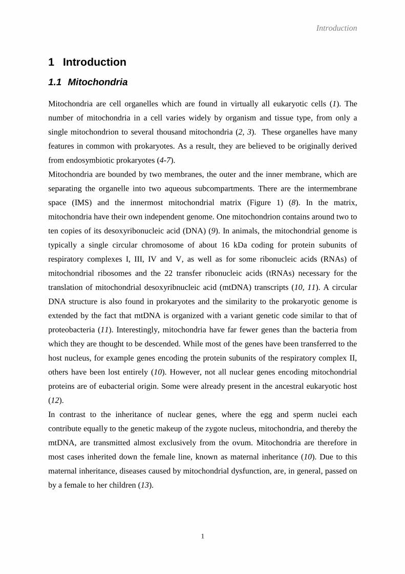
Mitochondria are bounded by two membranes, the outer and the inner membrane, which are
separating the organelle into two aqueous subcompartments. There are the intermembrane
space (IMS) and the innermost mitochondrial matrix (Figure 1) (8). In the matrix,
mitochondria have their own independent genome. One mitochondrion contains around two to
ten copies of its desoxyribonucleic acid (DNA) (9). In animals, the mitochondrial genome is
typically a single circular chromosome of about 16 kDa coding for protein subunits of
respiratory complexes I, III, IV and V, as well as for some ribonucleic acids (RNAs) of
mitochondrial ribosomes and the 22 transfer ribonucleic acids (tRNAs) necessary for the
translation of mitochondrial desoxyribnucleic acid (mtDNA) transcripts (10, 11). A circular
DNA structure is also found in prokaryotes and the similarity to the prokaryotic genome is
extended by the fact that mtDNA is organized with a variant genetic code similar to that of
proteobacteria (11). Interestingly, mitochondria have far fewer genes than the bacteria from
which they are thought to be descended. While most of the genes have been transferred to the
host nucleus, for example genes encoding the protein subunits of the respiratory complex II,
others have been lost entirely (10). However, not all nuclear genes encoding mitochondrial
proteins are of eubacterial origin. Some were already present in the ancestral eukaryotic host
(12).
In contrast to the inheritance of nuclear genes, where the egg and sperm nuclei each
contribute equally to the genetic makeup of the zygote nucleus, mitochondria, and thereby the
mtDNA, are transmitted almost exclusively from the ovum. Mitochondria are therefore in
most cases inherited down the female line, known as maternal inheritance (10). Due to this
maternal inheritance, diseases caused by mitochondrial dysfunction, are, in general, passed on
by a female to her children (13).
Introduction
2
Mitochondria are not synthesized de novo but derived from binary fission of pre-existing
organelles similar to bacterial cell division. Mitochondrial inheritance therefore depends on
mitochondrial fission during cytokinesis (14). Unlike bacteria however, mitochondria can also
fuse with other mitochondria (10). Fusion of several mitochondria results in extended
interconnected mitochondrial networks and serves to mix and unify the mitochondrial
compartment. In case of an accumulation of different somatic mutations in the mtDNA of
individual mitochondria this fusion can, for example, counteract the manifestation of
respiratory deficiencies by allowing the complementation of mtDNA gene products in the
heteroplasmic cells. Furthermore, the connectivity of the mitochondrial network is an
important factor in the cell´s calcium signaling response, embryonic development and
spermatogenesis (14).
The most prominent role of mitochondria is the production of adenosine triphosphate (ATP),
a source of chemical energy (15), through respiration. This is also reflected by the large
number of proteins involved in ATP synthesis in the inner membrane (3). The central set of
reactions involved in ATP production is collectively known as the citric acid cycle, or Krebs
Cycle, followed by the electron transport chain in the mitochondrial inner membrane. The
electrochemical gradient established across the inner mitochondrial membrane by the electron
transport chain is used by ATP-synthase to synthesize ATP from adenosine diphosphate
(ADP) and inorganic phosphate (Pi). This process is known as oxidative phosphorylation (3).
As mentioned above, mitochondria have many others functions in addition to the production
of ATP. They play a central role in calcium signaling (16), apoptosis (17), regulations of
membrane potential (3), cellular proliferation (18), and heme synthesis or the formation and
export of iron-sulfur (Fe/S) clusters (12).
Given the critical role mitochondria play in cell metabolism, damage and the resultant
dysfunction of these organelles are key components in a wide range of human diseases.
Classic mitochondrial disorders typically appear to affect brain and skeletal muscle functions,
often referred to as mitochondrial encephalomyophaties, but can also result in diabetes,
multiple endocrinopathy or a variety of other systemic manifestations (10, 13).
Mitochondrial encephalomyopathy with lactic acidosis and stroke-like episodes (MELAS)
syndrome, Kearns-Sayre syndrome, cardiomyophathy, and Leber´s hereditary opticus
neuropathy (LHON) are diseases caused by mutations in the mtDNA (12, 19). Additionally,
in early tumors of the bladder, prostate, liver or head and neck, mtDNA alterations could be
observed (12). Dysfunctions of mitochondrial proteins caused by defects in nuclear genes
evoke clinical observations such as Friedreich‟s ataxia, hereditary spastic paraplegia and

Introduction
3
Wilson´s disease (20). Moreover, cardiovascular disease, stroke, dementia, Alzheimer‟s
disease, epilepsy, Parkinson‟s disease and diabetes mellitus are examples of diseases
associated with defective mitochondrial functionality (21, 22). How exactly mitochondrial
dysfunction fits into the etiology of these pathologies has yet to be elucidated.
Taken together, mitochondria are associated with a variety of essential functions in the cell,
which, in the vast majority of the cases, are only poorly understood and whose disturbance
leads again to a variety of diseases. Therefore, the investigation of these organelles presents
an important field in cell biology.
Figure 1
The general organisation of a mitochondrion
OM: outer mitochondrial membrane, IMS: intermembrane space, IM: inner mitochondrial
membrane, M: matrix
(N. crassa, courtesy of F. Miller, LMU München, GER)

Introduction
4
1.2 Protein translocation into mitochondria
As already mentioned, the mitochondrial genome encodes only a rather small number of
proteins. Therefore, about 99% of the mitochondrial proteins are encoded in the nucleus of the
host (8) and synthesized in the cytosol as precursor proteins. These preproteins have to be
transported into the mitochondria and be targeted to their final submitochondrial destination,
the outer or inner mitochondrial membrane, the IMS or the matrix. The process of protein
sorting and export or transport to intracellular membranes or compartments is termed “protein
topogenesis” (23).
In contrast to the inner mitochondrial membrane, the outer mitochondrial membrane contains
porin protein channels. These channels allow the passage of molecules smaller than 5000
Daltons (2), whereas the inner membrane is impermeable to virtually all molecules (24).
Consequently, transport of larger proteins into the mitochondrial subcompartments is a
selective and controlled process, performed by a variety of complex molecular machineries
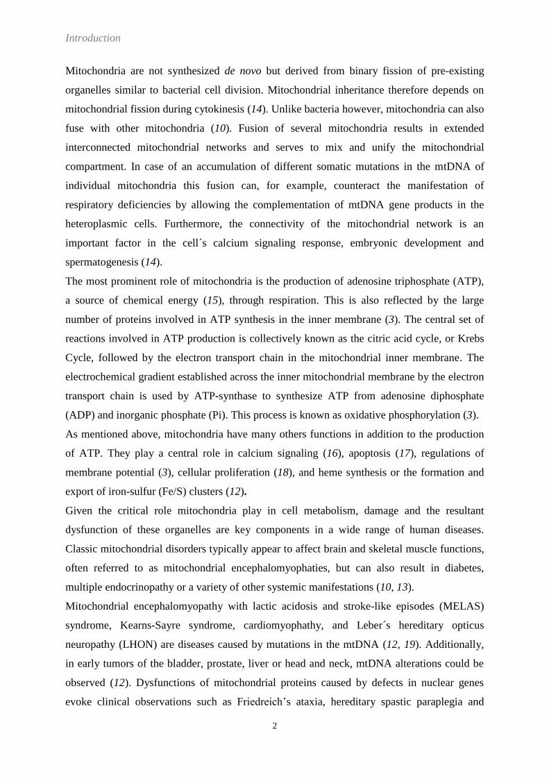
known collectively as mitochondrial protein translocases (Figure 2). Mitochondrial protein
translocases are able to recognize so called targeting signals or topogenic sequences (23) of
the precursor proteins. These targeting signals are present either as amino-terminal extensions,
which are usually proteolytically removed after import into mitochondria, or they are non-
cleavable internal elements which remain part of the mature protein. Targeting signals
facilitate recognition of the precursor proteins by receptors on the mitochondrial surface.
Thereafter, they are sorted to their appropriate destination within the mitochondria (8, 25).

Introduction
5
Figure 2
Protein translocases in the outer and inner mitochondrial membrane
Precursor proteins are imported into mitochondria via the TOM complex, some are further
transported into the matrix, or integrated into the inner membrane by the TIM23 complex (1),
directed to the IMS (2), inserted into the inner membrane by the TIM22 complex (3) or
embedded into the outer membrane via the TOB complex (4). The Oxa1 complex facilitates
the insertion into the inner mitochondrial membrane of a subset of preproteins approaching it
from the matrix (5).
IM: inner mitochondrial membrane, IMS: intermembrane space, OM: outer mitochondrial
membrane
The figure was adapted from Mokranjac et al., 2008 (26).
Introduction
6
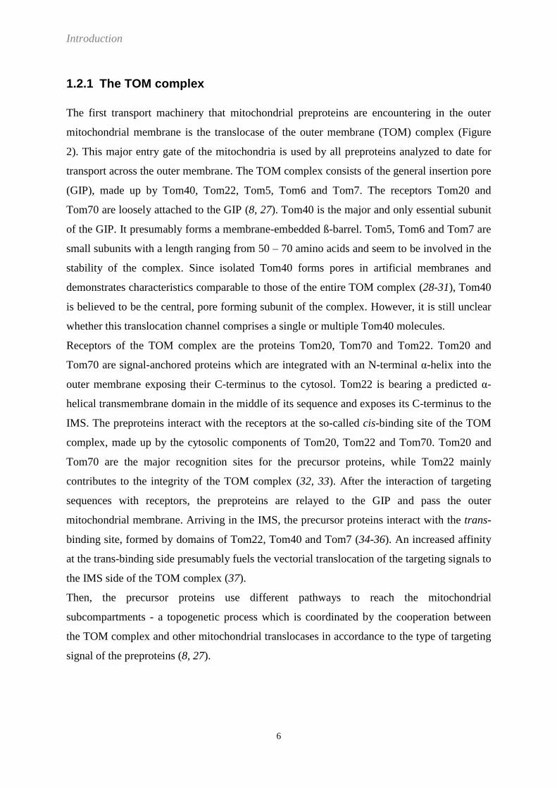
1.2.1 The TOM complex
The first transport machinery that mitochondrial preproteins are encountering in the outer
mitochondrial membrane is the translocase of the outer membrane (TOM) complex (Figure
2). This major entry gate of the mitochondria is used by all preproteins analyzed to date for
transport across the outer membrane. The TOM complex consists of the general insertion pore
(GIP), made up by Tom40, Tom22, Tom5, Tom6 and Tom7. The receptors Tom20 and
Tom70 are loosely attached to the GIP (8, 27). Tom40 is the major and only essential subunit
of the GIP. It presumably forms a membrane-embedded ß-barrel. Tom5, Tom6 and Tom7 are
small subunits with a length ranging from 50 – 70 amino acids and seem to be involved in the
stability of the complex. Since isolated Tom40 forms pores in artificial membranes and
demonstrates characteristics comparable to those of the entire TOM complex (28-31), Tom40
is believed to be the central, pore forming subunit of the complex. However, it is still unclear
whether this translocation channel comprises a single or multiple Tom40 molecules.
Receptors of the TOM complex are the proteins Tom20, Tom70 and Tom22. Tom20 and
Tom70 are signal-anchored proteins which are integrated with an N-terminal α-helix into the
outer membrane exposing their C-terminus to the cytosol. Tom22 is bearing a predicted α-
helical transmembrane domain in the middle of its sequence and exposes its C-terminus to the
IMS. The preproteins interact with the receptors at the so-called cis-binding site of the TOM
complex, made up by the cytosolic components of Tom20, Tom22 and Tom70. Tom20 and
Tom70 are the major recognition sites for the precursor proteins, while Tom22 mainly
contributes to the integrity of the TOM complex (32, 33). After the interaction of targeting
sequences with receptors, the preproteins are relayed to the GIP and pass the outer
mitochondrial membrane. Arriving in the IMS, the precursor proteins interact with the trans-
binding site, formed by domains of Tom22, Tom40 and Tom7 (34-36). An increased affinity
at the trans-binding side presumably fuels the vectorial translocation of the targeting signals to
the IMS side of the TOM complex (37).
Then, the precursor proteins use different pathways to reach the mitochondrial
subcompartments - a topogenetic process which is coordinated by the cooperation between
the TOM complex and other mitochondrial translocases in accordance to the type of targeting
signal of the preproteins (8, 27).

Introduction
7
1.2.2 Import of proteins into the matrix
Mitochondrial matrix proteins make up the majority of all mitochondrial proteins. Their
precursor forms are transported across both mitochondrial membranes by the concerted
interaction of two protein translocases, the aforementioned TOM complex in the outer
mitochondrial membrane and the TIM23 (translocase of the inner membrane) complex in the
inner mitochondrial membrane (27).
The TIM23 complex can be subdivided into the membrane component, comprising those
proteins which are forming the protein-conducting channel, and the import motor, which
drives the translocation of the precursor proteins into the matrix. The membrane component
consists of three essential proteins namely Tim23, Tim17 and Tim50. They, together with the
non-essential Tim21, are highly conserved throughout the eukaryotic kingdom. Tim50 forms
the receptor subunit of the membrane component, exposing a large C-terminal domain to the
IMS. The N-terminus is anchored to the inner membrane with a single transmembrane
domain. Tim50 is the first component of the inner membrane interacting with incoming
precursor proteins as they emerge from the trans-binding site at the TOM complex (38, 39).
Afterwards, the precursor proteins are presumably transferred to Tim23 (25, 27). Tim50 has
been proposed to block the protein-conducting channel of the TIM23 complex in the absence
of any precursor protein and thereby prevent the collapse of the membrane potential (Δψ) by
ion leakage (25, 40). Tim23 and its associated Tim17 form the translocation channel of the
TIM23 complex. The C-terminus of Tim23 is embedded by four transmembrane helices into
the inner mitochondrial membrane. Surprisingly, Tim23 can span the IMS and the outer
mitochondrial membrane, indicated by the accessibility of the N-terminus to proteases added
to intact mitochondria (41). It has been suggested that the N-terminus of Tim23 brings the
TIM23 translocase in proximity to the outer mitochondrial membrane to facilitate its interplay
with the TOM complex (41). Interestingly, a comparable function has been suggested for
Tim21, bringing together the TOM and the TIM23 complex due to its binding to the IMS-
exposed part of Tom22 (27, 42, 43). The segment of Tim23 spanning the IMS interacts with
Tim50 and serves as an additional presequence receptor of the TIM23 complex (44).
Comparable to Tim23, Tim17 has four C-terminal transmembrane helices anchoring it in the
inner mitochondrial membrane. Although these helices have sequence similarity to those of
Tim23, they seem to have diverse functions, as they are not interchangeable (8). The N-
terminal segment of Tim17 exposed to the IMS is rather short and contains several conserved

Introduction
8
negatively charged residues which play a crucial role in preprotein import and gating of the
translocation channel (45, 46).
The import motor is also called presequence translocase-associated motor (PAM) and sits at
the matrix site of the inner mitochondrial membrane. It is made up of the proteins Tim44,
Tim14 (Pam18), Tim16 (Pam16), mtHsp70 (matrix 70 kDa heat shock protein), and Mge1.
Tim44 is a peripherally attached membrane protein forming the interface of the membrane
component with the import motor unit of the TIM23 complex. The C-terminus of Tim44 is
embedded in the inner mitochondrial membrane. On the one side, Tim44 binds to the Tim23-
Tim17 core of the membrane embedded translocation channel, while the other side interacts
with mtHsp70 and its associated DnaJ-like proteins, Tim14 and Tim16 (8, 27). Tim14 is
anchored with its N-terminus in the inner membrane and forms a tight complex with Tim16
which is lacking a transmembrane segment (47). mtHsp70 is an exchangeable subunit of the
import motor, fluctuating between a bound and a released state. As found in all Hsp70 (heat
shock protein) chaperones, mtHsp70 contains an N-terminal nucleotide (ATP) binding
domain (NBD) and a C-terminal substrate or peptide binding domain (PBD) (48, 49). ATP
bound mtHsp70 is recruited by Tim44 and thereby enables the interaction of the incoming
polypeptide with the mtHsp70. The alternation between the binding to and release of the
translocating polypeptide by mtHsp70 is an ATP-dependent process which results in the
vectorial movement of the unfolded polypeptide chain into the matrix. The hydrolysis of ATP
to ADP as well as substrate binding to the mtHsp70 is regulated by the DnaJ-like proteins.
Upon binding of the polypeptide to mtHsp70, Tim14 stimulates ATP hydrolysis and thereby
triggers the release of the mtHsp70-precursor protein complex from Tim44. The mtHsp70-
precursor protein complex dissociates from the membrane and enables the binding of the next
ATP bound mtHsp70. After ATP hydrolysis, ADP is exchanged for ATP by the nucleotide
exchange factor Mge1. This leads to a release of the substrate (8, 27, 50, 51). Tim16 is not a
functional DnaJ-like protein, as it is missing the HPD (His-Pro-Asp) motif which is crucial for
the stimulation of mtHsp70. Rather it functions as a negative regulator of the import motor by
blocking the contact between Tim14 and mtHsp70 (52-54).
Roughly half of the mitochondrial proteins are synthesized with an N-terminal extension as a
targeting signal. This signal is also called the presequence, prepeptide or matrix targeting
sequence (MTS), since it directs the N-terminus across the inner mitochondrial membrane. To
date the DNA helicase Hmi1 is the only exception known, where the MTS is positioned at the
C-terminus (55). The MTSs do not share a conserved primary sequence but they all have the
propensity to form an amphipathic helix presenting one hydrophobic and one positively

Introduction
9
charged face. Tom20 possesses a binding groove for the hydrophobic site of those N-terminal
presequences (MTSs), where Tom22 recognizes the positively charged surface (8, 25). After
passing the TOM complex, the precursor proteins are transferred to the TIM23 complex. The
membrane component of the TIM23 complex only transports the MTS through the inner
mitochondrial membrane, a process driven by the membrane potential Δψ. The translocation
into the mitochondrial matrix of the polypeptide chain C-terminal to the MTS is performed by
the import motor unit of the TIM23 complex and fuelled by a second energy source, ATP. As
already mentioned above, this ATP is used by mtHsp70 to pull the polypeptide in a stepwise
manner into the matrix. In the absence of further sorting information, MTS-containing
precursor proteins are fully transferred into the matrix. The MTS is sufficient to direct a
preprotein into the matrix and is therefore referred to as the default mode of the TIM23
complex (56, 57). Currently this is under debate (8, 25). Once the precursor proteins have
reached the mitochondrial matrix, the MTS is removed by the matrix processing peptidase
(MPP).
1.2.3 Import of proteins into the inner mitochondrial membrane
For those nuclear encoded mitochondrial proteins which have the inner mitochondrial
membrane as their final destination there are three different pathways for their translocation
from the cytosol into the mitochondria: (1) the stop-transfer pathway, (2) the TIM22 pathway,
and (3) the conservative sorting pathway.
The stop-transfer pathway includes the already described protein translocases TOM and
TIM23. TIM23 is not only capable of transporting precursor proteins into the matrix
(translocation mode), but can also switch to a second mode, for the stop-transfer pathway, in
which the preproteins are stopped during the translocation and are laterally inserted into the
inner mitochondrial membrane (lateral insertion mode). In contrast to the preproteins directed
to the matrix, which only possess an MTS at the N-terminus, preproteins designated for lateral
insertion have an additional signal, a stop-transfer signal, in some distance C-terminal to the
MTS. This stop-transfer signal is a transmembrane domain which causes the arrest of the
precursor protein inside the TIM23 complex (27).
Depending on the type of targeting signal the TIM23 complex encounters, the complex
changes its internal conformation to set up either for the translocation mode or the lateral
insertion mode (58, 59).

Introduction
10
Another pathway for proteins to be transported from the cytosol to the inner mitochondrial
membrane is the TIM22 pathway. This pathway requires the concerted action of three
different consecutive mitochondrial protein translocases: (1) the TOM complex in the outer
membrane, (2) the complexes of the small Tim proteins in the IMS, and (3) the TIM22
translocase in the inner mitochondrial membrane.
The small Tim protein complexes Tim9-Tim10 and Tim8-Tim13 reside in the IMS and are
composed of polypeptides with a molecular weight between 8 and 12 kDa. They are
characterized by twin Cx3C motifs. The cysteine residues of these motifs form pairs of
intramolecular disulfide bridges which are crucial for the structure of the small Tim proteins.
Small Tim proteins oligomerize as tightly bound hexamers with a “jellyfish-like structure”.
The tentacles of this jellyfish-like structure might be able to bind to the hydrophobic regions
of incoming proteins from the TOM complex (60) and therefore are supposed to fulfill a
chaperone like function while transferring the precursor proteins in the IMS from the TOM to
the TIM22 complex (61, 62), although this is still hypothetical.
The TIM22 complex is made up of the membrane proteins Tim22, Tim54 and Tim18 and has
a combined molecular weight of around 300 kDa. Tim22 is the core subunit of the complex
and is embedded in the inner mitochondrial membrane with four transmembrane helices
which presumably form the translocation pore of the complex (63). Tim54 and Tim18 are
associated with Tim22 and seem to be important but non-essential components of the TIM22
complex, since preproteins are inserted even in the absence of Tim54 and Tim18, although at
strongly reduced levels (63). Tim54 exposes a large domain in the IMS and might serve as a
docking site for a small Tim protein complex consisting of Tim9, Tim10, and Tim12, which is
permanently bound to the TIM22 complex (63, 64). The essential small Tim protein Tim12 is
exclusively found in the TIM22 associated complex but not in the soluble chaperone
complexes of the IMS and might be involved in substrate recognition at the TIM22 complex.
Tim18 is supposed to play a role in the assembly of the TIM22 complex (25, 27).
The TIM22 pathway is responsible for the insertion of members of the solute carrier family,
such as the ATP/ADP or the phosphate carrier as well as the membrane embedded subunits of
TIM complexes such as Tim17, Tim22 and Tim23. All these proteins are lacking cleavable
presequences but contain internal non-cleavable targeting signals (8, 27). After their synthesis
in the cytosol, the carrier precursor proteins are bound by the chaperones Hsp70 and Hsp90
and guided to the Tom70 receptor at the TOM complex. Here, the chaperones dock to the
tetratricopeptide repeat (TPR) domain of the receptor (65). Following the ATP-dependent
release and transfer from the chaperones to the TOM complex, the precursor proteins are

Introduction
11
transferred through the Tom40 translocation channel. In contrast to the MTS bearing
precursor proteins, carrier precursor proteins are not transported as linear polypeptide chains
through the TOM complex but pass it in a loop structure (66). Subsequently, with the help of
the small Tim protein complex Tim9-Tim10, the preproteins are transferred to the Tim9-
Tim10-Tim12 chaperone complex at the surface of the TIM22 translocase. Finally, substrates
of the TIM22 complex are laterally inserted into the membrane in a membrane potential-
dependent process and result in an even-numbered transmembrane segment in the inner
mitochondrial membrane exposing both their N- and C-termini into the IMS. The import
process of the TIM subunits Tim17, Tim22, and Tim23 is comparable to that of the carrier
proteins, however, not well characterized. Instead of utilizing the Tim9-Tim10 complex, the
precursor proteins of the TIM subunits are interacting with an alternative Tim complex made
up by Tim8 and Tim13 (8, 27).
In the conservative sorting pathway, proteins destined for the inner mitochondrial membrane
are first transported from the cytosol into the matrix and from there inserted into the inner
membrane. Due to the resemblance of the export-like transport of the proteins from the matrix
side into the inner membrane to that of protein transport in prokaryotes, this pathway is
termed the „conservative sorting pathway‟. Precursor proteins following this pathway are for
example Oxa1 (67) and subunit 9 of the F0F1-ATPase of Neurospora crassa (N. crassa, N.c.),
(68). They are synthesized with an N-terminal cleavable presequence and consist of more than
one transmembrane domain. As they reach the matrix, they are bound by mtHsp70 to prevent
them from aggregating. Thereafter, the MTS is removed by the MPP. The molecular
mechanism by which these precursor proteins are integrated into the inner mitochondrial
membrane is still ill defined. In general, protein segments which are transported from the
matrix across the inner membrane are enriched in negatively charged amino acid residues.
Since the membrane insertion from the matrix strongly depends on the membrane potential, it
is likely that the negative charged regions are pulled to the IMS in an electrophoretic manner
(67-69). The Oxa1 (oxidase assembly) complex of the inner membrane facilitates the insertion
of at least some of these inner membrane proteins (69, 70). Oxa1 is also involved in the co-
translational insertion of proteins encoded in the mtDNA (70-72).

Introduction
12
1.2.4 Import of proteins into the intermembrane space
The biogenesis of proteins which reside in the IMS is diverse. Three different import
mechanisms are known: (1) the bipartite presequence coordinated pathway, (2) the folding
trap mechanism, and (3) the localization of proteins in the IMS due to affinity interactions.
Some of the proteins which are directed to the IMS have bipartite presequences or sorting
signals, consisting of an N-terminal MTS as well as a hydrophobic sorting sequence. The
precursor proteins follow the translocation path provided by the TOM and the TIM23
complex. In general, following the arrest of the incoming proteins in the inner mitochondrial
membrane, peptidases remove those targeting signals by proteolytic cleavage and thereby
release the proteins into the IMS. Proteins located in the IMS which are using this pathway
are for example the cytochrome c peroxidase (CCPO) or cytochrome b2 (27).
Another import pathway by which proteins are directed into the IMS is the folding trap
mechanism. Here, after passing the outer mitochondrial membrane, the proteins are stabilized
by cofactors or disulfide bridges in their folded state and are thereby trapped in the IMS.
Prominent examples are the folding of small Tim proteins mediated by the MIA
(mitochondrial intermembrane space import and assembly) machinery (73-76) or the covalent
addition of heme to apocytochrome c catalyzed by the cytochrome c heme lyase (CCHL) in
the IMS (77). The MIA machinery comprises the disulfide carrier Mia40, which is anchored
in the inner mitochondrial membrane, and the soluble sulfhydryl oxidase Erv1 (essential for
respiration and viability), and promotes the formation of intramolecular disulfide bonds in, for
example, the imported small Tim proteins.
Finally, the import of proteins such as cytochrome c heme lyase or the creatine kinase seems
to be driven by their high affinity to certain components in the IMS to which they are
permanently associated after they have reached the IMS (27). The mechanism of this import
pathway is largely obscure.
1.2.5 Import of proteins into the outer mitochondrial membrane
All proteins of the outer mitochondrial membrane known to date are nuclear encoded. They
are missing canonical cleavable N-terminal prepeptides, but contain non-cleavable targeting
and sorting signals within the protein sequence itself. The membrane-integrated proteins of
the outer mitochondrial membrane can be subdivided into two groups: α-helical proteins and
ß-barrel proteins. ß-barrel proteins can only be found in the outer membranes of Gram-

Introduction
13
negative bacteria, chloroplasts and mitochondria while all other membranes harbour α-helical
membrane proteins (78).
1.2.5.1 Insertion of α-helical proteins into the outer mitochondrial membrane
According to their topology, different classes of α-helical proteins can be distinguished in the
outer mitochondrial membrane. Characteristically, these proteins are anchored with one or
more α-helical transmembrane segments into the membrane. Signal anchored proteins carry
one α-helical transmembrane domain at the N-terminus. This group includes the primary
import receptors of the TOM complex, Tom20 and Tom70. When the transmembrane
segment is located at the C-terminus, such as in Tom5, Tom6, Tom7 or Fis1, the proteins are
referred to as tail-anchored proteins. Proteins of both categories expose their main part into
the cytosol and only a short segment into the IMS. Tom22 and mitochondrial import protein 1
(Mim1) have one central embedded transmembrane domain and are orientated with the
N-terminus to the cytosol and the C-terminus to the IMS. The peripheral benzodiazepine
receptor (PBR), Fzo1 and Ugo1 are examples of proteins which contain multiple α-helices in
the membrane, spanning the outer mitochondrial membrane five, two and three times,
respectively (79-82).
In all these proteins, the hydrophobic segments do not only serve as anchors within the
membrane, but also typically function as targeting signals of the proteins. However, no
sequence similarities could be found among those targeting sequences. The targeting
information is apparently encoded in structural elements such as the hydrophobicity and
charge of the transmembrane α-helix and its flanking regions (83). The mechanism by which
the different α-helical membrane proteins are inserted into the outer mitochondrial membrane
seems to differ between the individual members and is still ill defined. The insertion of the
signal-anchored proteins Tom20, Tom70 or Mcr1, for example, was shown to be independent
of the presence of import receptors while unaffected by the blocking of the translocation pore
of the TOM complex (84-87). However, in contrast to Mcr1, Tom20 seems to be dependent
on Tom40 for acquiring its correct topology. It has been suggested that the TOM translocase
can facilitate protein insertion at its protein-lipid interface (86-88). Moreover, both Tom20
and Tom70 were described to use a further outer membrane protein for membrane insertion,
namely the mitochondrial import protein 1 (Mim1) (89-91). Mim1 was also found to be
important for the insertion of the small TOM proteins (92). Other tail-anchored proteins do
not require any of the known outer membrane machineries and seem to be dependent on the
lipid composition of the membrane for their insertion (93, 94). The precursor of Tom22 needs

Introduction
14
the TOM receptors to be directed to the mitochondrial surface and seems to use the TOB
complex for its insertion into the outer mitochondrial membrane (95). Multiple spanning
membrane proteins were reported to use components of the IMS, Tom70, but no other TOM
complex proteins for efficient insertion into the outer mitochondrial membrane (96).
Taken together, the TOM complex was found to have two distinct functions: (1) the
aforementioned translocation of virtually all preproteins from the cytosol across the outer
mitochondrial membrane and (2) the direct integration of α-helical outer membrane proteins.
Our knowledge of how those α-helical transmembrane proteins are inserted into the outer
envelope of mitochondria is still elusive. Since the pore of the TOM complex seems not to be
needed for that process, at least in some cases, those proteins may not follow the canonical
route through the import channel, but are following a second pathway which awaits further
analysis.
1.2.5.2 Insertion of ß-barrel proteins into the outer mitochondrial membrane
ß-barrel proteins are embedded in the outer mitochondrial membrane by multiple antiparallel
ß-strands. The topogenesis of the mitochondrial outer membrane ß-barrel proteins (TOB)
complex (97), also termed the sorting and assembly machinery (SAM) (98), is specialized in
the insertion of the ß-barrel precursor proteins into the outer membrane and requires a
coordinated interaction with the TOM complex and small Tim protein complexes to fulfill its
task. Composition and function of the TOB complex are discussed in detail below.

Introduction
15
1.3 The TOB complex
1.3.1 Introduction
In eukaryotes, ß-barrel proteins are exclusively found in the outer membrane of organelles of
endosymbiotic origin, namely chloroplast and mitochondria (99, 100). Furthermore, ß-barrel
proteins can only be found in the outer membrane of Gram-negative bacteria (101), which
supports the idea that these organelles are derived from a bacterial ancestor (5, 102).
Membrane-embedded ß-barrel proteins are referred to as outer membrane proteins (OMPs)
(103). The TOB complex in the outer mitochondrial membrane is responsible for the correct
insertion of ß-barrel proteins and cooperates with the TOM complex, which is also sitting in
the outer mitochondrial membrane and with the small Tim protein complexes in the IMS.
1.3.2 Composition of the TOB complex
TOB is a hetero-oligomeric protein complex which comprises the proteins Tob55 (Sam50,
Tom50), Tob38 (Sam35, Tom38) and Tob37 (Mas37, Sam37, Tom37) (97, 98, 104-108).
Tob55 is the main component of this complex and was found in a proteomic screening of
outer mitochondrial membrane proteins from N. crassa by mass spectrometry analysis (97). It
was also found in copurification experiments with Tob37 (98), a known subunit of the TOB
complex (104). Sequence analysis revealed homologues of Tob55 not only in the genomes of
virtually all eukaryotes, but significant sequence similarity was also detected with the outer
membrane protein 85 (Omp85) from Gram-negative bacteria (97, 109). Omp85 (YaeT,
BamA) is the main subunit of the bacterial ß-barrel assembly machinery (BAM) and was
determined to be a ß-barrel protein itself (25, 110-113).
It is assumed that Tob55 is also a ß-barrel protein. This is mainly based on sequence analysis
and secondary structure prediction, since, to date, no high resolution structure of Tob55 and
its homologues could be solved (97, 98, 109, 113-116). Therefore, Tob55 is supposedly both a
substrate and subunit of the TOB complex. Besides Tob55, porin (also termed as voltage-
dependent anion-selective channel (VDAC)), Tom40, Mdm10 and Mmm2 are assigned to the
family of outer mitochondrial ß-barrel membrane proteins and thereby putative substrates of
the TOB complex (27). The membrane-embedded ß-barrel of Tob55 is predicted to be formed
by the C-terminus, whereas the hydrophilic amino acids at the N-terminus are facing the IMS
and fold into a characteristic structure, the polypeptide transport associated (POTRA) domain
(112, 113, 117). These POTRA domains were described to have receptor functions and were
not only found in Tob55 but also in other OMPs such as Omp85/YaeT (114, 118), although

Introduction
16
the amount of POTRA domains could vary between one (Tob55), two (FhaC (filamentous
haemagglutinin adhesin)), and even up to five (Omp85) (103, 117, 118).
Several experiments indicated a specific role of the TOB complex in the biogenesis of ß-
barrel precursor proteins in the outer membrane. The Tob55 gene was found to be essential
for cell viability (109, 119, 120) and downregulation of Tob55 (Tob55↓) resulted in low
levels of ß-barrel proteins such as Tom40, porin and Mdm10 in the outer mitochondrial
membrane. In contrast, the levels of α-helical proteins in the outer mitochondrial membrane,
the IMS, the inner mitochondrial membrane and the matrix remained unaffected (97, 120).
There is only one exception, the α-helical protein Tom22, which was decreased in Tob55↓
mitochondria in N. crassa (120). In accordance with that, import of the pre-proteins Tom40
and Tob55 itself in Tob55↓ mitochondria was also strongly reduced, whereas α-helical outer
mitochondrial membrane proteins or proteins designated to the IMS, the inner mitochondrial
membrane or the matrix were imported at roughly wild-type levels (97, 120). Studies on the
assembly process of the ß-barrel proteins Tom40 and porin also revealed that the biogenesis
of these proteins is severely impaired by the depletion of Tob55. Antibody supershift assays
presented direct evidence for the interaction of the TOB complex with ß-barrel precursor
proteins (98, 109, 120).
Consisting of a POTRA domain and a ß-barrel, having a high sequence similarity to Omp85,
and functioning as a transporter of ß-barrel precursor proteins makes it tempting to assign the
TOB complex to the Omp85-TpsB transporter superfamily (Tps – Two-Partner Secretion),
and there to the Omp85 subfamily (109, 115, 121-124).
The Omp85 family is a conserved family of protein transporters and includes Toc75
(translocon at the outer envelope membrane of chloroplasts) of chloroplasts, D15 of
Haemophilus influenza, Omp85 from Neisseria meningitides and YaeT from Escherichia coli.
So far, Tob55 is the only known mitochondrial ß-barrel protein with clear homologues outside
the kingdom of eukaryotes (78).
To date only one member of the Omp85-TpsB transporter superfamily could be crystallized,
FhaC (115, 121). FhaC belongs to the second subfamily of the Omp85-TpsB transporter
superfamily, the TpsB-transporter family. TpsB transporter can be found in a subset of Gram-
negative bacteria and are responsible for the secretion of their dedicated TpsA substrates
(124). Whereas Tob55 was primarily predicted to have 12 ß-strands (112, 113), recent
alignments with the FhaC sequence in combination with the resolved ß-barrel structure of
FhaC suggest a 16-stranded ß-barrel for members of the Omp85 transporter family (115, 121).

Introduction
17
Despite these similarities between Tob55 and the prokaryotic members of this transporter
family, the insertion mechanism of ß-barrel precursor proteins in prokaryotes and
mitochondria are expected to diverge due to the different additional components of the
transporter complexes (8).
The additional components to Tob55 of the TOB complex, Tob38 and Tob37, are located at
the cytosolic surface of the outer mitochondrial membrane (104-107). So far, a high
homology of both proteins could only be found among fungi (27). Only a moderate sequence
homology to the mammalian metaxin-1 was reported for Tob37 (125-127). Furthermore,
Tob38 was proposed to be a homologue of the mammalian metaxin-2. Convincing evidence
for homology of metaxin proteins with Tob38 and Tob37 is still lacking (105, 107, 126).
Tob38 is an essential component of the TOB complex in yeast and depletion of Tob38 results
in impaired ß-barrel import comparable to a loss of Tob55 (105-107). In contrast to Tob55
and Tob38, yeast Tob37 is not an essential protein, but the deletion of Tob37 compromises
the insertion of ß-barrel precursor proteins and results in growth defects (104). Similarly,
metaxin-1 and metaxin-2 were also indicated to play important roles in ß-barrel biogenesis
(126). Electron microscopy images of negative stained native and recombinant Tob55
revealed ring-shaped structures with a fivefold symmetry which displayed an inner pore size
of 4-5 nm and an outer diameter of 15 nm (97). To date, high resolution structures for Tob38
as well as Tob37 are still elusive.
1.3.3 Topogenesis of mitochondrial ß-barrel proteins
After their synthesis in the cytosol, ß-barrel precursor proteins interact with the receptors of
the TOM complex (83, 128) and subsequently pass through the TOM pore into the IMS (97,
104). There, they are transferred from the TOM pore to the TOB complex with the help of the
small Tim protein complexes Tim8-Tim13 and Tim9-Tim10. These complexes presumably
prevent backsliding of the ß-barrel precursor proteins and have a chaperone-like function
analogous to the bacterial chaperone Skp (129-131). As the ß-barrel precursors reach the TOB
complex, they are supposedly bound by the POTRA domain of Tob55 (114, 118) and inserted
into the outer mitochondrial membrane from the IMS side (97, 104). Interestingly, the
translocation across the TOM pore seems to be coupled to the membrane insertion of the ß-
barrel precursor proteins by the TOB complex, as a depletion of Tob55 leads to an
accumulation of preproteins within the TOM complex and prevents them from reaching the
IMS (27, 97). Interaction of the ß-barrel precursor proteins with the POTRA domain and their

Introduction
18
membrane insertion from the IMS side are corresponding to the insertion mechanism in
prokaryotes, where the preproteins are inserted into the outer envelope from the periplasm.
This reflects the evolutionary origin of mitochondria from bacteria.
The mechanism by which the ß-barrels are inserted into the outer-mitochondrial membrane by
the TOB complex is still ill defined. Tob38 and Tob37 are both contributing to the stability of
the TOB complex (105, 129, 132). Recently, Tob38 was reported to have a receptor-like
function for ß-barrel precursor proteins by binding to a conserved ß-signal peptide at the most
C-terminal ß-strand and thereby initiating their membrane-insertion (132, 133). Tob37 was
described to be responsible for the release of precursors into the lipid phase of the membrane
and thereby act downstream of Tob38 in ß-barrel assembly (129, 132). Moreover, recent
findings assigned diverse proteins a role in the membrane insertion of ß-barrel precursor
proteins. Mdm10, Mdm12 and Mmm1, constituents of the MDM (mitochondrial distribution
and morphology) complex, were determined to act downstream of the TOB complex in the
assembly pathway. Depletion of these proteins reduced the assembly of Tom40 and porin in
the outer mitochondrial membrane (108, 134, 135). Mdm10 was also suggested to be a
constituent of the TOB complex (134, 136, 137). Mim1 was found to associate with Tob55
and is essential in the late steps of the assembly pathway of Tom40 (91, 106, 138). In addition
to its role in the topogenesis of mitochondrial outer membrane ß-barrel proteins, TOB was
shown to participate in the insertion of α-helical subunits of the TOM complex such as Tom6
and Tom22 (92, 95).

Introduction
19
1.4 Aim of the present study
The TOB complex was proven to be responsible for the insertion of ß-barrel precursor
proteins into the outer membrane of mitochondria, a process which is crucial for the
functionality of these organelles and consequently the host cell. During the last years,
remarkable progress was made in the identification of proteins involved in the biogenesis of
ß-barrel proteins. However, our knowledge about their structures, mechanism of membrane
insertion and interplay is still fragmentary. The filamentous fungi N. crassa turned out to be
an excellent organism for studying the biogenesis of mitochondrial proteins, due to its simple
cultivation conditions and genetic manipulation procedures, and the fact that relatively large
amounts of functionally and structurally intact mitochondria can be easily obtained (139). The
aim of this study was to establish an isolation procedure of the TOB complex from N. crassa,
with a view to carefully identify its composition and biochemical characteristics and thereby
elucidating its functional mechanism.

Results
20
2 Results
2.1 Tob55 is expressed in three different isoforms due to alternative splicing
For the isolation of the TOB complex of N. crassa, it was planned to start with a Ni-NTA
(nickel-nitriloacetic acid) affinity purification from a strain expressing a His-tagged form
(bearing a stretch of attached histidinyl residues) of the Tob55 protein. Surprisingly, Tob55
appeared in two bands upon sodiumdodecylsulfate polyacrylamide gel electrophoresis (SDS-
PAGE) and Western blotting using antibodies directed against Tob55. Both bands were absent
when the mitochondria were isolated from a strain in which Tob55 was downregulated
(Tob55↓, Tob55KO-3) indicating two forms of Tob55 (Figure 3A).
Figure 3
N. crassa Tob55 appears in two bands upon SDS-PAGE
A: The control strain HP1 and the Tob55KO-3 strain were cultivated on non-selective
medium (-) or selective medium (+) with 400 mM fpa and histidine. Tob55KO-3 is a tob55
knockout sheltered heterokaryon strain. Its growth on selective medium forces the tob55-
knockout-bearing nucleus to predominate in the heterokaryon, leading to a severe reduction
of Tob55-levels in comparison to controls. Following SDS-PAGE, the separated proteins
were analyzed by immunodecoration against Tob55.
B: As in “A” except mitochondria were isolated from the control strain (HP1) and two
strains, T55his6-1 and T55his6-3, expressing Tob55 with an N-terminal hexahistidinyl tag.
Immunodecoration was performed with Tob55 antiserum or penta-His antiserum.
(Nargang group, University of Edmonton, Alberta, CA)
Results
21
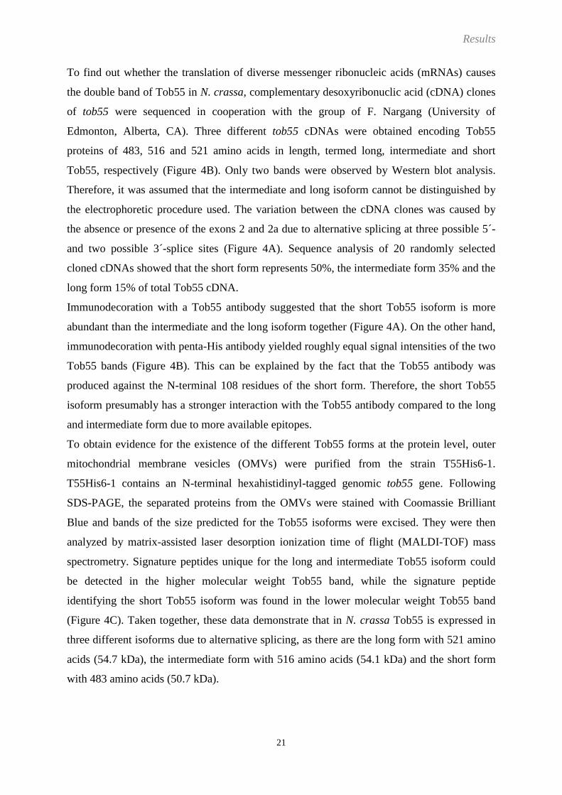
To find out whether the translation of diverse messenger ribonucleic acids (mRNAs) causes
the double band of Tob55 in N. crassa, complementary desoxyribonuclic acid (cDNA) clones
of tob55 were sequenced in cooperation with the group of F. Nargang (University of
Edmonton, Alberta, CA). Three different tob55 cDNAs were obtained encoding Tob55
proteins of 483, 516 and 521 amino acids in length, termed long, intermediate and short
Tob55, respectively (Figure 4B). Only two bands were observed by Western blot analysis.
Therefore, it was assumed that the intermediate and long isoform cannot be distinguished by
the electrophoretic procedure used. The variation between the cDNA clones was caused by
the absence or presence of the exons 2 and 2a due to alternative splicing at three possible 5´-
and two possible 3´-splice sites (Figure 4A). Sequence analysis of 20 randomly selected
cloned cDNAs showed that the short form represents 50%, the intermediate form 35% and the
long form 15% of total Tob55 cDNA.
Immunodecoration with a Tob55 antibody suggested that the short Tob55 isoform is more
abundant than the intermediate and the long isoform together (Figure 4A). On the other hand,
immunodecoration with penta-His antibody yielded roughly equal signal intensities of the two
Tob55 bands (Figure 4B). This can be explained by the fact that the Tob55 antibody was
produced against the N-terminal 108 residues of the short form. Therefore, the short Tob55
isoform presumably has a stronger interaction with the Tob55 antibody compared to the long
and intermediate form due to more available epitopes.
To obtain evidence for the existence of the different Tob55 forms at the protein level, outer
mitochondrial membrane vesicles (OMVs) were purified from the strain T55His6-1.
T55His6-1 contains an N-terminal hexahistidinyl-tagged genomic tob55 gene. Following
SDS-PAGE, the separated proteins from the OMVs were stained with Coomassie Brilliant
Blue and bands of the size predicted for the Tob55 isoforms were excised. They were then
analyzed by matrix-assisted laser desorption ionization time of flight (MALDI-TOF) mass
spectrometry. Signature peptides unique for the long and intermediate Tob55 isoform could
be detected in the higher molecular weight Tob55 band, while the signature peptide
identifying the short Tob55 isoform was found in the lower molecular weight Tob55 band
(Figure 4C). Taken together, these data demonstrate that in N. crassa Tob55 is expressed in
three different isoforms due to alternative splicing, as there are the long form with 521 amino
acids (54.7 kDa), the intermediate form with 516 amino acids (54.1 kDa) and the short form
with 483 amino acids (50.7 kDa).
Results
22
Figure 4
Three isoforms of Tob55 arise from alternative splicing
A: Overview of the intron/exon structure of the tob55 gene
Exons are sketched as rectangular boxes, introns as solid lines. The amount of codons
comprised by each exon is given in parentheses. Possible 5´- and 3´-splice sites are indicated
by numbered arrows above and below the line, respectively.
B: The three different Tob55 isoforms resulting from alternative splicing
The exons are shaded as in “A”. Predicted tryptic peptide fragments which are unique for
each isoform (signature peptides) are underlined by solid bars.
C: The signature peptides that are predicted to arise from the tryptic digestion
The initial methionine is followed by six histidinyl residues, since the analysis was performed
with the short Tob55 isoform bearing an N-terminal hexahistidinyl tag. Splice points between
exon 1 and 3 for the short form, between exon 2 and 3 for the intermediate (“interm”) form,
and between exon 2a and 3 for the long form of Tob55 are marked by arrows below the
peptide sequence. The underlined residues in the long Tob55 isoform represent exon 2a.
Coomassie blue-stained Tob55 bands were excised from a gel and analyzed by MALDI-TOF.
For each signature peptide the predicted („„P‟‟) mass and the mass that was determined
experimentally („„E‟‟) by mass spectrometry is given. The experimentally determined mass of
the signature peptide of the short Tob55 isoform indicates an oxidation, which presumably
took place at the N-terminal Met residue. For each signature peptide the tracings from the
appropriate region of the mass spectra are shown.

Results
23
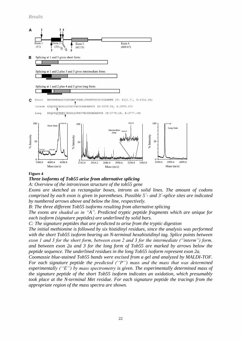
2.2 Isolation of the TOB complex
An isolation procedure of the TOB complex from the filamentous fungus N. crassa was
established using His-tagged variants of components of the TOB complex. The first step was
the purification of OMVs to enrich the amount of TOB complex in the starting material for
Ni-NTA affinity. The TOB complex is present at very low levels in mitochondria and
separation of the outer membrane leads to a strong enrichment as well as removal of potential
contaminating proteins.
A strain expressing the short Tob55 isoform with an N-terminal hexahistidinyl tag was chosen
to be used for Ni-NTA affinity purification. The tag had to be extended from six to nine
histidinyl residues to obtain efficient purification. Lysis of the OMVs was performed with the
detergents TX-100 (Figure 5) or digitonin (Figure 6). The lysates were passed over Ni-NTA
columns for affinity purification. The specifically bound proteins were eluted and subjected to
SDS-PAGE. They were identified by Western blotting and immunodecoration (Figure 5B,
Figure 6B) or Coomassie blue staining (Figure 5C, Figure 6C). Coomassie blue-stained bands
were excised and proteins identified by LS-MS/MS. With both detergents, Tob55, Tob38
(37.3 kDa) and Tob37 (48.6 kDa) were the only proteins, which were detectable in the eluate
(Figure 5, B and C, Figure 6, B and C). Very minor amounts of Mdm10 (52.7 kDa) were
detected by immunodecoration (Figure 5B, Figure 6B). The amounts of this protein were not
high enough to show up upon LC-MS/MS. In the preparations obtained with digitonin
sometimes traces of the very abundant outer membrane protein porin were present.
The same isolation procedure was carried out with OMVs from the strains His9-Tob38 and
His9-Tob37. These strains express Tob38 or Tob37 with an N-terminal ninefold His-tag and
all three untagged Tob55 isoforms. These preparations were performed to exclude loss of
TOB complex subunits caused by the absence of the intermediate and long Tob55 isoform.
The only proteins recovered in the eluate were Tob55, Tob37 and Tob38, and again, very
small amounts of Mdm10 (Figure 5, Figure 6). Thus, these results suggest that Tob55, Tob38
and Tob37 are the subunits of the TOB complex.
Results
24
Figure 5
Tob38, Tob37 and Tob55 copurify in the presence of TX-100
A: Schematic representation of TOB complexes with N-terminal ninefold His-tags at Tob55,
Tob38 or Tob37.
OM: outer mitochondrial membrane, IMS: intermembrane space
B and C: Outer mitochondrial membrane vesicles (OMVs) from N.c. strains with the His-
tagged TOB-subunit, 9His-Tob55, 9His-Tob38 or 9His-Tob37 were solubilized with TX-100;
proteins were isolated by Ni-NTA affinity purification and analyzed by SDS-PAGE followed
by immunodecoration (B) or Coomassie blue staining (C).
Solubilized outer mitochondrial membrane vesicles (OMVs) before (S) or after (L) clarifying
spin. FT: flowthrough with unbound proteins of the Ni-NTA column, E: eluate of bound
proteins, 55 lg and int: Tob55-intermediate and long isoform, 55sh: Tob55-short isoform, 37:
Tob37, 38: Tob38.
The strain with His-tagged Tob55 is only expressing the short isoform; hence only one band
can be seen in the immunodecorations. Note that only traces of Mdm10 can be copurified in
all three approaches.

Results
25
Figure 6
Tob38, Tob37 and Tob55 copurify in the presence of digitonin
A: Schematic representation of TOB complexes with N-terminal ninefold His-tags at Tob55,
Tob38 or Tob37. OM: outer mitochondrial membrane, IMS: intermembrane space
B and C: Outer mitochondrial membrane vesicles (OMVs) from N.c. strains with the His-
tagged TOB-subunit, 9His-Tob55, 9His-Tob38 or 9His-Tob37, were solubilized with
digitonin; proteins were isolated by Ni-NTA affinity purification and analyzed by SDS-PAGE
followed by immunodecoration (B) or Coomassie blue staining (C).
Solubilized outer mitochondrial membrane vesicles (OMVs) before (S) or after (L) clarifying
spin. FT: flowthrough with unbound proteins of the Ni-NTA column; E: eluate of bound
proteins, 55 lg and int: Tob55-intermediate and long isoform, 55sh: Tob55-short isoform, 37:
Tob37, 38: Tob38, Arrow head in “B” indicates a nonspecific interaction of a standard
protein in the immunodecoration.
The strain with His-tagged Tob55 is only expressing the short isoform; hence only one band
can be seen after immunodecoration. Note that only traces of Mdm10 can be copurified in all
three approaches.
Results
26
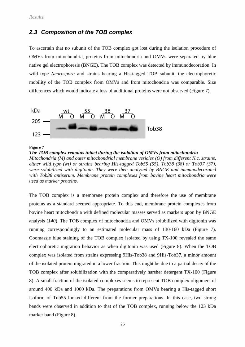
2.3 Composition of the TOB complex
To ascertain that no subunit of the TOB complex got lost during the isolation procedure of
OMVs from mitochondria, proteins from mitochondria and OMVs were separated by blue
native gel electrophoresis (BNGE). The TOB complex was detected by immunodecoration. In
wild type Neurospora and strains bearing a His-tagged TOB subunit, the electrophoretic
mobility of the TOB complex from OMVs and from mitochondria was comparable. Size
differences which would indicate a loss of additional proteins were not observed (Figure 7).
Figure 7
The TOB complex remains intact during the isolation of OMVs from mitochondria
Mitochondria (M) and outer mitochondrial membrane vesicles (O) from different N.c. strains,
either wild type (wt) or strains bearing His-tagged Tob55 (55), Tob38 (38) or Tob37 (37),
were solubilized with digitonin. They were then analyzed by BNGE and immunodecorated
with Tob38 antiserum. Membrane protein complexes from bovine heart mitochondria were
used as marker proteins.
The TOB complex is a membrane protein complex and therefore the use of membrane
proteins as a standard seemed appropriate. To this end, membrane protein complexes from
bovine heart mitochondria with defined molecular masses served as markers upon by BNGE
analysis (140). The TOB complex of mitochondria and OMVs solubilized with digitonin was
running correspondingly to an estimated molecular mass of 130-160 kDa (Figure 7).
Coomassie blue staining of the TOB complex isolated by using TX-100 revealed the same
electrophoretic migration behavior as when digitonin was used (Figure 8). When the TOB
complex was isolated from strains expressing 9His-Tob38 and 9His-Tob37, a minor amount
of the isolated protein migrated in a lower fraction. This might be due to a partial decay of the
TOB complex after solubilization with the comparatively harsher detergent TX-100 (Figure
8). A small fraction of the isolated complexes seems to represent TOB complex oligomers of
around 400 kDa and 1000 kDa. The preparations from OMVs bearing a His-tagged short
isoform of Tob55 looked different from the former preparations. In this case, two strong
bands were observed in addition to that of the TOB complex, running below the 123 kDa
marker band (Figure 8).

Results
27
Figure 8
TOB complex purification by affinity chromatography after lysis of OMVs with TX-100
Bovine heart mitochondria (BHM) were solubilized with TX-100. The TOB complex was
isolated from N.c. strains bearing His-tagged Tob55, Tob38 or Tob37 using TX-100. The
samples were subjected to BNGE and Coomassie blue staining. Membrane protein complexes
from bovine heart mitochondria were used as markers.
Asterisks indicate possible oligomeric forms of the TOB complex.
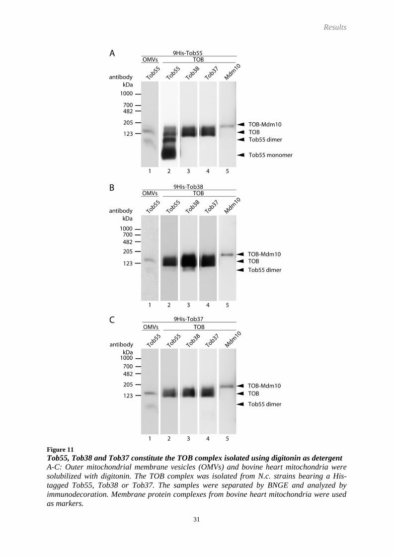
The isolated complexes were further analyzed by immunodecoration after BNGE. The
dominant band of the TOB complex isolated from strains harboring 9His-Tob38 or 9His-
Tob37 showed the same electrophoretic mobility as the complex from solubilized OMVs
without further affinity purification (Figure 9, B and C). This form of the TOB complex
contains Tob55, Tob38 and Tob37 and is therefore representing the TOB complex.
Decoration with antibodies against Mdm10 identified traces of copurified Mdm10 as part of
an apparent molecular mass species of around 200 kDa (Figure 9, B and C), and not as a
constituent of the TOB complex. In this 200 kDa complex, Tob55, Tob38 and Tob37 were
also detected. Considering the molecular weight of Mdm10 of 52.7 kDa, this complex most
likely represents one Mdm10 bound to the TOB complex. It was termed TOB-Mdm10
complex.

Results
28
Figure 9
Tob55, Tob38 and Tob37 constitute the TOB complex isolated using TX-100 as detergent
A-C: Outer mitochondrial membrane vesicles (OMVs) and bovine heart mitochondria were
solubilized with TX-100. The TOB complex was isolated from N.c. strains bearing His-tagged
Tob55, Tob38 or Tob37. The samples were separated by BNGE and analyzed by
immunodecoration. Membrane protein complexes from bovine heart mitochondria were used
as marker proteins.

Results
29
The protein complex running faster than the TOB complex (apparent molecular mass around
90 kDa) contained Tob55 and Tob38 (Figure 9). Tob37 was missing and therefore seems to
be a subunit which can be lost from the TOB complex during isolation. The 90 kDa complex
is referred to as Tob55-Tob38 complex in the following. The Tob55-Tob38 complex makes
up just a very minor species as judged by its intensity upon staining with Coomassie blue
(Figure 8, panel 2 and 3).
Upon comparing immunodecorations of the TOB complex isolated with 9His-Tob55 with
those in which 9His-Tob37 or 9His-Tob38 were present, most Tob55 was found at a complex
running at around 100 kDa, roughly the size of Tob55-Tob38 (Figure 9A). Hence, tagging
Tob55 could possibly have a destabilizing effect on the TOB complex causing a decay of the
complex. Nevertheless, the amount of Tob38 was higher in the TOB complex than in the
Tob55-Tob38 complex. This is inconsistent with the possibility that this 100 kDa species
might have arisen from the loss of Tob37 from the TOB complex.
In addition to the preparations performed with TX-100, the TOB complex was isolated using
digitonin as detergent for the solubilization of the OMVs. With strains expressing 9His-Tob38
or 9His-Tob37, the dominant TOB complex was running somewhat faster than that isolated
with TX-100 (Figure 10). A second, weaker band was present above the main complex,
presumably representing the TOB-Mdm10 complex. The Tob55-Tob38 complex is missing.
This supports the suggestion that the complex is more prone to the loss of Tob37 when the
solubilization is performed with TX-100 (Figure 10). Again, the elution pattern was very
different when the complex was isolated via His-tagged Tob55 in comparison to preparations
from the His9-Tob38 or His9-Tob37 strain (Figure 10, panel 1). Similar to the affinity
purifications performed with TX-100, monomeric Tob55 was found in addition to the TOB
and the TOB-Mdm10 complex. Moreover an enriched complex at around 100 kDa was
observed.
Results
30
Figure 10
TOB complex purification by affinity chromatography after lysis of OMVs with digitonin
Bovine heart mitochondria (BHM) were solubilized with digitonin. The TOB complex was
isolated from N.c. strains bearing His-tagged Tob55, Tob38 or Tob37 by the use of digitonin.
The samples were subjected to BNGE and Coomassie blue staining. Membrane protein
complexes from bovine heart mitochondria were used as markers.
Results
31
Figure 11
Tob55, Tob38 and Tob37 constitute the TOB complex isolated using digitonin as detergent
A-C: Outer mitochondrial membrane vesicles (OMVs) and bovine heart mitochondria were
solubilized with digitonin. The TOB complex was isolated from N.c. strains bearing a His-
tagged Tob55, Tob38 or Tob37. The samples were separated by BNGE and analyzed by
immunodecoration. Membrane protein complexes from bovine heart mitochondria were used
as markers.

Results
32
The TOB complex isolated from OMVs solubilized with digitonin was found to be made up
by the proteins Tob55, Tob38 and Tob37, only (Figure 11). No Tob55-Tob38 complex was
present in the eluate. Therefore, no Tob37 is lost from the TOB complex in course of the Ni-
NTA affinity purification. By the use of the strain, which expresses 9His-Tob55, an
enrichment of Tob55 in a complex with an apparent molecular mass of 100 kDa was observed
and in addition monomeric Tob55 (Figure 11A). This 100 kDa complex was not detected
when antibodies against Tob38, Tob37 or Mdm10 were used. Therefore, Tob55 exists as a
dimer. Interestingly, this Tob55 dimer was also present in solubilized OMVs from all various
strains without any further isolation. This demonstrates that the Tob55 dimer is not an artefact
caused in the course of the Ni-NTA purification. Mdm10 again was running in a complex
with a slightly higher apparent molecular mass than the TOB complex. Thereby it most likely
represents the TOB-Mdm10 complex. Taken together, the use of digitonin and TX-100 for the
solubilization of OMVs resulted in the isolation of complexes of very similar composition.
The only species which was only present in preparations using TX-100 was the Tob55-Tob38
complex. However, only very minor amounts of this complex were found. Therefore, both
detergents are suitable for the purification of the TOB complex.
Antibody supershift assays were performed to further analyze the diverse complexes observed
by BNGE after preparation of the TOB complex by affinity purification. To this end, OMVs
differing in the His-tagged TOB complex subunit were solubilized with digitonin.
Subsequently, penta-His antibody was added and BNGE was performed. Tob38, Tob37 and
Tob55 were present in the TOB complex as well as in the far less abundant TOB-Mdm10
complex running directly above the TOB complex at around 200 kDa. Both complexes could
be shifted with the penta-his antibody directed against Tob55, Tob38 or Tob37 (Figure 12).
Tob55 was further detected in a second abundant complex of around 100 kDa. This band
could only be shifted when the His-tag was attached to the Tob55, but not with His-tagged
Tob38 and Tob37 (Figure 12). Decoration with Tob38 and Tob37 antibody showed the
absence of these proteins in the 100 kDa complex. The 100 kDa complex was not the result of
a decay of the TOB complex due to the modification of Tob55 by the attachment of the His-
tag, since it was also present in the His9-Tob38 and His9-Tob37 strains and even in the wild
type strain (Figure 12A). In addition, a control with penta-his antibody alone excluded an
unspecific interaction with the Tob55 antibody during immunodecoration. When affinity
purification experiments were performed with His-tagged Tob55 and digitonin, only traces of
porin and Mdm10 could be found in addition to the subunits Tob55, Tob38 and Tob37

Results
33
(Figure 6B). Mdm10 and porin, as well as Tom40 are ß-barrels themselves. Therefore, they
are substrates of the TOB complex. The distribution of these proteins was determined by
immunodecoration following BNGE and Western blotting. An interaction of porin or Tom40
with the TOB complex was not detected in the antibody supershift assays (Figure 12, D and
F). Mdm10 was not detected together with Tob55 in the complex of around 100 kDa, but it
was present in the complex of 200 kDa which could be shifted with His-tagged Tob55, Tob38
or Tob37 (Figure 12B).
The 100 kDa complex most likely represents a Tob55 dimer, since none of the co-isolated
proteins was found to be part of it.

Results
34
Figure 12
Tob55 is not only present in the TOB complex but in addition forms a dimer and Mdm10 is
present in a 200 kDa complex.
A-F: Outer mitochondrial membrane vesicles from different N.c. strains, either wild type (wt)
or strains bearing a His-tagged Tob55 (55), Tob38 (38), or Tob37 (37), were solubilized with
digitonin. Where indicated, monoclonal His-antibody was added to the solubilized proteins or
loaded as controls (c) before the samples were separated by BNGE. Immunodecoration was
performed with antibodies as indicated. Membrane protein complexes from bovine heart
mitochondria were used as markers.

Results
35
When TX-100 was used only a very faint band representing a putative Tob55 dimer was
observed (Figure 13A). This is in contrast to the result of solubilisation of the OMVs with
digitonin (Figure 12A). Nonetheless, upon isolation of the TOB complex with His-tagged
Tob55 using TX-100, a substantial amount of the very same band at around 100 kDa was
present in the eluate (Figure 8, Figure 9A). This finding is comparable to preparations
performed with digitonin (Figure 10, Figure 11A). The unexpected appearance of the Tob55
dimer in the eluate of TOB complex preparations performed with TX-100 suggests that TX-
100 leads to disintegration of the Tob55 dimers. Ni-NTA affinity isolation of the TOB
complex includes loading of solubilized OMVs in presence of high detergent concentrations.
Upon elution the detergent concentration is reduced. Therefore, isolated Tob55 monomers
might reform dimers upon decrease of the detergent concentration or when their concentration
is increased.
The origin of the Tob55 dimers was to be analyzed in more detail. To this end, affinity
purification was performed with a strain expressing the short isoform of Tob55 with a
ninefold His-tag as well as the intermediate Tob55 isoform bearing a Flag-tag. Pulldowns
directed against one or the other tag only eluted one kind of tag. This indicates that Tob55 is
not present in dimers under the given conditions (Figure 14). However, with this result one
can only exclude the presence of Tob55 dimers formed by the short and intermediate Tob55
isoform. Tob55 dimers constituted by different Tob55 isoform combinations might still exist.
Monomeric Tob55 was found in addition to Tob55 dimers preparations performed with TX-
100 as well as with digitonin (Figure 8, Figure 10). It remains to be elucidated where these
Tob55 monomers originated from. In case of a decay of the TOB complex, there should also
be an equivalent amount of monomeric Tob38 and Tob37 which should also be enriched
when the TOB isolation is performed with 9His-Tob38 or 9His-Tob37. Such an enrichment of
Tob38 and Tob37 in the eluate was not observed (Figure 5, Figure 6). In conclusion, the
enriched Tob55 originates from an excess of Tob55 over Tob38 and Tob37 in mitochondria
rather than from a decay of the TOB complex.

Results
36
Figure 13
Analysis of the isolated TOB complex by Blue native shift experiments
Outer mitochondrial membrane vesicles from different N.c. strains, either wild type (wt) or
strains bearing His-tagged Tob55 (55), Tob38 (38), or Tob37 (37), were solubilized with TX-
100. Where indicated, His-antibody was added to the solubilized proteins or loaded alone as
a control (c). Samples were then analyzed by BNGE. Immunodecoration was performed with
diverse antibodies as indicated.
Membrane protein complexes from bovine heart mitochondria were used as marker proteins.
Figure 14
The TOB complex contains only one Tob55 subunit
Outer mitochondrial membrane vesicles bearing a His-tagged short isoform and a Flag-
tagged intermediate isoform of Tob55 were solubilized with TX-100. Proteins were isolated
by Ni-NTA or anti-Flag-tag affinity purification and analyzed by SDS-PAGE followed by
immunodecoration.
OMVs before (S) or after (L) clarifying spin. FT: flowthrough with unbound proteins of the
Ni-NTA column; E: eluate of bound proteins

Results
37
The antibody supershift assays demonstrated the presence of Mdm10 in a complex of around
200 kDa together with Tob55, Tob38 and Tob37. This 200 kDa complex therefore reflects the
association of Mdm10 with the TOB complex. Since porin and Tom40 were not observed to
bind to the TOB complex, unspecific co-purification of ß-barrel precursor proteins can be
excluded.
Taken together, Tob55, Tob38 and Tob37 exclusively constitute the TOB complex with an
estimated molecular mass of ca 150 kDa. Mdm10 is not part of the TOB complex. Very small
amounts of Mdm10 are recovered with Tob55, Tob38 and Tob37 in a complex of around 200
kDa, forming the TOB-Mdm10 complex. Furthermore, a substantial amount of Tob55 in the
outer mitochondrial membrane occurs in a form that is not associated with Tob38 and Tob37.

Results
38
2.4 Tob55, Tob38 and Tob37 are present in the TOB complex in a 1:1:1 stoichiometry
In order to further analyze the structure of the TOB complex it was necessary to determine the
stoichiometry of the three different subunits. This could not be achieved by determination of
the molecular mass of the complex since reliable methods for measuring the precise molecular
mass of membrane inserted complexes do not exist. Therefore, quantitative mass spectrometry
was employed to determine the stoichiometry of the subunits Tob55, Tob38 and Tob37 in the
TOB complex. TOB complexes were excised from gels after BNGE (Figure 8, Figure 10).
They were analyzed by isotope diluted mass spectrometry (IDMS) with quantified stable
isotope (13
C/15
N) labeled internal peptide standards of all three subunits (Figure 15G; see also:
Material and Methods, 6.1.7, “Peptides”). Figure 15 depicts representative measurements of
technical replicates of the different isolated complexes to illustrate the reproducibility of the
quantification by IDMS. A total of six biological replicates of the TOB complex, and four
biological replicates of the Tob55-Tob38 complex, isolated using TX-100 or digitonin, from
both strains, His9-Tob38 and His9-Tob37, were analyzed. The various analyses yielded the
same results, as described in the following: TOB complexes isolated via 9His-Tob38 or 9His-
Tob37, with either TX-100 or digitonin, had a 1:1:1 ratio between the subunits Tob55, Tob38
and Tob37 (Figure 15). Therefore, the slight difference in the electrophoretic mobility
between the TOB complex isolated with TX-100 and digitonin (Figure 8, Figure 10) was
caused by the use of the different detergents but not by different stoichiometries of the
subunits of the complex. The absence of Tob37 in the Tob55-Tob38 complex, found in
preparations performed with TX-100, could be confirmed by the IDMS measurements,
resulting in a 1:1:0 ratio (Figure 15, C and D). Moreover, protein identification by LC-
MS/MS could not detect any other proteins than Tob55, Tob38 and Tob37 in the TOB or
Tob55-Tob38 complex. A 1:1:1 ratio of Tob55, Tob38 and Tob37 monomers results in a
molecular weight of about 140 kDa. This is in accordance with the electrophoretic mobility of
the complex in relation to the protein complexes from bovine heart mitochondria that served
as standards (Figure 7 - Figure 13). Thus it is concluded that one Tob55 protein assembles
with one Tob38 and Tob37 molecule each.

Results
39
Figure 15
Tob55, Tob38 and Tob37 are present in the TOB complex in a 1:1:1 stoichiometry
A, C and E: TOB complex was isolated from N.c. strains bearing a His-tagged Tob38 using
TX-100 (A and C) or digitonin (E). It was subjected to BNGE and cut out after Coomassie
blue staining of the native gel. The complexes were analyzed by IDMS. By comparing the two
peak areas of the heavy and light peptides, the amount of each TOB complex subunit could be
determined. The relative amounts of the native peptides and thereby the native proteins are
depicted; Tob55 was set to 1. Representative experiments for each measured protein complex
are shown. The error bars depict the standard deviation of the measurements of several
diverse samples (n) originating from the very same isolated complex and illustrate the
reproducibility of the quantification by IDMS.
B, D and F: Same as in “A”, “C” and “E” with N.c. strains bearing a His-tagged Tob37. TX-
100 (B and D) or digitonin (F), was used for solubilization.
G: Quantified stable isotope (13
C/15
N) labeled internal peptide standards, AQUA
(AbsoluteQUAntification) peptides, for the proteins Tob55 (55), Tob38 (38) and Tob37 (37)
are shown; the amino acid substituted with a stable isotopic amino acid is written in gray.

Results
40
IDMS was also performed with the other complexes present as higher apparent molecular
mass species in the BNGE than the TOB complex (Figure 8, Figure 10). However, due to the
tailing of the protein bands in the upper part of the gels reproducible and therefore reliable
results could not be obtained.
In addition, the TOB complex which was isolated by affinity purification using His-tagged
Tob55 was mass-spectrometrically analyzed. The TOB complex was determined to have a
1:1:1 stoichiometry. Due to the high amounts of Tob55 in the isolate and the tailing of Tob55
in many replicates, the relative amounts of Tob55 were slightly higher (Figure 16, A and C).
The presence of more than one Tob55 in the TOB complex isolated with His-tagged Tob55
can be excluded, since this complex has the very same running behavior as the complexes
isolated via a His-tagged Tob37 or Tob38. Furthermore, the TOB complex did not show an
increased Tob55 upon immunodecoration with Tob55 antiserum (Figure 9, A-C, Figure 11,
A-C). The majority of Tob55 was detected in the complexes with an apparent molecular mass
of ca. 100 kDa (Figure 16, B and D). Only traces of Tob38 were found in this band when TX-
100 was used for the TOB complex preparation (Figure 16B). The excess of Tob55 over
Tob38 in preparations of the TOB complex obtained with TX-100 indicates that Tob55
dimers apparently overlay smaller amounts of the Tob55-Tob38 complex in the native gel.
When digitonin was used, no Tob55-Tob38 complex was observed and accordingly no Tob38
was detected in the 100 kDa band. The distinct bands on the BN-gels with an apparent
molecular mass of ca. 60 kDa (Figure 8, Figure 10) were identified as Tob55 monomers, since
only Tob55 was detected.
In summary, these studies support our findings that there is a large amount of Tob55 which is
not associated with Tob38 and Tob37.

Results
41
Figure 16
Dimer-formation of Tob55
A and B: TOB complex was isolated from N.c. strains bearing a His-tagged Tob55 using TX-
100 and was subjected to BNGE and then cut out after Coomassie blue staining of the native
gel. The complexes were analyzed by IDMS. By comparing the two peak areas of the heavy
and light peptides, the amount of each TOB complex subunit could be determined. The
relative amounts of the native peptides and thereby the native proteins are depicted; Tob55
was set to 1. Representative experiments for each protein complex analyzed are shown. The
error bars depict the standard deviation of the measurements of diverse samples originating
from the very same isolated complex and illustrate the reproducibility of the quantification by
IDMS.
C and D: Same as in “A” and “B”, but solubilization was performed with digitonin.
Results
42
2.5 Reconstruction of the isolated TOB complex by cryo-electron microscopy
In order to obtain further insights into the structure of the TOB complex cryo-electron
microscopy (cryo-EM) analysis was performed. This analysis was carried out by single
particle analysis in collaboration with Dennis Thomas (Baumeister group, MPI Martinsried,
GER). A 2D structure prediction of Tob55 was made since no crystal structure of a TOB
complex subunit was available. The Research Collaboratory for Structural Bioinformatics
Protein Data Bank (RCSB PDB) was searched for known structures with sequence homology
and similar predicted secondary structure. The secondary structure prediction for Tob55
showed remarkable similarity to the known structure of the FhaC ß-barrel protein. Tob55 was
suggested to have 16 ß-strands that form a transmembrane ß-barrel like FhaC and an N-
terminal region harbouring one POTRA domain, whereas FhaC has two.
Using MODELLER, a 3D model for Tob55 was constructed by essentially mapping the
predicted Tob55 secondary structure onto the FhaC 3D structure (PDB I.D. 2QDZ) (121). The
resulting model follows the 2D structure prediction well. The ß-strands are connected by short
turns or longer loops sticking out on both sides of the membrane (Figure 17A). Two long
defined loops (red and blue loops) are present at the cytoplasmic side in the model predicted
for Tob55 (Figure 17, Figure 18, Figure 19). Superposition of both protein structures revealed
that these two loops, comprising amino acid residues 267 to 291 (blue loop) and amino acid
residues 412 to 439 (red loop) of Tob55 correspond to loop 5 (L5) and loop 8 (L8),
respectively, in the FhaC structure (Figure 18, Figure 19). The loops at the cytoplasmic face
of the predicted Tob55 ß-barrel structure are longer and more exposed than those of FhaC.
However, the blue loop of FhaC in fact must be longer in reality since the published structure
lacks amino acid residues 384 to 398 (Figure 19A, red boxed residues in loop 5) (121).
Therefore, this loop might show higher similarity to the corresponding loop in the Tob55
structure. Another significant feature in the FhaC structure is a large loop between the extra-
cellular ends of strands 11 and 12. This loop is folded back into the lumen of the barrel (L6,
green loop) joining an N-terminal α-helix (pink helix) to occlude the ß-barrel (Figure 17,
Figure 18, Figure 19). The sequence corresponding to the N-terminal α-helix of FhaC was
missing in the predicted structure of Tob55 (Figure 17A). However, a long loop stretching
through the interior of the ß-barrel was predicted between the Tob55 strands 11 and 12,
containing amino acids 318 to 376 (green loop) (Figure 17, Figure 18, Figure 19). At the tip
of the loop the VRGY/F tetrad was localized (Figure 19B, grey framing). The VRGY/F tetrad
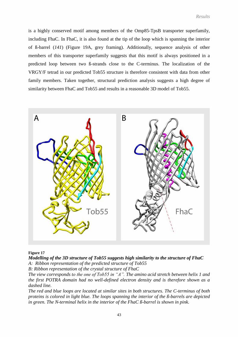
Results
43
is a highly conserved motif among members of the Omp85-TpsB transporter superfamily,
including FhaC. In FhaC, it is also found at the tip of the loop which is spanning the interior
of ß-barrel (141) (Figure 19A, grey framing). Additionally, sequence analysis of other
members of this transporter superfamily suggests that this motif is always positioned in a
predicted loop between two ß-strands close to the C-terminus. The localization of the
VRGY/F tetrad in our predicted Tob55 structure is therefore consistent with data from other
family members. Taken together, structural prediction analysis suggests a high degree of
similarity between FhaC and Tob55 and results in a reasonable 3D model of Tob55.
Figure 17
Modelling of the 3D structure of Tob55 suggests high similarity to the structure of FhaC
A: Ribbon representation of the predicted structure of Tob55
B: Ribbon representation of the crystal structure of FhaC
The view corresponds to the one of Tob55 in “A”. The amino acid stretch between helix 1 and
the first POTRA domain had no well-defined electron density and is therefore shown as a
dashed line.
The red and blue loops are located at similar sites in both structures. The C-terminus of both
proteins is colored in light blue. The loops spanning the interior of the ß-barrels are depicted
in green. The N-terminal helix in the interior of the FhaC ß-barrel is shown in pink.

Results
44
Figure 18
Superposition of Tob55 and FhaC illustrates corresponding elements in the architecture of
both transporters
A and B: Ribbon representation of two corresponding views of the predicted structure of
Tob55 (row 1, yellow) and the crystal structure of FhaC (row 2, grey) are illustrated. Row 3
depicts an overlay of both structures.
C: Insight into cutaway views of Tob55 and FhaC is given.
The red and blue loops are located on similar sites in both structures. The C-terminus of both
proteins is colored in light blue. The loops spanning the interior of the ß-barrels are depicted
in green. The N-terminal helix in the interior of the FhaC ß-barrel is shown in pink.

Results
45
Figure 19
Analysis of the secondary structural elements of FhaC and Tob55
A: Secondary structural elements of the FhaC structure
Helices and ß-strands of the POTRA domains are presented in yellow and grey, respectively.
ß-strands which are forming the ß-barrel are coloured in light blue. The FhaC model does not
include the first two N-terminal residues, the loop after helix 1 as well as the residues 384 –
398 of loop 5 (red framing). The VRGY/F tetrad in the green loop is framed in grey.
B: The predicted structure of the short isoform of Tob55 is illustrated in the same way as in
“A”. Amino acid residues which were not part of ß-strands in the 2D structure prediction of
Tob55 but turned out to contribute to the ß-strands in the 3D modelling are underlined.

Results
46
TOB complexes bearing a His-tag either at the Tob37 or Tob38 subunit were placed on grids
with a “lacey” carbon film. Lacey meaning the surface is mostly irregular holes in the film.
The samples form a thin layer of liquid in the holes during blotting. These complexes are
more homogenous than those isolated using His-tagged Tob55, as described earlier (chapter:
2.3, Figure 8, Figure 10).
Images of TOB complexes in vitreous ice were obtained (Figure 20). Particles visible in the
images were boxed from the images in an automated manner.
Figure 20
A typical micrograph of TOB complex particles obtained by cryo-electron microscopy
A micrograph of vitrified TOB complex, imaged at 20-25 e- per Å, -2.2 µm ΔF is depicted.
Since there were no models available to begin alignment and reconstruction with, the first step
of the image analysis was to perform a reference free alignment on the particles. Reference
free alignment potentially can produce classified images representing closely related views of
the particle whose orientations can be determined. At best, these views might be used to
generate a starting model. At a minimum, it gives some insight into the basic architecture of
the particle and non-particle images, such as carbon edges, can be identified (Figure 21C).
Reference free alignment averages were determined from three different subsets of particle
images collected in one session from one grid with TOB complex prepared from the His9-
Tob37 N. crassa strain (Figure 21A). These averages are essentially the same although they
appear shifted and rotated relative to one another. This shift and rotation is a result of each set
of images starting from a different randomly determined starting point. These averages have
one prominent globular protein mass.

Results
47
After classification of reference free aligned particle images, the individual class averages
show one or two additional masses occurring in different positions relative to the main mass
(Figure 21, B and C). These appear much like different projections of a three dimensional
complex containing three subunits. None of these class averages appear symmetric nor do
they have a visible channel.
Figure 21
Reference free alignment of single particle images suggests that the TOB complex
comprises three subunits
A: Particle images of the TOB complex collected in one session from one grid were divided
into three subsets and subjected to reference free alignment.
B: Classification of reference free aligned particle images
C: Class averages out of “B” are enlarged. Images 1, 3 and 4 depict class averages of TOB
complex particles; image 2 illustrates a class average of non-particle images, such as carbon
edges.

Results
48
A 3D model could not be obtained from the class averages of the reference free alignment.
Therefore, a starting model needed to be generated to be able to begin alignment and
reconstruction of the complexes. Since the reference free alignment classes strongly suggested
that there were three discrete domains or subunits, a reference with three subunits was an
obvious starting point. The reference was centered on a density map calculated from the
Tob55 model. Two subunits were expected to be found on the cytoplasmic surface of Tob55.
Thus it seemed reasonable to place two spheres in the model that are associated with Tob55.
However, there was no information to guide the placement of such spheres. Therefore, to
avoid any bias in the reconstruction by a defined prepositioning of spheres for the subunits
Tob38 and Tob37, an amorphous ring was placed above the cytoplasmic face of the Tob55 ß-
barrel.
With successive rounds of reconstruction, the ring shape converted into two defined spherical
structures that are facing each other, presumably presenting the subunits Tob38 and Tob37
(Figure 22). The bulky central part of the structure retained the original overall appearance of
the model of the ß-barrel protein Tob55. However, after the formation of the two peripheral
subunits and a progressing improvement of the reconstruction, conformational flexibility in
the structure started to increasingly influence the alignment, thereby blurring the structure
again (Figure 22, compare 5 and 6). A maximum-likelihood-analysis might be a good
approach in the future for the separation of complexes with slight internal conformational
differences.
In addition, the reconstruction is limited by the small molecular mass of the TOB complex.
The analysis of the TOB complex in this study revealed that the complex has an apparent
molecular mass of 140 kDa, which is much smaller than expected from previous results. The
small size of the protein complex presents severe difficulties in the cryo-EM analysis due to a
low signal-to-noise ratio in the image. The low signal-to-noise ratio is reflected in the minimal
difference between cross-correlation intensities resulting from images of the TOB complex
and those calculated from images of buffer background aligned against the same reference
data projections (Figure 23). The cross correlations of data containing images are consistently
above the background but by a much smaller margin than might be expected for larger
complexes. This low signal-to-noise ratio results in errors in alignment and therefore errors in
assignment of correct angles to images.

Results
49
Figure 22
Reconstruction of the isolated TOB complex
TOB complex was isolated from the His9-Tob37 N.c. strain and subjected to cryo-electron
microscopy single particle analysis. Progress with increasing rounds of alignment and
reconstruction is illustrated from the left to the right. A superposition of the predicted
structure of Tob55 and the reconstruction is shown (1, 4 and 6).

Results
50
Figure 23
The small molecular mass of the TOB complex results in low signal-to-noise ratios
The cross-correlation intensities of classified particle images are depicted. The green line
reveals the number of particle images assigned to a certain class number. The x-axis depicts
the class number, the y-axis the intensity of cross correlation. Cross-correlation intensities
originating from vitrified buffer are coloured in red, signals emerging from TOB complex
particles in blue. The average cross-correlation intensities of the TOB complex particles are
illustrated in pink.
Interestingly, fitting of the predicted structure of Tob55 into the reconstruction suggests that
the extended loops at the cytoplasmic face of Tob55 are sticking out into the spheres of the
subunits Tob37 and Tob38. They presumably form the connection sites between the latter
proteins and Tob55 (Figure 24, A and B).
Samples of isolated TOB complex were incubated with Ni-NTA-Nanogold. This was done in
order to identify and localize the His-tagged subunits and thereby determine the organization
of the complex. Reconstructions of the TOB complex containing labelled or unlabelled 9His-
Tob37 were obtained. After density difference calculations, extra mass could mainly be
detected in one of the two spheres, identifying it as Tob37 (Figure 24, A and B). Furthermore,
a mass strand between Tob37 and Tob55 or the membrane was observed. Such a structure
was not recognized with Tob38 (Figure 24). In conclusion, the results obtained in this study,
including analysis of stoichiometries, molecular masses, cryo-electron microscopy and
molecular modeling allow a preliminary organization of structure of the TOB complex to be
proposed (Figure 24C).
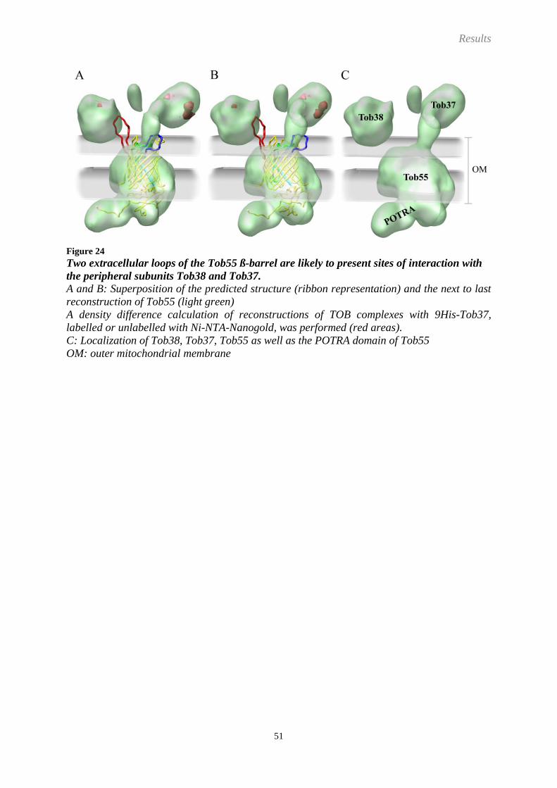
Results
51
Figure 24
Two extracellular loops of the Tob55 ß-barrel are likely to present sites of interaction with
the peripheral subunits Tob38 and Tob37.
A and B: Superposition of the predicted structure (ribbon representation) and the next to last
reconstruction of Tob55 (light green)
A density difference calculation of reconstructions of TOB complexes with 9His-Tob37,
labelled or unlabelled with Ni-NTA-Nanogold, was performed (red areas).
C: Localization of Tob38, Tob37, Tob55 as well as the POTRA domain of Tob55
OM: outer mitochondrial membrane

Results
52
2.6 The subunits of the TOB complex are tightly associated with the outer mitochondrial membrane
Cryo-electron microscopy analysis revealed a mass strand for Tob37 which might function as
a membrane anchor. To get some further insight into the interaction of Tob37 and the other
TOB subunits with the membrane, alkaline extraction was performed. By this, it should be
determined whether the subunits of the TOB complex are only loosely attached or tightly
anchored to the outer mitochondrial membrane. Tob37 turned out to be very resistant to
alkaline extraction. It could not be removed from the membrane at pH values up to 12.5. The
extra mass strand observed for Tob37 in the reconstruction of the TOB complex might
represent a membrane anchor. Thus, Tob37 can be classified as a membrane anchored protein.
Tob38 is slightly more sensitive to alkaline treatment than the signal-anchored protein Tom70
and roughly half of Tob38 is extracted at pH 12.5. Thus, Tob38 rather has the characteristics
of a peripheral membrane protein.
Tob55 is predicted to be a ß-barrel protein and thus is expected to have a tight interaction with
the membrane. In agreement with this, even under very basic conditions no extraction of
Tob55 could be observed (Figure 25). Mdm10 has also been assigned to the family of ß-barrel
membrane proteins and its membrane integration has been demonstrated (108). Taken
together, all subunits of the TOB complex reveal a tight interaction with the membrane.

Results
53
Figure 25
Tob55, Tob38 and Tob37 are tightly associated with the outer mitochondrial membrane
OMVs were subjected to alkali extraction with 0.1 M sodium carbonate adjusted to different
pH values of 10.8, 11.5 and 12.5. As a control, proteins were treated with HEPES buffer at
pH 7.4 (control). Membrane bound proteins were sedimented by centrifugation (P). Soluble
proteins in the supernatant were precipitated with trichloroacetic acid (S).

Results
54
2.7 The isolated TOB complex shows specific substrate binding behavior
In order to study the functionality of the isolated TOB complex, its ability to bind substrates
was analyzed. To this end, the TOB complex was immobilized on a Ni-NTA matrix,
thoroughly washed and incubated with radioactive mitochondrial precursor proteins. Tom40
precursor showed a weak binding, slightly above background level. In constrast, a distinct
binding to the TOB complex was observed for Tom22. No significant binding of porin was
found (Figure 26).
Figure 26
The immobilized TOB complex shows specific binding of certain substrates
TOB was affinity-purified from OMVs bearing a 9His-Tob37 on a Ni-NTA matrix by using
TX-100. The isolated complex was incubated with different radioactive precursor proteins
(TOB) before being eluted from the column. As a control, a mock isolation without OMVs was
performed and the matrix was treated the same way with radioactive precursor proteins
(mock). After washing, the bound precursor proteins were eluted and visualized by
autoradiography.
Radioactive precursor proteins: Tom40, Porin, Su9-DHFR, Tom22; mock, no plasmid was
added to the transcription/translation-reaction
The immobilization of the TOB complex on the matrix might have hindered the interaction
with its substrates. Therefore, a pepspot binding assay was performed, which allows the
isolated TOB to interact with peptide libraries of its substrates Tom40 and porin in a soluble
state. The peptide libraries used in the assay consisted of amino acid residues covering the
complete sequence of the respective substrates. The assay allowsThis enables the localization

Results
55
of specific binding sites on the substrates. With both peptide libraries, TOB presented a
distinct and reproducible binding pattern (Figure 27, Figure 28). Interestingly, binding to the
“ß-signal” of the ß-barrel substrate proteins could not be detected.
Figure 27
The isolated TOB complex interacts with distinct peptides of a porin peptide library
A: The TOB complex was isolated by Ni-NTA affinity purification from N. crassa by the use of
TX-100. The porin peptide library was incubated with the isolated TOB complex. Bound TOB
complex was blotted from the pepspot-membrane to a PVDF-membrane and
immunodecorated with penta-His antibodies.
B and C: Schematic view of the binding sites on the peptide library
Strong binding of the TOB complex is indicated by black pepspot dots (B) and black peptide
fragments in the peptide library overview (C). Weak interactions are in dark grey. The “ß-
signal” is underlined (# 68).
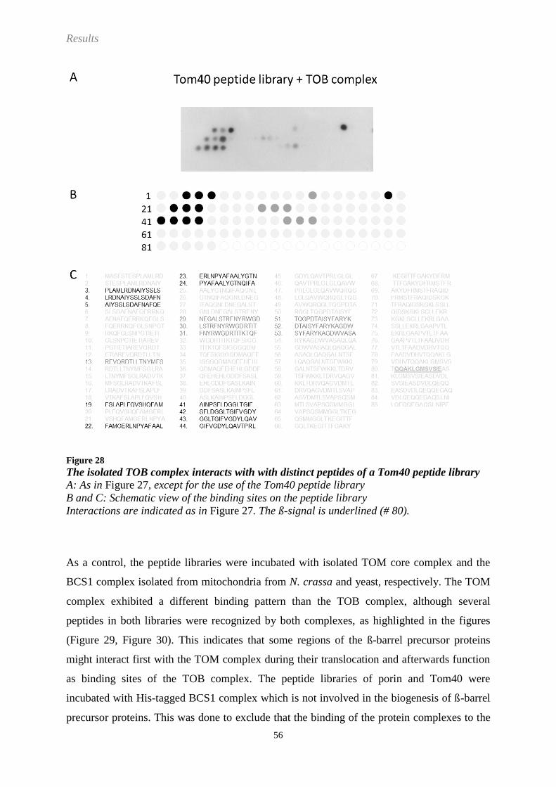
Results
56
Figure 28
The isolated TOB complex interacts with with distinct peptides of a Tom40 peptide library
A: As in Figure 27, except for the use of the Tom40 peptide library
B and C: Schematic view of the binding sites on the peptide library
Interactions are indicated as in Figure 27. The ß-signal is underlined (# 80).
As a control, the peptide libraries were incubated with isolated TOM core complex and the
BCS1 complex isolated from mitochondria from N. crassa and yeast, respectively. The TOM
complex exhibited a different binding pattern than the TOB complex, although several
peptides in both libraries were recognized by both complexes, as highlighted in the figures
(Figure 29, Figure 30). This indicates that some regions of the ß-barrel precursor proteins
might interact first with the TOM complex during their translocation and afterwards function
as binding sites of the TOB complex. The peptide libraries of porin and Tom40 were
incubated with His-tagged BCS1 complex which is not involved in the biogenesis of ß-barrel
precursor proteins. This was done to exclude that the binding of the protein complexes to the

Results
57
peptide libraries resulted from unspecific hydrophobic protein interactions or was caused by
the presence of the His-tag attached to the membrane protein complexes. There was a
considerable overlap of the binding pattern of the BCS1 complex with that observed for the
TOB complex (Figure 31, Figure 32). Therefore, the specificity of the binding of the TOB
complex to the substrate peptides has to be further verified.
Figure 29
The isolated TOM core complex interacts with distinct peptides of a porin peptide library
A: The TOM core complex was isolated by Ni-NTA affinity purification from N. crassa by the
use of DDM. The porin peptide library was incubated with the isolated TOB complex. Bound
TOM core complex was blotted from the pepspot-membrane to a PVDF-membrane and
immunodecorated with penta-His antibodies.
B and C: Schematic view of the binding sites on the peptide library
Strong binding of the TOM complex is depicted by black pepspot dots (B) and black peptide
fragments in the peptide library overview (C). Weak interactions are colored in dark grey.
Binding sites which are also recognized by the TOB complex are illustrated by open cyles (B)
and underlined peptide fragments (C). The “ß-signal” is underlined (# 68).

Results
58
Figure 30
The isolated TOM core complex interacts with distinct peptides of a Tom40 peptide library
A: As in Figure 29, except for the use of the Tom40 peptide library
B and C: Schematic view of the binding sites on the peptide library
Interactions are indicated as in Figure 29. The ß-signal is underlined (# 80).
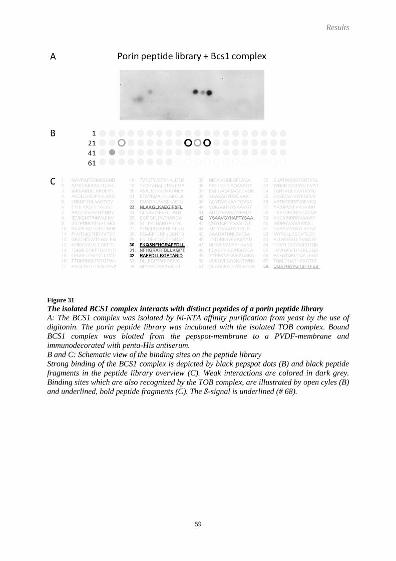
Results
59
Figure 31
The isolated BCS1 complex interacts with distinct peptides of a porin peptide library
A: The BCS1 complex was isolated by Ni-NTA affinity purification from yeast by the use of
digitonin. The porin peptide library was incubated with the isolated TOB complex. Bound
BCS1 complex was blotted from the pepspot-membrane to a PVDF-membrane and
immunodecorated with penta-His antiserum.
B and C: Schematic view of the binding sites on the peptide library
Strong binding of the BCS1 complex is depicted by black pepspot dots (B) and black peptide
fragments in the peptide library overview (C). Weak interactions are colored in dark grey.
Binding sites which are also recognized by the TOB complex, are illustrated by open cyles (B)
and underlined, bold peptide fragments (C). The ß-signal is underlined (# 68).

Results
60
Figure 32
The isolated BCS1 complex interacts with distinct peptides of a Tom40 peptide library
A: As in Figure 31, except for the use of the Tom40 peptide library
B and C: Schematic view of the binding sites on the peptide library
Interactions are indicated as in Figure 31. The ß-signal is underlined (# 80).
Discussion
61
3 Discussion
3.1 Composition of the TOB complex
The aim of the present study was to enhance our knowledge about the composition and
biochemical as well as functional characteristics of the TOB complex in the outer
mitochondrial membrane. Such an analysis necessitated the isolation of the complex in a pure
form. Up to now, the TOB complex has not been purified. So far, only small scale
preparations resulting from affinity columns were reported that could be analyzed by
decoration with antibodies. In a first step Neurospora strains were generated that had a His-
tag on one of its subunits. His-tagged Tob55 was used because it was predicted to form the
main component of the TOB complex as a membrane inserted ß-barrel protein (97, 105, 113,
114).
The TOB complex was isolated by Ni-NTA affinity purification from OMVs which were
solubilized either with the mild detergent digitonin or with TX-100. With both detergents
Tob55, Tob38 and Tob37 were the only proteins which were considerably enriched in the
eluate and only traces of Mdm10 were present. Therefore the loss of subunits due to the use of
the comparatively harsher detergent TX-100 could be excluded.
Since the His9-Tob55ST strain was only expressing the short but not the long and
intermediate Tob55 isoforms, the loss of further subunits due to the absence of these isoforms
had to be considered. Therefore, strains with His-tagged Tob38 or Tob37 were created which
are expressing all three Tob55 isoforms. Preparations of the TOB complex from these strains
yielded the same results as obtained by the use of His-tagged Tob55. Thus, Tob55, Tob38 and
Tob37 could be identified as the components of the TOB complex in N. crassa. This finding
is consistent with observations made in yeast (98, 105, 107). Since only minor amounts of
Mdm10 were detected in the eluate, it was suggested that Mdm10 is not a subunit of the TOB
complex, but rather functions as an interaction partner of the TOB complex. By being a ß-
barrel protein itself, Mdm10 might also copurify with the complex as a substrate. However,
no other substrates such as the ß-barrel proteins porin or Tom40 could be coisolated.
In a next step, the TOB complex isolated from all three different strains with His-tagged
Tob55, Tob38 or Tob37 was subjected to BNGE analysis. Several protein complexes could be
identified in the eluate. Indepent of the detergent used, two complexes were found with all
three strains, the TOB complex and very minor amounts of a TOB-Mdm10 complex. By
immunodecoration Tob55, Tob38 and Tob37 were identified as constituents of the TOB

Discussion
62
complex and Mdm10 was additionally found in the TOB-Mdm10 complex. Using membrane
protein complexes from the respiratory chain of bovine heart mitochondria as a protein
standard indicated an apparent molecular size between 130 and 160 kDa for the TOB
complex. Association of Mdm10 with the TOB complex resulted in a complex with an
apparent molecular mass of 200 kDa. This is in contrast to former publications suggesting a
molecular mass of 200 - 250 kDa for the TOB complex (97, 135, 136). However, in those
published studies, hydrophilic, soluble proteins were used as protein standards for the mass
estimation of the TOB complex in BNGE analysis. Considerable discrepancies between the
migration behavior of membrane integrated and soluble proteins demonstrate that soluble
markers are not appropriate for the mass estimation of membrane proteins (140). In contrast to
soluble proteins, membrane proteins contain substantial amounts of bound lipids, detergent
and Coomassie-Blue when subjected to BNGE. The latter dye binds preferentially to
hydrophobic surfaces of proteins and to basic amino acid residues. Therefore, soluble proteins
bind this anionic dye which enables anodic migration to different degrees and provide these
proteins with a different number of negative charges (142). Different amounts of protein-
bound lipids additionally bias the running behavior of membrane proteins and thereby
severely compromise mass estimations of membrane proteins by soluble markers. Thus,
respiratory chain complexes of bovine heart mitochondria represent a more reliable marker
for the mass determination of the TOB complex.
The apparent molecular masses determined by BNGE analysis in combination with membrane
protein markers are also highly consistent with the stoichiometry of the TOB complex. The
stoichiometry was measured by isotope diluted mass spectrometry which is a novel and highly
precise method to analyze the relative amounts of proteins within a mixture (143, 144). A
1:1:1 ratio between the TOB complex components Tob55, Tob38 and Tob37 was measured,
resulting in a calculated molecular protein mass for the TOB complex of 140 kDa. BNGE
analysis and volume estimations of the cryo-EM structure of the TOB complex exclude a
composition of two or more of each TOB complex subunit (e.g. a 2:2:2 stoichiometry).
Although the same stoichiometry for the TOB complex isolated either with TX-100 or
digitonin was measured, a slight difference in their running behavior could be observed in the
BNGE. This might be attributed to the different properties of the detergents, the relative harsh
TX-100 as compared to the mild digitonin. Although it was shown that protein-bound
detergent is replaced by Coomassie-dye during the BNGE (145), the detergent used causes
differences in the migration behavior of the protein. On the one hand, the proteins are
solubilized with different efficiencies, leaving different amounts of lipids on the protein (140),

Discussion
63
on the other hand, differences in the ratios of hydrophilic and hydrophobic, membrane
embedded parts of membrane complexes might also cause slight variations in the amount of
protein-bound lipids remaining on membrane proteins of comparable mass, although they
were solubilized with the same detergent. Thus, moderate differences in the migration
behavior between sample and standard proteins in various detergents are a common
observation, especially when proteins are analyzed that were solubilized with a mild detergent
such as digitonin.
Association of one Mdm10 molecule with the 140 kDa TOB complex, results in a complex of
calculated 190 kDa, which is in agreement with the migration behavior of this TOB-Mdm10
complex in BNGE. BN-shift analysis demonstrated that Mdm10, but no Tom40 or Porin, can
be shifted with the TOB complex. This suggests that Mdm10 is not a substrate, but rather an
interaction partner of the TOB complex.
In accordance with this, a regulatory function of Mdm10 in the biogenesis of ß-barrel
membrane proteins has been described. Mdm10 was suggested to be responsible for the
release of Tom40 from the TOB complex and thereby to coordinate its assembly process
(135-137). Moreover, the TOB-Mdm10 complex in yeast was found to promote the
membrane integration of Tom22 (92). Differences in the regulatory processes between the
organisms may exist, since Mdm10 was described to play a crucial role not only for the
assembly of Tom40, but also for porin in N. crassa (108), which conflicts with findings in
yeast (135-137).
Mim1 (91) and Tom40 (92) were also reported to associate with the TOB complex as
interaction partners in yeast, adjusting them for the specific interplay with different substrates.
However, even with the use of the mild detergent digitonin, the eluates of our TOB isolation
from N. crassa were entirely devoid of Tom40 and Mim1 and therefore do not support those
findings. Analysis from Waizenegger et al. in yeast also provides further evidence that Mim1
is not a constituent of the TOB complex (105), although the presence of precursor proteins
might be necessary for the association of Mim1 with the complex (92). Taken together these
data conclusively show that Tob55, Tob38 and Tob37 are the only stoichiometric subunits of
the TOB complex in N. crassa. The flexible interplay of this complex with other proteins, as
for example Mdm10, might provide a regulatory mechanism to facilitate the insertion and
assembly of different substrates in the outer mitochondrial membrane.
In preparations of the TOB complex performed with TX-100 a complex was present in
addition to the TOB and TOB-Mdm10 complex, which is composed by Tob55 and Tob38
only, the Tob55-Tob38 complex. Since isolation with digitonin did not yield a Tob55-Tob38

Discussion
64
complex form, it seems likely that by using the detergent TX-100 a small fraction of the TOB
complex is destabilized and Tob37 dissociates from the complex. In agreement with this,
Tob55 and Tob38 were described to form a tight complex in yeast, whereas the interaction of
these proteins with Tob37 was suggested to be more labile (105). Consistent with this,
complexes between Tob55 and Tob37 or Tob37 with Tob38 could not be detected in the
absence of Tob38 and Tob55, respectively. This indicates that Tob37 is the only dynamic
constituent of the TOB complex whose detachment does not lead to a complete disintegration
of the whole complex. Nevertheless, Tob37 is crucial for the functionality of the TOB
complex as it is involved in the release of substrate proteins from the complex in yeast (132)
and essential in N. crassa (F. Nargang, University of Edmonton, Alberta, CA; personal
communication). Moreover, Tob37 was found to be required and needed for embryonic
development in mice (146).
Furthermore, in preparations using the 9His-Tob55ST strain Tob55 dimers and small amounts
of monomeric Tob55 were present. This was not only observed upon immunodecoration of
the complexes but also by IDMS measurement. A striking excess of Tob55 over Tob38 and
Tob37 was measured in the 100 kDa species of preparations from the strain containing His-
tagged Tob55. Thus, Tob55 dimers exist beside small amounts of the Tob55-Tob38 in these
100 kDa species. BN-shift experiments revealed that Tob55 dimers are not generated in the
course of the isolation but are already present in the solubilized OMVs. They can be observed
not only in the strains with a His-tagged TOB subunit but also in the wild type strain.
Therefore, it is unlikely that they result from a destabilization and disintegration of the TOB
complex, caused by the modification of the TOB complex by the attachment of a His-tag.
Furthermore, as described earlier, disintegration of the TOB complex would lead to the
presence of monomeric Tob38 and Tob37. In preparations from strains carrying 9His-Tob38
or 9His-Tob37, such species should be observed but are not detectable. Therefore it was
concluded that mitochondria contain Tob55 which is not assembled in a TOB complex. The
function of those Tob55 monomers and dimers will be subject of experimental investigations
in the future.
Taken together, this study describes for the first time an isolation of a highly purified TOB
complex. Tob55, Tob38 and Tob37 were identified as the components of the complex. The
stoichiometry and molecular mass of a highly purified TOB complex were determined.
Isotope diluted mass spectrometry (IDMS) analysis revealed a ratio between these
constitutents of 1:1:1. The TOB complex comprises one of each subunit. This resulted in a
calculated molecular mass of 140 kDa which is in very good agreement with results from

Discussion
65
BNGE analysis and which is much smaller than expected before. Mdm10 is suggested to
function as an interaction partner of the complex. Moreover, a second population of Tob55
which is not associated with the TOB complex but is present predominantly in form of dimers
exists in the mitochondrial outer membrane. Its function is not clear so far.

Discussion
66
3.2 Structure of the TOB complex
Secondary structure prediction analysis demonstrates a striking similarity between the TpsB-
transporter FhaC from Bordetella pertussis and Tob55 from N. crassa. A crystal structure of
FhaC shows the presence of a 16-stranded ß-barrel (121). The same structure is suggested by
our prediction for Tob55. Moreover, additional traits, such as the presence of a conserved
VRGY/F tetrad at the tip of the loops of FhaC and Tob55 as well as the high similarity in the
arrangement of the loops at the cytosolic face in both proteins, support the predicted Tob55
model with a 16-stranded ß-barrel. In line with this, sequence alignments of Omp85 from
Neisseria meningitides and BamA from Escherichia coli also indicate a 16-stranded ß-barrel
for these proteins (115). This is in contrast to earlier studies, by which 12 antiparallel ß-
strands were predicted (113). However, recent structure prediction analyses of human Tob55
on the basis of the resolved FhaC structure also show a sufficiently clear similarity between
both proteins to assign Tob55 to the group of 16-stranded ß-barrel proteins (116).
Another common characteristic feature of FhaC and Tob55 is the POTRA domain. The
POTRA domain is a module of approximately 75 amino acid residues found in various
numbers at the N-terminus of all members of the Omp85-TpsB superfamily. The POTRA
domain is characterized by a fold including three ß-strands, the first and second strands being
separated by two α-helices (ß-α-α-ß-ß motive) (103, 117, 147). Slight variations exist, as seen
for example in the POTRA domains of FhaC, where the first helix of POTRA1 is replaced by
a loop (121). According to these results, the structure of Tob55 was suggested to be highly
similar to that described for TpsB transporters, in particular FhaC.
The cryo-electron microscopy analysis presented in this study together with the quantitative
mass spectrometry analysis is in agreement with the biochemically determined composition of
the TOB complex of one Tob55, Tob38 and Tob37 each. The POTRA domain at the N-
terminus of Tob55 is localized at the inner face of the outer membrane whereas Tob38
together with Tob37 are localized at the outer face of the ß-barrel pointing to the cytosol. As
indicated by the superposition of the secondary structure of Tob55 and the reconstruction
obtained from cryo-EM analysis, long loops between the ß-strands of Tob55 are most likely
representing the connections between the ß-barrel and Tob38 and Tob37.
The composition of the TOB complex of only three subunits with a resulting mass of 140 kDa
is perfectly in line with the stoichiometry and BNGE analysis. Nevertheless, these features are
not in accordance with the preliminary structure of the TOB complex from yeast presented by
Paschen et al. (97). In that study, cryo-negative staining analysis suggested a contribution of

Discussion
67
several Tob55 subunits to the TOB complex. Initial image processing steps of our study
included a reference free alignment of the particle images which can provide some insight
into the basic architecture of the particle analyzed. Our study suggests a complex of three
subunits and is therefore not supporting a pentameric structure. Furthermore, in contrast to
these previous studies of the TOB complex structure, evidence for the presence of the
subunits Tob55, Tob38 and Tob37 in stoichiometric amounts and high purity could be
demonstrated for the complex isolated from N. crassa. On the other hand, formation of higher
order complexes of TOB cannot be excluded. Of note, larger species of the TOB complex
were consistently observed upon BNGE of preparations gained by the use of TX-100.
In our final model of the TOB complex the structure was inserted in a 4-nm thick bilayer in a
way that the Tob55 ß-barrel protein is embedded in the membrane, its N-terminus is reaching
into the IMS and the subunits Tob38 and Tob37 are located at the cytosolic face of the
membrane. This orientation is based on several findings presented in the literature. Proteinase
K (PK) treatment in combination with osmotic shock or solubilization of the mitochondrial
envelope demonstrated that the POTRA domain at the N-terminus of Tob55 is exposed to the
IMS (114). The same results were obtained in studies with mitochondria from N. crassa (own
data, not shown). Furthermore, when Tob55 is embedded in such an orientation in the
membrane, the VRGF tetrad is at the tip of the loop which is spanning the interior of the ß-
barrel and reaching into the IMS. This is in line with findings for FhaC, where the VRGY
tetrad is also at the tip of this loop reaching into the periplasm.
Treatment of mitochondria at alkaline pH characterized Tob37 as well as Tob38 as peripheral
membrane proteins ((105-107, 148) and this study), while PK treatment additionally revealed
that they are exposed to the cytosolic side of the outer membrane (105-107) (F. Nargang,
University of Edmonton, Alberta, CA; personal communication).
The presence of the Tob55-Tob38 complex in our preparations of TOB demonstrates that
Tob55 and Tob38 still form a complex when Tob37 is detached, an observation which was
also made in yeast (105) and which most likely can be attributed to a direct connection
between these two proteins. Thus, this connection is probably made by a loop of the Tob55 ß-
barrel sticking out into the cytosol.
Tob37, on the other hand, is not forming a complex with Tob55 when Tob38 is missing (105)
and a direct interaction between Tob55 and Tob37 has not been demonstrated so far. In
contrast to Tob37, Tob38 does not include a predicted transmembrane segment (133).
Moreover, Tob37 is more resistant to alkali extraction than Tob38 suggesting that there is a
tighter connection to the membrane than in the case of Tob38. Therefore, Tob38 might need a

Discussion
68
strong bond to Tob55 to stay attached to the membrane, whereas Tob37 might be anchored
into the outer mitochondrial membrane by an additional transmembrane segment and
therefore does not depend on its association with Tob55 to stay in a membrane bound state.
Hence, it seems reasonable to suggest that a transmembrane segment anchors Tob37 in the
outer mitochondrial membrane and that a further interaction between Tob55 and Tob37 is
made by an exposed loop of the ß-barrel. An extra mass which might represent the membrane
anchor of Tob37 but is missing in Tob38 can be seen in our cryo-electron microscopy
reconstruction of the TOB complex (Figure 24).
Taken together, our detailed analysis of the TOB complex by cryo-electron microscopy
yielded a structure of the TOB complex with one Tob55, Tob38 and Tob37 each. It is
reflecting all the characteristics obtained from further analysis, as there are composition and
stoichiometry of the subunits, molecular size as well as the relative orientation of the subunits
towards each other. A superposition of the predicted Tob55 structure demonstrated a nice fit
into the cryo-electron microscopy reconstruction of the complex and indicated a specific role
of the loops sticking out from the cytoplasmic ß-barrel side in connecting the peripheral
subunits Tob38 and Tob37 to the ß-barrel of Tob55. An exemplary embedding of our final
cryo-electron microscopy structure of the TOB complex is consistent with all topological
characteristics of its subunits known so far and the dimension of a biological membrane with
a thickness of 3 – 5 nm.

Discussion
69
3.3 The functional mechanism of the TOB complex
According to our structural analysis, Tob55 bears a striking resemblance to the X-ray
structure of FhaC. Both proteins belong to the same Omp85-TpsB superfamily and epitomize
all the features which seem to be characteristic for this transporter family, as there are one or
several N-terminal POTRA domains, a C-terminal ß-barrel, and a loop between two ß-strands
of the barrel harboring a highly conserved and functional important VRGY/F tetrad at its tip
(109, 115, 121, 122, 124, 141). The similarity of the architecture of both protein transporters
suggests a comparable functionality of their components.
The first component which the substrates are encountering in the periplasmic or
intermembrane space is most likely the POTRA domain. In the TpsB-family, the POTRA
domain functions as a specific interface between the transporter and its dedicated TpsA
substrate (115, 149). The function of FhaC absolutely depends on the presence of both
POTRA domains. Binding of incoming substrates to the POTRA domain was demonstrated
(121, 149). In agreement with this, the POTRA domains of BamA were described to play a
key role in the recruitment of lipoproteins and deletion mutants depict severe phenotypes
(103). A construct encoding BamA without the fifth POTRA domain (ΔPOTRA5) could not
be introduced into a BamA-deletion strain, even in the presence of the expression of wild type
BamA. Therefore, Kim et al. suggested that the ΔPOTRA5 construct of BamA “mishandles
nascent ß-barrel substrates, producing harmful misfolded or aggregated OMPs” (103). A
similar important role of the most carboxy-terminal POTRA domain was demonstrated for
Omp85. POTRA5 of Omp85, preceding the ß-barrel, turned out to be essential, whereas
deletion of POTRA1-4 only had minor effects on the assembly of OMPs (118). Here, the
membrane integration of large OMPs by Omp85 was more sensitive to the deletions of the
POTRA domains 1-4 than that of smaller OMPs.
Conflicting results were reported for the role of the POTRA domain of Tob55. While Habib et
al. provided evidence for the involvement of the POTRA domain in binding of the precursor
proteins and their subsequent membrane insertion (114), Kutik et al. later described that the
POTRA domain can be removed with only a minor effect on the membrane insertion of ß-
barrel precursor proteins (133). However, the POTRA domain in yeast was predicted to
comprise the amino acids 29-120 (117, 133). Since deletion of the first N-terminal 102 amino
acids causes severe import defects of precursor proteins (114), but not the deletion of the N-
terminal 120 amino acids (133), it is most likely, that the remaining 18 amino acids (102 –
120) impair the insertion mechanism of the TOB complex. One explanation might be that the
Discussion
70
interaction between the POTRA domain of Tob55 and incoming precursor proteins facilitates
their membrane insertion rather than being essential for it and primes a conformational change
by which the POTRA domains are rearranged in favor of the following membrane integration
of the precursor protein. In case of only a partial deletion of the POTRA domain, such a
conformational rearrangement of the remaining POTRA stretch might not be possible.
Nevertheless, the strong and specific interaction of the POTRA domain with precursor
proteins (114) indicates a role of the POTRA domain as an anchor and interaction platform
for incoming substrate proteins in the IMS and thereby resembles the role of the POTRA
domains in the periplasmic space of the prokaryotic homologues.
A second component of the transporters which is conceivable to play an important role in the
subsequent processing of substrate proteins is a large loop connecting the ß-strands 11 and 12
of the ß-barrel. This loop is predicted to pass the interior of the ß-barrel and, together with the
first half of ß12, forms a conserved motif, motif3, in this transporter superfamily (115, 141).
Alignment analysis in representative members of the TpsB- as well as Omp85 family revealed
that this loop harbors the highly conserved VRGY/F tetrad. In FhaC and Tob55, this tetrad is
located at the tip of the loop reaching into the periplasmic or intermembrane space,
respectively. The arginine residue is invariant in all tetrads. Point mutation analysis in FhaC
revealed that substitution of this arginine suffices to inhibit secretion of FHA by FhaC and
thus indicates the importance of this loop and the tetrad for transport activity (141). In view of
the observation that loop6 of motif3 in FhaC is mobile and changes its conformation in the
course of the secretion process (121, 150) it has been suggested that the loop might be
expelled from the barrel after its interaction with the substrate at the periplasmic side (121).
The removal of the loop from the interior of the barrel is expected to result in considerable
structural changes, as deletion of the loop has substantial influence on the stability and
channel activity of the ß-barrel (115, 141). This is in line with the finding of a further
conserved motif, motif4, which corresponds to the ß-strands 14 and 15 and forms part of the
inner surface of the barrel covered by the loop of motif3 (121). Interaction between motif3
and motif4 might have a stabilizing effect on the barrel and removal of the loop of motif3
therefore might end up in a severe destabilization. Thus the binding of incoming substrates to
the POTRA domain and most likely subsequently to the tip of the ß-barrel spanning loop
seems to trigger rearrangement of the ß-barrel and induce transport activity. Considering the
high resemblance in the structural features, this mechanism might not only be true for FhaC,
but for all members of this transporter superfamily, including the TOB complex.

Discussion
71
The further processing of the substrates is thought to be different between those two
transporter families, since their substrates do not only differ in their final structure but are also
directed to different destinations. Members of the TpsB-family transport their substrates to the
membrane surface where they are secreted in form of ß-helical soluble proteins (123). In
contrast, Omp85 transporters mediate the membrane insertion of ß-barrel precursor proteins
(122). Furthermore, TpsB transporters consist only of a single ß-barrel protein (123), whereas
members of the Omp85 transporter family possess further subunits beside the ß-barrel protein
as the core component of the complex (122). The presence of additional subunits, as for
example Tob38 and Tob37 for the TOB complex or BamB-E for the BAM complex (147)
might be attributed to the fact that Omp85 transporters are responsible for membrane insertion
of a variety of substrates. In contrast to this, each TpsB transporter exclusively secretes its
dedicated TpsA substrate.
The model of the TOB complex presented here suggests possible mechanisms of how the
membrane insertion of ß-barrel proteins might function. On the one hand, the embedding of
the substrate into the membrane could be facilitated by the concerted action of several TOB
complexes. An oligomer of TOB complexes might form a cavity whose interior is lined by the
hydrophobic outer sheaths of the contributing ß-barrels and thereby could have a scaffolding
function in the formation of a new ß-barrel in the interior of the cavity. This functional
mechanism is reminiscent to the principle of an “Anfinsen cage”, which was described to
provide a protective environment for precursor folding (151, 152). A similar model was also
proposed for the insertion of α-helical transmembrane protein by the TOM complex (88). The
presence of a small amount of complexes with an estimated molecular mass of around 400
and 1000 kDa in our TOB preparations could support this possibility. However, complex
oligomers could only be seen upon preparations performed with TX-100 but not with
digitonin and could also result from partial aggregation.
With this latter model it is difficult to imagine how the hydrophilic inner face of the new
barrel can be integrated into the hydrophobic surrounding of the lipid bilayer. Nonetheless, an
instance which argues for such an insertion mechanism is that a lateral opening of the ß-barrel
is not required. Although channel activity and thereby the presence of a pore in Omp85
transporter has been described (97, 133, 153), the folding of a new ß-barrel within the pore
would require lateral opening for the release of the substrate into the membrane. Such a
scenario was suggested to be highly unlikely in respect to thermodynamics since the opening
of a ß-barrel requires significant displacement of the ß-strands which are tightly
interconnected by hydrogen bonds (102, 110, 154).
Discussion
72
On the other hand, diverse transporters of the Omp85 superfamily such as BamA, FhaC or
Tob55 exhibit pore activity that is responsive to substrate binding (110, 121, 133). This is
suggesting an active role of the pore in the transport process. The analysis of the conserved
motif4 and the loop of motif3 indicate a substantial rearrangement of the ß-barrel induced by
the binding of a substrate protein. Thus, it cannot be excluded that such a rearrangement of
the complex leads to a transient opening of the barrel, which may either take place in the
conserved motif4 or between ß-strands 1 and 16, as they are not connected by a turn within
the barrel. A new barrel might be generated by a sequence of antiparallel ß-strands from one
end of the ß-barrel. After the arrangement of all 16 antiparallel ß-strands, both ß-barrels, the
Tob55 and the client barrel, might close and separate. In this manner, the outer, hydrophobic
sheath of the old and new barrel would stay in a hydrophobic surrounding. The same would
be true for the hydrophilic inner sheath facing the hydrophilic interior of the barrel. Such a
mechanism would allow the TOB complex to act as a monomeric complex.
During the rearrangement of the ß-barrel after substrate binding, the loop flapping out of the
interior of the ß-barrel might still be connected to the incoming substrate, thereby pulling it
through the pore and bringing it in proximity of Tob38 at the outer face of the membrane.
This would be in line with an interaction of the ß-signal of incoming ß-barrel precursor
proteins and Tob38 which has been proposed in the recent literature (133). A similar
interaction might take place at the C-terminal signature sequence of bacterial ß-barrel
proteins, which presumably corresponds to the eukaryotic ß-signal (133, 155). In agreement
with this, a high conservation of this interaction mechanism is likely, since the C-terminal
signature sequence in precursor proteins was reported to be processed by the TOB complex in
yeast (156).
To date, our knowledge about what drives the membrane insertion of ß-barrel proteins is still
elusive. No obvious energy source required for the assembly of ß-barrel proteins could be
determined so far (26). The protein folding and insertion of ß-barrels into the hydrophobic
enviroment of a membrane are likely to be energetically favorable reactions, bringing the
protein into a free energy minimum. The free energy minimum of proteins is determined by
their surroundings, comprising interactions of the peptide chains with each other, with water,
the lipid bilayer hydrocarbon core and the bilayer interface (157). Thus, the free energy of
folding the soluble precursor form into the stable membrane-inserted form was suggested to
provide the energy needed for the whole process. Increasing affinities of the folding
intermediates with their binding partners might provide unidirectionality to the transport
pathway (26).

Discussion
73
Structural and functional analysis of Tob55 and FhaC shed light on the architecture of these
transporters and provide further indications of how they interact with and process their
substrates. However, the elements of the various constituents which are crucial for the
functionality of the transport process are just about to be determined and further analysis will
be needed to improve our knowledge about this enigmatic mechanism.

Discussion
74
3.4 Alternative splicing of tob55
In the course of our studies of the TOB complex N. crassa Tob55 was observed to be
expressed in three different isoforms due to alternative splicing. This was an observation
made for the first time for a component of the mitochondrial import machinery (120).
Expression of isoforms of one protein might serve the purpose of specialization, varying for
example in different tissues (158) or under certain growth condition, such as upon exposure to
chemical oxidants or environmental stress (159). Thus it was analyzed whether the different
Tob55 isoforms might have specialized functions in N. crassa (120). The strain exclusively
expressing the long Tob55 isoform had growth defects at elevated temperatures (37°C) and in
the presence of high salt concentrations. Furthermore, assembly of mitochondrial ß-barrel
proteins of this strain was severely impaired. However, strains expressing only the short or
intermediate Tob55 isoform showed no differences in growth or import efficiencies compared
to the wild type strain (120). The substrates of the TOB complex are either essential (120,
160, 161) or their deletion results in reduced growth rates or import defects of ß-barrel
proteins (108, 162). Therefore, substrate specific interaction of the different Tob55 isoforms
with incoming precursor proteins seems unlikely. Since the differences in the Tob55 isoforms
are at the N-terminus just in front of the POTRA domain, it was proposed that the interaction
of the TOB complex with incoming precursor proteins might be compromised under stress
conditions when only the long Tob55 isoform is present (120). An explanation for this might
lie in divergent affinities of the Tob55 isoforms to incoming substrates, with the long Tob55
isoform possessing the lowest affinity. The efficiency of membrane insertion of precursor
proteins by the TOB complex might be regulated in this way, a suggestion which has to be
verified. Altogether, our knowledge about possible differences in the functionality of the
different Tob55 isoforms is still elusive and awaits further analysis.
Discussion
75
3.5 α-Helical substrates of the TOB complex
Functionality of the isolated TOB complex was tested by diverse binding assays. Although
only a slightly increased binding above background level was observed for ß-barrel precursor
proteins, an especially strong binding of Tom22 could be detected. This supports the
surprising finding made by Stojanovski et al. in Saccharomyces cerevisiae that the TOB
complex is essential for the efficient membrane insertion and assembly of the α-helical outer
mitochondrial membrane protein Tom22 into the TOM complex (95). The involvement of the
TOB complex in the biogenesis of Tom22 is also reflected in the observations that a
downregulation of the essential protein Tob55 in N. crassa not only resulted in decreased
levels of ß-barrel proteins, but also severely reduced the steady state levels of Tom22 (120).
To our knowledge this is the first time a direct interaction between the TOB complex and
Tom22 is reported.
Additionally to Tom22, the α-helical proteins Tom5, Tom6 and Tom7 were found to depend
on the TOB complex (92, 95). However, in contrast to Tom22, the TOB complex is only
needed for the assembly of the small Tom proteins into the TOM complex, but not for their
membrane insertion (95). Thus, the substrate spectrum of the TOB complex is not only
restricted to ß-barrel precursor proteins but also includes α-helical proteins of the outer
mitochondrial membrane.
Akin to ß-barrel precursor proteins, Tom22 first interacts with the TOM complex (163) and is
subsequently integrated into the membrane with the help of the TOB complex. However, the
membrane interaction of Tom22 with the TOM complex is suggested to be “a loose
association with the mitochondrial surface followed by the SAM (TOB)-stimulated insertion
into the outer membrane” (95). Therefore, a passage of the TOM complex might not be
necessary for the assembly of Tom22. This is in contrast to ß-barrel precursor proteins which
have to be translocated to the IMS by the TOM complex before they interact with the TOB
complex (114). This indicates some differences in the interaction of α-helical and ß-barrel
substrates with the TOB complex during the import process.
Summary
76
4 Summary
ß-Barrel membrane proteins are exclusively present in the outer membrane of Gram-negative
bacteria and in the outer envelope of organelles of endosymbiotic origin, mitochondria and
chloroplasts. The assembly of ß-barrel precursor proteins into the outer mitochondrial
membrane is mediated by the TOB (topogenesis of mitochondrial outer membrane ß-barrel
proteins) complex. Tob55 together with Tob38 and Tob37 constitutes the TOB complex.
Tob55 is a putative ß-barrel protein and represents the core component of the complex. The
work presented here describes a functional and structural analysis of the TOB complex of
Neurospora crassa. Tob55 was found to be expressed in three different isoforms. It is the first
component of the mitochondrial import machinery described to be expressed in different
isoforms due to alternative splicing.
The stoichiometry analysis of the TOB complex was performed by isotope dilution mass
spectrometry (IDMS) and revealed a 1:1:1 ratio between these subunits. This, together with
the results obtained by electrophoretic analysis of the TOB complex led to the conclusion that
one Tob55 associates with one Tob38 and one Tob37 to a complex of 140 kDa molecular
mass. Association of Mdm10 with this complex results in the formation of the TOB-Mdm10
complex, which only makes up a minority of the isolated complexes. A second population of
Tob55 was detected which is not contributing to the TOB complex but was present
predominantly in form of dimers. The physiological role of this Tob55 population still has to
be determined.
Molecular modeling based on the X-ray structure of the FhaC transporter and cryo-electron
microscopy studies of the TOB complex revealed a high similarity between both proteins.
FhaC and Tob55 expose their N-terminal POTRA domain into the periplasmic or
intermembrane space, respectively. They are embedded into the membrane by their ß-barrel
domain. The interior of the ß-barrels of both proteins harbors a large loop with a conserved
VRGY/F tetrad at its tip, which plays a crucial role in the function of FhaC. Tob38 and Tob37
are associated with Tob55 at the cytosolic face of the outer membrane. Tob37 interacts firmly
with the membrane and with Tob55. Based on these results the mechanism of how the TOB
complex mediates the insertion of ß-barrel proteins into the outer membrane of mitochondria
is discussed.
The TOB complex was described not only to participate in the assembly of ß-barrel precursor
proteins, but also of α-helical proteins such as Tom22 of the TOM complex. Here, a direct
interaction of the isolated TOM complex with the Tom22 precursor protein is demonstrated.

Zusammenfassung
77
5 Zusammenfassung
ß-Barrel-Proteine kommen ausschließlich in der Außenmembran von Gram-negativen
Bakterien, Mitochondrien und Chloroplasten vor. Für die Insertion von ß-Barrel Proteinen in
die mitochondriale Außenmembran ist der TOB- (topogenesis of mitochondrial outer
membrane ß-barrel proteins) Komplex verantwortlich. Der TOB-Komplex umfaßt die drei
Untereinheiten Tob55, Tob38 und Tob37. Tob55 bildet die Hauptuntereinheit des TOB-
Komplexes. Die hier vorliegende Arbeit beschäftigt sich mit der funktionellen sowie
strukturellen Analyse des TOB-Komplexes von Neurospora crassa. Es konnte
herausgefunden werden, daß die Tob55-mRNA alternativ gespleißt wird und Tob55 dadurch
in drei verschiedenen Isoformen exprimiert wird. Tob55 ist damit die erste Komponente der
mitochondrialen Translokasen, für die eine Expression in verschiedenen Isoformen aufgrund
alternativen Spleißings bekannt ist.
Durch Isotopen verdünnte Massenspektrometry (IDMS) konnte ein stoichiometrisches
Verhältnis von Tob55, Tob38 und Tob37 von 1:1:1 ermittelt werden. Unter Berücksichtigung
der elektrophoretischen Mobilität des nativen Komplexes ergibt sich, daß jede Untereinheit
einmal im TOB Komplex vorliegt und dieser demnach ein errechnetes Molekulargewicht von
140 kDa hat. Ein geringfügiger Anteil des TOB-Komplexes assoziiert mit Mdm10 zum TOB-
Mdm10-Komplex. Zudem wurde gefunden, daß Tob55 nicht nur an der Bildung des TOB-
Komplexes beteiligt ist, sondern zudem auch in Dimeren vorkommt. Die funktionelle
Bedeutung dieser Dimere ist derzeit noch offen.
Mittels Strukturvorhersage und cryo-Elektronenmikroskopie konnte eine starke Ähnlichkeit
zwischen dem FhaC-Transporter und dem TOB-Komplex ermittelt werden. Beide Proteine
verfügen über eine N-terminale POTRA-Domäne und sind über ein C-terminales ß-Barrel in
der Membran verankert. Der Innenraum des ß-Barrels von FhaC als auch von Tob55 wird von
einer großen Schleife durchspannt, welche an ihrer Spitze das konserviert VRGY/F-Motif
besitzt. Dies ist essentiell für die Funktion von FhaC. Tob38 und Tob37 sind auf der
cytosolischen Membranseite mit Tob55 assoziiert, wobei Tob37 eine stärkere Verbindung mit
der Membran aufweist als Tob38. Anhand der bekannten strukturellen Eigenschaften des
TOB-Komplexes werden mögliche Funktionsmechanismen zur Insertion der ß-Barrel-
Proteine in die Membran diskutiert.
Neben der Insertion von ß-Barrel-Proteinen wurde auch eine Beteiligung des TOB Komplexes
bei der Assemblierung von α-helicalen Proteinen, wie z.B. Tom22, beschrieben. Eine direkte
Interaktion von Tom22 mit dem TOB Komplex wird in der vorliegenden Arbeit gezeigt.

Material and Methods
78
6 Material and Methods
6.1 Materials
6.1.1 Equipment
Equipment Prescription
Autoclave Bioclav Schütt, Göttingen, GER
Autoclave Varioclav 400E H + P Labortechnik, Oberschleißheim,
GER
Autoclave Systec DX-150, D1167 Systec GmbH, Wettenberg, GER
Cannulaes Braun, Melsungen, GER
Centrifuge Allegra X-22 R Beckman Instruments, München, GER
Centrifuge Avanti J-20 XP Beckman Instruments, München, GER
Centrifuge Avanti J-25 Beckman Instruments, München, GER
Centrifuge Optima Max-E
Ultracentrifuge
Beckman Instruments, München, GER
Centrifuge Optima LE-80 K
Ultracentrifuge
Beckman Instruments, München, GER
Centrifuge Optima L-90 K
Ultracentrifuge
Beckman Instruments, München, GER
Centrifuge L8-M Ultracentrifuge Beckman Instruments, München, GER
Centrifuge 5417 R Eppendorf, Hamburg, GER
Centrifuge 5810 R Eppendorf, Hamburg, GER
Centrifuge 5415 D Eppendorf, Hamburg, GER
Centrifuge 3 K 30 Sigma, München, GER
Centrifuge RC-3B
Refrigerated Centrifuge
Sorvall Instruments, Newtown, USA
Colloid mill Workshop, Institute for Physiological
Chemistry, LMU Munich
Developer machine AGFA Gevamatic
60
Agfa-Gevaert, Munich, GER
Erlenmeyer flask for N.c.-cultivation,
300 ml, wide-necked
Fisherbrand, Schwerte, GER
Exicator Duran, Wertheim/Main, GER
Foam Caps for N.c.-cultivation Schaumstoffe-Mayer, München, GER
Freezer -20°C Liebherr, Ochsenhausen, GER
Freezer -80°C GFL, Burgwedel, GER
Fridge 4°C Liebherr, Ochsenhausen, GER
Gas burner Fireboy eco
50/60 Hz, 5 W
Integra Biosciences AG, Wallisellen,
CH
Gelelectrophoresis chamber Workshop, Institute for Physiological
Chemistry, LMU Munich
Glass-teflon homogenizer Workshop, Institute for Physiological
Chemistry, LMU Munich
Glass ware Schott, Mainz, GER
Glas ware Duran, Wertheim/Main, GER

Material and Methods
79
Heating Cabinet Memmert, Hannover, GER
HPLC Ultimate 3000 system Dionex, Benelux B.V.
Incubator B 5042 E Heraeus Christ, Osterode, GER
Incubator BA3 Heraeus Christ, Osterode, GER
Magnetic stirrer MR 3001 K Heidolph, Schwabach, GER
Orbitrap mass spectrometer Thermo Electron, Rockford, USA
Overhead shaker Workshop, Institute for Physiological
Chemistry, LMU Munich
Peristaltic pump P-1 Amersham Biosciences, Freiburg, GER
pH-Meter Lab 850 Schott, Mainz, GER
Photometer BioPhotometer Eppendorf, Hamburg, GER
Photometer Biochrom Libra S11 Biochrom Ltd., Cambridge, GB
Pipettes Gilson, Inc., Middleton, WI, USA
Pipet tips Sarstedt, Bad Homburg, GER
Poly-Prep Chromatography Columns Bio-Rad Laboratories, Hercules, CA
Power supply EPS 600 Amersham Biosciences, Freiburg, GER
Power supply EPS 601 Amersham Biosciences, Freiburg, GER
PP-Test tubes, 15 and 50 ml (falcons) Greiner bio-one, Frickenhausen, GER
Pure water plant PURELAB plus
UV/UF
USF, Ransbach-Baumbach, GER
Pure water plant PURELAB classic
UVF
ELGA, Bucks, GB
Quarz precision cuvettes 105.201-QS Hellma GmbH & Co. KG, Müllheim,
GER
Reaction tubes Sarstedt, Bad Homburg, GER
Rotor HLR6 Sorvall Instruments, Newtown, USA
Rotor JA 10 Beckman Instruments, München, GER
Rotor SW28 Beckman Instruments, München, GER
Rotor Ti70 Beckman Instruments, München, GER
Semi-dry blotting chamber Workshop, Institute for Physiological
Chemistry, LMU Munich
Shaker Multitron II Infors AG, Bottmingen, CH
SpeedVac vacuum centrifuge ScanSpeed MaxiVac, Scanvac, Lynge
(DK)
Steampot Varioklav H + P Labortechnik, Oberschleißheim,
GER
Sterile Bench BDK, Genkingen, GER
Sterile filters Schleicher & Schüll, Kassel, GER
Table shaker Workshop, Institute for Physiological
Chemistry, LMU Munich
Table shaker LS10 Gerhardt, Bonn, GER
Thermomixer comfort Eppendorf, Hamburg, GER
Thermostat Julabo ED Julabo Labortechnik GmbH, Seelbach,
GER
Universal mixer GT,
4l, polycarbonate bucket, 500 – 17000 /
min, 800W
Carl Roth GmbH, Karlsruhe, GER
Venting filter Midisart 2000,
Sterile-EO, non-pyrogenic, PTFE-
membrane, PP-housing, 0.2 µm
Sartorius Stedim Biotech GmbH,
Göttingen, GER
Material and Methods
80
Vortex Mixer Vortex Genie 2 Bender & Hobein, Zurich, CH
Vortex Mixer VF2 Janke & Kunkel, IKA Labortechnik,
Staufen, GER
Voyager-DE STR Time Of Flight mass
spectrometer
Applied Biosystems, Carlsbad, CA,
USA
Washbottle without head, 10 l for N.c.-
cultifation
Duran, Wertheim/Main, GER
Washbottle-head 60/46 without filter
disc for 10 l Wash bottle without head
for N.c.-cultivation
Stricker, Tutzing, GER
Weighing machine CP2202 S Sartorius, Mainz, GER
Weighing machine QS 4000 Sartorius, Mainz, GER
Weighing machine L610-D Sartorius, Mainz, GER
Weighing machine A120S Sartorius, Mainz, GER
Weighing machine 1475A MP8-2 Sartorius, Mainz, GER
Weighing machine XS205 Dual Range Mettler Toledo, Giessen, GER
X-Cell SureLock Mini-Cell,
gelelectrophoresis chamber
Invitrogen, Carlsbad, CA
Material and Methods
81
6.1.2 Chemicals
All chemicals not listed below were obtained from Merck, Darmstadt, GER.
Chemical Manufacturer
ACN Rotisolv Ultra LC-MS, Roth, Karlsruhe
(GER)
Acrylamid-Bis (37.5 : 1), 30% (w/v) Serva, Heidelberg, GER
Agar-agar Serva, Heidelberg, GER
Amicon Ultra 30 k device Millipore, Cork, IRL
Amino Acids Sigma, München, GER
Amino acid minus methionine,
1 mM
Promega, Madison, USA
Aminohexanoic acid Sigma, St. Louis, MO, USA
Ammonium nitrate Grüssing, Filsum, GER
Ampicillin AppliChem, Darmstadt, GER
Anti-Guinea Pig IgG
(whole molecule) Peroxidase,
Developed in Goat, Affinity isolated
antigen specific antibody
Sigma, St. Louis, USA
APS AppliChem, Darmstadt, GER
Bacto-pepton DIFCO, Detroit, USA
Bacto-Trypton DIFCO, Detroit, USA
ß-DDM Glycon Biochemicals, Luckenwalde,
GER
ß-mercaptoethanol Sigma, München, GER
Bio-Rad Protein Assay (Bradford
reagent)
Bio-Rad Laboratories, München, GER
Bio-Rad Protein Assay Standard I Bio-Rad Laboratories, Hercules, CA
Bromphenolblue Serva, Heidelberg, GER
BSA Grade VIII (fatty acid free) Sigma, München, GER
C18 micro column (75 µm i.d. x 15 cm,
packed with C18 PepMap, 3 µm, 100 Å)
Dionex, Benelux B.V.
Coomassie Brilliant Blue R-250 Serva, Heidelberg, GER
DH5α E.coli Takara Bio Europe/Clontech, Saint-
Germain-en-Laye, FR
DMSO Sigma, St. Louis, MO, USA
Developer for medical X-ray film
processing G153 A + B
Agfa Healthcare NV, Mortsel, BEL
Digitonin,
high purity
Calbiochem, Darmstadt, GER
DTT Gerbu Biotechnik GmbH, Gaiberg,
GER
EDTA-free complete protease inhibitor Roche Mannheim GmbH, Mannheim,
GER
Ethanol Serva, Heidelberg, GER
Fuji medical X-ray film Super RX,
100 NIF
FujiFilm Deutschland, Düsseldorf, GER
Freund´s Adjuvant incomplete Sigma, St. Louis, MO, USA

Material and Methods
82
Gel Drying Film Promega Corporation, Madison, WI,
USA
Glycerol SIGMA, München, GER
Goat-Anti-Mouse
IgG (H + L)-HRP Conjugate
Bio-Rad Laboratories, Hercules, CA
Goat-Anti-Rabbit
IgG (H + L)-HRP Conjugate
Bio-Rad Laboratories, Hercules, CA
HEPES Serva, Heidelberg, GER
Hydrogen peroxide AppliChem, Darmstadt, GER
Imidazol AppliChem, Darmstadt, GER
Kodak Bio Max MR film,
X-ray film
Carestream Health, Rochester, USA
Lacey carbon film on molybdenum EM
grid
Plano GmbH, Wetzlar, GER
Luminol (5-Amino-2,3-dihydro-1,4-
phthalazinedione free acid)
Sigma, St. Louis, MO, USA
m7G(5´)ppp(5´) G RNA Cap Structure
Analog Amersham Biosciences, Freiburg, GER
Methanol Serva, Heidelberg, GER
Methionine,
non-radioactive
Sigma, St. Louis, MO, USA
Monoclonal Anti-FLAG,
antibody produced in mouse,
Clone M2, purified immunoglobulin
Sigma, St. Louis, MO, USA
NativePAGE Sample Buffer Invitrogen, Carlsbad, CA, USA
NativePAGE 5% G-250 Sample
Additive
Invitrogen, Carlsbad, CA, USA
NativeMark Unstained Protein Standard Invitrogen, Carlsbad, CA, USA
NativePAGE 4-16% Bis-Tris Gel,
1.0 mm x 10 well
Invitrogen, Carlsbad, CA, USA
NativePAGE Running Buffer (20x) Invitrogen, Carlsbad, CA, USA
NativePAGE Cathode Buffer Additive
(20x)
Invitrogen, Carlsbad, CA, USA
NHS-activated Sepharose 4 Fast Flow GE Healthcare, Piscataway, NJ, USA
Ni-NTA agarose QIAGEN, Hilden, GER
Ni-NTA-Nanogold Nanoprobes, Yaphank, NY, USA
NuPAGE Transfer Buffer (20x) Invitrogen, Carlsbad, CA
PageRuler Plus Prestained Protein
Ladder
Fermentas, St. Leon-Roth, GER
Parafilm American National Can, Neenah, WI,
USA
PCA Sigma, St. Louis, MO, USA
Penta-His Antibody, BSA-free,
mouse monoclonal IgG1
QIAGEN, Hilden, GER
Peptide libraries JPT Peptide Technologies GmbH,
Berlin, GER
PMSF Serva, Heidelberg, GER
Ponceau S Serva, Heidelberg, GER
Protease arrest Calbiochem, Darmstadt, GER
Protran nitrocellulose membrane B A83 Schleicher & Schüll, Kassel, GER

Material and Methods
83
PVDF membrane Carl Roth GmbH, Karlsruhe, GER
Rabbit Reticulocyte Lysate, nuclease
treated
Promega, Madison, USA
Rapit Fixer for medical x-ray film
processing G354
Agfa Healthcare NV, Mortsel, BEL
rNTPs,
Ribonucleosidtriphosphate set,
(100 mM lithium salt solution, 4 x 20
µmol (200 µl))
Boehringer Mannheim GmbH,
Mannheim, GER
RNasin Rnase inhibitor,
(ribonuclease inhibitor), 40 u/µl
Promega, Madison, USA
SDS Serva, Heidelberg, GER 35
S-methionine,
10 mCi/ml
ICN Pharmaceuticals, Eschwege, GER
Skimmed milk powder AppliChem, Darmstadt, GER
SP6-RNA-polymerase,
25 u/µl
Epicentre, Madison, WI, USA
Spermidine,
(N-[3-aminopropyl]-1,4-butanediamine)
Sigma, St. Louis, MO, USA
Sucrose, (Saccharose) Carl Roth GmbH, Karlsruhe, GER
Sulfo Link Coupling Gel Thermo Scientific, Rockford, USA
TEMED Serva, Heidelberg, GER
TFA,
for peptide synthesis
Roth, Karlsruhe (GER)
TiterMax Gold Adjuvant Sigma, St. Louis, MO, USA
TNT Reaction buffer Promega, Madison, USA
TNT Rabbit Reticulocyte Lysate Promega, Madison, USA
TNT SP6 Polymerase Promega, Madison, USA
Tris AppliChem GmbH, Darmstadt, GER
Triton X-100 Sigma, München, GER
Trypsin
(sequencing grade modified trypsin)
Promega, Madison, USA
Tween20
(Polyoxyethylene-sorbitan
Monolaurate)
Sigma, St. Louis, MO, USA
Unstained Protein Molecular Weight
Marker
Fermentas, St. Leon-Roth, GER
Yeast extract Serva, Heidelberg, GER
Whatman filter paper Schleicher & Schüll, Kassel, GER
Whatman´s #1 filter paper Whatman, Maidstone, GBR
Material and Methods
84
6.1.3 Preparation Kits
Preparation Kit Use Manufacturer
Flag-
Immunoprecipitation Kit
Affinity purification of
Flag-tagged proteins
Sigma-Aldrich,
Steinheim, GER
Promega Pure Yield
Plasmid Midiprep
System
Plasmid isolation out of
bacteria
Promega, Heidelberg,
GER
TNT SP6 Coupled
Reticulocyte Lysate
System
In vitro synthesis of
radioactive precursor
protein
Promega, Madison,
USA
6.1.4 Medias and buffers
In general, all solutions and medium were prepared with bidestillized water (ddH2O,
Millipore) and were autoclaved or sterile filtered if required. Unless stated otherwise, the pH-
value was adjusted with hydrochloric acid and sodium hydroxide solution.
NAME PRESCRIPTION
Neurospora crassa cultivation
Biotin solution dissolve 20 g biotin in 100 ml H2O + 100 ml 95% ethanol
Trace elements
solution
(dissolved in H2O)
50 mg/ml Citric acid, 50 mg/ml ZnSO4, 10 mg/ml Fe[(NH4)2
SO4], 2.5 mg/ml CuSO4, 0.5 mg/ml MnSO4·H2O, 0.5 mg/ml
water-free H3BO3, 0.5 mg/ml Na2MoO4
Vogel´s 50x stock
solution
(amounts for 1 l 50x stock solution)
150 g Na3-citrate·2H2O, 100 g KH2PO4, 10 g MgSO4, 5 g
CaCl2, 2.5 ml biotin solution and 5 ml stock solution of trace
elements
Histidine 100x stock
solution
2 mg/ml
Vogel´s minimal
liquid medium
1x Vogel´s, 2% Sucrose, 1x histidine if needed
Vitamin solution 0.01 mg/ml vitamin B1, 0.005 mg/ml vitamin B2, 0.005
mg/ml vitamin B6, 0.005 mg/ml, aminobenzoic acid 0.005
mg/ml nicotinamide, 0.1 mg/ml cholin-hydrochloride, 0.001
mg/ml folic acid, 0.1 mg/ml inositol, 0.05 mg/ml calcium
pantothenate
Vogel´s minimal
solid medium
(for 50 ml medium / Erlenmeyer flask)
1 ml Vogel´s 50x stock solution, 1 ml 50% sucrose solution,
0.5 ml 100x histidine stock solution if needed, 1 g agar
powder, autoclave and allow the medium to solify
Vogel´s full solid
medium
(for 50 ml medium / Erlenmeyer flask)
1 ml Vogel´s 50x stock solution, 1 ml 50% sucrose solution,
0.5 ml 100% glycerine, 0.25 ml vitamin solution, 0.125 g
yeast extract, 0.05 g casein hydrolysate, 0.5 ml 100x
histidine stock solution 1 g agar powder, autoclave and

Material and Methods
85
allow the medium to solify
10 % (w/v)
Skimmed milk for
silica stocks
10 g skimmed milk powder in 100 ml water, autoclave and
store at 4°C
Silica gel O2Si, ~ 0.2 – 1 mm Ø granules, aliquots of 1 g were heat
sterilized at 180°C for 3 hours in glas vials
Isolation of mitochondria and OMVs from Neurospora crassa
0.25 M SET-buffer 250 mM Sucrose, 1 mM EDTA, 1 mM PMSF, 20 mM Tris,
pH 8.5
0.25 M ST-buffer 250 mM Sucrose, 1 mM PMSF, 20 mM Tris, pH 8.5
0.7 M SET-buffer 0.7 M Sucrose, 1 mM EDTA, 20 mM Tris, pH 8.5
0.9 M SET-buffer 0.9 M Sucrose, 1 mM EDTA, 20 mM Tris, pH 8.5
2 M SET-buffer 2 M Sucrose, 1 mM EDTA, 20 mM Tris, pH 8.5
Tris/EDTA-buffer 1 mM EDTA, 20 mM Tris, pH 8.5
OMV-buffer 1mM PMSF, 20 mM Tris, pH 8.5
Cultivation of Saccharomyces cerevisiae
Lactate medium,
liquid
0.3% (w/v) yeast extract, 0.1% (w/v) KH2PO4, 0.1% (w/v)
NH4Cl, 0.05% (w/v) CaCl2 · 2 H2O, 0.05% (w/v) NaCl,
0.11% (w/v) MgSO4 · 6 H2O, 0.0003% (w/v) FeCl3, 2 %
(v/v) lactic acid, pH 5.5 adjusted with KOH
Lactate medium,
solid
0.3% (w/v) yeast extract, 0.1% (w/v) KH2PO4, 0.1% (w/v)
NH4Cl, 0.05% (w/v) CaCl2 · 2 H2O, 0.05% (w/v) NaCl,
0.11% (w/v) MgSO4 · 6 H2O, 0.0003% (w/v) FeCl3, 2 %
(v/v) lactic acid, 2% (w/v) agar, pH 5.5 adjusted with KOH
Isolation of mitochondria from Saccharomyces cerevisiae
Resuspension buffer 10 mM DTT, 100 mM Tris, pH 9.4
Sorbitol buffer 1.2 M sorbitol, 20 mM KH2PO4, pH 7.4 adjusted with KOH
Homogenization
buffer
0.6 M sorbitol, 1 mM EDTA, 0.2% (w/v) BSA, 1 mM
PMSF, 10 mM Tris, pH 7.4
SEH-buffer 0.6 M sorbitol, 1 mM EDTA, 20 mM HEPES, pH 7.4
adjusted with KOH
Chemical competent E. coli cells
CaCl2-solution 0.1 M CaCl2
LB-medium 1% (w/v) (bacto-)trypton, 0.5% (w/v) yeast extract, 1%
(w/v) NaCl (for culture plates ad 1.5% (w/v) agar
Plasmid transformation into E. coli cells
LB-medium with
selective antibiotics
in general: ampicillin, 100 µg/ml
In vitro synthesis of radioactive precursor proteins
DTT-solution 0.1 M
MgAc 15 mM
m7G(5´)ppp(5´) G
RNA Cap Structure
Analog
2.5 mM
methionine solution
(non-radioactive)
58 mM
ribonucleoside
triphosphates
(rNTPs)
2.5 mM ATP, GTP, UTP, CTP diluted in H2O
Sucrose solution 1.5 M

Material and Methods
86
Transcription buffer
(5x)
50 mM MgCl2, 10 mM spermidine, 200 mM Tris, pH 7.5
Isolation of the TOB complex by Ni-NTA affinity purfication
Ni-NTA
solubilization buffer
10% (v/v) glycerol, 1 mM PMSF, 15 mM imidazole, 50 mM
HEPES, pH 8.5
Ni-NTA wash buffer 1 mM PMSF, 50 mM HEPES, pH 8.5
Isolation of the TOB complex by Flag-tag affinity purification
Flag solubilization
buffer
10% (v/v) glycerole, 1 mM PMSF, 50 mM HEPES, pH 8.5
Flag washing buffer 1 mM PMSF, 0.084% (v/v) Triton X-100, 50 mM HEPES,
pH 8.5
Isolation of the TOM core complex
Solubilization buffer 300 mM NaCl, 20% (v/v) glycerol, 20 mM imidazole, 1%
(w/v) β-DDM, 1 mM PMSF (freshly added), 20 mM Tris-Cl
pH 8.5
Equilibration buffer 20% (v/v) glycerol, 0.2% (w/v) β-DDM, 1mM PMSF, 50
mM Tris-Cl pH 8.5
TOM washing
buffer 1
300 mM NaCl, 10% (v/v) glycerol, 20 mM imidazole, 0.1%
(w/v) β-DDM, 1 mM PMSF, 20 mM Tris-Cl pH 8.5
TOM washing
buffer 2
10% (v/v) glycerol, 40 mM imidazole, 0.1% (w/v) β-DDM,
1 mM PMSF, 50 mM KAc/MOPS pH 7.2
TOM elution buffer 10% (v/v) glycerol, 300 mM imidazole, 0.1% (w/v) β-DDM,
1 mM PMSF, 50 mM KAc/MOPS pH 7.2
ResQ low salt buffer 10% (v/v) glycerol, 0.03% (w/v) β-DDM, 50 mM
KAc/MOPS pH 7.2
ResQ high salt
buffer
10% (v/v) glycerol, 7.457% (w/v) KCl, 0.03% (w/v) β-
DDM, 50 mM KAc/MOPS pH 7.2
Gel filtration buffer 0.03% (w/v) β-DDM, 10 mM KAc/MOPS pH 7.2
Isolation of the BCS1-complex
NaPi-buffer 1 M Na2PO4 was titrated with 1M NaH2PO4 to pH 8.0
Protease arrest, 100x dissolved in DMSO according to instructions
Solubilisation buffer 3% (w/v) digitonin, 100 mM NaCl, 20 mM imidazole, 5%
(v/v) glycerol, 1x protease arrest, 50 mM NaPi-buffer, pH
8.0
Washing buffer 0.1% (w/v) digitonin, 100 mM NaCl, 20 mM imidazole, 5%
(v/v) glycerol, 50 mM NaPi-buffer, pH 8.0
Elution buffer 0.1% (w/v) digitonin, 100 mM NaCl, 400 mM imidazole,
5% (v/v) glycerol, 50 mM NaPi-buffer, pH 8.0
SDS PAGE and TCA precipitation
72% TCA 72% (w/v) TCA in H2O
4x Laemmli 8% (w/v) SDS, 20% (v/v) ß-mercaptoethanol, 240 mM Tris,
pH 6.8, 40% (v/v) glycerol, 0.02% (w/v) bromphenolblue
2x Laemmli 4% (w/v) SDS, 10% (v/v) ß-mercaptoethanol, 120 mM Tris,
pH 6.8, 20% (v/v) glycerol, 0.01% (w/v) bromphenolblue
5 % SDS-gel 5 % acrylamide-bis (37.5 : 1), 30% (w/v), 60 mM Tris, pH
6.8, 0.1% (w/v) SDS, 0.05% (w/v) APS, 0.1% (v/v) TEMED
12.5% SDS-gel 12.5 % acrylamide-bis (37.5 : 1), 30% (w/v), 380 mM Tris,
pH 8.8 0.1% (w/v) SDS, 0.06% (w/v) APS, 0.06% (v/v)
TEMED
SDS-running buffer 24.8 mM Tris, 1.9 M glycine, 0.1% (w/v) SDS

Material and Methods
87
Western blotting
SDS-transfer buffer 0.1% (w/v) SDS, 192 mM glycin, 25 mM Tris, pH not
adjusted
Staining of proteins
Coomassie
destaining solution
30% (v/v) methanol, 10% (v/v) acetic acid, pH not adjusted
Coomassie staining
solution
45% (v/v) methanol, 40% (v/v) acetic acid, 0.2% (w/v)
Coomassie Brilliant Blue R-250, pH not adjusted
Ponceau S solution 0.2% (w/v) Ponceau S, 3% (w/v) TCA, pH not adjusted
Pepspot protein interaction assay
Blocking buffer 3% BSA (w/v), 2.7 mM KCl, 137 mM NaCl, 50 mM Tris,
pH 8.0
Anode buffer I 20% (v/v) methanol, 30 mM Tris, pH not adjusted
Anode buffer II 20% (v/v) methanol, 300 mM Tris, pH not adjusted
Cathode buffer 20% (v/v) methanol, 40 mM aminohexanoic acid, 25 mM
Tris, pH 9.2
PBS-buffer (10x) 150 mM NaCl, 9.2 mM Na2HPO4, 1.6 mM NaH2PO4, pH
7.2
Regeneration buffer
IA
2% (w/v) SDS, 100 mM ß-mercaptoethanol, 62.5 mM Tris,
pH 6.7
TBS-buffer 2.7 mM KCl, 137 mM NaCl, 50 mM Tris, pH 8.0
T-TBS-buffer 0.05% (v/v) Tween20, 2.7 mM KCl, 137 mM NaCl, 50 mM
Tris, pH 8.0
Binding studies of mitochondrial precursor proteins to the isolated TOB complex
Beads binding buffer 0.084% (v/v) TX-100, 15 mM imidazole, 50 mM HEPES,
pH 7.8
Beads washing
buffer
0.084% (v/v) TX-100, 5 mM imidazole, 5 mM MgCl2, 50
mM HEPES, pH 7.8
Tryptic digestion of protein samples for mass spectrometry analysis
ACN/TFA-solution 50% (v/v) ACN, 0.25% TFA, pH not adjusted
DTT-solution 10 mM DTT, 25 mM NH4HCO3, pH not adjusted
IAA-solution 55 mM IAA, 25 mM NH4HCO3, pH not adjusted
NH4HCO3-buffer 25 mM NH4HCO3, pH 7.6
Trypsin-solution 200 ng/µl trypsin in 50 mM acetic acid, pH not adjusted
Mass spectrometry analysis of protein samples by electrospray ionisation
Buffer A 0.1% (v/v) FA in HPLC grade water, pH not adjusted
Buffer B 80% (v/v) ACN, 0.1% (v/v) FA in HPLC grade water, pH
not adjusted
Immunodecoration
TBS-buffer 154 mM NaCl, 10 mM Tris, pH 7.5
ECL-reagent 1.24 mM luminol, 0.2 mM PCA, 100 mM Tris, pH 8.5;
add H2O2 to a final concentration of 0.012 % (v/v) directly
before use
Flag-TBS-buffer 138 mM NaCl, 2.7 mM KCl, 50 mM Tris, pH 8.0
Luminol 248 mM luminol (= 5-Amino-2,3-dihydro-1,4-
phthalazinedione free acid) in DMSO
PCA 91.35 mM in DMSO
Affinity purification of antibodies
Antiserum solution 6.5 ml rabbit antiserum, 1.2 ml 25 x EDTA free complete
protease inhibitor solution, 22.3 ml 10 mM Tris, pH 7.5
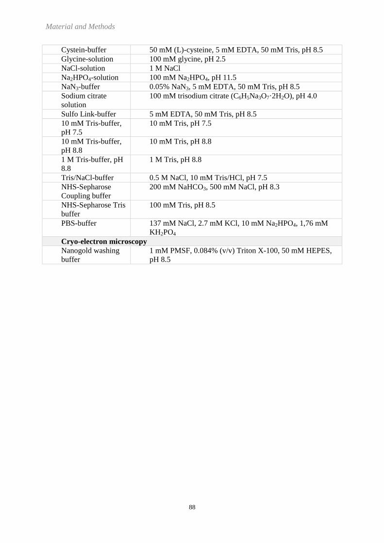
Material and Methods
88
Cystein-buffer 50 mM (L)-cysteine, 5 mM EDTA, 50 mM Tris, pH 8.5
Glycine-solution 100 mM glycine, pH 2.5
NaCl-solution 1 M NaCl
Na2HPO4-solution 100 mM Na2HPO4, pH 11.5
NaN3-buffer 0.05% NaN3, 5 mM EDTA, 50 mM Tris, pH 8.5
Sodium citrate
solution
100 mM trisodium citrate (C6H5Na3O7·2H2O), pH 4.0
Sulfo Link-buffer 5 mM EDTA, 50 mM Tris, pH 8.5
10 mM Tris-buffer,
pH 7.5
10 mM Tris, pH 7.5
10 mM Tris-buffer,
pH 8.8
10 mM Tris, pH 8.8
1 M Tris-buffer, pH
8.8
1 M Tris, pH 8.8
Tris/NaCl-buffer 0.5 M NaCl, 10 mM Tris/HCl, pH 7.5
NHS-Sepharose
Coupling buffer
200 mM NaHCO3, 500 mM NaCl, pH 8.3
NHS-Sepharose Tris
buffer
100 mM Tris, pH 8.5
PBS-buffer 137 mM NaCl, 2.7 mM KCl, 10 mM Na2HPO4, 1,76 mM
KH2PO4
Cryo-electron microscopy
Nanogold washing
buffer
1 mM PMSF, 0.084% (v/v) Triton X-100, 50 mM HEPES,
pH 8.5

Material and Methods
89
6.1.5 Neurospora crassa strains
Strain Genotype
Origin or source
and reference
(if applicable)
NCN251
(also called
74A)
A FGSC 2489
76-26 his-3 mtrR a
(mtrR imparts fpa resistance)
R.L. Metzenberg
71-18 pan-2 BmlR a (BmlR imparts benomyl
resistance)
R.L. Metzenberg
HP1 Heterokaryon of 76-26 plus 71-18 F. Nargang
Tob55
(Tob55KO-1)
Sheltered heterokaryon. As HP1, but
with replacement of the tob55 gene in
76-26 nucleus with a hygromycin
resistance (hygR) cassette.
F. Nargang
(120)
Tob55
(Tob55KO-3)
As Tob55KO-1 As Tob55KO-1
T55His6-1 his-3 mtrR a Δtob55 ::hygR contains an
ectopic copy of genomic tob55 with N-
terminal hexahistidinyl tag. Also
bleomycin resistant.
F. Nargang
(120)
T55His6-3 As T55His6-1, but different clone As T55His6-1
ST55-2 his3 mtrR a Δtob55 ::hygR contains an
ectopic copy of tob55 cDNA specific
for the short form.
F. Nargang
(120)
IT55-8 his3 mtrR a Δtob55 ::hygR contains an
ectopic copy of tob55 cDNA specific
for the intermediate form.
F. Nargang
(120)
LT55-2 his3 mtrR a Δtob55 ::hygR contains an
ectopic copy of tob55 cDNA specific
for the long form.
F. Nargang
(120)
His9-Tob55
(H6C4-5)
his-3 mtrR a Δtob55 hygR
Contains an ectopic copy of genomic
tob55 with an N-terminal nine His tag.
Also bleomycin resistant.
F. Nargang
(108)
His9-Tob55ST his-3 mtrR a Δtob55 ::hygR contains an
ectopic copy of tob55 cDNA specific
for the short form with an N-terminal
nine His-tag. Also bleomycin resistant.
F. Nargang

Material and Methods
90
55FH123 his-3 mtrR a Δtob55 hygR
Contains an ectopic copy of tob55
cDNA specific for the short form with
with an N-terminal nine His-tag, and an
ectopic copy of tob55 cDNA specific
for the intermediate form with an N-
terminal Flag-tag. Also bleomycin and
benomyl resistant.
F. Nargang
Tob37
(Tob37KO-5)
Sheltered heterokaryon. As HP1, but
with replacement of tob37 gene in 76-
26 nucleus with a hygromycin
resistence (hygR) cassette.
F. Nargang
His9-Tob37
(9His-Tob37-2)
his-3 mtrR a Δtob37 hygR
Contains an ectopic copy of genomic
tob37 with a C-terminal nine His tag.
Also bleomycin resistant.
F. Nargang,
(108)
Tob38
(Tob38KO-6)
Sheltered heterokaryon. As HP1, but
with replacement of tob38 gene in 76-
26 nucleus with a hygromycin
resistence (hygR) cassette.
F. Nargang
His9-Tob38
(9His-Tob38-3)
his-3 mtrR a Δtob38 hygR
Contains an ectopic copy of genomic
tob38 with a C-terminal nine His tag.
Also bleomycin resistant.
F. Nargang
(108)
mdm10 mdm10
his-3 mtrR hygR a
Replacement of mdm10 gene in 76-26
with hygromycin resistance (hygR)
cassette
F. Nargang
(108)
GR 107 expression of a Tom22 with a
hexahistidinyl tag at its C-terminus
W. Neupert (31,
164)

Material and Methods
91
6.1.6 Saccharomyces cerevisiae strain
6.1.7 Peptides
Name of
peptide
Sequence Use Origin
Tob38Nc-8
RDPEYTDLLDRFYI
TPASS
Injection into rabbits for
synthesis of polyclonal
antibodies against Tob38
from N. crassa; the peptide
was coupled to KLH before
injection.
Peptide corresponds
to the residues 146 –
182 of the Tob38
protein from N.
crassa.
Tob38Nc-4
C -
RDPEYTDLLDRFYI
TPASS
Used for affinity
purification of antibodies
against Tob38 from N.
crassa out of antiserum
Peptide corresponds
to the residues 146 –
182 of the Tob38
protein from N.
crassa with an extra
cystein at the N-
terminus.
Tob38Nc-9 KYMSDAEGEVEGN
MGFILASRK
Injection into rabbits for
synthesis of polyclonal
antibodies against Tob38
from N. crassa; the peptide
was coupled to KLH before
injection.
Peptide corresponds
to the residues 269 –
290 of the Tob38
protein from N.
crassa.
Tob38Nc-5
C-
KYMSDAEGEVEGN
MGFILASRK
Used for affinity
purification of antibodies
against Tob38 from N.
crassa out of antiserum
Peptide corresponds
to the residues 269 –
290 of the Tob38
protein from N.
crassa with an extra
cystein at the N-
terminus.
Tob37Nc-10
DTDAEMERLEREE
REREAAG
Injection into rabbits for
synthesis of polyclonal
antibodies against Tob37
from N. crassa; the peptide
was coupled to KLH before
injection.
Used for affinity
purification of antibodies
against Tob37 out of
antiserum.
Peptide corresponds
to the residues 165 –
184 of the Tob37
protein from N.
crassa.
Strain Genotype
Origin or source and reference
(if applicable)
N-His6-Bcs1 in Δbcs1 W303a Kai Hell, N. Wagener, W. Neupert
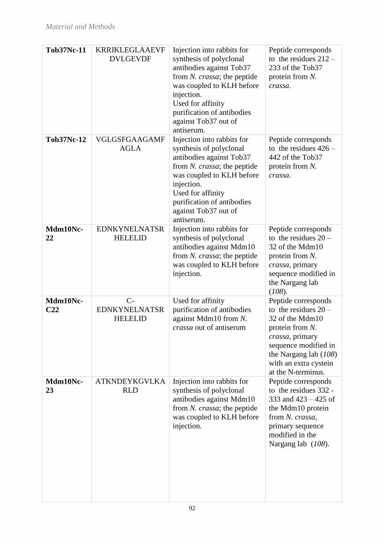
Material and Methods
92
Tob37Nc-11
KRRIKLEGLAAEVF
DVLGEVDF
Injection into rabbits for
synthesis of polyclonal
antibodies against Tob37
from N. crassa; the peptide
was coupled to KLH before
injection.
Used for affinity
purification of antibodies
against Tob37 out of
antiserum.
Peptide corresponds
to the residues 212 –
233 of the Tob37
protein from N.
crassa.
Tob37Nc-12
VGLGSFGAAGAMF
AGLA
Injection into rabbits for
synthesis of polyclonal
antibodies against Tob37
from N. crassa; the peptide
was coupled to KLH before
injection.
Used for affinity
purification of antibodies
against Tob37 out of
antiserum.
Peptide corresponds
to the residues 426 –
442 of the Tob37
protein from N.
crassa.
Mdm10Nc-
22
EDNKYNELNATSR
HELELID
Injection into rabbits for
synthesis of polyclonal
antibodies against Mdm10
from N. crassa; the peptide
was coupled to KLH before
injection.
Peptide corresponds
to the residues 20 –
32 of the Mdm10
protein from N.
crassa, primary
sequence modified in
the Nargang lab
(108).
Mdm10Nc-
C22
C-
EDNKYNELNATSR
HELELID
Used for affinity
purification of antibodies
against Mdm10 from N.
crassa out of antiserum
Peptide corresponds
to the residues 20 –
32 of the Mdm10
protein from N.
crassa, primary
sequence modified in
the Nargang lab (108)
with an extra cystein
at the N-terminus.
Mdm10Nc-
23
ATKNDEYKGVLKA
RLD
Injection into rabbits for
synthesis of polyclonal
antibodies against Mdm10
from N. crassa; the peptide
was coupled to KLH before
injection.
Peptide corresponds
to the residues 332 -
333 and 423 – 425 of
the Mdm10 protein
from N. crassa,
primary sequence
modified in the
Nargang lab (108).

Material and Methods
93
Mdm10Nc-
C23
C-
ATKNDEYKGVLKA
RLD
Used for affinity
purification of antibodies
against Mdm10 from N.
crassa out of antiserum
Peptide corresponds
to the residues 332 -
333 and 423 – 425 of
the Mdm10 protein
from N. crassa,
primary sequence
modified in the
Nargang lab (108)
with an extra cystein
at the N-terminus.
Mdm10Nc-
25
HKLGEPFRSLGEVQ
YSS
Injection into rabbits for
synthesis of polyclonal
antibodies against Mdm10
from N. crassa; the peptide
was coupled to KLH before
injection.
Peptide corresponds
to the residues 463 –
480 of the Mdm10
protein from N.
crassa, primary
sequence modified in
the Nargang lab
(108).
AQUA55 EDGFGVFISDAR Isotopic labelled peptide
(heavy peptide) used for the
quantification of the Tob55
subunit in the isolated TOB
complex; arginine
substituted by isotopic
arginine (bold) (13
C6, 15
N4),
mass shift +10 Da
Thermo Fisher
Scientific GmbH,
Ulm, GER
AQUA38 DPEYTDLLDR Isotopic labelled peptide
(heavy peptide) used for the
quantification of the Tob38
subunit in the isolated TOB
complex; arginine
substituted by isotopic
arginine (bold) (13
C6, 15
N4),
mass shift +10 Da
Thermo Fisher
Scientific GmbH,
Ulm, GER
AQUA37 VYADSQAYK Isotopic labelled peptide
(heavy peptide) used for the
quantification of the Tob37
subunit in the isolated TOB
complex; lysine substituted
by isotopic lysine (bold)
(13
C6, 15
N2), mass shift +8
Da
Thermo Fisher
Scientific GmbH,
Ulm, GER

Material and Methods
94
6.1.8 Whitehead codes and molecular masses of selected proteins from Neurospora crassa
Protein Whitehead code Molecular mass (kDa)
Tob55 NCU05593 54.7 (long),
54.1 (intermediate),
50.7 (short)
Tob37 NCU00923 48.6
Tob38 NCU01401 37.3
Tom40 NCU01179 38.1
Mdm10 NCU07824 52.7
porin NCU00431 30
For further information seek the complete N. crassa genome database:
http://www.broadinstitute.org/annotation/genome/neurospora/FeatureSearch.html
6.1.9 Antibodies
Primary
antibody
Blocking
buffer
Dilution
primary
antibody
Secondary
antibody
Dilution
secondary
antibody
anti-His Penta-His
Antibody, BSA-
free,
mouse
monoclonal IgG1
3%
(w/v)BSA
in TBS-
buffer
1 : 1000
in 3%
(w/v)
BSA in
TBS-
buffer
Goat-Anti-
Mouse
IgG
(H + L)-
HRP
Conjugate
1:3000 in
10% (w/v)
skimmed
milk
powder in
TBS-buffer
anti-Flag Monoclonal Anti-
FLAG,
antibody
produced in
mouse,
Clone M2,
purified
immunoglobulin
3% (w/v)
skimmed
milk
powder in
Flag-TBS-
buffer
1:1900 in
3% (w/v)
skimmed
milk
powder in
Flag-
TBS-
buffer
Goat-Anti-
Mouse
IgG
(H + L)-
HRP
Conjugate
1:3000 in
in 3%
(w/v)
skimmed
milk
powder in
Flag-TBS-
buffer
rabbit
antisera or
affinity
purified
antibodies Polyclonal
Antibodies risen
in rabbits by
antigen injection;
affinity
purification was
performed if
required
5% (w/v)
skimmed
milk
powder in
TBS-
buffer
1:30 –
1:24000
in 5%
(w/v)
skimmed
milk
powder in
TBS-
buffer
Goat-Anti-
Rabbit
IgG
(H + L)-
HRP
Conjugate
1:10000 in
5% (w/v)
skimmed
milk
powder in
TBS-buffer
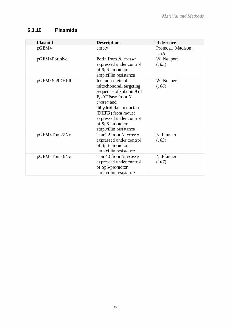
Material and Methods
95
6.1.10 Plasmids
Plasmid Description Reference
pGEM4 empty Promega, Madison,
USA
pGEM4PorinNc Porin from N. crassa
expressed under control
of Sp6-promotor,
ampicillin resistance
W. Neupert
(165)
pGEM4Su9DHFR fusion protein of
mitochondrail targeting
sequence of subunit 9 of
Fo-ATPase from N.
crassa and
dihydrofolate reductase
(DHFR) from mouse
expressed under control
of Sp6-promotor,
ampicillin resistance
W. Neupert
(166)
pGEM4Tom22Nc Tom22 from N. crassa
expressed under control
of Sp6-promotor,
ampicillin resistance
N. Pfanner
(163)
pGEM4Tom40Nc Tom40 from N. crassa
expressed under control
of Sp6-promotor,
ampicillin resistance
N. Pfanner
(167)

Material and Methods
96
6.2 Methods
Unless stated otherwise, methods were performed at room temperature.
6.2.1 Cell biology
6.2.1.1 Cultivation of Neurospora crassa
Growth and handling of N.crassa were performed as described previously (168). For the
cultivation of N. crassa, solid Vogel´s growth medium was inoculated with a few granules of
the silica stock of the strain of interest in Erlenmeyer flasks. These A-flasks were incubated
for 48 hours in the dark at 30°C and subsequently for around 190 hours at light at room
temperature. With mycelium from the A-flasks, a second lot of flasks was inoculated and
cultivated as described for the A-flasks. The fungus was harvested from the solid medium
with 50 ml sterile water per flask and vigorous swirling and used for inoculating liquid
medium.
For a 2 l-culture, 1.9 l of Vogel´s minimal liquid medium were mixed with 100 ml of
harvested N. crassa. For a 50 l-culture, a 10 l-preculture was started with 1 liter of harvested
mycelium and 9 l Vogel´s minimal liquid medium containing 40 µg/ml chloramphenicol.
After 6 hours, 40 liters of Vogel´s minimal medium were mixed with this preculture and
cultivated for another 14 – 16 hours. Cultivation in liquid medium was performed at 30°C
with constant aeration. The mycelium was harvested by filtration of the liquid medium.
6.2.1.2 Preparation of silica stocks from Neurospora crassa
Mycelium of one to two A-flasks was harvested with a total volume of 50 ml 10 % (w/v)
skimmed milk and vigorous swirling. The solution was filtered through an autoclaved funnel
filled with cotton batting. 300 µl of the filtrate were mixed with 1 g silica gel in a glass vial,
dried in an exicator at room temperature for 3 weeks and stored at -20°C.
6.2.1.3 Isolation of mitochondria from Neurospora crassa
Mycelia were ground in the presence of sand and 0.25 M SET-buffer. The sand was separated
from the cell extract by two sequential centrifugation steps at 2594 x g for 5 minutes. The cell
organells in the supernatant were sedimented at 10976 x g for 50 minutes at 4°C. The
mitochondria were scraped off from the very top of the resulting pellet and resuspended in
0.25 M ST-buffer. After homogenization of the mitochondria they were again sedimented at
10976 x g for 50 minutes at 4°C, resuspended in a smaller amount of 0.25 M ST-buffer and

Material and Methods
97
homogenized. The concentration of mitochondrial proteins was determined by Bradford
assay.
6.2.1.4 Isolation of outer mitochondrial membrane vesicles from Neurospora
crassa
Mycelia were ground in the presence of sand and 0.25 M SET-buffer. The sand was separated
from the cell extract by two sequential centrifugation steps at 2594 x g for 5 minutes. The cell
organelles in the supernatant were sedimented at 10976 x g for 50 minutes at 4°C. The
mitochondria were scraped off from the very top of the resulting pellet and resuspended in
0.25 M SET-buffer. After homogenization of the mitochondria the amount of mitochondrial
protein was determined by Bradford assay. The homogenized mitochondria were spun down
at 10976 x g for 50 minutes at 4°C, resuspended in 0.25 M SET-buffer and incubated at 37°C
for 5 minutes. Afterwards, mitochondria were transferred into swelling buffer under stirring at
4°C to a final concentration of 1 g mitochondrial proteins per litre of swelling buffer.
Following 30 minutes of stirring, the resulting mitoplasts were sedimented at 10976 x g for 30
minutes and resuspended in a smaller volume of 0.25 M SET-buffer. With an automated
glass-teflon-homogenizer, the outer mitochondrial membrane was sheared off the mitoplasts
for 40 minutes at 4 °C. 20 ml of the homogenate was applied on a step gradient of 20 ml 0.9
M - SET buffer at the bottom and 9 ml of 0.25 M SET-buffer and centrifuged for 1 hour at
141370 x g, 4°C. The vesicles of the outer mitochondrial membrane could be taken from a
thin layer between the 0.9 M SET-buffer and the 0.25 M SET-buffer and were mixed with
half of the volume of 2 M SET-buffer. 15 ml of these outer mitochondrial membrane vesicles
(OMVs) were overlaid with 20 ml 0.7 M SET-buffer and 3 ml Tris/EDTA-buffer and
centrifuged at 141370 x g overnight at 4°C. OMVs could be collected from the interface
between the Tris/EDTA-buffer and the 0.7 M SET-buffer. The double volume of OMV-buffer
was added and the OMVs were centrifuged for 1 hour at 183960 x g. Finally, the sedimented
OMVs were dissolved in OMV-buffer. The protein concentration was determined by
Bradford assay and aliquots of the OMVs were shock frozen in liquid nitrogen and stored at -
80°C.
Material and Methods
98
6.2.1.5 Cultivation of Saccharomyces cerevisiae
Cultivation of the Saccharomyces cerevisiae yeast strain “N-His6-Bcs1 in Δbcs1” was
performed according to standard procedures (169). Yeast from a glycerine-stock were struck
out on solid lactate complete medium and cultivated at 30°C. After 2 days of cultivation,
yeast from the solid medium were used for the inoculation of liquid lactate medium and
incubated under moderate shaking at 30°C.
6.2.1.6 Preparation of glycerol stocks from Saccharomyces cerevisiae
For the preparation of stocks of S. cerevisiae, yeast cells from solid medium plates were
mixed with 15% (v/v) glycerol in water and stored at -80°C.
6.2.1.7 Isolation of mitochondria from Saccharomyces cerevisiae
Mitochondria from S. cerevisiae were isolated according to standard procedures as described
before (170). Yeasts were cultivated in liquid medium to an OD600 of 1 – 1.5 and harvested by
centrifugation at 2800 x g for 5 min at room temperature (RT). The cells were washed with
sterile water and resuspended to a final concentration of 0.5 g/ml yeast cells in resuspension
buffer and incubated for 10 min at 30°C under moderate shaking. Following centrifugation
2000 x g for 5 min at 4°C, the cells were washed again with a final concentration of 0.16 g/ml
yeast cells in sorbitol buffer. For the digestion of the cell wall, 2.5 mg zymolyase were added
per g yeast cell and incubation was performed for 30 – 45 min at 30°C under moderate
shaking. Resulting spheroplasts were harvested by centrifugation at 2000 x g for 5 min at 4°C
and homogenized 10 times in a Dounce-homogenizer at 4°C with a final concentration of 0.16
g/ml spheroplasts in homogenization buffer. After centrifugation at 2000 x g for 5 min at 4°C,
the supernatant was stored on ice and the remaining pellet was again resuspended and
homogenized as described before. The supernatants of the first and second homogenization
were pooled and centrifuged at 2000 x g for another 5 min at 4°C to sediment cell remnants.
Mitochondria were spun down by centrifugation at 17000 x g for 12 min at 4°C and
resuspended in SEH-buffer. Cell remnants were again separated by centrifugation at 2000 x g
for 5 min at 4°C and the mitochondria in the supernatant were pelletized by centrifugation at
17000 x g for 12 min at 4°C. Mitochondria were resuspended in SEH-buffer with a final
concentration of 10 mg/ml protein, aliquotted, frozen in liquid nitrogen, and stored at -80°C.

Material and Methods
99
6.2.1.8 Preparation of cryostocks from Escherichia coli cells
For storage 1 ml of an Escherichia coli (E. coli) overnight culture was mixed with 1 ml 87%
glycerol and kept at -80°C.
6.2.1.9 Chemical competent Escherichia coli cells
500 ml of LB-medium were inoculated with 5ml overnight-culture of E. coli and grown at
37°C to an OD600 of 0.4. Following incubation on ice for 15 minutes the bacteria were spun
down at 1756 x g for 15 minutes at 4°C. The pellet was resuspended in 40 ml CaCl2-solution
and after 30 minutes on ice, the bacteria were harvested by centrifugation at 1756 x g for 15
minutes at 4°C. After resuspending the pellet in 20 ml CaCl2-solution, 4 ml glycerol, 100%
were added and the bacteria were incubated on ice for 2 hours. 200 µl aliquots of the
suspension were shock-frozen in liquid nitrogen and subsequently stored at -80°C.
Material and Methods
100
6.2.2 Molecular biology
6.2.2.1 Plasmid transformation into Escherichia coli cells
For amplification, in general plasmids were transformed into DH5α E. coli cells. For that, 100
µl of chemical competent E. coli cells were thawed on ice and mixed with 1 µl of purified
plasmid and incubated on ice for 15 minutes. After a heat shock for 30 seconds at 42°C, the
cells were placed on ice for 2 minutes. Subsequently, 800 µl LB-medium without any
selective antibiotics was added and the cells were shaken at 37°C for 45 minutes. A small
amount of the bacteria were outplated on LB-medium with and without selective antibiotics
and incubated overnight at 37°C to monitor the transformation efficiency. 55 ml of LB-
medium with selective antibiotics were inoculated with the remaining bacteria suspension and
incubated over night at 37°C for the following plasmid isolation.
6.2.2.2 Plasmid isolation out of Eschericia coli cells
Plasmids were isolated from 50 ml overnight bacteria culture with the Promega Pure Yield
Plasmid Midiprep System - Kit according to the instructions. For the determination of the
DNA-content, the eluate with the purified plasmid was diluted 1 : 50 with water and analyzed
photometrically as described (6.2.2.3). The yield of ~ 400 µl purified plasmids was stored at -
20°C.
6.2.2.3 Photometrical quantification of nucleic acids
The DNA-concentration was determined photometrically by measuring the absorption of the
solution at 260 nm in a quartz crystal cuvette. Absorption of 1 corresponds to 50 µg/ml
double strand DNA.

Material and Methods
101
6.2.2.4 In vitro synthesis of radioactive precursor proteins
6.2.2.4.1 In vitro transcription
For the synthesis of radioactive precursor proteins, plasmid DNA encoding for the protein of
interest under the control of the SP6-promotor was transcribed by the SP6-RNA-polymerase
to RNA. For that, a transcription mix (50µl) with 3 - 10 µg of purified plasmids, 10 mM
MgCl2, 2 mM spermidine, 10 mM DTT, 1.6 u/µl RNasin RNase (ribonuclease) inhibitor, 0.5
mM ribonucleoside triphophates (rNTPs), 0.13 mM m7G(5´)ppp(5´)G, 0.75 u/ml SP6-RNA-
polymerase and 40 mM Tris (pH 7.5) was incubated for 1 hour at 37°C. The RNA was
precipitated at -20°C for 30 minutes in the presence of 244 mM LiCl and 73% ethanol.
Following a centrifugation at 36000 x g for 20 minutes at 2°C, the pellet was washed with 150
µl cold 70% (v/v) ethanol and DNA was spun down again at 36000 x g for 20 minutes at 2°C.
After evaporation of the ethanol at RT, the pellet was resuspended in water supplemented
with 1.54 u/µl RNasin RNase inhibitor solution. RNA was stored at -20°C.
6.2.2.4.2 In vitro translation
For the translation of the protein of interest from the isolated mRNA, a translation mix (45 µl)
with 6.25 µl RNA, 25 µl rabbit reticulocyte lysate, 0.875 µl amino-acid mix without
methionine, 0.25 µl RNasin RNase inhibitor and 3 µl 35
S-methionine (radioactive methionine)
was incubated for one hour at 30°C. The synthesis of radioactive protein was stopped by
adding 3.2 mM methionine (non-radioactive methionine) and 167 mM sucrose. 1 µl and 3 µl
of the synthesized protein were analyzed by SDS-PAGE and Western blotting followed by
autoradiography. The protein was shock-frozen in aliquots in liquid nitrogen and stored at -
80°C.
6.2.2.4.3 TNT coupled reticulocyte lysate system
In the TNT coupled reticulocyte lysate system, transcription and translation were performed
in the same reaction mix. The TNT-mix (50 µl), containing 25 µl TNT rabbit reticulocyte
lysate, 2 µl TNT Reaction buffer, 1 µl TNT SP6 Polymerase, 1 µl amino-acid mix without
methionine, 4 µl 35
S-methionine (radioactive methionine), 1 µl RNasin RNase inhibitor and 1-
3 µg plasmid-DNA, was incubated for 1.5 hours at 30°C. When radioactive porin precursor
was synthesized, the reaction was already stopped after 40 minutes. Following the addition of
3.7 mM methionine (non-radioactive) and 194 mM sucrose, the protein was shock-frozen in

Material and Methods
102
aliquots in liquid nitrogen and stored at -80°C. 1 µl and 3 µl of the synthesized protein were
analyzed by SDS-PAGE and Western blotting followed by autoradiography.
Material and Methods
103
6.2.3 Protein biochemistry
6.2.3.1 Autoradiography
Radioactive proteins were detected by exposing a X-ray film (Kodak) on the dried membrane
with immobilized proteins for different time spans, ranging from a few hours to 15 days.
Exposure times depended on the signal intensities observed after developing the films
6.2.3.2 Isolation of the TOB complex by Ni-NTA affinity chromatography
OMVs from N. crassa were solubilized in Ni-NTA solubilization buffer for 2 hours in the
presence of 0.192% (v/v) Triton X-100 or 0.64% (w/v) digitonin, resulting in an
detergent/protein ratio of 6/1 (TX-100) and 20/1 (digitonin), respectively. After a
centrifugation step for clarification at 36000 x g for 20 minutes, the supernatant was loaded
on a Ni-NTA agarose column (1.66 mg solubilized protein / 100 µl matrix, 100%) for affinity
purification. The column was rinsed stepwise with 16 column volumes (CVs) of Ni-NTA
washing buffer with 15 mM imidazole, 25 CVs with 20 mM imidazole and 10 CVs with 40
mM imidazole. Bound proteins were eluted with Ni-NTA washing buffer with 200 mM
imidazole. Depending on the detergent used, the Ni-NTA washing buffer contained either
0.084 % Triton X-100 or 0.24% (w/v) digitonin. The isolation was performed at 4°C.
6.2.3.3 Isolation of the TOB complex by Flag-tag affinity purification
OMVs from N. crassa were solubilized in Flag solubilization buffer for 2 hours in the
presence of 0.192 % Triton X-100. After a centrifugation step for clarification at 36000 x g
for 20 minutes, the supernatant was loaded on ANTI-FLAG M2-Agarose Affinity Gel (1.66
mg solubilized protein / 10 µl matrix, 100%) and incubated for 2 hours with gentle agitation.
Following a centrifugation at 4000 x g for 3 minutes, the supernatant with the unbound
proteins was removed. The ANTI-FLAG M2-Agarose Affinity Gel with the bound proteins
was transferred to a Mini-frit column in an 2 ml reaction tube and rinsed 5 times with 10 CVs
of Flag washing buffer. After an incubation of 2 minutes, the washing buffer was removed by
a centrifugation step at 4000 x g for one minute each. For elution, 2.5 CVs of Flag washing
buffer with 5 µg/µl three fold Flag peptide was added to the matrix and incubated for 30
minutes with gentle agitation. Bound proteins were sedimented at 4000 x g for 1 minute. The
isolation was performed at 4°C.

Material and Methods
104
6.2.3.4 Isolation of the TOM core complex
The TOM core complex was isolated according to a protocol established by Patrick Schreiner
in the group of W. Neupert (LMU, Munich, GER), as listed below. The protocol was adapted
from “Structural investigation of two supramolecular complexes of the eukaryotic cell: the
proteasome and the mitochondrial TOM complex”, (171).
TOM core complex was isolated from a N. crassa strain (GR-107) that carried a version of
Tom22 with a hexahistidinyl tag at its C-terminus. The TOM core complex was isolated
according to Ahting et al., 1999 (172), with some modifications in the protocol. Isolated
mitochondria were solubilized in solubilization buffer at a protein concentration of 10 mg/ml for
30 min at 4°C. Insoluble material was removed by centrifugation, and the clarified extract was
loaded onto the equilibrated Ni-NTA agarose column using 4 ml resin per g of total mitochondrial
protein. The column was washed with 5 CVs of TOM washing buffer 1 and with 10 CVs of TOM
washing buffer 2. The bound protein was eluted with 4 CVs of TOM elution buffer. The eluted
fraction of the Ni-NTA column was loaded onto an equilibrated Resource Q anion exchange
column. The complex was eluted with a linear potassium chloride gradient. For further
purification, the fractions containing TOM core complex were pooled, concentrated to 500 μl
(spin filtration devices from Pall Corporation, 100 kDa cut-off) and loaded onto a Superose 6 size
exclusion column. Proteins were eluted in gel filtration buffer. Stock solutions of purified TOM
core complex were stored at a protein concentration of 10-20 mg/ml at 4°C. An average
preparation of the protein started with ~1.5 kg of Neurospora cells (wet weight) which yielded
about 5 g of mitochondrial proteins and 1-2 mg of purified TOM core complex.
6.2.3.5 Isolation of the BCS1complex
Mitochondria isolated from the “N-His6-Bcs1 in Δbcs1” Saccharomyces cerevisiae strain
were resuspended in solubilisation buffer by pipetting. After incubation for 10 min on ice in
the presence of 3% (w/v) digitonin, unsolubilized mitochondria were sedimented by
centrifugation at 10000 x g for 10 min at 4°C and the supernatant was loaded on a Ni-NTA
agarose batch (5 mg solubilized protein / 100 µl matrix, 100%) for affinity purification.
Following incubation of 1 hour at 4°C under gentle agitation, the beads were subjected to
three washing steps in which the matrix beads were resuspended in washing buffer, spun
down at 1000 x g for 1 min at 4°C and resuspended again in fresh buffer. For elution of the
bound proteins, the matrix was incubated fort 10 min in elution buffer at 4°C and eluted
proteins were separated from the matrix beads by centrifugation at 1000 x g for 1min at 4°C.
Material and Methods
105
The isolation of the BCS1 complex was performed according to a protocol established by N.
Wagener (Neupert group, LMU, Munich, GER).
6.2.3.6 Carbonate extraction
OMVs were resuspended in 100 mM Na2CO3 with different pH values of 10.8, 11.5 and 12.5
and incubated for 30 minutes on ice. As a control, proteins were treated with 10 mM HEPES-
KOH, pH 7.4. By centrifugation for one hour at 125800 x g, transmembrane proteins were
sedimented and could thereby be separated from the soluble and membrane associated
proteins in the supernatant. The proteins in the supernatant were precipitated with
trichloracetic acid as described (6.2.3.8).
6.2.3.7 Protein concentration
If needed, the isolated proteins of the TOB complex were concentrated by using an Amicon
Ultra 30k device. Centrifugation was performed at 4000 x g at 4°C.
6.2.3.8 Protein precipitation
Proteins in solution were mixed with 1/5th
volume of 72% trichloracetic acid (TCA) and
frozen overnight at -20°C. The precipitated proteins were sedimented at 36000 x g for 15
minutes at 4°C and washed with 100% acetone. The pellet was dried at 35°C for 30 minutes
and, unless stated otherwise, resolved by cooking in 2 x Laemmli at 95°C for 5 minutes.
6.2.3.9 Determination of the protein concentration by Bradford assay
The protein concentration was determined by adding 1ml of a 1:5 dilution of the “Bio-Rad
Protein assay” solution to the samples of same volume. Following 5 minutes of incubation at
RT, the absorbance at 595 nm was measured in a 10 mm path length microcuvette. The
protein solution of interest was diluted and 2.5 µl, 5 µl and 7.5 µl were measured twice. The
protein concentration was calculated according to a standard curve, measured in parallel with
48 µg, 24 µg, 12 µg, 6 µg, 3 µg, 1.5 µg and 0 µg of the “Bio-Rad Protein Assay Standard I”.
6.2.3.10 SDS-Polyacrylamide gel electrophoresis (SDS-PAGE)
By denaturing discontinuous sodiumdodecylsulfate polyacrylamide gelelectrophoresis (SDS-
PAGE) proteins were separated according to their molecular mass as described previously
Material and Methods
106
(173). Proteins were solved in 2 x Laemmli and cooked for 5 minutes at 95°C, applied on a
5% SDS-gel and separated on a 12.5% SDS-gel. The gels used had a size of 140 mm x 140
mm and a thickness of 1 mm. Standard running conditions were 30 mA for 2 – 3 hours at RT
in SDS-running buffer. By comparison with marker proteins of known size running on the
same gel, the molecular weight of the sample proteins could be estimated.
6.2.3.11 Blue Native-Polyacrylamide gel electrophoresis (BN-PAGE)
Proteins from mitochondria (60 – 120 µg) or OMVs (10 – 30 µg) were solubilized in 1 x
NativePAGE sample buffer containing 0.9% - 1.8% (v/v) TX-100 or 0.75% - 6% (w/v)
digitonin, resulting in a protein/detergent ratio of 6/1 (TX-100) and 10/1 – 40/1 (digitonin),
respectively. Following 1 hour of incubation on ice, the samples were centrifuged for
clarification at 20 000 x g for 30 minutes and the supernatant was mixed with NativePAGE 5
% G-250 sample additive to adjust the final G-250 concentration in the sample to at least 1/4th
of the detergent concentration. BN-PAGE was performed as previously described (142, 174)
with NativePAGE Novex 4-16 % Bis-Tris Gel according to the instructions. All buffers were
used and gelelectrophoresis was performed at 4°C. The bovine heart mitochondria used as
protein standard for the mass estimation in the BN-PAGE analysis were provided by Ilka
Wittig (Schägger group, Goethe-University Frankfurt, Frankfurt am Main, GER).
6.2.3.12 In gel staining of proteins with Coomassie brilliant blue
After SDS- or BN-PAGE, the separated proteins in the gel were stained with the Coomassie
Brilliant Blue dye. The gel was shaken in Coomassie staining solution for at least 30 minutes
at RT. Subsequently, the stained gel was rinsed several times with water and incubated with
Coomassie destaining solution until distinct bands became visible. After scanning for
documentation, the gel was dried overnight between two gel-drying films.
6.2.3.13 Transfer of proteins to a nitrocellulose membrane (Western blotting)
Unless stated otherwise, proteins separated by SDS-PAGE were transferred by the semi-dry
blotting procedure (175, 176) onto nitrocellulose membrane. SDS-gels, nitrocellulose
membrane and Whatman filter paper were incubated in SDS-transfer buffer for ca. 1 minute at
RT. One Whatman filter paper was placed on the anode electrode plate followed by the
nitrocellulose membrane, the SDS-gel, another Whatman filter paper and the cathode

Material and Methods
107
electrode plate. Transfer was performed at RT at 200 mA or 230 mA for 1 or 1.5 hours,
respectively.
6.2.3.14 Transfer of proteins to a PVDF membrane (Western blotting)
For transferring proteins from BN-gels by the semi-dry blotting procedure, a PVDF
(polyvinylidene fluoride) membrane was incubated in 100% methanol for 1 minute and
afterwards rinsed in 1 x NuPAGE Transfer Buffer. The BN-gel was incubated in SDS-running
buffer for 4 minutes and rinsed in 1 x NuPAGE Transfer Buffer. Whatman filter paper was
also soaked with 1 x NuPAGE Transfer Buffer for 1 minute. One Whatman filter paper was
placed on the anode electrode followed by the PVDF-membrane, the BN-gel, another
Whatman filter paper and the cathode electrode plate. Transfer was performed at 130 mA for
1 hour. All buffers were used and protein transfer was performed at 4°C. After the blotting,
the proteins were fixed on the membrane by shaking in 8% acetic acid for 15 minutes at RT
and subsequently air-dried.
6.2.3.15 Reversible staining of blotted proteins by Coomassie Brilliant Blue
solution
To visualize proteins transferred to a PVDF-membrane, the wet PVDF-membrane was
incubated in Coomassie staining solution for 30 to 60 seconds at RT and rinsed in Coomassie
destaining solution to remove the background staining. Visible marker proteins were marked
with a pen on the membrane. The membrane was then further destained by shaking in 100%
methanol for 1 to 2 minutes, rinsed in TBS-buffer and either air-dried or used for
immunodecoration.
6.2.3.16 Reversible staining of blotted proteins by Ponceau S solution
To verify the transfer of proteins on a nitrocellulose membrane, the membrane was shaken in
Ponceau S solution for two minutes at RT after blotting. The immobilized proteins were
reversibly stained by the Ponceau dye and the membrane was rinsed in water to remove the
background staining. Afterwards, the membrane was air-dryed.

Material and Methods
108
6.2.3.17 Pepspot protein interaction assay
To analyze specific binding of the isolated TOB complex to its substrates Tom40 and Porin
and to identify their specific binding sites, a pepspot binding assay was performed.
Peptide libraries of peptides with 15 amino acids in length and an overlap of 11 amino acids
were synthesized (JPT Peptide Technologies GmbH, Berlin, GER). Each peptide was
covalently bound to a spot on a cellulose-PEG-membrane by the C-terminus (pepspot
membrane). By acetylation the charged N-terminus was transformed into an uncharged
peptide end and thereby provided the peptide with higher stability towards degradation and
higher resemblance to the uncharged native peptide side. Cysteins were substituted with serins
to prevent oxidation and cyclization of the peptide.
For the protein interaction assay, the pepspot membrane was rinsed in 100% methanol for one
minute followed by three washing steps with TBS-buffer for 10 minutes at RT. After blocking
unspecific sites of the pepspot membrane with blocking buffer for three hours, the membrane
was washed in T-TBS-buffer for 10 minutes. Isolated TOB complex from the His9-Tob37
strain was added to the membrane at an estimated concentration of 5 µg/ml in blocking buffer
supplemented with 0.042% (v/v) TX-100 and incubated overnight at 4°C with gentle
agitation. The next day, the incubation was continued for three more hours at RT and
subsequently rinsed three times for one minute with T-TBS-buffer. Bound TOB complex was
transferred to a PVDF-membrane by the semi-dry blotting procedure as follows: two
Whatman filter papers were incubated in anode buffer II and placed on the anode electrode
followed by another two Whatman filter paper and a PVDF membrane soaked in anode buffer
I. Subsequently, the pepspot membrane was put onto the stack upside down and covered with
two Whatman filter paper pretreated with cathode buffer and the cathode electrode plate. The
blotting was performed three times with 1.0 mA / cm2 pepspot membrane; twice for 30
minutes and the last time for one hour at RT. After blotting, the PVDF-membranes were
transferred to T-TBS-buffer.
The bound TOB complex was detected by immunodecoration against the histidin-tagged
subunit of the TOB complex as described (6.2.4.1) with slight modifications: blocking was
performed for three hours at RT, incubation with primary antibody was done overnight at 4°C
and the secondary antibody was diluted 1:3000 in 5% (w/v) skimmed milk powder in TBS-
buffer.
Pepspot membranes were washed in T-TBS-buffer three times for 10 minutes and stored at
4°C. To prepare the pepspot membranes for another protein-interaction assay, they were
regenerated by shaking them three times for 10 minutes in water at RT, four times for 30

Material and Methods
109
minutes in regeneration buffer I at 50°C, three times for 10 minutes in 10x-PBS-buffer at RT,
three times for 20 minutes in T-TBS buffer at RT and finally three times for 10 minutes in
TBS at RT. Regeneration efficiency was tested by blotting the pepspot membrane once for 2
hours and subsequent anti-histidin immunodecoration as described above.
To control the specificity of the binding sites of the TOB complex, pepspot protein interaction
assays were performed with isolated TOM core complex out of N. crassa and BCS1 complex
out of S. cerevisiae. The assay was performed in the very same way except for the fact that
the complex was incubated with the peptide library in blocking buffer supplemented with
0.03% (w/v) DDM (TOM core complex) or 0.12% (w/v) Digitonin (BCS1 complex).
6.2.3.18 Binding studies of mitochondrial precursor proteins to the isolated TOB
complex
As described in 6.2.3.2 “Isolation of the TOB complex by Ni-NTA affinity chromatography”,
the TOB complex isolation was performed until the washing step of the bound proteins with
buffer containing 40 mM imidazole. Subsequently, the column was rinsed with 5 CVs of
Beads washing buffer. Aliquots of the beads with bound TOB complex were resuspended in
16 bead batch volumes (BVs) of Beads binding buffer supplemented with 2 mM ATP.
Radioactive precursor proteins were added to the beads and incubated for 30 minutes on ice.
Afterwards, the batch was washed twice with 16 BVs of Beads binding buffer. Beads were
sedimented at 1000 x g for 2 minutes during the washing steps. All steps were performed at
4°C.
Subsequently, bound proteins were eluted with 2 x Laemmli and cooking for 5 min at 95°C.
The eluted proteins were separated by SDS-PAGE and transferred on a nitrocellulose
membrane. Co-purified radioactive proteins were detected by autoradiography.
6.2.3.19 Tryptic digestion of protein samples for mass spectrometry analysis
After SDS- or BN-PAGE followed by Coomassie blue staining, protein bands of interest were
excised from the gel and frozen at -20°C in water until tryptic digestion. After thawing, the
gel pieces were washed twice with water and twice with NH4HCO3-buffer for 10 minutes at
RT. To dry the gel pieces, they were treated three times with acetonitrile (ACN) for 10
minutes at RT. Afterwards, remaining ACN was evaporated for 2 minutes. The gel pieces
were reduced in dithiothreitol (DTT) solution for one hour at RT and subsequently treated
with 2-Iodacetamide (IAA) solution for 30 minutes at RT avoiding light exposure. Next, they
were washed once with NH4HCO3-buffer and three times with ACN for 10 minutes at RT.

Material and Methods
110
Remaining ACN was again evaporated for 5 minutes. Trypsin (300 – 400 ng) was now added
to each dried gel piece. After 2 - 3 minutes, the gel pieces were covered with NH4HCO3-
buffer to a total volume of 50 µl and incubated for 12 – 16 hours at 37°C under moderate
shaking (550 rpm).
6.2.3.20 Mass spectrometry analysis of protein samples by matrix-assisted laser
desorption ionization-time of flight (MALDI-TOF)
For the identification of the different Tob55-protein isoforms, bands in the corresponding
molecular weight range were excised and digested overnight with trypsin as described.
Tryptic fragments in the range of 500 – 3500 Da were obtained by reflector matrix-assisted
laser desorption ionization-time of flight (MALDI-TOF), whereas peptides in the range of
3500 – 6000 Da were analyzed by linear MALDI-TOF. Measurements were performed on a
Voyager-DE STR Time Of Flight (TOF) mass spectrometer. With the PeptideMass program
set for isotope averaging, predicted peptide masses could be obtained (177, 178). The
automated database search of peptide spectra was performed with the MASCOT software
(Matrix Science) on the complete N. crassa genome database
(http://www.broad.mit.edu/annotation/fungi/neurospora/). MALDI-TOF analysis was
performed in collaboration with Lars Israel (Imhof group, LMU Munich, GER).
6.2.3.21 Mass spectrometry analysis of protein samples by nano-electrospray
ionisation-LC-tandem MS (ESI-LC-MS/MS)
For protein identification, the tryptic fragments were separated on a C18 reverse phase
column by a linear acetonitrile gradient running from 5 – 60% (v/v) buffer B in buffer A
within 40 minutes followed by 60 – 95% (v/v) within 10 minutes. The peptides eluting from
the HPLC were directly analyzed by nano-electrospray ionization-LC-tandem MS (ESI-LC-
MS/MS), recorded on an Orbitrap mass spectrometer. Spectra analysis was performed with
the MASCOT software (Matrix Science) on the complete N. crassa genome database
(http://www.broad.mit.edu/annotation/fungi/neurospora/). ESI-LC-MS/MS analysis was
performed in collaboration with Lars Israel (Imhof group, LMU Munich, GER).
6.2.3.22 Peptide quantification by Isotope Dilution Mass Spectrometry (IDMS)
To determine the absolute and relative amounts the TOB complex subunits Tob55, Tob38 and
Tob37 in the isolated TOB complex, the isolated complex was excised from a BN-gel and the
Material and Methods
111
gel piece was dried as described (6.2.3.21, p. 110). A defined amount of AQUA-
(AbsoluteQuantification-) peptides, quantified stable isotope (13
C/15
N) labelled internal
peptide standards, were mixed with the shrunken gel pieces before tryptic digestion. Digested
proteins were analyzed as described in 6.2.3.21 by ESI-LC-MS/MS. By comparison of the
peak areas in the extracted ion chromatogram (EIC) of each AQUA peptide and the native
counterpart, the amount of each native peptide was calculated. IDMS analysis was performed
in collaboration with Lars Israel (Imhof group, LMU Munich, GER).
Material and Methods
112
6.2.4 Immunology
6.2.4.1 Immunodecoration of immobilized proteins
By immunodecoration with specific antibodies, immobilized proteins of interest could be
detected on a membrane. PVDF-membranes were prepared in 100% methanol for one minute
and rinsed with TBS-buffer. Nitrocellulose- as well as PVDF-membranes with immobilized
proteins were incubated for 10 minutes in TBS-buffer. Blocking of membrane sites not
associated with immobilized proteins was performed by incubation with blocking buffer for
30 to 60 minutes. Afterwards, membranes were shaken in a solution of primary antibody for 1
to 2 hours at RT or overnight at 4°C, rinsed three times for 10 minutes with TBS-buffer,
incubated for 45 minutes in a solution with horseradish peroxidase (HRP) coupled secondary
antibody and washed again three times for 10 minutes with TBS-buffer. Following one
minute of incubation with ECL-reagent, luminescence was recorded with X-ray films
(FujiFilm). Blocking buffer, dilution of primary and secondary antibodies are listed in 6.1.9
“Antibodies” above. The dilution of the primary antibodies was dependant from the titer of
the antiserum or antibody solution.
6.2.4.2 Synthesis of polyclonal antibodies in rabbits
For the synthesis of specific polyclonal antibodies, rabbits were injected with artificially
synthesized antigen peptides from the protein of interest (see 6.1.7 “Peptides”). The antigen
was injected subcutaneously into the upper shoulder area. The first injection was performed
with 500 µg of peptide mixed with an equal volume of TiterMax Gold Adjuvant. After two
weeks, the boost immunization was started, meaning that the rabbit was injected every four
weeks with 250 – 500 µg of antigen peptide mixed with the equal volume of Freund´s
Adjuvant Incomplete. 8 – 10 Days after injection 20 – 30 ml blood were taken from the ear
artery of the rabbit. Following a centrifugation of the clotted blood at 3220 x g for 5 minutes
at RT, the serum was transferred to a new tube and centrifuged again at 27200 x g for 15
minutes at RT. The clarified serum was incubated for 20 – 30 minutes at 56°C in a water bath
to inactivate the complement system. The serum was stored at -20°C. For testing the sera,
proteins from mitochondria and OMVs from N.c. strains expressing, as well as knock-out
strains, lacking the protein of interest, were separated by SDS-PAGE, blotted and
immunodecorated with the antisera.

Material and Methods
113
6.2.4.3 Affinity purification of antibodies
To increase titer and specifity of the used antibody solution, specific antibodies were
extracted out of antisera by affinity purification. For that, antigens were coupled to a matrix.
In case of a Sulfo-Link matrix, antigens with an extra cystein at one terminus were solved in
Sulfo-Link buffer in a concentration of 1 mg/ml protein. The Sulfo-Link matrix was packed in
a column and rinsed with 6 CVs Sulfo-Link buffer. Antigen peptides were given to the
equilibrated matrix in the column and allowed to bind for 15 minutes at RT with gentle
agitation. Afterward the column was fixed in a stand clamp and the matrix was left to
sediment for 30 more minutes without agitation. Unbound antigens were removed by washing
the column with 3 CVs of Sulfo-Link buffer. For blocking unspecific sites, the matrix was
incubated with one CV of cystein-buffer for 15 minutes at RT with gentle agitation and
incubated for 30 more minutes without shaking. Then the matrix was washed with 16 CVs
NaCl-solution. Before storage at 4°C, the column was washed with NaN3-buffer.
In case of a NHS-activated sepharose matrix, peptides were bound by their amino group to an
agarose matrix. For swelling, 1 g NHS-acitvated Sepharose 4 Fast Flow was incubated with 1
mM HCl at 4°C for 20 minutes. After mild centrifugation at 1000 x g for 15 seconds at 4°C,
the supernatant was discarded and the beads were washed three times with NHS-Sepharose
coupling buffer. 5-10 mg of peptides were solved in NHS-Sepharose coupling buffer in a
concentration of 1-3 mg/ml and were immobilized on the Sepharose for 2 – 4 hours at RT
with gentle agitation. Free binding sites on the Sepharose were blocked by incubation with
NHS-Sepharose Tris-buffer for 1 hour at RT. The beads were intensively washed with PBS-
buffer and stored in 20% ethanol at 4°C.
For antibody purification, the antigen-coupled matrix was equilibrated by rinsing with 10 CVs
of 10 mM Tris-buffer, pH 7.5. Weakly bound antigen was removed by washing subsequently
with 10 CVs of glycine-solution, 10 mM Tris-buffer (pH 8.8), Na2HPO4-solution, 10 mM
Tris-buffer (pH 8.8) and 10 mM Tris-buffer (pH 7.5), each. The matrix was incubated with
the antiserum solution for 1.5 hours and subsequently washed with 10 CVs 10 mM Tris-
buffer (pH 7.5) and Tris/NaCl-buffer, each.
Elution of the antibodies was performed stepwise by washing with elution buffers of different
pH-values. First, 10 CVs of sodium citrate solution were added. 1ml fractions were collected
in tubes with 200 µl of 1M Tris-buffer (pH 8.8). Afterwards, 10 CVs of glycine-solution were
loaded on the column and fractions were taken as described above. Subsequently, the column
was rinsed with 10 CVs of 10 mM Tris-buffer (pH 8.8) and the last elution step consisted of
rinsing the matrix with 10 CVs of Na2HPO4-solution. 1-ml fractions were collected in tubes
Material and Methods
114
with 200 µl of glycine-solution. Finally, the column was washed with 10 CVs 10 mM Tris-
buffer (pH 7.5) and 2 CVs of NaN3-buffer and stored at 4°C.
The antibody content was initially assessed by application of 20 µl of each fraction on a
nitrocellulose membrane and subsequent staining with Ponceau S solution. The best fractions
were tested in an immunodecoration of membranes with immobilized test proteins.
6.2.4.4 Antibody supershifts of proteins in Blue Native-Polyacrylamide gel
electrophoresis (BN-PAGE)
OMVs were treated with detergent as described above (6.2.3.11, BN-PAGE). Where
indicated, 1 µg QUIAGEN Penta-His antibody was added to the solubilized proteins.
Following one hour of incubation on ice, the samples were centrifuged for clarification at
36000 x g for 30 minutes and the supernatant was mixed with NativePAGE 5 % G-250
sample additive (Invitrogen, Carlsbad, CA) to adjust the final G-250 concentration in the
sample to at least 1/4th
of the detergent concentration and separated by BN-PAGE as stated
before.

Material and Methods
115
6.2.5 Cryo-electron microscopy
The TOB complex was isolated as described earlier (6.2.3.2) using TX-100. 4 µl were applied
to a lacey carbon film on molybdenum EM grids blotted with Whatman‟s #1 filter paper and
flash frozen in liquid ethane or an ethane-propane mixture cooled by liquid nitrogen. To
identify a given subunit the His-tag used for isolation was labelled with Ni-NTA-Nanogold
with gold particles of 1.8 nm. The Ni-NTA nanogold was mixed with the purified complex in
ratios of 1:4, 1:6 and 1:9 (v/v). After incubation for 45 min on ice, the TOB/nanogold mixture
was applied to EM grids as described above and serially washed three times on droplets of
aqueous buffer to remove excess of gold label before they were blotted and flash frozen. The
plunging of the grids was either performed at RT or at 4°C.
The samples are stored under liquid nitrogen and microscopy is performed at liquid nitrogen
termperature (~ -175°C). Data acquisition was performed on a Tecnai F20 microscope
operating at 120 kV, using a Gatan 656 cryo-holder. Images were recorded with the
TOM_acquisition package (179) acquiring images on a 4K FEI EAGLE CCD (charge-
coupled device) camera at 84270x magnification, corresponding to 1.78 Å/pixel. The images
were collected with a defocus range (ΔF) from -0.7 and 3.5 µm with an electron dose of 15-25
e-/Å2. Initial selection of the images was done with WEB (180). The defocus contrast transfer
function (CTF) for each image was determined and the image phases were corrected using
TOM_ctffindgui. The power spectra could be examined and compared with the determined
defocus. Images with incorrectly determined defocus or drift were excluded from analysis.
Astigmatism was corrected. Particles were picked automatically from the phase corrected
micrographs using a set of scripts written for SPIDER (System for Processing Image Data
from Electron microscopy and Related fields) (180, 181). The picking algorithm calculates a
band pass filter based on the expected size of the particle. The filter includes data from ½ to 4
times the particle diameter. A 2D average of unaligned particles is used as a reference for
alignment. A number of cross correlation peaks in excess of the expected particle number per
image is requested. This number is reduced by accepting only peaks corresponding to
particles which are separated by more than 1.5 times the diameter, particles which overlap are
excluded. The particles boxes are cut from the image and are then masked and realigned in x
and y to the original reference. Particles which needed to be shifted more than 5 pixels were
excluded from the data set. It was empirically determined that false positives tend to require
significant shifts when masked and realigned.
Material and Methods
116
Further data processing was performed with the SPIDER software package. Initially, the
particle images underwent several iterations of reference free alignment and averaging.
Reference free alignment starts with an arbitrary blob for a reference and then aligns all
images to this. An average is calculated which then becomes the new reference. All images
are sequentially aligned to the current average after the current image has been subtracted
from the average. When the images no longer change alignment parameters the iterations
stop. The aligned images were then subject to classification.
The particle images underwent several rounds of alignment by cross correlation to a set of
reference projections. No high resolution structure of any component of the TOB complex has
been determined to date. FhaC from Bordetella pertussis, a related transport machinery
belonging to the same Omp85-TpsB superfamily as Tob55, has been solved (121) and is
structurally homologous to Tob55. A 3D structural homology model of the Tob55 protein was
made based on the FhaC structure and Tob55 secondary structure prediction (HHpred and
MODELLER, LMU gene center). This 3D model was used as a starting point for building
models of the complex. With these complex models starting references were generated.
Models with spheres of uniform density were added on the cytoplasmic side of the Tob55
model approximating the volumes for Tob37 and Tob38. Two spheres, 6 spheres and a
continuous ring of density were tried as starting models; the continuous ring seemed to bias
the results the least.
Images are aligned to 2D projections through a 3D model along predetermined views. The
views are calculated to be equally separated in distance on a hemi-sphere representing the
approximate diameter of the object. The direction of the projection has a known Euler angle
associated with it. When the images are aligned to the model projections the Euler angle of
the best matching model projection is assigned to the image and used in generating the 3D
reconstruction. Thereby, an improvement of the reconstruction of the TOB complex could be
achieved with increasing rounds of alignment and reconstruction.
Superposition of structures was done with the UCSF Chimera program (Resource for
Biocomputing, Visualization, and Informatics, University of California, San Francisco, CA,
USA).

Abbreviations
117
7 Abbreviations
AAC – ATP-ADP carrier
ACN – acetonitrile
APS – ammonium peroxo disulphate
AQUA – AbsoluteQuantification (peptides)
ADP – adenosine diphosphate
ATP – adenosine triphosphate
BAM – bacterial ß-barrel assembly machinery
ß-DDM - n-dodecyl-ß-D-maltoside
BHM – bovine heart mitochondria
BN – blue native
BNGE – blue native gel electrophoresis
BN-PAGE – blue native polyacrylamide gel electrophoresis
BSA – bovine serum albumin
BV – batch volume
CCD – charged-coupled device
CCHL – cytochrome c heme lyase
CCPO – cytochrome c peroxidase
CTF – contrast transfer function
cDNA – complementary desoxyribonucleic acid
cryo-EM – cryo-electron microscopy
CTP – cytidine triphosphate
CV – column volume
Da – Dalton
ΔF – defocus range
Δψ – membrane potential

Abbreviations
118
DHFR – dihydrofolate reductase
DLD – D-lactate dehydrogenase
DMSO – dimethyl sulfoxide
DNA – desoxyribonucleic acid
DTT – dithiothreitol
E. coli – Escherichia coli
ECL – electrochemical luminescence
EDTA – ethylenedinitrilotetraacetic acid (also Titriplex III)
Erv – essential for respiration and viability
ESI-LC-MS/MS – nano-electrospray ionization-LC-tandem mass spectrometry
F1ß – ß-subunit of ATP synthase
Fe/S cluster – iron-sulfur cluster
FGSC – Fungal Genetics Stock Center
Fha – filamentous haemagglutinin adhesin
FPA – p-fluorophenylalanine
GIP – general insertion pore
GTP – guanosine triphosphate
His – histidine
HEPES – 2-[4-(2-hydroxyethyl)piperazin-1-yl]ethanesulfonic acid
HPD – His-Pro-Asp
HRP – horseradish peroxidase
Hsp – heat shock protein
Hz – hertz
IAA – 2-Iodacetamide
IDMS – isotope diluted mass spectrometry
IMS – intermembrane space
kDa – kilodalton

Abbreviations
119
KLH – keyhole limpet hemocyanin
LB – Luria-Bertani
LHON – Leber´s hereditary opticus neuropathy
LMU – Ludwig-Maximilians-Universität München
MALDI-TOF – matrix-assisted laser desorption ionization time of flight
MELAS – mitochondrial encephalomyopathy with lactic acidosis and stroke-like episodes
MDM – mitochondrial distribution and morphology
MIA – mitochondrial intermembrane space import and assembly (machinery)
Mim – mitochondrial import (protein)
min – minute
MPP – matrix processing peptidase
mRNA – messenger ribonucleic acid
mtHsp – (mitochondrial) matrix heat shock protein
mtDNA – mitochondrial desoxyribonucleic acid
MS – mass spectrometry
mtHsp – matrix heat shock protein
MTS – matrix targeting signal
N.c. – Neurospora crassa
N. crassa – Neurospora crassa
NHS – N-hydroxysuccinimide
Ni-NTA – nickel-nitriloacetic acid
NBD – nucleotide binding domain
NTP – nucleoside triphophate
Omp – outer membrane protein
OMV – outer mitochondrial membrane vesicle
OXA – oxidase assembly (complex)
PAGE – polyacrylamide gel electrophoresis

Abbreviations
120
PAM – presequence translocase-associated motor
pH – potentia Hydrogenii
Pi – inorganic phosphate
PBD – peptide binding domain
PBR – peripheral benzodiazepine receptor
PCA – p-coumaric acid
PMSF – phenylmethylsulfonylfluoride
POTRA – polypeptide transport associated (domain)
PVDF – polyvinylidene fluoride
RCSB PDB – Research Collaboratory for Structural Bioinformatics Protein Data Bank
RNA – ribonucleic acid
RNase – ribonuclease
rNTP – ribonucleoside triphophate
RT – room temperature
SAM – sorting and assembly machinery
SDS – sodiumdodecylsulfate
SDS-PAGE – sodiumdodecylsulfate polyacrylamide gel electrophoresis
SPIDER – System for Processing Image Data from Electron microscopy and Related fields
Su9 - subunit 9 of Fo-ATPase from Neurospora crassa
TBS – Tris buffered saline
TCA – trichloracetic acid
TEMED - N, N, N‟, N‟-Tetramethylethylendiamine
TFA – trifluoroacetic acid
TIM – translocase of the inner (mitochondrial) membrane
TOB – topogenesis of mitochondrial outer membrane ß-barrel proteins (complex)
TOC – translocon at the outer envelope membrane of chloroplasts
TOF – Time of flight

Abbreviations
121
TOM – translocase of the outer (mitochondrial) membrane
TPR – tetratricopeptide repeat
Tps – Two-partner secretion
tRNA – transfer ribonucleic acid
Tris – trishydroxyaminomethan
Tween20 – polyoxyethylene-sorbitan monolaurate
TX-100 – Triton X-100
UTP – uridine triphospate
VDAC – voltage-dependent anion-selective channel
W – Watt
wt – wild type
References
122
8 References
1. Henze, K., and Martin, W. (2003) Evolutionary biology: essence of mitochondria, Nature 426, 127-128.
2. Alberts, B. (1994) Molecular biology of the cell, 3rd ed., Garland Pub., New York.
3. Voet, D., Voet, J. G., and Pratt, C. W. (2006) Fundamentals of biochemistry : life at the molecular level, 2nd ed., Wiley, Hoboken, N.J.
4. Emelyanov, V. V. (2003) Mitochondrial connection to the origin of the eukaryotic cell, Eur J Biochem 270, 1599-1618.
5. Gray, M. W., Burger, G., and Lang, B. F. (1999) Mitochondrial evolution, Science 283, 1476-1481.
6. Kurland, C. G., and Andersson, S. G. (2000) Origin and evolution of the mitochondrial proteome, Microbiol Mol Biol Rev 64, 786-820.
7. Martin, W., and Muller, M. (1998) The hydrogen hypothesis for the first eukaryote, Nature 392, 37-41.
8. Mokranjac, D., and Neupert, W. (2009) Thirty years of protein translocation into mitochondria: unexpectedly complex and still puzzling, Biochim Biophys Acta 1793, 33-41.
9. Wiesner, R. J., Ruegg, J. C., and Morano, I. (1992) Counting target molecules by exponential polymerase chain reaction: copy number of mitochondrial DNA in rat tissues, Biochem Biophys Res Commun 183, 553-559.
10. Chan, D. C. (2006) Mitochondria: dynamic organelles in disease, aging, and development, Cell 125, 1241-1252.
11. Futuyma, D. J. (2005) On Darwin's shoulders., Natural History 114, 64-68.
12. Reichert, A. S., and Neupert, W. (2004) Mitochondriomics or what makes us breathe, Trends Genet 20, 555-562.
13. Zeviani, M. (2004) Mitochondrial disorders, Suppl Clin Neurophysiol 57, 304-312.
14. Westermann, B. (2008) Molecular machinery of mitochondrial fusion and fission, J Biol Chem 283, 13501-13505.
15. Campbell, N. A., Williamson, B., and Heyden, R. J. (2006) Biology: Exploring Life, Pearson Prentice Hall, Boston, Massachusetts.
16. Hajnoczky, G., Csordas, G., Das, S., Garcia-Perez, C., Saotome, M., Sinha Roy, S., and Yi, M. (2006) Mitochondrial calcium signalling and cell death: approaches for assessing the role of mitochondrial Ca2+ uptake in apoptosis, Cell Calcium 40, 553-560.
17. Green, D. R. (1998) Apoptotic pathways: the roads to ruin, Cell 94, 695-698.
18. McBride, H. M., Neuspiel, M., and Wasiak, S. (2006) Mitochondria: more than just a powerhouse, Curr Biol 16, R551-560.
19. Taylor, R. W., and Turnbull, D. M. (2005) Mitochondrial DNA mutations in human disease, Nat Rev Genet 6, 389-402.
References
123
20. Chinnery, P. F., and Schon, E. A. (2003) Mitochondria, J Neurol Neurosurg Psychiatry 74, 1188-1199.
21. Schapira, A. H. (2006) Mitochondrial disease, Lancet 368, 70-82. 22. Pieczenik, S. R., and Neustadt, J. (2007) Mitochondrial dysfunction and
molecular pathways of disease, Exp Mol Pathol 83, 84-92. 23. Blobel, G. (1980) Intracellular protein topogenesis, Proceedings of the
National Academy of Sciences of the United States of America 77, 1496-1500.
24. Herrmann, J. M., and Neupert, W. (2000) Protein transport into mitochondria, Curr Opin Microbiol 3, 210-214.
25. Schmidt, O., Pfanner, N., and Meisinger, C. (2010) Mitochondrial protein import: from proteomics to functional mechanisms, Nat Rev Mol Cell Biol 11, 655-667.
26. Mokranjac, D., and Neupert, W. (2008) Energetics of protein translocation into mitochondria, Biochim Biophys Acta 1777, 758-762.
27. Neupert, W., and Herrmann, J. M. (2007) Translocation of proteins into mitochondria, Annu Rev Biochem 76, 723-749.
28. Hill, K., Model, K., Ryan, M. T., Dietmeier, K., Martin, F., Wagner, R., and Pfanner, N. (1998) Tom40 forms the hydrophilic channel of the mitochondrial import pore for preproteins [see comment], Nature 395, 516-521.
29. Becker, L., Bannwarth, M., Meisinger, C., Hill, K., Model, K., Krimmer, T., Casadio, R., Truscott, K. N., Schulz, G. E., Pfanner, N., and Wagner, R. (2005) Preprotein translocase of the outer mitochondrial membrane: reconstituted Tom40 forms a characteristic TOM pore, J Mol Biol 353, 1011-1020.
30. Kunkele, K. P., Juin, P., Pompa, C., Nargang, F. E., Henry, J. P., Neupert, W., Lill, R., and Thieffry, M. (1998) The isolated complex of the translocase of the outer membrane of mitochondria. Characterization of the cation-selective and voltage-gated preprotein-conducting pore, J Biol Chem 273, 31032-31039.
31. Ahting, U., Thieffry, M., Engelhardt, H., Hegerl, R., Neupert, W., and Nussberger, S. (2001) Tom40, the pore-forming component of the protein-conducting TOM channel in the outer membrane of mitochondria, J Cell Biol 153, 1151-1160.
32. van Wilpe, S., Ryan, M. T., Hill, K., Maarse, A. C., Meisinger, C., Brix, J., Dekker, P. J., Moczko, M., Wagner, R., Meijer, M., Guiard, B., Honlinger, A., and Pfanner, N. (1999) Tom22 is a multifunctional organizer of the mitochondrial preprotein translocase, Nature 401, 485-489.
33. Mayer, A., Nargang, F. E., Neupert, W., and Lill, R. (1995) MOM22 is a receptor for mitochondrial targeting sequences and cooperates with MOM19, EMBO J 14, 4204-4211.
34. Bolliger, L., Junne, T., Schatz, G., and Lithgow, T. (1995) Acidic receptor domains on both sides of the outer membrane mediate translocation of precursor proteins into yeast mitochondria, The EMBO journal 14, 6318-6326.
References
124
35. Esaki, M., Shimizu, H., Ono, T., Yamamoto, H., Kanamori, T., Nishikawa, S., and Endo, T. (2004) Mitochondrial protein import. Requirement of presequence elements and tom components for precursor binding to the TOM complex, The Journal of biological chemistry 279, 45701-45707.
36. Mayer, A., Neupert, W., and Lill, R. (1995) Mitochondrial protein import: reversible binding of the presequence at the trans side of the outer membrane drives partial translocation and unfolding, Cell 80, 127-137.
37. Komiya, T., Rospert, S., Koehler, C., Looser, R., Schatz, G., and Mihara, K. (1998) Interaction of mitochondrial targeting signals with acidic receptor domains along the protein import pathway: evidence for the 'acid chain' hypothesis, EMBO J 17, 3886-3898.
38. Mokranjac, D., Paschen, S. A., Kozany, C., Prokisch, H., Hoppins, S. C., Nargang, F. E., Neupert, W., and Hell, K. (2003) Tim50, a novel component of the TIM23 preprotein translocase of mitochondria, The EMBO journal 22, 816-825.
39. Yamamoto, H., Esaki, M., Kanamori, T., Tamura, Y., Nishikawa, S., and Endo, T. (2002) Tim50 is a subunit of the TIM23 complex that links protein translocation across the outer and inner mitochondrial membranes, Cell 111, 519-528.
40. Meinecke, M., Wagner, R., Kovermann, P., Guiard, B., Mick, D. U., Hutu, D. P., Voos, W., Truscott, K. N., Chacinska, A., Pfanner, N., and Rehling, P. (2006) Tim50 maintains the permeability barrier of the mitochondrial inner membrane, Science 312, 1523-1526.
41. Donzeau, M., Kaldi, K., Adam, A., Paschen, S., Wanner, G., Guiard, B., Bauer, M. F., Neupert, W., and Brunner, M. (2000) Tim23 links the inner and outer mitochondrial membranes, Cell 101, 401-412.
42. Chacinska, A., Lind, M., Frazier, A. E., Dudek, J., Meisinger, C., Geissler, A., Sickmann, A., Meyer, H. E., Truscott, K. N., Guiard, B., Pfanner, N., and Rehling, P. (2005) Mitochondrial presequence translocase: switching between TOM tethering and motor recruitment involves Tim21 and Tim17, Cell 120, 817-829.
43. Mokranjac, D., Popov-Celeketic, D., Hell, K., and Neupert, W. (2005) Role of Tim21 in mitochondrial translocation contact sites, J Biol Chem 280, 23437-23440.
44. Bauer, M. F., Sirrenberg, C., Neupert, W., and Brunner, M. (1996) Role of Tim23 as voltage sensor and presequence receptor in protein import into mitochondria, Cell 87, 33-41.
45. Meier, S., Neupert, W., and Herrmann, J. M. (2005) Conserved N-terminal negative charges in the Tim17 subunit of the TIM23 translocase play a critical role in the import of preproteins into mitochondria, J Biol Chem 280, 7777-7785.
46. Martinez-Caballero, S., Grigoriev, S. M., Herrmann, J. M., Campo, M. L., and Kinnally, K. W. (2007) Tim17p regulates the twin pore structure and voltage gating of the mitochondrial protein import complex TIM23, J Biol Chem 282, 3584-3593.
References
125
47. Marom, M., Azem, A., and Mokranjac, D. (2011) Understanding the molecular mechanism of protein translocation across the mitochondrial inner membrane: still a long way to go, Biochimica et biophysica acta 1808, 990-1001.
48. Bukau, B., Weissman, J., and Horwich, A. (2006) Molecular chaperones and protein quality control, Cell 125, 443-451.
49. Young, J. C., Agashe, V. R., Siegers, K., and Hartl, F. U. (2004) Pathways of chaperone-mediated protein folding in the cytosol, Nat Rev Mol Cell Biol 5, 781-791.
50. Schneider, H. C., Westermann, B., Neupert, W., and Brunner, M. (1996) The nucleotide exchange factor MGE exerts a key function in the ATP-dependent cycle of mt-Hsp70-Tim44 interaction driving mitochondrial protein import, EMBO J 15, 5796-5803.
51. Mokranjac, D., Sichting, M., Neupert, W., and Hell, K. (2003) Tim14, a novel key component of the import motor of the TIM23 protein translocase of mitochondria, EMBO J 22, 4945-4956.
52. Kozany, C., Mokranjac, D., Sichting, M., Neupert, W., and Hell, K. (2004) The J domain-related cochaperone Tim16 is a constituent of the mitochondrial TIM23 preprotein translocase, Nat Struct Mol Biol 11, 234-241.
53. Frazier, A. E., Dudek, J., Guiard, B., Voos, W., Li, Y., Lind, M., Meisinger, C., Geissler, A., Sickmann, A., Meyer, H. E., Bilanchone, V., Cumsky, M. G., Truscott, K. N., Pfanner, N., and Rehling, P. (2004) Pam16 has an essential role in the mitochondrial protein import motor, Nat Struct Mol Biol 11, 226-233.
54. Mokranjac, D., Bourenkov, G., Hell, K., Neupert, W., and Groll, M. (2006) Structure and function of Tim14 and Tim16, the J and J-like components of the mitochondrial protein import motor, EMBO J 25, 4675-4685.
55. Lee, C. M., Sedman, J., Neupert, W., and Stuart, R. A. (1999) The DNA helicase, Hmi1p, is transported into mitochondria by a C-terminal cleavable targeting signal, J Biol Chem 274, 20937-20942.
56. Hurt, E. C., Pesold-Hurt, B., and Schatz, G. (1984) The amino-terminal region of an imported mitochondrial precursor polypeptide can direct cytoplasmic dihydrofolate reductase into the mitochondrial matrix, EMBO J 3, 3149-3156.
57. Ostermann, J., Horwich, A. L., Neupert, W., and Hartl, F. U. (1989) Protein folding in mitochondria requires complex formation with hsp60 and ATP hydrolysis, Nature 341, 125-130.
58. Popov-Celeketic, D., Mapa, K., Neupert, W., and Mokranjac, D. (2008) Active remodelling of the TIM23 complex during translocation of preproteins into mitochondria, EMBO J 27, 1469-1480.
59. Popov-Celeketic, D., Waegemann, K., Mapa, K., Neupert, W., and Mokranjac, D. (2011) Role of the import motor in insertion of transmembrane segments by the mitochondrial TIM23 complex, EMBO reports.
References
126
60. Webb, C. T., Gorman, M. A., Lazarou, M., Ryan, M. T., and Gulbis, J. M. (2006) Crystal structure of the mitochondrial chaperone TIM9.10 reveals a six-bladed alpha-propeller, Mol Cell 21, 123-133.
61. Curran, S. P., Leuenberger, D., Oppliger, W., and Koehler, C. M. (2002) The Tim9p-Tim10p complex binds to the transmembrane domains of the ADP/ATP carrier, EMBO J 21, 942-953.
62. Sirrenberg, C., Endres, M., Folsch, H., Stuart, R. A., Neupert, W., and Brunner, M. (1998) Carrier protein import into mitochondria mediated by the intermembrane proteins Tim10/Mrs11 and Tim12/Mrs5, Nature 391, 912-915.
63. Kovermann, P., Truscott, K. N., Guiard, B., Rehling, P., Sepuri, N. B., Muller, H., Jensen, R. E., Wagner, R., and Pfanner, N. (2002) Tim22, the essential core of the mitochondrial protein insertion complex, forms a voltage-activated and signal-gated channel, Mol Cell 9, 363-373.
64. Koehler, C. M. (2004) New developments in mitochondrial assembly, Annu Rev Cell Dev Biol 20, 309-335.
65. Young, J. C., Hoogenraad, N. J., and Hartl, F. U. (2003) Molecular chaperones Hsp90 and Hsp70 deliver preproteins to the mitochondrial import receptor Tom70, Cell 112, 41-50.
66. Wiedemann, N., Pfanner, N., and Ryan, M. T. (2001) The three modules of ADP/ATP carrier cooperate in receptor recruitment and translocation into mitochondria, EMBO J 20, 951-960.
67. Herrmann, J. M., Neupert, W., and Stuart, R. A. (1997) Insertion into the mitochondrial inner membrane of a polytopic protein, the nuclear-encoded Oxa1p, EMBO J 16, 2217-2226.
68. Rojo, E. E., Stuart, R. A., and Neupert, W. (1995) Conservative sorting of F0-ATPase subunit 9: export from matrix requires delta pH across inner membrane and matrix ATP, EMBO J 14, 3445-3451.
69. Herrmann, J. M., Koll, H., Cook, R. A., Neupert, W., and Stuart, R. A. (1995) Topogenesis of cytochrome oxidase subunit II. Mechanisms of protein export from the mitochondrial matrix, J Biol Chem 270, 27079-27086.
70. Hell, K., Herrmann, J. M., Pratje, E., Neupert, W., and Stuart, R. A. (1998) Oxa1p, an essential component of the N-tail protein export machinery in mitochondria, Proc Natl Acad Sci U S A 95, 2250-2255.
71. Hell, K., Neupert, W., and Stuart, R. A. (2001) Oxa1p acts as a general membrane insertion machinery for proteins encoded by mitochondrial DNA, EMBO J 20, 1281-1288.
72. Dolezal, P., Likic, V., Tachezy, J., and Lithgow, T. (2006) Evolution of the molecular machines for protein import into mitochondria, Science 313, 314-318.
73. Banci, L., Bertini, I., Cefaro, C., Ciofi-Baffoni, S., Gallo, A., Martinelli, M., Sideris, D. P., Katrakili, N., and Tokatlidis, K. (2009) MIA40 is an oxidoreductase that catalyzes oxidative protein folding in mitochondria, Nat Struct Mol Biol 16, 198-206.
References
127
74. Herrmann, J. M., and Kohl, R. (2007) Catch me if you can! Oxidative protein trapping in the intermembrane space of mitochondria, J Cell Biol 176, 559-563.
75. Hell, K. (2008) The Erv1-Mia40 disulfide relay system in the intermembrane space of mitochondria, Biochim Biophys Acta 1783, 601-609.
76. Stojanovski, D., Muller, J. M., Milenkovic, D., Guiard, B., Pfanner, N., and Chacinska, A. (2008) The MIA system for protein import into the mitochondrial intermembrane space, Biochim Biophys Acta 1783, 610-617.
77. Diekert, K., de Kroon, A. I., Ahting, U., Niggemeyer, B., Neupert, W., de Kruijff, B., and Lill, R. (2001) Apocytochrome c requires the TOM complex for translocation across the mitochondrial outer membrane, EMBO J 20, 5626-5635.
78. Walther, D. M., and Rapaport, D. (2009) Biogenesis of mitochondrial outer membrane proteins, Biochim Biophys Acta 1793, 42-51.
79. Rojo, M., Legros, F., Chateau, D., and Lombes, A. (2002) Membrane topology and mitochondrial targeting of mitofusins, ubiquitous mammalian homologs of the transmembrane GTPase Fzo, J Cell Sci 115, 1663-1674.
80. Fritz, S., Rapaport, D., Klanner, E., Neupert, W., and Westermann, B. (2001) Connection of the mitochondrial outer and inner membranes by Fzo1 is critical for organellar fusion, J Cell Biol 152, 683-692.
81. Coonrod, E. M., Karren, M. A., and Shaw, J. M. (2007) Ugo1p is a multipass transmembrane protein with a single carrier domain required for mitochondrial fusion, Traffic 8, 500-511.
82. Joseph-Liauzun, E., Delmas, P., Shire, D., and Ferrara, P. (1998) Topological analysis of the peripheral benzodiazepine receptor in yeast mitochondrial membranes supports a five-transmembrane structure, J Biol Chem 273, 2146-2152.
83. Rapaport, D. (2003) Finding the right organelle. Targeting signals in mitochondrial outer-membrane proteins, EMBO Rep 4, 948-952.
84. Schlossmann, J., and Neupert, W. (1995) Assembly of the preprotein receptor MOM72/MAS70 into the protein import complex of the outer membrane of mitochondria, J Biol Chem 270, 27116-27121.
85. Schneider, H., Sollner, T., Dietmeier, K., Eckerskorn, C., Lottspeich, F., Trulzsch, B., Neupert, W., and Pfanner, N. (1991) Targeting of the master receptor MOM19 to mitochondria, Science 254, 1659-1662.
86. Ahting, U., Waizenegger, T., Neupert, W., and Rapaport, D. (2005) Signal-anchored proteins follow a unique insertion pathway into the outer membrane of mitochondria, J Biol Chem 280, 48-53.
87. Meineke, B., Engl, G., Kemper, C., Vasiljev-Neumeyer, A., Paulitschke, H., and Rapaport, D. (2008) The outer membrane form of the mitochondrial protein Mcr1 follows a TOM-independent membrane insertion pathway, FEBS Lett 582, 855-860.

References
128
88. Rapaport, D. (2005) How does the TOM complex mediate insertion of precursor proteins into the mitochondrial outer membrane?, J Cell Biol 171, 419-423.
89. Popov-Celeketic, J., Waizenegger, T., and Rapaport, D. (2008) Mim1 functions in an oligomeric form to facilitate the integration of Tom20 into the mitochondrial outer membrane, J Mol Biol 376, 671-680.
90. Hulett, J. M., Lueder, F., Chan, N. C., Perry, A. J., Wolynec, P., Likic, V. A., Gooley, P. R., and Lithgow, T. (2008) The transmembrane segment of Tom20 is recognized by Mim1 for docking to the mitochondrial TOM complex, J Mol Biol 376, 694-704.
91. Becker, T., Pfannschmidt, S., Guiard, B., Stojanovski, D., Milenkovic, D., Kutik, S., Pfanner, N., Meisinger, C., and Wiedemann, N. (2008) Biogenesis of the mitochondrial TOM complex: Mim1 promotes insertion and assembly of signal-anchored receptors, J Biol Chem 283, 120-127.
92. Thornton, N., Stroud, D. A., Milenkovic, D., Guiard, B., Pfanner, N., and Becker, T. (2010) Two modular forms of the mitochondrial sorting and assembly machinery are involved in biogenesis of alpha-helical outer membrane proteins, J Mol Biol 396, 540-549.
93. Setoguchi, K., Otera, H., and Mihara, K. (2006) Cytosolic factor- and TOM-independent import of C-tail-anchored mitochondrial outer membrane proteins, EMBO J 25, 5635-5647.
94. Kemper, C., Habib, S. J., Engl, G., Heckmeyer, P., Dimmer, K. S., and Rapaport, D. (2008) Integration of tail-anchored proteins into the mitochondrial outer membrane does not require any known import components, J Cell Sci 121, 1990-1998.
95. Stojanovski, D., Guiard, B., Kozjak-Pavlovic, V., Pfanner, N., and Meisinger, C. (2007) Alternative function for the mitochondrial SAM complex in biogenesis of alpha-helical TOM proteins, J Cell Biol 179, 881-893.
96. Otera, H., Taira, Y., Horie, C., Suzuki, Y., Suzuki, H., Setoguchi, K., Kato, H., Oka, T., and Mihara, K. (2007) A novel insertion pathway of mitochondrial outer membrane proteins with multiple transmembrane segments, J Cell Biol 179, 1355-1363.
97. Paschen, S. A., Waizenegger, T., Stan, T., Preuss, M., Cyrklaff, M., Hell, K., Rapaport, D., and Neupert, W. (2003) Evolutionary conservation of biogenesis of beta-barrel membrane proteins, Nature 426, 862-866.
98. Kozjak, V., Wiedemann, N., Milenkovic, D., Lohaus, C., Meyer, H. E., Guiard, B., Meisinger, C., and Pfanner, N. (2003) An essential role of Sam50 in the protein sorting and assembly machinery of the mitochondrial outer membrane, J Biol Chem 278, 48520-48523.
99. Rapaport, D. (2002) Biogenesis of the mitochondrial TOM complex, Trends Biochem Sci 27, 191-197.
100. Schleiff, E., Eichacker, L. A., Eckart, K., Becker, T., Mirus, O., Stahl, T., and Soll, J. (2003) Prediction of the plant beta-barrel proteome: a case study of the chloroplast outer envelope, Protein Sci 12, 748-759.
101. Schulz, G. E. (2002) The structure of bacterial outer membrane proteins, Biochim Biophys Acta 1565, 308-317.

References
129
102. Paschen, S. A., Neupert, W., and Rapaport, D. (2005) Biogenesis of beta-barrel membrane proteins of mitochondria, Trends Biochem Sci 30, 575-582.
103. Kim, S., Malinverni, J. C., Sliz, P., Silhavy, T. J., Harrison, S. C., and Kahne, D. (2007) Structure and function of an essential component of the outer membrane protein assembly machine, Science 317, 961-964.
104. Wiedemann, N., Kozjak, V., Chacinska, A., Schonfisch, B., Rospert, S., Ryan, M. T., Pfanner, N., and Meisinger, C. (2003) Machinery for protein sorting and assembly in the mitochondrial outer membrane, Nature 424, 565-571.
105. Waizenegger, T., Habib, S. J., Lech, M., Mokranjac, D., Paschen, S. A., Hell, K., Neupert, W., and Rapaport, D. (2004) Tob38, a novel essential component in the biogenesis of beta-barrel proteins of mitochondria, EMBO Rep 5, 704-709.
106. Ishikawa, D., Yamamoto, H., Tamura, Y., Moritoh, K., and Endo, T. (2004) Two novel proteins in the mitochondrial outer membrane mediate beta-barrel protein assembly, J Cell Biol 166, 621-627.
107. Milenkovic, D., Kozjak, V., Wiedemann, N., Lohaus, C., Meyer, H. E., Guiard, B., Pfanner, N., and Meisinger, C. (2004) Sam35 of the mitochondrial protein sorting and assembly machinery is a peripheral outer membrane protein essential for cell viability, J Biol Chem 279, 22781-22785.
108. Wideman, J. G., Go, N. E., Klein, A., Redmond, E., Lackey, S. W., Tao, T., Kalbacher, H., Rapaport, D., Neupert, W., and Nargang, F. E. (2010) Roles of the Mdm10, Tom7, Mdm12, and Mmm1 proteins in the assembly of mitochondrial outer membrane proteins in Neurospora crassa, Mol Biol Cell 21, 1725-1736.
109. Gentle, I., Gabriel, K., Beech, P., Waller, R., and Lithgow, T. (2004) The Omp85 family of proteins is essential for outer membrane biogenesis in mitochondria and bacteria, J Cell Biol 164, 19-24.
110. Knowles, T. J., Scott-Tucker, A., Overduin, M., and Henderson, I. R. (2009) Membrane protein architects: the role of the BAM complex in outer membrane protein assembly, Nat Rev Microbiol 7, 206-214.
111. Wu, T., Malinverni, J., Ruiz, N., Kim, S., Silhavy, T. J., and Kahne, D. (2005) Identification of a multicomponent complex required for outer membrane biogenesis in Escherichia coli, Cell 121, 235-245.
112. Voulhoux, R., Bos, M. P., Geurtsen, J., Mols, M., and Tommassen, J. (2003) Role of a highly conserved bacterial protein in outer membrane protein assembly, Science 299, 262-265.
113. Voulhoux, R., and Tommassen, J. (2004) Omp85, an evolutionarily conserved bacterial protein involved in outer-membrane-protein assembly, Res Microbiol 155, 129-135.
114. Habib, S. J., Waizenegger, T., Niewienda, A., Paschen, S. A., Neupert, W., and Rapaport, D. (2007) The N-terminal domain of Tob55 has a receptor-like function in the biogenesis of mitochondrial beta-barrel proteins, J Cell Biol 176, 77-88.
References
130
115. Jacob-Dubuisson, F., Villeret, V., Clantin, B., Delattre, A. S., and Saint, N. (2009) First structural insights into the TpsB/Omp85 superfamily, Biol Chem 390, 675-684.
116. Zeth, K. (2010) Structure and evolution of mitochondrial outer membrane proteins of beta-barrel topology, Biochimica et biophysica acta 1797, 1292-1299.
117. Sanchez-Pulido, L., Devos, D., Genevrois, S., Vicente, M., and Valencia, A. (2003) POTRA: a conserved domain in the FtsQ family and a class of beta-barrel outer membrane proteins, Trends Biochem Sci 28, 523-526.
118. Bos, M. P., Robert, V., and Tommassen, J. (2007) Functioning of outer membrane protein assembly factor Omp85 requires a single POTRA domain, EMBO Rep 8, 1149-1154.
119. Brachat, A., Liebundguth, N., Rebischung, C., Lemire, S., Scharer, F., Hoepfner, D., Demchyshyn, V., Howald, I., Dusterhoft, A., Mostl, D., Pohlmann, R., Kotter, P., Hall, M. N., Wach, A., and Philippsen, P. (2000) Analysis of deletion phenotypes and GFP fusions of 21 novel Saccharomyces cerevisiae open reading frames, Yeast 16, 241-253.
120. Hoppins, S. C., Go, N. E., Klein, A., Schmitt, S., Neupert, W., Rapaport, D., and Nargang, F. E. (2007) Alternative splicing gives rise to different isoforms of the Neurospora crassa Tob55 protein that vary in their ability to insert beta-barrel proteins into the outer mitochondrial membrane, Genetics 177, 137-149.
121. Clantin, B., Delattre, A. S., Rucktooa, P., Saint, N., Meli, A. C., Locht, C., Jacob-Dubuisson, F., and Villeret, V. (2007) Structure of the membrane protein FhaC: a member of the Omp85-TpsB transporter superfamily, Science 317, 957-961.
122. Gentle, I. E., Burri, L., and Lithgow, T. (2005) Molecular architecture and function of the Omp85 family of proteins, Mol Microbiol 58, 1216-1225.
123. Jacob-Dubuisson, F., Fernandez, R., and Coutte, L. (2004) Protein secretion through autotransporter and two-partner pathways, Biochim Biophys Acta 1694, 235-257.
124. Jacob-Dubuisson, F., Locht, C., and Antoine, R. (2001) Two-partner secretion in Gram-negative bacteria: a thrifty, specific pathway for large virulence proteins, Mol Microbiol 40, 306-313.
125. Armstrong, L. C., Komiya, T., Bergman, B. E., Mihara, K., and Bornstein, P. (1997) Metaxin is a component of a preprotein import complex in the outer membrane of the mammalian mitochondrion, J Biol Chem 272, 6510-6518.
126. Kozjak-Pavlovic, V., Ross, K., Benlasfer, N., Kimmig, S., Karlas, A., and Rudel, T. (2007) Conserved roles of Sam50 and metaxins in VDAC biogenesis, EMBO Rep 8, 576-582.
127. Abdul, K. M., Terada, K., Yano, M., Ryan, M. T., Streimann, I., Hoogenraad, N. J., and Mori, M. (2000) Functional analysis of human metaxin in mitochondrial protein import in cultured cells and its relationship with the Tom complex, Biochem Biophys Res Commun 276, 1028-1034.

References
131
128. Model, K., Meisinger, C., Prinz, T., Wiedemann, N., Truscott, K. N., Pfanner, N., and Ryan, M. T. (2001) Multistep assembly of the protein import channel of the mitochondrial outer membrane, Nat Struct Biol 8, 361-370.
129. Habib, S. J., Waizenegger, T., Lech, M., Neupert, W., and Rapaport, D. (2005) Assembly of the TOB complex of mitochondria, J Biol Chem 280, 6434-6440.
130. Hoppins, S. C., and Nargang, F. E. (2004) The Tim8-Tim13 complex of Neurospora crassa functions in the assembly of proteins into both mitochondrial membranes, J Biol Chem 279, 12396-12405.
131. Wiedemann, N., Truscott, K. N., Pfannschmidt, S., Guiard, B., Meisinger, C., and Pfanner, N. (2004) Biogenesis of the protein import channel Tom40 of the mitochondrial outer membrane: intermembrane space components are involved in an early stage of the assembly pathway, J Biol Chem 279, 18188-18194.
132. Chan, N. C., and Lithgow, T. (2008) The peripheral membrane subunits of the SAM complex function codependently in mitochondrial outer membrane biogenesis, Mol Biol Cell 19, 126-136.
133. Kutik, S., Stojanovski, D., Becker, L., Becker, T., Meinecke, M., Kruger, V., Prinz, C., Meisinger, C., Guiard, B., Wagner, R., Pfanner, N., and Wiedemann, N. (2008) Dissecting membrane insertion of mitochondrial beta-barrel proteins, Cell 132, 1011-1024.
134. Meisinger, C., Pfannschmidt, S., Rissler, M., Milenkovic, D., Becker, T., Stojanovski, D., Youngman, M. J., Jensen, R. E., Chacinska, A., Guiard, B., Pfanner, N., and Wiedemann, N. (2007) The morphology proteins Mdm12/Mmm1 function in the major beta-barrel assembly pathway of mitochondria, EMBO J 26, 2229-2239.
135. Meisinger, C., Rissler, M., Chacinska, A., Szklarz, L. K., Milenkovic, D., Kozjak, V., Schonfisch, B., Lohaus, C., Meyer, H. E., Yaffe, M. P., Guiard, B., Wiedemann, N., and Pfanner, N. (2004) The mitochondrial morphology protein Mdm10 functions in assembly of the preprotein translocase of the outer membrane, Dev Cell 7, 61-71.
136. Yamano, K., Tanaka-Yamano, S., and Endo, T. (2010) Mdm10 as a dynamic constituent of the TOB/SAM complex directs coordinated assembly of Tom40, EMBO Rep 11, 187-193.
137. Meisinger, C., Wiedemann, N., Rissler, M., Strub, A., Milenkovic, D., Schonfisch, B., Muller, H., Kozjak, V., and Pfanner, N. (2006) Mitochondrial protein sorting: differentiation of beta-barrel assembly by Tom7-mediated segregation of Mdm10, J Biol Chem 281, 22819-22826.
138. Waizenegger, T., Schmitt, S., Zivkovic, J., Neupert, W., and Rapaport, D. (2005) Mim1, a protein required for the assembly of the TOM complex of mitochondria, EMBO Rep 6, 57-62.
139. Nargang, F. E., and Rapaport, D. (2007) Neurospora crassa as a model organism for mitochondrial biogenesis, Methods Mol Biol 372, 107-123.
140. Wittig, I., Beckhaus, T., Wumaier, Z., Karas, M., and Schagger, H. (2010) Mass estimation of native proteins by blue native electrophoresis: principles and practical hints, Mol Cell Proteomics 9, 2149-2161.

References
132
141. Delattre, A. S., Clantin, B., Saint, N., Locht, C., Villeret, V., and Jacob-Dubuisson, F. (2010) Functional importance of a conserved sequence motif in FhaC, a prototypic member of the TpsB/Omp85 superfamily, FEBS J 277, 4755-4765.
142. Schagger, H., Cramer, W. A., and von Jagow, G. (1994) Analysis of molecular masses and oligomeric states of protein complexes by blue native electrophoresis and isolation of membrane protein complexes by two-dimensional native electrophoresis, Anal Biochem 217, 220-230.
143. Burkitt, W. I., Pritchard, C., Arsene, C., Henrion, A., Bunk, D., and O'Connor, G. (2008) Toward Systeme International d'Unite-traceable protein quantification: from amino acids to proteins, Anal Biochem 376, 242-251.
144. Arsene, C. G., Ohlendorf, R., Burkitt, W., Pritchard, C., Henrion, A., O'Connor, G., Bunk, D. M., and Guttler, B. (2008) Protein quantification by isotope dilution mass spectrometry of proteolytic fragments: cleavage rate and accuracy, Anal Chem 80, 4154-4160.
145. Heuberger, E. H., Veenhoff, L. M., Duurkens, R. H., Friesen, R. H., and Poolman, B. (2002) Oligomeric state of membrane transport proteins analyzed with blue native electrophoresis and analytical ultracentrifugation, Journal of molecular biology 317, 591-600.
146. Bornstein, P., McKinney, C. E., LaMarca, M. E., Winfield, S., Shingu, T., Devarayalu, S., Vos, H. L., and Ginns, E. I. (1995) Metaxin, a gene contiguous to both thrombospondin 3 and glucocerebrosidase, is required for embryonic development in the mouse: implications for Gaucher disease, Proceedings of the National Academy of Sciences of the United States of America 92, 4547-4551.
147. Knowles, T. J., Jeeves, M., Bobat, S., Dancea, F., McClelland, D., Palmer, T., Overduin, M., and Henderson, I. R. (2008) Fold and function of polypeptide transport-associated domains responsible for delivering unfolded proteins to membranes, Molecular microbiology 68, 1216-1227.
148. Ryan, M. T., Muller, H., and Pfanner, N. (1999) Functional staging of ADP/ATP carrier translocation across the outer mitochondrial membrane, The Journal of biological chemistry 274, 20619-20627.
149. Hodak, H., Clantin, B., Willery, E., Villeret, V., Locht, C., and Jacob-Dubuisson, F. (2006) Secretion signal of the filamentous haemagglutinin, a model two-partner secretion substrate, Molecular microbiology 61, 368-382.
150. Guedin, S., Willery, E., Tommassen, J., Fort, E., Drobecq, H., Locht, C., and Jacob-Dubuisson, F. (2000) Novel topological features of FhaC, the outer membrane transporter involved in the secretion of the Bordetella pertussis filamentous hemagglutinin, The Journal of biological chemistry 275, 30202-30210.
151. Ellis, R. J. (1996) Revisiting the Anfinsen cage, Fold Des 1, R9-15. 152. Ellis, R. J. (2001) Molecular chaperones: inside and outside the
Anfinsen cage, Current biology : CB 11, R1038-1040.
References
133
153. Stegmeier, J. F., and Andersen, C. (2006) Characterization of pores formed by YaeT (Omp85) from Escherichia coli, J Biochem 140, 275-283.
154. Wimley, W. C. (2003) The versatile beta-barrel membrane protein, Curr Opin Struct Biol 13, 404-411.
155. Robert, V., Volokhina, E. B., Senf, F., Bos, M. P., Van Gelder, P., and Tommassen, J. (2006) Assembly factor Omp85 recognizes its outer membrane protein substrates by a species-specific C-terminal motif, PLoS Biol 4, e377.
156. Walther, D. M., Papic, D., Bos, M. P., Tommassen, J., and Rapaport, D. (2009) Signals in bacterial beta-barrel proteins are functional in eukaryotic cells for targeting to and assembly in mitochondria, Proceedings of the National Academy of Sciences of the United States of America 106, 2531-2536.
157. White, S. H., and Wimley, W. C. (1999) Membrane protein folding and stability: physical principles, Annu Rev Biophys Biomol Struct 28, 319-365.
158. Likic, V. A., Perry, A., Hulett, J., Derby, M., Traven, A., Waller, R. F., Keeling, P. J., Koehler, C. M., Curran, S. P., Gooley, P. R., and Lithgow, T. (2005) Patterns that define the four domains conserved in known and novel isoforms of the protein import receptor Tom20, Journal of molecular biology 347, 81-93.
159. Lister, R., Chew, O., Lee, M. N., Heazlewood, J. L., Clifton, R., Parker, K. L., Millar, A. H., and Whelan, J. (2004) A transcriptomic and proteomic characterization of the Arabidopsis mitochondrial protein import apparatus and its response to mitochondrial dysfunction, Plant Physiol 134, 777-789.
160. Taylor, R. D., McHale, B. J., and Nargang, F. E. (2003) Characterization of Neurospora crassa Tom40-deficient mutants and effect of specific mutations on Tom40 assembly, J Biol Chem 278, 765-775.
161. Nargang, F. E., Kunkele, K. P., Mayer, A., Ritzel, R. G., Neupert, W., and Lill, R. (1995) 'Sheltered disruption' of Neurospora crassa MOM22, an essential component of the mitochondrial protein import complex, EMBO J 14, 1099-1108.
162. Summers, W. A. T. (2010) An in vivo approach to elucidating the function of mitochondrial porin by the characterisation of Neurospora crassa strains deficient in porin, In Microbiology, Manitoba.
163. Keil, P., and Pfanner, N. (1993) Insertion of MOM22 into the mitochondrial outer membrane strictly depends on surface receptors, FEBS Lett 321, 197-200.
164. Kunkele, K. P., Heins, S., Dembowski, M., Nargang, F. E., Benz, R., Thieffry, M., Walz, J., Lill, R., Nussberger, S., and Neupert, W. (1998) The preprotein translocation channel of the outer membrane of mitochondria, Cell 93, 1009-1019.
165. Mayer, A., Lill, R., and Neupert, W. (1993) Translocation and insertion of precursor proteins into isolated outer membranes of mitochondria, J Cell Biol 121, 1233-1243.

References
134
166. Pfanner, N., Tropschug, M., and Neupert, W. (1987) Mitochondrial protein import: nucleoside triphosphates are involved in conferring import-competence to precursors, Cell 49, 815-823.
167. Keil, P., Weinzierl, A., Kiebler, M., Dietmeier, K., Sollner, T., and Pfanner, N. (1993) Biogenesis of the mitochondrial receptor complex. Two receptors are required for binding of MOM38 to the outer membrane surface, J Biol Chem 268, 19177-19180.
168. Davis, R. H., and de Serres, F. J. (1970) [4] Genetic and microbiological research techniques for Neurospora crassa, In Methods in Enzymology (Herbert Tabor, C. W. T., Ed.), pp 79-143, Academic Press.
169. Sambrook, J., Fritsch, E. F., and Maniatis, T. (1989) Molecular cloning : a laboratory manual, 2nd ed. ed., Cold Spring Harbor Laboratory, Cold Spring Harbor, N.Y.
170. Daum, G., Bohni, P. C., and Schatz, G. (1982) Import of proteins into mitochondria. Cytochrome b2 and cytochrome c peroxidase are located in the intermembrane space of yeast mitochondria, The Journal of biological chemistry 257, 13028-13033.
171. Schreiner, P. (2008) Structural investigation of two supramolecular complexes of the eukaryotic cell: the proteasome and the mitochondrial TOM complex.
172. Ahting, U., Thun, C., Hegerl, R., Typke, D., Nargang, F. E., Neupert, W., and Nussberger, S. (1999) The TOM core complex: the general protein import pore of the outer membrane of mitochondria, The Journal of cell biology 147, 959-968.
173. Laemmli, U. K. (1970) Cleavage of structural proteins during the assembly of the head of bacteriophage T4, Nature 227, 680-685.
174. Schagger, H., and von Jagow, G. (1991) Blue native electrophoresis for isolation of membrane protein complexes in enzymatically active form, Anal Biochem 199, 223-231.
175. Kyhse-Andersen, J. (1984) Electroblotting of multiple gels: a simple apparatus without buffer tank for rapid transfer of proteins from polyacrylamide to nitrocellulose, J Biochem Biophys Methods 10, 203-209.
176. Towbin, H., Staehelin, T., and Gordon, J. (1979) Electrophoretic transfer of proteins from polyacrylamide gels to nitrocellulose sheets: procedure and some applications, Proceedings of the National Academy of Sciences of the United States of America 76, 4350-4354.
177. Gasteiger, E., Hoogland, C., Gattiker, A., Duvaud, S. e., Wilkins, M. R., Appel, R. D., and Bairoch, A. (2005) Protein Identification and Analysis Tools on the ExPASy Server, pp 571-607.
178. Wilkins, M. R., Lindskog, I., Gasteiger, E., Bairoch, A., Sanchez, J. C., Hochstrasser, D. F., and Appel, R. D. (1997) Detailed peptide characterization using PEPTIDEMASS – a World-Wide-Web-accessible tool, ELECTROPHORESIS 18, 403-408.
179. Nickell, S., Forster, F., Linaroudis, A., Net, W. D., Beck, F., Hegerl, R., Baumeister, W., and Plitzko, J. M. (2005) TOM software toolbox:

135
acquisition and analysis for electron tomography, J Struct Biol 149, 227-234.
180. Frank, J., Radermacher, M., Penczek, P., Zhu, J., Li, Y., Ladjadj, M., and Leith, A. (1996) SPIDER and WEB: processing and visualization of images in 3D electron microscopy and related fields, J Struct Biol 116, 190-199.
181. Frank, J., Shimkin, B., and Dowse, H. (1981) Spider - a Modular Software System for Electron Image-Processing, Ultramicroscopy 6, 343-357.