Embed Size (px)
Citation preview

Stroke Guidelines of the Bern Stroke Network
Physicians on duty Phone numbers Miscellaneous Phone numbers
Neurology Resuscitation (CPR)
Neuroradiology Laboratory results
Neurosurgery Bed scheduling
Radiology Stroke Unit
Anesthesia
Intenive Care Unit
Cardiology
Internal Medicine
Infectiology
S. Jung, H. Mattle, T. Horvath, D. Seiffge, M. Heldner, U. Prange, S. Renaud, S. Salmen, A. Humm, R. Bühler,
J. Gralla, G. Schroth, P. Mordasini, M. El-Koussy, A. Angelillo-Scherrer, A. Raabe, W. Z`Graggen,
C. Bassetti, U. Fischer, M. Arnold, Stroke-Team Bern
www.strokecenter.ch Version 2019

2
Contents
Contact information ………………………………………...………………………...……. 3 Treatment plan……..……………………………………...…………………………………… 4-5 Indications and choice of therapy………………………………………………………. 6 Contraindications ………………..…………………………………………………………….7 IVT alteplase dosage…….…………………………………………………………………... 8 IVT in patients treated with DOAC..…………………………………………………….8 Monitoring during IVT……………………………………………………………………….. 9 Antihypertensive medication.…...………………………………………………………. 9 Vasopressor therapy…………………………………………………………………………..9 Mobilization………………………………………………………………………………………. 10 Agitation/delirium………….…………………………………………………………………. 10 Prevention of deep vein thrombosis….………………………………………………. 10 Stroke Unit treatment………………...…………………………………………………..... 11 Daily checklist…...…………….....……………………………………………………………. 12 DD neurological deterioration…………………………………………………………. 12 DD myocardial infarction DD stress cardiomyopathy……………………...…. 12 TIA and minor stroke…………………………………………………………………………. 13 Pathway for patients with TIA..………………………………………………………….. 13 ABCD2 score..……………………………………………………………………………………..13 Risk factors and causes of stroke…..…………………………………………………... 14 Diagnostic workup.……………………………………………………………………………. 15 Secondary prevention……………………………………………………………………….. 16 Secondary prevention in special situations……………………………………….. 17 (A)symptomatic artery stenosis…….……….…………………………………………..18 Dissections…….………………………………………………………………………………….. 19 PFO………….…….………………………………………………………………………………….. 19 Vasculitis workup…………..………………………………………………………………….. 19 Silent stroke……...…………..………………………………………………………………….. 20 Direct oral anticoagulants (DOAC)…..…………………………………………………. 21 Risk factors treatment……………………………………………………………………….. 22 Malignant infarcts…………….....……………………………………………………………. 23 Non-traumatic intracerebral hemorrhage….……………………………………... 24 Anticoagulation associated ICH ………………………………………………………... 24 Diagnostic algorhythm for ICH ..………………………………………………………... 25 Re-initiation of antithrombotic medication after ICH……………………... 25 Microbleeds…………………………...………………………………………………………... 26 Cerebral amyloid angiopathy….………………………………………………………... 26 Fig. Scheme of functional systems….…………………………………………….….. 27 Fig. Brain supplying arteries…………………………………………………………………28-29 Fig. Vascular territories…………………………………………….…………………………30-31 Cerebral venous and sinus thrombosis….…………………………………………... 32 Therapeutic heparinization………………….…………………………………………... 32 Fig. Cerebral veins and sinuses..……………………………………………….…………33 Pictures for assessment of naming and spatial recognition..……………….34-35 Reading sample..………………………………………………………………………………...36 GCS, CHA2DS2-VASc score, modified Rankin Scale…...………………………..37 NIHSS………………………………..………………………………………………………………..38-39 Table for vision assessment...…………………………………………………………….. 40

Contact information Prof. Dr. med. S. Jung, Leiter Neurologischer Notfall, Tel. +41 (0)31 632 78 32, email: [email protected]
Prof. Dr. med. M. Arnold, Leiter Stroke Center, Tel. +41 (0)31 632 78 32, email: [email protected]
Prof. Dr. med. U. Fischer, Leiter Akutneurologie, +41 (0)31 632 78 32, email: [email protected]
Administration Stroke Center: Pia Kupferschmid, Tel. +41 (0)31 632 78 32, email: [email protected]
The guidelines are also available free of charge as an app for android and Apple smartphones.
Links to additional documents including pediatric stroke guidelines
www.strokecenter.ch
University of Bern, Department of Neurology
Prof. M. Arnold, Prof. H. Mattle, Prof. U. Fischer, Prof. S. Jung, Dr. S. Seiffge, PD Dr. M. Heldner, PD Dr. H. Sarikaya,
Dr. T. Horvath, Dr. M. Oberholzer, Dr. M. Bühlmann, Dr. T. Meinel, Dr. M. Göldlin, Stephanie Wittwer, Irène
Kaeser, Marie-Therese Probst, Prof. W. Z`Graggen, Prof. R. Müri, Prof. C. Bassetti
University of Bern, Department of Diagnostic and Interventional Neuroradiology
Prof. J. Gralla, Prof. G. Schroth, PD P. Mordasini, Dr. E. Piechowiak, Dr. T. Dobrocky, Dr. J. Kaesmacher, Dr. P.
Mosimann, PD M. El-Koussy, Prof. R. Wiest, M. Mordasini
University of Bern, Department of Neurosurgery
Prof. A. Raabe, Prof. J. Beck, Prof. P. Schucht, Prof. W. Z`Graggen
University of Bern, Department of Anesthesia and Pain Therapy
Prof. F. Stüber, Dr. F. Neff
University of Bern, Department of Intensive Care
Prof. S. Jakob, PD M. Hänggi
University of Bern, Department of Emergency Medicine
Prof. A. Exadaktylos
University of Bern, Department of Cardiology
Prof. S. Windecker, Prof. C. Seiler, Prof. H. Tanner, Prof. T. Pilgrim, Prof. M. Wilhelm, Prof. J.P. Pfammatter
University of Bern, Department of General Medicine
Prof. D. Aujeski, Dr. M. Perrig, Prof. N. Rodondi
University of Bern, Department of Hematology
Prof. A. Angelillo-Scherrer, Dr. M. Nagler
Drawings from Anja Giger, may be freely distributed with appropriate source citation.
Eye chart: PD M. Abegg, S. Küng; Translation corrections: S. Kaplan
All information is supplied without guarantee. This version replaces the guidelines from 08/2018. 3
Stroke Guide

Patient selection for acute therapy (IVT/EVT) - emergency diagnosis with fastest transportation of patients with o neurological deficits with symptom onset within 24h o wake-up stroke and unknown symptom onset - fast diagnosis also in case of rapid symptom improvement (CAVE: persistent vessel occlusion with secondary clinical worsening possible) - Triage of patients Symptom onset < 4.5h: transport to the nearest hospital with possibility for IVT (if IVT can be initiated with 4.5h) => eventually IVT and transport to stroke center in case of large vessel occlusion (ICA, carotis T, M1, M2, BA, P1, A1) Symptom onset 4.5-24h, wake up stroke and unknown symptom onset o direct transfer to stroke center or triage in stroke unit with possibility for MRI Patient treated with (D)OAC: direct transport to stroke center, irrespective of time since symptom onset
IVT: intravenous thrombolysis, EVT: endovascular treatment ICA: internal carotid artery, BA: basilar artery, M1-2: middle cerebral artery, A1: anterior cerebral artery, P1: posterior cerebral artery
Treatment plan
4

Treatment plan
Prehospital phase - control of respiration, BP, heart rate, Biox, temperature - GCS and FAST or NIHSS (without losing time) - ask about: symptom onset? Previous history/medication? Pacemaker/artificial heart valve? Phone number of GP/relatives - supine position—max. 30°, venous line - aim Biox > 92%; aim BB 120-220 sys, < 120 diast. - BP > 220mmHg syst. or >120mmHg diast.: lower carefully - BP < 120mmHg sys: 500ml NaCl - early information transmitted to Stroke Center/Unit to decide triage, fastest transportation
5
Hospital phase Prehospital information Surname, first name, date of birth
ABCD
Main symptom
Time of symptom onset
Previous history/medication
(D)OAC/heparin?
Pacemaker/artificial heart valve?
Phone number GP/relatives
After registration
Inform:
- Emergency room
- Neuroradiology
- Anesthesiology
- Other specialists if necessary
Neuroradiography & Decision
MRI or CT with MRA/CTA
Immediate treatment decision
Patient information
Do not wait for laboratory results
Before IVT: BP target ≤185mmHg syst. + ≤105mmHg diast.
On arrival in the ER Start time measurement (e.g. „Stroke Clock“ App)
Supine position
1 venous line; second line after start of IVT, no bladder catheter
Blood tests (including Troponin, BNP, differential BB)
Chest pain → ECG, otherwise ECG after acute treatment for stroke
Fever (endocarditis?)
Foot-/inguinal pulse/-temperature; BP left/right (aortic dissection?)
Short (!) NIHSS score /neurological examination
Monitor stable patients only in CT/MRI/before IVT
Sym
pto
ms
NIH
SS s
core
≥ 4
or
NIH
SS <
4 w
ith
rel
e-
van
t d
efici
ts (
e.g.
aph
asia
, an
op
sia,
dis
tal p
ares
is, e
tc.)
or
con
sid
er in
cas
e o
f
min
or
defi
cits
an
d/o
r
rap
idly
imp
rovi
ng
sym
pto
ms
wit
h p
ersi
s-
ten
t ve
ssel
occ
lusi
on
+
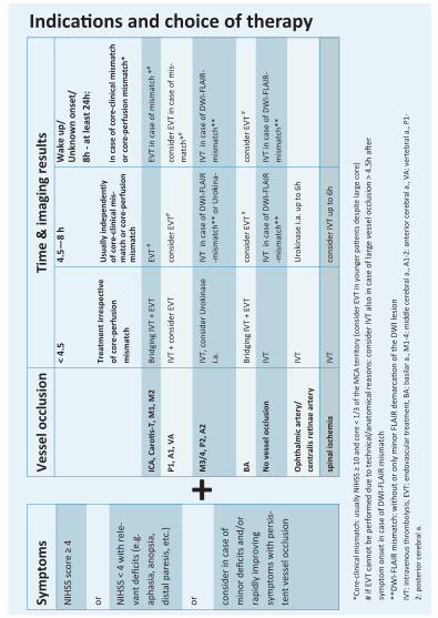
Indications and choice of therapy V
ess
el o
cclu
sio
n
Tim
e &
imag
ing
resu
lts
< 4
.5
Trea
tmen
t ir
resp
ecti
ve
of
core
-per
fusi
on
mis
mat
ch
4.5
—8
h
Usu
ally
ind
epen
den
tly
of
core
-cli
nic
al m
is-
mat
ch o
r co
re-p
erfu
sio
n
mis
mat
ch
Wak
e u
p/
Un
kno
wn
on
set/
8h
- a
t le
ast
24
h:
in c
ase
of
core
-cli
nic
al m
ism
atch
or
core
-per
fusi
on
mis
mat
ch*
ICA
, Car
oti
s-T,
M1
, M2
B
rid
gin
g IV
T +
EVT
EVT
# EV
T in
cas
e o
f m
ism
atch
*#
P1
, A1,
VA
IV
T +
con
sid
er E
VT
con
sid
er E
VT#
con
sid
er E
VT
in c
ase
of
mis
-
mat
ch*
#
M3
/4, P
2, A
2
IVT,
co
nsi
der
Uro
kin
ase
i.a.
IVT
in c
ase
of
DW
I-FL
AIR
-mis
mat
ch**
or
Uro
kin
a-
IVT
in c
ase
of
DW
I-FL
AIR
-
mis
mat
ch**
BA
B
rid
gin
g IV
T +
EVT
con
sid
er E
VT
# co
nsi
der
EV
T #
No
ves
sel o
cclu
sio
n
IVT
IVT
in c
ase
of
DW
I-FL
AIR
-mis
mat
ch**
IVT
in c
ase
of
DW
I-FL
AIR
-
mis
mat
ch**
Op
hth
alm
ic a
rte
ry/
cen
tral
is r
etin
ae a
rte
ry
IVT
Uro
kin
ase
i.a. u
p t
o 6
h
spin
al is
chem
ia
IVT
co
nsi
der
IVT
up
to
6h
*Co
re-c
linic
al m
ism
atch
: usu
ally
NIH
SS ≥
10
an
d c
ore
< 1
/3 o
f th
e M
CA
ter
rito
ry (
con
sid
er E
VT
in y
ou
nge
r p
atien
ts d
esp
ite
larg
e co
re)
# if
EV
T ca
nn
ot
be
per
form
ed d
ue
to t
ech
nic
al/a
nat
om
ical
rea
son
s: c
on
sid
er IV
T al
so in
cas
e o
f la
rge
vess
el o
cclu
sio
n >
4.5
h a
fter
sym
pto
m o
nse
t in
cas
e o
f D
WI-
FLA
IR m
ism
atch
**D
WI-
FLA
IR m
ism
atch
: wit
ho
ut
or
on
ly m
ino
r FL
AIR
dem
arca
tio
n o
f th
e D
WI l
esi
on
IVT:
intr
aven
ou
s th
rom
bo
lysi
s, E
VT:
en
do
vasc
ula
r tr
eatm
ent,
BA
: bas
ilar
a., M
1-4
: mid
dle
cer
ebra
l a.,
A1
-2: a
nte
rio
r ce
reb
ral a
., V
A: v
erte
bra
l a.,
P1
-
2: p
ost
erio
r ce
reb
ral a
.

Contraindications IVT EVT
Ab
solu
te
Re
lative
Septic embolization, endocarditis, encephalitis, pancreatitis
Intracranial hemorrhage
INR > 1.7
Surgery at non-compressible sites within the last 10d
Severe trauma
Intraparenchymal hemorrhage within the last 3 months
Pregnancy (IVT may be considered as off-label treatment)
Delivery within the last 14d
Gastrointestinal hemorrhage within the last 21d
Blood pressure above 185 mmHg sys/105 mmHg dias after BP treatment
Re
lative
Coagulopathy, incl. tumor associated (e.g. in case of leukemia) and prolonged aPTT
Thrombocytopenia < 100,000
Ischemic stroke within the last 2 months
Septicaemia
Hypoglycemia < 2.7 mmol/l or hyperglycemia > 22.2 mmol/l
Sodium < 120 mmol/l or > 150 mmol/l
Severe underlying disease, short life expectancy
Notes - IVT in patients previously treated with antiplatelet aggregation therapy
- Monotherapy Aspirin/clopidogrel/Aspirin+dipyridamol/ticagrelor: no restrictions
- Dual therapy: Aspirin+clopidogrel: no restrictions; other combinations: consider IVT carefully
- Monotherapy or combination therapy with prasugrel: consider IVT carefully
- Triple therapies: no IVT
- Bridging (IVT + EVT):
- normally full dose Alteplase 0.9mg/kg KG
- normally no control-imaging before EVT except in case of clinical deterioration
- large infarction DWI/CBV (> 100mL): consider EVT in younger patients (< 75 years, and especially if < 60
years)
7

IVT Alteplase dosage
Weight (kg) Total dose
0.9mg/kg body weight
Bolus
10% in 1min
Perfusor
90% within 60min
44-47 40mg = 40ml 4 ml 36 ml/h
48-51 44mg = 44ml 4 ml 40 ml/h
52-54 47mg = 47ml 5 ml 42 ml/h
55-57 50mg = 50ml 5 ml 45 ml/h
58-62 54mg = 54ml 5 ml 49 ml/h
63-67 59mg = 59ml 6 ml 53 ml/h
68-72 63mg = 63ml 6 ml 57 ml/h
73-77 68mg = 68ml 7 ml 61 ml/h
78-82 70mg = 70ml 7 ml 63 ml/h
83-88 77mg = 77ml 8 ml 69 ml/h
89-92 80mg = 80ml 8 ml 72 ml/h
93-97 86mg = 86ml 9 ml 77 ml/h
≥98 90mg = 90ml 9 ml 81 ml/h
Note: stop perfusor after 40min for 2/3 dosage
IVT in patients treated with DOAC Dabigatran
(Pradaxa®) Rivaroxaban (Xarelto®)
Apixaban (Eliquis®)
Edoxaban (Lixiana®)
Emergency measu-rement of:
- anti-IIa activity - thrombin time - aPTT
- Anti-Xa activity for Rivaroxaban - INR - aPTT
- Anti-Xa activity for Apixaban - INR - aPTT
- Anti-Xa activity for Edoxaban - INR - aPTT
IVT without restric-tions possible when last dose was taken more than 48h ago, or if:
Thrombin time normal
or anti-IIa activity not detectable
Anti-Xa activity not detectable
Anti-Xa activity not detectable
Anti-Xa activity not detectable
Consider IVT in individual situation with possibly higher bleeding risk if:
(these recommen-dations require in addition normal coagulation)
Anti-IIa acitivity < 100 ng/ml*
_________________
If Anti-IIa >100 ng/ml a./o. aPTT prolon-ged: consider antagonising with Idarucizumab (s.p. 12) and then start IVT
Anti-Xa activity < 100 ng/ml*
Anti-Xa activity < 100 ng/ml*
Anti-Xa activity < 100 ng/ml*
Note: *Consider timepoint of measurement (potential further increase of activity if peak activity is not reached yet) + these recommendations require normal renal function (Clearance > 30 ml/min)

Monitoring during IVT 1. Measure BP every 5 minutes: target sys ≤ 185 mmHg, diast ≤ 105 mmHg - in case of > 185/105: re-check after 5 minutes - if BP persists > 185/105: BP lowering (see Antihypertensive medications below) 2. Respiration: Control of oxygen saturation: target Biox > 92% 3. Evaluation of pupils: 3 x per hour - in case of clinical deterioration: stop Alteplase; CT: hemorrhage? - in case of allergic reaction: stop Alteplase administer Clemastin 2mg, methylprednisolone 250mg i.v. for extreme anaphylaxis: adrenalin 0.3.-0.5 mg s.c. for very extreme anaphylaxis: adrenalin 0.05-0.1 mg i.v. - in case of plasma glucose > 11 mmol/l: reduce with insulin carefully
Antihypertensive medication (iv) Use (standard values) Medication Dosage Maximum
effect CAVE/Side effects
Urapidil 50mg/vial
2.5-10mg (1ml=5mg) max 50mg/d
10 min Vertigo, headache, dyspnea, arrhythmia (tachycardia or bradycardia)
bolus administ-ration for HR > 70/min
Labetolol 100mg/vial
5-10mg (1ml=5mg) max 200mg/d
15 min Bradycardia, AV-block, hypotensi-on, vertigo, nausea, paresthesia, bronchial spasm
bolus administ-ration for HR > 70/min
Metoprolol 5mg/vial
1-2.5mg (1ml=1mg) max 15mg/d
5 min Bradycardia, AV-block, low output syndrome, bronchial spasm
bolus administ-ration for HR < 70/min
Dihydralazin 25mg/vial
6.25mg slowly over 2 minutes (1ml=12.5mg) max 100mg/d
20 min
Edema, tachycardia, angina pectoris, excercise caution in case of liver or renal failure
CI: Coronary insufficiency
Perfusion therapy
Urapidil 50mg/vial
5-10 mg/h max. 40mg/h - Restricted to 48h therapy.
Perfusion therapy Labetolol
10-40 mg/h max 100 mg/h (1ml = 1mg)
- Bradycardia, AV-block, hypotensi-on, vertigo, nausea, paresthesia, bronchial spasm
Vasopressor therapy (iv) Use (standard values) Medication Dosage Start CAVE/Side effects
Perfusion therapy
Noradrenalin®
Noradrenalin
10 mg/vial
20-400 g/h
Start with 20 g/h
then titrate
CI: Hyperthyreosis, tachycardia arrhythmias, angle-closure glau-coma, pheochromocytoma, cardiomyopathy (esp. hypertro-phic)
Compensate hypovolemia first
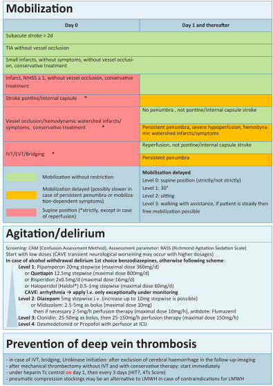
Agitation/delirium Screening: CAM (Confusion Assessment Method), Asssessment parameter: RASS (Richmond Agitation Sedation Scale) Start with low doses (CAVE transient neurological worsening may occur with higher dosages) In case of alcohol withdrawal delirium 1st choice benzodiazepines, otherwise following scheme: Level 1: Pipamperon 20mg stepwise (maximal dose 360mg/d) or Quetiapin 12.5mg stepwise (maximal dose 800mg/d) or Risperidon 2x0.5mg/d (maximal dose 16mg/d) or Haloperidol (Haldol®) 0.5-1mg stepwise (maximal dose 60mg/d) CAVE: arrhythmia → apply i.v. only exceptionally under monitoring Level 2: Diazepam 5mg stepwise i.v. (increase up to 10mg stepwise is possible) or Midazolam: 2.5-5mg as bolus (maximal dose 10mg) then if necessary 2-5mg/h perfusion therapy (maximal dose 10mg/h), antidote: Flumazenil Level 3: Clonidin: 25-50mg as bolus, then 25-150mg/h perfusion therapy (maximal dose 150mg/h) Level 4: Dexmedetomid or Propofol with perfusor at ICU
Prevention of deep vein thrombosis - in case of IVT, bridging, Urokinase initiation: after exclusion of cerebral haemorrhage in the follow-up-imaging - after mechanical thrombectomy without IVT and with conservative therapy: start immediately - under heparin Tc control on day 1, then every 3 days (HIT?, 4Ts Score) - pneumatic compression stockings may be an alternative to LMWH in case of contraindications for LMWH
Mobilization Day 0 Day 1 and thereafter
Subacute stroke > 2d
TIA without vessel occlusion
Small infarcts, without symptoms, without vessel occlusi-on, conservative treatment
Infarct, NIHSS ≥ 1, without vessel occlusion, conservative
treatment
Stroke pontine/internal capsule *
Vessel occlusion/hemodynamic watershed infarcts/symptoms, conservative treatment *
No penumbra , not pontine/internal capsule stroke
Persistent penumbra, severe hypoperfusion, hemodyna-mic watershed infarcts/symptoms
IVT/EVT/Bridging *
Reperfusion, not pontine/internal capsule stroke
Persistent penumbra
Mobilization delayed
Level 0: supine position (strictly/not strictly)
Level 1: 30°
Level 2: sitting
Level 3: walking with assistance, if patient is steady then
free mobilization possible
Mobilization without restriction
Mobilization delayed (possibly slower in case of persistent penumbra or mobiliza-tion-dependent symptoms)
Supine position (*strictly, except in case of reperfusion)

Stroke Unit treatment First neurological examination immediately after arrival Cardiovascular monitoring: - BP upper limits during the early phase: ≤ 185/105 mmHg after IVT/EVT ≤ 220/110 mmHg in conservatively treated patients - BP lower limit: only in selected cases in case of hypoperfusion/symptom worsening with drop of BP => to increase BP: only temporary administration of a limited volume of infusion solution (max. 500ml); in other cases use vasopressors (e.g. Noradrealine) - Tachycardia > 100 bpm => usually beta blockers; in case of tachycardic atrial fibrillation consider to add digoxin - frequent ventricular extrasystole => magnesium 2g i.v. - bursts of ventricular extrasystole (more than 3 beats): usually beta blocker + magnesium; ≥10 beats or polymorph or >120/min or clinically symptomatic => consultation with cardiologist - Bradycardia: during sleep in asymptomatic patients, usually up to 35 bpm is tolerable - pause > 3 seconds => consultation with cardiologist Respiration: target Biox ≥ 92; screening for sleep apnea - if > 4l O2/min is necessary or respiration frequency > 20 => clinical examination, arterial blood gas analysis, chest X-ray (pulmonary embolism? cardiac failure? pneumonia?) - if respiration frequency > 25-30 there may be danger of respiratory exhaustion Body temperature: ≥ 38° -> antipyretics (1st choice paracetamol) + 2x2 blood cultures, empirical/causal treat-ment Neurological evaluation: usually every 2h during the first 24h Clinical general medical evaluations: cardiac compensation, lung, abdomen to be checked daily Prescription of medication: - Do not prescribe antiplatelet aggregation therapy after IVT/i.a Urokinase before exclusion of hemorrhage in control CT/MRI after 24h - general cardiac premedication should be continued, with potential reduction of dose (CAVE cardiac failure/rebound tachycardia after stop) - stop any antihypertensive medication in case of hemodynamic stroke
Laboratory controls: (24h after IVT/EVT) - Hb, Lc, Tc, CRP, glucose, Na, K, creatinin, INR - hs-Troponin T and ECG after 1 h if initially abnormal - anemia: transfusion if Hb < 90 g/l - Tc daily under heparin therapy; further laboratory examinations individually determined
Neuroradiological control: - 24h after IVT/EVT, MRI (or CT), including MRA (CTA) except in cases with severe renal insufficiency - in case of neurological deterioration immediately
Swallowing: in case of dysphagia, reduced consciousness , facial palsy or relevant neuropsychological deficits: swallowing test (GUSS: Gugging Swallowing Screen)
Nutrition and fluid balance: - Daily fluid intake requirement: 30-35 ml/kg body weight - daily energy demand: 35kcal x body weight - if sufficient oral energy supply cannot be given within 3 days after stroke: enteral feeding via nasogastric tube with high caloric fibrous enteral feeding as bolus application 3-4x/d; control of electrolytes (incl. magnesium and phosphate) - if fasting period > 7 days: delayed feeding (CAVE refeeding syndrome)
11

Daily checklist - visiting stroke patients 1 Neurological evaluation
NIHSS and symptom-orientated functional examination (results of physio-, ergotherapy, speech therapy) Depression? Sleep-wake disorder?
2 Clinical evaluation Cardiac compensation Lung Abdomen Fever?
3 Monitoring Relevant rhythmic disorders (regarding reason, hemodynamic, cardiac pathology) BP target value? BP actual value?
4 Mobilization?
5 Nutrition, Dysphagia?
6 Laboratory controls ? (especially electrolytes, inflammation parameters, kidney, hemostasis)
7 Medication Antithrombotic therapy? Deep vein thrombosis prophylaxis? BP therapy?
DD Neurological deterioration ? Reinfarction ? Infarct localization: e.g. secondary deterioration more frequent in internal capsula or pontine infarctions ? Hemodynamic: BP associated? Associated with mobilization? ? Bleeding ? Rising ICP ? Epileptic seizure ? Infection ? Sedation ? psychogenic and other less frequent causes
DD Myocardial inf. DD stress cardiomyopathy hsTnT-elevation in approx. 20% of ischemic stroke patients, DD: MI, stress cardiomyopathy (SCM), renal failure, hypertensive crisis, tachycardia, aortic dissection
Variable manifestation of SCM: hsTnT ↑ < regional hypokinesia < transient apical ballooning
- the extent of hsTnT-elevation does not discriminate between MI and SCM - SCM is an exclusion diagnosis - in case of doubt consider cardiac MRI (best discrimination) or ergometry
Possible practical approach in case of hsTnT-elevation: - clinical correlate for MI (repolarization disturbance a./o. angina pectoris) → coronary angiography - no clinical correlate: repeat ECG and hsTnT after 1 and 3h, and if necessary after 6h: - hsTnt without relevant change (<20%): renal failure? heart failure? hypertensive state? - hsTnT change >20%: consider cardiac MRI or coronary angiography

TIA and minor stroke Perform MRI in clinical TIA patients whenever possible Pathologic definition of TIA: transient neurological deficit without diffusion restriction in MRI Time- dependent definition of TIA: transient neurological deficits of < 24h duration Definition of minor stroke: NIHSS ≤ 4, symptoms stable or improving
ABCD2 score (stroke risk after TIA) Risk factor Points
Age ≥ 60 years 1
Systolic BP ≥ 140 or diastolic ≥ 90 1
Unilateral weakness with/without speech disturbance Speech disturbance without weakness
2 1
TIA duration ≥ 60 min TIA duration 10-59 min
2 1
Diabetes mellitus 1
6-7 points: high 2-days risk (8%) 4-5 points: intermediate 2-days risk (4%) 0-3 points: low 2-days risk (1%)
Management for patients with TIA
13

Frequent risk factors and causes
Ris
k fa
cto
rs (n
ot
dir
ectl
y ca
usa
l)
Spe
cifi
c ca
use
s (p
ote
nti
al c
ausa
tive
ris
k fa
cto
rs)
Hyp
erte
nsi
on
(>1
40
/90
mm
Hg)
C
ard
ioe
mb
olis
m/p
arad
oxi
cal e
mb
olis
m
Dia
bet
es m
ellit
us
(fas
tin
g b
loo
d s
uga
r ≥7
mm
ol/
l, H
bA
1c
≥
6.5
%);
imp
aire
d f
asti
ng
glu
cose
: 5.6
-6.9
mm
ol/
l
- A
tria
l fib
rilla
tio
n/fl
utt
er
Dys
lipid
emia
(LD
L ≥2
.5 m
mo
l/l,
TG >
5.2
mm
ol/
l, H
DL
<1.0
mm
ol/
l)
- M
yoca
rdia
l in
farc
tio
n
Fam
ily h
isto
ry (
m <
55
yea
rs, f
<65
yea
rs)
- O
ther
dys
rhyt
hm
ia (
e.g.
Sic
k-Si
nu
s, s
ilen
t at
riu
m)
Pre
-str
oke
/TIA
-
Val
ve d
isea
se
Smo
kin
g (i
ncl
. pip
e, c
igar
s)
- En
do
card
itis
Lack
of
ph
ysic
al a
ctivi
ty
(< 1
50
min
/wee
k m
od
erat
e o
r <7
5 m
in in
ten
sive
exe
rcis
e)
- P
FO/A
SD
Wei
ght
(BM
I >2
5, a
bd
om
inal
gir
th >
m:9
4cm
/f:8
8 c
m)
Larg
e ar
tery
dis
eas
e
Un
he
alth
y d
iet
- A
rter
io-a
rter
ial e
mb
olis
m
Alc
oh
ol a
bu
se (
> 30
dri
nks
/mo
nth
; f>1
5g/
d, m
>30
g/d
)
- A
orti
c ar
ch e
mb
olis
m
Slee
p r
elat
ed b
reat
hin
g d
iso
rder
s -
No
n a
ther
oth
rom
bo
tic
vasc
ulo
pat
hy
(e.g
. FM
D)
Dep
ress
ion
O
ther
cau
ses
Mig
rain
e w
ith
au
ra (
at le
ast
2 au
ras
in a
life
tim
e)
- D
isse
ctio
n (
incl
. ao
rta)
Pre
gnan
cy
- Sm
all a
rter
y d
isea
se (
lacu
nar
<1
.5cm
+ B
G)
Atr
ial t
ach
ycar
dia
-
Vas
culiti
s
Tach
ycar
dia
at
rest
-
Ch
ron
ic in
fecti
on
(in
par
ticu
lar
HIV
, Hep
B/C
, syp
hili
s)
Incr
ease
d v
aria
bili
ty in
blo
od
pre
ssu
re
- Fa
cto
r V
Lei
den
/Th
rom
bo
ph
ilia/
anti
Car
dio
lipin
/Lu
pu
s an
tico
agu
lan
t
Car
dia
c w
all
mo
tio
n a
bn
orm
aliti
es
- A
cute
co
agu
lati
on
dis
ord
ers
(esp
. DIC
)
Co
ntr
acep
tio
n
- C
oag
ula
tio
n d
iso
rder
s as
soci
ated
wit
h t
um
or
Ho
rmo
ne
rep
lace
men
t th
erap
y -
Fab
ry d
isea
se
Acu
te in
fecti
on
(es
p. i
nfl
uen
za)
- Si
ckle
cel
l dis
ease
/oth
er h
aem
oly
tic
cris
es
Ch
ron
ic r
enal
fai
lure
-
Po
lygl
ob
ulia
/th
rom
bo
cyto
sis
-
Dru
gs
-
Iatr
oge
nic
(e.
g. p
eriin
terv
enti
on
al)
14
Bo
ld: t
he
5 m
ost
imp
ort
ant
risk
fac
tors
, co
ntr
ibu
tin
g to
80
% o
f is
chem
ic s
tro
kes

15
Diagnostic work-up MRI incl. MRA (for a reliable evaluation of the distribution pattern of acute/chronic infarction and deter-
mination of the etiology, esp. in view of a CEA!), if not possible, CT incl. CTA
consider neurovascular ultrasound
12-lead ECG
3x 7-day-ECG or 30-day ECG
TEE (or poss. TTE), in case of known etiology (e.g. symptomatic ICA stenosis) usually only TTE
Routine laboratory testing: Na, K, CRP, ESR, glucose, HbA1c, creatine, Urea, hs-Troponin T, CK, CK-MB, AST, ALT, GGT, TSH, pro-BNP, D-Dimer, complete blood count, coagulation state, blood lipids
DD according to medical history and physical examination ? Circumstance at onset (e.g.. Valsalva?) ? Positive familial history with onset < 40 years (Fabry, Coagulopathy) ? < 50 years, previous art/ven thrombosis, abortion (anti-Phospholipid syndrome), Fabry disease ? Throat/neck/eye pain, Trauma (Dissection ICA/VA) ? Headache (vasculitis), thunderclap headache (reversible vasoconstriction syndrome) ? Heart murmurs (endocarditis, valvular calcification) ? Angina pectoris (acute or in the past) ? Acute chest/back pain (aortic dissection!, coronary syndrome) ? Peripheral vascular examination incl. BP-difference left-right (aortic dissection) ? Skin lesions (septic emboli, Fabry: angiokeratoma, Sneddon: Livedo racemosa)) ? Vision disturbance + hearing disturbance (Susac`s syndrome => corpus callosum affected?) ? Signs for systemic rheumatic disease ? B symptoms ? Acute or chronic infection
Embolic stroke of
unknown origin
Known cause
Distribution on MRI:
1 vascular territory
Distribution on MRI:
Multiple vascular
territories
Small, multiple/recurring
ischemic lesions
Large/cortical lesions
Arteriogenic
embolism ?
Cardiogenic embo-
lism? D-Dimer ↑
frequent
Paraneoplastic
coaguloathy?
D-Dimer frequently ↑↑
Coagulopathy ?
Aortic arch embolism?
Prolonged
ECG monitoring
Cardiac MRI?
< 50 years
Embolic stroke of
undetermined source Cause still unknown:
Laboratory tests (phospholipid: repeat after 3 months)
Lupus-anticoagulant, anti-cardiolipin, anti-2GPI
APC resistence (Factor V), protein C/S in PFO
Tumor screening
CTA/MRA
Plaque-MRI/-CT

Secondary prevention Etiology First stroke Re-Stroke → always repeat or escalate
examinations for etiology
no reason determined (specially no cardiac embolism source, no symptomatic stenosis)
ASS 100mg or Clopidogrel 75mg or ASS+Dipyridamole (Asasantin®)Ticagrelor (Brilique®) in case of intolerance to the other agents
Change to Clopidogrel 75mg or ASS+Dipyridamole (Asasantin®) Ticagrelor (Brilique®) in case of intolerance to the other agents
Initial therapy: in case of TIA or minor stroke within 24h after symptom onset and NIHSS < 4, small infarct): 4 weeks ASS 100mg + Clopidogrel 75mg (loading 600mg) when hemorr-hagic transformation is excluded and individual bleeding risk is not elevated
non-valvular AF - DOAC - occurence under sufficient or insufficient OAC => change to DOAC - occurence under DOAC: change sub-stance class (Xa ↔ IIa) -consider atrial appendage closure
- occurence under sufficient or insufficient OAC => change to DOAC - occurence under DOAC: change substance class (Xa ↔ IIa) -consider atrial appendage closure
valvular AF (Def: AF with rheumatic mitral stenosis)
OAC INR 2-3 1. optimize dosage if neccessary 2. consider OAC INR 2.5-3.5 3. consider OAC + ASS 100mg
symptomatic extracra-nial carotid stensosis
>50% degree of stenosis: CEA/CAS < 50% with radiologically proven plaque rupture: individual decision (e.g. dual antiplatelet therapy) + always statin at high dose
< 50% stenosis with radiologically proven plaque rupture: consider CEA/CAS
symptomatic extracra-nial vertebral artery stensosis
ASS 100mg + 4 weeks Clopidogrel 75mg + statin at high dose Contralateral hypoplasia: consider stenting
consider stenting
symtomatic intracrani-al stenosis
ASS 100mg + Clopidogrel 75mg for 3 months, then monotherapy + statin at high dose
ASS 100mg + Clopidogrel 75mg (duration individually) + statin at high dose + consider stenting
Instructions for the initiation of antiplatelet aggregation therapy after ischemic stroke
- in case of conservative treatment: immediately - after mechanical EVT: usually immediately with loading (250-500mg ASS or 300-600mg Clopidogrel) - after IVT, Bridging, Urokinase i.a.: after exclusion of bleeding in 24h control imaging - in case of imminent space-occupying brain edema neurosurgeons should be involved immediately. If a potential craniectomy is considered, no administration of antiplatelets (see separate guidelines).
Instructions for the earliest initiation of (D)OAC after ischemic stroke
- CAVE: Exclude hemorrhagic transformation and endocarditis (=contraindications) TIA/smallest infarctions: immediate initiation Small infarction (≈ <40ml): after 3d (with BG involvement 6d) Middle-sized infarction (≈ 40-100ml): after 6d (with BG involvement 9d) after exclusion of hemorrhage Large infarction (≈ >100ml): after 12d (with BG involvement 15d) after exclusion of hemorrage - usually no intermediate antiplatelet therapy until start of (D)OAC - in case of medication switch: consider transient „dual therapy“ depending on the delayed loss of therapeutic effect depending on T1/2 - highly embolic source of embolism (e.g. mechanical heart valve): consider immediate initiation of a therapeutic heparinization except if infarction is very large or hemorrhagic - in case of hemorrhagic transformation, initiation usually after 2 weeks (following CT scan)
≈40ml
≈100ml

Secondary prevention in special situations Myocardial infarction (sub)acute
- consider DOAC application for 3 months also without thrombus finding, esp. with embolic infarct distribution - stenting in patients with (D)OAC indication → (D)OAC + clopidogrel (consider DOAC low dose in large infarctions), triple therapy in acute stroke only in exceptional cases (esp. in-stent-thrombosis, stent main stem) If AF is indication for (D)OAC: consider atrial appendage closure, afterwards only dual antiplatelet therapy STEMI: Coro immediately; NSTEMI: Coro as soon as clopidogrel + ASS or (D)OAC + clopidogrel is possible (depending on indication); Coro immediately in case of severe arrhythmia, hemodynamic instability, persistent pain
Detection of AF or atrial thrombus in patients taking aspirin + clopidogrel due to coronary stent
DOAC long-term therapy + usually 1 year clopidogrel; during dual therapy consider (transient) DOAC low dose in dependence on infarct size
Intracardial thrombus
Ventricular: (D)OAC for 3 months, then control TEE and consider change to antiplatelet therapy Atrial appendage thrombus: DOAC therapy life long also without proven AF
Symptomatic stenosis
see page 18
Coronary heart disease or peripheral arterial occlusive disease + high risk for ischemic events
Consider Rivaroxaban 2x2.5mg + ASS 100mg/d
Severe heart failure with severe hypokinesia/akinesia
No DOAC escept in case of intra cardial thrombus
Infectious Endocarditis
No antiplatelet therapy/heparin/(D)OAC; if valvular replacement is indicated, early operation seems to be beneficial
Pulmonary embolism
DOAC, start depends on infarct size; duration: 6 months in case of uniquivocal provocative factors (surgery, immobilization >48h, plaster cast on leg), otherwise long-term therapy; PFO occlusion in case of long-term DOAC therapy not indicated, otherwise PFO closure also with low RoPE score
Paraneoplastic Coagulopathy
LMWH therapeutic dosage (2x/d, not 1x/d) or Edoxaban or Rivaroxaban
17

(A)symptomatic artery stenosis Criteria for the classification of a symptomatic carotid artery stenosis: - confirmation by a neurologist - very likely: proof of a plaque rupture with apposition thrombus in CT/MR-angiography - probable: internal carotid artery stenosis of at least 50% + typical stroke distribution pattern in MRI, with no other cause of the stroke (TEE/TTE and at least 24-hour ECG monitoring test) In general: CEA/stenting usually within few days after symptom onset - always high-dose statin therapy, for antiplatelet aggregation therapy see below - in case of additional atrial fibrillation as long as anticoagulation is possible (depending upon infarct size): o CEA: begin aspirin 100mg 1d preoperatively, therapeutic heparinization until surgery after surgery: 7d aspirin 100mg + prophylactic heparin, then stop aspirin/heparin + begin (D)OAC o Stenting: normally N(OAC) + aspirin 100mg; start aspirin at least 1 day before intervention
in case of CEA: normally pre- and postoperative aspirin 100mg or clopidogrel 75mg monotherapy (stroke occurrence under aspirin or clopidogrel: consider aspirin 100mg + clopidogrel 75mg peri-operatively)
ICA stenosis extracranial
in case of stenting: preinterventional aspirin 100mg + clopidogrel 75mg (possibly loading dose); postinterventional aspirin 100mg + clopidogrel 75mg for at least 6 months (depending on device type, result after stenting, follow up results), then monotherapy
In case of apposition thrombus: Stenosis > 50%: CEA/CAS as early as possible, consider transient therapeutic heparinization (1st choice LMWH) + statin high dose (for example atorvastatin 80mg) Stenosis > 50%: therapeutic heparinization (1st choice LMWH) + statin high dose; control MRI after 2 and 7 days; CEA/CAS in case of new ischemia or persistent thrombus; in case of decrease of thrombus consider conservative treatment
Stenosis of vertebral artery origin
Stenting normally only in case of failure of best medical treatment (including transient therapy with aspirin + clopidogrel) preinterventional aspirin 100mg + clopidogrel 75mg (possibly as loading dose) postinterventional aspirin 100mg + clopidogrel 75mg usually for 12 months, then monotherapy
Intracranial artery stenosis
aspirin 100mg + clopidogrel 75mg for 3 months, then de-escalate to monotherapy + statin at a high dose (for example atorvastatin 80mg) Stenting should be performed only in exceptional cases and after failure of medical therapy
Hyperperfusion syndrome: -after revascularization of hemodynamically relevant stenosis there is a danger of hyperperfusion syndrome - risk factors: high grade stenosis, bilateral stenosis, perioperative hypertension, diabetes, woman, age > 75 years, reduced reserve capacity - clinically: headache, seizures, neurological deficits; risk: intracerebral hemorrhage - occurrence 12h-7d after revascularization → therefore BP should normally be kept < 140/100 mmHg postoperatively/postinterventionally - in case of pronounced edema poss. additional dexamathasone
18

PFO Occlusion of PFO in case of cryptogenic stroke (at least TTE/TEE and one 7-day ECG negative) and/or RoPE Score > 5 in patients < 60 years. The decision should be made individually and RoPE score serves as orientation. Consider circumstances that may facilitate paradoxical embolism (e. g. deep vein thrombosis, onset of neurological symp-toms after valsalva maneuver, co-existence of atrial septal aneurysm or eustachian tube (increase possibly recur-rent risk) and poss. psychological factors). Antiplatelet aggregation inhibitors should be continued lifelong after occlusion of PFO.
RoPE score (risk of paradoxical embolism)
No hypertension 1 Age 18-29 5 Sum 0-3 0% PFO attributable risk
No diabetes mellitus 1 Age 30-39 4 Sum 4 38% PFO attributable risk
No previous stroke/TIA 1 Age 40-49 3 Sum 5 34% PFO attributable risk
Non-smoker 1 Age 50-59 2 Sum 6 62% PFO attributable risk
Cortical infarct localization 1 Age 60-69 1 Sum 7 72% PFO attributable risk
Age ≥ 70 0 Sum 8 84% PFO attributable risk
Sum 9 88% PFO attributable risk
Dissections - according to current data the preventive effect of aspirin and OAC are propably comparable - in case of higher grade extracranial stenosis due to dissection or occlusions without large infarction or hemorrha-gic transformations consider OAC/therapeutic heparinizaton followed by OAC - OAC is generally contraindicated in case of intradural dissections or dissections extending intradurally (elevated risk for SAH) - in case of uncertain diagnosis with fat-suppressed T1 sequences in MRI: extend to regular diagnostic work-up after stroke - off-label use of DOAC can be considered in single cases if OAC cannot be adjusted
- duration of secondary prevention with aspirin/OAC: switch OAC to aspirin after 3-6 months, continue aspirin 100mg/d as long term prophylaxis
Vasculitis workup (most relevant clarifications, see DGN guidelines for complete list)
- history: thunderclap headache? B symptoms? other symptoms? stay abroad? impaired vision/hearing? - physical examination: internal, dermatological or rheumatic manifestations? - additional examinations: TEE, MRI with dark-blood-sequences, cerebral angiography, CT chest/abdomen - laboratory testing: blood culture, complete blood count, ESR, CRP, ANA, ANCA, dsDNS, rheumatoid factor, SS-A, SS-B, anti-phospholipid-ab, lupus-anticoagulant, drug screening, LDH, CK, creatinine, AST, ALT, TSH, coagulati-on state, C3, C4, protein electrophoresis - serology: hepatitis B, C, HIV, syphilis, Borrelia, VZV, HSV, Mycoplasma, Chlamydia, toxoplasmosis, cysticercosis - consider Quantiferon test, procalcitonin, cryoglobulins, ferritin, soluble Interleukin receptor, fluorescein angio-graphy eyes - cerebrospinal fluid: microscopy, cytology, culture, VZV cerebrospinal fluid-serum-index, consensus-PCR fungus-bacteria/mycobacteria - biopsy brain (meninges+parenchyma) or nasal mycosa
19

Silent Strokes - most frequent incidental finding in CT/MRI (no TIA or stroke suspicious episodes in medical history) - prevelence depending on cardiovascular risk profile and age (~30% in people aged 70) - increased stroke risk and severity, risk for dementia, depresssion and subclinical deficites
Definition by MRI - acute or subacute ischemia (see A, p.e. acute diffusion lesion with signal decrease in ADC and without symptoms and without otherwise explanation - chronic ischemia:
T2/FLAIR hyperintense lesion, T1 hypointense lesion non-lacunar (see B) cerebellar or supratentorieal cortical, or supratentorial subcortical >3mm with affection of deep gray matter and without otherwise explana-
tion lacunar lesion (see C): ≥3mm, not corresponding to enlarged perivasculuar space
Definition by CT - cortical defect zone or lacunar lesion
Diagnostics - search for risk factors - complete vessel imaging if not already done with initial imaging - 3x 7d ECG - TTE/TEE
Therapy - risk factor treatmemt - ASS with consideration of risk/benefit value - treatment of blood pressure equal to secondary prevention guidelines, - consider treatment of stenosis > 60% of the depending vessel after consideration of risk/benefit value, in case of - acute ischemia, or - multiple chronic ischemia in the corresponding vessel territory
A B B C
20

Direct oral anticoagulants (DOAC) - indicated in strokes with evidence of non-valvular AF - in cerebral venous thrombosis and dissection: phenprocoumon/acenocoumarol, no DOAC (only off label use) - not recommended in anti-phospholipid-antibody syndrome, paraneoplastic coagulopathy or valvular AF (valvular: rheumatic mitral stenosis) - in case of known elevated GIT bleeding risk: preferable lower doses of DOAK especially in patients > 75 years
Factor II-inhibitor Factor X-inhibitors
Dabigatran (Pradaxa®)
Apixaban (Eliquis®)
Rivaroxaban (Xarelto®)
Edoxaban (Lixiana®)
General informa-tion
CI: Child-Pugh A-C
CI: Child-Pugh C CI: Child-Pugh B+C
CI: Child-Pugh C
Dose if CrCl ≥ 50 ml/min
2 x 150mg (≥ 80 years: 2x110mg)
2 x 5mg (2 x 2.5mg if two of the following criteria are fullfilled: ≥80 years, ≤60kg, crea-tinine ≥ 133 mol/l)
1 x 20mg 1 x 60mg (1 x 30mg if bw < 60kg)
Dose if CrCl 30-49 ml/min
2 x 110mg 1 x 15mg 1 x 30mg
Dose if CrCl 15-29 ml/min
contraindicated 1 x 15mg, control of plasma coagulation recommended
1 x 30mg
Dose if CrCl <15 ml/min
contraindicated not recommended contraindicated not recommended
Inductors (effect diminis-hed) (bold print: contraindication)
Rifampicin, St John's wort, carbamazepine
Rifampicin (edoxaban: dosage reduction not necessary), phenytoin, carbamazepine, phenobarbital, St John's wort
Inhibitors (effect enhanced) (bold print: contraindication)
Verampil, ketocona-zole, itraconazole, voriconazole, HIV-protease inhibitors, quinidine, droneda-rone, ciclosporin, tacrolimus, amioda-rone
Verapamil, ketoconazole, itraconazole, voriconazole, posaconazole HIV-protease inhibitors
T1/2 12-17h 9-14h 5-9h 10-14h
Set off time before surgery (in agreement with surgeon)
24h up to 72h in case of large operations 4d with CrCl < 50ml/min
24h 48h in case of high bleeding risk, renal failure, elderly patients
24h 48h in case of high bleeding risk, renal failure, elderly patients
24h before 48h in case of high bleeding risk, renal failure, elderly patients
21

Risk factor treatment 5 important: hypertension, physical activity, nutrition, smoking, obesity Arterial hypertension
1st choice: combination ACE inhibitor + diuretics or sartan + diuretics; aim BP < 140/90 mmHg
Dyslipidemia
- no arteriosclerosis : target LDL-C levels: < 2.6 mmol/l - in case of symptomatic stenosis: high dose (e.g. Atorvastatin 80mg), target LDL-C levels: < 1.8 mmol/l - strength of action : fluvastatin < pravastatin < simvastatin < atorvastatin < rosuvastatin - alternatively in case of intolerance: ezetimib in case of only hypercholesterinemia, otherwise fibrate - combination therapy in case of missed target value with monotherapy: statin + ezetimib, fibrate + ezetemib, statin + evolocumab
Sleep-apnea syndrome
- Screening with respiratory polygraphiy or Apnea link - Treatment with CPAP/APAP/ASV indicated with 1. AHI ≥ 5/h in symptomatic SAS (preexisting sleepiness) 2. AHI ≥ 30/h also in asymptomatic SAS 3. AHI 5-29/h + relevant general medical indications (e.g. severe heart failure, untreatable arterial hypertension)
22

Malignant infarcts General - usually 30° supine position - BP aim: MAP > 85 mmHg, sys < 220 mmHg - in case of imminent craniectomy: stop antiplatelet therapy - pneumatic compression stockings for prevention of deep vein thrombosis - consider as emergency medication until craniectomy: - mannitol/hypertonic saline solution (dosage control mannitol via osmotic gap, hypertonic saline solution via Na and osmolality) - Hyperventilation Decompressive craniectomy - craniectomy if possible within 24-48h and before relevant neurological deterioration - critical phase with risk for neurological deterioration: 24-96h (rarely up to as late as 10d) - signs of rising ICP: decreasing consciousness, disturbance of pupillomotor function usually with dilatation in case of supratentorial swelling and miosis in case of infratentorial swelling, increasing paresis, new ipsilateralparesis, pathological breathing pattern, rhythmic disorders - possible practical approach: o general actions see above o strict clinical control and CT control o in case of decreasing consciousness or other signs of rising ICP with corresponding increase of edema, craniectomy as early as possible o in very young patients and predictable course (very large infarcts) prophylactic craniectomy
Malignant infarctions of the middle cerebral artery territory
Predictors for malignant infarction: young patient, persistent vessel occlusion, early midline shift ≥ 4mm, critical infarct volume dependent upon age/atrophy but >>80ml or >1/2 media territory, additional infarction in anterior– or posterior territory
Indications for craniectomy: 1. usually < 60 years, in rare cases up to 70 years 2. symptom onset within the last 48 h (in exceptional cases this may be longer) 3. infarction of at least half of the middle cerebral artery territory 4. consent of patient or family 5. no contraindication for intervention Contraindications 1. Bilateral fixed pupils and coma 2. More than 3 of the following unfavorable prognostic factors: a. age >50 years b. infarction extends beyond the middle cerebral artery territory c. unilateral dilated pupil d. GCS <8 3. severe comorbidity; severe preexisting disability
Malignant cerebellar infarctions
Predictors for malignant infarction: young patient, persistent vessel occlusion, bilateral infarction, the size has less predictive value because small infarcts may induce large edema Indications for craniectomy 1. neurological signs of progressively increasing pressure in the brainstem 2. imaging shows space occupying infarction 3. consent of patient or family Contraindications 1. clinical or imaging signs of severe irreversible brainstem damage 2. severe comorbidity, severe preexisting disability

Non-traumatic intracerebral hemorrhage (ICH) - elevate upper body at least 30°, mobilization free - in case of severe thrombocytopenia (<70.000/l) or severe platelet dysfunction: platelet infusion (platelet dysfunction as a result of antiplatelet therapy is not an indication for platelet infusion) - in case of hemophilia: substitution of coagulation factors - not effective in studies: steroid treament, tranexamic acid, activated factor VIIa
Blood pressure management Blood pressure aim ≤ 140/90mmHg within 1h after admission important: a) Avoid fluktuations >20% → early perfusion treatment b) Avoid lowering >60mmHg within the first hour Medication: 1st. Choice: Uradipil 5-10mg i.v. bolus wise, 5-40mg/h perfusion therapy 2nd. Choice: Labetalol 20-80mg i.v. bolus wise, 1-2mg/min perfusion therapy, max 2.4g/d 3rd. Choice: Clonidin 25-500g i.v. bolus wise Avoid: i.v. Nitrate derivates (possible negative effect)
Interdisciplinary decision for neurosurgical intervention: - individual decision for hematoma evacuation in case of bleedings outside basal ganglia in patients with GCS 9-13 - basal ganglia bleeding: no indication for surgery, consider SWITCH study - ventricular drainage in case of impaired CSF drainage
Anticoagulation/antiplatelet associated ICH
Always: stop antiplatelet therapy/(D)OAC/heparines
Anticoagulant Therapy Note
Alteplase In case of occurence under IVT: stop Alteplase Involve hematology
Phenprocoum-on
Prothrombin complex concentrate: 2400 IE (if < 50 kg body weight: 30 IE/kg body weight) + vitamin K: if INR ≥ 1.5 → 10 mg i.v., then dosage in depen-dence on INR; onset of drug effect approx. 4-6h
Repeat prothrombin complex concentrate in case of insuffi-cient INR decrease after 15min. Then INR at least 1x/d (and eventually repeat vitamin K
Heparin UFH
Protamine sulfate:
If Heparin was stopped ≤1h or anti-Xa acitivity ≥ 0.35: 1000 E i.v. (1ml) per 1000 E heparin given during the last 3 hours (max. 5000E); If Heparin was stopped 1-3h before or anti-Xa acitivity 0.15-0.35: 500 E i.v. (0.5ml) per 1000 E heparine given during the last 3 hours (max. 5000E)
Involve hematology; beware of contraindications!
Heparin LMWH
Protamine sulfate: Last therapeutic dosage given ≤8h or anti-Xa acitivity ≥ 0.5: 5000 E protamine sulfate Last therapeutic dosage given 8-12h or anti-Xa acitivity 0.3-0.5: 2500 E protamine sulfate
Involve hematology; beware of contraindications!
Xa-Inhibitors Apixaban/Edoxaban/Rivaroxaban/
No evidence based therapy Prothrombin complex concentrate (see under Phenprocoumon) as option
measure anti-Xa of Apixaban/Rivaroxaban/Edoxaban before and after application
IIa-Inhibitor Dabigatran
No evidence based therapy Idarucizumab (2x2.5g) as specific antidot available
Antiplatelet No specific treatment (thrombocyte infusion potentially harm-ful)
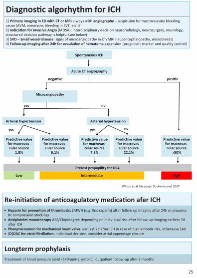
Diagnostic algorhythm for ICH 1) Primary imaging in ED with CT or MRI always with angiography – suspicision for macrovascular bleeding cause (AVM, aneurysm, bleeding in SVT, etc.)? 2) Indication for invasive Angio (IADSA): interdisciplinary decision neuroradiology, neurosurgery, neurology, structured decision pathway is helpful (see below) 3) SVD – Small vessel disease: signs of microangiopathy in CT/MRI (leucencephalopathy, microbleeds) 4) Follow-up imaging after 24h for evaulation of hematoma expansion (prognostic marker and quality control)
Spontaneous ICH
Acute CT angiography
Microangiopathy
Arterial hypertension Arterial hypertension
Predictive value for macrovas-cular source
1.8%
Predictive value for macrovas-cular source
6.1%
Predictive value for macrovas-cular source
7.3%
Predictive value for macrovas-cular source
22.1%
Predictive value for macrovas-cular source
>50%
Low Intermediate High
Pretest propapility for DSA
negative positiv
yes no
yes yes no
Wilson et al, European Stroke Journal 2017
Re-initiation of anticoagulatory medication afer ICH
Heparin for prevention of thrombosis: LMWH (e.g. Enoxaparin) after follow up imaging after 24h or pneuma-tic compression stockings
Antiplatelet monotherapy ASS/Clopidogrel: depending on individual risk after follow up imaging earliest 7d after ICH
Phenprocoumon for mechanical heart valve: earliest 7d after ICH in case of high embolic risk, otherwise 14d (D)OAC for atrial fibrillation: individual decision, consider atrial appendage closure
Longterm prophylaxis
Treatment of blood pressure (aiml <140mmHg systolic), outpatient follow-up after 3 months
25

26
Microbleeds differential diagnosis of incidental „microbleeds“ findings in SWI: thrombus, metastasis, microangiopathy, vasculi-
tis, cerebral amyloid angiopathy, rub off metallic waves most frequent origin: microangiopathy consider always cerebral amyloid angiopathy (s. below)
Microbleeds & Antiplatelet therapy/(D)OAC Effect of secondary prophylaxis with antiplatelet therapy and (D)OAC outweights bleeding risk Bleeding risk and risk for ischemia rises with number of microbleeds, but risk for ischemia remains higher
Cerebral amyloid angiopathy (CAA) Progressive dementia Frequently one or multiple small ischemic strokes or microbleeds in follow up images
MRI: modified Boston criteria for age >55 y Possible CAA Singular bleeding lobar, cortical or cortical-subcortical
localisation (cerebellar allowed) or focal or disseminiated superficial siderosis exclusion of other causes of ICB Probable CAA multiple bleedings lobar, cortical or cortical-subcortical loacalosation (cerebellar allowed) or singular, cortical-subcortical bleeding and focal or disseminiated superficial siderosis exclusion of other causes of ICB Definitive CAA Autoptic proven
CT: Edinburgh criteria Finger-like projections (FLP): elongated extension from the hematoma (longer than wider) Subarachnoid hemorrhage (SAH): extension of the bleeding in subarachnoid space
Hostettler, Seiffge & Werring, Expert Rev Neuroth 2019
Amyloid angiopathy & Antiplatelet therapy/(D)OAC with probable CAA: stop antiplatelet therapy/(D)OAC consider atrial appendage closure in case of atrial fibrillation in case of mehanical waves individual decision (reports of low embolic risk without OAC in some types of waves)

27
Motor areas
Speech areas
Visual areas
Sensory areas
Functional systems

Subclavian artery
Common carotid artery
Internal carotid artery
Vertebral artery
V0
Basilar artery
V1
V2
V3
V4
Brachiocephalic trunk
Cervical segment
Petrous segment
Cavernous segment
lower
mid
upper
border: skull base
border: Entrance cavernous sinus
border: outlet cavernous sinus/
ramification ophthalmic artery
(from here on intradurally)
Ophthalmic artery
Supraophthalmic segment
28
External carotid artery

Anterior cerebral artery
Middle cerebral artery
Posterior cerebral artery
M1 M2 M3
Superior cerebellar artery
Lenticulostriatal branches
Anterior inferior cerebellar artery
Posterior inferior cerebellar artery
Anterior choroidal artery
Pontine arteries
Posterior communicating artery
Anterior communicating artery
Basilar artery
Vertrebral artery
Anterior spinal artery
A1
A2
P1 P2
29
Internal carotid artery

Medial pontine a. of basilar a.; branches of posterior cerebral artery
Lateral pontine a. of basilar a.
Lateral pontine a. of basilaris a. Anterior inferior cerebellar a.(Fig. 8: Superior cerebellar a.)
Collicular and choroidal posterior medial a. of posterior cerebral a., superior cerebellar a.
Superior cerebellar artery
Posterior inferior cerebellar a.
Anterior inferior cerebellar a.
Pons (Fig. 5-8)
10 9
8 7
6 5
4 3
2 1
Anterior spinal artery
Anterior spinal artery Vertebral artery Posterior inferior cerebellar a.
Posterior spinal artery
Vertebral artery
Medulla oblongata (Fig. 1-4)
Anterior inferior cerebellar a.
Superior cerebellar a.
Central posteromedial a. of posterior cerebral artery Collicular and choroidal posterior medial a. of posterior cerebral artery
Collicular and choroidal posterior medial a. of posterior cerebral a.
Mesencephalon (Fig. 9-10)
Posterior inferior cerebellar a.

Lateral pontine a. of basilaris a.
posterior medial a. of posterior cerebral a., superior cerebellar a.
Posterior inferior cerebellar a.
posterior medial a. of posterior
Middle cerebral a.
Middle cerebral a., lenticuolostriate branches
Anterior cerebral a.
Posterior cerebral a.
Anterior choroidal a.
Posterior communi-cating artery
Anterior communi- cating artery
Posterior choroidal a. (from P2)
Thalamogeniculata a. (from P2)
Thalamoperforating A. (from P1 or BA; if jointly main trunk: Percheron artery)
Internal carotid a.
31

Cerebral venous and sinus thrombosis - LMWH in therapeutic dosage: e.g. enoxaparin (1mg/kg bw, 2x/d) (a non-randomized study even showed superiority in respect to efficacy and hemorrhagic com-plications; especially in patients with congestion hemorrhage) - alternatively therapeutic heparinization (aPTT 1.5-2.5x baseline aPTT) particularly in patients with risk of craniectomy; switch to OAC in the course of time - off-label use of DOAC can be considered in single cases if OAC cannot be adjusted (case series of dabigatran and rivaroxaban in SVT patients were positive)
- continue therapeutic heparinization/LMWH also after occurrence of congestion hemorrhages - IVT or mechanical recanalization in exceptional cases or in studies (e.g. TO-ACT) - in case of large hemorrhagic infarctions and impending lateral herniation: decompressive craniectomy as early as possible without removal of hematoma or infarcted tissue
- duration of OAC 6 months (except in case of progressive thrombosis at follow-up MRI or known thrombophilia) - usually examination for coagulation disorders after stopping OAC
Therapeutic heparinization with unfractio-nated heparin - complete baseline coagulation status before start of therapeutic heparinization - if baseline aPTT is abnormal (normal: 26-37sec) or in case of extensive thrombosis, consult a hematologist and control anti-factor-Xa-activity (aim 0.3-0.6 U/ml) - usual aPTT aim: 1.5-2.5x baseline aPTT - strictly check thrombocytes every 2 days during the course of therapy (HIT? => 4Ts score) The following dosage scheme is for patients at the Inselspital with low bleeding risk. Depending on infarct size, the dosage should be reduced individually.
Therapy start
Bolus 60-70 U/kg (max. 5000U) i.v. continuously 12-15 U/kg/h (max. 1000 U/h)
Re-evaluation after 6h
aPTT Anti-Xa
< 35 sec < 0.2 U/ml Bolus 40 U/kg Increase infusion rate by 3 U/kg/h
Re-evaluation after 6h
36-45 sec 0.2-0.29 U/ml No bolus, increaase infusion rate by 1.5 U/kg/h
Re-evaluation after 6h
46-70 sec 0.3-0.7 U/ml No change Re-evaluation after 6h, then 1x/day
71-90 sec 0.71-1.0 U/ml Reduce infusion rate by 1.5 U/kg/h Re-evaluation after 6h
> 90 sec > 1.0 U/ml Pause infusion for 1 h then reduce by 2-3U/kg/h (if aPTT >200sec pause infusion for 2h)
Re-evaluation after 6h

Superior sagittal sinus Cortical vein
Inferior sagittal sinus
Superior anastomotic vein
(Vein of Trolard)
Transverse sinus
Sigmoid sinus
Jugular vein
Basal vein of
Rosenthal
Inferior anastomotic vein
(Labbe)
Middle superficial
cerebral vein
Superior petrosal sinus
Anterior & posterior
intercavernous sinus
Cavernosus sinus
Sinus sphenoparietalis
Ophthalmic V.
Vein of Galen
Internal cerebral vein
Straight sinus
33


35
Close your eyes
He‘s a chip off the old block.
Harm set, harm get.
HUCKLEBERRY
BASEBALL PLAYER
36

Glasgow Coma Scale Eye opening response 4 Spontaneously
3 To speech 2 To pain 1 No response
Best verbal response 5 Oriented to time, place, and person 4 Confused 3 Inappropriate words 2 Incomprehensible sounds 1 No response
Best motor response 6 Obeys commands 5 Moves to localized pain 4 Flexion withdrawal from pain 3 Abnormal flexion (decorticate) 2 Abnormal extension (decerebrate) 1 No response
Modified Rankin Scale (mRS) 0 No symptoms at all
1 No significant disability despite symptoms; able to carry out all usual duties and activities
2 Slight disability; unable to carry out all previous activities, but able to look after own affairs
3 Moderate disability, requiring some help, but able to walk without assistance
4 Moderately severe disability; unable to walk without assistance and unable to attend own bodily needs
5 Severe disability; bedridden, incontinent and requiring constant nursing care and attention
6 Dead
CHA2DS2-VASc-Score (stroke risk with atrial f.) Risk factor Points (N)
O A C I F >1 P O I N T
Sum Risk/year taking Aspirin
Congestive heart failure 1 0 0%
Hypertension 1 2 2.2%
Age > 75 2 3 3.2%
Diabetes mellitus 1 4 4.8%
Stroke/TIA/thromboembolism 2 5 7.2%
Vascular disease (heart, peripheral) 1 6 9.2%
Age 65-74 years 1 7 11.2%
Woman 1 9 12.2%
37

NIH Stroke Scale Points Category Explanation
Level of conscious-ness
0 Alert 1 Not alert, but arousable by minor stimulation 2 Not alert, requires repeated stimulation to attend. Or, ob-tunded and requires painful stimuli to make movements 3 Makes only reflexive posturing movements to repeated painful stimuli. Or, they are totally unresponsive
Orientation anarthria, intubation=1, coma=2
Ask the current month and the patient‘s age. 0 Answered both questions correctly 1 Answered one correctly 2 Answered neither question correctly or aphasia
Commands
Ask the patient to open/close the eyes and make a fist/relax the non-paretic hand. 0 Performed both correctly 1 Performed one correctly 2 Performed neither correctly
Best gaze uncooperative=1, coma=2
0 Normal 1 Partial gaze palsy = Conjugate gaze deviation that can be overcome with voluntary or reflexive activity 2 Forced deviation
Visual Fields not evaluable=0, neglect=1, coma=3, in case of aphasia, evaluate reaction
0 No visual loss 1 Partial hemianopia 2 Complete hemianopia 3 Bilateral hemianopia
Facial palsy coma=3
0 Normal 1 Minor paralysis (flattened nasolabial fold or mild asymmetry while smiling) 2 Partial paralysis (total or near total paralysis of lower face) 3 Complete paralysis of upper and lower face
Left: Motor arm coma=4
0 No drift, remains in position for 10 sec. after an initial dip 1 Jerks or drifts to an intermediate position without encoun-tering support before the full 10 sec. 2 Some effort against gravity. Drifts down before 10 sec. 3 No effort against gravity and the arm falls 4 No voluntary movement Right:
Left: Motor leg coma=4
0 No drift, remains in position for 5 sec. after an initial dip 1 Jerks or drifts to an intermediate position without encoun-tering support before the full 5 sec. 2 Some effort against gravity. Drifts down before 5 sec. 3 No effort against gravity and the leg falls 4 No voluntary movement Right:
38

NIH Stroke Scale (part 2) Points Category Explantion
Limb ataxia coma, aphasia, paralyzed=0
0 Absent 1 Present in one limb 2 Present in two limbs
Sensory bilateral loss=2, coma=2 aphasia=rather 1
0 Normal 1 Mild to moderate sensory loss, patient feels asymmetry between the two sides but is still aware of being touched 2 Severe or total sensory loss, patient is not aware of being touched on the face, arm, and leg
Best language Intubated patients should be asked to write, coma=3
0 No aphasia 1 Mild to moderate aphasia; some obvious loss of fluency or facility of comprehension without significant limitation on ideas expressed or form of expression 2 Severe aphasia; all communication is fragmentary; great need for inference, questioning, and guessing by the examiner 3 Mute or global aphasia; globally aphasic patients have no usable speech or auditory comprehension
Dysarthria coma=2
0 Normal 1 Mild to moderate dysarthria; patient can still be understood 2 Severe dysarthria; patients are either mute or speech is so slurred they cannot be understood out of proportion to any dysphasia that is present
Extinction and inattention coma=2
0 Absence of neglect 1 Inattention to one modality only (visual, tactile, auditory, spatial, or personal inattention) 2 Profound hemi-inattention or extinction to more than one modality; does not recognize own hand or orients only to one side of space
39