Embed Size (px)
Citation preview

6/4/2018
1
Stroke, Cerebrovascular Imaging, and Anatomy
Dawn Tymianski, NP-A, PhD, CNNC
AGENDA
Provisional diagnosis
Observation*
Presentation
Diagnostics
Care requirements Predictive
DIAGNOSTIC TESTING: REASONING
Allows prompt diagnostic information
Correlates/confirms physical presentation (functional location)
To rule in/out pathology or disease process
Guides care, practice, conversation
Common stroke diagnostic imaging includes: CT, MRI (and their variants), digital subtraction angiography (DSA), PET, SPECT
We have come a long way……The 1st DSA included injections of petroleum, quicklime and mercury

6/4/2018
2
DIAGNOSTIC TESTS
Considerations prior to determining test:
Need to determine:
the pre-test probability of the disease-how likely the patient has something
if the test will change the decision to treat
what test provides the best information with the least harm
cost
skill set of the reader/quality of the image
50% of all acquired personal radiation exposure comes from diagnostic imaging
DIAGNOSTIC TIMING
Mr. S. is a 72 yr. old male admitted for ER via ambulance after collapsing on the street. On his way to ER he loses his airway. Upon arrival, his pupils are large, the (L) unreactive. During your assessment his (R) pupil ‘blows’. His BP is 280/124. He is urgently treated with Mannitol/3% prior to his DI.
The decision to treat without a DI is based upon:
High probability his has high ICP
The medication benefit outweighs the risk
A CT is ordered because:
CT is sensitive to rapid neurological deterioration
The result will change the decision to treat and type of treatment required
DI = diagnostic image

6/4/2018
3
CNS IMAGING: CT AND MRI
QUESTION
Plain CT of head is always ordered first on a potential stroke patient because:
A. Most institutions have a CT and can administer timely TPA
B. CTs always shows areas of ischemic infarction
C. CTs rule out/in non-ischemic causes of stroke
D. CTs allow clear visualization of the whole brain
QUESTION
Plain CT of head is always ordered first on a potential stroke patient because:
A. Most institutions have a CT and can administer timely TPA
B. CTs always shows areas of ischemic infarction
C. CTs rule out/in non-ischemic causes of stroke
D. CTs allow clear visualization of the whole brain

6/4/2018
4
Diagnostic Test
Good for Not great for Benefits or limitations
CT: plain acute blood, trauma, edema,
acute stroke evaluation
worsening of neurology
parenchyma
definition, limited
ability in early
ischemia
B: available, rapid, cheap,
tolerated, can be used to
determine treatment
options, high reliability
L: poor visualization of Bst
and post fossa
CTA/MRA vascular imaging
MRA better for large vessels
(carotids)
Can be used to guide treatment
MRI parenchyma, previous infarction
micro-hemorrhage (GRE)
worsening of neurology (ICP)
acute blood (can
be confusing)
B: can determine age of
hemorrhage
L: less tolerated, less
available, may exclude
some patients (pacemaker)
Diffusion-
weight image
(DWI)
ischemic changes visual within
minutes of stroke onset (can
separate out acute and chronic
stroke)
can be falsely positive
>90% reliability
L: may be positive in non-
stroke ( migraine,
seizures, acute MS, TIA)
Cerebral angiography
vascular requiring finite image
can be used singularly or in
conjunction with other treatments
(clot retrieval, embolization)
L: small risk of stroke,
side effects, invasive,
inconvenient for patient
TEST TYPE
Axial CT-plain Axial MRI-plain (T1)
CAT SCAN
6/4/2018
5
CAT SCAN (CT): 3D X-RAYS
Overall principles
X-rays are absorbed by different degrees by different tissues
The ‘colour’ of the tissue produced is the result of attenuation (rate at which the x-ray passes through the tissue or how much the radiation is absorbed)
Produces a 3D image
CT language = ‘density’ ‘dens----See--tee’ (CT) ‘hypodensity’, ‘hyperdensity’
Limitations: Beam hardening: when high density tissue abuts low density tissue
in a small space (post fossa)
Volume averaging: pictures produced include a variety of tissues with different densities (early stroke)
CT DENSITY
Very black
Black Grey Isodense Whitish WhiteBright white
Air CSFFatEdema
White matter
Old blood
Acute bloodcalcification
BoneContrast
Gray matter
Low attenuation High attenuation
Hypodense Hyperdense
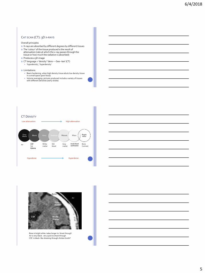
AIR
Bone
Gray matter
White matter
CSF
Bone is bright white: takes longer to ‘shoot through’ Air is very black: very quick to shoot through CSF: is black- like shooting through chicken broth*
Air

6/4/2018
6
CT plainGood for looking at ‘stuff’Great for blood, deterioration
CT with contrast: is iodine-basedGood for abscess, compromised BBB
CTA (is contrast): great for blood vessels
CT VARIATION
CTA
SPOT SIGN
The presence of contrast enhancement within
ICH, visible on CTA . Suggests active, dynamic
hemorrhage. Is a predictor of ICH growth and
poorer outcomes.

6/4/2018
7
Left MCA territory swelling (loss of sulcus, gyri flattening)Loss of gray-white borders
24 hours post stroke-hypodensity
CT on admission
STROKE AND CT
Stroke involves both gray and white matter
Deprived of blood, cells will take on water (cytotoxic edema)
Gray matter takes on water faster (more metabolically active)
Water influx into cells begins at 6 hours
Leads to lose of gray-white differentiation
a 1% increase in water content will reveal hypodensity
60% of infarcts are seen within 3-6 hours (all seen within 24)
CT LIMITATIONS
Volume averaging: tissues of different densities look the same
Beam hardening: stuff is jammed in together

6/4/2018
8
MRI
MRI: RADIO WAVES
Bursts of radio waves (magnet) are sent to the
head
Radio waves disrupt Hydrogen (proton) ion
Hydrogen (protons) moves towards the
magnet position
Once radio waves stop proton relax
Protons re-align and ‘relax’: T1-T2 produce variations in relaxation
and re-alignment (time)
Images produced are dependent upon proton
density and re-transit time
MRI COLOURS
MRI
BrightWhite
White Gray Black Very black
T1 BoneGadolinium
Fat
Orbits
Blood (with
contrast only)
Light: white
matter
Dark: Gray
matter
Water
Fluid (edema)
CSF
dense bone, Calcium
Eye globes
Most lesions
Air
T2 CSF Water, fat
Fluid
New blood
Most lesion
Light: Gray
matter
Dark: white
matter
Blood vessels
Dense bone, calcium
Flow
Air

6/4/2018
9
COMMON TYPES OF MRI
T1 CSF is ‘black’, white matter is white
T2 CSF is white and white matter is black
Produces picture of intensity (not density)
MRI: DWI (DIFFUSION WEIGHTED IMAGE)
Is the most sensitive sequence
Image is the result of the loss of Brownian motion of water (water that can move freely has no signal)
Swollen tissue (cytotoxic edema) has restricted-no movement = signal
Can be positive within minutes of stroke
Produces a high intensity signal for 7 days, then settles
Maximizes b/w 7-30 days (positive in early stages, the fades)
Can be positive for MS and migraine
FLUID ATTENUATION INVERSION RECOVERY : FLAIR

6/4/2018
10
DIGITAL SUBTRACTION ANGIOGRAM
(DSA)
MCA
ACA
ACA infarct
MCA infarct

6/4/2018
11
Moyamoya
Disease
AVM
VASCULAR ANATOMY AND PRESENTATION

6/4/2018
12
ANATOMY
Managing a patient with vascular disease requires an understanding of where that vessel goes and areas that vessel supplies
Allows predictability, care, and knowing when patient’s are running into trouble
Remember that:
BV variations are common and frequently non-pathological
Significant anastomosis exist
Presentation is related to functional disturbance, not always to the cause of the disturbance
(L) CCA
(R) CCA
ICA
MCA
ACA
Basilar
Aortic
arch
MCA

6/4/2018
13
1. Clyde is 2-days post clipping of an asymptomatic p.comm aneurysm. On rounds, the student nurse informs you that the patient has a ‘blown’ pupil on the same side.
2. Susan is admitted for a right parietal AVM resection. She is experiencing changes in proprioception, agnosia and acalculia
3. Rahmin has been diagnosed with a thalamic hemorrhagic stroke. He has difficulty keeping awake and is experiencing allodynia
4. Michael is admitted with a PCA ischemic stroke. He is suffering from significant nausea, is disoriented to place and year
Which one of the above vignette(s) is correct:
A. One of themB. Two of themC. Three of them D. All of them E. None of them
1. Clyde is 2-days post clipping of an asymptomatic p.comm aneurysm. On rounds, the student nurse informs you that the patient has a ‘blown’ pupil on the same side.
2. Susan is admitted for a right parietal AVM resection. She is experiencing changes in proprioception, agnosia and acalculia
3. Rahmin has been diagnosed with a thalamic hemorrhagic stroke. He has difficulty keeping awake and is experiencing allodynia
4. Michael is admitted with a PCA ischemic stroke. He is suffering from significant nausea, is disoriented to place and year
Which one of the above vignette(s) is correct:
A. One of themB. Two of themC. Three of them (2-left side for math and face)D. All of them E. None of them
CASE PRESENTATION
Carol is a healthy 42 yr. old female that was at the shopping mall with her husband and children. Carol had a 15 minute neck massage at a pop-up massage place. Shortly after, Carol experienced a bad headache and dizziness. By the time she met her husband, Carol was a bit off-balance and nauseated. When not better 5 hours later, they went to the hospital, thinking Carol had a bad virus.
In ER, Carol’s BP was 182/97. Neurologically, Carol was found to have 6th nerve palsy, a small pupil and facial weakness on the right, ataxia, dysdiadochokinesia left upper extremity, and difficulty with word articulation (NIHSS 10).

6/4/2018
14
Vertebral
PICA
SCA
PCA
Basilar
‘Shower’ into her brainstem and cerebellum
Cerebellum: Body coordination, muscle tone • Upper: Axial equilibrium • Middle: Peripheral coordination and planning (sides)• Bottom: Ear, eye balance
Brain stem: functional presentation location dependent • information relay (tracts), CN function, RAS, cardiac
respiratory center coordination, processing of visual and auditory data
IN
IN
CASE PRESENTATION
Mary is a 78 year old female driving in CDN tire parking lot. Was noted to be hitting parked cars. Stopped car. Police/ambulance were called. Upon arrival Mary when she was found to be densely hemiplegic on left side. Speech normal. No past history.
Transferred to stroke center at 14:40. NIHSS on arrival 13, right lateral gaze. BP 220/124.
CT plain at 1450-normal

6/4/2018
15
MCA
ACA
MCA supplies the greatest territoryand is the most often occluded
? Why the left MCA more?
Mary: treated with TPA, off to
radiology
DSA Thrombectomy wire
Post stroke CTPost thrombectomy DSA

6/4/2018
16
CASE PRESENTATION
Clyde is a 52 yr. old janitor with a known history of HTN. He is found with mild hemiplegia with hemianaesthesia and broca’s aphasia. EMS is called. Clyde loses consciousness and requires airway support upon transfer to ER.
On admission to ER his BP is 268/128, HR 110. GCS 6 (eyes 1, verbal 1, motor 4 (withdraws)
QUESTION
What statement is true regarding thalamic hemorrhage?
1. Left-sided hemorrhage is more common than right
2. Hallucinations, agitation and dementia can occur
3. Hemianaesthesia is the most common presentation
4. A significant percentage of patients have Type I or II diabetes
QUESTION
What statement is true regarding thalamic hemorrhage
1. Left-sided hemorrhage is more common than right (equal distribution)
2. Hallucinations, agitation and dementia can occur
3. Hemianaesthesia is the most common presentation (motor weakness)
4. A significant percentage of patients have Type I or II diabetes (approx. 10%)
4 thalamic stroke variations exist-symptoms location dependent (anterior-posterior), can also ‘mimic’ cortical function

6/4/2018
17
R MCA
Leticulostriate arteries
Thalamic Hemorrhage outcomes:
Anterior > posterior or if associated
with hydrocephalus
CASE PRESENTATION
Brittany is a 24 yr. receptionist at a dental. On Monday she doesn’t show up for work. Her colleagues try to reach her, but to no avail. At 10:00 pm after continuing to be unsuccessful, her colleague goes to her apartment where her superintendent opens the door. Her history includes Type I DM. Brittany is on the floor unconscious. EMS is called. Her pulse is thready. Both pupils are large. She is provided airway support and transported to ER. GCS on arrival 4 (extension to pain)
BRAIN EDEMA

6/4/2018
18
AGENDA
Provisional diagnosis
Observation*
Presentation
Diagnostics
Care requirements Predictive