Embed Size (px)
Citation preview

Research ArticleStress-Activated Degradation of Sphingolipids RegulatesMitochondrial Function and Cell Death in Yeast
Sara Manzanares-Estreder,1 Amparo Pascual-Ahuir,2 and Markus Proft1
1Department of Molecular and Cellular Pathology and Therapy, Instituto de Biomedicina de Valencia (IBV-CSIC), Jaime Roig 11,46010 Valencia, Spain2Department of Biotechnology, Instituto de Biología Molecular y Celular de Plantas, Universitat Politècnica de València-CSIC,Ingeniero Fausto Elio s/n, 46022 Valencia, Spain
Correspondence should be addressed to Markus Proft; [email protected]
Received 14 March 2017; Revised 24 May 2017; Accepted 11 June 2017; Published 6 August 2017
Academic Editor: Cungui Mao
Copyright © 2017 Sara Manzanares-Estreder et al. This is an open access article distributed under the Creative CommonsAttribution License, which permits unrestricted use, distribution, and reproduction in any medium, provided the original workis properly cited.
Sphingolipids are regulators of mitochondria-mediated cell death in higher eukaryotes. Here, we investigate how changes insphingolipid metabolism and downstream intermediates of sphingosine impinge on mitochondrial function. We found in yeastthat within the sphingolipid degradation pathway, the production via Dpl1p and degradation via Hfd1p of hexadecenal arecritical for mitochondrial function and cell death. Genetic interventions, which favor hexadecenal accumulation, diminishoxygen consumption rates and increase reactive oxygen species production and mitochondrial fragmentation and vice versa.The location of the hexadecenal-degrading enzyme Hfd1p in punctuate structures all along the mitochondrial network dependson a functional ERMES (endoplasmic reticulum-mitochondria encounter structure) complex, indicating that modulation ofhexadecenal levels at specific ER-mitochondria contact sites might be an important trigger of cell death. This is furthersupported by the finding that externally added hexadecenal or the absence of Hfd1p enhances cell death caused by ectopicexpression of the human Bax protein. Finally, the induction of the sphingolipid degradation pathway upon stress is controlledby the Hog1p MAP kinase. Therefore, the stress-regulated modulation of sphingolipid degradation might be a conserved way toinduce cell death in eukaryotic organisms.
1. Introduction
Sphingolipids are specialized bioactive lipid molecules foundin the membranes of all eukaryotic cells. They have criticalfunctions in the control of cell growth, senescence, differenti-ation, and programmed cell death. Different intermediates ofsphingolipid pathways can have opposing effects on cellsignaling. Therefore, imbalances in sphingolipid metabolismcan deregulate key cellular processes and contribute tohuman disorders [1–3]. A critical cellular homeostasis path-way regulated by sphingolipids is mitochondria-mediatedapoptosis [4]. In this process, multiple pathways convergeon mitochondria and induce mitochondrial outer membranepermeabilization (MOMP). MOMP causes the release ofproteins from the mitochondrial intermembrane space intothe cytosol, where they execute the programmed cell death
via the caspase cascade in higher eukaryotes. Therefore,MOMP is considered the initial irreversible trigger of mito-chondrial apoptosis [5]. This death decision needs to betightly controlled and depends on the interplay of severalpro- and antiapoptotic members of the Bcl-2 family [6, 7].Some Bcl-2 protein members such as Bax have to gain accessto mitochondria in response to apoptotic stimulation, as theyare normally cytosolic proteins [8, 9]. During apoptotic acti-vation, Bax structural conformation is altered in a regulatedmanner, which facilitates its insertion into the outermitochondrial membrane followed by dimerization and theformation of higher order oligomeric pores to induce MOMP[10, 11]. Specific sphingolipid levels at mitochondria areknown to regulate the entry into mitochondrial apoptosis[4, 12]. Ceramide has been identified as a prodeath molecule,as it can induce apoptosis when applied externally [13], and
HindawiOxidative Medicine and Cellular LongevityVolume 2017, Article ID 2708345, 14 pageshttps://doi.org/10.1155/2017/2708345

the regulation of ceramide biosynthesis and degradation isimportant for the entry into the intrinsic apoptotic pathway[14–16]. However, recent studies point to importantproapoptotic functions for metabolites of the sphingolipiddegradation pathway, such as sphingosine-1-phosphate (S1-P) or hexadecenal [17, 18]. S1-P and hexadecenal are efficientinducers of Bax-mediated apoptosis and MOMP in vitro[17]. Since sphingolipid metabolism takes place in the ER,the regulated production and transfer of sphingolipid metab-olites to mitochondria might be an important, but yet unex-plored, upstream event in the initiation of cell death [19].
Despite their simplicity, yeast cells are an informativemodel to understand the regulation of programmed celldeath [20, 21]. Importantly, yeast undergoes MOMP, cyto-chrome c release, and mitochondrial fragmentation inresponse to apoptotic stimuli [22–24], as well as upon theheterologous expression of the human apoptosis executerBax [25, 26]. As for higher eukaryotes, also in yeast, ourinformation about the effect of sphingolipid metabolism onmitochondrial function and death is limited to ceramidelevels [27, 28]. The lack of enzyme functions leading to cer-amide production has been shown to decrease programmedcell death in response to acetic acid in yeast [29]. Theenzymes involved are Isc1p and Lag1p, which contribute toceramide formation by the breakdown of complex sphingoli-pids or by de novo synthesis, respectively. However, therecent identification of the precise enzymatic steps involvedin the degradation of sphingolipids and the formation ofthe key intermediates S1-P and hexadecenal in yeast [30]has enabled us to study the effect of this metabolic pathwayon mitochondrial-mediated cell death. We show here thatthe key enzyme in this pathway, the hexadecenal metaboliz-ing Hfd1p dehydrogenase, has a stress-regulated functionassociated with mitochondria, which might be decisive forthe entry into mitochondrial cell death.
2. Materials and Methods
2.1. Yeast Strains and Plasmids. A complete list of yeaststrains used in this work can be found in Table 1. Constitu-tive expression of Hfd1-GFP was obtained by cloning fulllength HFD1 into the yeast Gateway vector pAG426-GPD-ccdB-GFP [31]. Fusion of full length OM14 with dsRed wasperformed in yeast Gateway vector pAG415-GPD-ccdB-dsRed [31]. Galactose inducible overexpression of Dpl1pwas achieved by cloning full length DPL1 into yeast Gatewayvector pAG426-GAL1p-ccdB-HA [31]. Plasmids pCM189-Bax and pCM184-Bcl-xL for Tetoff-controlled Bcl-2 memberprotein expression were a kind gift from S. Manon [32].GFP-Ybh3 fusion protein expression was achieved from plas-mid pUG36-GFP-YBH3 (a kind gift from F. Madeo [33]).For the galactose inducible expression of a Mmm1-mCherry fusion protein, plasmid pAG413-GAL1p-MMM1-mCherry was used (a kind gift from W. Prinz [34]).
2.2. Growth Conditions. Yeast strains were grown at 28°C inyeast extract peptone containing 2% dextrose (YPD) or 2%galactose (YPGal) for agar plate assays. Synthetic dextrose(SD) or galactose (SGal) media contained 0.67% yeast
nitrogen base, 50mM succinic acid (pH5.5), and 2% ofthe respective energy source. According to the auxotro-phies of each strain, methionine (10mg/l), histidine(10mg/l), leucine (10mg/l), or uracil (25mg/l) were added.As indicated, growth media were supplemented with men-adione, valinomycin, or NaCl from appropriate stocks inDMSO (menadione) or water (valinomycin, NaCl). Yeaststrains transformed with Bax- or Bcl-xL-expression plas-mids were pregrown in the presence of 10μg/ml doxycy-cline (Sigma), and protein expression was induced byswitching cells to doxycycline-free medium. For sensitivityassays with externally added sphingolipid metabolic inter-mediates, serial dilutions of the cells were treated in syn-thetic liquid medium with the indicated doses of thecompounds from DMSO stock solutions and then platedonto YPD agar plates. Palmitic acid, hexadecenoic acid,and sphingosine-1-phosphate were purchased from Sigma;hexadecenal was purchased from Avanti Polar Lipids. Forquantitative colony assays, fresh overnight yeast cultureswere diluted in triplicate to OD600 = 0.1 and allowed togrow for 24h on the indicated media. Alternatively, forthe hexadecenal toxicity assays, fresh overnight cultures inSD medium were adjusted to the same OD600 and thentreated with the indicated concentrations of hexadecenalfor 2 h. The number of colony-forming units was thendetermined by plating the cultures onto YPD agar platesat appropriate dilutions.
2.3. Reverse Transcriptase Assays. Total RNA was isolated byacid phenol extraction from yeast cells grown in the indicatedconditions. RNA samples were digested with DNaseI andpurified with the RNeasy Mini kit (Qiagen). A total of 5μgof RNA was converted into DNA using the SuperScript IIIFirst-Strand cDNA Synthesis kit (Invitrogen). The amountof DNA was determined with the indicated gene-specificprimers by quantitative PCR in real time using the EvaGreenqPCR Master Mix (Biotium) on an Applied Biosystems 7500sequence detection system. The ACT1 gene was used as a ref-erence. Relative expression levels were calculated in triplicatefrom three independent cDNA samples. Primer sequencesare available upon request.
2.4. Microscopy. Mitochondria were visualized with Mito-Tracker Red CMXRos (Invitrogen). Cells were incubatedwith 1μM MitoTracker dye for 60min, washed once, andfinally resuspended in fresh culture medium. Cells wereobserved on a Leica confocal microscope TCS SP8 usingthe following excitation and emission wavelengths: GFP(excitation 488nm; emission 509nm), mCherry (excitation587 nm; emission 610nm), dsRed (excitation 560 nm; emis-sion 592 nm), and MitoTracker Red (excitation 578 nm;emission 600nm).
2.5. ROS Determination. Exponential cell culture aliquotsupon the indicated growth conditions were incubatedfor 30min with 2′,7′-dichlorodihydrofluorescein diacetate(Sigma) at a final concentration of 10μM. The cells werewashed with water and resuspended in 1ml of 50mM Tris/HCl pH7.5. After the addition of 10μl of chloroform and
2 Oxidative Medicine and Cellular Longevity

5μl of 0.1% SDS, cell extracts were prepared by rigorous agi-tation with glass beads. Fluorescence was quantified in thesupernatant in a microplate reader at 492nm excitation and525nm emission wavelengths and normalized for the fluo-rescence of the same number of mock-treated cells.
2.6. Measurement of Oxygen Consumption. For measure-ments of respiration rates, yeast cells were grown to exponen-tial phase in synthetic galactose-containing medium, washedwith water, and finally resuspended at the same OD in 40mMNaPO4 pH7.4 with 1% glucose. Oxygen consumption was
Table 1: Yeast strains used in this study.
Name Relevant genotype Source
BY4741 MATa; his3Δ1; leu2Δ0; met15Δ0; ura3Δ0 EUROSCARF
BY4741 hfd1 BY4741 with hfd1Δ::KanMX EUROSCARF
BY4741 faa1 BY4741 with faa1Δ::KanMX EUROSCARF
BY4741 faa4 BY4741 with faa4Δ::KanMX EUROSCARF
BY4741 dpl1 BY4741 with dpl1Δ::KanMX EUROSCARF
BY4741 fzo1 BY4741 with fzo1Δ::KanMX EUROSCARF
BY4741 dnm1 BY4741 with dnm1Δ::KanMX EUROSCARF
BY4741 fis1 BY4741 with fis1Δ::KanMX EUROSCARF
BY4741 hog1 BY4741 with hog1Δ::KanMX EUROSCARF
BY4741 om14 BY4741 with om14Δ::KanMX EUROSCARF
BY4741 mcr1 BY4741 with mcr1Δ::KanMX EUROSCARF
BY4741 rdl1 BY4741 with rdl1Δ::KanMX EUROSCARF
BY4741 mdm10 BY4741 with mdm10Δ::KanMX EUROSCARF
BY4741 mdm12 BY4741 with mdm12Δ::KanMX EUROSCARF
BY4741 ybh3 BY4741 with ybh3Δ::KanMX EUROSCARF
BY4741 msn2msn4 BY4741 with msn2Δ::Nat msn4Δ::KanMX E. de Nadal
BY4741 Hfd1-GFPOm14-dsRed
BY4741 with plasmids pAG426-GPD-HFD1-GFP (URA3) + pAG415-GPD-OM14-dsRed This study
BY4741 GAL1-ccdB-HA BY4741 with plasmid pAG426-GAL1p-ccdB-HA (URA3) This study
BY4741 GAL1-DPL1-HA BY4741 with plasmid pAG426-GAL1p-DPL1-HA (URA3) This study
hfd1 GAL1-ccdB-HA BY4741 hfd1Δ::KanMX with plasmid pAG426-GAL1p-ccdB-HA (URA3) This study
hfd1 GAL1-DPL1-HA BY4741 hfd1Δ::KanMX with plasmid pAG426-GAL1p-DPL1-HA (URA3) This study
W303-1A MATa; ade2-1; his3-11,-15; leu2,3-112; ura3-1; trp1 R. Rothstein
W303-1A Bax W303-1A with plasmid pCM189-Bax (TetOff-Bax-c-myc, URA3) This study
W303-1A Bcl-xL W303-1A with plasmid pCM184-Bcl-xL (TetOff-Bcl-xL-c-myc, TRP1) This study
W303-1A pCM189 W303-1A with plasmid pCM189 This study
W303-1A pCM184 W303-1A with plasmid pCM184 This study
BY4741 Bax BY4741 with plasmid pCM189-Bax (TetOff-Bax-c-myc, URA3) This study
hfd1 Bax BY4741 hfd1Δ::KanMX with plasmid pCM189-Bax (TetOff-Bax-c-myc, URA3) This study
hog1 Bax BY4741 hog1Δ::KanMX with plasmid pCM189-Bax (TetOff-Bax-c-myc, URA3) This study
BY4741 pCM189 BY4741 with plasmid pCM189 This study
hfd1 pCM189 BY4741 hfd1Δ::KanMX with plasmid pCM189 This study
hog1 pCM189 BY4741 hog1Δ::KanMX with plasmid pCM189 This study
BY4741 Ybh3-GFP BY4741 with plasmid pUG36-GFP-YBH3 (URA3) This study
BY4741 Hfd1-GFPMmm1-mCherry
BY4741 with plasmids pAG426-GPD-HFD1-GFP (URA3) + pAG413-GAL1-MMM1-mCherry(HIS3)
This study
mdm10 Hfd1-GFP BY4741 mdm10Δ::KanMX with plasmid pAG426-GPD-HFD1-GFP (URA3) This study
mdm12 Hfd1-GFP BY4741 mdm12Δ::KanMX with plasmid pAG426-GPD-HFD1-GFP (URA3) This study
mmm1 Hfd1-GFP BY4741 mmm1Δ::KanMX with plasmid pAG426-GPD-HFD1-GFP (URA3) This study
BY4741 mt-GFP BY4741 with plasmid pVT100U-mtGFP (URA3)B.
Westermann
dnm1 mt-GFP BY4741 dnm1Δ::KanMX with plasmid pVT100U-mtGFP (URA3) This study
fis1 mt-GFP BY4741 fis1Δ::KanMX with plasmid pVT100U-mtGFP (URA3) This study
3Oxidative Medicine and Cellular Longevity
then quantified in intact cells using a Mitocell S200 Respi-rometry System (Strathkelvin Instruments) with a Clarketype oxygen electrode. Oxygen consumption rates weredetermined from at least three independent yeast culturesfor each strain background.
2.7. Statistical Analysis. Student’s t-test was performed tocompare the mean values between two different condi-tions or strains. A P value < 0.05 was considered statisticallysignificant.
3. Results and Discussion
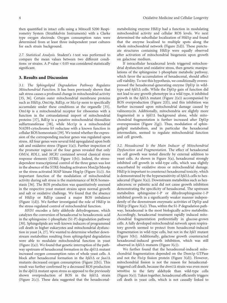
3.1. The Sphingolipid Degradation Pathway RegulatesMitochondrial Function. It has been previously shown thatsalt stress causes a profound change in mitochondrial activity[35, 36]. Certain outer mitochondrial membrane proteinssuch as Hfd1p, Om14p, Rdl1p, or Mcr1p seem to specificallyaccumulate under these conditions at the organelle [35].Om14p is a mitochondrial receptor for ribosomes with afunction in the cotranslational import of mitochondrialproteins [37], Rdl1p is a putative mitochondrial thiosulfatesulfurtransferase [38], while Mcr1p is a mitochondrialNADH-cytochrome b5 reductase with a known function incellular ROS homeostasis [39]. We tested whether the expres-sion of the corresponding nuclear genes was regulated uponstress. All four genes were several fold upregulated upon bothsalt and oxidative stress (Figure 1(a)). Further inspection ofthe promoter regions of the four genes revealed that onlyOM14, RDL1, and MCR1 contained several classical stressresponse elements (STRE; Figure 1(b)). Indeed, the stress-dependent transcriptional control of the three genes was lostin the absence of the STRE-binding activators Msn2p/Msn4por the stress-activated MAP kinase Hog1p (Figure 1(c)). Animportant function of the modulation of mitochondrialactivity during salt stress is the maintenance of ROS homeo-stasis [36]. The ROS production was quantitatively assessedin the respective yeast mutant strains upon normal growthand salt or oxidative challenge. We found that the absenceof Hfd1p or Rdl1p caused a major ROS imbalance(Figure 1(d)). We further investigated the role of Hfd1p inthe stress-regulated control of mitochondrial function.
HFD1 encodes a fatty aldehyde dehydrogenase, whichcatalyzes the conversion of hexadecenal to hexadecenoic acidin the sphingosine-1-phosphate (S1-P) degradation pathway[30]. Sphingolipids are well-known inducers of programmedcell death in higher eukaryotes and mitochondrial dysfunc-tion in yeast [4, 27]. We wanted to determine whether down-stream metabolites resulting from sphingolipid degradationwere able to modulate mitochondrial function in yeast(Figure 2(a)). We found that genetic interruption of the path-way upstream of hexadecenal formation in the dpl1Δmutantincreased oxygen consumption rates of whole yeast cells. Ablock after hexadecenal formation in the hfd1Δ or faa1Δmutants decreased oxygen consumption (Figure 2(b)). Thisresult was further confirmed by a decreased ROS productionin the dpl1Δmutant upon stress as opposed to the previouslyshown overproduction of ROS in the hfd1Δ strain(Figure 2(c)). These data suggested that the hexadecenal-
metabolizing enzyme Hfd1p had a function in modulatingmitochondrial activity and cellular ROS levels. We nextdetermined the subcellular localization of Hfd1p and foundthat the enzyme localized to multiple spots along thewhole mitochondrial network (Figure 2(d)). These punctu-ate structures containing Hfd1p were equally observedafter activation of mitochondrial biogenesis upon growthon galactose medium.
If intracellular hexadecenal levels triggered mitochon-drial dysfunction and oxidative stress, then genetic manipu-lations of the sphingosine 1-phosphate metabolic pathway,which favor the accumulation of hexadecenal, should affectcell viability. To test this hypothesis, we conditionally overex-pressed the hexadecenal-generating enzyme Dpl1p in wild-type and hfd1Δ cells. While the Dpl1p gain of function didnot lead to any growth phenotype in a wild type, it inhibitedgrowth in the hfd1Δ mutant (Figure 2(e)) accompanied byROS overproduction (Figure 2(f)), and this inhibition wasfurther increased upon mitochondrial damage caused byvalinomycin. Additionally, mitochondria are slightly morefragmented in a hfd1Δ background alone, while mito-chondrial fragmentation is further increased after Dpl1poverexpression (Figure 2(g)). Thus, modulation of sphin-golipid metabolism, and in particular the hexadecenalintermediate, seemed to regulate mitochondrial functionand cell growth.
3.2. Hexadecenal Is the Main Inducer of MitochondrialDysfunction and Fragmentation. The effect of hexadecenalon cell growth was tested directly by external addition toyeast cells. As shown in Figure 3(a), hexadecenal stronglyinhibited cell growth in wild-type cells, which was slightlyexacerbated by oxidative stress or mitochondrial damage.Hfd1p is important to counteract hexadecenal toxicity, whichis demonstrated by the hypersensitivity of hfd1Δ cells to hex-adecenal (Figure 3(a)). Downstream metabolites such as hex-adecenoic or palmitic acid did not cause growth inhibitiondemonstrating the specificity of hexadecenal. The upstreammetabolites sphingosine-1-P or dihydrosphingosine-1-Pinhibited growth in a significantly weaker manner indepen-dently of the downstream enzymatic activities of Dpl1p andHfd1p (Figure 3(a)). Thus, within the S1-P degradation path-way, hexadecenal is the most biologically active metabolite.Accordingly, hexadecenal treatment rapidly induced mito-chondrial fragmentation preferentially in glucose-growncells. A fully developed mitochondrial network upon respira-tory growth seemed to protect from hexadecenal-inducedfragmentation in wild-type cells, but not in the hfd1 mutant(Figure 3(b)). Additionally, galactose growth counteractedhexadecenal-induced growth inhibition, which was stillobserved in hfd1Δ mutants (Figure 3(c)).
We further found that the hexadecenal-induced mito-chondrial fragmentation depended on the Dnm1p GTPaseand not the Fis1p fission protein (Figure 3(d)). However,mitochondrial fission is not the reason for hexadecenal-triggered cell death, because the dnm1Δ strain was even moresensitive to the fatty aldehyde than wild-type cells(Figure 3(e)). Taken together, hexadecenal efficiently triggerscell death in yeast cells, which is not causally linked to
4 Oxidative Medicine and Cellular Longevity
0
2
4
6
8
10
12
14
16
OM14 RDL1 MCR1 HFD1 GRE2
Rela
tive m
RNA
leve
l
30
NaCl
0 1020 300 1020 300 1020 300 1020 300 1020 min0
24
6
810
1214
1618
20
Rela
tive m
RNA
leve
l
Menadione
OM14 RDL1 MCR1 HFD1 GRE2300 1020 300 1020 300 1020 300 1020 300 1020 min
(a)
OM14
RDL1
MCR1
HFD1
‒950 ‒363 ‒180
‒21
‒98
‒203
‒301
‒752
‒888
‒988 ‒308 ‒268
‒225
STRE (CCCCT)
AP‒1 (TT/GACTA/CA)
Gcn4 (TGACTGA)
(b)
0
2
4
6
8
10
Relat
ive m
RNA
leve
l
01234567
8
Relat
ive m
RNA
leve
l
wt hog1∆ msn2/4∆
01
2
3
4
5
6
7
Relat
ive m
RNA
leve
l
⁎⁎⁎⁎
⁎
⁎⁎
⁎⁎⁎⁎
⁎⁎
⁎⁎
⁎⁎
⁎⁎
⁎⁎
OM14
RDL1
MCR1
0 10 20 min0 10 20 0 10 20
0 10 20min0 10 20 0 10 20
0 10 20min0 10 20 0 10 20
(c)
0
1
2
3
4
5
6
7
wt om14∆ mcr1∆ rdl1∆ hfd1∆
Rela
tive R
OS
leve
l
Control
NaCl
⁎⁎
0
1
2
3
4
5
6
wt om14∆ mcr1∆ rdl1∆ hfd1∆
Rela
tive R
OS
leve
l
Control
Menadione
⁎
⁎⁎
⁎⁎
⁎⁎
⁎⁎⁎⁎
⁎⁎
⁎⁎ ⁎
(d)
Figure 1: Stress-induced expression of genes encoding outer mitochondrial membrane proteins. (a) Reverse transcriptase determination ofmRNA induction of four outer mitochondrial membrane protein encoding genes upon salt shock (0.4M NaCl) and oxidative stress (50 μMmenadione) for the indicated times. The GRE2 gene was included as a positive marker for salt and oxidative stress. Data are presented asmean± SD. Three biological replicates were analyzed. The mRNA level was normalized in all cases for the ACT1 control, and theuninduced level was arbitrarily set to 1. (b) Representation of consensus binding sites for stress-activated transcription factors in thepromoter regions of OM14, RDL1, MCR1, and HFD1. (c) Reverse transcriptase determination of mRNA induction of the same genes inthe indicated strain backgrounds upon salt shock (0.4M NaCl) as described in (a). Significantly different mRNA levels as compared to wtare marked. ∗P < 0 05; ∗∗P < 0 01 (Student’s t-test). (d) Reactive oxygen species (ROS) production in mutants affected in specificmitochondrial outer membrane proteins. 2′,7′-dichlorodihydrofluorescein diacetate assay in the indicated yeast strains before or after salt(1M NaCl, 2 h) or oxidative shock (50 μM menadione, 2 h). Data are presented as mean± SD. Three biological replicates were analyzed.Significantly different ROS levels as compared to wt are marked. ∗P < 0 05; ∗∗P < 0 01 (Student’s t-test).
5Oxidative Medicine and Cellular Longevity

Palmitoyl‒CoA + serine
DihydrosphingosineDHS
Dihydrosphingosine‒1‒P
DHS-P
HexadecenalDihydroceramide
Complex sphingolipids
Hexadecenoic acid
Palmitoyl‒coA
HFD1
FAA1,4
DPL1
(a)
0
0.2
0.4
0.6
0.8
1
1.2
1.4
1.6
WT hfd1∆ faa1∆ faa4∆ dpl1∆ fzo1∆
Rela
tive O
2 cons
umpt
ion
⁎⁎
⁎
(b)
0
1
2
3
4
5
6
7
8
wt dpl1∆ hfd1∆
Rela
tive R
OS
leve
ls
‒NaCl+NaCl
⁎
⁎
(c)
GFP dsRed Overlay Nomarski
Glu
cG
al
5�휇m
(d)
SD SGalSD + Val SGal + Val
WT control
WT GAL1p-DPL1
hfd1∆ control
hfd1∆ GAL1p-DPL1
DHS
DHS-P
Hexadecenal
Hexadecenoicacid
DPL1
HFD1
0
20
40
60
80
100
120
140
SD SGal
Wt control Wt DPL1
hfd1∆control hfd1∆DPL1
⁎
⁎⁎
Col
ony-
form
ing
units
⁎⁎
SD + Val SGal + Val
(e)
Figure 2: Continued.
6 Oxidative Medicine and Cellular Longevity

mitochondrial fragmentation. Accordingly, although mito-chondrial fragmentation generally accompanies apoptosisin different cellular models, it is not a prerequisite for celldeath [40]. In mammalian cells, the Drp1 fission protein isresponsible for apoptotic mitochondrial fragmentation, butsimilarly to what is reported here for yeast, its function inmitochondrial fission is not decisive for MOMP and subse-quent cell death [41, 42].
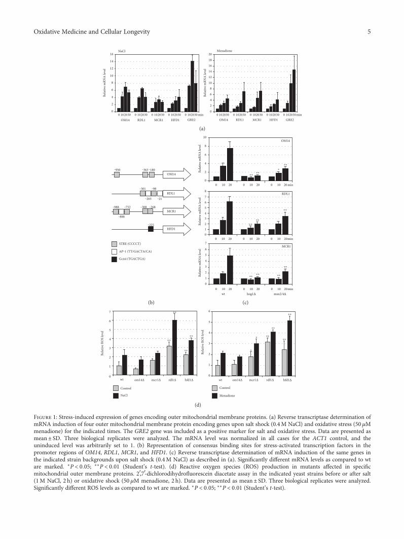
3.3. Modulation of Sphingolipid Metabolism TriggersMitochondrial Cell Death in Yeast. We next wanted to testwhether the sphingolipid degradation pathway and especiallythe hexadecenal intermediate triggered cell death via themitochondrial death pathway. In higher eukaryotes, mito-chondrial apoptosis is irreversibly induced by the perme-abilization of the outer mitochondrial membrane. Thisprocess is highly controlled by pro- and antiapoptotic mem-bers of the Bcl-2 family. In yeast, only one Bcl-2 ortholog hasbeen identified, Ybh3p, which has both pro- and antiapopto-tic properties [33, 43]. We tested whether Ybh3p was func-tionally involved in hexadecenal-mediated growth arrest. Asexpected, Ybh3p translocated to mitochondria upon the apo-ptotic stimulus of acetic acid treatment. However, hexadece-nal treatment did not induce mitochondrial localization ofYbh3p (Figure 4(a)). Additionally, the lack of Ybh3p
function did not change the susceptibility of yeast cells toexternally added hexadecenal (Figure 4(b)). Ybh3p is there-fore not the executor of hexadecenal-induced cell death.
Alternatively, we examined the death-promoting anddeath-preventing functions of human Bcl-2 members inyeast cells with an altered sphingolipid metabolism. HumanBax protein has been shown to efficiently activate mitochon-drial cell death in yeast [25, 44]. Here, we found that theinduced Bax expression arrested cell growth in a synergisticmanner with external hexadecenal (Figure 5(a)). On theother hand, antiapoptotic human Bcl-xL expression largelyprevented hexadecenal-induced growth inhibition. Thus,hexadecenal seemed to arrest cell growth via proteins of theBcl-2 family when expressed ectopically in yeast. This wasfurther supported by the fact that Bax was a more efficientinhibitor in cells with a block in the hexadecenal-metabolizing Hfd1p enzyme (Figure 5(b)). Thus, Bcl-2 pro-teins such as Bax promote growth arrest in yeast cells in aprocess, which is modulated by sphingolipid degradationproducts such as hexadecenal. Our results support the ideathat high mitochondrial hexadecenal levels would favor theproapoptotic action of Bax and subsequently promote mito-chondrial fragmentation. In this model, the Hfd1p enzymewould counteract hexadecenal accumulation at mitochon-dria and prevent the induction of cell death. It remains to
0
0.5
1
1.5
2
ControlGAL1p-DPL1
Rela
tive R
OS
leve
ls
wt hfd1�훥 wt hfd1�훥SD SGal
⁎
(f)
wt
hfd1�훥
Control GAL1p-DPL1
5 �휇m
(g)
Figure 2: Sphingolipid degradation modulates mitochondrial activity in yeast. (a) Schematic overview of the enzymatic conversions ofsphingolipid degradation. Downstream enzymatic activities of dihydrosphingosine-1-phosphate are depicted. Only the conversions ofdihydrosphingosine and not of other sphingosine species such as phytosphingosine are shown. (b) Oxygen consumption rates of mutantsaffected in the sphingolipid degradation pathway. Cells were grown in synthetic galactose medium. The fzo1Δ mutant was included as anegative control. The O2 consumption rate of the wild type was arbitrarily set to 1. (c) Reactive oxygen species (ROS) production inmutants affected in hexadecenal production (dpl1Δ) or degradation (hfd1Δ). 2′,7′-dichlorodihydrofluorescein diacetate assay in theindicated yeast strains before or after salt shock (1M NaCl, 2 h). ROS levels upon normal growth conditions were set to 1 for each strainbackground. (d) Intracellular localization of Hfd1p. Cells expressing constitutive Hfd1-GFP and Om14-dsRed fusion proteins were grownin synthetic glucose- or galactose-containing medium. (e, f) Genetic manipulation of the sphingolipid degradation pathway affects cellviability and ROS production. The hexadecenal-producing Dpl1p enzyme was overexpressed under control of the GAL1 promoter in yeastwild type or the hfd1Δ mutant. (e) Growth efficiency was assessed on synthetic agar medium-containing glucose (SD) or galactose (SGal)supplemented or not with 4 μM valinomycin. Alternatively, colony formation was quantified in the same strains (lower panel). Cells fromfresh overnight cultures in synthetic glucose medium were diluted in the indicated media to an OD600 of 0.1, and growth was allowed foran additional 24 h. Colony-forming units were determined by plating the cells onto YPD agar medium. The colony number obtained forthe wt upon the different growth conditions was set to 100. (f) Quantification of ROS production in the same strains grown in syntheticglucose or galactose medium by the 2′,7′-dichlorodihydrofluorescein diacetate assay. (g) Overexpression of Dpl1p causes mitochondrialfragmentation in hfd1Δ mutants. MitoTracker-stained mitochondria were visualized in the indicated yeast cells containing the emptyvector or the galactose-inducible DPL1 expression on synthetic galactose medium. Data information: in (b, c, e, and f), data are presentedas mean± SD. Three biological replicates were analyzed. Significant changes with respect to the wild type are marked. ∗P < 0 05,∗∗P < 0 01 (Student’s t-test).
7Oxidative Medicine and Cellular Longevity

YPD Mena Val
Control
25 �휇M
50 �휇M
Control
25 �휇M
50 �휇M
Hexadecenal
wt
hfd1∆
YPD Mena ValHexadecenoic acid
YPD Mena ValPalmitic acid
wt
hfd1∆
wt
hfd1∆
Control
25 �휇M
50 �휇M
Control
25 �휇M
50 �휇M
Control
25 �휇M
50 �휇M
Control
25 �휇M
50 �휇M
Control
100 �휇M
200
Hexadecenal
S1-P
wt
hfd1∆
wt
dpl1∆
Control
100 �휇M
200 �휇M
Control
100 �휇M
200 �휇M
Control
100 �휇M
200 �휇M
DHS1-P
(a)
wt
hfd1∆
GFP GFP NomarskiNomarski‒ + Hexadecenal
Glu
cose
Gly
c/Et
OH
wt
hfd1∆
5 �휇m
(b)
Control
25 �휇M
50 �휇M
Control
25 �휇M
50 �휇M
100 �휇M
100 �휇M
Gluc GalHexadecenal
wt
hfd1
∆
(c)
‒ + Hexadecenal
wt
dnm1∆
fis1∆
5 �휇m
(d)
Control
100 �휇M
200 �휇M
Hexadecenalwt dnm1∆ fis1∆
(e)
Figure 3: Hexadecenal is the most biologically active intermediate of the sphingolipid degradation pathway. (a) Hexadecenal and thedownstream metabolites hexadecenoic and palmitic acid were tested for growth inhibition of the indicated yeast strains (left panel). Theindicated doses were applied for 2 h, and colony formation was subsequently assessed on YPD plates containing or not 50 μM menadioneor 4 μM valinomycin. The upstream metabolites sphingosine-1-phosphate (S1-P) and dihydrosphingosine-1-phosphate were tested forgrowth inhibition of the indicated yeast strains in the right panel. (b) External hexadecenal addition causes mitochondrial fragmentationdependent on Hfd1p function. Mitochondria were visualized by expression of mt-GFP in yeast wild type and the hfd1Δ mutant insynthetic glucose or glycerol/ethanol medium before and after the exposure (1 h) to 50 μM hexadecenal. (c) Galactose growth counteractshexadecenal growth inhibition. Hexadecenal was applied for 1 h to the indicated yeast strains grown on glucose- or galactose-containingsynthetic medium. Colony formation was then assessed on YPD agar plates. (d) Hexadecenal induces mitochondrial fragmentationthrough Dnm1p. The indicated yeast strains expressing mt-GFP were treated or not with 50μM hexadecenal for 1 h before visualization ofmitochondria. (e) Suppression of mitochondrial fission does not counteract hexadecenal-mediated growth inhibition. The indicated yeaststrains were assayed for hexadecenal inhibition as in (a).
8 Oxidative Medicine and Cellular Longevity
be experimentally addressed whether changes in hexadecenalmetabolism trigger mitochondrial outer membrane perme-abilization in yeast.
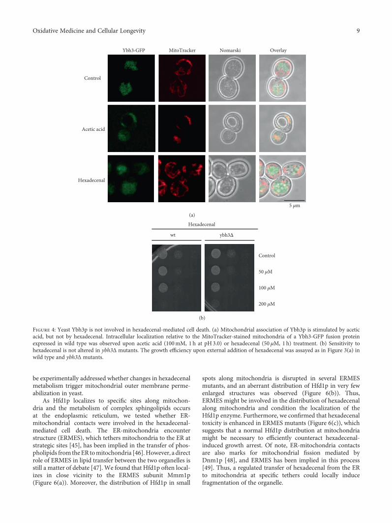
As Hfd1p localizes to specific sites along mitochon-dria and the metabolism of complex sphingolipids occursat the endoplasmic reticulum, we tested whether ER-mitochondrial contacts were involved in the hexadecenal-mediated cell death. The ER-mitochondria encounterstructure (ERMES), which tethers mitochondria to the ER atstrategic sites [45], has been implied in the transfer of phos-pholipids from theER tomitochondria [46].However, a directrole of ERMES in lipid transfer between the two organelles isstill a matter of debate [47]. We found that Hfd1p often local-izes in close vicinity to the ERMES subunit Mmm1p(Figure 6(a)). Moreover, the distribution of Hfd1p in small
spots along mitochondria is disrupted in several ERMESmutants, and an aberrant distribution of Hfd1p in very fewenlarged structures was observed (Figure 6(b)). Thus,ERMES might be involved in the distribution of hexadecenalalong mitochondria and condition the localization of theHfd1p enzyme. Furthermore, we confirmed that hexadecenaltoxicity is enhanced in ERMES mutants (Figure 6(c)), whichsuggests that a normal Hfd1p distribution at mitochondriamight be necessary to efficiently counteract hexadecenal-induced growth arrest. Of note, ER-mitochondria contactsare also marks for mitochondrial fission mediated byDnm1p [48], and ERMES has been implied in this process[49]. Thus, a regulated transfer of hexadecenal from the ERto mitochondria at specific tethers could locally inducefragmentation of the organelle.
MitoTracker Nomarski Overlay
Control
Acetic acid
Hexadecenal
5 �휇m
Ybh3‑GFP
(a)
Control
50 �휇M
100 �휇M
Hexadecenal
wt ybh3∆
200 �휇M
(b)
Figure 4: Yeast Ybh3p is not involved in hexadecenal-mediated cell death. (a) Mitochondrial association of Ybh3p is stimulated by aceticacid, but not by hexadecenal. Intracellular localization relative to the MitoTracker-stained mitochondria of a Ybh3-GFP fusion proteinexpressed in wild type was observed upon acetic acid (100mM, 1 h at pH 3.0) or hexadecenal (50 μM, 1 h) treatment. (b) Sensitivity tohexadecenal is not altered in ybh3Δ mutants. The growth efficiency upon external addition of hexadecenal was assayed as in Figure 3(a) inwild type and ybh3Δ mutants.
9Oxidative Medicine and Cellular Longevity

3.4. Genetic Control of the Sphingolipid Degradation Pathwayvia the Hog1p MAP Kinase. The expression of theHFD1 geneis activated by salt and oxidative stress. Thus, we testedwhether the expression of genes encoding the enzymes ofsphingolipid degradation was generally regulated by stress(Figure 7(a)). We found that all enzymatic steps from cer-amide to hexadecenoic acid were inducible by NaCl stressat the mRNA level (Figure 7(b)). Moreover, the Hog1pstress-activated kinase, which is the key regulator of theadaptive response of yeast to osmotic stress [50], wasindispensable for this transcriptional activation. In turn,most of the reverse enzymatic reactions from S1-P to cer-amide were transcriptionally repressed under the sameconditions (Figure 7(c)). Thus, yeast cells employ a geneticregulation, which seems to favor the degradation of sphin-golipids over its biosynthesis upon stress. This switch iscontrolled by the Hog1p MAP kinase, which could
therefore be a modulator of sphingolipid metabolic inter-mediates during exposure to salt stress. Accordingly, wefound that hog1Δ mutants were more sensitive to externalhexadecenal (Figure 7(d)), which could be explained by adecreased detoxification capacity of this strain-lackingHfd1p activation.
We further investigated the correlation between saltstress, sphingolipid metabolism, and mitochondrial function.As salt stress induced the genes encoding sphingolipid-metabolizing enzymes, a block after hexadecenal formationcould be deleterious especially upon NaCl stress. We foundindeed that hfd1Δ mutant cells showed complete mitochon-drial fragmentation upon salt shock, which was not observedin the wild type (Figure 7(e)). Finally, we tested whetherHog1p function was important for Bax toxicity. We foundthat hog1Δ mutants were more resistant to Bax expressionthan wild-type cells (Figure 7(f)). Taken together, the Hog1p
Con
trol
Bax
0 h 6 h 24 h
0
25 �휇M
50 �휇M
0
25 �휇M
50 �휇M
Con
trol
Bcl X
L
0 h 6 h 24 h
0
25 �휇M
50 �휇M
0
25 �휇M
50 �휇M
Hexadecenal
(a)
0 h 24 h
+
‒
wt
hfd1
∆w
thf
d1∆
+
‒
+
‒
+
‒
Con
trol
Bax
Dox
ycyc
line
(b)
Figure 5: Bax-mediated growth inhibition is modulated by hexadecenal levels. (a) External hexadecenal potentiates Bax function. Humanproapoptotic Bax and antiapoptotic Bcl-xL were expressed under control of the Tetoff promoter for the indicated times in the presence ornot of the indicated hexadecenal concentrations. Control strains contained the respective empty vectors. Colony formation was thenassessed on YPD agar plates. (b) Bax inhibition is enhanced in hfd1Δ mutants. Human Bax expression was induced for 24 h in theindicated yeast strains by the removal of doxycycline.
10 Oxidative Medicine and Cellular Longevity

stress-activated kinase is involved in the induction of sphin-golipid metabolic enzymes and this activation could lead tothe accumulation of bioactive intermediates, especially hexa-decenal, at mitochondria. In the absence of detoxification bythe Hfd1p enzyme, this can induce the mitochondrial deathpathway by favoring the activity of proapoptotic proteins suchasBax.Ofnote, hyperosmotic andNaCl stress are knownenvi-ronmental conditions, which induce mitochondria-mediatedcell death in yeast [51, 52]. In this scenario, stress-activatedHog1p could trigger mitochondrial dysfunction by activatingsphingolipid degradation. This model is in agreement withthe previous finding that sustained activation of Hog1pimpairsmitochondrial respiration, increases ROSproduction,and induces cell death [53].
It is important to note that the sphingolipid metabolitehexadecenal also inhibits mitochondrial function in highereukaryotes. Hexadecenal addition to murine mitochondriaefficiently permeabilizes the outer membrane [17], which
has been postulated as an early irreversible decision toenter the mitochondria-mediated death pathway [19].Accordingly, hexadecenal is a potent activator of apoptosisin mouse and human cell lines [18].
4. Conclusions
Our work suggests that the modulation of mitochondrialactivity and induction of cell death by hexadecenal are aconserved feature of eukaryotic cells. However, apart fromits proapoptotic function when externally applied to cells ormitochondria, it was not known whether intracellular regula-tion of hexadecenal metabolism was involved in modulatingmitochondrial function and death. Importantly, we showhere that the regulation of the sphingolipid degradation path-way is a decisive trigger of mitochondrial function and cellgrowth in yeast. Hexadecenal is the intermediate in this path-way with the highest impact on mitochondrial function.
Hfd1-GFP OverlayMmm1-mCherry Nomarski
5 �휇m
(a)
Hfd1-GFP OverlayMitoTracker Nomarski
wt
mdm10∆
mdm12∆
mmm1∆
5 �휇m
(b)
Control
50 �휇M
100 �휇M
Hexadecenalwt mdm12∆mdm10∆
0
20
40
60
80
100
120
SD 50 �휇M HD 100 �휇M HD
wt mdm10∆mdm12∆
⁎⁎
⁎⁎
⁎⁎
Colo
ny-fo
rmin
g un
its
(c)
Figure 6: Functional connection between the ERMES complex and hexadecenal-mediated cell death. (a) Colocalization study of Hfd1-GFPwith the Mmm1-mCherry ERMES complex subunit. Cells were grown in synthetic galactose medium for the induced expression of theMmm1-mCherry fusion. (b) Intracellular distribution of Hfd1p is affected in ERMES complex mutants. Hfd1-GFP was expressed in theindicated yeast strains and localized relative to MitoTracker-stained mitochondria. Cells were grown in synthetic glucose medium. (c)ERMES complex mutants are hypersensitive to hexadecenal. The growth efficiency upon external addition of hexadecenal (HD) wasassayed as in Figure 3(a) in wild type and the indicated ERMES deletion mutants (upper panel). Quantitative colony assays are shown forthe same strains in the lower panel. Cells from fresh overnight cultures in synthetic glucose medium were diluted to OD600 0.5 and thenincubated with the indicated hexadecenal doses for 2 h. Colony-forming units were determined by plating the cells onto YPD agarmedium. Data are presented as mean± SD. Three biological replicates were analyzed. The colony number obtained for the wt uponcontrol conditions was set to 100. Significant changes with respect to the wild type upon the same growth condition are marked.∗P < 0 05, ∗∗P < 0 01 (Student’s t-test).
11Oxidative Medicine and Cellular Longevity

Palmitoylg-CoA + serine
3-Ketohydrosphingosine
Dihydrosphingosine
DHS
Dihydrosphingosine-1-P
DHS-P
HexadecenalDihydroceramide
Inositol-P-ceramide
IPC
Complex sphingolipids
Hexadecenoic acid
Palmityl-CoA
LCB2
TSC10
LAG1
LAC1YDC1
AUR1
LCB3
LCB4
LCB5
DPL1
HFD1
FAA1, 4
(a)
00.5
11.5
22.5
33.5
44.5
5
LCB4 LCB5 DPL1 HFD1 YDC1 LCB4 LCB5 DPL1 HFD1 YDC1
Rela
tive m
RNA
leve
l
ControlNaCl
wt hog1∆
⁎⁎ ⁎⁎⁎⁎
⁎⁎
⁎⁎
(b)
0
0.5
1
1.5
2
AUR1 LAG1 LCB3 LAC1
Rela
tive m
RNA
leve
l
ControlNaCl
⁎ ⁎
⁎
(c)
Control
50 �휇M
100 �휇M
Control
50 �휇M
100 �휇M
wt
hog1�훥
Hexadecenal
(d)
hfd1�훥
NaCl
wt
Control
5 �휇m(e)
0 h 24 h
+
−
wt
hog1�훥
wt
hog1�훥
+
−
+
−
+
−
Cont
rol
Bax
Doxycycline
(f)
Figure 7: Stress regulation of the sphingolipid degradation pathway via Hog1p and its impact on mitochondrial integrity. (a) Schematicoverview of the enzymatic conversions implied in sphingolipid biosynthesis and degradation in yeast. (b) Expression of sphingolipiddegradation enzymes is stimulated upon salt stress in a Hog1p-dependent manner. RT-PCR analysis of gene expression in the indicatedyeast strains upon salt shock (0.4M NaCl, 20min). Relative mRNA levels of the indicated genes were normalized for the ACT1 control.(c) Expression of sphingolipid biosynthesis enzymes is generally repressed upon salt stress. Yeast wild-type cells were analyzed by RT-PCRas in (b). In (b, c), data are presented as mean± SD. Three biological replicates were analyzed. Significant changes with respect to the wildtype (b) or to the nonstress condition (c) are marked. ∗P < 0 05; ∗∗P < 0 01 (Student’s t-test). (d) Loss of Hog1p function causeshexadecenal sensitivity. Growth inhibition of the indicated yeast strains by hexadecenal was assessed as in Figure 3(a). (e) Salt stressinduces mitochondrial fragmentation in hfd1Δ mutant cells. Yeast cells expressing mt-GFP on synthetic glucose medium were treated ornot with 1M NaCl before visualization of mitochondria. (f) Loss of Hog1p function counteracts Bax inhibition. Human Bax expressionwas induced for 24 h as in Figure 5(b) in wild type and hog1Δ mutants. Growth was then recorded on YPD agar plates.
12 Oxidative Medicine and Cellular Longevity
Genetic interventions, which favor intracellular hexadecenalproduction, induce ROS imbalance, decrease respiration,and arrest cell growth. Moreover, stress-activated signalingpathways, such as HOG, are implied in the regulation ofsphingolipid degradation, which in turn can be decisive forthe entry into cell death pathways upon environmental stress.Mutations in the ALDH3A2 gene, encoding the humanortholog of yeast Hfd1p fatty aldehyde dehydrogenase, causeSjögren-Larsson syndrome, a rare neurocutaneous disorder[54]. In the light of our results, the implication of the accu-mulation of toxic fatty aldehydes and their effect on mito-chondrial function should be further investigated in humancells with impaired ALDH3A2 enzymatic activity.
Conflicts of Interest
The authors declare that they have no competing interests.
Acknowledgments
The authors thank Eulalia de Nadal, William Prinz, BenoitKornmann, Stephen Manon, Benedikt Westermann, andFrank Madeo for the kind gift of yeast strains and plasmids.The authors thank Alba Calatayud for her help with Baxexpression experiments and Benito Alarcón for his help withthe confocal microscopy. This work was supported by thegrants from the Ministerio de Economía y Competitividad(BFU2011-23326 and BFU2016-75792-R).
References
[1] N. J. Haughey, “Sphingolipids in neurodegeneration,” Neuro-molecular Medicine, vol. 12, no. 4, pp. 301–305, 2010.
[2] X. Huang, B. R. Withers, and R. C. Dickson, “Sphingolipidsand lifespan regulation,” Biochimica et Biophysica Acta(BBA) - Molecular and Cell Biology of Lipids, vol. 1841, no. 5,pp. 657–664, 2014.
[3] M.MaceykaandS. Spiegel, “Sphingolipidmetabolites in inflam-matory disease,”Nature, vol. 510, no. 7503, pp. 58–67, 2014.
[4] G. A. Patwardhan, L. J. Beverly, and L. J. Siskind, “Sphingoli-pids and mitochondrial apoptosis,” Journal of Bioenergeticsand Biomembranes, vol. 48, no. 2, pp. 153–168, 2016.
[5] L. Galluzzi, O. Kepp, and G. Kroemer, “Mitochondria: masterregulators of danger signalling,” Nature Reviews. MolecularCell Biology, vol. 13, no. 12, pp. 780–788, 2012.
[6] J. E. Chipuk and D. R. Green, “How do BCL-2 proteins inducemitochondrial outer membrane permeabilization?” Trends inCell Biology, vol. 18, no. 4, pp. 157–164, 2008.
[7] J. E. Chipuk, T. Moldoveanu, F. Llambi, M. J. Parsons, andD. R. Green, “The BCL-2 family reunion,” Molecular Cell,vol. 37, no. 3, pp. 299–310, 2010.
[8] M. Nomura, S. Shimizu, T. Ito, M. Narita, H. Matsuda, andY. Tsujimoto, “Apoptotic cytosol facilitates Bax translocationto mitochondria that involves cytosolic factor regulated byBcl-2,” Cancer Research, vol. 59, no. 21, pp. 5542–5548, 1999.
[9] K. G. Wolter, Y. T. Hsu, C. L. Smith, A. Nechushtan, X. G. Xi,and R. J. Youle, “Movement of Bax from the cytosol to mito-chondria during apoptosis,” The Journal of Cell Biology,vol. 139, no. 5, pp. 1281–1292, 1997.
[10] J. F. Lovell, L. P. Billen, S. Bindner et al., “Membrane bindingby tBid initiates an ordered series of events culminating inmembrane permeabilization by Bax,” Cell, vol. 135, no. 6,pp. 1074–1084, 2008.
[11] D. Westphal, R. M. Kluck, and G. Dewson, “Building blocks ofthe apoptotic pore: how Bax and Bak are activated and oligo-merize during apoptosis,” Cell Death and Differentiation,vol. 21, no. 2, pp. 196–205, 2014.
[12] M. J. Hernandez-Corbacho, M. F. Salama, D. Canals,C. E. Senkal, and L. M. Obeid, “Sphingolipids in mitochon-dria,” Biochimica et Biophysica Acta (BBA) - Molecular andCell Biology of Lipids, vol. 1862, no. 1, pp. 56–68, 2017.
[13] L. M. Obeid, C. M. Linardic, L. A. Karolak, and Y. A. Hannun,“Programmed cell death induced by ceramide,” Science,vol. 259, no. 5102, pp. 1769–1771, 1993.
[14] B. J. Kroesen, B. Pettus,C. Luberto et al., “Inductionof apoptosisthrough B-cell receptor cross-linking occurs via de novo gener-ated C16-ceramide and involves mitochondria,” The Journal ofBiological Chemistry, vol. 276, no. 17, pp. 13606–13614, 2001.
[15] C. Rodriguez-Lafrasse, G. Alphonse, P. Broquet, M. T. Aloy,P. Louisot, and R. Rousson, “Temporal relationships betweenceramide production, caspase activation and mitochondrialdysfunction in cell lines with varying sensitivity to anti-Fas-induced apoptosis,” The Biochemical Journal, vol. 357,Part 2, pp. 407–416, 2001.
[16] R. L. Thomas Jr., C. M. Matsko, M. T. Lotze, and A. A.Amoscato, “Mass spectrometric identification of increasedC16 ceramide levels during apoptosis,” The Journal of Bio-logical Chemistry, vol. 274, no. 43, pp. 30580–30588, 1999.
[17] J. E. Chipuk, G. P. McStay, A. Bharti et al., “Sphingolipidmetabolism cooperates with BAK and BAX to promote themitochondrial pathway of apoptosis,” Cell, vol. 148, no. 5,pp. 988–1000, 2012.
[18] A. Kumar,H. S. Byun, R. Bittman, and J. D. Saba, “The sphingo-lipid degradation product trans-2-hexadecenal induces cyto-skeletal reorganization and apoptosis in a JNK-dependentmanner,”Cellular Signalling, vol. 23, no. 7, pp. 1144–1152, 2011.
[19] T. T. Renault and J. E. Chipuk, “Inter-organellar communica-tion with mitochondria regulates both the intrinsic and extrin-sic pathways of apoptosis,” Communicative & IntegrativeBiology, vol. 6, no. 2, article e22872, 2013.
[20] D. Carmona-Gutierrez, T. Eisenberg, S. Buttner, C. Meisinger,G. Kroemer, and F. Madeo, “Apoptosis in yeast: triggers, path-ways, subroutines,” Cell Death and Differentiation, vol. 17,no. 5, pp. 763–773, 2010.
[21] C. Falcone and C. Mazzoni, “External and internal triggers ofcell death in yeast,” Cellular and Molecular Life Sciences,vol. 73, no. 11-12, pp. 2237–2250, 2016.
[22] S. Giannattasio, N. Guaragnella, M. Zdralevic, and E. Marra,“Molecular mechanisms of Saccharomyces cerevisiae stressadaptation and programmed cell death in response to aceticacid,” Frontiers in Microbiology, vol. 4, p. 33, 2013.
[23] P. Ludovico, F. Rodrigues, A. Almeida, M. T. Silva, A.Barrientos, and M. Corte-Real, “Cytochrome c release andmitochondria involvement in programmed cell death inducedby acetic acid in Saccharomyces cerevisiae,” Molecular Biologyof the Cell, vol. 13, no. 8, pp. 2598–2606, 2002.
[24] C. Pereira, R. D. Silva, L. Saraiva, B. Johansson, M. J. Sousa,and M. Corte-Real, “Mitochondria-dependent apoptosis inyeast,” Biochimica et Biophysica Acta (BBA) - Molecular CellResearch, vol. 1783, no. 7, pp. 1286–1302, 2008.
13Oxidative Medicine and Cellular Longevity

[25] S. Manon, B. Chaudhuri, and M. Guerin, “Release ofcytochrome c and decrease of cytochrome c oxidase inBax-expressing yeast cells, and prevention of these effectsby coexpression of Bcl-xL,” FEBS Letters, vol. 415, no. 1,pp. 29–32, 1997.
[26] T. T. Renault, L. M. Dejean, and S. Manon, “A brewingunderstanding of the regulation of Bax function by Bcl-xLand Bcl-2,” Mechanisms of Ageing and Development,vol. 161, Part B, pp. 201–210, 2017.
[27] P. Spincemaille, N. Matmati, Y. A. Hannun, B. P. Cammue,and K. Thevissen, “Sphingolipids and mitochondrial functionin budding yeast,” Biochimica et Biophysica Acta (BBA) -General Subjects, vol. 1840, no. 10, pp. 3131–3137, 2014.
[28] A. Rego, D. Trindade, S. R. Chaves et al., “The yeast modelsystem as a tool towards the understanding of apoptosisregulation by sphingolipids,” FEMS Yeast Research, vol. 14,no. 1, pp. 160–178, 2014.
[29] A. Rego, M. Costa, S. R. Chaves et al., “Modulation of mito-chondrial outer membrane permeabilization and apoptosisby ceramide metabolism,” PLoS One, vol. 7, no. 11, articlee48571, 2012.
[30] K. Nakahara, A. Ohkuni, T. Kitamura et al., “The Sjogren-Larsson syndrome gene encodes a hexadecenal dehydrogenaseof the sphingosine 1-phosphate degradation pathway,”Molec-ular Cell, vol. 46, no. 4, pp. 461–471, 2012.
[31] S. Alberti, A. D. Gitler, and S. Lindquist, “A suite of Gatewaycloning vectors for high-throughput genetic analysis in Sac-charomyces cerevisiae,” Yeast, vol. 24, no. 10, pp. 913–919,2007.
[32] M. Priault, N. Camougrand, B. Chaudhuri, and S. Manon,“Role of the C-terminal domain of Bax and Bcl-XL in theirlocalization and function in yeast cells,” FEBS Letters,vol. 443, no. 2, pp. 225–228, 1999.
[33] S. Buttner, D. Ruli, F. N. Vogtle et al., “A yeast BH3-only pro-tein mediates the mitochondrial pathway of apoptosis,” TheEMBO Journal, vol. 30, no. 14, pp. 2779–2792, 2011.
[34] S. Lahiri, J. T. Chao, S. Tavassoli et al., “A conserved endoplas-mic reticulum membrane protein complex (EMC) facilitatesphospholipid transfer from the ER to mitochondria,” PLoSBiology, vol. 12, no. 10, article e1001969, 2014.
[35] M. Martinez-Pastor, M. Proft, and A. Pascual-Ahuir, “Adap-tive changes of the yeast mitochondrial proteome in responseto salt stress,” Omics, vol. 14, no. 5, pp. 541–552, 2010.
[36] M. M. Pastor, M. Proft, and A. Pascual-Ahuir, “Mitochondrialfunction is an inducible determinant of osmotic stress adapta-tion in yeast,” The Journal of Biological Chemistry, vol. 284,no. 44, pp. 30307–30317, 2009.
[37] C. Lesnik, Y. Cohen, A. Atir-Lande, M. Schuldiner, andY. Arava, “OM14 is a mitochondrial receptor for cytosolicribosomes that supports co-translational import into mito-chondria,” Nature Communications, vol. 5, p. 5711, 2014.
[38] S. L. Melideo, M. R. Jackson, andM. S. Jorns, “Biosynthesis of acentral intermediate in hydrogen sulfide metabolism by anovel human sulfurtransferase and its yeast ortholog,” Bio-chemistry, vol. 53, no. 28, pp. 4739–4753, 2014.
[39] J. S. Lee, W. K. Huh, B. H. Lee et al., “MitochondrialNADH-cytochrome b5 reductase plays a crucial role in thereduction of D-erythroascorbyl free radical in Saccharomycescerevisiae,” Biochimica et Biophysica Acta (BBA) - GeneralSubjects, vol. 1527, no. 1-2, pp. 31–38, 2001.
[40] C. Sheridan and S. J. Martin, “Mitochondrial fission/fusiondynamics and apoptosis,” Mitochondrion, vol. 10, no. 6,pp. 640–648, 2010.
[41] P. A. Parone, D. I. James, S. D. Cruz et al., “Inhibiting the mito-chondrial fission machinery does not prevent Bax/Bak-depen-dent apoptosis,”Molecular and Cellular Biology, vol. 26, no. 20,pp. 7397–7408, 2006.
[42] C. Sheridan, P. Delivani, S. P. Cullen, and S. J. Martin, “Bax- orBak-induced mitochondrial fission can be uncoupled fromcytochrome C release,” Molecular Cell, vol. 31, no. 4,pp. 570–585, 2008.
[43] J. Cebulski, J. Malouin, N. Pinches, V. Cascio, and N.Austriaco, “Yeast Bax inhibitor, Bxi1p, is an ER-localizedprotein that links the unfolded protein response andprogrammed cell death in Saccharomyces cerevisiae,” PLoSOne, vol. 6, no. 6, article e20882, 2011.
[44] M. Priault, N. Camougrand, K. W. Kinnally, F. M. Vallette,and S. Manon, “Yeast as a tool to study Bax/mitochondrialinteractions in cell death,” FEMS Yeast Research, vol. 4,no. 1, pp. 15–27, 2003.
[45] B. Kornmann, E. Currie, S. R. Collins et al., “An ER-mitochondria tethering complex revealed by a synthetic biol-ogy screen,” Science, vol. 325, no. 5939, pp. 477–481, 2009.
[46] K. O. Kopec, V. Alva, and A. N. Lupas, “Homology of SMPdomains to the TULIP superfamily of lipid-binding proteinsprovides a structural basis for lipid exchange between ER andmitochondria,” Bioinformatics, vol. 26, no. 16, pp. 1927–1931, 2010.
[47] A. Lang, A. T. John Peter, and B. Kornmann, “ER-mitochon-dria contact sites in yeast: beyond the myths of ERMES,” Cur-rent Opinion in Cell Biology, vol. 35, pp. 7–12, 2015.
[48] J.R. Friedman,L. L. Lackner,M.West, J. R.DiBenedetto, J.Nun-nari, and G. K. Voeltz, “ER tubules mark sites of mitochondrialdivision,” Science, vol. 334, no. 6054, pp. 358–362, 2011.
[49] A. Murley, L. L. Lackner, C. Osman et al., “ER-associ-ated mitochondrial division links the distribution of mito-chondria and mitochondrial DNA in yeast,” eLife, vol. 2,article e00422, 2013.
[50] H. Saito and F. Posas, “Response to hyperosmotic stress,”Genetics, vol. 192, no. 2, pp. 289–318, 2012.
[51] G. H. Huh, B. Damsz, T. K. Matsumoto et al., “Salt causes iondisequilibrium-induced programmed cell death in yeast andplants,” The Plant Journal, vol. 29, no. 5, pp. 649–659, 2002.
[52] R. D. Silva, R. Sotoca, B. Johansson et al., “Hyperosmotic stressinduces metacaspase- and mitochondria-dependent apoptosisin Saccharomyces cerevisiae,” Molecular Microbiology, vol. 58,no. 3, pp. 824–834, 2005.
[53] A. Vendrell, M. Martinez-Pastor, A. Gonzalez-Novo et al.,“Sir2 histone deacetylase prevents programmed cell deathcaused by sustained activation of the Hog1 stress-activatedprotein kinase,” EMBO Reports, vol. 12, no. 10, pp. 1062–1068, 2011.
[54] W. B. Rizzo, “Sjogren-Larsson syndrome: molecular geneticsand biochemical pathogenesis of fatty aldehyde dehydrogenasedeficiency,”Molecular Genetics and Metabolism, vol. 90, no. 1,pp. 1–9, 2007.
14 Oxidative Medicine and Cellular Longevity

Submit your manuscripts athttps://www.hindawi.com
Stem CellsInternational
Hindawi Publishing Corporationhttp://www.hindawi.com Volume 2014
Hindawi Publishing Corporationhttp://www.hindawi.com Volume 2014
MEDIATORSINFLAMMATION
of
Hindawi Publishing Corporationhttp://www.hindawi.com Volume 2014
Behavioural Neurology
EndocrinologyInternational Journal of
Hindawi Publishing Corporationhttp://www.hindawi.com Volume 2014
Hindawi Publishing Corporationhttp://www.hindawi.com Volume 2014
Disease Markers
Hindawi Publishing Corporationhttp://www.hindawi.com Volume 2014
BioMed Research International
OncologyJournal of
Hindawi Publishing Corporationhttp://www.hindawi.com Volume 2014
Hindawi Publishing Corporationhttp://www.hindawi.com Volume 2014
Oxidative Medicine and Cellular Longevity
Hindawi Publishing Corporationhttp://www.hindawi.com Volume 2014
PPAR Research
The Scientific World JournalHindawi Publishing Corporation http://www.hindawi.com Volume 2014
Immunology ResearchHindawi Publishing Corporationhttp://www.hindawi.com Volume 2014
Journal of
ObesityJournal of
Hindawi Publishing Corporationhttp://www.hindawi.com Volume 2014
Hindawi Publishing Corporationhttp://www.hindawi.com Volume 2014
Computational and Mathematical Methods in Medicine
OphthalmologyJournal of
Hindawi Publishing Corporationhttp://www.hindawi.com Volume 2014
Diabetes ResearchJournal of
Hindawi Publishing Corporationhttp://www.hindawi.com Volume 2014
Hindawi Publishing Corporationhttp://www.hindawi.com Volume 2014
Research and TreatmentAIDS
Hindawi Publishing Corporationhttp://www.hindawi.com Volume 2014
Gastroenterology Research and Practice
Hindawi Publishing Corporationhttp://www.hindawi.com Volume 2014
Parkinson’s Disease
Evidence-Based Complementary and Alternative Medicine
Volume 2014Hindawi Publishing Corporationhttp://www.hindawi.com