Embed Size (px)
Citation preview
461
Korean J Physiol PharmacolVol 18: 461-467, December, 2014http://dx.doi.org/10.4196/kjpp.2014.18.6.461
ABBREVIATIONS: NMJ, neuromuscular junction; NDMRs, non-de-polarizing muscle relaxants; ACh, acetylcholine; AChRs, acetyl-choline receptors; EDL, Extensor digitorum longus muscle; SOL, soleus muscle; STZ, streptozotocin; IC50, concentration giving 50% of maximal inhibition.
Received February 25, 2014, Revised March 20, 2014, Accepted October 7, 2014
Corresponding to: Shitong Li, Department of Anesthesiology, The Affiliated First People’s Hospital, School of Medicine, Shanghai Jiaotong University, 100Haining Road, Shanghai 200080, China. (Tel) 86-21-63240090-3023, (Fax) 86-21-63240090-3021, (E-mail) [email protected]
This is an Open Access article distributed under the terms of the Creative Commons Attribution Non-Commercial License (http://
creativecommons.org/licenses/by-nc/3.0) which permits unrestricted non-commercial use, distribution, and reproduction in any medium, provided the original work is properly cited.
Streptozotocin Diabetes Attenuates the Effects of Nondepolarizing Neuromuscular Relaxants on Rat Muscles
Lina Huang, Dan Chen, and Shitong Li
Department of Anesthesiology, The Affiliated First People’s Hospital, School of Medicine, Shanghai Jiaotong University, Shanghai 200080, China
The hypothesis of this study was that diabetes-induced desensitization of rat soleus (SOL) and extensor digitorum longus (EDL) to non-depolarizing muscle relaxants (NDMRs) depends on the stage of diabetes and on the kind of NDMRs. We tested the different magnitude of resistance to vecuronium, cisatracurium, and rocuronium at different stages of streptozotocin (STZ)-induced diabetes by the EDL sciatic nerve-muscle preparations, and the SOL sciatic nerve-muscle preparations from rats after 4 and 16 weeks of STZ treatment. The concentration-twitch tension curves were significantly shifted from those of the control group to the right in the diabetic groups. Concentration giving 50% of maximal inhibition (IC50) was larger in the diabetic groups for all the NDMRs. For rocuronium and cisatracurium in both SOL and EDL, IC50 was significantly larger in diabetic 16 weeks group than those in the diabetic 4 weeks group. For SOL/EDL, the IC50 ratios were significantly largest in the diabetic 16 weeks group, second largest in the diabetic 4 weeks group, and smallest for the control group. Diabetes-induced desensitization to NDMRs depended on the stage of diabetes and on the different kind of muscles observed while was independent on different kind of NDMRs. The resistance to NDMRs was stronger in the later stage of diabetes (16 versus 4 weeks after STZ treatment). Additionally, when monitoring in SOL, diabetes attenuated the actions of neuromuscular blockade more intensely than that in EDL. Nonetheless, the hyposensitivity to NDMRs in diabetes was not relevant for the kind of NDMRs.
Key Words: Diabetes, Extensor digitorum longus, Non-depolarizing muscle relaxants, Soleus, Streptozotocin
INTRODUCTION
Diabetes mellitus is characterized by peripheral neuro-pathy of sensory and motor nerves. Peripheral nerves and muscles dysfunctions have been demonstrated in both hu-mans and rodents [1-3]. The morphological destabilization of neuromuscular junction (NMJ) in diabetes such as axo-nal degeneration, axonal atrophy and demyelination was has been verified [4,5]. It has been reported that the hyposensitivity to d- Tubocurarine and other non-depolarizing muscle relaxants (NDMRs) in diabetic state [6,7]. However, the hyper-sensitivity to acetylcholine (ACh) and succinylcholine due to the decrease of ACh release and the augmentation of
presynaptic acetylcholine receptors (AChRs) sensitivity was has been investigated [8,9]. The profound response to mus-cle relaxants in diabetes needs further investigation. Mammalian skeletal muscle fibers are commonly divided into three types: slow-oxidative (type I), fast-glycolytic (type IIb), and fast-oxidative-glycolytic (type IIa). The typing of muscle fibers is based upon oxidative and glycolytic capaci-ties, twitch characteristics, and ATPase activity [10]. Ex-tensor digitorum longus muscle (EDL), and soleus muscle (SOL) are typical fast and slow twitch muscles, composed of predominately fast and slow twitch fibers respectively. Many previous studies have focused on the effects of dia-betes on the muscle contractile properties, which depends on the alteration of muscle types [11]. The fiber-type switching from fast to slow isoforms was manifested in dia-betes rodents [12]. Regarding the contractile properties, an increase in the contraction and relaxation times in SOL in part reflecting a shift in the isomyosin composition with diabetes, which was indicated by an increased percentage of histochemically demonstrable slow fibers, at the expense of fast fibers, was testified. Moreover, a decrease in tetanic
462 L Huang, et al
tension in EDL with no effect on muscle strength and per-formance, which was relative to atrophy of the fast-glyco-lytic fibers normally making the largest contribution to ten-sion, was proved [13]. Findings of previous studies have indicated the differ-ences in muscle relaxation in slow and fast twitch muscles. It has been reported that type I muscle fiber is more re-sistant to NDMRs [14-17]. Motor neurons distributed to each muscle have muscle-specific characteristics that are reflected in differences in ACh release [18]. The switching of muscle fiber types affects motor neurons and NMJs subsequently. NMJs at type I and IIa fibers are smaller and less complex compared to NMJs at type IIx and/or IIb fibers [19]. The safety factor for neuromuscular trans-mission is higher in fast- than in slow-twitch muscles. In comparison with NMJs in slow-twitch muscles, those in fast muscles are reported to have a higher quantal content, more functional AChRs, a higher density of voltage-gated sodium channels in both junctional and extrajunctional regions. Postsynaptic folds are shorter, broader and sparser in slow-twitch muscles than in fast-twitch muscles [20]. The fiber-type switching results in different performances of SOL and EDL in diabetes, which may underpin the mecha-nisms of different effects to NDMRs in these two muscles. Whilst the contractile and histochemical properties of SOL and EDL have been studied extensively, little is known about the pharmacodynamics of muscle relaxants in those muscles. Of note the fact is that NMJs are the very early target in diabetes, the effects of diabetes on NDMRs actions in SOL and EDL may be changed [5]. It is worth notice noticing that the progress in muscle func-tions and fiber alteration occurs in a time-dependent man-ner [4]. It has not been investigated whether Whether the effects of diabetes on the actions of NDMRs depends on the stage of this disease or not has not been investigated. The aim of this study was to test the differences in the magnitude of resistance to vecuronium, cisatracurium, and rocuronium in SOL and EDL at different stages of strepto-zotocin (STZ)-induced diabetes. The hypothesis of this study was that diabetes-induced desensitization to NDMRs depends on the stage of diabetes, different muscles ob-served, and on the kind of NDMRs. We investigated the actions of vecuronium, cisatracurium, and rocuronium on twitch tensions of the EDL sciatic nerve-muscle prepara-tions, and the SOL sciatic nerve-muscle preparations from rats after 4weeks and 16 weeks of STZ treatment.
METHODS
Animals
The study was approved by the Animal Care Committee in Shanghai Jiaotong University (Shanghai, China). Forty male Sprague-Dawley rats (Experimental Animal Center of the School of Medicine, Shanghai Jiaotong University, Shanghai, China), weighing 200 to 240 g, were housed in groups of three. They were fasted but allowed to have free access to water and food prior to the experiments.
Induction of diabetes
Rats were randomly divided into two groups. One group (n=20) was made diabetic group by a single injection of STZ 60 mg/kg intraperitoneally after 12 hours fasting. Another
group (n=20) was given the same volumes of citric acid buf-fer solution, which was made up of normal animals match-ed in age. 2 days after injection, blood sugar was measured twice in series by cutting tails. Successful models were de-termined if the blood sugar values were above 16.7 mmol/L. The rats with blood sugar tested below 16.7 mmol/L were rejected from the study. After modeling successfully, the diabetic rats and the age-matched controls were randomly divided into two subgroups, diabetic 4 weeks group(dia 4 wk) and diabetic 16 weeks (dia 16 wk) group, control 4 weeks group (con 4 wk) and control 16 weeks group(con 16 wk) (n=10 each group)respectively. The rats were sacri-ficed at 4 and 16 weeks after STZ administration.
Nerve-muscle preparations
12 rats were killed with 60 mg/kg pentobarbital intra-peritoneally. The isolated EDL sciatic nerve-muscle prepa-rations and the SOL sciatic nerve-muscle preparations were established for indirectly electrical stimulation as described previously [21,22]. Body temperature was maintained at 37oC using a heating blanket and radiant heat. Either EDL or SOL was exposed in one leg. After meas-urements were completed for one muscle, the other was then exposed in the other leg. The SOL was exposed by sectioning the tendons connecting the plantaris and gastro-cnemius muscles to the heel, and reflecting the muscles back. Silk thread was attached to the distal tendon of the SOL and the tendon was sectioned. The muscle was then carefully freed of surrounding tissues, ensuring the blood supply remained intact, and the sciatic nerve was sectioned. The EDL was prepared in the similar manner after first exposing the muscle by reflection of the anterior tibialis muscle. The silk sutures were tied to the proximal and distal tendons of the EDL and/or soleus muscles and the muscles were removed, tendon to tendon. The isolated nerve-muscle preparations were dipped im-mediately into plexiglass chambers filled with Krebs sol-ution, maintained at 37oC and bubbled with 95% oxygen/5% CO2. The composition of the Krebs solution was as follows: 137 mM NaCl, 4 mM KCl, 2 mM CaCl2, 1 mM MgCl2, 1 mM KH2PO3, 12 mM NaHCO3 and 6.5 mM glucose, with a pH 7.40±0.05 during bubbling.
NDMRs potency
The indirect electrical stimulation-evoked twitch tension was recorded with MPA Multiple Channel Biological Signal Analysis System (provided by the Department of Anesthe-siology, Shanghai First People’s Hospital of SJTU). Each isolated strip was mounted vertically in a tissue chamber, inferiorly positioned. The EDL and SOL preparations were aligned vertically with distal tendon attached to the force displacement transducer (ALCM System for Isolated TissueOrgan Research; 40 ml in volume), proximal tendon fixed to the stainless steel fixed-post. The chamber was fil-led with Krebs solution as mentioned above. The nerves of the preparations were positioned on wire bipolar plati-num electrodes for indirect stimulation. Isometric tension was elicited by indirect supramaximal constantvoltage stimulation at 0.1 Hz for 0.05 ms, using a stimulator and a constantvoltage unit. The twitch tension was recorded via the force transducer on a recorder (ALCMPA 2000m, Acquisition and Analysis System for Life Science Research, Shanghai Alcott Biotech, Shanghai, China). The stimulator
Diabetes Attenuates the Effects of NDMRs 463
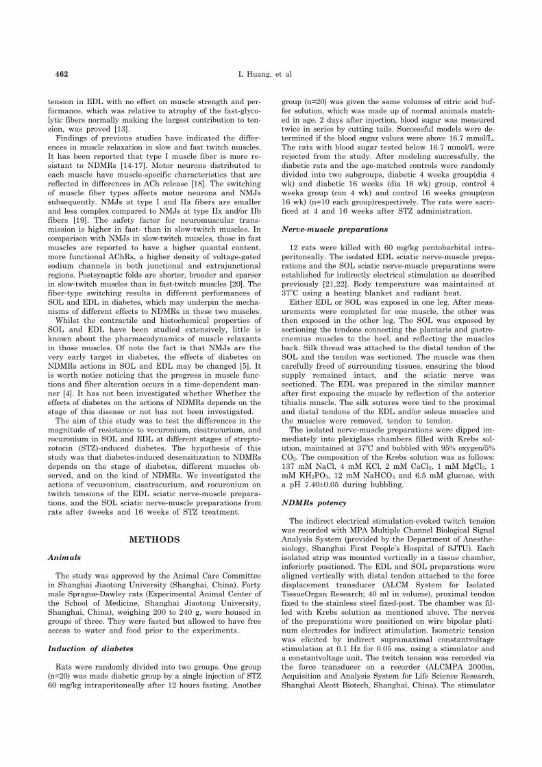
Fig. 1. Twitch tension height elicited by indirect stimulation in Extensor digitorum longus (EDL, A) and Soleus (SOL, B). There was a significant difference between diabetic groups and the age-matched control groups (p<0.05), and between dia 4 wk and dia 16 wk groups (p<0.05), while no significant difference between con 4 wk group and con 16 wk group. *p<0.05 versus control groups, &p<0.05 versus diabetic 4 weeks group. con 4 wk, control 4 weeks group; dia 4 wk, diabetic 4 weeks group; con 16 wk, control 16 weeks group; dia 16 wk, diabetic 16 weeks group.
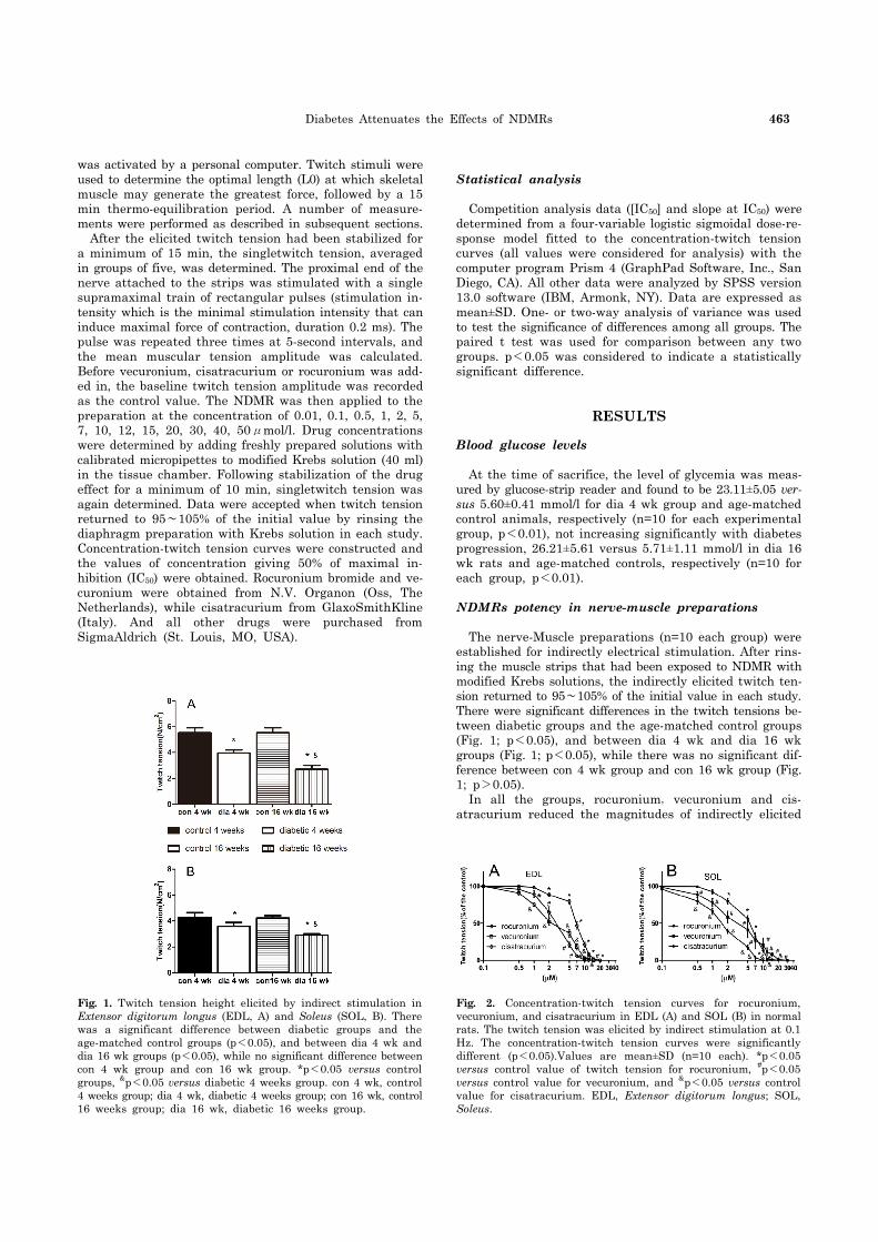
Fig. 2. Concentration-twitch tension curves for rocuronium, vecuronium, and cisatracurium in EDL (A) and SOL (B) in normal rats. The twitch tension was elicited by indirect stimulation at 0.1 Hz. The concentration-twitch tension curves were significantly different (p<0.05).Values are mean±SD (n=10 each). *p<0.05 versus control value of twitch tension for rocuronium, #p<0.05 versus control value for vecuronium, and &p<0.05 versus control value for cisatracurium. EDL, Extensor digitorum longus; SOL, Soleus.
was activated by a personal computer. Twitch stimuli were used to determine the optimal length (L0) at which skeletal muscle may generate the greatest force, followed by a 15 min thermo-equilibration period. A number of measure-ments were performed as described in subsequent sections. After the elicited twitch tension had been stabilized for a minimum of 15 min, the singletwitch tension, averaged in groups of five, was determined. The proximal end of the nerve attached to the strips was stimulated with a single supramaximal train of rectangular pulses (stimulation in-tensity which is the minimal stimulation intensity that can induce maximal force of contraction, duration 0.2 ms). The pulse was repeated three times at 5-second intervals, and the mean muscular tension amplitude was calculated. Before vecuronium, cisatracurium or rocuronium was add-ed in, the baseline twitch tension amplitude was recorded as the control value. The NDMR was then applied to the preparation at the concentration of 0.01, 0.1, 0.5, 1, 2, 5, 7, 10, 12, 15, 20, 30, 40, 50μmol/l. Drug concentrations were determined by adding freshly prepared solutions with calibrated micropipettes to modified Krebs solution (40 ml) in the tissue chamber. Following stabilization of the drug effect for a minimum of 10 min, singletwitch tension was again determined. Data were accepted when twitch tension returned to 95∼105% of the initial value by rinsing the diaphragm preparation with Krebs solution in each study. Concentration-twitch tension curves were constructed and the values of concentration giving 50% of maximal in-hibition (IC50) were obtained. Rocuronium bromide and ve-curonium were obtained from N.V. Organon (Oss, The Netherlands), while cisatracurium from GlaxoSmithKline (Italy). And all other drugs were purchased from SigmaAldrich (St. Louis, MO, USA).
Statistical analysis
Competition analysis data ([IC50] and slope at IC50) were determined from a four-variable logistic sigmoidal dose-re-sponse model fitted to the concentration-twitch tension curves (all values were considered for analysis) with the computer program Prism 4 (GraphPad Software, Inc., San Diego, CA). All other data were analyzed by SPSS version 13.0 software (IBM, Armonk, NY). Data are expressed as mean±SD. One- or two-way analysis of variance was used to test the significance of differences among all groups. The paired t test was used for comparison between any two groups. p<0.05 was considered to indicate a statistically significant difference.
RESULTS
Blood glucose levels
At the time of sacrifice, the level of glycemia was meas-ured by glucose-strip reader and found to be 23.11±5.05 ver-sus 5.60±0.41 mmol/l for dia 4 wk group and age-matched control animals, respectively (n=10 for each experimental group, p<0.01), not increasing significantly with diabetes progression, 26.21±5.61 versus 5.71±1.11 mmol/l in dia 16 wk rats and age-matched controls, respectively (n=10 for each group, p<0.01).
NDMRs potency in nerve-muscle preparations
The nerve-Muscle preparations (n=10 each group) were established for indirectly electrical stimulation. After rins-ing the muscle strips that had been exposed to NDMR with modified Krebs solutions, the indirectly elicited twitch ten-sion returned to 95∼105% of the initial value in each study. There were significant differences in the twitch tensions be-tween diabetic groups and the age-matched control groups (Fig. 1; p<0.05), and between dia 4 wk and dia 16 wk groups (Fig. 1; p<0.05), while there was no significant dif-ference between con 4 wk group and con 16 wk group (Fig. 1; p>0.05). In all the groups, rocuronium,vecuronium and cis-atracurium reduced the magnitudes of indirectly elicited
464 L Huang, et al
Table 1. IC50 values and slopes of the concentration-twitch tension curves of NDMRs
Control Diabetic 4 weeks Diabetic 16 weeks
EDL SOL EDL SOL EDL SOL
Rocuronium IC50 7.170
(4.705∼8.667)6.686
(5.148∼8.684)7.970
(7.361∼8.667)8.461
(7.590∼9.432)*9.832
(9.441∼10.24)*&12.84
(11.06∼14.91)*&#
Slope −3.727±0.395 −1.667±0.227# −4.737±0.673 −4.030±0.651* −4.245±0.306 −3.179±0.630*#
Vecuronium IC50 4.798
(1.778∼12.95)$4.620
(1.354∼15.77)$8.163
(7.253∼9.186)*8.227
(4.215∼17.37)*8.532
(6.308∼11.54)*10.43
(8.801∼12.37)*&#$
Slope −0.932±0.193$ −0.930±0.303$ −2.336±0.845*$ −1.278±0.500#$ −1.973±0.473*$ −2.639±0.477*$
Cisatracurium IC50 2.962
(2.383∼3.681)[email protected]
(1.361∼2.782)#@$3.806
(2.177∼6.652)*@$2.776
(1.098∼7.020)*#@$4.887
(2.713∼8.802)*&@$5.074
(3.213∼8.010)*&@$
Slope −1.980±0.300@$ −1.221±0.194@$ −1.224±0.219@$ −1.080±0.325@$ −1.334±0.262@$ −1.689±0.359@$
IC50, and slope at log IC50 were determined from a four-variable logistic sigmoidal dose-response model fitted to the concen-tration-twitch tension curves. The twitch tension was elicited by indirect stimulation at 0.1 Hz. Values are expressed as means with 95% confidence intervals in IC50 (μM) and mean±SD in slope at log IC50 (n=10 each). Statistical analysis was performed by oneANOVA with postBonferroni testing. *p<0.05 versus control group (Data of 16 weeks control group not presented in this table), &p<0.05 versus diabetic 4 weeks group; #p<0.05 versus EDL in the same group; $p<0.05 versus Rocuronium, @p<0.05 versus Vecuronium. EDL, Extensor digitorum longus; SOL, Soleus.
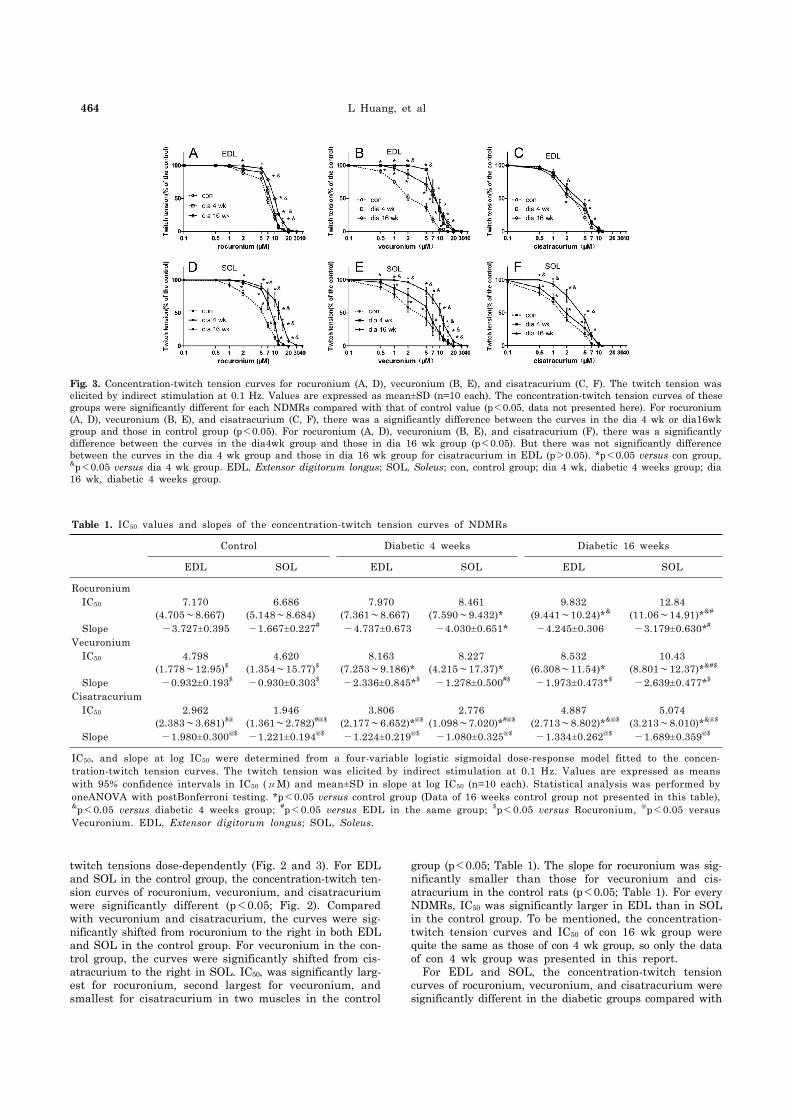
Fig. 3. Concentration-twitch tension curves for rocuronium (A, D), vecuronium (B, E), and cisatracurium (C, F). The twitch tension was elicited by indirect stimulation at 0.1 Hz. Values are expressed as mean±SD (n=10 each). The concentration-twitch tension curves of thesegroups were significantly different for each NDMRs compared with that of control value (p<0.05, data not presented here). For rocuronium (A, D), vecuronium (B, E), and cisatracurium (C, F), there was a significantly difference between the curves in the dia 4 wk or dia16wk group and those in control group (p<0.05). For rocuronium (A, D), vecuronium (B, E), and cisatracurium (F), there was a significantly difference between the curves in the dia4wk group and those in dia 16 wk group (p<0.05). But there was not significantly difference between the curves in the dia 4 wk group and those in dia 16 wk group for cisatracurium in EDL (p>0.05). *p<0.05 versus con group, &p<0.05 versus dia 4 wk group. EDL, Extensor digitorum longus; SOL, Soleus; con, control group; dia 4 wk, diabetic 4 weeks group; dia 16 wk, diabetic 4 weeks group.
twitch tensions dose-dependently (Fig. 2 and 3). For EDL and SOL in the control group, the concentration-twitch ten-sion curves of rocuronium, vecuronium, and cisatracurium were significantly different (p<0.05; Fig. 2). Compared with vecuronium and cisatracurium, the curves were sig-nificantly shifted from rocuronium to the right in both EDL and SOL in the control group. For vecuronium in the con-trol group, the curves were significantly shifted from cis-atracurium to the right in SOL. IC50, was significantly larg-est for rocuronium, second largest for vecuronium, and smallest for cisatracurium in two muscles in the control
group (p<0.05; Table 1). The slope for rocuronium was sig-nificantly smaller than those for vecuronium and cis-atracurium in the control rats (p<0.05; Table 1). For every NDMRs, IC50 was significantly larger in EDL than in SOL in the control group. To be mentioned, the concentration- twitch tension curves and IC50 of con 16 wk group were quite the same as those of con 4 wk group, so only the data of con 4 wk group was presented in this report. For EDL and SOL, the concentration-twitch tension curves of rocuronium, vecuronium, and cisatracurium were significantly different in the diabetic groups compared with
Diabetes Attenuates the Effects of NDMRs 465
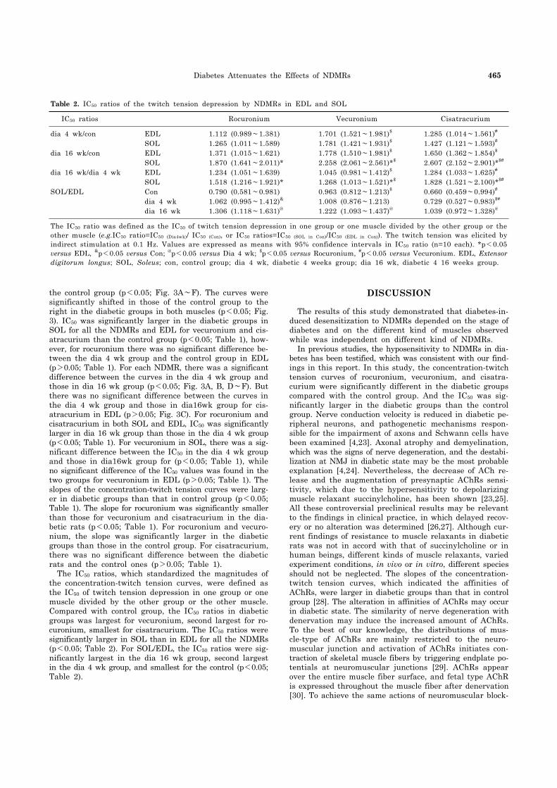
Table 2. IC50 ratios of the twitch tension depression by NDMRs in EDL and SOL
IC50 ratios Rocuronium Vecuronium Cisatracurium
dia 4 wk/con EDL 1.112 (0.989∼1.381) 1.701 (1.521∼1.981)$ 1.285 (1.014∼1.561)#
SOL 1.265 (1.011∼1.589) 1.781 (1.421∼1.931)$ 1.427 (1.121∼1.593)#
dia 16 wk/con EDL 1.371 (1.015∼1.621) 1.778 (1.510∼1.981)$ 1.650 (1.362∼1.854)$
SOL 1.870 (1.641∼2.011)* 2.258 (2.061∼2.561)*$ 2.607 (2.152∼2.901)*$#
dia 16 wk/dia 4 wk EDL 1.234 (1.051∼1.639) 1.045 (0.981∼1.412)$ 1.284 (1.033∼1.625)#
SOL 1.518 (1.216∼1.921)* 1.268 (1.013∼1.521)*$ 1.828 (1.521∼2.100)*$#
SOL/EDL Con 0.790 (0.581∼0.981) 0.963 (0.812∼1.213)$ 0.660 (0.459∼0.994)#
dia 4 wk 1.062 (0.995∼1.412)& 1.008 (0.876∼1.213) 0.729 (0.527∼0.983)$#
dia 16 wk 1.306 (1.118∼1.631)@ 1.222 (1.093∼1.437)@ 1.039 (0.972∼1.328)@
The IC50 ratio was defined as the IC50 of twitch tension depression in one group or one muscle divided by the other group or the other muscle (e.g.IC50 ratio=IC50 (Dia4wk)/ IC50 (Con), or IC50 ratios=IC50 (SOL in Con)/IC50 (EDL in Con)). The twitch tension was elicited by indirect stimulation at 0.1 Hz. Values are expressed as means with 95% confidence intervals in IC50 ratio (n=10 each). *p<0.05 versus EDL, &p<0.05 versus Con; @p<0.05 versus Dia 4 wk; $p<0.05 versus Rocuronium, #p<0.05 versus Vecuronium. EDL, Extensor digitorum longus; SOL, Soleus; con, control group; dia 4 wk, diabetic 4 weeks group; dia 16 wk, diabetic 4 16 weeks group.
the control group (p<0.05; Fig. 3A∼F). The curves were significantly shifted in those of the control group to the right in the diabetic groups in both muscles (p<0.05; Fig. 3). IC50 was significantly larger in the diabetic groups in SOL for all the NDMRs and EDL for vecuronium and cis-atracurium than the control group (p<0.05; Table 1), how-ever, for rocuronium there was no significant difference be-tween the dia 4 wk group and the control group in EDL (p>0.05; Table 1). For each NDMR, there was a significant difference between the curves in the dia 4 wk group and those in dia 16 wk group (p<0.05; Fig. 3A, B, D∼F). But there was no significant difference between the curves in the dia 4 wk group and those in dia16wk group for cis-atracurium in EDL (p>0.05; Fig. 3C). For rocuronium and cisatracurium in both SOL and EDL, IC50 was significantly larger in dia 16 wk group than those in the dia 4 wk group (p<0.05; Table 1). For vecuronium in SOL, there was a sig-nificant difference between the IC50 in the dia 4 wk group and those in dia16wk group for (p<0.05; Table 1), while no significant difference of the IC50 values was found in the two groups for vecuronium in EDL (p>0.05; Table 1). The slopes of the concentration-twitch tension curves were larg-er in diabetic groups than that in control group (p<0.05; Table 1). The slope for rocuronium was significantly smaller than those for vecuronium and cisatracurium in the dia-betic rats (p<0.05; Table 1). For rocuronium and vecuro-nium, the slope was significantly larger in the diabetic groups than those in the control group. For cisatracurium, there was no significant difference between the diabetic rats and the control ones (p>0.05; Table 1). The IC50 ratios, which standardized the magnitudes of the concentration-twitch tension curves, were defined as the IC50 of twitch tension depression in one group or one muscle divided by the other group or the other muscle. Compared with control group, the IC50 ratios in diabetic groups was largest for vecuronium, second largest for ro-curonium, smallest for cisatracurium. The IC50 ratios were significantly larger in SOL than in EDL for all the NDMRs (p<0.05; Table 2). For SOL/EDL, the IC50 ratios were sig-nificantly largest in the dia 16 wk group, second largest in the dia 4 wk group, and smallest for the control (p<0.05; Table 2).
DISCUSSION
The results of this study demonstrated that diabetes-in-duced desensitization to NDMRs depended on the stage of diabetes and on the different kind of muscles observed while was independent on different kind of NDMRs. In previous studies, the hyposensitivity to NDMRs in dia-betes has been testified, which was consistent with our find-ings in this report. In this study, the concentration-twitch tension curves of rocuronium, vecuronium, and cisatra-curium were significantly different in the diabetic groups compared with the control group. And the IC50 was sig-nificantly larger in the diabetic groups than the control group. Nerve conduction velocity is reduced in diabetic pe-ripheral neurons, and pathogenetic mechanisms respon-sible for the impairment of axons and Schwann cells have been examined [4,23]. Axonal atrophy and demyelination, which was the signs of nerve degeneration, and the destabi-lization at NMJ in diabetic state may be the most probable explanation [4,24]. Nevertheless, the decrease of ACh re-lease and the augmentation of presynaptic AChRs sensi-tivity, which due to the hypersensitivity to depolarizing muscle relaxant succinylcholine, has been shown [23,25]. All these controversial preclinical results may be relevant to the findings in clinical practice, in which delayed recov-ery or no alteration was determined [26,27]. Although cur-rent findings of resistance to muscle relaxants in diabetic rats was not in accord with that of succinylcholine or in human beings, different kinds of muscle relaxants, varied experiment conditions, in vivo or in vitro, different species should not be neglected. The slopes of the concentration- twitch tension curves, which indicated the affinities of AChRs, were larger in diabetic groups than that in control group [28]. The alteration in affinities of AChRs may occur in diabetic state. The similarity of nerve degeneration with denervation may induce the increased amount of AChRs. To the best of our knowledge, the distributions of mus-cle-type of AChRs are mainly restricted to the neuro-muscular junction and activation of AChRs initiates con-traction of skeletal muscle fibers by triggering endplate po-tentials at neuromuscular junctions [29]. AChRs appear over the entire muscle fiber surface, and fetal type AChR is expressed throughout the muscle fiber after denervation [30]. To achieve the same actions of neuromuscular block-
466 L Huang, et al
ing, the doses of NDMRs need to be increased to com-petitively bind to AChRs in diabetes. Further investigation should be done to testify the hypothesis. Moreover, there was a significantly difference between the IC50 in the dia 4 wk group and those in dia 16 wk group for rocuronium and cisatracurium in both SOL and EDL in this study. IC50 was significantly larger in dia 16 wk group than those in the dia 4 wk group for vecuronium in SOL. Although there was no significantly difference be-tween the IC50 values in the dia 4 wk group and those in dia 16 wk group for vecuronium in EDL, IC50 was larger in dia 16 wk group. In a word, regardless of the different muscle observed, diabetes induced the resistance to each NDMRs depends on the stage of diabetes for rocuronium, vecuronium and cisatracurium. The progress in muscle functions and fiber alteration occurs in a time-dependent manner [4]. In Brotto et al’s report, the alteration in dia-phragm contractility depended on the time after the onset of STZ-induced diabetes, which at least in part, relied on the switch in fiber type [12]. Although NMJs are the very early target in diabetes, the course of disease may play an important role in the final performance of changes in mus-cle contractility and alterations of response to NDMRs. Compared with SOL in control group, IC50 in EDL was larger for rocuronium, vecuronium and cisatracurium than that in EDL. While in diabetic groups, IC50 in SOL was larger for every NDMRs. Additionally, IC50 ratio of SOL/EDL in dia 16 wk group was larger than that in dia 4 wk group. The results meant that the response to NDMRs depended on the muscles observed. STZ diabetes had differ-ential effects on skeletal muscles accompanying the course of diabetes. It has been shown that diabetic SOL muscles had a higher proportion of slow-oxidative fibers than weight-matched controls while no obvious changes in fiber composition in diabetic EDL muscles [13]. For the slow SOL of the diabetic animals, muscle weight was preserved with respect to body weight. However, preferential atrophy of fast fibers in EDL was reflected in fiber area measurements [28,31]. Whereas SOL was protected from diabetic effects in terms of muscle mass and force production, both speed- related properties and oxidative capacity were impaired. In contrast, EDL underwent profound wasting and had de-creased strength, while the speed-related properties and ox-idative capacity was unchanged. The differences in the con-tractile and histochemical properties of slow and fast mus-cles may account for the differences in the resistance to NDMRs. The changes in fiber-type proportions of SOL which involved a change in muscle phenotypic expression, may induce more type I muscle fibers, which may result in much more resistance to NDMRs in SOL than that in EDL. Compared with control rats, the IC50 in diabetic groups were different among the neuromuscular blockers. In addi-tion, the IC50 ratios among the neuromuscular blockers were quite different with each other. However, there was no obvious relevance between the different kind of NDMRs and IC50 ratios. E. Narimatsu et al. concluded that the mag-nitude of sepsis-induced attenuation of potencies to NDMRs depended on the different molecular structures of neuro-muscular blockers [28]. To the best of our knowledge, there are two kinds of NDMRs,: one is benzylisoquinolines (cisatracurium) and the other is bis-quaternary ammonium steroids (vecuronium and rocuronium). Our results demon-strated that diabetes-induced resistance to NDMRs was in-dependent on the kind of NDMRs.
The results of our study seemed to be of great clinical relevance. Though the experiment was obtained in vitro by nerve-muscle preparations, the findings can be extra-polated to clinical studies. From the relevance in the con-ditions of different stages of diabetes between rat models and human beings, it is assumed that in clinical practice, the hyposensitivity to NDMRs may also develop to different extents during the course of diabetes. Moreover, now that diabetes-induced desensitization to NDMRs depended on the different kind of muscles observed, extra caution should be taken in clinical practice when monitoring for muscle relaxation in anesthetic management using different muscles. It is worth noticing that when choosing drugs for diabetic patients in anesthesia, NDMRs do not need further consideration as diabetes-induced resistance to NDMRs was independent on the kind of NDMRs. In summary, diabetes-induced desensitization to NDMRs depended on the stage of diabetes and on the different kind of muscles observed while was independent on different kind of NDMRs. The resistance to NDMRs was stronger in the later stage of diabetes (16 weeks verus 4 weeks after STZ treatment in this report). Additionally, when monitor-ing in SOL, diabetes attenuated the actions of neuro-muscular blockade than that in EDL. Nonetheless, the hy-posensitivity to NDMRs in diabetes was not relevant for the kind of NDMRs.
ACKNOWLEDGEMENTS
This study is supported by the National Natural Science Foundation of China (grant number 81171845) and Songjiang District Foundation of Shanghai (grant number 2011PD13). The authors declare no conflict of interest.
REFERENCES
1. Clements RS Jr. Diabetic neuropathy--new concepts of its etiology. Diabetes. 1979;28:604-611.
2. Diabetic peripheral neuropathies. Physiopathology and clinical guidelines. Diabetes Res Clin Pract. 1986;2:183-256.
3. Harati Y. Diabetic peripheral neuropathies. Ann Intern Med. 1987;107:546-559.
4. Fahim MA, Hasan MY, Alshuaib WB. Early morphological remodeling of neuromuscular junction in a murine model of diabetes. J Appl Physiol (1985). 2000;89:2235-2240.
5. Marques MJ, Santo Neto H. Acetylcholine receptors and nerve terminal distribution at the neuromuscular junction of non-obese diabetic mice. Anat Rec. 2002;267:112-119.
6. Schofield GG, Furman BL, Marshall IG. The effect of acute alloxan diabetes on the sensitivity of the rat skeletal neuro-muscular junction to drugs. Acta Diabetol Lat. 1978;15:287- 293.
7. Minker E, Kac P, Blazsó G, Koltai M. A study of the origin of altered pharmacological reactivity of synaptic structures caused by diabetes and pretreatment with contrainsular agents. Acta Physiol Hung. 1984;63:175-183.
8. Kimura M, Fujihara M, Nojima H, Kimura I. Hypersensitivity of acetylcholine receptor in diabetic skeletal muscle to neuromuscular blockers: the effect of myotubes cultured with spinal cord or its extract. J Pharmacobiodyn. 1986;9:29-38.
9. Kimura M, Kimura I, Nojima H, Muroi M. Diabetes mellitus- induced hypersensitivity of mouse skeletal muscles to acetyl-choline and succinylcholine. Jpn J Pharmacol. 1986;40:251- 256.
10. Peter JB, Barnard RJ, Edgerton VR, Gillespie CA, Stempel KE. Metabolic profiles of three fiber types of skeletal muscle in
Diabetes Attenuates the Effects of NDMRs 467
guinea pigs and rabbits. Biochemistry. 1972;11:2627-2633.11. Cotter M, Cameron NE, Lean DR, Robertson S. Effects of
long-term streptozotocin diabetes on the contractile and histochemical properties of rat muscles. Q J Exp Physiol. 1989;74:65-74.
12. Brotto M, Brotto L, Jin JP, Nosek TM, Romani A. Temporal adaptive changes in contractility and fatigability of diaphragm muscles from streptozotocin-diabetic rats. J Biomed Biotechnol. 2010;2010:931903.
13. Cotter M, Cameron NE, Lean DR, Robertson S. Effects of long-term streptozotocin diabetes on the contractile and histo-chemical properties of rat muscles. Q J Exp Physiol. 1989; 74:65-74.
14. Secher NH, Rube N, Secher O. Effect of tubocurarine on human soleus and gastrocnemius muscles. Acta Anaesthesiol Scand. 1982;26:231-234.
15. Kitajima T, Ishii K, Kobayashi T, Ogata H. Differential effects of vecuronium on the thumb and great toe as measured by accelography and electromyography. Anaesthesia. 1995;50:76- 78.
16. Saitoh Y, Tanaka H, Toyooka H, Amaha K. Recovery of post-tetanic and train-of-four responses at the first dorsal interosseous and adductor pollicis muscles in patients receiving vecuronium. Can J Anaesth. 1996;43:362-367.
17. Saitoh Y, Koitabashi Y, Makita K, Tanaka H, Amaha K. Train-of-four and double burst stimulation fade at the great toe and thumb. Can J Anaesth. 1997;44:390-395.
18. Taquahashi Y, Yonezawa K, Nishimura M. Influence of motor activities on the release of transmitter quanta from motor nerve terminals in mice. J Vet Med Sci. 1999;61:513-516.
19. Sieck DC, Zhan WZ, Fang YH, Ermilov LG, Sieck GC, Mantilla CB. Structure-activity relationships in rodent diaphragm muscle fibers vs. neuromuscular junctions. Respir Physiol Neurobiol. 2012;180:88-96.
20. Wood SJ, Slater CR. The contribution of postsynaptic folds to the safety factor for neuromuscular transmission in rat fast- and slow-twitch muscles. J Physiol. 1997;500:165-176.
21. McGuire M, MacDermott M. The influence of streptozotocin diabetes and metformin on erythrocyte volume and on the membrane potential and the contractile characteristics of the extensor digitorum longus and soleus muscles in rats. Exp
Physiol. 1999;84:1051-1058.22. Oishi PE, Cholsiripunlert S, Gong W, Baker AJ, Bernstein HS.
Myo-mechanical analysis of isolated skeletal muscle. J Vis Exp. 2011;(48).
23. Kimura I, Okazaki M, Kimura M. Streptozocin-diabetes modifies acetylcholine release from mouse phrenic nerve terminal and presynaptic sensitivity to succinylcholine. Jpn J Pharmacol. 1993;62:35-41.
24. Medina Sánchez M, Rodríguez Sánchez C, Vega Alvarez JA, Menéndez Peláez A. Ultrastructural study of neuromuscular junction in rectus femoris muscle of streptozotocin-diabetic rats. Histol Histopathol. 1992;7:607-610.
25. Kimura M, Kimura I, Fujihara M, Hoshino N. Diabetic state-induced modifications of succinylcholine binding mode in the microsomal fractions of mouse skeletal muscles. Life Sci. 1988;42:1029-1036.
26. Saitoh Y, Hattori H, Sanbe N, Nakajima H, Akatu M, Murakawa M. Reversal of vecuronium with neostigmine in patients with diabetes mellitus. Anaesthesia. 2004;59:750-754.
27. Alper I, Ulukaya S, Makay O, Balcioglu T. The pharma-codynamic effects of rocuronium during general anesthesia in patients with type 2 diabetes mellitus. Minerva Anestesiol. 2010;76:115-119.
28. Narimatsu E, Niiya T, Kawamata M, Namiki A. Sepsis stage dependently and differentially attenuates the effects of nondepolarizing neuromuscular blockers on the rat diaphragm in vitro. Anesth Analg. 2005;100:823-829.
29. Lee BH, Shin TJ, Hwang SH, Choi SH, Kang J, Kim HJ, Park CW, Lee SH, Nah SY. Inhibitory effects of quercetin on muscle-type of nicotinic acetylcholine receptor-mediated ion currents expressed in Xenopus Oocytes. Korean J Physiol Pharmacol. 2011;15:195-201.
30. Salpeter MM, Loring RH. Nicotinic acetylcholine receptors in vertebrate muscle: properties, distribution and neural control. Prog Neurobiol. 1985;25:297-325.
31. McGuire M, MacDermott M. The influence of streptozotocin diabetes and metformin on erythrocyte volume and on the membrane potential and the contractile characteristics of the extensor digitorum longus and soleus muscles in rats. Exp Physiol. 1999;84:1051-1058.