Embed Size (px)
Citation preview

Stem anatomy at various developmental stages of secondary growth in Turbina corymbosa (Convolvulaceae)
Author(s): Manoj M. Lekhak, Amit D. Gondaliya, Shrirang R. Yadav and Kishore S. Rajput
Source: Plant Ecology and Evolution , 2018, Vol. 151, No. 2 (2018), pp. 219-230
Published by: Royal Botanical Society of Belgium and the Botanic Garden Meise
Stable URL: https://www.jstor.org/stable/44945380
JSTOR is a not-for-profit service that helps scholars, researchers, and students discover, use, and build upon a wide range of content in a trusted digital archive. We use information technology and tools to increase productivity and facilitate new forms of scholarship. For more information about JSTOR, please contact [email protected]. Your use of the JSTOR archive indicates your acceptance of the Terms & Conditions of Use, available at https://about.jstor.org/terms
and Royal Botanical Society of Belgium are collaborating with JSTOR to digitize, preserve and extend access to Plant Ecology and Evolution
This content downloaded from �������������86.59.13.237 on Mon, 21 Jun 2021 10:53:31 UTC��������������
All use subject to https://about.jstor.org/terms
Plant Ecology and Evolution 151 (2): 219-230, 2018 https://doi.org/10.5091/plecevo.2018.1389
REGULAR PAPER
Stem anatomy at various developmental stages of secondary growth in Turbina corymbosa (Convolvulaceae)
Manoj M. Lekhak1, Amit D. Gondaliya2, Shrirang R. Yadav1 & Kishore S. Rajput2 *
1 Angio sperm Taxonomy Laboratory, Department of Botany, Shivaji University, Kolhapur (MS) 416 004, India department of Botany, Faculty of Science, The Maharaja Sayajirao University of Baroda, Vadodara 390002, India * Author for correspondence: [email protected]
Background - Population growth of lianas in the tropical forest is credited to their ability of C02 sequestration and efficiency of the narrow stems to supply water required for the amount of foliage it bears. Turbina corymbosa (L.) Raf. (Convolvulaceae Juss.) is one of the fast-growing invasive species of scrambling woody lianas. It covers trees entirely within a short period to compete with above-ground resources (particularly sunlight). However, no information is available on how it manages to cope up with an increasing demand of water supply and mineral nutrients. What are the structural and developmental patterns adapted by this species to expand the stem diameter for efficient supply of below-ground resources? Therefore, our aim was to investigate the secondary growth patterns and structure of secondary xylem and phloem in T. corymbosa. Methods - Several samples of the stem with various diameters were studied using a histological method. Morphological and anatomical analyses were carried out using light microscopy. Key results - With the initiation of secondary growth, stems lose their circular outline rapidly due to unequal deposition of secondary xylem and formation of successive cambia. New successive cambia initiate from parenchymatous cells as small crescent-shaped fragments on asymmetric/opposite sides and result in a different stem conformation. Though several segments of successive cambia are formed, very few stem samples form complete cambium rings. The secondary xylem formed by successive cambia is diffuse porous with indistinct growth rings and is composed of both wide and narrow (fibriform) vessels, tracheids, fibres, axial and ray parenchyma cells. The secondary phloem consists of sieve tube elements, companion cells, axial and ray parenchyma cells. In fully grown plants, cambial action (internal cambium) occurrs between the intraxylary phloem and protoxylem and produces secondary xylem and phloem near the pith region.
Conclusion - Structural alterations and unequal deposition of conducting elements, occurrence of intraxylary phloem and flattening of the stem are suggested to facilitate rapid growth of the plants by providing required minerals and nutrients. Internal cambium formed at the periphery of the pith is bidirectional and produces secondary xylem externally and intraxylary phloem internally. Continued development of intraxylary phloem from the internal cambium provides an additional path for rapid and safe translocation of photosynthates.
Key words - Cambial variant, Christmas vine, intraxylary phloem, internal cambium, successive cambia, secondary xylem and phloem.
INTRODUCTION
Climbing plants are important components of forest ecosys- tems and contribute to many vital aspects of the forest eco- system particularly, in species diversity, carbon, nutrients and water sequestration (Isnard & Silk 2009, Schnitser & Bongers 2011). Their increasing population in tropical for-
est is correlated with growing forest disturbances, forest fragmentation and elevated concentration of atmospheric
C02 (Philips et al. 2002, Schnizer & Bongers 2011). They represent about 10-25% of woody biomass of some tropical forests (Gentry & Dodson 1987, Schnitzer et al. 2005). Their expansion in tropical forests lies within their successful ad- aptation to the climbing habit by modifying their mechanical
All rights reserved. © 2018 Meise Botanic Garden and Royal Botanical Society of Belgium ISSN: 2032-3913 (print) - 2032-3921 (online)
This content downloaded from �������������86.59.13.237 on Mon, 21 Jun 2021 10:53:31 UTC��������������
All use subject to https://about.jstor.org/terms

Pl. Ecol Evol 151 (2), 2018
architecture (Rowe et al. 2004, Rowe & Speck 2005). Prior to climbing habit, they are self-supporting during early de- velopmental stages, which are reflected in large anatomical changes, especially vessel diameter and the amount of ray and axial parenchyma (Rowe et al. 2004, Rowe & Speck 2005, Isnard & Silk 2009, Rajput et al. 2013). Since their load is shared by the host, the energy required for radial growth and production of mechanical tissue is diverted to- wards extension growth. Therefore, they are able to develop a significantly large canopy, which may cover the crown of the host trees entirely and make the climbing species more competitive compared to the host trees for the above ground (sunlight) and below ground (water, mineral and nutrients) resources (Schnitzer & Bongers 2011).
The radial stem diameter in several climbing species re- lies on the development of successive cambia (Isnard & Silk 2009, Rajput et al. 2013). In most of them the successive cambia form a complete cylinder and the stem is circular in outline. In some species, however, stems lose their circular outline and show a variable shape. It may be lobed, flat- tened or irregular in shape in cross sectional outline, which is species specific (Rajput et al. 2012a, 2012b). Interestingly, no such consistent stem conformation in cross section was
observed in Turbina corymbosa (L.) Raf. (Convolvulaceae Juss.).
Turbina corymbosa is native to the West Indies, Mexico, Central America and the tropical portion of South America (Liogier 1995). It is a scrambling woody liana and can com- pletely cover the vegetation by forming a blanket over trees. It grows over 8 m high with smooth stem when young and it becomes deeply fissured, reaching above 13 cm in diameter and is a serious threat to trees growing on rainforest margin (Anonymous 2016). Various outlines of the stems in different species of lianas attracted the attention of several research- ers searching for the answer that how the different species of plants are achieving such conformations (Basson & Bi- erhost 1967, Mennega 1982, Carlquist 1988, Jacques & De Franceschi 2007, Rajput et al. 2012a, 2012b). Therefore, the main aim of the present study was to investigate the pattern of secondary growth and formation of its derivatives and their significance in relation to the climbing habit.
MATERIAL AND METHODS
Plant materials
Turbina corymbosa (Convolvulaceae) grows in the field as an invasive species in various part of the Maharashtra state. However, in the present study, samples were collected from the main stems of five individuals growing in the botanical garden of the Shivaji University, Kolhapur (Maharashtra state). Samples of various diameters measuring from 5 mm to maximum thickness (40 mm) available were cut into 60 mm long pieces. Voucher material (No. KSR/INSA/9) were submitted to the BARO herbarium of the Department of Botany, Faculty of Science, The M. S. University of Baro- da, Vadodara (India). Eight to ten segments each were col- lected from the base, middle and top portion of the stem and immediately fixed in FAA (Berlyn & Miksche 1976). After
12 h of fixation these samples were transferred in 70% alco- hol for further processing and fixation.
Macroscopic methods
For macro-morphological preparations, main stem samples of various thicknesses were cut into 3-4 cm long pieces. Fresh/semi-dried or completely dried stem pieces were pol- ished under running tap water by using waterproof sand pa- pers of various grain sizes. Running tap water prevents ac- cumulation of dust or wood particles formed due to polishing of the wood samples. Sometimes, accumulation of dust or wood particles was observed even after polishing under run- ning tap water. These wood blocks were cut directly by using a sharp knife. Stem blocks with fine finishing and open ves- sels were used for photography using a Cannon SLR 1200D camera.
Histological preparations
Transverse, radial and tangential longitudinal sections of 12-15 pm thickness were directly obtained with a Leica SM2010R sliding microtome and stained with Safranin- Astra blue combination (Srebotnik & Messner 1994). After dehydration through ethanol-xylene series the sections were mounted in Dibutyl Phthalate Xylene (DPX). Some of the samples of young stems (for primary growth) were also pro- cessed by routine method of paraffin embedding as described by Johansen (1940) and stained with the above-mentioned staining combination. Permanent slides were observed under a Leica DME 2000 trinocular research microscope and pho- tographed with a Cannon DC 150 Digital Camera.
Maceration
A small portion of the xylem, immediately next to the cam- bium was cut into 2-3 mm small slices and macerated with
Jeffery's fluid (Berlyn & Miksche 1976) at 55 to 60°C for 8-12 h. Macerated slices were gently washed with distilled water and stained with 1% aqueous Safranin to measure the length and width of vessel elements and xylem fibres. The length of the sieve tube elements was measured directly from the tangential longitudinal sections while their width was obtained from the transverse sections. Thirty measurements were taken randomly to obtain mean and standard deviations for each cell type.
RESULTS
Stem morphology
Turbina corymbosa is a perennial liana with various stem shapes ranging from circular, lobed, flat to dumbbell or crescent-shaped (fig. 1A-H). In the early stages of second- ary growth, stems are circular in outline but initiation of suc- cessive cambia and unequal deposition of secondary xylem results in various shapes. Crescent-shaped successive seg- ments of cambia develop on two opposite sides (fig. 1A & B), irregularly (fig. 1C), on three unequal or equidistance (fig. ID & F). In thick stems, several segments initiate which
220
This content downloaded from �������������86.59.13.237 on Mon, 21 Jun 2021 10:53:31 UTC��������������
All use subject to https://about.jstor.org/terms

Lekhak et al., Stem anatomy of Turbina corymbosa (Convolvulaceae)
Figure 1 - Macro-morphological cross-sectional view of Turbina corymbosa stems showing various conformations: A & B, development of successive cambia only on opposite sides of the stem, resulting in an elongated stem shape; C, irregular conformation of stem due to formation of successive cambia; D, formation of a tri-lobed stem due to development of cambia on three sides; E-H, development of successive cambia on two opposite sides forming various stem shapes. Scale bars = 1 5 mm.
221
This content downloaded from �������������86.59.13.237 on Mon, 21 Jun 2021 10:53:31 UTC��������������
All use subject to https://about.jstor.org/terms

PL Ecol Evol. 151 (2), 2018
Figure 2 - Transverse view of stem and secondary xylem of Turbina corymbosa : A, portion of young stem showing epidermis (arrowhead) and cortex; the arrow indicates isolated pericyclic fibres; B, simultaneous origin of external (arrow), intraxylary phloem (arrowhead) and differentiating metaxylem (MX) elements; note the already differentiated phloem on both sides; C, cross section of young stem showing newly formed cambium (arrows) on opposite sides; arrowheads indicate the side of the stem that fails to develop successive cambia; note the pattern of vessel distribution; D, development of successive cambia on opposite sides leads to an elongated, oval shape of the stem; E, pattern of vessel distribution on a portion of the stem that fails to develop successive cambia (shown with arrowheads in figure 2C); F, note the number of vessels on the stem side that develop successive cambia (shown with arrows in figure 2C). Scale bars: A, E & F = 250 pm; B = 150 pm; C & D = 12 mm.
222
This content downloaded from �������������86.59.13.237 on Mon, 21 Jun 2021 10:53:31 UTC��������������
All use subject to https://about.jstor.org/terms

Lekhak et al., Stem anatomy of Turbina corymbosa (Convolvulaceae)
do not fuse together, therefore resulting in an irregularly shaped outline of the stems in cross sections (fig. IG & H).
Anatomy of young stem and development of successive cambia
The young stems possess thin-walled, barrel-shaped epider- mal cells followed by a hypodermis comprising few layers of cells. It is followed by a thin walled oval to polygonal par- enchymatous cortex. Though, the endodermis and pericycle appear indistinct (fig. 2A), they can be discerned by the pres- ence of pericyclic fibres. Several conjoint collateral vascular bundles get interconnected by interfascicular cambium and form a complete cylinder of vascular cambium. Simultane- ously, differentiation of intraxylary phloem occurs along with the initiation of procambium (fig. 2B). The cambium is functionally bidirectional and produces secondary xylem centripetally and phloem centrifugally. The site for the ori- gin of the first successive cambium may be identified on the basis of vessel distribution pattern. As evidenced from the figure 2C and 2D, the first successive segment of cambium initiates as crescent-shaped arcs on two opposite sides of the stem (fig. 2C). In the young stem, a certain portion of the stem shows a complete absence of wide vessels, and they be- come rare when present (fig. 2E). In contrast, the other side of the stem shows a normal distribution of wide vessels (fig. 2F). Development of successive cambia always remains con- fined to the portion of the stem that shows normal distribu- tion of vessels (fig. 2D & F) whereas in the portion of stem lacking vessels, fail to develop successive cambia even in the 50-60 mm thick stems (fig. 3A). Occasionally, development of small arcs of cambia is seen in thick stems (fig. 3B).
As the secondary growth progresses (in 5-6 mm thick stems), thin-walled parenchyma (located on the outer side of the protophloem and on the inner side of isolated, or a group of 2-3 pericyclic fibres) dedifferentiate and acquire meris- tematic activity to form small segments of cambia. These segments may initiate on two or three sides at equidistance to form flat or tri-lobed stems, respectively (fig. 1A & D). However, in some individuals, several segments originate and form the first successive ring of cambium (figs IC & 3C). After the development of few xylem derivatives, these cam- bial arcs interconnect themselves by small segments of the cambium, which form thin walled derivatives on either side of the cambium (fig. 3C). Development of further successive cambial segments is seen only outside of the successive cam- bia, which form various shapes.
Structure of secondary xylem
Secondary xylem is diffuse porous with indistinct growth rings and composed of fibriform and wide vessels, trac- heids, fibres, ray and scanty vasicentric axial parenchyma cells (fig. 3A, B & D). In the first normal ring of second- ary xylem, rays are mostly uniseriate and thick-walled with few exceptionally large, thin-walled and multiseriate rays (fig. 3E). The scanty vasicentric axial parenchyma cells are thick-walled but their walls are relatively thinner than those of the fibres (fig. 3D). Therefore, it becomes difficult to dis- tinguish them in cross section due to their thick walls and more or less similar diameter as xylem fibres. They also form
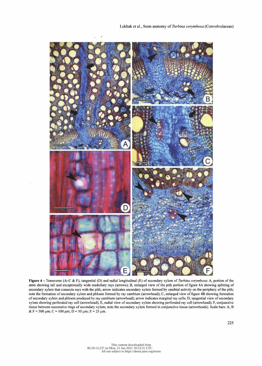
a sheath around the vessels. As the secondary growth pro- gresses, the ray parenchyma cells undergo proliferation by acquiring meristematic character. Repeated divisions in the ray cells add several cells and they become several cells wide. Widening of rays push adjacent thick-walled axial ele- ments; thus, connecting the pith and internal phloem to outer phloem (fig. 4A). Subsequently, cell division and differentia- tion of pith parenchyma (fig. 4A & B) and marginal ray cells differentiate into vessels (both wide and fibriform), tracheids and fibres (fig. 4B & C).
Rays are heterocellular and composed of oval oblong and upright cells. The multiseriate rays were several cells in height and width, measuring 177-1011 jam in height and 16-374 jam in width. Perforated ray cells also occur in the thick-walled lignified rays (fig. 4D & E). Perforated ray cells are isodiametric and similar to other ray cells except for the presence of simple perforation plate.
In thick stems, successive rings of the secondary xylem alternate with the phloem rings. Each ring of the secondary xylem and phloem is separated by relatively wider bands of conjunctive tissues (fig. 4F). Proliferation of conjunc- tive tissue between successive rings of secondary xylem and phloem results in the formation of small segments of radi- ally arranged meristematic cells appearing like cambium (fig. 5A). Subsequent divisions in these small segments give rise to secondary xylem and phloem on either side (fig. 5B). Therefore, formation of secondary xylem and phloem on ei- ther side looks like a "vascular bundle" embedded within the
bands of conjunctive tissue (fig. 5C). However, there is no specific orientation of these vascular bundles and they may be arranged radially, tangentially or diagonally. Structurally the secondary xylem formed by the successive cambia re- mains similar to the first ring of secondary xylem formed by the vascular cambium.
Vessels are dimorphic, in which wider ones are oval to circular in outline and mostly solitary, while radial or tan- gential multiples are rarely observed (fig. 3A & D). A simple perforation plate at the vessel element tip is slightly oblique to transverse. Vessel elements 178-310 jam long and 144- 292 jam wide. In contrast, fibriform vessels possess a nar- row lumen diameter, such as imperforated tracheary elements with a very small sub-terminal perforation plate (fig. 4D & E). Therefore, in transverse view it was difficult to distinguish them from the adjacent fibres and thick- walled parenchyma. Fibres or fibre tracheids are the longest cell types in the sec- ondary xylem, measuring 654-933 jam in length.
Structure of secondary phloem
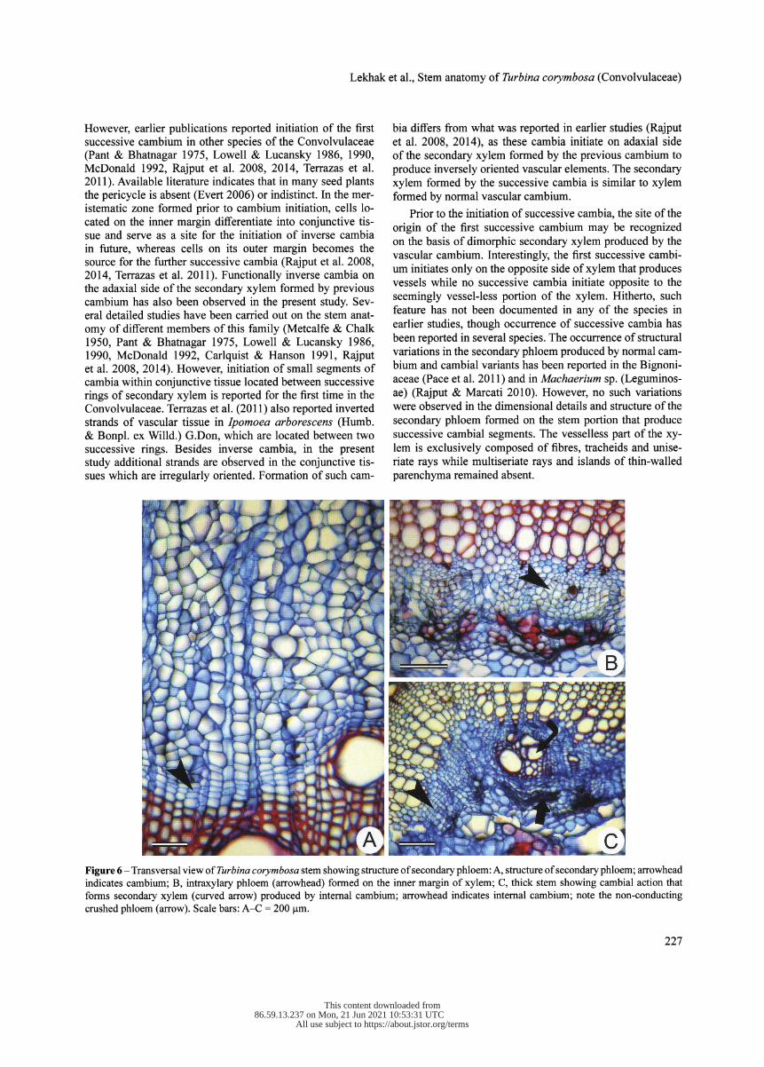
The secondary phloem is composed of sieve tube elements, companion cells axial and ray parenchyma cells (fig. 6A). Sieve tube elements possess transverse to lightly oriented simple sieve plate. Non-conducting sieve elements are char- acterized by the heavy accumulation of callose, subsequently followed by obliteration (fig. 6C).
Development of intraxylary phloem
Discrete strands of intraxylary phloem occur from the begin- ning of primary growth (fig. 6B). Intraxylary protophloem
223
This content downloaded from �������������86.59.13.237 on Mon, 21 Jun 2021 10:53:31 UTC��������������
All use subject to https://about.jstor.org/terms

PI Ecol Evol 151 (2), 2018
Figure 3 - Transverse (A-D) and longitudinal (E) view of secondary xylem of Turbina corymbosa : A, thick stems showing a portion of stem that fails to develop successive cambia; note the pattern of vessel distribution (arrowheads); B, newly formed cambium showing newly formed secondary xylem (arrowheads); note that newly formed xylem is lacking wide vessels; C, formation of small cambial segment resulting into vascular bundle-like arrangement (arrowheads) of vascular tissues; D, structure of secondary xylem; note the uniseriate rays and solitary vessels; E, longitudinal view of secondary xylem; note the vertically upright cells in both uniseriate (arrowhead) and bi- to multiseriate (arrow) rays. Scale bars: A-D = 500 pm; E = 250 pm.
224
This content downloaded from �������������86.59.13.237 on Mon, 21 Jun 2021 10:53:31 UTC��������������
All use subject to https://about.jstor.org/terms
Lekhak et al., Stem anatomy of Turbina corymbosa (Convolvulaceae)
Figure 4 - Transverse (A-C & F), tangential (D) and radial longitudinal (E) of secondary xylem of Turbina corymbosa : A, portion of the stem showing tall and exceptionally wide medullary rays (arrows); B, enlarged view of the pith portion of figure 4A showing splitting of secondary xylem that connects rays with the pith; arrow indicates secondary xylem formed by cambial activity on the periphery of the pith; note the formation of secondary xylem and phloem formed by ray cambium (arrowhead); C, enlarged view of figure 4B showing formation of secondary xylem and phloem produced by ray cambium (arrowhead); arrow indicates marginal ray cells; D, tangential view of secondary xylem showing perforated ray cell (arrowhead); E, radial view of secondary xylem showing perforated ray cell (arrowhead); F, conjunctive tissue between successive rings of secondary xylem; note the secondary xylem formed in conjunctive tissue (arrowheads). Scale bars: A, B & F = 500 ļum; C = 100 ļum; D = 50 pm; E = 25 pm.
225
This content downloaded from �������������86.59.13.237 on Mon, 21 Jun 2021 10:53:31 UTC��������������
All use subject to https://about.jstor.org/terms
PL Ecol EvoL 151 (2), 2018
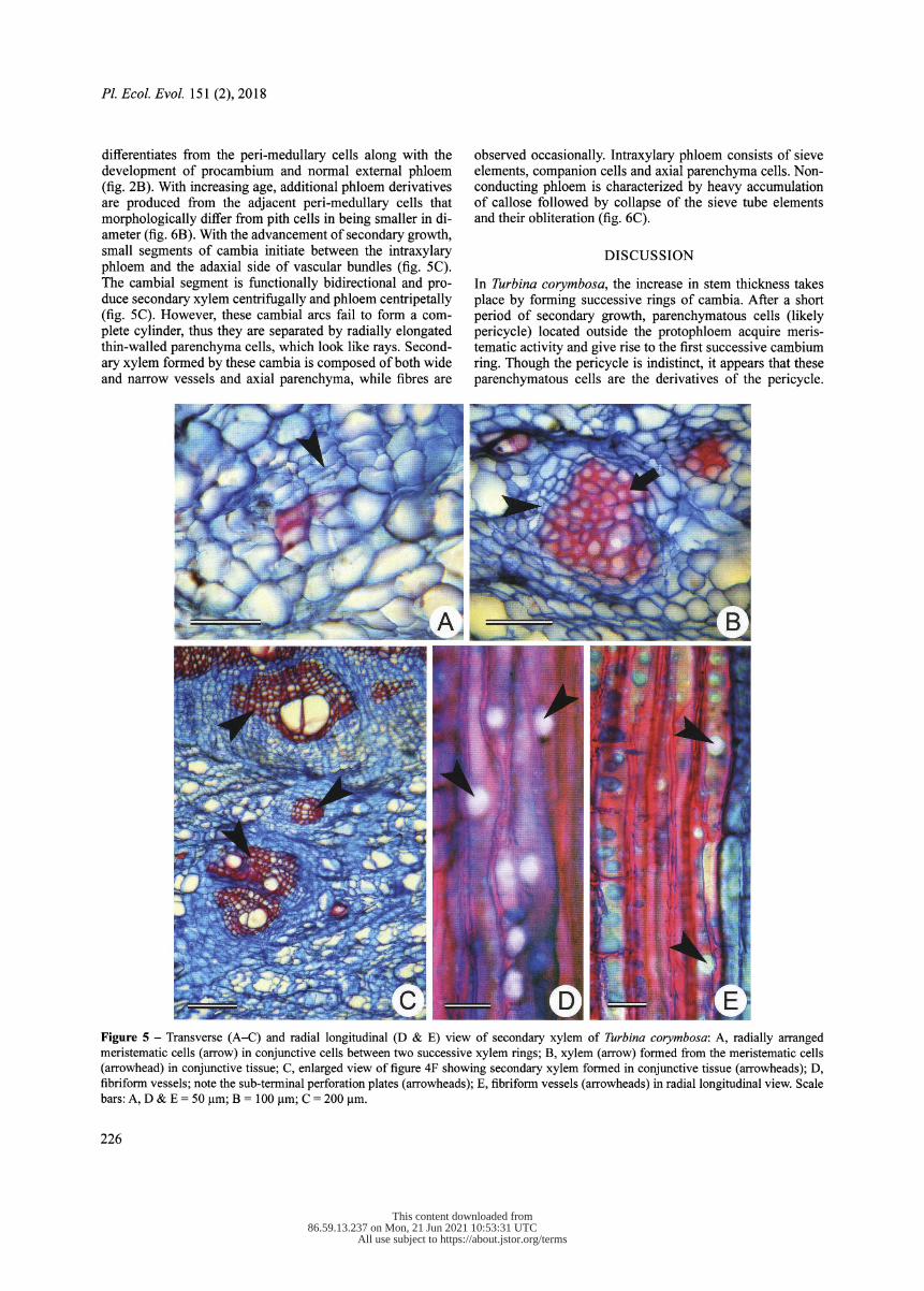
differentiates from the peri-medullary cells along with the development of procambium and normal external phloem (fig. 2B). With increasing age, additional phloem derivatives are produced from the adjacent peri-medullary cells that morphologically differ from pith cells in being smaller in di- ameter (fig. 6B). With the advancement of secondary growth, small segments of cambia initiate between the intraxylary phloem and the adaxial side of vascular bundles (fig. 5C). The cambial segment is functionally bidirectional and pro- duce secondary xylem centrifugally and phloem centripetally (fig. 5C). However, these cambial arcs fail to form a com- plete cylinder, thus they are separated by radially elongated thin-walled parenchyma cells, which look like rays. Second- ary xylem formed by these cambia is composed of both wide and narrow vessels and axial parenchyma, while fibres are
observed occasionally. Intraxylary phloem consists of sieve elements, companion cells and axial parenchyma cells. Non- conducting phloem is characterized by heavy accumulation of callose followed by collapse of the sieve tube elements and their obliteration (fig. 6C).
DISCUSSION
In Turbina corymbosa , the increase in stem thickness takes place by forming successive rings of cambia. After a short period of secondary growth, parenchymatous cells (likely pericycle) located outside the protophloem acquire meris- tematic activity and give rise to the first successive cambium ring. Though the pericycle is indistinct, it appears that these parenchymatous cells are the derivatives of the pericycle.
Figure 5 - Transverse (A-C) and radial longitudinal (D & E) view of secondary xylem of Turbina corymbosa : A, radially arranged meristematic cells (arrow) in conjunctive cells between two successive xylem rings; B, xylem (arrow) formed from the meristematic cells (arrowhead) in conjunctive tissue; C, enlarged view of figure 4F showing secondary xylem formed in conjunctive tissue (arrowheads); D, fibriform vessels; note the sub-terminal perforation plates (arrowheads); E, fibriform vessels (arrowheads) in radial longitudinal view. Scale bars: A, D & E = 50 gm; B = 100 gm; C = 200 gm.
226
This content downloaded from �������������86.59.13.237 on Mon, 21 Jun 2021 10:53:31 UTC��������������
All use subject to https://about.jstor.org/terms
Lekhak et al., Stem anatomy of Turbina corymbosa (Convolvulaceae)
However, earlier publications reported initiation of the first successive cambium in other species of the Convolvulaceae (Pant & Bhatnagar 1975, Lowell & Lucansky 1986, 1990, McDonald 1992, Rajput et al. 2008, 2014, Terrazas et al. 2011). Available literature indicates that in many seed plants the pericycle is absent (Evert 2006) or indistinct. In the mer- istematic zone formed prior to cambium initiation, cells lo- cated on the inner margin differentiate into conjunctive tis- sue and serve as a site for the initiation of inverse cambia
in future, whereas cells on its outer margin becomes the source for the further successive cambia (Rajput et al. 2008, 2014, Terrazas et al. 2011). Functionally inverse cambia on the adaxial side of the secondary xylem formed by previous cambium has also been observed in the present study. Sev- eral detailed studies have been carried out on the stem anat-
omy of different members of this family (Metcalfe & Chalk 1950, Pant & Bhatnagar 1975, Lowell & Lucansky 1986, 1990, McDonald 1992, Carlquist & Hanson 1991, Rajput et al. 2008, 2014). However, initiation of small segments of cambia within conjunctive tissue located between successive rings of secondary xylem is reported for the first time in the Convolvulaceae. Terrazas et al. (2011) also reported inverted strands of vascular tissue in Ipomoea arbores cens (Humb. & Bonpl. ex Willd.) G.Don, which are located between two successive rings. Besides inverse cambia, in the present study additional strands are observed in the conjunctive tis- sues which are irregularly oriented. Formation of such cam-
bia differs from what was reported in earlier studies (Rajput et al. 2008, 2014), as these cambia initiate on adaxial side of the secondary xylem formed by the previous cambium to produce inversely oriented vascular elements. The secondary xylem formed by the successive cambia is similar to xylem formed by normal vascular cambium.
Prior to the initiation of successive cambia, the site of the origin of the first successive cambium may be recognized on the basis of dimorphic secondary xylem produced by the vascular cambium. Interestingly, the first successive cambi- um initiates only on the opposite side of xylem that produces vessels while no successive cambia initiate opposite to the seemingly vessel-less portion of the xylem. Hitherto, such feature has not been documented in any of the species in earlier studies, though occurrence of successive cambia has been reported in several species. The occurrence of structural variations in the secondary phloem produced by normal cam- bium and cambial variants has been reported in the Bignoni- aceae (Pace et al. 2011) and in Machaerium sp. (Leguminos- ae) (Rajput & Marcati 2010). However, no such variations were observed in the dimensional details and structure of the
secondary phloem formed on the stem portion that produce successive cambial segments. The vesselless part of the xy- lem is exclusively composed of fibres, tracheids and mise- riate rays while multiseriate rays and islands of thin-walled parenchyma remained absent.
Figure 6 - Transversal view of Turbina corymbosa stem showing structure of secondary phloem: A, structure of secondary phloem; arrowhead indicates cambium; B, intraxylary phloem (arrowhead) formed on the inner margin of xylem; C, thick stem showing cambial action that forms secondary xylem (curved arrow) produced by internal cambium; arrowhead indicates internal cambium; note the non-conducting crushed phloem (arrow). Scale bars: A-C = 200 pm.
227
This content downloaded from �������������86.59.13.237 on Mon, 21 Jun 2021 10:53:31 UTC��������������
All use subject to https://about.jstor.org/terms

PL Ecol Evol. 151 (2), 2018
In thick stems, marginal ray cells of the multiseriate rays acquire meristematic activity and form xylem derivatives to- wards the ray margins, while phloem towards the centre of the rays. Formation of xylem and phloem exerts a pressure on the central cells that leads to crushing of the cells. Similar behaviour of marginal rays is observed in Coccinia (Patii et al. 2011). Formation of such cambial segments in rays (i.e. ray cambium as reported by Patii et al. 2011) is a rare fea- ture and is reported in Thladiantha dubia Bunge (Metcalfe & Chalk 1950) and in some other members of Cucurbitaceae (Carlquist 1992b). Lev-Yadun & Aloni (1991) reported for- mation of vascular elements in the tall and large heterocel- lular rays, referred to as 'polycentric rays'. In the present study, rays forming vascular elements are not polycentric. In polycentric rays there are several meristematic centres distributed throughout the large rays (Lev-Yadun & Aloni 1991). However, in the present study only marginal cells be- come meristematic and differentiate into xylem and phloem derivatives. Formation of xylem in the ray cells may be en- hancing the mechanical strength to protect the vessel from damage, while phloem may be supporting the rapid translo- cation of photosynthates.
Secondary xylem showed vessel dimorphism, i.e. wide and fibriform vessels, islands of thin-walled parenchyma, scanty vasicentric axial parenchyma and uni- to multiseriate rays. All these features are characteristic of lianoid members of the Convolvulaceae (Lowell & Lucansky 1986, 1990, Carlquist & Hanson 1991, Rajput et al. 2008, 2014, Terrazas et al. 2011). As other representatives of the family, Turbina xylem is also characterized by the presence of fibriform ves- sels, which are similar to non-perforated tracheary elements and said to be highly resistant against air embolism (Ellmore & Ewers 1985, Carlquist 1992a, Carlquist & Hanson 1991, Rajput et al. 2008, 2013). Rays are mostly uniseriate, while multiseriate rays are observed occasionally. Abundance of thin-walled parenchyma either in the form of patches or as conjunctive tissue in the stems of climbing plants reduces the stem stiffness to provide flexibility against stem torsion (Rowe & Speck 1996, 1998, 2005, Rowe et al. 2004). Be- sides stem flexibility, parenchyma cells play a crucial role in storage of food reserve material and water (Patii et al. 2011, Morris et al. 2016, Plavcova et al. 2016). It appears to be an important feature that helps T. corymbosa to thrive even dur- ing the drier parts of the year. In some species of Ipomoea , islands of thin-walled parenchyma embedded within ligni- fied xylem form interxylary phloem as an alternative path for the conduction of photosynthates (Carlquist & Hanson 1991, Rajput et al. 2008, 2013). Though thin-walled parenchyma patches are found in the present study, no interxylary phloem was observed in thick sections available to us.
Occurrence of vessel restriction is not uncommon in the
secondary xylem of the Convolvulaceae (Carlquist & Han- son 1991, Rajput et al. 2013). Vessel restriction refers to the condition where vessels are rarely in contact with the rays (Carlquist & Zona 1988) or to the condition where vessels are either completely absent or remain restricted to a certain portion of the xylem (Carlquist & Hanson 1991, Rajput et al. 2013). They may be absent in the early phase of the sec- ondary xylem development, or may be absent initially but produced later on (Rajput et al. 2014). As shown in fig. 2E,
vessels are formed in an early and later phase of the second- ary xylem from the normal vascular cambium while in suc- cessive cambia, vessels are found absent in the early xylem derivatives. Restriction of such vessel distribution pattern has been recorded for Ipomoea arborescens and I. pauciflora M.Martens & Galeotti by Carlquist & Hanson (1991) and in I. hederifolia L., by Rajput et al. (2013).
Ray cells with a perforation are observed frequently in the samples investigated. They differentiate from the ray ini- tials and develop lateral wall pits like vessels (Rajput et al. 2014). Chalk & Chattaway (1933) observed such perforated ray cells for the first time in several genera belonging to dif- ferent families. Subsequently, several reports appeared on its occurrence (McLean & Richardson 1973, Nazma & Vijendra Rao 1981, Ceccantini & Angyalossy-Alfonso 2000, Serdar et al. 2004, Merev et al. 2005, Sonsin et al. 2008) and assumed that perforated ray cells are associated with a variable cam- bial activity. Their exact function is not known but they are said to play an important role in short distance transportation of water (Sonsin et al. 2008).
Presence of intraxylary or peri-medullary phloem is a characteristic of the family and is reported in all Convolvu- laceae members studied so far (Solerederl908, Carlquist & Hanson 1991). Intraxylary phloem development occurs from various cell types, e.g. from the marginal pith cells (Singh 1943), from the normal procambium (Kennedy & Crafts 1931), from procambially derived cells (Fukuda 1967, Mike- sell & Schroeder 1984, Patii et al. 2009), from peri-medullary tissue (Worsdell 1915, Artschwager 1918, Woodcock 1935), or from the ground meristem as in Nicotiana tabacum L. (Esau 1938). We studied formation of intraxylary phloem in I. hederifolia (Patii et al. 2009) which develops from the pro- cambial derivatives. The present study shows that it develops from the procambially derived cells, while development of secondary intraxylary phloem takes place from the paren- chymatous cells, which are dimensionally smaller than the pith cells and are located on the outer margin of the pith. In thick stems, additional phloem is added by the cambial action (internal cambium) of the meristem initiated on the periphery of the pith. Earlier studies also documented a similar mode of intraxylary phloem development in different members of the Convolvulaceae (Lowell & Lucansky 1986, 1990, Rajput et al. 2008, Patii et al. 2009, Terrazas et al. 2011). Presence of intraxylary phloem may be associated with increased translo- cation of photosynthates, as it is enclosed within the second- ary xylem, which may be protecting the phloem from exter- nal injury as compared to external phloem.
CONCLUSION
Successful invasion of T. corymbosa in any given area might be associated with plasticity of vegetative parts, particularly stem conformation starting from circular, flat or variable in cross section. Development of successive cambia in particu- lar portions of the stem and accordingly modification of sec- ondary xylem structure contributes to a proper supply of pho- tosynthates and nutrients for fast, axial growth. Abundance of axial parenchyma, lignified axial parenchyma around the vessels, and wide, large rays contribute to stem flexibility to protect the hydraulic conductivity and repair the damage in
228
This content downloaded from �������������86.59.13.237 on Mon, 21 Jun 2021 10:53:31 UTC��������������
All use subject to https://about.jstor.org/terms

Lekhak et al., Stem anatomy of Turbina corymbosa (Convolvulaceae)
response to internal and external injury due to stem swing- ing. The first ring of successive cambia develops from the parenchymatous cells located outside the phloem produced by the previous cambium. Further development of succes- sive cambia follows a similar pattern. Occurrence of vessel dimorphism, development of intra- and interxylary phloem, internal cambium, and development of xylem and phloem from this meristematic tissue seems to provide additional pathways to fulfil the increased demand of a large crown size against a narrow stem diameter. Proliferation of ray cells results in stem splitting that connects the pith to the cortex. Marginal ray cells may become meristematic and provide an additional pathway for the transport of water and minerals by xylem and photosynthates by phloem.
ACKNOWLEDGEMENTS
The author is thankful to Science and Engineering Research Council (SERB, Grant No SR/SO/PS- 179/20 12), Govern- ment of India for the financial support. Thanks are also due to Indian National Science Academy (INSA, SP/VF- 14/2013-14/656) for Visiting Scientist Fellowship to KSR, and to both anonymous reviewer and Prof. Elmar Robbrecht (Editor) for their valuable suggestions on the previous ver- sion of the manuscript.
REFERENCES
Anonymous (2016) Turbina corymbosa: Fact sheet, pest plants. The State of Queensland, Department of Agriculture and Fisher- ies. Available from https://www.daf.qld.gov.au/
pdf_file/0008/75428/IPA-Turbina-PP 105.pdf [accessed 10 Aug. 2017].
Artschwager E. (1918) Anatomy of the potato plant, with special reference to the ontogeny of the vascular system. Journal of Ag- riculture Research 14: 221-252.
Basson P. W., Bierhost D.W. (1967) An analysis of differential lateral growth in the stem of Bauhinia surinamensis. Bul- letin of the Torrey Botanical Club 94: 404-411. https://doi. org/10.2307/2483510
Berlyn G.P., Miksche J.P. (1976) Botanical microtechnique and cy- tochemistry. Ames, The Iowa State University Press.
Carlquist S. (1988) Comparative wood anatomy, systematic eco- logical and evolutionary aspect of dicotyledonous wood. Berlin, Springer- Verlag, https://doi.org/! 0. 1 007/978-3-662-2 1 714-6
Carlquist S. (1992a) Anatomy of vines and lianas: a review and synthesis. In: Putz F.E., Mooney H.A., Bullock S.H. (eds) Biol- ogy of vines: 53-71. Cambridge, Cambridge University Press. https://doi.org/10.1017/CB09780511897658.004
Carlquist S. (1992b) Wood anatomy of selected Cucurbitaceae and its relationship to habit and systematics. Nordic Journal of Botany 12: 347-355. https://doi.Org/10.llll/j.1756-1051.1992. tb01312.x
Carlquist S., Zona S. (1988) Wood anatomy of Papaveraceae, with comments on vessel restriction patterns. International Asso- ciation of Wood Anatomists Bulletin 9: 253-267. https ://doi. org/1 0. 1 1 63/2294 1 932-9000 1 073
Carlquist S., Hanson M.A. (1991) Wood and stem anatomy of Con- volvulaceae: a survey. Aliso 13: 51-94. https://doi.org/10.5642/ aliso. 1991 1301 .03
Ceccantini G.C.T., Angyalossy-Alfonso V. (2000) Perforated ray cells in Bathys ameridionalis (Rubiaceae). International As- sociation of Wood Anatomists Journal 21: 77-82. https://doi. org/ 10.11 63/2294 1 932-9000023 8
Chalk L., Chattaway M.M. (1933) Perforated ray cells. Proceedings of the Royal Society of London series B 1 13: 82-92. https://doi. org/1 0. 1 098/rspb. 1 933 .0032
Ellmore G.S., Ewers F.W. (1985) Hydraulic conductivity in trunk xylem of elm, Ulmus americana. International Asso- ciation of Wood Anatomists Journal 6: 303-307. https://doi. org/ 1 0. 1 1 63/2294 1 932-9000095 8
Esau K. (1938) Ontogeny and structure of the phloem of to- bacco. Hilgardia 11: 343^424. https://doi.org/10.3733/hilg. vlln08p343
Evert R.F. (2006) Esau's plant anatomy. 3rd Ed. Hoboken, New Jer- sey, John Wiley and Sons.
Fukuda Y. (1967) Anatomical study of the internal phloem in the stems of dicotyledons, with special reference to its histogen- esis. Journal of Faculty of Science, University of Tokyo sect. Ill Botany 9: 313-375.
Gentry A.H., Dodson C. (1987) Contribution of nontrees to spe- cies richness of a tropical rain forest. Biotropica 19: 149-156. https://doi.org/10.2307/2388737
Isnard S., Silk W.K. (2009) Moving with climbing plants from Charles Darwin's time into the 21st century. American Journal of Botany 96: 1205-1221. https://doi.org/10.3732/ajb.0900045
Jacques F.M.B. , De Franceschi D. (2007) Menispermaceae wood anatomy and cambial variants. International Associa- tion of Wood Anatomists Journal 28: 139-172. https://doi. org/ 10.11 63/2294 1 932-9000 1 63 1
Johansen D. A. (1940) Plant microtechnique. New York, McGraw Hill.
Kennedy P.B., Crafts A.S. (1931) The anatomy of Convolvulus arvensis, wild morning-glory or field bind weed. Hilgardia 5: 591-622. https://doi.org/10.3733/hilg.v05nl8p591
Lev-Yadun S., Aloni R. (1991) Poly centric vascular rays in Suaeda monoica and the control of ray initiation and spacing. Trees 5: 22-29. https://doi.org/10. 1007/BF0022533 1
Liogier H.A. (1995) Descriptive flora of Puerto Rico and adjacent islands. Vol. 4. Rio Piedras, Editorial de la Universidad de Puer- to Rico.
Lowell C., Lucansky T.W. (1986) Vegetative anatomy and mor- phology of Ipomoea hederifolia (Convolvulaceae). Bulle- tin of the Torrey Botanical Club 113: 382-397. https://doi. org/10.2307/2996431
Lowell C., Lucansky T.W. (1990) Vegetative anatomy and Mor- phology of Ipomoea quamoclit (Convolvulaceae). Bulle- tin of the Torrey Botanical Club 117: 232-246. https://doi. org/10.2307/2996692
McDonald J. A. (1992) Evolutionary implications of typical and anomalous secondary growth in arborescent Ipomoea (Convol- vulaceae). Bulletin of the Torrey Botanical Club 119: 262-267. https://doi.org/10.2307/2996757
McLean J.D., Richardson P.E. (1973) Vascular ray cells in woody stems. Phytomorphology 23: 59-64.
Mennega A.M.W. (1982) Stem structure of the new world Meni- spermaceae. Journal of the Arnold Arboretum 63: 145-171.
Merev N., Gercek Z., Serdar B., Ersen B.F., Birturk T. (2005) Wood anatomy of some Turkish plants with special reference to perfo- rated ray cells. Turkish Journal of Botany 29: 269-281.
Metcalfe C.R., Chalk L. (1950) Anatomy of the dicotyledons. Ox- ford, Clarendon Press.
229
This content downloaded from �������������86.59.13.237 on Mon, 21 Jun 2021 10:53:31 UTC��������������
All use subject to https://about.jstor.org/terms

Pl. Ecol Evol. 151 (2), 2018
Mikesell J., Schroeder A.C. (1984) Internal phloem development in Pharbitis nil Chois. (Convolvulaceae). Botanical Gazette 145: 1 96-203 . https://doi.org/! 0. 1 086/337446
Morris H., Plavcova L., Cvecko P., Fichtler E., Gillingham M.A.F., Martínez-Cabrera H.I., McGlynn D.J., Wheeler E., Zheng J., Ziemińska K., Jansen S. (2016) A global analysis of paren- chyma tissue fractions in secondary xylem of seed plants. New Phytologist 209: 1553-1565. https://doi.org/10.llll/nph.13737
Nazma B.S., Vijendra Rao R. (1981) Occurrence of perforated ray cells in the wood of Dryptesroxburghii (Wall.) Hurusava. Inter- national Association of Wood Anatomist Bulletin 2: 201-202.
Pace M.R., Lohmann L.G., Angyalossy V. (2011) Evolution dispar- ity between the regular and variant secondary phloem in Big- nonieae (Bignoniaceae). American Journal of Botany 98: 602- 618. https://doi.org/10.3732/ajb.1000269
Pant D.D., Bhatnagar S. (1975) Morphological studies in Argyreia Lour. (Convolvulaceae). Botanical Journal of the Linnaean Society 70: 45-69. https://doi.Org/10.llll/j.1095-8339.1975. tb00678.x
Patii V.S., Rao K.S., Rajput K.S. (2009) Development of intraxy- lary phloem and internal cambium in Ipomoea hederifolia (Convolvulaceae). Journal of the Torrey Botanical Society 136: 423-432. https://doi.org/10.3159/09-RA-033.!
Patii V.S., Marcati C.R., Rajput K.S. (2011) Development of intra- and interxylary secondary phloem in Coccinia indica (Cucurbi- taceae). International Association of Wood Anatomists Journal 32: 475-491.
Phillips O.L., Vásquez Martínez R., Arroyo L., Baker T.R., Killeen T., Lewis S.L., Malhi Y., Monteagudo Mendoza A., Neill D., Núñez Vargas P., Alexiades M., Cerón C., Di Fiore A., Erwin T., Jardim A., Palacios W., Saldias M., Vinceti B. (2002) Increasing dominance of large lianas in Amazonian forests. Nature 418: 770-774. https://doi.org/! 0. 1 03 8/nature00926
Plavcova L., Hoch G., Morris H., Ghiasi S., Jansen S. (2016) The amount of parenchyma and living fibers affects storage of non- structural carbohydrates in young stems and roots of temperate trees. American Journal of Botany: 103: 603-612. https://doi. org/10.3732/ajb. 1500489
Rajput K.S., Raole V.M., Gandhi D. (2008) Radial secondary growth, formation of successive cambia and their products in Ipomoea hederifolia L. (Convolvulaceae). Botanical Journal of the Linnaean Society 158: 30^40. https://doi.org/10.llll/ j. 1095-8339.2008.00854.X
Rajput K.S., Marcati C.R. (2010) Pattern of secondary growth and formation of asymmetrical successive cambia in Machaerium sp., (Fabaceae). Oral presentation at Pan-American Regional Group of IAWA and IUFRO joint conference, June 23-27, 2010, USD A Forest Product Laboratory, Madison- Wisconsin, USA.
Rajput K.S., Fiamengui M.B., Marcati C.R. (2012a) Stem anatomy and development of successive cambia in the Neotropical liana Securidaca rivinifolia A. St-Hil (Polygalaceae). International Association of Wood Anatomists Journal 33: 391-402. https:// doi.org/1 0. 1 1 63/2294 1 932-90000 1 02
Rajput K.S., Nunes O.M., Brandes A.F.N., Tamaio N. (2012b) Suc- cessive cambia and pattern of secondary growth in the stem of the Neotropical liana Rhynchosia phaseoloides (SW) DC (Fabaceae). Flora 206: 607^614. https://doi.org/10. 1016/j. flo- ra.20 12.04.001
Rajput K.S., Patii VS., Rao K.S. (2013) Wood anatomy and the de- velopment of interxylary phloem of Ipomoea hederifolia Linn. (Convolvulaceae). Journal of Plant Growth Regulation 32: 654-662. https://doi.org/10.1007/s00344-013-9334-8
Rajput K.S., Patii VS., Rao K.S. (2014) Multiple cambia and sec- ondary xylem of Ipomoea pes-caprae (L.) R. Br. (Convolvu- laceae). Acta Botanica Gallica 161: 13-19. https://doi.org/10.10 80/12538078.2013.847020
Rowe N.P., Speck T. (1996) Biomechanical characteristics of the ontogeny and growth habit of the tropical liana Condylocarpon guianense (Apocynaceae). International Journal of Plant Sci- ence 157: 406-417. https://doi.org/10.1086/297357
Rowe N.P., Speck T. (1998) Biomechanics of plant growth forms: the trouble with fossil plants. Review of Palaeobotany and Palynology 102: 43-62. https://doi.org/10.1016/S0034- 6667(98)000 13-X
Rowe N.P., Isnard S., Speck T. (2004) Diversity of mechanical architecture in climbing plants: an evolutionary perspective. Journal of Plant Growth Regulation 23: 108-128. https://doi. org/1 0. 1 007/s00344-004-0044-0
Rowe N., Speck T. (2005) Plant growth forms: an ecological and evolutionary perspective. New Phytologist 166: 61-72. https:// doi.org/10.1111/j.l469-8137.2004.01309.x
Schnitzer S.A., Kuzee M.E., Bongers F. (2005) Disentangling above- and below-ground competition between lianas and trees in a tropical forest. Journal of Ecology 93: 1115-1125. https:// doi.org/10.1111/j.l365-2745.2005.01056.x
Schnitzer S.A., Bongers F. (2011) Increasing liana abundance and biomass in tropical forests: emerging patterns and puta- tive mechanisms. Ecology Letters 14: 397-406. https://doi. org/10.1 lll/j.1461-0248.201 1.01590.x
Serdar B., Geręek Z., Merev N. (2004) Perforated ray cells in Salix rizeensis (Salicaceae). International Association of Wood Anat- omists Journal 25: 119-120. https://doi.org/10.1163/22941932- 90000354
Singh B. (1943) Origin and distribution of inter- and intraxylary phloem in Leptadenia. Proceedings of the Indian Academy of Sciences - Section B 18: 14-19.
Sonsin J.O., Machado S.R., Marcati C.R. (2008) Perforated ray cells in the wood of roots and branches of Cerrado species from Brazil. International Association of Wood Anatomists Journal
29: 291-299. https://doi.org/10.1163/22941932-90000187
Solereder H. (1908) Systematic anatomy of the dicotyledons. Vol. I. Oxford, Clarendon Press.
Srebotnik E., Messner K. (1994) A simple method that uses differ- ential staining and light microscopy to assess the selectivity of wood delignification by white rot fungi. Applied & Environ- mental Microbiology 60: 1383-1386.
Terrazas T., Aguilar-Rodríguez S., Ojanguren C.T. (2011) Develop- ment of successive cambia, cambial activity, and their relation- ship to physiological traits in Ipomoea arborescens (Convolvu- laceae) seedlings. American Journal of Botany 98: 765-774. https ://doi . org/ 10.373 2/ajb.l 000 182
Woodcock E.F. (1935) Vegetative anatomy of the tomato (Lycoper- sicon esculentum Mill). I. Stem structure. Papers of the Michi- gan Academy of Science, Arts and Letters 21 : 215-222.
Worsdell W.C. (1915) The origin and meaning of medullary (intrax- ylary) phloem in the stems of dicotyledons. I. Cucurbitaceae. Annals of Botany 29: 567-590. https://doi.org/10.1093/oxford- journals.aob.a089564
Manuscript received 10 Aug. 2017; accepted in revised version 29 Jan. 2018.
Communicating Editor: Elmar Robbrecht.
230
This content downloaded from �������������86.59.13.237 on Mon, 21 Jun 2021 10:53:31 UTC��������������
All use subject to https://about.jstor.org/terms