Embed Size (px)
Citation preview
Cancer Chemother Pharmacol (1993) 33:48-52 ancer hemotherapyand harmacology
�9 Springer-Verlag 1993
Steady-state plasma concentrations and effects of taxol for a 250 mg/m 2 dose in combination with granulocyte-colony stimulating factor in patients with ovarian cancer
Carlos A. Jamis-Dowl, 2, Raymond W. Kleckerl, Gisele Sarosy 2, Eddie Reed 2, Jerry M. Collinsl
1 Division of Clinical Pharmacology, Office of Research Resources, Center for Drug Evaluation and Research, Food and Drug Administration, Rockville, MD 20850, USA 2 Medicine Branch, Clinical Oncology Program, Division of Cancer Treatment, National Cancer Institute, Bethesda, MD 20892, USA
Received: 19 April 1993/Accepted: 22 June 1993
Abstract. Taxol, a natural product initially isolated from the stem bark of the western yew Taxus brevifolia, is undergoing phase II and III evaluation due to its reported activity against a variety of tumors. Previous studies have described correlations between plasma concentrations and toxicity when taxol is given (a) at lower doses, (b) for shorter infusion times, and (c) without granulocyte-colony- stimulating factor. Because the 24-h infusion schedule is most commonly used in current clinical trials, we at- tempted to correlate steady-state plasma concentrations of taxol achieved with a 24-h continuous i.v. infusion with toxicities and responses. Plasma samples from 48 re- fractory ovarian cancer patients were obtained 1-2 h prior to the end of the first taxol infusion. Taxol concentrations were measured by high-performance liquid chromatogra- phy (HPLC). Interpatient variation of taxol plasma concen- trations was small (mean ___ SD, 0.85 _ 0.21 gM. Total taxol body clearance was 256 _+ 72 ml rain-1 m-2 (mean ___ SD). Taxol plasma protein binding was 88.4% _ 1.3% (mean __ SD, n = 9). Grade 3-4 hematologic toxicity, mainly leukopenia, occurred in 92% of the patients. The leukopenia was transient and did not warrant a reduction in the dose of taxol. Grade 3-4 nonhematologic toxicity oc- curred in 8% of the patients. No severe hypersensitivity reaction or grade 3-4 neurotoxicity was observed. Correla- tions of plasma concentrations and toxicities were not feasible due to the high frequency of hematologic effects and the low frequency of nonhematologic toxicity. The low degree of interpatient variation in plasma concentrations hindered the development of correlations with response.
Introduction
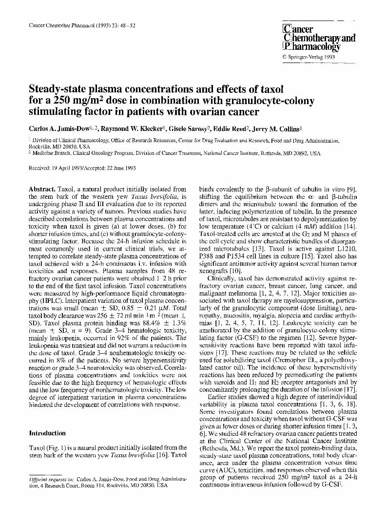
Taxol (Fig. 1) is a natural product initially isolated from the stem bark of the western yew Taxus brevifolia [16]. Taxol
Offprint requests to: Carlos A. Jamis-Dow, Food and Drug Administra- tion, 4 Research Court, Room 314, Rockville, MD 20850, USA
binds covalently to the ~3-subunit of tubulin in vitro [9], shifting the equilibrium between the o~- and ~3-tubulin dimers and the microtubule toward the formation of the latter, inducing polymerization of tubulin. In the presence of taxol, microtubules are resistant to depolymerization by low temperature (4~ or calcium (4 raM) addition [14]. Taxol-treated cells are arrested at the G2 and M phases of the cell cycle and show characteristic bundles of disorgan- ized microtubules [13]. Taxol is active against L1210, P388 and P1534 cell lines in culture [15]. Taxol also has significant antitumor activity against several human tumor xenografts [10].
Clinically, taxol has demonstrated activity against re- fractory ovarian cancer, breast cancer, lung cancer, and malignant melanoma [1, 2, 4, 7, 12]. Major toxicities as- sociated with taxol therapy are myelosuppression, particu- larly of the granulocytic component (dose limiting), neu- ropathy, mucositis, myalgia, alopecia and cardiac arrhyth- mias [1, 2, 4, 5, 7, 11, 12]. Leukocyte toxicity can be ameliorated by the addition of granulocyte-colony stimu- lating factor (G-CSF) to the regimen [12]. Severe hyper- sensitivity reactions have been reported with taxol infu- sions [17]. These reactions may be related to the vehicle used for solubilizing taxol (Cremophor EL, a polyethoxy- lated castor oil). The incidence of these hypersensitivity reactions has been reduced by premedicating the patients with steroids and H1 and H2 receptor antagonists and by concomitantly prolonging the duration of the infusion [17].
Earlier studies showed a high degree of interindividual variability in plasma taxol concentrations [1, 3, 6, 18]. Some investigators found correlations between plasma concentrations and toxicity when taxol without G-CSF was given at lower doses or during shorter infusion times [1, 3, 6]. We studied 48 refractory ovarian cancer patients treated at the Clinical Center of the National Cancer Institute (Bethesda, Md.). We report the taxol protein-binding data, steady-state taxol plasma concentrations, total body clear- ance, area under the plasma concentration versus time curve (AUC), toxicities, and responses observed when this group of patients received 250 mg/m 2 taxol as a 24-h continuous intravenous infusion followed by G-CSF.
49
O
H C"X'~ H3C,
0 ~ " J~" OH 0",,~ --X=/ 'b o=(
cua
TAXOL
NSC-12S973
0
o
O Y O ~ '~ OH
HaC" ~ . y "N" .Y "0','~ ~.
M__/ 'b o=( CH 3
INTERNAL STANDARD
NSC-318735 Fig. 1. Chemical structures of taxol and the internal standard cephalomannine
Patients and methods
Patient eligibility. Only patients with histologic proof of ovarian cancer of epithelial histology, International Federation of Gynecology and Ob- stetrics (FIGO) stage III or IV, treated with at least one prior cisplatin or carboplatin-based regimen, and objective bidimensionally measurable disease were admitted. Eligibility criteria included an age of _> 18 years; an Eastern Cooperative Oncology Group (ECOG) performance status of _<2; no prior chemo- or radiotherapy within 4 weeks of entry to the study; no prior radiation except for intraperitonea132p; recovery from all toxicities of prior treatment; and adequate bone marrow, renal, and hepatic function as documented by laboratory parameters. Not eligible were patients with cerebral metastasis; a prior history of myocardial infarction, congestive heart failure, asymptomatic first-degree atrio- ventricular (AV) block, asymptomatic left anterior hemiblock (LAHB) and right bundle branch block (RBBB), or a cardiac arrhythmia requiring medication; a preexisting peripheral neuropathy of > grade 1; any coex- isting malignancy or a history of a malignancy other than ovarian carci- noma (exceptions: local basal cell carcinoma of the skin or carcinoma in situ of the cervix); or an active, uncontrolled infection. All patients gave written informed consent before entering the study.
Each patient provided a medical history and a physical examination was completed prior to taxol administration. Other tests performed before therapy included a complete blood cell count with differential and platelet count, determinations of serum electrolytes, glucose, blood urea nitrogen (BUN), creatinine, bilirubin, alkaline phosphatase, SGOT,
SGPT, and lactate dehydrogenase (LDH), urinalysis, an EKG; electro- myograph (EMG)/nerve conduction tests; chest Xrays, other radiologic studies to measure disease; and determinations of CA 125 levels. Platelet and complete blood cell counts were performed biweekly until two successive determinations after the nadir showed a total granulocyte count of >1500/gI. If toxicity was acceptable and there was no disease progression, the cycle of therapy was repeated every 3 weeks. Patients were evaluated for response after each cycle if the disease was measur- able by physical examination, every two cycles if radiologic studies were necessary to evaluate disease response, and every four cycles if peri- toneoscopy or laparoscopy was required. Responding patients received two cycles beyond the maximal response. Patients with stable or progres- sive disease were removed from study after two cycles of therapy. Patients who required evaluation by an invasive procedure were removed from study if they had stable or progressive disease after four cycles of therapy.
Taxolformulation and administration. Taxol was supplied by the Divi- sion of Cancer Treatment, National Cancer Institute (Bethesda, Md.) as a concentrated sterile solution of 6 mg/ml in 5-ml ampules (30 rag/ampule) in 50% polyethoxylated castor oil (Cremophor EL; BASF, Ludwigs- hafen, Germany) and 50% Dehydrated Alcohol, USP. Just prior to ad- ministration the taxol infusion was prepared by diluting one-third of the total dose in each of three 1000-ml bottles of 5% Dextrose Injection, USP. In-line filtration of the infusion solution was accomplished by incorporating a hydrophilic filter of pore size 0.22 gM into the i.v. fluid path.
Taxol (250 mg/m 2) was given through a central vein as a 24-h continuous infusion, repeated every 21 days. Patients were premedicated with 20 mg dexamethasone given p.o. or i.v. 14 and 7 h prior to taxol and with 300 mg cimetidine and 25 mg diphenhydramine given i.v. 30 min prior to taxol. Recombinant G-CSF was supplied by the Division of Cancer Treatment, National Cancer Institute (Bethesda, Md.). G-CSF was given s.c. at a dose of 10 btg/kg daily, beginning 24 h after the completion of the taxol infusion and ending after the total granulocyte count had reached >~ 1500/gl on two successive determinations.
Taxol measurement. Taxol plasma concentrations were measured by reverse-phase high-performance liquid chromatography (HPLC) using cephalomannine (Fig. 1) as the internal standard. Taxol and cephaloman- nine reference standards were obtained from the Division of Cancer Treatment, National Cancer Institute, (Bethesda, Md.). Cephalomannine was further purified by HPLC. An aliquot of cephalomannine was added to 0.5 ml standard or sample and mixed. Samples were extracted on a C18 solid-phase extraction column (1 ml Bond Elut; Varian Sample Prepara- tion Products, Harbor City, Calif.). Each column was conditioned with 1 ml acetonitrile followed by 1 ml water. The sample was applied and the column was washed with 3 ml water. The extract was collected in a 2-ml acetonitrile elution. The eluent was dried in a vacuum concentrator (Savant Instruments, Farmingdale, NY). The extract was reconstituted in 150 bt145% acetonitrile in water and 70 btl were injected onto the HPLC column.
Extracts were chromatographed isocratically with 45% acetonitrile in water at a flow of 1 ml/min. Separation was accomplished on an ODS Hypersyl Cl8, 5 btm, 100 • 4.6 mm column (Hewlett-Packard Co., Palo Alto, Calif.) with a C~8 precolumn insert (Millipore Corp., Milford, Mass.). Precolumn inserts were replaced after ever3, 16 plasma sample injections to maintain optimal resolution. Taxol and cephalomannine were quantitated at 230 nm with peak purity and the identity was con- firmed by diode-array detection. After each sample run the column was washed for 2 min with 95% aeetonittile in water and equilibrated for 7 min under the initial conditions. The taxol concentration in plasma samples was quantitated by linear regression analysis of the peak area ratio (taxol/cephalomannine) versus taxol standards prepared in ethy- lenediaminetetraacetic acid (EDTA)-treated normal human plasma. The typical plasma standard curve was in the range of expected plasma concentrations, between 0.125 and 2.5 btM (0.107-2.13 btg/ml).
Pharmacokinetics. For measurement of steady-state plasma concentra- tions, blood samples were collected in EDTA-treated tubes prior to and 1 and 2 h before the end of the first taxol infusion. The samples were
50
20
15 g E c
O
. o 5 8
.cl <
I.S. A
i i i i
2 4 6 8 Time (minutes)
20
" 7
�9 ~ 15 g
O e~ 10
5
<
B I.S.
i t i
0 10 12 0 12
taxo~
i i i i J
2 4 6 8 10 Time (minutes)
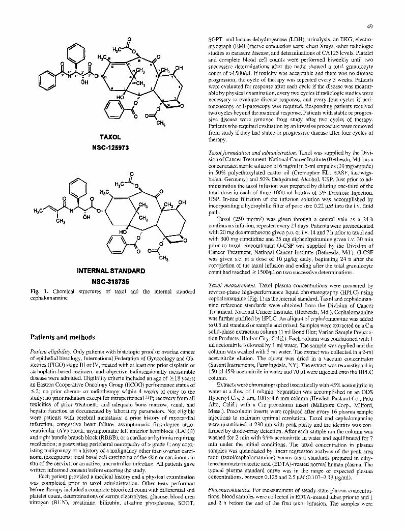
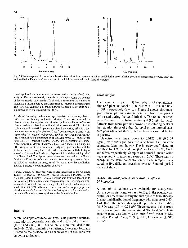
Fig. 2. Chromatograms of plasma sample extracts obtained from a patient A before and B during taxol infusion (t = 22 h). Plasma samples were analyzed as described in Patients and methods, mA. U., milliabsorbance units; LS., internal standard
centrifuged and the plasma was separated and stored at -20~ until analysis. The reported steady-state plasma value represents the average of the two steady-state samples. Total body clearance was calculated by dividing the infusion rate by the average steady-state taxol concentration. The AUC was calculated by multiplying the average steady-state taxol concentration by the infusion time (24 h).
Taxolprotein binding. Preliminary experiments in our laboratory showed extensive taxol binding to filtration devices. Thus, we estimated the plasma protein binding of taxol in vitro by equilibrium dialysis of human plasma against a phosphate-buffered saline solution (PBS; 0.144 M sodium chloride + 0.01 M potassium phosphate buffer, pH 7.46). Pre- treatment plasma samples obtained from 9 ovarian cancer patients were spiked with [3H]-taxol (23 Ci/mmol, 1 mCi/ml; Moravek Biochemicals, Inc., Brea, Calif.) to a concentration of 1 ~tCi/ml (0.043 gM) and dialyzed for 5.5 h at 37~ through a 12,000-14,000 MWCO Spectra/Por 2 mem- brane (Spectrum Medical Industries, Inc., Los Angeles, Calif.) against PBS using a Spectrum Equilibrium Dialyzer (Spectrum Medical In- dustries, Inc., Los Angeles, Calif.). After incubation, a 100-gl aliquot was taken from each cell side and dispensed into a vial containing 10 ml scintillation fluid. The pipette tip was rinsed two times with scintillation fluid to avoid any loss of taxol to the tip, Another aliquot was analyzed by HPLC to confirm the integrity of [3H]-taxol after the equilibrium dialysis. Samples were measured in duplicate.
Clinical effects. All toxicities were graded according to the Common Toxicity Criteria of the Cancer Therapy Evaluation Program of the National Cancer Institute. Disease response was assessed according to
the following definitions: clinical complete response, the complete reso- lution of all evaluable disease, lasting at least 1 month; partial response, a reduction of >_ 50% in the sum of the products of the longest perpendic- ular diameters of all measurable lesions, lasting at least 1 month; and no response, all cases not meeting either of the above definitions.
Resul t s
A total of 49 patients received taxol. One patient's replicate taxol plasma concentrations showed a 6.1-fold difference (10.98 and 1.81 W1//). This patient was not included in the analysis. Of the remaining 48 patients, 5 were not formally treated on the protocol and as such were not evaluable for response to therapy.
Taxol analysis
The mean recovery (+ SD) from plasma of cephaloman- nine (2.5 gM) and taxol (1 gM) was 90% + 7% and 88% + 5%, respectively (n = 11). Figure 2 shows chromato- grams from plasma extracts obtained from one patient before and during the taxol infusion. The retention times were 7.9 rain for cephalomannine and 9.6 rain for taxol. Extracts from blank plasma showed no interfering peaks at the retention times of either the taxol or the internal stan- dard peak (data not shown). No metabolites were detected in plasma.
Detection was linear down to 0.0125 W14 (0.0107 gg/ml), with the signal-to-noise ratio being 3 at this con- centration (data not shown). The interday coefficients of variation for 1.9, 1.2, and 0.49 gMtaxol were 1.6%, 3.4%, and 6.3%, respectively. Samples of normal human plasma were spiked with taxol and stored at -20~ There was no change in the taxol concentration of these samples mea- sured on five different occasions over an 8-month period (data not shown).
Steady-state taxol plasma concentrations after a 24-h infusion
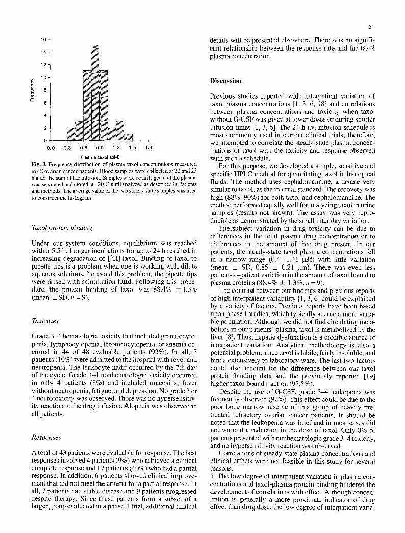
A total of 48 patients were evaluable for steady-state plasma concentrations. As seen in Fig. 3, the plasma con- centrations measured during the first cycle of taxol therapy fit a normal distribution of frequency with a range of 0.40- 1.41 gM. The mean steady-state plasma concentration (+ SD) was 0.85 + 0.21 gM. Three patients in this group had only one concentration measured. The total body clear- ance for taxol was 256 + 72 ml rain-1 m-2 (mean + SD, n = 48). The AUC was 20.3 + 5.1 gM h (mean + SD, n = 48).
5t
16
14
12
10
8
4
0 0.0 0.3 0.6 0.9 1.2 1.5 1.8
Plasma taxol (gM)
Fig. 3. Frequency distribution of plasma taxol concentrations measured in 48 ovarian cancer patients. Blood samples were collected at 22 and 23 h after the start of the infusion. Samples were centrifuged and the plasma was separated and stored at -20~ until analyzed as described in Patients and methods. The average value of the two steady-state samples was used to construct the histogram
Taxol protein binding
Under our system conditions, equilibrium was reached within 5.5 h. Longer incubations for up to 24 h resulted in increasing degradation of [3H]-taxol. Binding of taxol to pipette tips is a problem when one is working with dilute aqueous solutions. To avoid this problem, the pipette tips were rinsed with scintillation fluid. Following this proce- dure, the protein binding of taxol was 88.4% + 1.3% (mean + SD, n = 9).
Toxicities
Grade 3-4 hematologic toxicity that included granulocyto- penia, lymphocytopenia, thrombocytopenia, or anemia oc- curred in 44 of 48 evaluable patients (92%). In all, 5 patients (10%) were admitted to the hospital with fever and neutropenia. The leukocyte nadir occurred by the 7th day of the cycle. Grade 3-4 nonhematologic toxicity occurred in only 4 patients (8%) and included mucositis, fever without neutropenia, fatigue, and depression. No grade 3 or 4 neurotoxicity was observed. There was no hypersensitiv- ity reaction to the drug infusion. Alopecia was observed in all patients.
Responses
A total of 43 patients were evaluable for response. The best responses involved 4 patients (9%) who achieved a clinical complete response and 17 patients (40%) who had a partial response. In addition, 6 patients showed clinical improve- ment that did not meet the criteria for a partial response. In all, 7 patients had stable disease and 9 patients progressed despite therapy. Since these patients form a subset of a larger group evaluated in a phase II trial, additional clinical
details will be presented elsewhere. There was no signifi- cant relationship between the response rate and the taxol plasma concentration.
Discussion
Previous studies reported wide interpatient variation of taxol plasma concentrations [1, 3, 6, 18] and correlations between plasma concentrations and toxicity when taxol without G-CSF was given at lower doses or during shorter infusion times [1, 3, 6]. The 24-h i.v. infusion schedule is most commonly used in current clinical trials; therefore, we attempted to correlate the steady-state plasma concen- trations of taxol with the toxicity and response observed with such a schedule.
For this purpose, we developed a simple, sensitive and specific HPLC method for quantitating taxol in biological fluids. The method uses cephalomannine, a taxane very similar to taxol, as the internal standard. The recovery was high (88%-90%) for both taxol and cephalomannine. The method performed equally well for analyzing taxol in urine samples (results not shown). The assay was very repro- ducible as demonstrated by the small inter day variation.
Intersubject variation in drug toxicity can be due to differences in the total plasma drug concentration or to differences in the amount of free drug present. In our patients, the steady-state taxol plasma concentrations fell in a narrow range (0.4-1.41 ~tM) with little variation (mean +_ SD, 0.85 + 0.21 gm). There was even less patient-to-patient variation in the amount of taxol bound to plasma proteins (88.4% + 1.3%, n = 9).
The contrast between our findings and previous reports of high interpatient variability [1, 3, 6] could be explained by a variety of factors. Previous reports have been based upon phase I studies, which typically accrue a more varia- ble population. Although we did not find circulating meta- bolites in our patients' plasma, taxol is metabolized by the liver [8]. Thus, hepatic dysfunction is a credible source of interpatient variation. Analytical methodology is also a potential problem, since taxol is labile, fairly insoluble, and binds extensively to laboratory ware. The last two factors could also account for the difference between our taxol protein binding data and the previously reported [19] higher taxol-bound fraction (97.5 %).
Despite the use of G-CSF, grade 3-4 leukopenia was frequently observed (92%). This effect could be due to the poor bone marrow reserve of this group of heavily pre- treated refractory ovarian cancer patients. It should be noted that the leukopenia was brief and in most cases did not warrant a reduction in the dose of taxol. Only 8% of patients presented with nonhematologic grade 3-4 toxicity, and no hypersensitivity reaction was observed.
Correlations of steady-state plasma concentrations and clinical effects were not feasible in this study for several reasons: 1. The low degree of interpatient variation in plasma con- centrations and taxol-plasma protein binding hindered the development of correlations with effect. Although concen- tration is generally a more proximate indicator of drug effect than drug dose, the low degree of interpatient varia-

52
tion indicates that dose itself may be an adequate indicator in relatively homogeneous groups of patients with normal liver function. 2. The low frequency of nonhematologic toxicity pre- cluded correlations. 3. Conversely, the high frequency of hematologic effects was a barrier to discovery of relationships. Since the basic motivation for this study design was to push the taxol dose to the point where intervention with G-CSF was required, nearly every patient had grade 3 or 4 hematologic toxicity. 4. The inability to find a relationship between plasma con- centration and antitumor response may indicate that a max- imal response could be obtained at lower doses of taxol, but it is equally possible that the range of plasma concen- trations was simply too narrow for a definitive statement.
Ongoing trials of taxol doses randomized to 135 versus 170 versus 250 mg/m 2 provide a more suitable opportunity to explore the lower end of the curves of taxol concentra- tion versus antitumor response or hematologic toxicity.
References
1. Brown T, Havlin K, Weiss G, Cagnola J, Koeller J, Kuhn J, Rizzo J, Craig J, Phillips J, Von Hoff D (1991) A phase I trial of taxol given by a 6-hour intravenous infusion. J Clin Oncol 9:1261
2. Einzig AI, Hochster H, Wiernik PH, Trump DL, Dutcher JP, Garowski E, Sasloff J, Smith TJ (1991) A phase II study of taxol in patients with malignant melanoma. Invest New Drugs 9:59
3. Greta JL, Tutsch KD, Simon KJ, Alberti DB, Willson JKV, Tormey DC, Swaminathan S, Trump DL (1987) Phase I study of taxol admin- istered as a short i.v. infusion daily for 5 days. Cancer Treat Rep 71: 1179
4. Holmes FA, Walters RS, Theriault RL, Forman AD, Newton LK, Raber MN, Buzdar AU, Frye DK, Hortobagyi GN (1991) Phase II trial of taxol, an active drug in the treatment of metastatic breast cancer. J Natl Cancer Inst 83:1797
5. Lipton RB, Apfel SC, Dutcher JP, Rosenberg R, Kaplan J, Berger A, Einzig AI, Wiernik P, Schaumburg HH (1989) Taxol produces a predominantly sensory nenropathy. Neurology 39:368
6. Longnecker SM, Donehower RC, Cates AE, Chen TL, Brundrett RB, Grochow LB, Ettinger DS, Colvin M (1987) High-performance liq- uid chromatographic assay for taxol in human plasma and urine and pharmacokinetics in a phase I trial [published erratum in Cancer Treat Rep 71: 3401. Cancer Treat Rep 71: 53
7. McGuire WP, Rowinsky EK, Rosenshein NB, Grumbine FC, Ettinger DS, Armstrong DK, Donehower RC (1989) Taxol: a unique antineoplastic agent with significant activity in advanced ovarian epithelial neoplasms. Ann Intern Med 111:273
8. Monsarrat B, Mariel E, Cros S, Garbs M, Gu6nard D, Gu6ritte- Voegelein F, Wright M (1990) Taxol metabolism. Isolation and identification of three major metabolites of taxol in rat bile. Drug Metab Dispos 18:895
9. Rao S, Horwitz SB, Ringel I (1992) Direct photoaffinity labeling of tubulin with taxol. J Nail Cancer Inst 84:785
10. Riondel J, Jacrot M, Picot F, Beriel H, Mouriquand C, Potier P (1986) Therapeutic response to taxol of six human tumors xeno- grafted into nude mice. Cancer Chemother Pharmacol 17:137
11. Rowinsky EK, McGuire WP, Guarnieri T, Fisherman JS, Christian MC, Donehower RC (1991) Cardiac disturbances during the admin- istration of taxol. J Clin Oncol 9:1704
12. Sarosy G, Kohn E, Stone DA, Rothenberg M, Jacob J, Adamo DO, Ognibene FP, Cunnion RE, Reed E (1992) Phase I study of taxol and granulocyte colony-stimulating factor in patients with refractory ovarian cancer. J Clin Oncol 10:1165
13. Schiff PB, Horwitz SB (1980) Taxol stabilizes microtubules in mouse fibroblast cells. Proc Natl Acad Sci USA 77:1561
14. Schiff PB, Horwitz SB (1981) Taxol assembles tubulin in the ab- sence of exogenous guanosine 5'-triphosphate or microtubule-as- sociated proteins. Biochemistry 20:3247
15. Wall ME, Wani MC (1977) Antineoplastic agents from plants. Annu Rev Pharmacol Toxicol 17:117
16. Wani MC, Taylor HL, Wall ME, Coggon P, McPhail AT (1971) Plant antitumor agents. VI. The isolation and structure of taxol, a novel antilenkemic and antitumor agent from Taxus brevifoIia. J Am Chem Soc 93:2325
17. Weiss RB, Donehower RC, Wiernik PH, Ohnuma T, Gralla RJ, Trump DL, Baker JR Jr, Van Echo DA, Von Hoff DD, Leyland- Jones B (1990) Hypersensitivity reactions from taxol. J Clin Oncol 8:1263
18. Wiernik PH, Schwartz EL, Strauman JJ, Dutcher JP, Lipton RB, Paietta E (1987) Phase I clinical and pharmacokinetic study of taxol. Cancer Res 47:2486
19. Wiernik PH, Schwartz EL, Einzig A, Strauman JJ, Lipton RB, Dutcher JP (1987) Phase I trial of taxol given as a 24-hour infusion every 21 days: responses observed in metastatic melanoma. J Clin Oncol 5:1232

![Granulocyte colony-stimulating factor exacerbates hematopoietic … · successfully managed by the use of hematopoietic growth factors (HGFs) [4]. However, even though some irradiated](https://img.dokumen.tips/doc/110x75/5f4fbff2a006440ac9114981/granulocyte-colony-stimulating-factor-exacerbates-hematopoietic-successfully-managed.jpg)