Embed Size (px)
Citation preview
Epilepsia, 47(4):805–808, 2006Blackwell Publishing, Inc.C© 2006 International League Against Epilepsy
Comment Letters
Similarity of Balloon Cells in Focal CorticalDysplasia to Giant Cells in Tuberous Sclerosis
To the Editor:In a very recent article in Epilepsia (2005; 46:1716–
1723), Ying et al. studied balloon cells from focal cor-tical dysplasia (FCD), coming to the conclusion that thecells form a heterogeneous group with differential expres-sion of glial/astrocytic markers [microtubule-associatedprotein 2 (MAP2), glial fibrillary acidic protein (GFAP)]and several other tissue-specific proteins. Such a resultresembles our and other authors’ conclusions from im-munohistochemical and ultrastructural research on giantcells in tuberous sclerosis (TS). We found characteristiccells present in all three types of TS-associated lesions:subependymal giant cell astrocytoma (SEGA), cortical tu-bers, and subependymal nodules (SENs) share a similarprofile of protein expression and are a group of undif-ferentiated cells that, depending on individual determina-tion, can show neural or glial features (Jozwiak et al. Im-munohistochemical and microscopic studies on giant cellsin tuberous sclerosis. Histol Histopathol 2005;20:1321–1326).
Thus we recently evaluated the presence of two TS-associated proteins, hamartin and tuberin, in FCD type I(in which balloon cells are not found) and IIB (with bal-loon cells). Hamartin and tuberin are products of tumor-suppressor genes TSC1 and TSC2, inevitable for properdevelopment of tissues and lethal if both alleles are sub-ject to germ-line mutation. Inactivating mutation of TSC1or TSC2, found in TS, leads to upregulation of mTOR(mammalian target of rapamycin), the kinase being a cen-tral regulator of protein transcription and subsequent un-controlled cell proliferation. As a result, the clinical man-ifestation of TS is associated with tumor development inthe brain, heart, kidneys, lungs, liver, and/or skin.
Based on similarities between balloon and giant cells,our hypothesis was that FCD may be a form of focal TS,limited to selected regions of the brain tissue. Accordingto our expectations, in an immunohistochemical study, wefound loss of tuberin and hamartin expression, as well asstrong immunoreactivity for mTOR in FCD type IIB. Spe-cific kinases implicated in TS progression (pS6K1, pAkt,and pErk) also were hyperactivated in the same samples.Cortical balloon cells in these samples were NSE positive,whereas most subcortical giant cells were labeled withGFAP. In FCD type I and normal brain tissue, tuberin andhamartin were detected, but immunoreactivity for mTOR,the active form of S6K1, Akt, and Erk was subtle. Nestinexpression in all samples was weak or absent.
These findings suggest activation of the same pathwaysunderlying both FCD IIB and TS and argue for recogni-tion of the similarities in both pathologies and for consid-ering FCD IIB as a focal form of TS. Such a conclusioncould also potentially have a practical outcome: currentresearch with the application of rapamycin as an effectivedrug limiting activity of mTOR kinase are under way and,in animal models of TS, show effectiveness of this therapy.Nevertheless, further elucidation of accompanying muti-ations that probably predispose patients to FCD must beperformed.
∗Jaroslaw Jozwiak†Katarzyna Kotulska
†Sergiusz Jozwiak∗Department of Histology and Embryology
Center for Biostructure Research,Medical University of Warsaw
†Department of Pediatric NeurologyThe Children’s Memorial
Health Institute Warsaw, Poland
Status Epilepticus in Idiopathic Generalized Epilepsy
To the Editor:Epilepsia recently published a fascinating supplement
devoted to many aspects (epidemiologic, genetic, clinical,electroencephalographic, etc.) of idiopathic generalizedepilepsy (IGE). Certain important questions can be raisedpertaining to the terminology and concepts proposed byShorvon and Walker (1) in that supplement in their articleon status epilepticus.
First, they pointed out that de novo absence statusepilepticus (ASE) of late onset is a form of status epilep-ticus in IGE (see Table 1). Although several publicationshave discussed this entity (2–4), Thomas et al. (5) maybe considered one of the first research groups that delin-eated in detail the clinical and electroencephalographicfeatures of de novo ASE. Thomas and Andermann (6)reported 14 additional cases. Eight of 25 subjects had pre-existing epilepsy, and only four of these eight patients hadIGE. Recurrences of status was rare is settings for whichpsychotropic treatment was appropriately supervised andcofactors such as alcohol abuse or hyponatremia wereavoided. Therefore de novo ASE of late onset has beenconsidered most often a situation-related form of general-ized nonconvulsive status epilepticus, and in the majorityof cases, no further epileptic events will occur in patient’s
805

806 LETTERS
life. Therefore it seems questionable to include this par-ticular entity in the types of status epilepticus in IGE (asproposed in Table 1).
In Shorvon and Walker’s view, the term “typical ASE”should be restricted to ASE that occurs in patients withIGE, therefore, ASE in the syndrome of phantom absenceswith generalized tonic–clonic seizures should be consid-ered typical ASE, because this condition is part of IGE(7,8). The term “de novo ASE of late onset,” used in Ta-ble 1, may be misleading.
Second, in the section about nosology and diagnosis,these authors mentioned the term “complex partial ab-sence status” (see Table 2) for designing a category of ASEin partial epilepsy. They did not clarify completely its ictalelectroencephalographic correlate. Included were patientswith partial epilepsy and episodes of altered mental statewith ictal bilateral or generalized epileptiform dischargeson the EEG. From a clinical-EEG standpoint, this termi-nology may be confusing. Bilateral or generalized epilep-tiform activity during the course of an episode of noncon-vulsive status epilepticus in a subject with probable partialepilepsy does not inevitably imply ASE. The term “com-plex partial ASE” achieves the contrary effect, becauseany localization-related nonconvulsive status epilepticuscould be designed as ASE if the EEG does not show afocal ictal onset
Third, the authors considered generalized electro-graphic ASE a form of status epilepticus in IGE. In thearticle by Panayiotopoulos et al. (7), all six patients withabsence status were mildly to moderately confused, andtherefore, a variable degree of mental alteration was ob-served. Furthermore, the term “generalized electrographicstatus” is frequently used to describe a type of nonconvul-sive status epilepticus most often (but not always) asso-ciated with severe neurologic or medical illness and pro-found alteration of consciousness level (9,10).
In conclusion, some confusing and debatable catego-rizations are established in the article by Shorvon andWalker (1) about status epilepticus in IGE.
Jose L. Fernandez-TorreDepartment of Clinical Neurophysiology
University Hospital “Marques de Valdecilla”Santander, Cantabria, Spain
E-mail: [email protected]; [email protected]
REFERENCES
1. Shorvon S, Walker M. Status epilepticus in idiopathic generalizedepilepsy. Epilepsia 2005;46(suppl 9):73–79.
2. Thomas P, Lebrun C, Chatel M. De novo absence status epilepticusas a benzodiazepine withdrawal syndrome. Epilepsia 1993;34:355–358.
3. Kanemoto K, Miyamoto T, Abe R. Ictal catatonia as a manifesta-tion of de novo absence status epilepticus following benzodiazepinewithdrawal. Seizure 1999;8:364–366.
4. Fernandez-Torre JL. De novo absence status epilepticus of lateonset following withdrawal of lorazepam: a case report. Seizure2001;10:433–437.
5. Thomas P, Beaumanoir A, Genton P, et al. “De novo” absence statusof late onset: report of 11 cases. Neurology 1992;42:104–110.
6. Thomas P, Andermann F. Late-onset absence status epilepticus ismost often situation-related. In: Malafosse A, Genton P, Hirsch E,eds. Idiopathic Generalized Epilepsies. London: John Libbey &Company, 1994:95–109
7. Panayiotopoulos CP, Koutroumanidis M, Giannakodimos S, et al.Idiopathic generalised epilepsy in adults manifested by phantomabsences, generalised tonic-clonic seizures, and frequent absencestatus. J Neurol Neurosurg Psychiatry 1997;63:622–627.
8. Agathonikou A, Panayiotopoulos CP, Giannakodimos S, et al. Typ-ical absence status in adults: diagnostic and syndromic considera-tions. Epilepsia 1998;39:1265–1276.
9. Drislane FW, Schomer DL. Clinical implications of generalizedelectrographic status epilepticus. Epilepsy Res 1994;19:111–121.
10. Drislane FW. Presentation, evaluation, and treatment of nonconvul-sive status epilepticus. Epilepsy Behav 2000;1:301–314.
“Migralepsy” and the Significance of DifferentiatingOccipital Seizures from Migraine
To the Editor:I wish to take advantage of the report by Drs. Milli-
gan and Bromfield (1) on migralepsy to emphasize thefollowing:
1. Occipital seizures are frequently misdiagnosed asmigraine, although they are distinctly different.
2. Important clinical criteria are used in differentiatingbetween elementary visual hallucinations of occip-ital seizures and migraine.
3. “Occipital epilepsy–migraine” is far more commonthan the traditional view of a “migraine–epilepsy”(migralepsy) sequence.
The authors present a woman with visual symptoms ofleft-sided, brief “2- to 3-min” episodes of “fluorescent-colored flashing lights, black spots, and squiggly lines”alone or followed by bifrontal headaches and nausea.These “scintillations” and headaches, diagnosed as mi-graine, were sometimes followed by focal seizures andgeneralized convulsions; hence the diagnosis of “mi-gralepsy” is made. The discussion on “migralepsy” isbased on traditional views (1). Evidence that “migralepsy”is often an erroneous diagnosis (2–4) are not considered,as indeed happens in most related publications.
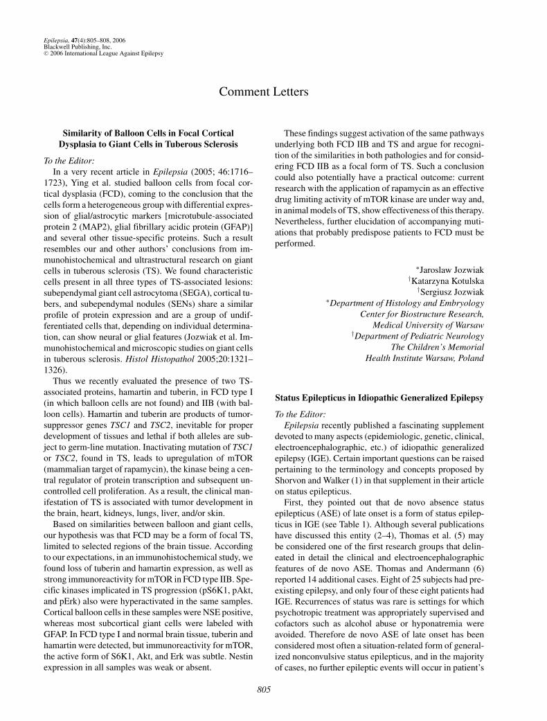
1. Misdiagnosis of visual seizures as migraine with vi-sual aura is frequent and costly. The major contrib-utory factor to error is that the description of visualhallucinations is often abbreviated in terms suchas scintillations, fortification spectrum, teichopsia,phosphenes, and their variations. Elementary vi-sual hallucinations of occipital seizures are funda-mentally different from the visual aura of migraine(Fig. 1).
2. In occipital seizures, elementary visual halluci-nations are usually the first and often the only
Epilepsia, Vol. 47, No. 4, 2006

LETTERS 807
FIG. 1. Schematic presentation of typical elementary visual hallucinations in occipital seizures (left) and migraine visual aura (right) at theirpeak. In their clinical differentiation, considerations should be made in regard to their different onsets, speed and location of development,duration, frequency, and associated and following symptoms (see also other illustrations of elementary visual hallucinations in occipitalseizures in references 2–4. Tables of differential diagnosis are detailed in references 3 and 4).
ictal symptom and may progress to other oc-cipital and extraoccipital manifestations and con-vulsions (2–4). Ictal elementary visual hallucina-tions are defined by color, shape, size, location,movement, speed of appearance and duration, fre-quency, and associated symptoms of progression.Elementary visual hallucinations are mainlycolored and circular, develop rapidly within sec-onds, and are brief in duration (2–3 min). Theyoften appear in the periphery of a temporalvisual hemifield, becoming larger and multi-plying in the course of the seizure, and fre-quently moving horizontally toward the other side.Significantly, postictal headache often indistin-guishable from migraine, occurs in more thanhalf of patients, even after brief visual seizures(2,3). Postictal headache frequently occurs 3–15min after the seizure ends, a situation known
in migraine as the “asymptomatic interval” (endof migraine aura to the onset of headache).Thus occipital seizures often generate migraine-like attacks, “epilepsy–migraine sequence” (2–4).In migraine, the visual aura usually starts as a flick-ering, uncolored, zigzag line in the center of the vi-sual field and affects the central vision. It graduallyprogresses over >4 min, usually lasting <30 min,toward the periphery of one hemifield, often leav-ing a scotoma (5). The total duration of visual aurasis 60 min. Acute onset of visual aura is very rare (5).Furthermore, migraine visual aura rarely has dailyfrequency, nonvisual ictal occipital symptoms, suchas eye and head deviation and repetitive eye-lid closures, do not occur; it is probably excep-tional to progress to nonvisual epileptic seizures.Less typical features of migraine visual aura, suchas spots, circles, and beads, with or without colors,
Epilepsia, Vol. 47, No. 4, 2006

808 LETTERS
may be experienced during the migraine visualaura, but usually they are not dominant. Clusteringof other symptoms, as above, betray their migrainenature.
3. No reason exists that epileptic seizures, whichare so vulnerable to precipitating factors, couldnot be susceptible to cortical changes induced bymigraine. However, by probability of prevalence,these cases should have symptoms of a typical vi-sual aura (>90% in comparison with atypical cases)followed by epileptic seizures. Contrary to this arethe findings in my review of reported cases of “mi-gralepsy,” in which the majority of them had symp-toms of visual seizures as defined earlier, whichwere interpreted as migraine aura (3). Thus it isprobable that these were genuine occipital seizuresimitating migraine (epilepsy–migraine sequence).Some cases had symptoms of atypical presenta-tions or bizarre symptoms that could be diagnosedas either migraine or epilepsy.
More important, in clinical practice, occipital seizuresof visual symptoms associated with headache are oftenerroneously identified as migraine with significant diag-nostic and therapeutic adverse effects (3,4). The differ-entiation of migraine from epilepsy should not be diffi-cult if clinical data are properly examined and synthesized(2–4).
C. P. PanayiotopoulosDepartment of Clinical
Neurophysiology and EpilepsiesSt. Thomas’ Hospital, London, United Kingdom
E-mail: [email protected]
REFERENCES
1. Milligan TA, Bromfield E. A case of “migralepsy.” Epilepsia2005;46(suppl 10):2–6.
2. Panayiotopoulos CP. Elementary visual hallucinations, blind-ness, and headache in idiopathic occipital epilepsy: differentia-tion from migraine. J Neurol Neurosurg Psychiatry 1999;66:536–540.
3. Panayiotopoulos CP. Visual phenomena and headache in occipitalepilepsy: a review, a systematic study and differentiation from mi-graine. Epileptic Disord 1999;1:205–216.
4. Panayiotopoulos CP. Occipital lobe epilepsies. In: PanayiotopoulosCP, ed. The Epilepsies: Seizures, Syndromes and Management. Ox-ford: Bladon Medical Publishing, 2005:416–429.
5. Russell MB, Olesen J. A nosographic analysis of the migraine aurain a general population. Brain 1996;119:355–361.
Response: “Migralepsy” and the Significance ofDifferentiating Occipital Seizures from Migraine
To the Editor:We thank Dr. Panayiotopoulos for his interest in our
case and for emphasizing features that distinguish occipi-tal lobe epilepsy from migraine with visual aura. We agree
that it is important to avoid misdiagnosis and that occipitalseizures should be considered in the differential diagnosisof a visual aura followed by headache. Our case empha-sizes the question of whether migraines lead to seizures,or vice versa, and was selected to highlight the geneticcomplexity of epilepsy as well as clinical aspects of the“borderlands of epilepsy.”
Our patient does have focal epilepsy, most likely ofright occipital origin, but she also has a strong familyhistory of migraine with visual aura and no family his-tory of seizures. Her visual symptoms and headaches pre-ceded the diagnosis of epilepsy by 3 years. During thisperiod, was she having simple partial seizures, as Dr.Panayiotopoulos suggests, or were the episodes migrainewith aura that evolved into seizures?
The visual symptoms of migraine aura and occipitalseizures may often be distinguished, as outlined by Dr.Panayiotopoulos, but an overlap is found between the twoconditions. Some patients with migraine do have visualauras that are colorful and may even be rapid in onset (1).The acute onset and relatively short duration of our pa-tient’s auras at the time of evaluation were consistent withoccipital seizures, but it was difficult to obtain a more accu-rate, retrospective description of her initial symptoms. Asa child, she had several EEGs that were all normal. Whenconvulsions later developed, these were always precededby both a visual aura and then a headache, never by visualaura alone. That her visual symptoms and headaches canbe provoked by missing meals is more typical of migrainethan of seizures, although her other provoking factors—the premenstrual phase, lack of sleep, and stress—are alsofairly common seizure triggers. Interestingly, since wepresented this case, she has also reported a typical spellwith an aura of fluorescent colors that stopped with drink-ing coffee.
Regardless of the particular diagnoses of our patient, itis likely that in some patients with migraine and epilepsy,the two processes share common mechanisms, as well ascommon genetic risk factors that may be elucidated byevolving methods of investigating complex genetic dis-eases (2).
Tracey MilliganEdward B. Bromfield
Brigham and Women’s HospitalHarvard Medical School
Boston, Massachusetts, U.S.A.E-mail: [email protected]
REFERENCES
1. Russell MB, Olesen J. A nosographic analysis of the migraine aurain a general population. Brain 1996;119:355–361.
2. Ottman R. Analysis of genetically complex epilepsies. Epilepsia2005;46(suppl 10):7–14.
Epilepsia, Vol. 47, No. 4, 2006