Embed Size (px)
Citation preview

Cell Metabolism
Article
Stat3 Activation Links a C/EBPdto Myostatin Pathwayto Stimulate Loss of Muscle MassLiping Zhang,1,5,* Jenny Pan,1,5 Yanjun Dong,1,4 David J. Tweardy,2 Yanlan Dong,1 Giacomo Garibotto,3
and William E. Mitch11Nephrology Division, Department of Medicine2Section of Infectious Diseases, Department of MedicineBaylor College of Medicine, Houston, TX 77030, USA3Nephrology Division, Department of Internal Medicine, Genoa University, Genoa 16132, Italy4Beijing Institute of Heart, Lung, and Blood Vessel Diseases, An Zhen Hospital Affiliated to Capital Medical University, Beijing 100029, China5These authors contributed equally to this work*Correspondence: [email protected]
http://dx.doi.org/10.1016/j.cmet.2013.07.012
SUMMARY
Catabolic conditions like chronic kidney disease(CKD) cause loss of muscle mass by unclear mecha-nisms. In muscle biopsies from CKD patients, wefound activated Stat3 (p-Stat3) and hypothesizedthat p-Stat3 initiates muscle wasting. We createdmice with muscle-specific knockout (KO) that pre-vents activation of Stat3. In these mice, losses ofbody and muscle weights were suppressed inmodels with CKD or acute diabetes. A small-mole-cule that inhibits Stat3 activation produced similarresponses, suggesting a potential for translationstrategies. Using CCAAT/enhancer-binding proteind (C/EBPd) KO mice and C2C12 myotubes withknockdown of C/EBPd or myostatin, we determinedthat p-Stat3 initiates muscle wasting via C/EBPd,stimulating myostatin, a negative muscle growthregulator. C/EBPd KO also improved survival ofCKD mice. We verified that p-Stat3, C/EBPd, andmyostatin were increased in muscles of CKDpatients. The pathway from p-Stat3 to C/EBPd tomyostatin and muscle wasting could identify thera-peutic targets that prevent muscle wasting.
INTRODUCTION
Muscle wasting is a debilitating complication of catabolic condi-
tions, including chronic kidney disease (CKD), diabetes, cancer,
or serious infections. Unfortunately, there are few reliable strate-
gies that block the loss of muscle protein initiated by these con-
ditions. Previously, we found that myostatin, a negative regulator
of muscle growth, is increased in muscles of mice with CKD, and
when we inhibited myostatin with a humanized myostatin pepti-
body, CKD-induced muscle wasting was blocked (Zhang et al.,
2011). A similar conclusion was reached in studies of mouse
models of cancer cachexia (Zhou et al., 2010). In exploring why
blocking myostatin is beneficial for muscle metabolism, we
368 Cell Metabolism 18, 368–379, September 3, 2013 ª2013 Elsevie
found that its inhibition reduced circulating levels of interleukin-
6 (IL-6) and tumor necrosis factor a (TNF-a), suggesting that
there is a link between inflammation and muscle wasting as
reported in clinical studies (Carrero et al., 2008; Hung et al.,
2011). The evidence that inflammation stimulates muscle
wasting includes reports that infusion of TNF-a, IL-6, IL-1b, or
interferon (IFN)-g into rodents results in muscle wasting, while
neutralization of cytokines using genetic or pharmacological
approaches attenuates muscle wasting (Cheung et al., 2010).
For example, we treated rodents with a constant infusion of
angiotensin II (AngII), which caused muscle wasting plus
increased circulating levels of IL-6 and increased expression of
suppressor of cytokine signaling 3 (SOCS3), leading to sup-
pressed insulin/insulin-like growth factor 1 (IGF-1) signaling.
Knockout IL-6 from mice suppressed AngII-induced muscle
wasting (Zhang et al., 2009; Rui et al., 2001; Rui et al., 2002).
Responses to IL-6 or IFN-g involve stimulation of intracellular
signaling pathways, including activation of Janus protein tyro-
sine kinases (JAKs). Subsequently, JAKsmediate tyrosine phos-
phorylation of signal transducer and activator of transcription
(STAT) factors, followed by their dimerization, nuclear transloca-
tion, and activation of target genes (Horvath, 2004). Among the
seven members of the STAT family, Stat3 is the major member
that is activated by the IL-6 family of cytokines (Hirano et al.,
1997; Kishimoto et al., 1994). Recently, Bonetto et al. (2011)
reported the results of a microarray analysis of muscles from
mice with cancer-induced cachexia. Components of 20
signaling pathways were upregulated, including IL-6, Stat3,
JAK-STAT, and SOCS3 complement and coagulation pathways
(Bonetto et al., 2011). Although this suggests that the Stat3
pathway could be linked to loss of muscle mass, a pathway
from Stat3 to muscle wasting has not been reported.
A potential target of activated Stat3 is CCAAT/enhancer-bind-
ing protein d (C/EBPd). The C/EBP transcription factors (C/EBPa,
-b, -g, -d, -u, and -z) are expressed in several tissues and act to
regulate inflammatory andmetabolic processes (Ramji andFoka,
2002). C/EBPb or -d can stimulate intracellular signaling in hepa-
tocytes or inflammatory cells (Poli, 1998; Akira et al., 1990; Alonzi
et al., 1997) and in muscles of mice responding to an excess
of glucocorticoids, the expression and binding activity of C/
EBPb and -d are increased (Penner et al., 2002; Yang et al., 2005).
r Inc.

Figure 1. Inflammatory Cytokines and
p-Stat3 Are Elevated in Muscles of Patients
with CKD
(A) Immunostaining of muscle sections for IL-6 and
TNF-a (brown color) from biopsies of age- and
gender-matched, healthy control subjects (left
panel) and CKD patients (middle panel). Staining
quantification is calculated as the percentage of
muscle fibers that are immunostained (right panel;
n = 3 control subjects; n = 4 CKD patients; ruler =
50 mm).
(B) Representative western blots for p-Stat3 in
control subjects and CKD patients (upper panel)
and the ratio of the intensity of p-Stat3 to total
Stat3 (lower panel) (n = 6 control subjects; n = 6
CKD patients).
(C) Muscle sections from control subjects and
CKD patients were immunostained for p-Stat3
(upper panel). Brown nuclei are p-Stat3 positive
(arrows). Percentage of p-Stat3-positive nuclei in a
total of 550 nuclei (lower panel; n = 4 control
subjects; n = 6 CKD patients). Values are means ±
SEM. *p < 0.05 versus control subjects. See also
Figure S1 and Table S1.
Cell Metabolism
p-Stat3 to C/EBPd to Myostatin and Muscle Wasting
A potential mechanism that includes C/EBPd involves
increased myostatin expression because the myostatin
promoter contains recognition sites for members of the C/EBP
family of transcription factors (Ma et al., 2001; Allen et al.,
2010). In the present report, we have uncovered an intracellular
signaling pathway in cultured myotubes that could bridge the
gaps between p-Stat3 and myostatin and loss of muscle mass.
To examine if the pathway was operative in vivo, we studied
how two catabolic conditions, CKD or streptozotocin-induced
acute diabetes, affected muscle metabolism in a muscle-spe-
cific Stat3 knockout (KO) mouse. We also tested whether a
small-molecule inhibitor of Stat3 activation would correctmuscle
wasting. Interruption of Stat3 improved muscle metabolism and
strength in mice with CKD. We also found evidence for this cata-
bolic pathway in muscle biopsies from patients with CKD.
RESULTS
Muscle Biopsies of Patients with CKD RevealInflammation and Stat3 ActivationTo address the mechanisms underlying muscle wasting, we
studied 18 CKD patients scheduled for peritoneal dialysis cath-
eter insertion and a control group of 16 age- and gender-
matched healthy subjects. All subjects led a sedentary lifestyle.
In the 18 CKD patients, the blood urea nitrogen (BUN) and
serum creatinine were increased 4- and �8-fold, respectively,
over control subjects (Table S1 available online). All CKD pa-
tients experienced unintentional weight loss in the 3 months
before muscle biopsies were obtained. In CKD patients, the
mean estimated protein and calorie intakes were 0.9 g/kg and
Cell Metabolism 18, 368–379, S
28 kcal/kg, respectively, compared to
�1 g/kg and 30–32 kcal/kg, respectively,
in control healthy subjects (from diet di-
aries). Even though these intakes of pro-
tein and calorie exceed the recommen-
ded daily allowance (RDA), 13 of the 18 patients were
malnourished, signified by a subjective global assessment level
of >2, and serum albumin was low in 11 patients (<3.8 g/100 ml)
(Fouque et al., 2008). Even though the body mass index was low
(<23 kg/m2) in only 4 subjects, all patients had evidence of pro-
tein losses; there was a marked reduction in muscle fiber cross-
sectional area (CSA) (CKD patients median = 1,003 mm2, range
717–1,601; control median = 1,873 mm2, range 1,100–3,389; p <
0.003 Mann-Whitney). We also calculated fat-free mass (FFM)
from skinfold thickness (Avesani et al., 2004). Over 3 months,
the FFM in CKD patients declined from 45.9 ± 2 kg to 44.1 ±
2 kg (p < 0.05). Regarding drugs that might influence muscle
metabolism, no patient was receiving steroids, but 14 patients
were treated with statins; these patients did not have signs of
myopathy. Characteristics of the CKD patients and control sub-
jects are shown in Table S1. Patients were treated with diuretic
(furosemide; all patients) at different dosages, lisinopril or dox-
azosin (17 patients), proton-pump inhibitors (14 patients),
platelet aggregation inhibitors (14 patients), insulin therapy (4
patients), oral anticoagulant therapy (1 patient), and erythropoi-
etin (10 patients). Diabetes was well controlled, with hemoglobin
A1c values < 6.5% and fasting plasma glucose levels < 110 mg/
dl. There were increased levels of inflammatory markers in CKD
patients, including circulating C-reactive protein (CRP; control
[3.21 ± 0.22 mg/dl] versus CKD [10.46 ± 2.98 mg/dl]; p <
0.05) and fibrinogen (control [291 ± 31.7 mg/dl] versus CKD
[579 ± 37.5 mg/dl]; p < 0.005) (Table S1). There were also
increased levels of IL-6 and TNF-a in muscle biopsies
compared to results from control subjects (Figure 1A). Finally,
TNF-a messenger RNA (mRNA) was increased (Figure S1)
eptember 3, 2013 ª2013 Elsevier Inc. 369

Figure 2. Muscle-Specific Stat3 Knockout
in Mice Suppresses CKD or Streptozoto-
cin-Induced Muscle Wasting
(A) Density of p-Stat3 corrected for total Stat3 in
lysates of gastrocnemius muscles (upper panel;
n = 5 mice/group; *p < 0.05 versus sham control
mice). Also shown are representative western
blots of p-Stat3 (lower panel).
(B) Changes in body weights of Stat3 KO and
Stat3flox/flox control mice over 5 weeks following
creation of CKD (n = 10 pairs of mice; *p < 0.05
versus Stat3flox/flox).
(C and D) Average weights of mixed fiber tibialis
anterior (TA) (C) and gastrocnemius (D) muscles
(n = 10 mice/group).
(E and F) Extensor digitorum longus (EDL) muscles
from sham or CKD mice and either Stat3flox/flox or
Stat3 KO were isolated. Rates of protein synthesis
(E) and protein degradation (F) weremeasured (n =
20 EDL muscles from 10 mice/group).
(G) Muscle force of each mouse used in Figure 2B
was measured on four consecutive days. The
averagemuscle force (in newtons) is shown (n = 10
mice/group).
(H) Representative western blots of p-Stat3 in
lysates of gastrocnemius muscles of acutely dia-
betic (STZ) and control mice. Bar graph shows the
densities of p-Stat3 corrected for that of Stat3 (n =
10 mice/group; *p < 0.05 versus CTRL mice).
(I and J) Average weights of the mixed fiber tibialis
anterior (TA) (I) and gastrocnemius (J) muscles
from both legs (n = 10 mice/group; *p < 0.05
versus control Stat3flox/flox). Values are means ±
SEM. See also Figure S2.
Cell Metabolism
p-Stat3 to C/EBPd to Myostatin and Muscle Wasting
and, as noted previously, so was IL-6 mRNA (Verzola et al.,
2011).
Activated Stat3 protein was significantly increased in muscles
of CKD patients versus healthy subjects (Figure 1B); p-Stat3 was
principally located in nuclei of biopsies, as �40% of nuclei in
muscle biopsies of CKDpatients were positive for p-Stat3 versus
�20% in healthy subjects (Figure 1C). Thus, significant increases
in the expressions of inflammatory cytokines, IL-6 and TNF-a,
were associatedwith Stat3 activation inmuscles of CKDpatients
who expressed evidence of muscle wasting.
Muscle-Specific Stat3 KO Suppresses Loss of Muscle,Despite CKD or Type 1 DiabetesIn gastrocnemius muscles of mice with CKD, the level of p-Stat3
was increased compared to results inmuscles of pair-fed, sham-
370 Cell Metabolism 18, 368–379, September 3, 2013 ª2013 Elsevier Inc.
operated, control mice (Figure 2A). To
explore if the activation of Stat3 triggers
muscle wasting in vivo, we studied mice
with muscle-specific deletion of the
Stat3 tyrosine phosphorylation site
(Stat3 KO) compared to results in control
Stat3flox/flox mice (Takeda et al., 1998).
Mice with muscle-specific Stat3 KO did
not differ from control mice in terms of
development, food intake (data not
shown), and body weight (Figure S2A).
However, with CKD, body weights of Stat3 KO mice increased
versus results in pair-fed Stat3flox/flox mice with CKD (Figure 2B).
The gain in weight was due in part to increased muscle mass;
after 5 weeks of CKD, the weights of gastrocnemius and tibialis
anterior muscles were significantly greater than muscles from
Stat3flox/flox mice (Figures 2C and 2D). To determine why loss
of muscle mass was blunted in Stat3 KO mice with CKD, we
measured rates of muscle protein synthesis and degradation
and found a significant improvement in both indices of protein
metabolism in Stat3 KOmicewith CKD (Figures 2E and 2F). Like-
wise, there was an increase in grip strength of Stat3 KO mice
versus Stat3flox/flox mice (Figure 2G).
Muscle atrophy in several catabolic conditions is characterized
as an increase in circulating inflammatory cytokines, impaired
insulin/IGF-1 signaling, and an increase in muscle protein

Figure 3. A Small-Molecule Inhibitor of
Stat3 Activation, C188-9, Blocks CKD-
Induced Muscle Wasting
(A) Sham or CKDmicewere treated with C188-9 or
D5W (diluent) for 14 days. Representative western
blots of p-Stat3, Stat3, andGAPDH from lysates of
gastrocnemius muscles are shown (n = 8 mice/
group).
(B) Differences in body weights of pair-fed, sham,
or CKD mice treated with C188-9 or D5W at
baseline and after 7 or 14 days of treatment (*p <
0.05 versus D5W sham).
(C and D) Average weights of mixed fiber
gastrocnemius (C) and tibialis anterior (TA) (D)
muscles from both legs (n = 7 mice/group).
(E) Cryosections of TA muscles were immuno-
stained with anti-laminin to identify the muscle
basement membrane. The myofiber areas were
measured, and the myofiber size distribution was
calculated from the areas of �500 myofibers
assessed by an observer blinded to treatment
group (n = 4 pairs of mice).
(F) Muscle force of each mouse studied in Fig-
ure 3C was measured on four consecutive days
(Experimental Procedures; n = 7 mice/group).
(G and H) At 2 weeks of C188-9 or D5W treatment,
protein synthesis (G) and degradation (H) were
measured (n = 8 pairs of mice; *p < 0.05 versus
D5W). Values are means ± SEM.
Cell Metabolism
p-Stat3 to C/EBPd to Myostatin and Muscle Wasting
degradation via the ubiquitin-proteasome system (UPS) (Zhang
et al., 2011; Lecker et al., 2004). To determine if results present
in mice with CKD occur in another model of muscle wasting, we
studied streptozotocin-treated, acutely diabetic mice (Price
et al., 1996). Therewasan increase inp-Stat3plushighcirculating
andmuscle levels of IL-6 in acutely diabeticmice (Figures 2H and
S2B). IL-6mRNA inmuscles of streptozotocin (STZ)-treatedmice
was increased 2-fold over control mice (data not shown). Stat3
KO mice expressed a slower decrease in body weight versus
results in acutely diabetic Stat3flox/flox mice (Figure S2C). The
slower loss of body weight in acutely diabetic Stat3 KO mice
was associatedwith a greatermass of gastrocnemius and tibialis
anterior muscles versus results in Stat3flox/flox mice (Figures 2I
and 2J). In the absence of CKD- or diabetes-induced catabolism,
muscle-specificStat3KOdid not significantly affect bodyweight,
muscle mass, protein metabolism, or grip strength compared to
results in Stat3flox/flox mice (Figure 2). Thus, p-Stat3 can trigger
muscle wasting in certain catabolic conditions.
Inhibition of Stat3 Activation Blocks CKD-InducedMuscle WastingTo determine if a translational strategy might be developed to
interfere with muscle wasting when Stat3 is activated, we evalu-
ated C188-9, a small-molecule inhibitor of Stat3 that targets the
phosphotyrosyl (pY) peptide binding pocket within the Src
homology (SH) 2 domain of Stat3, thereby blocking Stat3 recruit-
ment to activated receptors, its phosphorylation on tyrosine, and
Cell Metabolism 18, 368–379, S
tail-to-tail dimerization (Xu et al., 2009;
Redell et al., 2011). After 2 weeks of
CKD, mice were paired for their BUN
and body weights and injected with either
C188-9 or the diluent, 5%dextrose in water (D5W). C188-9 treat-
ment decreased the level of p-Stat3 in muscle without affecting
levels of total Stat3 (Figure 3A). Consistent with results from
Stat3 KO mice with CKD, the body weights of CKD mice treated
with the Stat3 inhibitor were significantly greater than weights of
the control, CKD mice (Figure 3B). After 14 days of C188-9, we
found that the increase in body weight included more muscle,
as the weights of gastrocnemius and tibialis anterior muscles
were greater (Figures 3C and 3D). The increase in muscle
mass was confirmed by an analysis of the size distribution of
myofibers in muscles of CKD mice treated with C188-9 (Fig-
ure 3E). This improvement in muscle mass was accompanied
by improved grip strength in CKD mice treated with C188-9
(Figure 3F). Consistent with results from the Stat3 KO mice,
blocking Stat3 with C188-9 in control, wild-type mice did not
significantly affect their food intake (data not shown), body
weight, muscle mass, or grip strength (Figures 3B–3D and 3F).
The mechanism underlying the C188-9-induced increase in
muscle weight included improved muscle protein synthesis
and decreased protein degradation (Figures 3G and 3H). We
conclude that inhibiting Stat3 activation suppresses CKD-
induced loss of both muscle mass and strength.
In C2C12 Myotubes, Stat3 Activation Increases theExpression of C/EBPd and MyostatinWe evaluated the signaling pathway from activated Stat3 to
muscle wasting. We studied myostatin because its expression
eptember 3, 2013 ª2013 Elsevier Inc. 371

Figure 4. Stat3 Activation in C2C12 Myo-
tubes Increases the Expression of C/EBPd
and Myostatin
(A) Representative western blots from C2C12
myotubes treated with IL-6 (100 ng/ml) for
different times (left panel). Fold changes in the
densities of proteins corrected for GAPDH at
different times was calculated from values at time
zero (right panel), n = 3 repeats; *p < 0.05 versus
time zero.
(B) C2C12 myotubes were infected with a lenti-
virus expressing constitutively active Stat3
(Stat3C-GFP). A representative western blot for
the indicated proteins is shown.
(C) C2C12 myotubes were treated with or without
C188-9 for 2 hr before adding IL-6 (100 ng/ml) for
24 hr. A representative western blot for the indi-
cated proteins is shown.
(D) C2C12 myoblasts were cotransfected with
a plasmid expressing the C/EBPd promoter-
driven luciferase and a plasmid expressing
Renilla luciferase. A lentivirus expressing Stat3C-
GFP was also transfected into the cell myo-
blasts. The cells were treated with or without
IL-6. Dual luciferase activity was measured (n = 3
repeats; *p < 0.05 versus respective GFP
control).
(E) C2C12 myoblasts were transfected with
control siRNA or C/EBPd siRNA and, after differ-
entiation to myotubes, were treated with or
without IL-6. Representative western blots of
Stat3, C/EBPd, and myostatin are shown.
(F) C2C12 myoblasts were cotransfected with a
plasmid expressing the myostatin promoter-
driven luciferase plus plasmids (cDNA3 control,
Stat3C, C/EBPd, C/EBPd siRNA, or Stat3C plus C/
EBPd siRNA) and treated with or without IL-6.
Luciferase activity was measured (n = 3 repeats;
*p < 0.05 versus cDNA3 CTRL).
(G) C2C12 myoblasts were transfected with
lentivirus expressing a siRNA to myostatin. Myo-
blasts exhibiting suppression of myostatin were
selected and then differentiated after they had
been transfected with plasmids expressing
Stat3C, C/EBPd, or Stat3C plus C/EBPd. In these
cells, we measured protein degradation (upper panel; n = 6 repeats, #p < 0.05 versus GFP control, *p < 0.05 versus siRNA CTRL). Western blots of proteins
expressed in response to transfections were shown in Figure S3E. Values are means ± SEM. See also Figure S3.
Cell Metabolism
p-Stat3 to C/EBPd to Myostatin and Muscle Wasting
is increased in muscles of CKD mice, and we have found that
myostatin inhibition overcomes the decrease in protein synthesis
and the increase in protein degradation stimulated by CKD
(Zhang et al., 2011). To determine how CKD leads to myostatin
expression, we evaluated C/EBPd because the myostatin pro-
moter has several C/EBP recognition sites (Ma et al., 2001)
and Stat3 can regulate C/EBPd, at least in epithelial cells (Zhang
et al., 2007). First, we treated C2C12 myotubes with IL-6 to
activate Stat3. After 3 hr, there was an increase in the C/EBPd
protein in myotubes responding to activated Stat3. After 24 hr,
myostatin protein was increased, and changes in mRNAs were
consistent with the western blotting results (Figures 4A, S3A,
and S3B). These results show that p-Stat3, C/EBPd, and myo-
statin were activated sequentially.
Next, we infected C2C12 myotubes with a lentivirus that
expresses a constitutively active Stat3-GFP (Stat3C-GFP). The
higher level of p-Stat3 expression resulted in an increase in
372 Cell Metabolism 18, 368–379, September 3, 2013 ª2013 Elsevie
C/EBPd and myostatin plus a decrease in p-Akt and myosin
heavy chain (MHC) as compared to results from myotubes ex-
pressing GFP alone (Figure 4B). Other evidence that Stat3 acti-
vation stimulates myostatin expression was uncovered when
we used the inhibitor of Stat3 (C188-9) to block p-Stat3 in
C2C12 myotubes. After 24 hr of exposure to IL-6, there was an
increase in p-Stat3, C/EBPd, and myostatin, and C188-9
blocked these responses. The inhibitor also increased p-Akt
(Figure 4C) and suppressed C/EBPd and myostatin mRNAs in
IL-6-treated C2C12 myotubes (Figure S3C). Notably, C188-9
not only suppressed p-Stat3, but also prevented the decrease
in myotube size induced by exposure to IL-6 (Figure S3D).
To assess whether Stat3 affects C/EBPd expression, we
cotransfected C2C12 myoblasts with a plasmid expressing a
C/EBPd promoter-driven luciferase plus a lentivirus expressing
the constitutively active Stat3C-GFP. Overexpression of Stat3C
increased C/EBPd promoter activity compared to that in
r Inc.

Figure 5. Stat3 Activation in Mouse Mus-
cles Increases C/EBPd and Myostatin
Expression
(A) Representative western blots of the indicated
proteins from lysates of gastrocnemius muscles of
control (Stat3flox/flox) or Stat3 KO sham or CKD
mice.
(B and C) mRNAs of myostatin (B) and C/EBPd (C)
in muscles of sham or CKD mice analyzed by
RT-PCR (n = 4 mice/group; *p < 0.05 versus
Stat3flox/flox sham).
(D) Representative western blots of the indicated
proteins in lysates of gastrocnemius muscles of
STZ versus WT control mice (n = 5 pairs).
(E) Sham or CKDmice were treated with C188-9 or
D5W (diluent) for 14 days. Representative western
blots of indicated proteins from lysates of
gastrocnemius muscles are shown (n = 8 mice/
group).
(F and G) mRNA levels of myostatin (F) and
C/EBPd (G) analyzed byRT-PCR and corrected for
GAPDH (n = 3 mice/group: wild-type mice without
CKD; sham mice treated with C188-9 or D5W;
mice with CKD treated with C188-9 or D5W;
*p < 0.05 versus WT non-CKD). Values are
means ± SEM.
Cell Metabolism
p-Stat3 to C/EBPd to Myostatin and Muscle Wasting
lentivirus expressing GFP control; addition of IL-6 stimulated C/
EBPd promoter activity in myoblasts (Figure 4D).
To identify whether p-Stat3 acts through C/EBPd to stimulate
myostatin, we knocked down C/EBPd using small interfering
RNA (siRNA). In this case, the IL-6-induced increase inmyostatin
expression was blocked when C/EBPd was suppressed, even
though p-Stat3 was increased (Figure 4E). Next, we cotrans-
fected C2C12 myoblasts with a plasmid expressing myostatin
promoter-driven luciferase plus one of the following: (1) a
plasmid expressing Stat3C; (2) a plasmid expressing C/EBPd;
(3) C/EBPd siRNA oligonucleotide; or (4) a plasmid expressing
Stat3C and the C/EBPd siRNA. Constitutively active Stat3C
moderately increased myostatin promoter activity, while trans-
fection with C/EBPd alone significantly increased myostatin pro-
moter activity. Knockdown of C/EBPd blocked myostatin pro-
moter activity that was stimulated by IL-6 or Stat3C (Figure 4F).
We also tranfected C2C12 myoblasts with a lentivirus that
expresses myostatin siRNA; it decreased myostatin expression
and reduced protein degradation even in cells expressing Stat3C
or C/EBPd (Figures 4G and S3E). Thus, the Stat3 to C/EBPd to
Cell Metabolism 18, 368–379, S
myostatin pathway provides a mecha-
nism causing loss of muscle mass.
CKD-Induced Muscle WastingIn Vivo Is Mediated by a Pathwayfrom p-Stat3 to C/EBPd toMyostatinIn muscles of CKD or acutely diabetic
mice, there were increases in the expres-
sion of p-Stat3, C/EBPd, and myostatin
(Figures 5A and 5D). The C/EBPd and
myostatin proteins in muscles of Stat3
KO mice with CKD were significantly
below responses in muscles of Stat3flox/flox mice with CKD.
Expression of p-Smad2 and p-Smad3, the downstream signal
of myostatin, was also increased in muscles of CKD mice,
consistent with reports that p-Smad2 and p-Smad3 mediates
myostatin-induced muscle atrophy (Trendelenburg et al.,
2009). We note that the increase in p-Smad2 and p-Smad3 in
muscle of mice with CKD was sharply decreased in muscles of
Stat3 KO mice with CKD. This suggests that in CKD, Stat3 acti-
vation results in myostatin expression and activation of its down-
stream signaling pathway (Figure 5A). Similar results were found
when we examined C/EBPd andmyostatin mRNAs in muscles of
the Stat3 KO mice with CKD; levels in Stat3 KO mice with CKD
were below those of control, Stat3flox/flox mice with CKD (Figures
5B and 5C). We also note that activated Stat3 in muscles of CKD
mice was not completely blocked by muscle-specific KO of
Stat3 when compared to p-Stat3 in muscles of non-CKD mice.
Possibly, the remaining p-Stat3 in muscle lysates of Stat3 KO
mice could reflect p-Stat3 in blood cells, blood vessels, or the in-
terstitium since the results were obtained from western blots of
gastrocnemius muscle lysates.
eptember 3, 2013 ª2013 Elsevier Inc. 373

Cell Metabolism
p-Stat3 to C/EBPd to Myostatin and Muscle Wasting
When we treated mice with CKD using the inhibitor of Stat3,
both C/EBPd and myostatin proteins were decreased, and the
CKD-induced phosphorylation of p-Smad2 and p-Smad3 was
blocked. In this case, the Akt phosphorylation was higher (Fig-
ure 5E). Notably, C188-9 suppressed the CKD-stimulated
mRNA expressions of C/EBPd and myostatin (Figures 5F and
5G). In control mice without CKD, muscle-specific Stat3 KO or
C188-9 treatment did not change either C/EBPd or myostatin
mRNAs or proteins in muscle.
To demonstrate a link from p-Stat3 to C/EBPd to myostatin
in vivo, we studied C/EBPd-deficient mice that have normal
embryonic development, are fertile, and do not display overt
developmental or physiological defects (Sterneck et al., 1998).
We created CKD in heterozygous and homozygous C/EBPd
KO and wild-type mice and fed the different groups the same
amount of chow as eaten by wild-type mice with CKD. In homo-
zygous C/EBPd KO mice with CKD, the losses of body and
muscle weights were prevented. There was also improved sur-
vival in pair fed, homozygousC/EBPdKOmicewith CKD (Figures
6A–6C). Despite the increase in p-Stat3 in muscles of homo- and
heterozygous C/EBPd KO or wild-typemice with CKD, there was
no increase in expression of myostatin in mice with homozygous
C/EBPd KO (Figure 6D). The degrees of survival and myostatin
expression in muscles of heterozygous C/EBPd KO mice were
intermediate between homozygous KO and wild-type mice.
To examine whether Stat3-induced muscle wasting in vivo is
mediated by myostatin, we injected a lentivirus expressing
constitutively active Stat3-GFP (Stat3C-GFP) into the right hin-
dlimb of newborn mice. The injection was repeated 2 weeks
later. At the same time, a lentivirus expressing GFP was injected
into the left hindlimb (control). At 2 weeks after the second injec-
tion of lentiviruses into hindlimbs, one group ofmicewas injected
with anti-myostatin peptibody for 2 weeks and compared to
mice injected with PBS (Figure 6E). In mice treated with PBS,
the overexpression of Stat3C induced a significant reduction in
myofiber sizes versus results in the contralateral hindlimb, which
was injected with GFP (Figure 6F; compare bar 2 with bar 1).
Notably, myostatin inhibition slightly increased the sizes of myo-
fibers inmuscles treated with GFP alone (Figure 6F; compare bar
3 with bar 1). Myostatin inhibition also eliminated the myofiber
atrophy induced by Stat3C: there was no difference in myofiber
sizes in muscles expressing GFP versus Stat3C-GFP (Figure 6F;
compare bar 3 with bar 4). Next, we immunostained muscle
cross-sections with p-Smad2 and p-Smad3 and found high
levels of p-Smad2 and p-Smad3 in myofibers overexpressing
Stat3C-GFP. Similar to results in CKDmice with muscle-specific
Stat3 KO or following treatment with the Stat3 inhibitor, the
increase in p-Smad2 and p-Smad3 was blocked by the anti-
myostatin peptibody (Figures 6E, 6G, and S4), consistent with
a catabolic pathway from p-Stat3 to C/EBPd to myostatin-
induced muscle protein loss.
In CKD Patients, There Is Evidence for the p-Stat3 toC/EBPd to Myostatin Pathway in MuscleMuscle biopsies from patients with advanced CKD had signifi-
cantly decreased sizes of myofibers and levels of p-Akt (Table
S1; Figure 7A). There was, however, increased mRNA and pro-
tein levels of p-Stat3, C/EBPd, and myostatin in muscles of
CKD patients (Figures 7B–7D).
374 Cell Metabolism 18, 368–379, September 3, 2013 ª2013 Elsevie
DISCUSSION
Many catabolic conditions, including CKD, diabetes, cancer,
and serious infections, are complicated by progressive muscle
wasting, which decreases the quality of life and raises the risk
of morbidity and mortality. The complications of CKD (excess
angiotensin II, glucocorticoids, acidosis, and impaired insulin/
IGF-1 signaling) stimulate protein degradation and loss of mus-
cle mass. CKD also increases inflammatory markers, including
IL-6, TNF-a, and CRP, which can activate p-Stat3 (Zhang
et al., 2009, 2011; May et al., 1987; Hu et al., 2009). Still, the
molecular mechanisms causing muscle loss are poorly under-
stood, which hampers the development of drug or other treat-
ment strategies. In the present experiments, we have identified
that activated Stat3 triggers a pathway from p-Stat3 to myosta-
tin, which causes the progressivemuscle wasting that is induced
by CKD or acute diabetes.
Evidence for the p-Stat3-dependent pathway that initiates
loss of muscle mass was obtained in five experimental models:
cultured C2C12 myotubes; muscle-specific p-Stat3 KO mice;
mice treated with a small molecule that inhibits Stat3 activation;
C/EBPdKOmice; andmuscle biopsies of patients with CKD. Our
results show that CKD activates Stat3, leading to increased
expression of C/EBPd and transcriptional regulation ofmyostatin
expression. When this pathway is activated, there is a decrease
in p-Akt, which we have shown will activate caspase-3 and the
ubiquitin-proteasome system (UPS) to degrade muscle protein
(Zhang et al., 2011; Du et al., 2004;Wang et al., 2010). Our results
demonstrate that: (1) CKD or acute diabetes activates Stat3 in
muscle, causing loss of muscle mass; (2) targeted knockout of
Stat3 in muscle or pharmacologic inhibition of Stat3 suppresses
the muscle wasting that is induced by CKD or acute diabetes,
which leads to an increase in muscle protein synthesis and a
decrease in protein degradation with improvement in muscle
mass and grip strength; and (3) C/EBPd is a mediator of the
pathway from p-Stat3 to myostatin because its KO inhibits
myostatin expression and suppresses muscle wasting. In addi-
tion, C/EBPd KO was associated with an improvement in sur-
vival. In muscle biopsies of patients with CKD, there are similar
changes in the levels of the same mediators, suggesting that
the results could form the basis for developing translation strate-
gies to suppress muscle wasting in CKD.
Presently, there are no clinically available drugs that directly
target Stat3. We initiated a small-molecule, drug development
program that targets the pY-peptide binding site within the
Stat3 SH2 domain and identified C188 (Xu et al., 2009); further
hit-to-led development yielded C188-9 (Redell et al., 2011).
C188-9 blocks Stat3 binding to its pY-peptide ligand with Ki =
136 nM; C188-9 does not inhibit upstream JAK or Src kinases
(Redell et al., 2011) and is well tolerated in mice even after pro-
longed administration. Results obtained using C188-9 in our
mouse CKD cachexia model suggest that it has promise for
development into a drug that can be administered safely to treat
or prevent cachexia in patients.
HowdoesC188-9 influencemuscle proteinwasting?One pos-
sibility is suggested by the findings that injection of IL-6 into ro-
dents activates Stat3 and stimulates muscle proteolysis
(Goodman, 1994). Indeed, we found increased IL-6 in muscles
of CKD patients and in STZ-induced acute diabetic mice. The
r Inc.
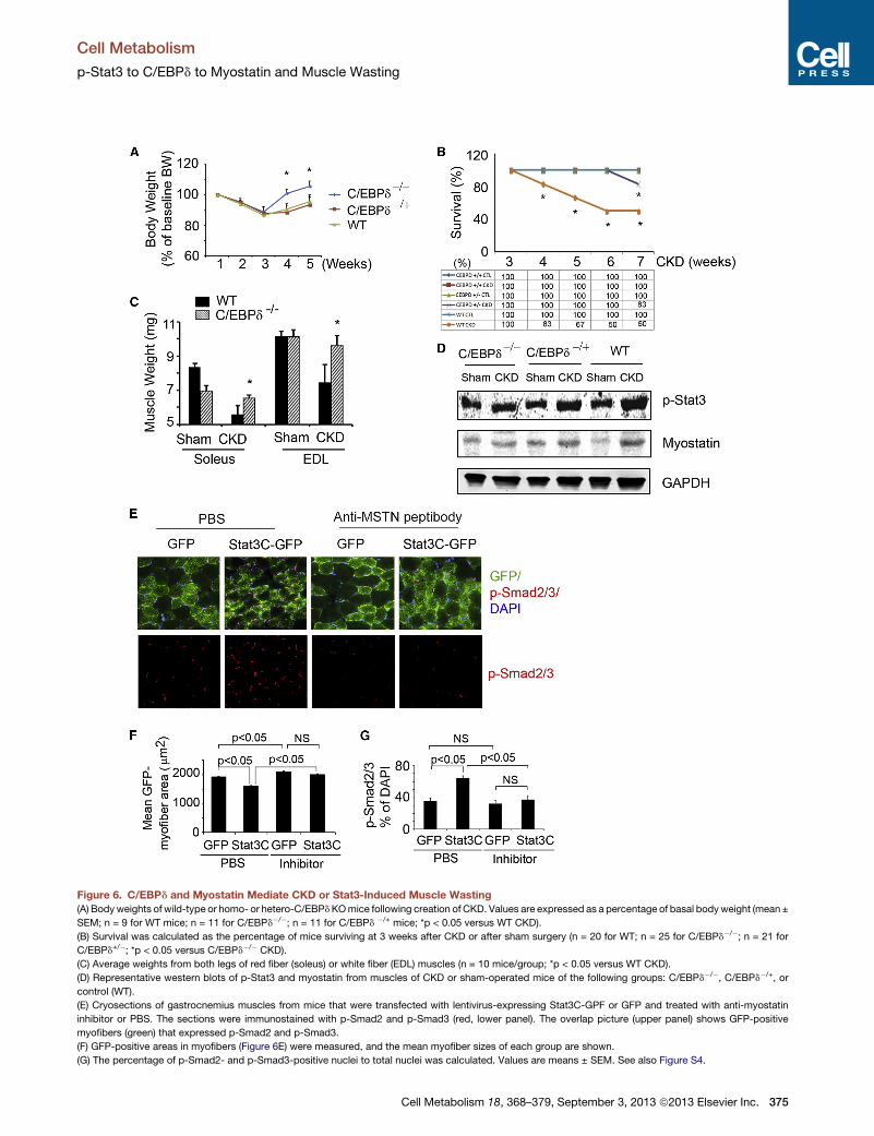
Figure 6. C/EBPd and Myostatin Mediate CKD or Stat3-Induced Muscle Wasting
(A) Bodyweights of wild-type or homo- or hetero-C/EBPdKOmice following creation of CKD. Values are expressed as a percentage of basal bodyweight (mean ±
SEM; n = 9 for WT mice; n = 11 for C/EBPd�/�; n = 11 for C/EBPd �/+ mice; *p < 0.05 versus WT CKD).
(B) Survival was calculated as the percentage of mice surviving at 3 weeks after CKD or after sham surgery (n = 20 for WT; n = 25 for C/EBPd�/�; n = 21 for
C/EBPd+/�; *p < 0.05 versus C/EBPd�/� CKD).
(C) Average weights from both legs of red fiber (soleus) or white fiber (EDL) muscles (n = 10 mice/group; *p < 0.05 versus WT CKD).
(D) Representative western blots of p-Stat3 and myostatin from muscles of CKD or sham-operated mice of the following groups: C/EBPd�/�, C/EBPd�/+, or
control (WT).
(E) Cryosections of gastrocnemius muscles from mice that were transfected with lentivirus-expressing Stat3C-GPF or GFP and treated with anti-myostatin
inhibitor or PBS. The sections were immunostained with p-Smad2 and p-Smad3 (red, lower panel). The overlap picture (upper panel) shows GFP-positive
myofibers (green) that expressed p-Smad2 and p-Smad3.
(F) GFP-positive areas in myofibers (Figure 6E) were measured, and the mean myofiber sizes of each group are shown.
(G) The percentage of p-Smad2- and p-Smad3-positive nuclei to total nuclei was calculated. Values are means ± SEM. See also Figure S4.
Cell Metabolism
p-Stat3 to C/EBPd to Myostatin and Muscle Wasting
Cell Metabolism 18, 368–379, September 3, 2013 ª2013 Elsevier Inc. 375

Figure 7. Evidence for a p-Stat3, C/EBPd,
and Myostatin Pathway in Muscles of Pa-
tients with CKD
(A) Representative western blots of p-Akt from
muscle biopsies of healthy control or CKD
patients. Bar graph shows the densities of p-Akt
corrected for GAPDH (lower panel; n = 4 CKD
patients and 3 healthy subjects).
(B) Levels of mRNAs of C/EBPd or myostatin were
analyzed by RT-PCR from muscle biopsies of
healthy control or CKD patients (n = 5 control
subjects and 9 CKD patients).
(C) Representative western blots of the indicated
proteins from muscle biopsies from healthy con-
trol or CKD patients.
(D) The band densities in (C) were quantified after
correction for GAPDH (n = 3 pairs for CEBPd; n = 8
pairs for myostatin). Values are means ± SEM. *p <
0.05 versus healthy controls.
Cell Metabolism
p-Stat3 to C/EBPd to Myostatin and Muscle Wasting
latter is consistent with reports from type 1 diabetic patients
(My�sliwiec et al., 2006; My�sliwiec et al., 2008; Shelbaya et al.,
2012). The potential origin of IL-6 in type 1 diabetes includes
peripheral blood mononuclear cells and/or T helper 17 (Th17)
cells (Bradshaw et al., 2009; Foss-Freitas et al., 2006; Ryba-Sta-
nis1awowska et al., 2013). However, others find that IL-6 does not
stimulate muscle loss in the absence of another illness, such as
cancer (Baltgalvis et al., 2008). Thus, it is unclear how cytokines
cause muscle proteolysis. We propose that the increase in IL-6
stimulated byCKD (Kimmel et al., 1998), and possibly other cyto-
kines, activates p-Stat3, which triggers muscle wasting. Indeed,
whenwedeletedStat3 frommuscle orwhenwe studied the Stat3
inhibitor, C188-9, CKD-induced muscle wasting was inhibited.
How could p-Stat3 stimulate muscle wasting? We have demon-
strated that p-Stat3 upregulates C/EBPd and increases the tran-
scription of myostatin, a potent negative regulator of muscle
mass. Others have implicated C/EBPd in the pathogenesis of
catabolic disorders. For example, based onmicroarray analyses,
there was upregulation of multiple genes, including C/EBPd in
muscles of mice with cancer cachexia or in muscle biopsies of
hemodialysis patients (Bonetto et al., 2011; Gutierrez et al.,
2008). In addition, there are reports that p-Stat3 stimulates
C/EBPd expression in cancer, immune, or liver cells. This is rele-
vant because theC/EBPdpromoter contains a Stat3 binding site,
making it a likely participant in the pathway (Zhang et al., 2007;
Sanford and DeWille, 2005). Indeed, we found that exposure of
C2C12 myotubes to IL-6 stimulates p-Stat3 and sequentially in-
creases the expression of C/EBPd. Likewise, expression of
constitutively active Stat3 in myotubes increased C/EBPd pro-
moter activity and the expression of C/EBPd protein. Conversely,
376 Cell Metabolism 18, 368–379, September 3, 2013 ª2013 Elsevier Inc.
Stat3 inhibition in C2C12 myotubes or in
CKD mice suppressed the expression of
C/EBPd. A likely target of C/EBPd is myo-
statin. For example, when we knockdown
C/EBPd by siRNA in myotubes, the in-
crease in myostatin stimulated by IL-6
was blocked. In addition, knockout of
C/EBPd in mice with CKD prevented their
loss of muscle mass and expression of
myostatin. These results are consistent with reports that myosta-
tin is expressed in a wide variety of catabolic conditions associ-
ated with muscle wasting, including cancer, CKD, diabetes, or
weightlessness (spaceflight) (Zhou et al., 2010; Zhang et al.,
2011; Feldman et al., 2006; Lalani et al., 2000). In mice with
CKD, the activation of Stat3 leads to expression of myostatin
and its downstream signals, p-Smad2 and p-Smad3, plus accel-
erated protein degradation. Overexpression of Stat3C in muscle
of mice causes a decrease in myofiber sizes; knocking down
myostatin resolves the phenotype of Stat3 activation because
myofiber sizes are increased and p-Smad2 and p-Smad3 levels
are reduced.Our results that Stat3 inducesmyostatin expression
could apply to other members of the TGF-b superfamily, such as
Activin A. Lipopolysaccharides and cytokines, including IL-6 and
IL-1b, induce expression of Activin A (Okuma et al., 2005); high
levels of Activin A have been shown to induce muscle atrophy
(Zhou et al., 2010). Importantly, treatment of mice with cancer-
induced cachexia using a soluble Activin A receptor (actRIIB)
blocked the loss of muscle mass (Zhou et al., 2010). Additional
studies will be necessary to determine if Activin A induction by
cytokines is mediated through Stat3.
The mechanism by which an increase in myostatin leads to
loss of muscle mass could be a decrease in p-Akt in muscle. A
decrease in p-Akt activates caspase-3, leading to cleavage of
the complex structure of muscle proteins and activation of
proteolysis by the 26S proteasome (Du et al., 2004; Wang
et al., 2010). In addition, a low p-Akt level would reduce phos-
phorylation of forkhead transcription factors, which stimulate
the expression of the muscle-specific E3 ubiquitin ligases, Atro-
gin-1/MAFbx or MuRF-1, and accelerate proteolysis in the UPS

Cell Metabolism
p-Stat3 to C/EBPd to Myostatin and Muscle Wasting
(Sandri et al., 2004; Lee et al., 2004; Stitt et al., 2004; Lecker
et al., 2006). In the present experiments, inhibition of p-Stat3
with C188-9 decreased myostatin expression and the activation
of its downstream signaling mediators, p-Smad2 and p-Smad3;
there also was an increase in p-Akt. There was also a sharp
decrease in p-Akt in muscle of CKD patients, with increased
mRNA and protein expressions of C/EBPd and myostatin.
In summary, our results have uncovered a pathway that stim-
ulates muscle wasting in response to activation of Stat3. The
pathway is activated by CKD or acute diabetes and provides
insights into the relationships among the signaling molecules,
Stat3, C/EBPd, and myostatin. Results from our studies of
cultured skeletal muscle cells or mice are consistent with
changes in the levels of the same signaling molecules in muscle
biopsies of CKD patients. Consequently, these results might be
translated into treatment strategies for catabolic conditions like
CKD that cause muscle wasting. Development of a safe and
potent small-molecule Stat3 inhibitor may represent a therapeu-
tic approach to muscle wasting in catabolic conditions.
EXPERIMENTAL PROCEDURES
Mouse Models
All animal experiments and procedures were approved by the Baylor College
of Medicine Institutional Animal Care and Use Committee (IACUC). Subtotal
nephrectomy was used to create CKD in mice (Zhang et al., 2011; May
et al., 1987). To induce diabetes, we injected 12-week-old Stat3flox/flox
and Stat3 KO mice intraperitoneally with 2 doses of 150 mg/kg/d STZ
(Sigma-Aldrich) in 0.1 M citrate buffer (pH 4). Control mice were injected
with the citrate buffer. Mice were housed in individual cages, and the diabetic
Stat3flox/flox mice were pair-fed with diabetic Stat3 KO mice for 9 days.
Muscle Biopsies
During placement of a peritoneal dialysis catheter in CKD patients, the rectus
abdominis muscle was biopsied, frozen at �80�C, and stored until analyzed.
Biopsy of this muscle was obtained from healthy subjects during abdominal
hernia surgeries. The procedures were approved by the Ethical Committee
of the Department of Internal Medicine of the University of Genoa, in accor-
dance with the Helsinki declaration regarding ethics of human research.
mRNA Analyses
mRNAs were analyzed by RT-PCR as described (Takeda et al., 1998). Primers
are listed in Table S2. Relative mRNA levels were calculated from cycle
threshold (Ct) values using glyceraldehyde 3-phosphate dehydrogenase
(GAPDH) as the internal control (relative expression = 2[sample Ct � GAPDH Ct]).
See Table S2 for primer sequences.
Muscle Force Measurement
Mouse grip strength was measured daily for 4 consecutive days using a Grip
Strength Meter (Columbus Instruments). Each day, 5 grip strengths were
assessed at 1 min intervals, and the average grip strength over 4 days was
calculated.
Statistical Analysis
Data were expressed as the mean ± SEM. Differences between two groups
were analyzed by the t test; multiple comparisons were analyzed by ANOVA
with a post hoc analysis by the Student-Newman-Keuls test for multiple com-
parisons. Results were considered statistically significant at p < 0.05.
SUPPLEMENTAL INFORMATION
Supplemental Information includes Supplemental Experimental Procedures,
four figures, and two tables and can be found with this article online at
http://dx.doi.org/10.1016/j.cmet.2013.07.012.
Cell Me
ACKNOWLEDGMENTS
These experiments were supported by the generous support of Dr. and Mrs.
Harold Selzman, grants from the Norman S. Coplon Extramural Research
Foundation and the American Diabetic Association to L.Z., NIH Grants R37-
DK37175 and T32-DK62706 to W.E.M., and NIH grants R21-CA149783 and
R41-CA153658 and grant RP100421from the Cancer Prevention and
Research Institute of Texas to D.J.T. G.G. is supported by grants from theMin-
istero dell’Universita e della Ricerca Scientifica e Tecnologica and fromGenoa
University. The authors thank Dr. E. Sterneck (NIH-NCI, Fredericksburg, MD)
for the C/EBPd knockout mice and the C/EBPd promoter-luciferase reporter
construct. Dr. D. Allen (University of Colorado) kindly provided the myostatin
promoter-luciferase reporter construct.
Received: April 9, 2012
Revised: March 17, 2013
Accepted: July 24, 2013
Published: September 3, 2013
REFERENCES
Akira, S., Isshiki, H., Sugita, T., Tanabe, O., Kinoshita, S., Nishio, Y., Nakajima,
T., Hirano, T., and Kishimoto, T. (1990). A nuclear factor for IL-6 expression
(NF-IL6) is a member of a C/EBP family. EMBO J. 9, 1897–1906.
Allen, D.L., Cleary, A.S., Hanson, A.M., Lindsay, S.F., and Reed, J.M. (2010).
CCAAT/enhancer binding protein-delta expression is increased in fast skeletal
muscle by food deprivation and regulates myostatin transcription in vitro. Am.
J. Physiol. Regul. Integr. Comp. Physiol. 299, R1592–R1601.
Alonzi, T., Gorgoni, B., Screpanti, I., Gulino, A., and Poli, V. (1997). Interleukin-6
and CAAT/enhancer binding protein beta-deficient mice act as tools to dissect
the IL-6 signalling pathway and IL-6 regulation. Immunobiology 198, 144–156.
Avesani, C.M., Draibe, S.A., Kamimura, M.A., Dalboni, M.A., Colugnati, F.A.,
and Cuppari, L. (2004). Decreased resting energy expenditure in non-dialysed
chronic kidney disease patients. Nephrol. Dial. Transplant. 19, 3091–3097.
Baltgalvis, K.A., Berger, F.G., Pena, M.M., Davis, J.M., Muga, S.J., and
Carson, J.A. (2008). Interleukin-6 and cachexia in ApcMin/+ mice. Am. J.
Physiol. Regul. Integr. Comp. Physiol. 294, R393–R401.
Bonetto, A., Aydogdu, T., Kunzevitzky, N., Guttridge, D.C., Khuri, S., Koniaris,
L.G., and Zimmers, T.A. (2011). STAT3 activation in skeletal muscle links mus-
cle wasting and the acute phase response in cancer cachexia. PLoS ONE 6,
e22538.
Bradshaw, E.M., Raddassi, K., Elyaman, W., Orban, T., Gottlieb, P.A., Kent,
S.C., and Hafler, D.A. (2009). Monocytes from patients with type 1 diabetes
spontaneously secrete proinflammatory cytokines inducing Th17 cells.
J. Immunol. 183, 4432–4439.
Carrero, J.J., Chmielewski, M., Axelsson, J., Snaedal, S., Heimburger, O.,
Barany, P., Suliman, M.E., Lindholm, B., Stenvinkel, P., and Qureshi, A.R.
(2008). Muscle atrophy, inflammation and clinical outcome in incident and
prevalent dialysis patients. Clin. Nutr. 27, 557–564.
Cheung, W.W., Paik, K.H., andMak, R.H. (2010). Inflammation and cachexia in
chronic kidney disease. Pediatr. Nephrol. 25, 711–724.
Du, J., Wang, X., Miereles, C., Bailey, J.L., Debigare, R., Zheng, B., Price, S.R.,
and Mitch, W.E. (2004). Activation of caspase-3 is an initial step triggering
accelerated muscle proteolysis in catabolic conditions. J. Clin. Invest. 113,
115–123.
Feldman, B.J., Streeper, R.S., Farese, R.V., Jr., and Yamamoto, K.R. (2006).
Myostatin modulates adipogenesis to generate adipocytes with favorable
metabolic effects. Proc. Natl. Acad. Sci. USA 103, 15675–15680.
Foss-Freitas, M.C., Foss, N.T., Donadi, E.A., and Foss, M.C. (2006). In vitro
TNF-alpha and IL-6 production by adherent peripheral blood mononuclear
cells obtained from type 1 and type 2 diabetic patients evaluated according
to the metabolic control. Ann. N Y Acad. Sci. 1079, 177–180.
Fouque, D., Kalantar-Zadeh, K., Kopple, J.D., Cano, N., Chauveau, P.,
Cuppari, L., Franch, H.A., Guarnieri, G., Ikizler, T.A., Kaysen, G.A., et al.
tabolism 18, 368–379, September 3, 2013 ª2013 Elsevier Inc. 377

Cell Metabolism
p-Stat3 to C/EBPd to Myostatin and Muscle Wasting
(2008). A proposed nomenclature and diagnostic criteria for protein-energy
wasting in acute and chronic kidney disease. Kidney Int. 73, 391–398.
Goodman, M.N. (1994). Interleukin-6 induces skeletal muscle protein break-
down in rats. Proc. Soc. Exp. Biol. Med. 205, 182–185.
Gutierrez, O.M., Mannstadt, M., Isakova, T., Rauh-Hain, J.A., Tamez, H., Shah,
A., Smith, K., Lee, H., Thadhani, R., Juppner, H., and Wolf, M. (2008).
Fibroblast growth factor 23 and mortality among patients undergoing hemodi-
alysis. N. Engl. J. Med. 359, 584–592.
Hirano, T., Nakajima, K., and Hibi, M. (1997). Signaling mechanisms through
gp130: a model of the cytokine system. Cytokine Growth Factor Rev. 8,
241–252.
Horvath, C.M. (2004). The Jak-STAT pathway stimulated by interleukin 6. Sci.
STKE 2004, tr9.
Hu, Z., Wang, H., Lee, I.H., Du, J., and Mitch, W.E. (2009). Endogenous gluco-
corticoids and impaired insulin signaling are both required to stimulate muscle
wasting under pathophysiological conditions in mice. J. Clin. Invest. 119,
3059–3069.
Hung, A.M., Ellis, C.D., Shintani, A., Booker, C., and Ikizler, T.A. (2011). IL-1b
receptor antagonist reduces inflammation in hemodialysis patients. J. Am.
Soc. Nephrol. 22, 437–442.
Kimmel, P.L., Phillips, T.M., Simmens, S.J., Peterson, R.A., Weihs, K.L.,
Alleyne, S., Cruz, I., Yanovski, J.A., and Veis, J.H. (1998). Immunologic func-
tion and survival in hemodialysis patients. Kidney Int. 54, 236–244.
Kishimoto, T., Taga, T., and Akira, S. (1994). Cytokine signal transduction. Cell
76, 253–262.
Lalani, R., Bhasin, S., Byhower, F., Tarnuzzer, R., Grant, M., Shen, R., Asa, S.,
Ezzat, S., and Gonzalez-Cadavid, N.F. (2000). Myostatin and insulin-like
growth factor-I and -II expression in the muscle of rats exposed to the micro-
gravity environment of the NeuroLab space shuttle flight. J. Endocrinol. 167,
417–428.
Lecker, S.H., Jagoe, R.T., Gilbert, A., Gomes,M., Baracos, V., Bailey, J., Price,
S.R., Mitch, W.E., and Goldberg, A.L. (2004). Multiple types of skeletal muscle
atrophy involve a common program of changes in gene expression. FASEB J.
18, 39–51.
Lecker, S.H., Goldberg, A.L., and Mitch, W.E. (2006). Protein degradation by
the ubiquitin-proteasome pathway in normal and disease states. J. Am. Soc.
Nephrol. 17, 1807–1819.
Lee, S.W., Dai, G., Hu, Z., Wang, X., Du, J., and Mitch, W.E. (2004). Regulation
of muscle protein degradation: coordinated control of apoptotic and ubiquitin-
proteasome systems by phosphatidylinositol 3 kinase. J. Am. Soc. Nephrol.
15, 1537–1545.
Ma, K., Mallidis, C., Artaza, J., Taylor, W., Gonzalez-Cadavid, N., and Bhasin,
S. (2001). Characterization of 50-regulatory region of human myostatin gene:
regulation by dexamethasone in vitro. Am. J. Physiol. Endocrinol. Metab.
281, E1128–E1136.
May, R.C., Kelly, R.A., andMitch,W.E. (1987). Mechanisms for defects in mus-
cle protein metabolism in rats with chronic uremia. Influence of metabolic
acidosis. J. Clin. Invest. 79, 1099–1103.
My�sliwiec, M., Balcerska, A., Zorena, K., My�sliwska, J., Lipska, B.S.,
Wi�sniewski, P., and My�sliwski, A. (2006). Serum and urinary cytokine homeo-
stasis and renal tubular function in children with type 1 diabetes mellitus.
J. Pediatr. Endocrinol. Metab. 19, 1421–1427.
My�sliwiec, M., Balcerska, A., Zorena, K., My�sliwska, J., Lipowski, P., and
Raczy�nska, K. (2008). The role of vascular endothelial growth factor, tumor
necrosis factor alpha and interleukin-6 in pathogenesis of diabetic retinopathy.
Diabetes Res. Clin. Pract. 79, 141–146.
Okuma, Y., O’Connor, A.E., Muir, J.A., Stanton, P.G., de Kretser, D.M., and
Hedger, M.P. (2005). Regulation of activin A and inhibin B secretion by inflam-
matory mediators in adult rat Sertoli cell cultures. J. Endocrinol. 187, 125–134.
Penner, G., Gang, G., Sun, X., Wray, C., and Hasselgren, P.O. (2002). C/EBP
DNA-binding activity is upregulated by a glucocorticoid-dependent mecha-
nism in septic muscle. Am. J. Physiol. Regul. Integr. Comp. Physiol. 282,
R439–R444.
378 Cell Metabolism 18, 368–379, September 3, 2013 ª2013 Elsevie
Poli, V. (1998). The role of C/EBP isoforms in the control of inflammatory and
native immunity functions. J. Biol. Chem. 273, 29279–29282.
Price, S.R., Bailey, J.L., Wang, X., Jurkovitz, C., England, B.K., Ding, X.,
Phillips, L.S., and Mitch, W.E. (1996). Muscle wasting in insulinopenic rats
results from activation of the ATP-dependent, ubiquitin-proteasome proteo-
lytic pathway by a mechanism including gene transcription. J. Clin. Invest.
98, 1703–1708.
Ramji, D.P., and Foka, P. (2002). CCAAT/enhancer-binding proteins: structure,
function and regulation. Biochem. J. 365, 561–575.
Redell, M.S., Ruiz, M.J., Alonzo, T.A., Gerbing, R.B., and Tweardy, D.J. (2011).
Stat3 signaling in acute myeloid leukemia: ligand-dependent and -indepen-
dent activation and induction of apoptosis by a novel small-molecule Stat3
inhibitor. Blood 117, 5701–5709.
Rui, L., Fisher, T.L., Thomas, J., and White, M.F. (2001). Regulation of
insulin/insulin-like growth factor-1 signaling by proteasome-mediated
degradation of insulin receptor substrate-2. J. Biol. Chem. 276, 40362–
40367.
Rui, L., Yuan, M., Frantz, D., Shoelson, S., andWhite, M.F. (2002). SOCS-1 and
SOCS-3 block insulin signaling by ubiquitin-mediated degradation of IRS1 and
IRS2. J. Biol. Chem. 277, 42394–42398.
Ryba-Stanis1awowska, M., Skrzypkowska, M., My�sliwska, J., and My�sliwiec,
M. (2013). The serum IL-6 profile and Treg/Th17 peripheral cell populations
in patients with type 1 diabetes. Mediators Inflamm. 2013, 205284.
Sandri, M., Sandri, C., Gilbert, A., Skurk, C., Calabria, E., Picard, A., Walsh, K.,
Schiaffino, S., Lecker, S.H., and Goldberg, A.L. (2004). Foxo transcription fac-
tors induce the atrophy-related ubiquitin ligase atrogin-1 and cause skeletal
muscle atrophy. Cell 117, 399–412.
Sanford, D.C., and DeWille, J.W. (2005). C/EBPdelta is a downstream medi-
ator of IL-6 induced growth inhibition of prostate cancer cells. Prostate 63,
143–154.
Shelbaya, S., Amer, H., Seddik, S., Allah, A.A., Sabry, I.M., Mohamed, T., and
El Mosely, M. (2012). Study of the role of interleukin-6 and highly sensitive C-
reactive protein in diabetic nephropathy in type 1 diabetic patients. Eur. Rev.
Med. Pharmacol. Sci. 16, 176–182.
Sterneck, E., Paylor, R., Jackson-Lewis, V., Libbey, M., Przedborski, S.,
Tessarollo, L., Crawley, J.N., and Johnson, P.F. (1998). Selectively enhanced
contextual fear conditioning in mice lacking the transcriptional regulator
CCAAT/enhancer binding protein delta. Proc. Natl. Acad. Sci. USA 95,
10908–10913.
Stitt, T.N., Drujan, D., Clarke, B.A., Panaro, F., Timofeyva, Y., Kline, W.O.,
Gonzalez, M., Yancopoulos, G.D., and Glass, D.J. (2004). The IGF-1/PI3K/
Akt pathway prevents expression of muscle atrophy-induced ubiquitin ligases
by inhibiting FOXO transcription factors. Mol. Cell 14, 395–403.
Takeda, K., Kaisho, T., Yoshida, N., Takeda, J., Kishimoto, T., and Akira, S.
(1998). Stat3 activation is responsible for IL-6-dependent T cell proliferation
through preventing apoptosis: generation and characterization of T cell-spe-
cific Stat3-deficient mice. J. Immunol. 161, 4652–4660.
Trendelenburg, A.U., Meyer, A., Rohner, D., Boyle, J., Hatakeyama, S., and
Glass, D.J. (2009). Myostatin reduces Akt/TORC1/p70S6K signaling, inhibiting
myoblast differentiation and myotube size. Am. J. Physiol. Cell Physiol. 296,
C1258–C1270.
Verzola, D., Procopio, V., Sofia, A., Villaggio, B., Tarroni, A., Bonanni, A.,
Mannucci, I., De Cian, F., Gianetta, E., Saffioti, S., and Garibotto, G. (2011).
Apoptosis and myostatin mRNA are upregulated in the skeletal muscle of
patients with chronic kidney disease. Kidney Int. 79, 773–782.
Wang, X.H., Zhang, L., Mitch, W.E., LeDoux, J.M., Hu, J., and Du, J. (2010).
Caspase-3 cleaves specific 19 S proteasome subunits in skeletal muscle stim-
ulating proteasome activity. J. Biol. Chem. 285, 21249–21257.
Xu, X., Kasembeli, M.M., Jiang, X., Tweardy, B.J., and Tweardy, D.J. (2009).
Chemical probes that competitively and selectively inhibit Stat3 activation.
PLoS ONE 4, e4783.
Yang, H., Mammen, J., Wei, W., Menconi, M., Evenson, A., Fareed, M.,
Petkova, V., and Hasselgren, P.O. (2005). Expression and activity of
r Inc.

Cell Metabolism
p-Stat3 to C/EBPd to Myostatin and Muscle Wasting
C/EBPbeta and delta are upregulated by dexamethasone in skeletal muscle.
J. Cell. Physiol. 204, 219–226.
Zhang, Y., Sif, S., and DeWille, J. (2007). The mouse C/EBPdelta gene
promoter is regulated by STAT3 and Sp1 transcriptional activators, chromatin
remodeling and c-Myc repression. J. Cell. Biochem. 102, 1256–1270.
Zhang, L., Du, J., Hu, Z., Han, G., Delafontaine, P., Garcia, G., and Mitch, W.E.
(2009). IL-6 and serum amyloid A synergy mediates angiotensin II-induced
muscle wasting. J. Am. Soc. Nephrol. 20, 604–612.
Cell Me
Zhang, L., Rajan, V., Lin, E., Hu, Z., Han, H.Q., Zhou, X., Song, Y., Min, H.,
Wang, X., Du, J., and Mitch, W.E. (2011). Pharmacological inhibition of myo-
statin suppresses systemic inflammation and muscle atrophy in mice with
chronic kidney disease. FASEB J. 25, 1653–1663.
Zhou, X., Wang, J.L., Lu, J., Song, Y., Kwak, K.S., Jiao, Q., Rosenfeld, R.,
Chen, Q., Boone, T., Simonet, W.S., et al. (2010). Reversal of cancer cachexia
and muscle wasting by ActRIIB antagonism leads to prolonged survival. Cell
142, 531–543.
tabolism 18, 368–379, September 3, 2013 ª2013 Elsevier Inc. 379


![Bt354 as a new STAT3 signaling pathway inhibitor against ...download.xuebalib.com/38cqWocZQRSY.pdf · carcinoma, prostate cancer, melanoma, multiple myeloma, and leukemia [3]. Aberrant](https://img.dokumen.tips/doc/110x75/5c873e1809d3f2d8348badbc/bt354-as-a-new-stat3-signaling-pathway-inhibitor-against-carcinoma-prostate.jpg)