Embed Size (px)
Citation preview

The 18th International Congress of Dento-Maxillo-Facial Radiology May 25-29, 2011, Hiroshima, Japan
Standardization of diagnostic images
- How DICOM works with Dental -
Japan Industries Association of Radiological Systems ( JIRA )DICOM-Committee Chair Makoto Suzuki

The 18th International Congress of Dento-Maxillo-Facial Radiology May 25-29, 2011, Hiroshima, Japan 2
preface• This presentation is made to introduce
– the basics of DICOM Standard which is already a de-facto standard in medical imaging industry.
– how standards help you work smoothly in your environment.
– Some other guidelines to standardize your work environment.
AcknowledgementSome of Dental images were provided by Mr. Yamamoto of Osaka University.

The 18th International Congress of Dento-Maxillo-Facial Radiology May 25-29, 2011, Hiroshima, Japan 3
contents
1) What DICOM defines
2) Application of DICOM to Dentalvs. mammography
3) Other related Standards

The 18th International Congress of Dento-Maxillo-Facial Radiology May 25-29, 2011, Hiroshima, Japan 4
DICOM Standard overview
• Digital Imaging and COmmunication in Medicine• Medical equipment vendors and users world-wide
contribute the progress of DICOM standard, and it is officially supported by NEMA,USA.
• Corrections and Supplements are accepted any time, and the latest version including them is issued every year as DICOM2008, DICOM2009,,,,,
• DICOM standard is written in English, and the Japanese translation is located at JIRA homepage.

The 18th International Congress of Dento-Maxillo-Facial Radiology May 25-29, 2011, Hiroshima, Japan 5
DICOM Standard Contribution from JapanDSC: DICOM Standard Committee
NEMA
MedicalEquipmentManufacturers
JIRA
MITA
Radiological Societies(ACR etc)
DICOM Committee
Medical EquipmentManufacturers
DSC&
WG’s
JSRTJAHIS JRS
Rad. vendors Societies
(DRG, etc)
DICOM Promoting org.(MISAT, K-PACS, etc)
Medical Informatics(HIMSS、etc)
some Manufacturers
Japan

The 18th International Congress of Dento-Maxillo-Facial Radiology May 25-29, 2011, Hiroshima, Japan 6
DICOM Standards LocationMITA:http://medical.nema.org/

The 18th International Congress of Dento-Maxillo-Facial Radiology May 25-29, 2011, Hiroshima, Japan 7
DICOM Standards Japanese translationJIRA Home-page http://www.jira-net.or.jp/index.htm
①
②
③

The 18th International Congress of Dento-Maxillo-Facial Radiology May 25-29, 2011, Hiroshima, Japan 8
DICOM Standard Object structure
data data data
data element
order of transmission
data
Tag ValueLength Value Field VR
data data data
One DICOM object( one image , one patient info.)
pixel is one of the data
Patient (0010,0010) PN 32 Yamada^taro=山田^太郎name

The 18th International Congress of Dento-Maxillo-Facial Radiology May 25-29, 2011, Hiroshima, Japan 9
DICOM Standard example ( MR image tags)IMAGE STORAGE from MR to PACS
(0000,0100) Command Field 1 0x0001 C-STORE-RQ (0008,0005) Specific Character Set “¥ISO 2022 IR 87 " (0008,0008) Image Type "DERIVED¥PRIMARY¥OTHER " (0008,0016) SOP Class UID "1.2.840.10008.5.1.4.1.1.4 " (0008,0018) SOP Instance UID "1.2.840.113701.4.2.9673.0.14415.0.1 " (0008,0020) Study Date "20110527" (0008,0030) Study Time "123000" (0008,0050) Accession Number "201105270203451" (0008,0060) Modality "MR" (0010,0010) Patient‘s Name “緊急S222" (0010,0020) Patient ID "1048010120"(0018,0087) Magnetic Field Strength "0.35"
(7FE0,0010) Pixel Data 524288Bytes
IMAGE STORAGE response from PACS(0000,0100) Command Field 32769 0x8001 C-STORE-RSP (0000,0900) Status "0 0x0000"

The 18th International Congress of Dento-Maxillo-Facial Radiology May 25-29, 2011, Hiroshima, Japan 10
DICOM Standard example ( exam query tags)MWM-SCU requests scheduled exam information
(0010,0010) Patient's Name 0 "" (0010,0020) Patient ID 0 "" (0010,0030) Patient's Birth Date 0 "" (0010,0040) Patient's Sex 0 "" (0040,0002) Start Date 18 "20110527-20110527" (0040,0003) Start Time 12 "000000-235959 "
MWM-SCP returns patient information
(0010,0010) Patient‘s Name 18 " testdata^inpatient" (0010,0020) Patient ID 10 "0000010508" (0010,0030) Patient's Birth Date 8 "19750520" (0010,0040) Patient's Sex 2 "M "(0040,0002) Start Date 8 "20110527" (0040,0003) Start Time 6 “094500"

The 18th International Congress of Dento-Maxillo-Facial Radiology May 25-29, 2011, Hiroshima, Japan 11
DICOM Standard PS 3.3• PS 3.3 defines information objects.
– DICOM defines all activities with combination of Service and Object Pairs (SOP)
– Defined objects are found in PS3.3 contents page.

The 18th International Congress of Dento-Maxillo-Facial Radiology May 25-29, 2011, Hiroshima, Japan 12
DICOM Standard PS 3.4• PS3.4 defines Services.
– DICOM defines all activities with combination of Service and Object (SOP)
– Defined services are found in PS3.4 contents page.– Service is activated by Service Class User (SCU),
and responded by Service Class Provider (SCP).– each SOP is numbered for easy acknowledgement.
(SOP Class UID)• MR Image Storage : 1.2.840.10008.5.1.4.1.1.1.2• IO Image Storage : 1.2.840.10008.5.1.4.1.1.1.3
– each object is numbered with unique ID ( SOP Instance UID)

The 18th International Congress of Dento-Maxillo-Facial Radiology May 25-29, 2011, Hiroshima, Japan 13
DICOM Standard PS 3.2• Each system needs to declare which DICOM
SOP classes are supported in it.
SCP
SCU
This document is calledConformance Statement.It is defined in PS3.2

The 18th International Congress of Dento-Maxillo-Facial Radiology May 25-29, 2011, Hiroshima, Japan 14
DICOM Standard 3 steps in DICOM
(2) DICOM
message -exchange
=== Association established ===
SCU SCP
==== Association released ====
(1) Request Association
(3) Request Closing Association
Provide Service
IP address,PORT,AET,SOP
Termination(Success,Fail)
StorageMWM etc (Syntax,
characters)
RequestService

The 18th International Congress of Dento-Maxillo-Facial Radiology May 25-29, 2011, Hiroshima, Japan 15
DICOM Standard Object structure• PS3.5 defines the structure and representation
of objects.
Image ObjectI I
SOP Common ModulePatient ModuleImage Info. Module
:
Image Pixel Module
Patient Name (0010,0010) optional PNPatient ID (0010,0020) optional LOPatient Birth date (0010,0030) optional DA
:
Pixel Data (7FE0,0010) always OW
SOP Instance UID (0008,0018) alwaysSOP Class UID (0008,0016) always
:
Patient Module
Pixel Module
Common Module
set of specific information tags required to each object ( modality )
Image Info. Module

The 18th International Congress of Dento-Maxillo-Facial Radiology May 25-29, 2011, Hiroshima, Japan 16
Application of DICOM Mammo vs. Dental• Mammography and Oral images are defined
as Objects in DICOM
Mammo images Dental images# of objects 2 (R/L) 32 max.(4
quadrants)# of images 2 or 4 3 to 14imaging method standardized local standarddisplay method standardized local standardobject MG (CR) IO (CR)

The 18th International Congress of Dento-Maxillo-Facial Radiology May 25-29, 2011, Hiroshima, Japan 17
Application of DICOM Mammo images
Info. module of MG images (Mammography)
name tag value
image type (0008,0008)
MG
image laterality (0020,0062)
RIGHT
view code (0054,0220)
yes
>code value (0008,0100)
R-10226
>code designator (0008,0102)
SNM3
>code meaning (0008,0104)
MLO
MG
LEFT
yes
R-10226
SNM3
MLO
MG
RIGHT
yes
R-10242
SNM3
CC
MG
LEFT
yes
R-10242
SNM3
CC
image#1 #2 #3 #4
“SNM” defines how images should be taken.It also defines how these images should be aligned on the monitor screen.
The 18th International Congress of Dento-Maxillo-Facial Radiology May 25-29, 2011, Hiroshima, Japan 18
Application of DICOM Mammo images
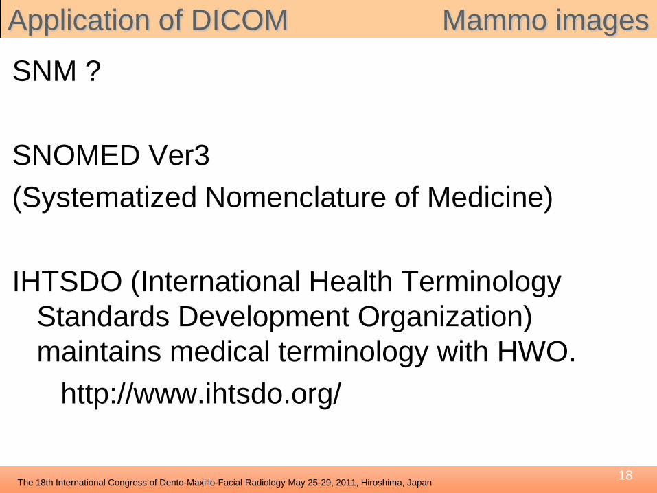
SNM ?
SNOMED Ver3(Systematized Nomenclature of Medicine)
IHTSDO (International Health Terminology Standards Development Organization) maintains medical terminology with HWO.
http://www.ihtsdo.org/

The 18th International Congress of Dento-Maxillo-Facial Radiology May 25-29, 2011, Hiroshima, Japan 19
Application of DICOM Mammo imagesTypical Imaging Technique: MLO & CCTypical Display format: MLO(R/L) + CC(R/L)Standardized by : SNM
MLO:medio-lateral oblique 内外斜位方向
CC:cephalad cranial 頭尾方向
MLO MLO CC CCRIGHT LEFT RIGHT LEFT

The 18th International Congress of Dento-Maxillo-Facial Radiology May 25-29, 2011, Hiroshima, Japan 20
Application of DICOM Mammo images• The method of taking and displaying
mammography images is standardized.– same # of images per exam.– same imaging method ( exposure angle )– same display format.– they are controlled / maintained by proper society
and international standardization organization.
• DICOM can refer these external standards and make use of standardized imaging method and display format.

The 18th International Congress of Dento-Maxillo-Facial Radiology May 25-29, 2011, Hiroshima, Japan 21
Application of DICOM Mammo vs. Dental• DICOM data structure
– Image information module• CR : No tags to specify how to take and display images
(CR is used in various exams, there is no standard)
• MG : SNM defines how to take and display imagesAnd there are tags to specify these.( pretty much standardized exam)
• IO : There are tags to specify these…. but none defines how to take and display images.

The 18th International Congress of Dento-Maxillo-Facial Radiology May 25-29, 2011, Hiroshima, Japan 22
Application of DICOM Dental
attribute TAG examplePhotometric Interpretation (0028,0004) MONOCHROME2kV (0018,0060) 150mA (0018,1151) 80Cassette Size (0018,1403) 35CMX43CM
Typical CR image Information tags
If a Intra-Oral image (IO) is taken by CR, these tags are added to the image.
name tag valuePositioner type (0018,1508) CEPHALOSTATimage laterality (0020,0062) RIGHT
anatomic region sequence (0008,2218) 11code sequence macro yes>coding scheme designator (0008,0102) ISO 3950-1984>primary anatomic structure seq. (0008,2228) 11¥12¥13

The 18th International Congress of Dento-Maxillo-Facial Radiology May 25-29, 2011, Hiroshima, Japan 23
Application of DICOM Dental- Image Info. module contains modality-specific
information. By using it, post-processing or display format can be automatically definedIF THERE IS A STANDARD and DATA IS THERE.
So, What is defined as a standard in DICOMrelated to DENTAL ?

The 18th International Congress of Dento-Maxillo-Facial Radiology May 25-29, 2011, Hiroshima, Japan 24
Application of DICOM DentalIf dental images are taken as CR ( not IO ),the position ( anatomic region resolution ) will be one ofRIGHT / BOTH / LEFT.BUT WE NEED MORE RESOLUTION ! teeth by teeth
If these images are taken as CR,there is no tag to tell upper or lower teeth.

The 18th International Congress of Dento-Maxillo-Facial Radiology May 25-29, 2011, Hiroshima, Japan 25
Application of DICOM Dental
DICOM accepts teeth indexing method.That is ISO 3950-1984.
Problem No1:Each domain uses local naming system.

The 18th International Congress of Dento-Maxillo-Facial Radiology May 25-29, 2011, Hiroshima, Japan 26
Application of DICOM DentalProblem No2:- Which image in the set covers which tooth is not
standardized. (physical size , overlap, missing ones )
- Also its variety is not registered / maintained. ( internationally )

The 18th International Congress of Dento-Maxillo-Facial Radiology May 25-29, 2011, Hiroshima, Japan 27
Application of DICOM Dental
• what is IDEAL ?These 10 images must align to correct positionAUTOMATICALLY. ( By using some tag info.)

The 18th International Congress of Dento-Maxillo-Facial Radiology May 25-29, 2011, Hiroshima, Japan 28
Application of DICOM DentalTo automatically display these images in correct
order, there must be some info. to tell…(1) Which imaging method is used
10-image / 14-image / etc(2) image index according to (1)
01-05/11-15 in 10-image method
01 02 03 04 05
11 12 13 14 15

The 18th International Congress of Dento-Maxillo-Facial Radiology May 25-29, 2011, Hiroshima, Japan 29
Application of DICOM Dental
If we add some new tags to DICOM…(0008,22XX) : Imaging method (10-image/14-
image..)(0008,22YY) : Image position index(0008,2228) : Anatomic Structure Sequence
tag data(0008,0008) IO(0020,0062) RIGHT(0008,2218) yes>(0008,22XX) IMG10>(0008,22YY) 01>(0008,2228) 18¥17¥
16¥15
dataIORIGHTyesIMG100215¥14¥13¥12
dataIOBOTHyesIMG100312¥11¥21¥22
dataIOLEFTyesIMG100422¥23¥24¥25
dataIOLEFTyesIMG100525¥26¥27¥28

The 18th International Congress of Dento-Maxillo-Facial Radiology May 25-29, 2011, Hiroshima, Japan 30
Application of DICOM Dental
tag data(0008,0008) IO(0020,0062) RIGHT(0008,2218) yes>(0008,22XX) IMG10>(0008,22YY) 01>(0008,2228) 18¥17¥
16¥15
dataIORIGHTyesIMG100215¥14¥13¥12
dataIOBOTHyesIMG100312¥11¥21¥22
dataIOLEFTyesIMG100422¥23¥24¥25
dataIOLEFTyesIMG100525¥26¥27¥28
01 02 03 04 05

The 18th International Congress of Dento-Maxillo-Facial Radiology May 25-29, 2011, Hiroshima, Japan 31
01
Application of DICOM DentalAnd if teeth index are somehow entered in (0008,2228) Primary Anatomic Structure Sequence,you can find which image each teeth is imaged in. This information can be used to find/retrieve images of specificteeth.PROBLEM : How to detect and index each teeth in an image.
tag data(0008,0008) IO(0020,0062) RIGHT(0008,2218) yes>(0008,22XX) IMG10>(0008,22YY) 01>(0008,2228) 18¥17¥16¥15
18 17 16 15

The 18th International Congress of Dento-Maxillo-Facial Radiology May 25-29, 2011, Hiroshima, Japan 32
Application of DICOM Dental
• We need to do these AUTOMATICALLY– set imaging method ( 10-image, 14-image,,)– set image index number ( out of 10/14 images )– set tooth index number ( out of 32 teeth )
– and enable all related equipments to handle these information.
• Then all Intra-oral images will be displayed correctly on monitor. And the same tooth in previous exam can be found ( retrieved ).

The 18th International Congress of Dento-Maxillo-Facial Radiology May 25-29, 2011, Hiroshima, Japan 33
Other related Standards IHE profile(1) IHE : Integrating Healthcare Enterprise- IHE is NPO from medical and manufacturers.- IHE defines many profiles.
(standardized relationship of related personnel (Actor) and information (Object))
- Profiles are created from practical routine work.
Some profiles can be applied to dental procedures.
・ Scheduled Workflow (SWF) ・ Consistent Presentation of Images (CPI) ・ Portable Data for Imaging (PDI) and more

The 18th International Congress of Dento-Maxillo-Facial Radiology May 25-29, 2011, Hiroshima, Japan 34
Other related Standards IHE profile
• Profiles defined by IHE are found at– IHE homepage http://www.ihe.net/– Japanese translation
http://www.ihe-j.org/links/index.html

The 18th International Congress of Dento-Maxillo-Facial Radiology May 25-29, 2011, Hiroshima, Japan 35
FIN1) DICOM defines most of medical image
format, and its communication method.2) Dental images has wide variety of taking
images, and showing them, mainly due to physical diversity. This makes it difficult to standardize the exam.
3) DICOM provides tags to arrange images on monitor, or specify each small structure.
4) DICOM can refer external standards to coordinate with them.
Thank You