Embed Size (px)
Citation preview
STANDARD TREATMENT GUIDELINES
Management of Osteoarthritis Knee
January 2017
Ministry of Health & Family Welfare Government of India

1
Index
SCOPE ..................................................................................................................................2
INTRODUCTION ...................................................................................................................3
NEED FOR A STG/ GUIDELINE PURPOSE ..............................................................................4
RECOMMENDATIONS ..........................................................................................................5 4.1. Diagnosis of Osteoarthritis Knee - ........................................................................................ 5 4.2 Role of imaging ...................................................................................................................... 6 4.3Treatment ............................................................................................................................ 10 FOLLOW UP AND REVIEW : ....................................................................................................... 15 ARTHROPLASTY : ....................................................................................................................... 16 4.7 Summary ............................................................................................................................. 19 How this guideline was developed ............................................................................................ 21 7. Research Needs ..................................................................................................................... 23 8. Annexure (Adopt/Adapt Table) ............................................................................................ 24 References ................................................................................................................................ 45

2
SCOPE :
Approach to Management of Osteoarthritis of Knee
SCOPE POPULATION :
Groups Covered :
Adults with suspected Osteoarthritis (OA) of Knee
persistent joint pain that becomes worse with use
predominantly in people age 45 years or older
morning stiffness lasting no more than half an hour
Pathology
Primary OA Knee
Secondary OA
Post traumatic
Healed sequelae of infection or inflammatory disease
Groups Not Covered :
Acute / subacute Infection : TB, Pyogenic septic arthritis, Fungal, Hansen
Acute Trauma
Acute Inflammatory diseases : RA, AS, Psoriatic, seronegative arthrititidis, Spondyloarthropathy
Crystal arthritis (gout or pseudo-gout)
Hemophilic arthropathy
Bone and soft tissue tumours
Health Care Settings :
Primary Health Care

3
Management : Pharmacology, Physiotherapy
Secondary
Management : Pharmacology, Physiotherapy, Occupational Therapy, Orthosis
Tertiary
Management : Pharmacology, Physiotherapy, Occupational Therapy, Orthosis, Surgery
Management Issue not covered :
Alternative therapies (Ayurvedic,Unani and Homeopathic medications) and Yoga, Tai Chi
INTRODUCTION 1.1 Definitions-
1.2 Burden of disease-

4
Knee OA is one of the leading causes of global disability and the real burden of osteoarthritis (OA) has been underestimated. The global age-standardized prevalence of knee OA was reported as 3.8%, higher in females than males (Cross et al, 2010).
The prevalence of OA knee in India in a report by ICMR in 2012 was 3.28% in Delhi ; 5.81% in Dibrugarh and 6.52% in Jodhpur (ICMR study, 2012). However a community based cross sectional study across five sites in India conducted in big city, small city, town, and village was reported in 2016 to be as high as 28.7% (Pal et al, 2016).
Morbidity and mortality –
CDC has reported an annual average of 0.2 to 0.3 deaths per 100,000 population due to OA. OA deaths are likely highly underestimated. For example, gastrointestinal bleeding due to treatment with NSAIDs is not counted (Sacks et al, 2004).
The hospitalization rate per 100,000 in the US, for total knee replacement increased by 217% from 1992 to 2011 from 203.6 to 6455.
OA of the knee is one of five leading causes of disability among non-institutionalized adults6. About 80% of patients with OA have some degree of movement limitation. About 25% cannot perform major activities of daily living (ADL's), 11% of adults with knee OA need help with personal care and 14% require help with routine needs and about 40% of adults with knee OA reported their health "poor" or "fair."
2. CURRENT PRACTICES IN INDIA
Pharmacological modalities like analgesics, NSAID’s, topical applications, supplementations like glucosamine along with physiotherapy including exercises, massage, TENS, thermotherapy and braces along with acupuncture and ayurvedic as well as homeopathic and other alternate therapies are widely used by different care givers.
Surgical interventions besides intra-articular injections including arthroscopy, osteotomy, unicondylar and total knee replacements are commonly done by orthopaedic surgeons across the country specially in different cities.
NEED FOR A STG/ GUIDELINE PURPOSE
5
Osteoarthritis of the knee, a common chronic problem makes the patients try out a wide variety of therapeutic modalities hopping from doctors to physiotherapists to alternative therapists for pain alleviation and sometimes to avoid surgery even in advanced osteoarthritis.
This manuscript would be useful for the patients as well as the primary level and secondary level health care professionals, for diagnosing a case of osteoarthritis of the knee followed by guidelines regarding the non operative management with special reference to prevent unnecessary prescriptions of glucosamine or chondroitin products while emphasizing on self management strategies such as weight loss, exercise, suitable footwear, braces, walking aids and thermotherapy as well as other physiotherapy modalities including electrotherapy and acupuncture. Role of pharmacological management including analgesics, topical ointments and intrarticular injections of steroids and hyaluronan are also elaborated.
For the tertiary level orthopaedic surgeons, this manual would be a useful guideline for controversial and confusing trends sometimes practiced differently by various surgeons. These guidelines include indications of osteotomy and arthroplasty and unicondylar replacement versus osteotomy with no role of arthroscopic lavage and debridement. Details of arthroplasty including prognostic factors, intra-operative considerations of analgesia, nerve blocks, tourniquets, tranexemic acid, surgical drains as well as choice of implants, cement, bilateral replacement, navigation, patient specific instrumentation and post operative protocols of cryotherapy, CPM, rehabilitation and hospital stay guidelines would also be of interest to the orthopaedic and arthroplasty surgeons.
RECOMMENDATIONS 4.1. Diagnosis of Osteoarthritis Knee - 4.1.1 Clinical features: symptoms and signs (Hasan and Shuckett, 2010)

6
Algorithm of approach to joint pain (Harrison, 19th Ed)
Symptoms
The main symptoms of OA include pain around the knee, stiffness, and altered joint function. Initially this tends to be worse with weight bearing and ambulation. Eventually this can progress to pain day and night once cartilage loss leads to bone-on-bone contact. In contrast to inflammatory arthritides such as rheumatoid arthritis, with their prolonged morning stiffness and worsened pain in the morning, OA tends to worsen as the day progresses. The stiffness in OA is termed “inactivity stiffness” and contrasts with the prolonged “morning stiffness” of rheumatoid arthritis. Inactivity stiffness in osteoarthritic lower limb joints lasts about 5 to 10 minutes and occurs when the patient gets up and bears weight after prolonged immobility.
Signs
On physical examination, a small effusion with a fluid bulge sign can be present in OA of the knee. Larger effusions can occur but are less frequent than in the inflammatory arthropathies. There may be cartilaginous crepitus or a crackling feeling on palpation of the knee with motion. Eventually there may be coarse bone-on-bone crepitus whereby the opposing bone ends, denuded of cartilage, seem to grate against one another. There is often a loss of range of motion of the involved knee,particularly with progression of OA. Loss of cartilage of the knee can lead to malalignment of the leg with a varus deformity or bow-legged positioning of the leg being evident. This angulation of the knee applies to medial compartment OA of the knees. Less commonly, patients may present with a valgus or knock-knee deformity, indicative of more advanced disease in the lateral compartment of the knee. On occasion, and much less commonly, patients may present with isolated OA in the patellofemoral joint, which itself may be very symptomatic.
4.2 Role of imaging 4.2.1 X Rays :
Since the 1970s, the standard view for radiographic assessment of the tibiofemoral joint has been the extended-knee radiograph, which is a bilateral antero-posterior image acquired while the patient is weight-bearing,with both knees in full extension.

7
The primary utility of radiography in the diagnosis of OA is for evaluation of joint space width (JSW). JSW and subsequent joint space narrowing (JSN) were originally assessed using manual techniques that required minimal additional equipment or processing software (Chondrometry, 1995; Ravaud et al,1996). However, these methods were time consuming and subjective and have since been largely abandoned in favour of automated assessment, which provides quick and precise measurements of joint space width (JSW). In addition to improving reproducibility of semi-quantitative scoring or manual measurements, automated assessment has also sparked additional characterizations of joint space, including minimum JSW, mean JSW, joint space area, and location-specific JSW (Roemer et al, 2011). Several studies have shown minimum JSW to be most reproducible and most sensitive to OA-related changes (Conrozier et al, 2001 ; Vignon, 2004).
On plain X-ray of an osteoarthritic joint, in addition to joint space narrowing, there tends to be subchondral sclerosis or an appearance of whitening of the subchondral bone. Osteophytes, which reflect a regenerative process with formation of fibrocartilaginous extensions or hooks at the joint margins, are common. Interestingly, the presence of osteophytes in one compartment, such as the lateral compartment in a patient with medial compartment OA, is not indicative of disease in that compartment. It is simply indicative of the body’s reparative response to the abnormal stresses and presence of disease in the medial compartment. Currently, the Kellgren-Lawrence (KL) grading scheme is the most widely used and accepted standard for diagnosis of radiographic OA (Kellgren and Lawrence,1957 ; Bauer et al,2006).
Fig 1 : Standing (weight bearing) AP view of both Knee

8
A KL grade of 0 indicates that no radiographic features of OA are present while a KL grade of 1 is defined as doubtful JSN and possible osteophytic lipping (Kellgren and Lawrence,1957). Radiographic OA receives a KL grade of 2, denoting the presence of definite osteophytes and possible joint space narrowing (JSN) on anteroposterior weight-bearing radiograph (Kellgren and Lawrence,1957). Further disease progression is graded as KL 3, characterized by multiple osteophytes, definite JSN, sclerosis, possible bony deformity and KL grade 4, which is defined by large osteophytes, marked JSN, severe sclerosis and definitely bony deformity (Kellgren and Lawrence,1957). The KL grading scheme has been criticized for characterizing the progression of OA as a linear process and combining osteophyte and JSN measurements (Roemer et al, 2011). More recently, the Osteoarthritis Research Society International atlas has developed OA classification scores that evaluate tibiofemoral JSN and osteophytes separately in each compartment (Altman, 1995 ; Altman and Gold, 2007).
While radiography is useful for evaluation of JSW, a 2005 study by Amin et al. revealed that a significant number of symptomatic patients show cartilage loss on MRI even when JSN or disease progression is not visualized using radiography. In this study, radiographic progression was 91% specific but only 23% sensitive for cartilage loss (Amin et al, 2005).
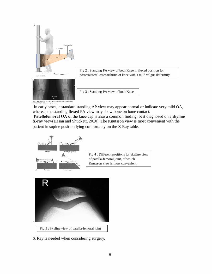
In patients with suspected posterolateral OA with a mild valgus deformity, a 30 degree flexed standing posteroanterior (PA) view with the beam directed 15 degrees from cephalad to caudad may be valuable in showing the disease in the posterior aspect of the lateral compartment of the knee (Leach et al, 1970 ; Cibere, 2006).
9
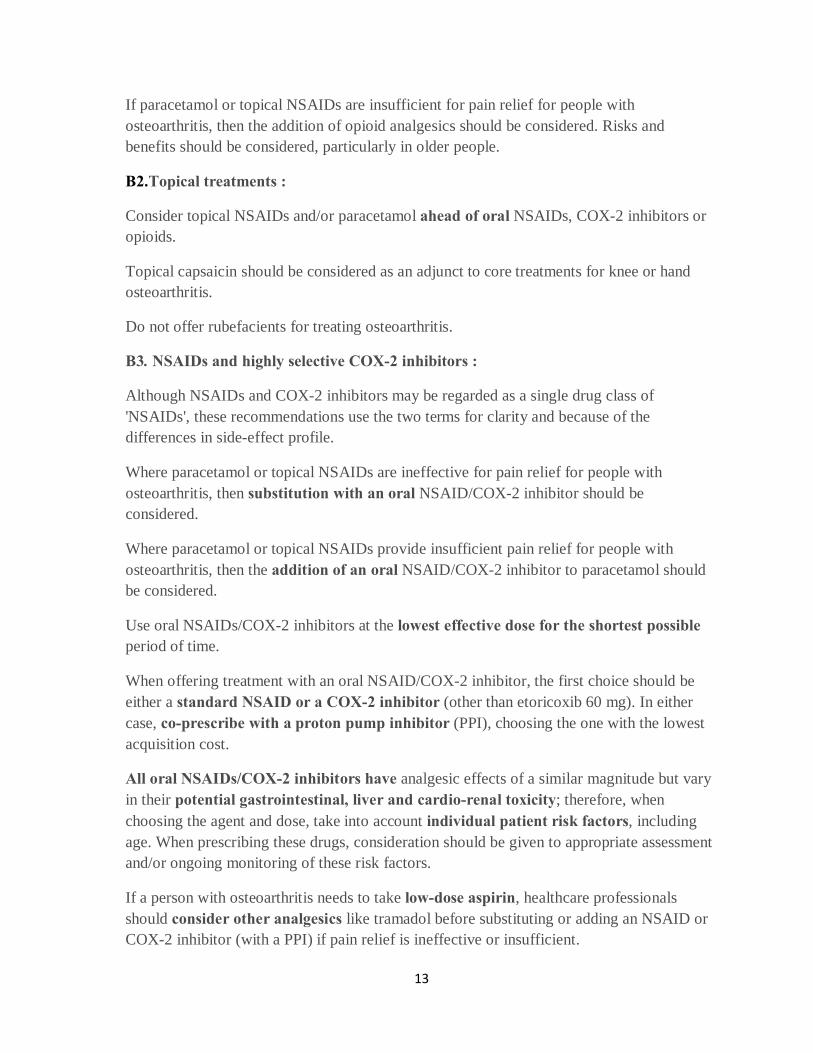
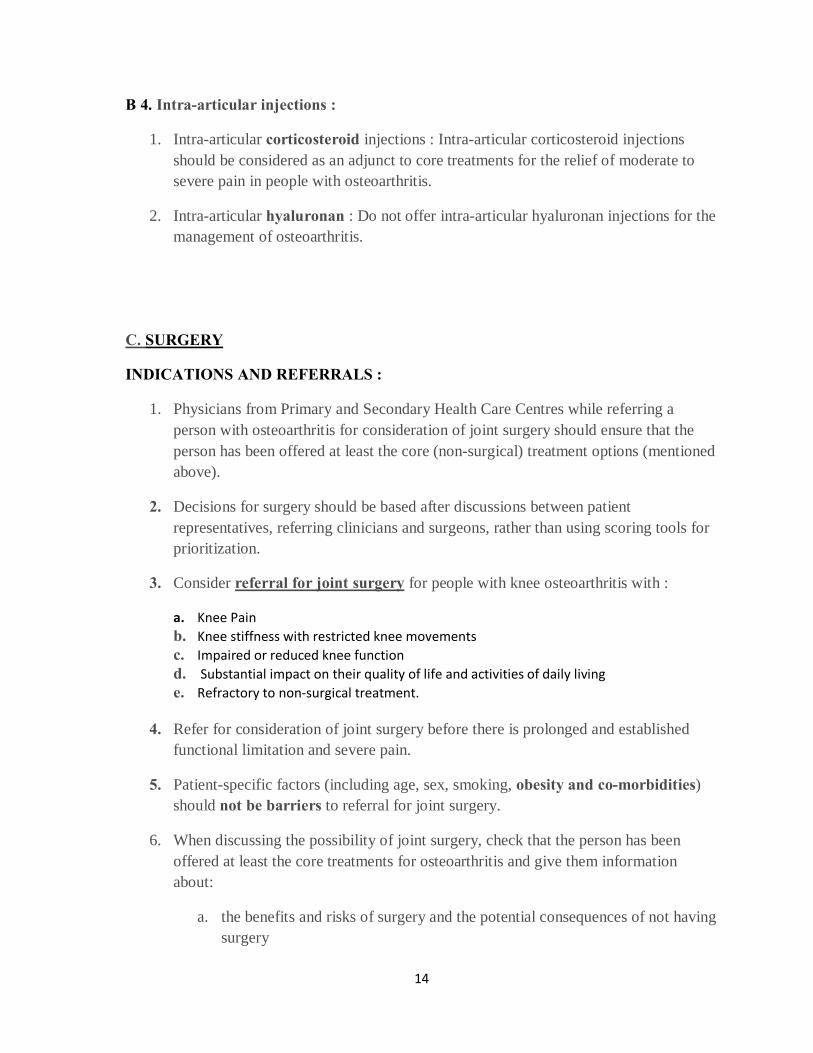
In early cases, a standard standing AP view may appear normal or indicate very mild OA, whereas the standing flexed PA view may show bone on bone contact. Patellofemoral OA of the knee cap is also a common finding, best diagnosed on a skyline X-ray view(Hasan and Shuckett, 2010). The Knutsson view is most convenient with the patient in supine position lying comfortably on the X Ray table.
.
X Ray is needed when considering surgery.
Fig 4 : Different positions for skyline view of patella-femoral joint, of which Knutsson view is most convenient.
Fig 5 : Skyline view of patella-femoral joint
Fig 2 : Standing PA view of both Knee in flexed position for posterolateral osteoarthritis of knee with a mild valgus deformity
Fig 3 : Standing PA view of both Knee

10
X Ray will identify radio-opaque loose bodies, a less frequent cause of locking. (Royal College of Radiologists Recommendations)
4.2.2 Indications of Ultrasound
US is useful for anterior knee pain with suspected tendinopathy or associated bursitis. (Royal College of Radiologists Recommendations)
4.2.3 Indications of MRI
MRI is useful for persistent undiagnosed pain of knee joint.
MRI is the investigation of choice to identify meniscal tears and loose bodies.( Royal College of Radiologists Recommendations)
MRI has emerged as an excellent modality for detection of OA when the plain radiographs indicate no disease or mild disease, and the patient’s symptoms are out of keeping with the apparent severity of disease.MRI can detect large focal articular cartilage lesions that cannot be detected on plain films (Boegard et al, 1998)
4.3Treatment A. GENERAL CORE TREATMENT

11
A 1.Patient Information :
Offer accurate verbal and written information to all people with osteoarthritis to enhance understanding of the condition and its management, and to counter misconceptions, such as that it inevitably progresses and cannot be treated. Ensure that information sharing is an ongoing, integral part of the management plan rather than a single event at time of presentation.
A 2.Patient self-management interventions :
Agree individualised self-management strategies with the person with osteoarthritis. Ensure that positive behavioural changes, such as exercise, weight loss, use of suitable footwear and pacing, are appropriately targeted.
A3.Thermotherapy :
The use of local heat or cold should be considered as an adjunct to core treatments.
A4. Exercise and manual therapy :
Advise people with osteoarthritis to exercise as a core treatment. Advise people with osteoarthritis to exercise as a core treatment irrespective of age, comorbidity, pain severity or disability. Exercise should include:
1. Local muscle strengthening : Exercise has been found to be beneficial but the clinician needs to make a judgement in each case on how to effectively ensure participation. This will depend upon the person's individual needs, circumstances and self-motivation, and the availability of local facilities.
2. Manipulation and stretching : It should be considered as an adjunct to core treatments.
A 5. Weight loss : Offer interventions to achieve weight loss as a core treatment for people who are obese or overweight.
A 6. Electrotherapy : Should consider the use of transcutaneous electrical nerve stimulation (TENS) as an adjunct to core treatments for pain relief.
A7. Nutraceuticals : Do not offer glucosamine or chondroitin products for the management of osteoarthritis.
A 8. Acupuncture : Do not offer acupuncture for the management of osteoarthritis.
A 9. Aids and Devices :

12
1. Footwear : Offer advice on appropriate footwear (including shock-absorbing properties) as part of core treatments for people with lower limb osteoarthritis.
2. Brace / Insole : People with osteoarthritis who have biomechanical joint pain or instability should be considered for assessment for bracing/joint supports/insoles as an adjunct to their core treatments.
3. Walking Aid : Assistive devices (for example, walking sticks and tap turners) should be considered as adjuncts to core treatments for people with osteoarthritis who have specific problems with activities of daily living. If needed, seek expert advice in this context
B. PHARMACOLOGICAL MANAGEMENT
B1.Oral analgesics:
Paracetamol for pain relief in addition to core treatments regular dosing may be required. Paracetamol and/or topical non-steroidal anti-inflammatory drugs (NSAIDs) should be considered ahead of oral NSAIDs, cyclo-oxygenase 2 (COX-2) inhibitors or opioids like tramadol.
Usual Adult Paracetamol Dose for Pain:
General Dosing Guidelines: 325 to 650 mg every 4 to 6 hours or 1000 mg every 6 to 8 hours orally. Paracetamol 500mg tablets: Two 500 mg tablets orally every 4 to 6 hours
Tramadol Dose : Drugs.com
Immediate-Release: -Initial dose: 25 mg orally once a day; titrate in 25 mg increments every 3 days to reach a dose of 25 mg four times a day; thereafter increase by 50 mg as tolerated every 3 days to reach a dose of 50 mg four times a day -Maintenance dose: After titration, 50 to 100 mg orally as needed for pain every 4 to 6 hours -Maximum dose: 400 mg per day Extended-Release: -Initial dose (tramadol-naive): 100 mg orally once a day; titrate upwards in 100 mg increments every 5 days as needed and as tolerated. -Maximum Dose: 300 mg orally per day
.
13
If paracetamol or topical NSAIDs are insufficient for pain relief for people with osteoarthritis, then the addition of opioid analgesics should be considered. Risks and benefits should be considered, particularly in older people.
B2.Topical treatments :
Consider topical NSAIDs and/or paracetamol ahead of oral NSAIDs, COX-2 inhibitors or opioids.
Topical capsaicin should be considered as an adjunct to core treatments for knee or hand osteoarthritis.
Do not offer rubefacients for treating osteoarthritis.
B3. NSAIDs and highly selective COX-2 inhibitors :
Although NSAIDs and COX-2 inhibitors may be regarded as a single drug class of 'NSAIDs', these recommendations use the two terms for clarity and because of the differences in side-effect profile.
Where paracetamol or topical NSAIDs are ineffective for pain relief for people with osteoarthritis, then substitution with an oral NSAID/COX-2 inhibitor should be considered.
Where paracetamol or topical NSAIDs provide insufficient pain relief for people with osteoarthritis, then the addition of an oral NSAID/COX-2 inhibitor to paracetamol should be considered.
Use oral NSAIDs/COX-2 inhibitors at the lowest effective dose for the shortest possible period of time.
When offering treatment with an oral NSAID/COX-2 inhibitor, the first choice should be either a standard NSAID or a COX-2 inhibitor (other than etoricoxib 60 mg). In either case, co-prescribe with a proton pump inhibitor (PPI), choosing the one with the lowest acquisition cost.
All oral NSAIDs/COX-2 inhibitors have analgesic effects of a similar magnitude but vary in their potential gastrointestinal, liver and cardio-renal toxicity; therefore, when choosing the agent and dose, take into account individual patient risk factors, including age. When prescribing these drugs, consideration should be given to appropriate assessment and/or ongoing monitoring of these risk factors.
If a person with osteoarthritis needs to take low-dose aspirin, healthcare professionals should consider other analgesics like tramadol before substituting or adding an NSAID or COX-2 inhibitor (with a PPI) if pain relief is ineffective or insufficient.
14
B 4. Intra-articular injections :
1. Intra-articular corticosteroid injections : Intra-articular corticosteroid injections should be considered as an adjunct to core treatments for the relief of moderate to severe pain in people with osteoarthritis.
2. Intra-articular hyaluronan : Do not offer intra-articular hyaluronan injections for the management of osteoarthritis.
C. SURGERY
INDICATIONS AND REFERRALS :
1. Physicians from Primary and Secondary Health Care Centres while referring a person with osteoarthritis for consideration of joint surgery should ensure that the person has been offered at least the core (non-surgical) treatment options (mentioned above).
2. Decisions for surgery should be based after discussions between patient representatives, referring clinicians and surgeons, rather than using scoring tools for prioritization.
3. Consider referral for joint surgery for people with knee osteoarthritis with :
a. Knee Pain b. Knee stiffness with restricted knee movements c. Impaired or reduced knee function d. Substantial impact on their quality of life and activities of daily living e. Refractory to non-surgical treatment.
4. Refer for consideration of joint surgery before there is prolonged and established functional limitation and severe pain.
5. Patient-specific factors (including age, sex, smoking, obesity and co-morbidities) should not be barriers to referral for joint surgery.
6. When discussing the possibility of joint surgery, check that the person has been offered at least the core treatments for osteoarthritis and give them information about:
a. the benefits and risks of surgery and the potential consequences of not having surgery
15
b. recovery and rehabilitation after surgery
c. how having a prosthesis might affect them
d. how care pathways are organised in their local area.
FOLLOW UP AND REVIEW : Offer regular reviews to all people with symptomatic osteoarthritis. Agree the timing of the reviews with the person Reviews should include:
a. monitoring the person's symptoms and the ongoing impact of the condition on their everyday activities and quality of life
b. monitoring the long-term course of the condition
c. discussing the person's knowledge of the condition, any concerns they have, their personal preferences and their ability to access services
d. reviewing the effectiveness and tolerability of all treatments
e. support for self-management.
Consider an annual review for any person with one or more of the following:
a. troublesome joint pain
b. more than one joint with symptoms
c. more than one co-morbidity
d. taking regular medication for their osteoarthritis.
ARTHROSCOPY :
Do not refer for arthroscopic lavage and debridement as part of treatment for osteoarthritis.
Indication for arthroscopy : If the person with knee osteoarthritis has a clear history of mechanical locking (as opposed to morning joint stiffness, 'giving way' or X-ray evidence of loose bodies).
OSTEOTOMY :
16
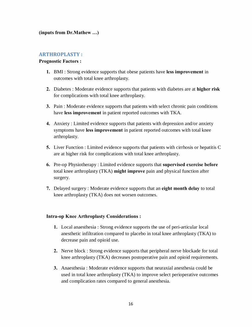
(inputs from Dr.Mathew …)
ARTHROPLASTY : Prognostic Factors :
1. BMI : Strong evidence supports that obese patients have less improvement in outcomes with total knee arthroplasty.
2. Diabetes : Moderate evidence supports that patients with diabetes are at higher risk for complications with total knee arthroplasty.
3. Pain : Moderate evidence supports that patients with select chronic pain conditions have less improvement in patient reported outcomes with TKA.
4. Anxiety : Limited evidence supports that patients with depression and/or anxiety symptoms have less improvement in patient reported outcomes with total knee arthroplasty.
5. Liver Function : Limited evidence supports that patients with cirrhosis or hepatitis C are at higher risk for complications with total knee arthroplasty.
6. Pre-op Physiotherapy : Limited evidence supports that supervised exercise before total knee arthroplasty (TKA) might improve pain and physical function after surgery.
7. Delayed surgery : Moderate evidence supports that an eight month delay to total knee arthroplasty (TKA) does not worsen outcomes.
Intra-op Knee Arthroplasty Considerations :
1. Local anaesthesia : Strong evidence supports the use of peri-articular local anesthetic infiltration compared to placebo in total knee arthroplasty (TKA) to decrease pain and opioid use.
2. Nerve block : Strong evidence supports that peripheral nerve blockade for total knee arthroplasty (TKA) decreases postoperative pain and opioid requirements.
3. Anaesthesia : Moderate evidence supports that neuraxial anesthesia could be used in total knee arthroplasty (TKA) to improve select perioperative outcomes and complication rates compared to general anesthesia.

17
4. Tourniquet and blood loss : Moderate evidence supports that the use of a tourniquet in total knee arthroplasty (TKA) decreases intraoperative blood loss.
5. Tourniquet and Pain : Strong evidence supports that tourniquet use in total knee arthroplasty (TKA) increases short term post-operative pain.
Limited evidence supports that tourniquet use in total knee arthroplasty (TKA) decreases short term post-operative function.
6. Tranexemic Acid : Strong evidence supports that, in patients with no known contraindications, treatment with tranexamic acid decreases postoperative blood loss and reduces the necessity of postoperative transfusions following total knee arthroplasty (TKA).
7. Bone Cement : Limited evidence does not support the routine use of antibiotics in the cement for primary total knee arthroplasty.
8. Implant Design : Strong evidence supports no difference in outcomes or complications between posterior stabilized and posterior cruciate retaining arthroplasty designs. Strong evidence supports use of either all-polyethylene or modular tibial components in knee arthroplasty (KA) because of no difference in outcomes.
Strong evidence supports no difference in pain or function with or without patellar resurfacing in total knee arthroplasty.
Moderate evidence supports that patellar resurfacing in total knee arthroplasty (TKA) could decrease cumulative reoperations after 5 years when compared to no patellar resurfacing in total knee arthroplasty (TKA).
Strong evidence supports the use of tibial component fixation that is cemented or cementless in total knee arthroplasty due to similar functional outcomes and rates of complications and reoperations.
Moderate evidence supports the use of either cemented femoral and tibial components or cementless femoral and tibial components in knee arthroplasty due to similar rates of complications and reoperations.
Moderate evidence supports the use of either cementing all components or hybrid fixation (cementless femur) in total knee arthroplasty due to similar functional outcomes and rates of complications and reoperations.
9. Cement : Limited evidence supports the use of either all cementless components or hybrid fixation (cementless femur) in total knee arthroplasty due to similar rates of complications and reoperations.

18
10. Bilateral Arthroplasty Indications : Limited evidence supports simultaneous bilateral total knee arthroplasty (TKA) for patients aged 70 or younger or ASA status 1-2, because there are no increased complications.
11. Unicondylar Knee : Moderate evidence supports that total knee arthroplasty (TKA) could be used to decrease revision surgery risk compared to unicompartmental knee arthroplasty (UKA) for medial compartment osteoarthritis.
Limited evidence supports that unicompartmental knee arthroplasty might be used to decrease the risk of deep vein thrombosis (DVT) and manipulation under anesthesia compared to total knee arthroplasty (TKA) for medial compartment osteoarthritis.
12. Unicondylar vs Osteotomy : Moderate evidence supports no difference between unicompartmental knee arthroplasty (UKA) or valgus-producing proximal tibial osteotomy in outcomes and complications in patients with medial compartment knee osteoarthritis.
13. Navigation : Strong evidence supports not using intraoperative navigation in total knee arthroplasty (TKA) because there is no difference in outcomes or complications.
14. Patient Specific : Strong evidence supports not using patient specific instrumentation compared to conventional instrumentation for total knee arthroplasty (TKA) because there is no difference in pain or functional outcomes.
Moderate evidence supports not using patient specific instrumentation compared to conventional instrumentation for total knee arthroplasty (TKA) because there is no difference in transfusions or complications.
15. Drains : Strong evidence supports not using a drain with total knee arthroplasty.
Post op Knee Arthroplasty Considerations :
1. Cryotherapy : Moderate evidence supports that cryotherapy devices after knee arthroplasty (KA) do not improve outcomes.
2. CPM : Strong evidence supports that CPM after knee arthroplasty (KA) does not improve outcomes.
3. Hospital Stay : Strong evidence supports that rehabilitation started on the day of the total knee arthroplasty (TKA) reduces length of hospital stay.
19
4. Rehabilitation : Moderate evidence supports that rehabilitation started on day of total knee arthroplasty (TKA) compared to rehabilitation started on postop day 1 reduces pain and improves function.
Moderate evidence supports that a supervised exercise program during the first two months after total knee arthroplasty (TKA) improves physical function.
Limited evidence supports that a supervised exercise program during the first two months after total knee arthroplasty (TKA) decreases pain.
Limited evidence supports that selected patients might be referred to an intensive supervised exercise program during late stage post total knee arthroplasty (TKA) to improve physical function.
4.7 Summary General Core Treatment : Individualised self-management strategies with the person with osteoarthritis. Ensure that positive behavioural changes, such as exercise, weight loss, use of suitable footwear, braces, walking aids and pacing, are appropriately targeted.
DO’s : Weight loss, Thermotherapy, Electrotherapy (TENS), Physiotherapy (Exercises and manipulation and stretchings), Footwear, Braces, Walking Aids And Pacing
Don’t’s : Nutraceuticals (glucosamine or chondroitin), Acupuncture
Pharmacological Management :
Consider topical NSAIDs and/or paracetamol ahead of oral NSAIDs, COX-2 inhibitors or opioids which should be given at the lowest dose for shortest period along with lowest cost PPI, taking into account individual patient risk factors.
DO’s : Topical capsaicin , Intra-articular corticosteroid injections
Don’t’s : Rubefacients , Intra-articular hyaluronan
Warning : Patient on low-dose aspirin, consider other analgesics before substituting or adding an NSAID or COX-2 inhibitor (with a PPI).
Surgery:

20
Appropriate referral for surgery in patients having pain, stiffness and reduced function that have a substantial impact on their quality of life and are refractory to non-surgical treatment.
Do’s : Age, sex, smoking, obesity and co-morbidities not barriers for surgery. Explain before surgery the benefits and risks of surgery and the potential consequences of not having surgery, recovery and rehabilitation after surgery.
Osteotomy or unicondylar same outcome.
Don’t’s : No surgery to be done without initial core treatment. Arthroscopic lavage and debridement (unless mechanical locking of knee). Bilateral knee replacement above age 70 years or ASA status more than 2. Navigation and patient specific instrumentation has no difference in outcome.
21
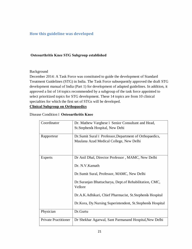
How this guideline was developed
Osteoarthritis Knee STG Subgroup established
Background December 2014: A Task Force was constituted to guide the development of Standard Treatment Guidelines (STG) in India. The Task Force subsequently approved the draft STG development manual of India (Part 1) for development of adapted guidelines. In addition, it approved a list of 14 topics recommended by a subgroup of the task force appointed to select prioritized topics for STG development. These 14 topics are from 10 clinical specialties for which the first set of STGs will be developed. Clinical Subgroup on Orthopaedics
Disease Condition – Osteoarthritis Knee
Coordinator Dr. Mathew Varghese – Senior Consultant and Head, St.Stephen’s Hospital, New Dehi
Rapporteur Dr.Sumit Sural – Professor,Department of Orthopaedics, Maulana Azad Medical College, New Delhi
Experts Dr Anil Dhal, Director Professor , MAMC, New Delhi
Dr. N.V.Kamath
Dr.Sumit Sural, Professor, MAMC, New Delhi
Dr.Suranjan Bhattacharya, Dept.of Rehabilitation, CMC, Vellore
Dr.A.K.Adhikari, Chief Pharmacist, St.Stephen’s Hospital
Dr.Kora, Dy.Nursing Superintendent, St.Stephen’s Hospital
Physician Dr.Gurtu
Private Practitioner Dr Shekhar Agarwal, Sant Parmanand Hospital,New Delhi
22
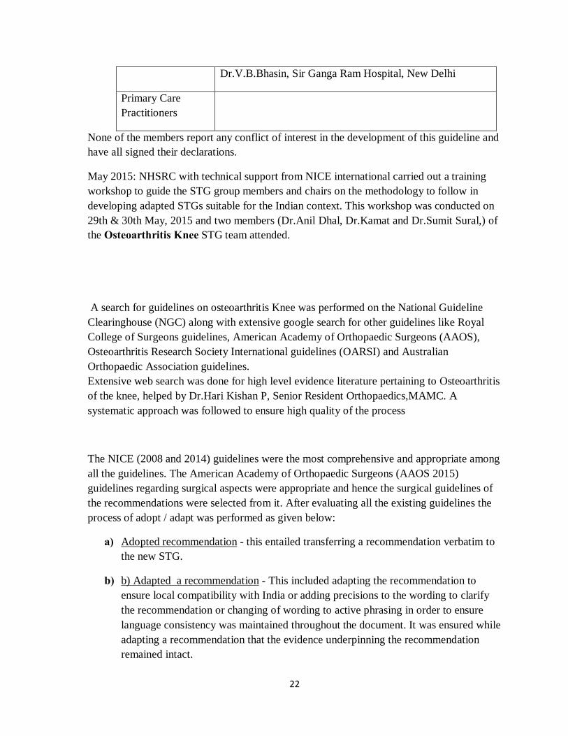
None of the members report any conflict of interest in the development of this guideline and have all signed their declarations.
May 2015: NHSRC with technical support from NICE international carried out a training workshop to guide the STG group members and chairs on the methodology to follow in developing adapted STGs suitable for the Indian context. This workshop was conducted on 29th & 30th May, 2015 and two members (Dr.Anil Dhal, Dr.Kamat and Dr.Sumit Sural,) of the Osteoarthritis Knee STG team attended.
A search for guidelines on osteoarthritis Knee was performed on the National Guideline Clearinghouse (NGC) along with extensive google search for other guidelines like Royal College of Surgeons guidelines, American Academy of Orthopaedic Surgeons (AAOS), Osteoarthritis Research Society International guidelines (OARSI) and Australian Orthopaedic Association guidelines. Extensive web search was done for high level evidence literature pertaining to Osteoarthritis of the knee, helped by Dr.Hari Kishan P, Senior Resident Orthopaedics,MAMC. A systematic approach was followed to ensure high quality of the process
The NICE (2008 and 2014) guidelines were the most comprehensive and appropriate among all the guidelines. The American Academy of Orthopaedic Surgeons (AAOS 2015) guidelines regarding surgical aspects were appropriate and hence the surgical guidelines of the recommendations were selected from it. After evaluating all the existing guidelines the process of adopt / adapt was performed as given below:
a) Adopted recommendation - this entailed transferring a recommendation verbatim to the new STG.
b) b) Adapted a recommendation - This included adapting the recommendation to ensure local compatibility with India or adding precisions to the wording to clarify the recommendation or changing of wording to active phrasing in order to ensure language consistency was maintained throughout the document. It was ensured while adapting a recommendation that the evidence underpinning the recommendation remained intact.
Dr.V.B.Bhasin, Sir Ganga Ram Hospital, New Delhi
Primary Care Practitioners

23
After going through the available guidelines, the group adopted the majority of existing guidelines and only two guidelines were adapted from NISE.
For this process, the STG subgroup met initially twice at Maulana Azad Medical College and later 3 times at St.Stephen’s Hospital where each time a video-conferencing was set up to include Dr.Suranjan Bhattacharya. One of the meetings was attended by Dr.Nikhil from the Ministry of Health and Family Welfare. The draft was written by Dr.Sumit Sural and all circulated on all experts on the e mail and subsequently the major recommendations were discussed point wise in the videoconferencing meetings.
An internal peer review meeting was held on 7th January 2016 attended by Dr.Sumit Sural and the draft was appropriately amended and circulated again on e mail to all concerned experts for their final comments.
7. Research Needs a. Evidence of alternate therapies as comparative study for pain management.
b. Future Recommendations :

24
8. Annexure (Adopt/Adapt Table) S.No STG India
Recommendations Adopted/
Adapted
Recommendations in the Original Guidelines
Reasons for Adaptation
A : General Core Treatment
A1:Patient Information
A2 :Self management
A3-6 :Physiotherapy
A7 – Supplements
A8: Alternative therapy
A9 :Orthotic devices
A1
Patient Information
Offer accurate verbal and written information to all people with osteoarthritis to enhance understanding of the condition and its management, and to counter misconceptions, such as that it inevitably progresses and cannot be treated. Ensure that information sharing is an ongoing, integral part of the management plan rather than a single event at time of presentation.
Adopt Nice (2008)
A2
Patient self-management interventions
Agree individualised self-management strategies with the person with osteoarthritis. Ensure that positive behavioural
Adapt Nice (2008)
Agree individualised self-management strategies with the
Reason for Adapting (Dr.Suranjan Bhattacharya)

25
changes, such as exercise, weight loss, use of suitable footwear are appropriately targeted.
person with osteoarthritis. Ensure that positive behavioural changes, such as exercise, weight loss, use of suitable footwear and pacing, are appropriately targeted.
A3
Thermotherapy
The use of local heat or cold should be considered as an adjunct to core treatments
Adopt Nice (2008)
A4
Exercise and manual therapy
Advise people with osteoarthritis to exercise as a core treatment irrespective of age, comorbidity, pain severity or disability. Exercise should include:
A4.1
local muscle strengthening
Exercise has been found to be beneficial
Adapt Nice (2008)
Exercise has been found to be beneficial but the clinician needs to make a judgement in each case on how to effectively ensure participation. This will depend upon the person's individual needs,
Reason for Adapting (Dr.Suranjan Bhattacharya)

26
circumstances and self-motivation, and the availability of local facilities.
A4.2
Manipulation and stretching
Nice (2008)
should be considered as an adjunct to core treatments, particularly for osteoarthritis of the hip.
A 5
Weight loss
Offer interventions to achieve weight loss as a core treatment for people who are obese or overweight.
Adopt Nice (2008)
A6
Electrotherapy
Should consider the use of transcutaneous electrical nerve stimulation (TENS) as an adjunct to core treatments for pain relief.
Adopt Nice (2008)
A7
Nutraceuticals
Do not offer glucosamine or chondroitin products for the management of osteoarthritis.
Adopt Nice (2014)
A8
Acupuncture
Do not offer acupuncture for the management of osteoarthritis
Adopt Nice (2014)
A9

27
Aids and devices
A9.1
Footwear
Offer advice on appropriate footwear (including shock-absorbing properties) as part of core treatments for people with lower limb osteoarthritis.
Adopt Nice (2008)
A9.2
Brace / Insole
People with osteoarthritis who have biomechanical joint pain or instability should be considered for assessment for bracing/joint supports/insoles as an adjunct to their core treatments.
Adopt Nice (2008)
A9.3
Walking Aid
Assistive devices (for example, walking sticks and tap turners) should be considered as adjuncts to core treatments for people with osteoarthritis who have specific problems with activities of daily living. If needed, seek expert advice in this context
Adopt Nice (2008)
B PHARMACOLOGICAL MANAGEMENT
B1 :Analgesics
B2 :Topical

28
B3 : NSAID
B4 : Intra-articular injections
B1
Oral analgesics
B1.1 Paracetamol for pain relief in addition to core treatments regular dosing may be required. Paracetamol and/or topical non-steroidal anti-inflammatory drugs (NSAIDs) should be considered ahead of oral NSAIDs, cyclo-oxygenase 2 (COX-2) inhibitors or opioids
Adopt Nice (2008)
B1.2 If paracetamol or topical NSAIDs are insufficient for pain relief for people with osteoarthritis, then the addition of opioid analgesics should be considered. Risks and benefits should be considered, particularly in older people.
Adopt Nice (2008)
B2
Topical treatments
B2.1 Consider topical NSAIDs and/or
Adopt Nice (2008)
29
paracetamol ahead of oral NSAIDs, COX-2 inhibitors or opioids.
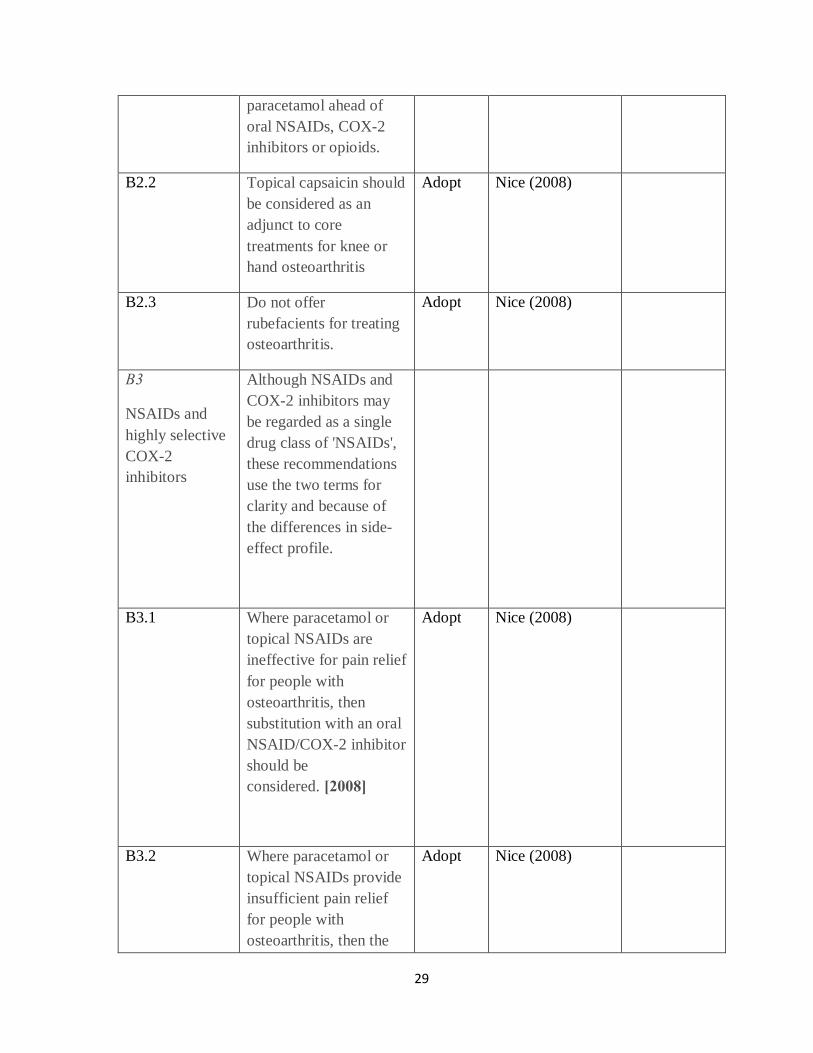
B2.2 Topical capsaicin should be considered as an adjunct to core treatments for knee or hand osteoarthritis
Adopt Nice (2008)
B2.3 Do not offer rubefacients for treating osteoarthritis.
Adopt Nice (2008)
B3
NSAIDs and highly selective COX-2 inhibitors
Although NSAIDs and COX-2 inhibitors may be regarded as a single drug class of 'NSAIDs', these recommendations use the two terms for clarity and because of the differences in side-effect profile.
B3.1 Where paracetamol or topical NSAIDs are ineffective for pain relief for people with osteoarthritis, then substitution with an oral NSAID/COX-2 inhibitor should be considered. [2008]
Adopt Nice (2008)
B3.2 Where paracetamol or topical NSAIDs provide insufficient pain relief for people with osteoarthritis, then the
Adopt Nice (2008)

30
addition of an oral NSAID/COX-2 inhibitor to paracetamol should be considered.
B3.3 Use oral NSAIDs/COX-2 inhibitors at the lowest effective dose for the shortest possible period of time
Adopt Nice (2008)
B3.4 When offering treatment with an oral NSAID/COX-2 inhibitor, the first choice should be either a standard NSAID or a COX-2 inhibitor (other than etoricoxib 60 mg). In either case, co-prescribe with a proton pump inhibitor (PPI), choosing the one with the lowest acquisition cost.
Adopt Nice (2008)
B3.5 All oral NSAIDs/COX-2 inhibitors have analgesic effects of a similar magnitude but vary in their potential gastrointestinal, liver and cardio-renal toxicity; therefore, when choosing the agent and dose, take into account individual patient risk factors, including age. When prescribing these drugs, consideration should be given to
Adopt Nice (2008)
31
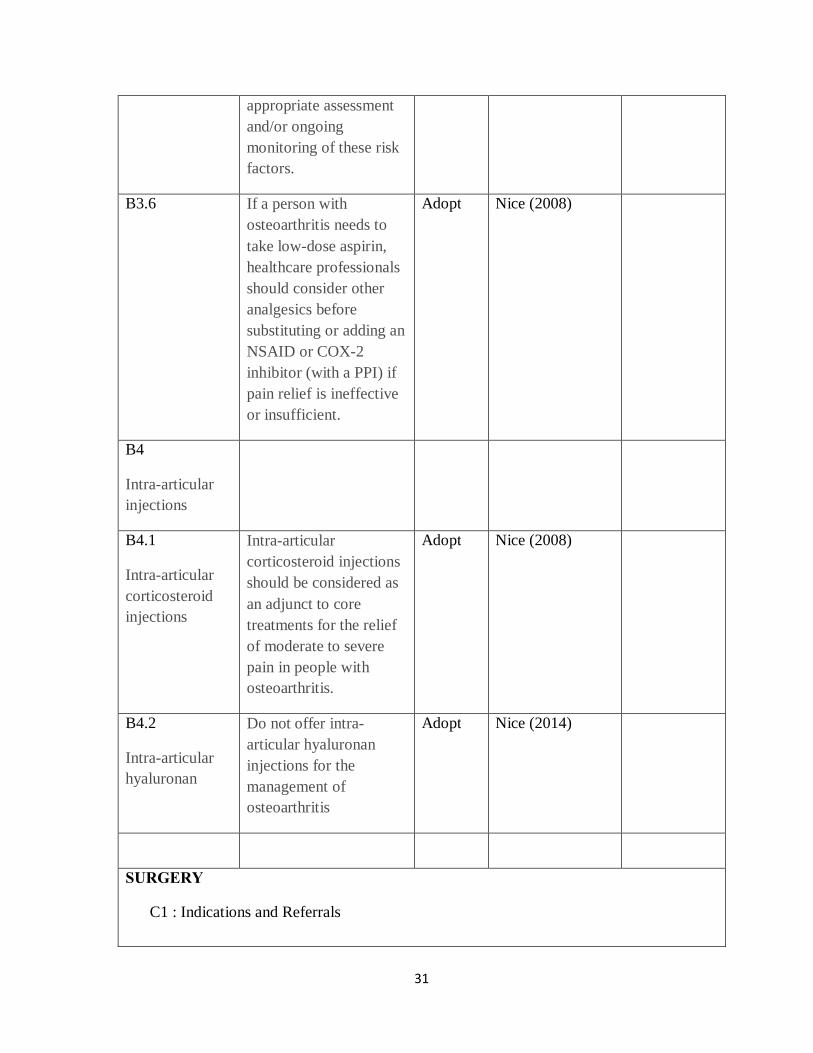
appropriate assessment and/or ongoing monitoring of these risk factors.
B3.6 If a person with osteoarthritis needs to take low-dose aspirin, healthcare professionals should consider other analgesics before substituting or adding an NSAID or COX-2 inhibitor (with a PPI) if pain relief is ineffective or insufficient.
Adopt Nice (2008)
B4
Intra-articular injections
B4.1
Intra-articular corticosteroid injections
Intra-articular corticosteroid injections should be considered as an adjunct to core treatments for the relief of moderate to severe pain in people with osteoarthritis.
Adopt Nice (2008)
B4.2
Intra-articular hyaluronan
Do not offer intra-articular hyaluronan injections for the management of osteoarthritis
Adopt Nice (2014)
SURGERY
C1 : Indications and Referrals

32
C2 : Follow up and Reviews
C3: Arthroscopy
C4 : Osteotomy
C5 : Arthroplasty
Clinicians with responsibility for referring a person with osteoarthritis for consideration of joint surgery should ensure that the person has been offered at least the core (non-surgical) treatment options
Adopt Nice (2008)
Base decisions on referral thresholds on discussions between patient representatives, referring clinicians and surgeons, rather than using scoring tools for prioritisation.
Adopt Nice (2008, amended 2014)
Consider referral for joint surgery for people with osteoarthritis who experience joint symptoms (pain, stiffness and reduced function) that have a substantial impact on their quality of life and are refractory to non-surgical treatment.
Adopt Nice (2008, amended 2014)
Refer for consideration of joint surgery before there is prolonged and
Adopt Nice (2008, amended 2014)
33
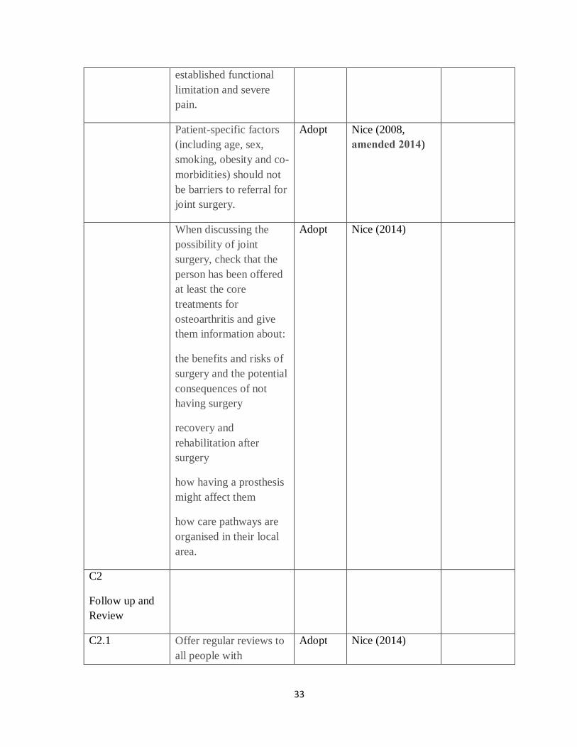
established functional limitation and severe pain.
Patient-specific factors (including age, sex, smoking, obesity and co-morbidities) should not be barriers to referral for joint surgery.
Adopt Nice (2008, amended 2014)
When discussing the possibility of joint surgery, check that the person has been offered at least the core treatments for osteoarthritis and give them information about:
the benefits and risks of surgery and the potential consequences of not having surgery
recovery and rehabilitation after surgery
how having a prosthesis might affect them
how care pathways are organised in their local area.
Adopt Nice (2014)
C2
Follow up and Review
C2.1 Offer regular reviews to all people with
Adopt Nice (2014)

34
symptomatic osteoarthritis. Agree the timing of the reviews with the person Reviews should include:
monitoring the person's symptoms and the ongoing impact of the condition on their everyday activities and quality of life
monitoring the long-term course of the condition
discussing the person's knowledge of the condition, any concerns they have, their personal preferences and their ability to access services
reviewing the effectiveness and tolerability of all treatments
support for self-management.
C2.2 Consider an annual review for any person with one or more of the following:
troublesome joint pain
more than one joint with symptoms
more than one co-
Adopt Nice (2014)

35
morbidity
taking regular medication for their osteoarthritis.
C3 :
Arthroscopy
Do not refer for arthroscopic lavage and debridement as part of treatment for osteoarthritis, unless the person has knee osteoarthritis with a clear history of mechanical locking (as opposed to morning joint stiffness, 'giving way' or X-ray evidence of loose bodies).
Adopt Nice (2008, amended 2014)
C4:
Osteotomy
C5 :Arthroplasty
Prognostic Factors :
BMI Strong evidence supports that obese patients have less improvement in outcomes with total knee arthroplasty
Adopt AAOS (2015)
Strong Evidence
Diabetes Moderate evidence supports that patients with diabetes are at
Adopt AAOS (2015)
Moderate

36
higher risk for complications with total knee arthroplasty
Evidence
Pain Moderate evidence supports that patients with select chronic pain conditions have less improvement in patient reported outcomes with TKA.
Adopt AAOS (2015)
Moderate Evidence
Anxiety Limited evidence supports that patients with depression and/or anxiety symptoms have less improvement in patient reported outcomes with total knee arthroplasty
Adopt AAOS (2015)
Limited Evidence
Liver Function Limited evidence supports that patients with cirrhosis or hepatitis C are at higher risk for complications with total knee arthroplasty
Adopt AAOS (2015)
Limited Evidence
Pre-op Physiotherapy
Limited evidence supports that supervised exercise before total knee arthroplasty (TKA) might improve pain and physical function after surgery.
Adopt AAOS (2015)
Limited Evidence
Delayed surgery Moderate evidence supports that an eight month delay to total knee arthroplasty (TKA) does not worsen
Adopt AAOS (2015)
Moderate Evidence

37
outcomes.
Intra-op Considerations
Local anaesthesia
Strong evidence supports the use of peri-articular local anesthetic infiltration compared to placebo in total knee arthroplasty (TKA) to decrease pain and opioid use.
Adopt AAOS (2015)
Strong Evidence
Nerve block Strong evidence supports that peripheral nerve blockade for total knee arthroplasty (TKA) decreases postoperative pain and opioid requirements.
Adopt AAOS (2015)
Strong Evidence
Anaesthesia Moderate evidence supports that neuraxial anesthesia could be used in total knee arthroplasty (TKA) to improve select perioperative outcomes and complication rates compared to general anesthesia.
Adopt AAOS (2015)
Moderate Evidence
Tourniquet and blood loss
Moderate evidence supports that the use of a tourniquet in total knee arthroplasty (TKA) decreases intraoperative blood loss.
Adopt AAOS (2015)
Moderate Evidence
Tourniquet and Pain
Strong evidence supports that tourniquet use in total knee arthroplasty (TKA)
Adopt AAOS (2015)
Strong Evidence

38
increases short term post-operative pain.
Limited evidence supports that tourniquet use in total knee arthroplasty (TKA) decreases short term post-operative function.
Adopt AAOS (2015)
Limited Evidence
Tranexemic Acid
Strong evidence supports that, in patients with no known contraindications, treatment with tranexamic acid decreases postoperative blood loss and reduces the necessity of postoperative transfusions following total knee arthroplasty (TKA).
Adopt AAOS (2015)
Strong Evidence
Bone Cement Limited evidence does not support the routine use of antibiotics in the cement for primary total knee arthroplasty
Adopt AAOS (2015)
Limited Evidence
Implant Design Strong evidence supports no difference in outcomes or complications between posterior stabilized and posterior cruciate retaining arthroplasty designs.
Adopt AAOS (2015)
Strong Evidence
Strong evidence supports use of either all-polyethylene or
Adopt AAOS (2015)
Strong Evidence

39
modular tibial components in knee arthroplasty (KA) because of no difference in outcomes.
Strong evidence supports no difference in pain or function with or without patellar resurfacing in total knee arthroplasty.
Adopt AAOS (2015)
Strong Evidence
Moderate evidence supports that patellar resurfacing in total knee arthroplasty (TKA) could decrease cumulative reoperations after 5 years when compared to no patellar resurfacing in total knee arthroplasty (TKA).
Adopt AAOS (2015)
Moderate Evidence
Strong evidence supports the use of tibial component fixation that is cemented or cementless in total knee arthroplasty due to similar functional outcomes and rates of complications and reoperations.
Adopt AAOS (2015)
Strong Evidence
Moderate evidence supports the use of either cemented femoral and tibial components or cementless femoral and tibial components in knee arthroplasty due to
Adopt AAOS (2015)
Moderate Evidence

40
similar rates of complications and reoperations.
Moderate evidence supports the use of either cementing all components or hybrid fixation (cementless femur) in total knee arthroplasty due to similar functional outcomes and rates of complications and reoperations.
Adopt AAOS (2015)
Moderate Evidence
Cement Limited evidence supports the use of either all cementless components or hybrid fixation (cementless femur) in total knee arthroplasty due to similar rates of complications and reoperations.
Adopt AAOS (2015)
Limited Evidence
Bilateral Limited evidence supports simultaneous bilateral total knee arthroplasty (TKA) for patients aged 70 or younger or ASA status 1-2, because there are no increased complications.
Adopt AAOS (2015)
Limited Evidence
Unicondylar Moderate evidence supports that total knee arthroplasty (TKA) could be used to decrease revision surgery risk compared to
Adopt AAOS (2015)
Moderate Evidence
41
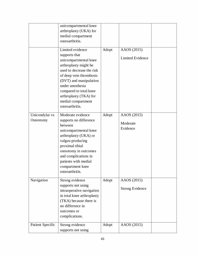
unicompartmental knee arthroplasty (UKA) for medial compartment osteoarthritis.
Limited evidence supports that unicompartmental knee arthroplasty might be used to decrease the risk of deep vein thrombosis (DVT) and manipulation under anesthesia compared to total knee arthroplasty (TKA) for medial compartment osteoarthritis.
Adopt AAOS (2015)
Limited Evidence
Unicondylar vs Osteotomy
Moderate evidence supports no difference between unicompartmental knee arthroplasty (UKA) or valgus-producing proximal tibial osteotomy in outcomes and complications in patients with medial compartment knee osteoarthritis.
Adopt AAOS (2015)
Moderate Evidence
Navigation Strong evidence supports not using intraoperative navigation in total knee arthroplasty (TKA) because there is no difference in outcomes or complications.
Adopt AAOS (2015)
Strong Evidence
Patient Specific Strong evidence supports not using
Adopt AAOS (2015)

42
patient specific instrumentation compared to conventional instrumentation for total knee arthroplasty (TKA) because there is no difference in pain or functional outcomes.
Strong Evidence
Moderate evidence supports not using patient specific instrumentation compared to conventional instrumentation for total knee arthroplasty (TKA) because there is no difference in transfusions or complications.
Adopt AAOS (2015)
Moderate Evidence
Drains Strong evidence supports not using a drain with total knee arthroplasty
Adopt AAOS (2015)
Strong Evidence
Post op
Cryotherapy Moderate evidence supports that cryotherapy devices after knee arthroplasty (KA) do not improve outcomes.
Adopt AAOS (2015)
Moderate Evidence
CPM Strong evidence supports that CPM after knee arthroplasty (KA) does not improve
Adopt AAOS (2015)
Strong Evidence

43
outcomes.
Stay Strong evidence supports that rehabilitation started on the day of the total knee arthroplasty (TKA) reduces length of hospital stay.
Adopt AAOS (2015)
Strong Evidence
Rehab Moderate evidence supports that rehabilitation started on day of total knee arthroplasty (TKA) compared to rehabilitation started on postop day 1 reduces pain and improves function.
Adopt AAOS (2015)
Moderate Evidence
Moderate evidence supports that a supervised exercise program during the first two months after total knee arthroplasty (TKA) improves physical function.
Adopt AAOS (2015)
Moderate Evidence
Limited evidence supports that a supervised exercise program during the first two months after total knee arthroplasty (TKA) decreases pain.
Adopt AAOS (2015)
Limited Evidence
Limited evidence supports that selected patients might be referred to an intensive
Adopt AAOS (2015)
Limited Evidence

44
supervised exercise program during late stage post total knee arthroplasty (TKA) to improve physical function.

45
References 1. Cross M, Smith E, Hoy D, Nolte S, Ackerman I, Fransen M, Bridgett L, Williams
S, Guillemin F, Hill CL, Laslett LL, Jones, Cicuttini F, Osborne R, Vos , Buchbinder R, Woolf A, March L. The global burden of hip and knee osteoarthritis: estimates from the global burden of disease 2010 study. Ann Rheum Dis. 2014 Jul;73(7):1323-30.
2. http:// icmr.nic.in/final/S.J.H.%20Final%20Project%20Report%202012.
3. Chandra Prakash Pal, Pulkesh Singh, Sanjay Chaturvedi, Kaushal Kumar Pruthi, and Ashok Vij. Epidemiology of knee osteoarthritis in India and related factor. Indian J Orthop. 2016 Sep; 50(5): 518–522.
4. Sacks JJ, Helmick CG, Langmaid G. Deaths from arthritis and other rheumatic conditions, United States, 1979–1998. J Rheumatol. 2004;31:1823-1828.
5. Arthritis and Related Conditions Chapter 4. http://www.boneandjointburden.org/2013-report/iv-arthritis/iv. Accessed 07-23-2015.
6. Guccione AA, Felson DT, Anderson JJ, Anthony JM, Zhang Y, Wilson PW, Kelly-Hayes M, Wolf PA, Kreger BE, Kannel WB. The effects of specific medical conditions on the functional limitations of elders in the Framingham Study. Am J Pub Health. 1994;84(3):351-358.
7. Leach RE, Gregg T, Siber FJ. Weight-bearing radiography in osteoarthritis of the knee. Radiology 1970;97: 265–8.
8. Chondrometry Lequesne M. Quantitative evaluation of joint space width and rate of joint space loss in osteoarthritis of the hip. Rev Rhum Engl Ed 1995;62: 155–8.
9. Ravaud P, Chastang C, Auleley GR, Giraudeau B, Royant V, Amor B, et al. Assessment of joint space width in patients with osteoarthritis of the knee: a comparison of 4 measuring instruments. J Rheumatol 1996;23:1749–55.
10. Roemer FW, Crema MD, Trattnig S, Guermazi A. Advances in imaging of osteoarthritis and cartilage. Radiology 2011;260:332–54.
11. Conrozier T, Lequesne M, Favret H, Taccoen A, Mazieres B, Dougados M, et al. Measurement of the radiological hip joint space width. An evaluation of various methods of measurement. Osteoarthritis Cartilage 2001;9:281–6.
46
12. Vignon E. Radiographic issues in imaging the progression of hip and knee osteoarthritis. J Rheumatol Suppl 2004;70:36–44.
13. Bauer DC, Hunter DJ, Abramson SB, Attur M, Corr M, Felson D, et al. Classification of osteoarthritis biomarkers: a proposed approach. Osteoarthritis Cartilage.2006;14:723–7.
14. Kellgren JH, Lawrence JS. Radiological assessment of osteo-arthrosis. Ann Rheum Dis 1957 : 16:494-502.
15. Altman RD. Gold GE. Atlas of individual radiographic features in osteoarthritis, revised. Osteoarthritis Cartilage 2007;15(Suppl A):A1–A56.
16. Altman RD, Hochberg M, Murphy Jr WA, Wolfe F, Lequesne M. Atlas of individual radiographic features in osteoarthritis. Osteoarthritis Cartilage 1995;3(Suppl A):
17. Amin S, LaValley MP, Guermazi A, Grigoryan M, Hunter DJ, Clancy M, et al. The relationship between cartilage loss on magnetic resonance imaging and radiographic progression in men and women with knee osteoarthritis. Arthritis Rheum 2005;52:3152–9.
18. Leach RE, Gregg T, Siber FJ. Weightbearing radiography in osteoarthritis of the knee. Radiology 1970;97:265-268.
19. Cibere J. Do we need radiographs to diagnose osteoarthritis? Best Pract Res Clin Rheumatol 2006;20:27-30.
20. Manal Hasan, Rhonda Shuckett. Clinical features and pathogenetic mechanisms of osteo -arthritis of the hip and kneewww.bcmj.org VOL. 52 NO. 8, October 2010 BC Medical Journal 399.
21. Boegard TL, Rudling O, Petersson IF, et al. Correlation between radiographically diagnosed osteophytes and magnetic resonance detected cartilage defects in the patellofemoral joint. Ann Rheum Dis 1998;57:395-400.
9. List of Abbreviations OA : Osteoarthritis STG : Standard Treatment Guidelines NICE : National Institute of Health and Care Excellence, United Kingdom AAOS : American Association of Orthopaedic Surgeons / American Academy of Orthopaedic Surgeons OARSI : Osteoarthritis Research Society International guidelines AP View :Anterop-posterior view PA View : Postero-anterior view
47
JSN : Joint space narrowing JSW : Joint space width KL grading : Kellgren-Lawrence grading TKR : Total Knee Replacement TKA : Total Knee Arthroplasty UKA : Unicompartmental knee arthroplasty NSAID : Nonsteroidal anti-inflammatory drug COX 2 : Cyclooxygenase-2 (COX-2 Inhibitors – a type of NSAID) PPI : Proton Pump Inhibitors (Pantoprazole, Omeprazole, Rabreprazole) TENS : Transcutaneous electrical nerve stimulation (Type of electrotherapy) DVT : Deep Vein Thrombosis