Embed Size (px)
DESCRIPTION
Staging and Less Invasive Tx of Esophageal Cancer. Jun Haeng Lee Sungkyunkwan University School of Medicine Samsung Medical Center, Seoul, Korea,. Diagnosis and staging of esophageal cancer. Siersema. Gastroenterol Clin N Am 2008:37:943-964. Different modalities have different roles. - PowerPoint PPT Presentation
Citation preview

Staging and Less Invasive Tx of Esophageal Cancer
Jun Haeng Lee
Sungkyunkwan University School of MedicineSamsung Medical Center, Seoul, Korea,


Siersema. Gastroenterol Clin N Am 2008:37:943-964
Diagnosis and staging of esophageal cancer

Siersema. Gastroenterol Clin N Am 2008:37:943-964
Different modalities have different roles
• For the evaluation of distant metastases, FDG-PET may have a higher sensitivity than CT.
• For the detection of regional and celiac lymph node metastases, EUS is most sensitive, whereas CT and FDG-PET are more specific tests.
• The combined use of FDG-PET and CT, which is increasingly being applied, could be of clinical value, with FDG-PET detecting possible metastases and CT confirming or excluding their presence and precisely determining their location.

Why accurate staging is important?
• Accurate staging is essential to select patients who will benefit from aggressive therapy and to avoid aggressive therapy in patients with distant metastases.
• Despite these efforts, metastatic spread is encountered during operation in up to 60% of patients.
• No one technology can completely stage all aspects of esophageal carcinoma with high accuracy.
Sleisenger & Fordtran's Gastrointestinal and Liver Disease, 8th ed. Ch 44

Comparative study will not be coming.
• The current enthusiasm for neoadjuvant therapy makes it unlikely that definitive studies comparing accuracy of specific or combination staging modalities will be forthcoming.
• Staging of newly diagnosed esophageal cancer may incorporate crosssectional imaging, EUS, positron emission tomography (PET) scanning, transcutaneous ultrasound scanning of the neck, laparoscopy, and video-assisted thoracoscopy (VATS) staging.
Sleisenger & Fordtran's Gastrointestinal and Liver Disease, 8th ed. Ch 44

Role of Endoscopy
Jun Haeng Lee
Sungkyunkwan University School of MedicineSamsung Medical Center, Seoul, Korea,
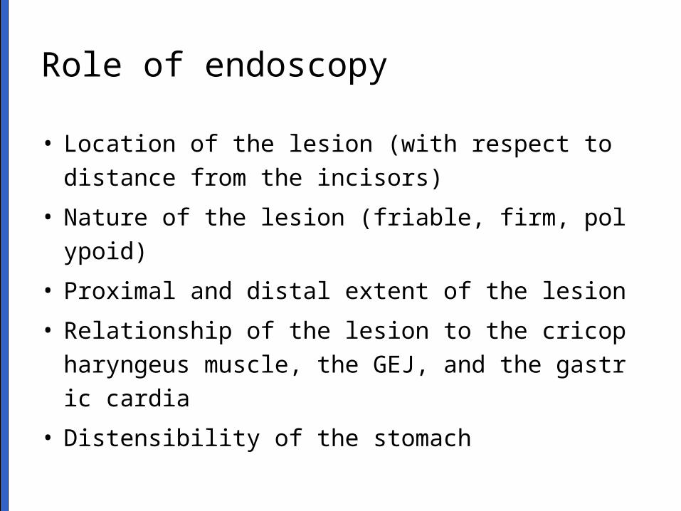
Role of endoscopy
• Location of the lesion (with respect to distance fr
om the incisors)
• Nature of the lesion (friable, firm, polypoid)
• Proximal and distal extent of the lesion
• Relationship of the lesion to the cricopharyngeus
muscle, the GEJ, and the gastric cardia
• Distensibility of the stomach

Early esophageal cancer- carcinoma in-situ. 1.2x0.4 cm. confined to the basement membrane in ESD

Esophageal cancer- usefulness of Lugol chromoendoscopy
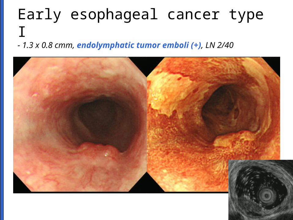
Early esophageal cancer type I- 1.3 x 0.8 cmm, endolymphatic tumor emboli (+), LN 2/40

Advanced esophageal cancer- extension to perimuscular adventitia, LN 3/62

Esophageal cancer + EGC (M/60)

Esophageal cancer after lye stricture (F/48)
Invasive SCC (M/D), 2x1.8 cm, PM, 0/24

CT, MRI, EUS
Jun Haeng Lee
Sungkyunkwan University School of MedicineSamsung Medical Center, Seoul, Korea,

CT and MRI
• CT is much less accurate in detecting lymph node metastases and is more accurate for subdiaphragmatic lymph nodes than for mediastinal ones.
• MRI can assess mediastinal invasion and liver metastasis as well as CT can but has not demonstrated any significant advantages. Because of accessibility and its lower cost, CT is preferred.
• The main limitations of CT are its insensitivity to the identification of irresectability (T4) and its inability to identify metastatic disease in normal-sized lymph nodes.
Sleisenger & Fordtran's Gastrointestinal and Liver Disease, 8th ed. Ch 44
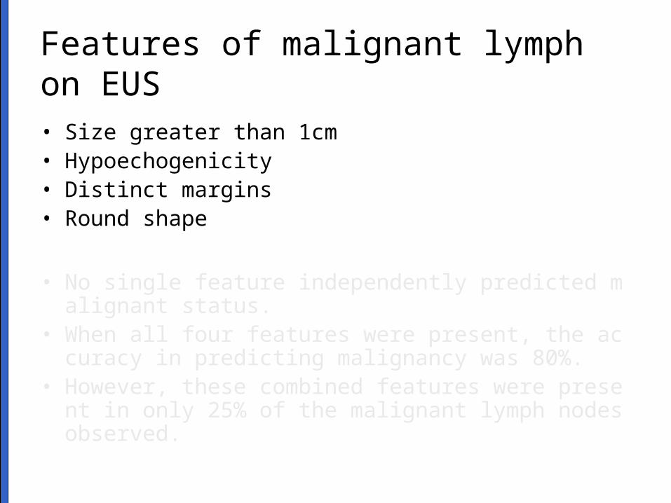
Features of malignant lymph on EUS
• Size greater than 1cm• Hypoechogenicity• Distinct margins• Round shape
• No single feature independently predicted malignant status.
• When all four features were present, the accuracy in predicting malignancy was 80%.
• However, these combined features were present in only 25% of the malignant lymph nodes observed.

Accuracy of EUS in early studies
Number Accuracy (%)
T1 185 81
T2 153 76
T3 419 92
T4 153 86
N0 231 69
N1 343 89
Rosch. Gastrointest Endosc Clin N Am 1995;5:537

Eloubeidi. Am J Gastroenterol 2009;104:53-56
Limitation of EUS (1): safety concerns

Limitation of EUS (2): publication bias- positive results 만 보고되는 경향이 있다 .
Harewood GC. Am J Gastroenterol 2005:100;808-816

Limitation of EUS (3): publication bias- 직장암에서 EUS 의 성적이 점점 나쁘게 보고되고 있다
Harewood GC. Am J Gastroenterol 2005:100;808-816

Limitation of EUS (4): too subjective- T-staging by EUS is strongly influenced by the endoscopic impression
Yanai. Intern J Gastointest Cancer 2003;34:1-8

Limitation of EUS (5): lack of experience
• 식도암 병기 결정에 있어서 EUS 의 정확도 (국립암센터 , 2008 대한 Hp 학회 추계학술대회 )
• Overall T 병기 정확도 : 73.9%– 분화암보다 미분화암에서 유의하게 낮은 결과 – 표재성 식도암에서 점막암 및 점막하층암 진단
정확도 : 53.8% (miniprobe 67.3%, radial 38.8%)
• Overall N 병기 정확도 : 72.7%
Korean J Helicobacter and Upper GI Gastrointest Res 2008;8(Suppl 2): 68

Twine. Surg Endosc 2009 May 14. [Epub ahead of print]
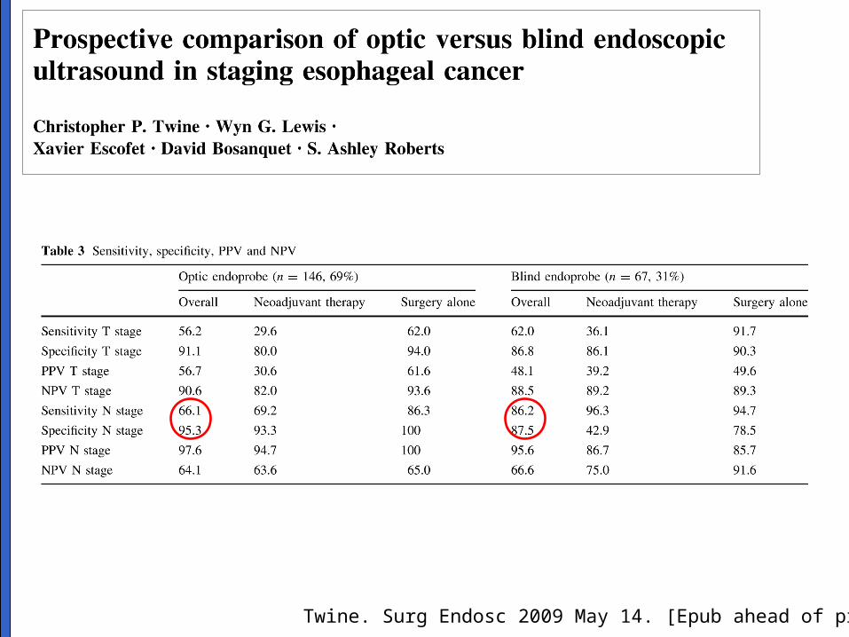
Twine. Surg Endosc 2009 May 14. [Epub ahead of print]

Role of PET
Jun Haeng Lee
Sungkyunkwan University School of MedicineSamsung Medical Center, Seoul, Korea,

NORMAL TUMOR
• Overexpression of Glucose transporters• Higher levels of Hexokinase• Down-regulation of Glucose-6-phosphatase• Anaerobic glycolysis, less ATP per glucose molecule, more glucose molecules needed for ATP production• General increase in metabolism from high growth rates

SMC experience
Yoon YC. Radiology 2003;227:764-770

PET is very useful for distant mets
van Westreenen HL. J Clin Oncol 2004;22:3805-3812

Esophageal cancer with LN mets

BUT, less useful for locoregional mets
van Westreenen HL. J Clin Oncol 2004;22:3805-3812
♠ In the included studies, change in patient management ranged from 3% to 20% due to the addition of PET to preoperative workup.

EMR/ESD for early esophageal cancer
Jun Haeng Lee
Sungkyunkwan University School of MedicineSamsung Medical Center, Seoul, Korea,

ESD (n=243)
Intramucosal differentiated cancer (n=196)
submucosal invasion orundifferentiated cancer (n=47)
Surgery (n=34) Follow up (n=13)
No recurrence
Median follow-up: 17 months (range: 4-37 months)
Complete resection (n=182) Not assessable (n=10) Incomplete resection (n=4)
Surgery (n=3)
Follow up (n=5)
Follow up (n=2)
Surgery (n=2)
Metachronousrecurrence
(n=9)
ESD (n=8)
Surgery (n=1)
Local recurrence
(n=1)
Surgery (n=1)
ESD(n=2)
Less than two EGD
follow up (n=7)
F/U after ESD for EGC at SMC
Min. Dig Liver Dis. 2009 Mar;41(3):201-9
Surgery for mucosal cancer: 7 (2.9%)

EMR was not considered as a Tx option- NCCN treatment guideline 2008
http://www.nccn.org/

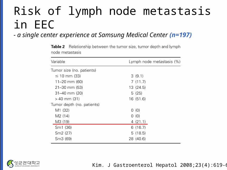
Risk of lymph node metastasis in EEC- a single center experience at Samsung Medical Center (n=197)
Kim. J Gastroenterol Hepatol 2008;23(4):619-625

Kodama. Surgery 1998;123:432-439
Risk of lymph node metastasis in EEC- a multicenter study in Japan (n=1740)

Risk of lymph node metastasis depends on the gross type of EEC
Endo. Endoscopy 1993;25:672-674

Indications for EMR for SCC
Gotoda. GIE 2008;67:805-807

Methods of endoscopic treatments - tissue retrieval techniques
• Techniques without suction
– Conventional snare polypectomy without injection
– Inject and cut
– Inject, lift and cut
– Inject, precut and cut: EMR-P
– ESD: needle knife, IT kinfe, hook knife, Flex knife
• Techniques with suction
– Suction and cut: EMR-C
– Suction and ligate: EMR-L
Modified from Endoscopy 2001;33:271-275

ESD 에 대한 용어의 정리- ESD 를 EMR 에서 분리하여 새로운 시술로 봄
• ER = EMR + ESD한시적 인정 비급여

Recurrence after EMR for EEC
Ishihara. GIE 2008;67:799-804

Esophageal ESD- M/D squamous cell carcinoma (M2)

Early esophageal cancer 0.7 cm

ESD for EEC with perforation

Pech. Gut 2007;56:1625-1634
Endoscopic resection for early squamous cell carcinoma

Take home message
• 식도암의 치료 전 병기판정의 방법은 어떠한 치료를 염두에 두는가에 따라 달라질 수 있다 .
• 각 검사법이 서로 다른 역할을 가지고 있으므로 경쟁적이기보다는 보완적인 관계로 이해할 필요가 있다 .
• 조기식도암의 내시경치료는 아직 개발단계의 시술로 향후 많은 발전이 예상된다 . 현재로서는 합병증을 줄이기 위한 다양한 노력이 필요하다 .