Embed Size (px)
Citation preview

ARTICLE
Stages of Embryonic Development in the AmphipodCrustacean, Parhyale hawaiensisWilliam E. Browne,1 Alivia L. Price,1,2 Matthias Gerberding,2 and Nipam H. Patel2*1University of Chicago, Department of Molecular Genetics and Cell Biology, Committee on Developmental Biology,Chicago, Illinois2University of California – Berkeley, Departments of Molecular Cell Biology, Integrative Biology,Center for Integrative Genomics, and HHMI, Berkeley, California
Received 1 March 2005; Accepted 12 May 2005
Summary: Studying the relationship between develop-ment and evolution and its role in the generation of bio-logical diversity has been reinvigorated by new techni-ques in genetics and molecular biology. However,exploiting these techniques to examine the evolution ofdevelopment requires that a great deal of detail beknown regarding the embryonic development of multiplespecies studied in a phylogenetic context. Crustaceansare an enormously successful group of arthropods andextant species demonstrate a wide diversity of morphol-ogies and life histories. One of the most speciose orderswithin the Crustacea is the Amphipoda. The embryonicdevelopment of a new crustacean model system, theamphipod Parhyale hawaiensis, is described in a seriesof discrete stages easily identified by examination of liv-ing animals and the use of commonly available molecu-lar markers on fixed specimens. Complete embryogene-sis occurs in �250 h at 268C and has been divided into30 stages. This staging data will facilitate comparativeanalyses of embryonic development among crustaceansin particular, as well as between different arthropodgroups. In addition, several aspects of Parhyale embry-onic development make this species particularly suit-able for a broad range of experimental manipulations.genesis 42:124–149, 2005. VVC 2005 Wiley-Liss, Inc.
Key words: Crustacean; amphipod; arthropod; evolution;development; embryogenesis; segmentation; Engrailed;Distalless
INTRODUCTION
The fields of development, genetics, and evolution haveadvanced dramatically in the past 25 years. This is inlarge part due to forward genetics approaches in suchspecies as Drosophila melanogaster and Caenorhabdi-
tis elegans. The information gathered from this work hasgiven us insight into developmental mechanisms andtheir underlying genetic architecture. Only a few addi-tional animal taxa (such as Xenopus laevis, Gallusdomestica, Mus musculus, Danio rerio) have comeunder the intense scrutiny received by Drosophila and
C. elegans. Thus, many critical questions remain withregard to how evolution operates over time and how ithas come to generate the full range of extant biologicaldiversity. Although we have accumulated significant datain model species, we are currently left with more ques-tions than answers regarding the tempo and mode of evo-lutionary change due to the vast evolutionary distancesbetween the current model systems. This information def-icit is largely due to the paucity of developmental, genetic,and evolutionary data available in nonmodel organisms.The descriptive analyses of gene expression in a widerange of taxa are still relatively few in number. Likewise,our ability to examine the functional basis of observedchanges in gene expression is restricted to a few species.This problem can be addressed by ‘‘filling in the gaps’’with new model systems more closely related to the cur-rent systems used by researchers. Indeed, the ultimategoal should be to ‘‘cover’’ the Tree of Life with a sufficientspread of well-studied taxa that the process of evolutioncan be approached and studied with several levels of reso-lution in all major organismal lineages. Ideally, these newtaxa should be systems in which embryos are readilyobtainable year-round and permit the design and imple-mentation of functional studies with relative ease.
Several recent studies have examined the evolutionaryrelationships among the major groups of arthropods.The data suggest two possible relationships between theInsecta (hexapods) and the Crustacea. One possibility isthat the two groups are sister taxa (Boore et al., 1995,1998; Friedrich and Tautz, 1995; Eernisse, 1997; Giribetet al., 2001). The other possibility is a ‘‘Pancrustacea’’
Present address for W.E. Browne: Kewalo Marine Lab, University of
Hawaii.
Present address for M. Gerberding: Max-Planck-Institut fur Entwicklungs-
biologie, Tubingen, Germany.
* Correspondence to: M. Gerberding, University of California, Berkeley,
Department of Integrative Biology, 3060 VLSB # 3140, Berkeley, CA 94720-3140.
E-mail: [email protected] online in Wiley InterScience (www.interscience.wiley.com).
DOI: 10.1002/gene.20145
' 2005 Wiley-Liss, Inc. genesis 42:124–149 (2005)

clade in which the insects branch from within the Crus-tacea (Regier and Shultz, 1997; Hwang et al., 2001). Inthis scenario the insects would represent a terrestrializedbranch of crustaceans. In either case the crustaceansform a key outgroup to the insects and in this contextwill help enormously in our understanding of arthropodevolution. The amphipod Parhyale hawaiensis (Dana,1853) is a crustacean species that is particularly wellsuited for developmental, genetic, and evolutionary anal-ysis and has the potential of filling an important taxo-nomic gap in current comparative studies.
While detailed staging information is available for Dro-sophila (Campos-Ortega and Hartenstein, 1985; Harten-stein, 1993), only relatively simple staging systems existfor the crustaceans Cherax (Sandeman and Sandeman,1991), and Nebalia (Olesen and Walossek, 2000).Amphipods (Peracarida; Malacostraca; Crustacea) havebeen the subject of study by developmental biologistssince the late 19th century (e.g., Langenbeck, 1898;Weygoldt, 1958). However, detailed descriptive staginginformation covering the complete course of embryo-genesis in any amphipod is largely lacking. Commonlyreferred to as beachhoppers or scuds, amphipods aremalacostracan crustaceans and thus closely affiliatedwith more familiar Crustacea such as krill, lobsters, andcrabs. Within the Crustacea, amphipods rank as one ofthe most ecologically successful and speciose extantorders and occur in nearly all known marine, fresh, andbrackish water environments as well as in high humidityterrestrial ecosystems (such as tidal zones, coastal floodplains, and forest leaf litter) (e.g., Barnard and Karaman,1991; Vinogradov et al., 1996; also Lindeman, 1991;Sherbakov et al., 1999; Kamaltynov, 1999; Vainola andKamaltynov, 1999; Sheader et al., 2000; Poltermannet al., 2000; Gasca and Haddock, 2004). They have pre-dominately exploited scavenging niches and thus an aptanalogy for the group would be ‘‘the flies of the sea.’’This ecological diversity is matched by a high level ofmorphological diversity. Several thousand amphipodspecies have been described and the current rate of sev-eral new species descriptions per year suggests that theupper limit of extant species is far higher than the cur-rent species count.
Importantly, several aspects of Parhyale hawaiensis
life history make this particular species amenable tomany types of classical and modern laboratory analysesand techniques. Parhyale hawaiensis are detritovoresthat have a circumtropical, worldwide, intertidal, andshallow water marine distribution (Shoemaker, 1956;Barnard, 1965), possibly as a species complex (Myers,1985). They have been reported to aggregate in largepopulations (>3,000/m2) on decaying mangrove leafmaterial in environments subjected to rapid changes insalinity (Poovachiranon, 1986). The ability to toleratethe rapid temperature and osmotic changes that occurin their preferred shallow-water habitat allows thisrobust species to thrive under typical laboratory condi-tions. Females brood their young in a ventral pouch andproduce embryos every 2 weeks once they reach sexual
maturity. Fertilized eggs can be removed from femalesprior to their first cleavage and are sufficiently large toperform microinjections (Gerberding et al., 2002) andblastomere isolations (Extavour, 2005) with relative ease.Eggs collected can be hatched individually and the matureanimals can subsequently be used in pairwise sister–brother or mother–son matings to generate inbred lines.The length of embryogenesis is relatively short, lastingroughly 10 days. Interestingly, the P. hawaiensis bodyplan is established by distinct and invariant cell lineagesvery early in embryogenesis (Gerberding et al., 2002).
Parhyale hawaiensis Body Plan
The basic body plan of P. hawaiensis follows that of thetypical arthropod in that the ground plan is organizedaround a series of repeating segmental units along theanterior–posterior (A-P) axis. Several synapomorphiccharacters clearly unite the Amphipoda (Schram, 1986;Schmitz, 1992). Most recognizable among these charac-ters are the lateral compression of the body, sessile com-pound eyes, and the relative orientation of the pereo-pods (appendages of thoracic segments T4–T8) to thebody axis (pereopods of T4 and T5 orient anteriorly, per-eopods of T6–T8 orient posteriorly, thus the name forthe group, amphipod) (Fig. 1). Additionally, amphipodshave large coxal plates (expanded plates attached dor-sally to the base of thoracic appendages) (Fig. 1).
Typical of most amphipods, the P. hawaiensis cepha-lon (head) is composed of the anteriormost six seg-ments. The anteriormost preantennal segment bears nopaired appendage. The remaining five segments do pos-sess paired appendages. From anterior to posterior, thepaired appendages of the head are the uniramous firstantenna (An1), uniramous second antenna (An2), gna-thobasic mandibles (Mn), biramous first maxillae (Mx1),and the biramous second maxillae (Mx2). In addition,the first thoracic segment (T1) is fused to the cephalon.The T1 appendage pair, the maxillipeds, are triramous,fused at their base, and extensively modified to assist infeeding (Fig. 1). There is a close arrangement of the gna-thal appendages, including the maxillipeds, in a basketshape around the mouth to form a highly compact buc-cal mass (Fig. 1b).
The next seven segments of the thoracic region, thesecond through eighth thoracomeres (T2–T8), eacharticulate independently and bears a pair of appendages.The T2–T6 appendages possess a modified dorsalappendage branch, an epipod, that articulates under-neath the animal posteriorly, medially, and ventrally as athin, laterally flattened sheet that functions as a respira-tory organ (Divakaran and Pillai, 1975, 1981) (Fig. 11).Sexually mature females possess an additional ventralappendage branch, termed an oosteogite, on appen-dages T2–T5 that interlock to form the protected ven-tral brood pouch into which fertilized eggs are shed andincubated until hatching (Fig. 1b). The T2 and T3appendages, termed gnathopods, are subchelate andfunction in grasping and mating. The T3 appendage is
125STAGES OF DEVELOPMENT P. HAWAIENSIS

sexually dimorphic (larger on mature males) (Fig. 1).The T4–T8 appendages, functioning in locomotion, aretermed pereopods (Fig. 1).
The abdominal region is composed of the next six seg-ments and is divided into two regions: segments A1–A3constitute the pleon and segments A4–A6 constitute the
FIG. 1. The Parhyale hawaiensis body plan. a: Schematic of the adult body plan. By convention the head (white) consists of the first sixsegments (termed the cephalon) plus the first segment of the thorax (T1). All segments from the second cephalic segment posterior bear apair of appendages. For the head these appendages from anterior to posterior are as follows: antennae 1 (An1), antennae 2 (An2), mandi-bles (Mn), first maxillae (Mx1), second maxillae (Mx2), and the maxillipeds of the first thoracic segment (T1). The pereon, composed of T2–T8, is colored dark gray. Each thoracic segment of the pereon possesses a pair of appendages and the proximal-most element of eachappendage, the coxa, has a dorsal branch which is compressed and expanded into a structure called the coxal plate (CP). The appen-dages of T2 and T3 are distinctly subchelate (clawed) in form and termed gnathopods. The gnathopod of T3 is sexually dimorphic; inmature males it is greatly enlarged (compare male to female in b). T4–T8 possess appendages termed pereopods. The abdominal seg-ments A1–A6 are colored light gray. A1–A3 form the pleon, and each bears a pair of appendages termed pleopods. The final three seg-ments of the abdomen (A4–A6) form the urosome, and each of these segments bears a pair of appendages termed uropods. At the veryposterior is the telson, which is a cleft flap of cuticle just posterior and dorsal of the anus. b: Sexually mature animals possess a number ofdimorphic characters. Males are larger than females. The appendages of T3 are greatly enlarged in males. Females possess a ventralbrood pouch in which they incubate their eggs until hatching (arrowhead). All amphipods retain a highly compact arrangement of mouth-parts termed the buccal mass (arrow).
126 BROWNE ET AL.

urosome. Each abdominal segment bears a pair of bira-mous appendages. The appendages of abdominal seg-ments one through three (A1–A3) are termed pleopodsand function in both swimming locomotion as well as inmoving water over the ventrally located thoracic gills.Each pair of pleopods is fused at its base along the ven-tral midline of the animal and is highly setose (Fig. 1).The appendages of abdominal segments four through six(A4–A6) are termed uropods. These appendages arereduced, thickened, and bear a number of stout spikesalong their proximal–distal axis (Fig. 1). The terminalstructure, the telson, is cleft and reduced in size relativeto comparable structures of other types of crustaceans.
Staging Methods
We used in vivo light microscopy and DAPI visualizationof nuclei in fixed embryos to create reference stages ofParhyale hawaiensis embryonic development. In vivostaging of living embryos is important for recognitionand identification of developmental stages prior tomanipulation. Companion DAPI images facilitate stagingof manipulated embryos through nuclear staining.Appendage development is included for accurate stagingof older embryos and studies specific to appendagedevelopment. We also used two antibodies that arebroadly cross-reactive across species as molecularmarkers during P. hawaiensis embryonic development.The product of the segment polarity gene engrailed (en)is required for proper segmentation in Drosophila andexhibits a conserved expression pattern in the posteriorcompartment of developing segments and appendagesin all arthropod species studied thus far. The en gene isalso expressed in selected sets of cells in the developingCNS (Patel et al., 1989a,b; Duman-Scheel and Patel,1999). Thus, the cross-reactive anti-EN antibody is usefulfor following the progression of segmentation as well asneurogenesis. The product of the distal appendage pat-terning gene, Distalless (Dll), possesses a conservedexpression pattern in developing appendages (Pangani-ban et al., 1997), and thus the cross-reactive anti-Dll anti-body is useful for following the establishment and devel-opment of appendage fields.
RESULTS
Parhyale hawaiensis (Fig. 1b) is small (<2 cm) and easyto maintain in very dense cultures in the laboratory.They have a high tolerance to variations in water quality,salinity, and temperature. Mature adults breed approxi-mately every 2–3 weeks, year-round. In addition, theyhave a relatively short egg-to-egg generation time of 7–8weeks when maintained at 268C. From egg deposition toegg hatching, embryogenesis takes �250 h (10 days) toreach completion (Fig. 2). All eggs in a single brood areroughly synchronous in development, as all eggs aredeposited within 1 h. The average brood size is sixembryos, with a range of as few as one and as many as25 embryos per ovigerous (egg-bearing) female. Asdirect developers, the hatchlings possess a complete
complement of segments and appendages morphologi-cally similar to those of adult animals. Females normallybrood the embryos in a ventral brood pouch. Embryoscan be rapidly and easily removed from the brood pouchand maintained in clean seawater. Furthermore, P.
hawaiensis embryos are easy to manipulate via microin-jection and thus can be subjected to a number of experi-ments designed to analyze their development (Gerberd-ing et al., 2002).
Developing P. hawaiensis embryos are optically clear,thus allowing for both detailed microscopic analysis insitu and the use of fluorescently tagged molecules (fluo-rescent dextrans, GFP, or DsRed) in live embryos. Theyolk, while opaque, is sequestered early in developmentto the center of the developing egg and then later to thedeveloping midgut of the embryo. The presence of thecolored yolk facilitates identification of embryonic cellsduring the early stages of development. In addition, earlycleavage is holoblastic (total), allowing the fates of indi-vidual early cells to be explored through experimentalmanipulation (Gerberding et al., 2002; Extavour, 2005).
Mating and Fertilization
Sexually mature male and female P. hawaiensis formmating pairs in which the males grasp and hold thesmaller females with their second thoracic appendages(T2, gnathopods) until mating occurs. The pairs remainin premating amplexus until the female molts. At thistime the male deposits sperm into the females pairedoviducts and then releases her. Before the female’s newcuticle hardens she sheds her eggs into a ventral broodpouch through two bilaterally symmetric oviducts, fertil-izing them in the process. The brood pouch itself is com-posed of several modified, flattened, and interlacing ven-tral appendage branches (endites termed oosteogites)from the second through the fifth thoracic appendages(T2–T5).
Mature Oocytes
In vivo. Mature oocytes, visible in females throughthe dorsal thoracic cuticle, are arranged in a row in eachof two symmetrical tubes parallel to the A-P axis flankingthe midgut. The oocyte nucleus is visible as a whitishoval near the middle of each individual mature oocyte(Fig. 3a). The eggs are fertilized during extrusionthrough the oviducts located in the posterior ventralregion of the fifth thoracic segment (T5).
S1 Stage 1 (0–4 h; 0–1.6%)
One-cellIn vivo. Each ovary releases its eggs in a thinly
sheathed transparent sac that expands and disintegrateswithin �2 h of egg release. Freshly shed eggs initiallyadhere to one another and have no distinct morphology.Within the first hour of egg lay the eggs lose their adhe-sion to one another and the outer chorion shell hardens.Individual eggs, once hardened, are generally elliptical
127STAGES OF DEVELOPMENT P. HAWAIENSIS

and tend to have a more rounded end and a morepointed end along the long axis of the egg (Fig. 3b).
The oocyte/egg yolk color ranges between shades ofbrown, green, gray, and purple. Within a single brood,however, yolk color is uniform and appears to be largelyinfluenced by diet. The yolk granules initially appearhomogenous in size and shape.
Fixed + DAPI. While difficult to visualize due tointerference from the surrounding opaque yolk, thenucleus appears faintly at approximately the center ofthe egg. The two polar bodies are visible as brightlystained nuclei at the surface of the egg, and their posi-tion was used to determine the early animal and vegetalpoles. As in most animals, the polar bodies (animal pole)are associated with the cells that will give rise to theectoderm and are opposite the mesendodermal cell line-ages associated with the vegetal pole (see below for line-age information). Unlike most other animals, however,
the yolk-rich cells are associated with the animal pole ofParhyale. The polar bodies remain fixed in position atthe animal pole on the egg surface through the thirdtotal cell cleavage cycle (S1–S4; Fig. 4a).
S2 Stage 2 (4–6 h; 1.6–2.4%)
Two-cellIn vivo. The first cleavage is total. It is always meri-
dional and perpendicular to the long axis of the egg andthus runs parallel to the animal–vegetal axis. This cleav-age is slightly unequal and results in a two-cell embryo(Fig. 3c).
Fixed + DAPI. As with the one-cell embryo, it is diffi-cult to visualize the nuclei of the two-cell embryo due tointerference from the surrounding yolk. Initially, the sis-ter nuclei are faintly discernable along the approximateequator of the embryo and do not appear to move far
FIG. 2. P. hawaiensis embryogenesis 0–100%. Axis representing percentage of embryonic development and elapsed time in days andhours runs from left to right along the top of panels a and b. Stage number, stage schematic, and short descriptor run from top to bottomalong the left side of panels a and b. Vegetal view is indicated by VG, lateral view is indicated by L, and ventral view is indicated by V.a: Embryonic events and stages from 0–50% of development. Events persisting past 50% of development are marked by arrowheads at farright. b: Embryonic events and stages from 50–100% of development. Events beginning earlier than 50% of development are marked byarrowheads at far left.
128 BROWNE ET AL.

from one another. The two nuclei then begin to migratetowards the vegetal pole. The polar bodies continue to bevisible as two brightly stained nuclei at the animal poleadjacent to the completed cleavage furrow (Fig. 4a).
S3 Stage 3 (6–7.5 h; 2.4–3.0%)
Four-cellIn vivo. The second cleavage is total. It is always meri-
dional and parallel to both the long axis of the egg andthe animal–vegetal axis. This cleavage is slightly unequaland results in a four-cell embryo (Fig. 3d). The secondcleavage furrow always initiates at the vegetal pole ofthe egg and spreads towards the animal pole (Fig. 4a).
Fixed + DAPI. The nuclei at the four-cell stage areclearly visible through the surrounding yolk. The polarbodies remain visible as two bright nuclei at the animalpole adjacent to the two completed cleavage furrows(Fig. 4a,b).
S4 Stage 4 (7.5–9 h; 3.0–3.6%)
Eight-cellIn vivo. Immediately preceding the third cleavage the
egg changes shape somewhat and becomes flattenedalong the animal pole and progressively dome-like alongthe vegetal pole. The third cleavage is total. It is alwaysperpendicular to the plane of cleavage of both the firstand second cleavage, and thus equatorial. This cleavageis always highly unequal. Completion of this cleavageplane establishes a set of four macromeres and fourmicromeres (Figs. 3e, 4). The four micromeres arecleaved off towards the dome-like vegetal pole.
Each of the eight cells can be uniquely identifiedbased on their relative size and shape and each bears adistinct fate (Gerberding et al., 2002). The four macro-mere fates are the following: right ectoderm (Er), leftectoderm (El), posterior ectoderm (Ep), and visceral andanterior mesoderm (Mav) (Fig. 4c). In Gerberding et al.
FIG. 3. P. hawaiensis oocytes and Stages 1–4 (S1–S4). a: Right ovary with six mature oocytes (anterior up, posterior down). The oocytenucleus (arrow) is visible as a white oval in the yolk-laden oocyte. b: Stage 1 (S1), single uncleaved cell. c: Stage 2 (S2), the first cleavageplane is meridional and slightly asymmetric generating a smaller and a larger cell. d: Stage 3 (S3), the second cleavage plane is meridionaland also slightly asymmetric generating a smaller cell, a larger cell, and two intermediate-sized cells. e: Stage 4 (S4), vegetal view, the thirdcleavage plane is equatorial and highly asymmetric generating an eight cell embryo composed of four macromeres and four micromeres.
129STAGES OF DEVELOPMENT P. HAWAIENSIS

(2002) the macromere Mav was called Mv, but our morerecent findings show that Mav also produces some ante-rior somatic mesoderm, hence the name alteration. Thefour micromere fates are the following: right mesoderm(mr), left mesoderm (ml), germline (g), and endoderm(en) (Fig. 4d).
Fixed + DAPI. The nuclei at the eight-cell stage areclearly visible. As the third cleavage begins the fourdividing nuclei move into the vegetal hemisphere of theegg. Once cleavage is complete the nuclei become cen-trally located within each of the eight newly formedcells. The polar bodies remain visible as two brightnuclei at the animal pole (Fig. 4b).
S5 Stage 5 (9.5–12 h; 3.6–4.8%)
Yolk segregation, 16–128 cellsIn vivo. Following the eight-cell stage the developing
embryo begins to transition to an asymmetric cleavageprogram which leads to the segregation of the yolk cen-trolecithally (towards the interior of the egg). During thenext few hours the E macromere lineages (Er, El, Ep)appear to have higher rates of proliferation relative tothe other five cell lineages. The partitioning of the yolktowards the center of the embryo correlates with a con-tinuing decrease in the size of nucleated cells and theincreased visibility of their associated cytoplasmicislands (see Sholtz and Wolff, 2002, for an example of
FIG. 4. P. hawaiensis S1–S4 cleavage patterns. a: Columns from left to right show lateral, animal, and vegetal views (animal and vegetalviews are in mirror image orientation) of S1–S4 embryos. An arrow indicates the initiating position of each cleavage plane (black line), andthe position of the completed cleavage plane is indicated by the continuation of a black dashed line. Nuclei are in light gray (arrowhead) andthe two polar bodies are represented by two black dots (arrowhead). The position of the polar bodies was used to define the animal–vegetalaxis. b: A lateral view of an S4 embryo stained with DAPI showing the relative positions of the four nuclei (at the top of the image) and thetwo polar bodies (at the bottom of the image). Animal (c) and vegetal (d) schematics indicating fates of the four macromeres and four micro-meres. Er, El, and Ep give rise to the anterior right, anterior left, and posterior ectoderm, respectively; Mav gives rise to the visceral meso-derm plus anterior head somatic mesoderm; mr and ml give rise to the right and left somatic trunk mesoderm; g gives rise to the germline;and en gives rise to the endoderm. For further details on the lineage patterns, see Gerberding et al. (2002).
130 BROWNE ET AL.

this process in the related amphipod, Orchestia
cavimana).Fixed + DAPI. Approximately 2 h after the third
cleavage (eight cell, S4) the polar bodies appear to beswallowed by later cleavage furrows and vanish from theembryo exterior.
S6 Stage 6 (12 h; 4.8%)
Soccerball stageIn vivo. At about 12 h of development, all cells of
the developing embryo are approaching the same
approximate size due to the accelerated rates of divi-sion of macromere progeny. We call this the ‘‘soccer-ball’’ stage (Fig. 5a). By this stage, most of the yolkappears to have been partitioned centrolecithallytowards the center of the developing embryo. The
center of the egg is mostly, although not completely,devoid of nuclei at this stage. At the egg surface cellbodies can be clearly seen as whitish cytoplasmicisland clusters containing a central and relatively clearhole (the cell nucleus).
FIG. 5. P. hawaiensis light microscopy and DAPI part I. a: Stage 6 (S6), ‘‘soccerball’’ stage; incident light brightfield photo of living embryo(left) and matching DAPI image after fixation (right). Most cells are approximately the same size and evenly distributed around the eggperiphery. b: Stage 7 (S7), ‘‘rosette’’ stage; same embryo shown at various orientations with brightfield and DAPI staining. Top image pair(lateral view) shows position and distribution of cells and their nuclei in the ‘‘rosette.’’ White arrows indicate the position of rosette cells andnuclei. Middle image pair (animal view) shows the large cluster of cells and nuclei migrating into the position of the future germdisc (there isa slight rotation between brightfield and DAPI images). Bottom image pair (vegetal view) shows the relative lack of cells along the vegetalsurface of the embryo as cells migrate towards the future anterior pole of the egg to form the germdisc. c: Stage 8 (S8), formation of thegermdisc at the anterior end of the egg. Gastrulation and the establishment of multiple germ layers occur as the germdisc is condensing.d: Stage 11 (S11), formation of ectodermal cell rows; same embryo shown at various orientations. Top image pair (lateral view) shows theaggregation of cells forming the midgut anlagen (white arrow indicates midgut anlagen nuclei in the DAPI field). Middle image pair (ventralview) shows the organization of the ectoderm into transverse cell rows. Bottom image pair (dorsal view) shows the aggregation of cells form-ing the dorsal organ (the cluster of nuclei forming the dorsal organ are indicated by the white arrowhead in the DAPI image).
131STAGES OF DEVELOPMENT P. HAWAIENSIS

Fixed + DAPI. Most of the nuclei are clearly visibleand have a fairly even distribution around the eggperiphery (Fig. 5a).
S7 Stage 7 (18 h; 7.2%)
Rosette stageIn vivo. After the soccerball stage the first major cell
migration events begin and are associated with early gas-trulation of the embryo (12–20 h). All visible embryoniccells during this time are largely yolk-free and bear a dis-tinct flattened and sheet-like morphology in which thecytoplasm has an opaque, whitish appearance surround-ing a clear nucleus. This morphology is particularlyprominent among the actively migrating El, Er, and Epmacromere descendant cells that form the ectoderm
anlagen. As these cells aggregate towards the presump-tive ventral side of the embryo, they begin to form asheet of cells with regular, tight, and generally hexago-nally shaped borders. This migration of cells continuesto �18 h of development (Fig. 5b).
A second distinct cluster of cells, the ‘‘rosette,’’ is alsoforming at this time by the aggregation of Mav and gdescendants. The rosette is distinctly visible at 18 h of devel-opment and marks the position of the first group of cellsthat will move beneath the peripheral surface of theembryo. The rosette marks the future anterior end of theembryonic A-P axis and, once it hasmigrated under the ecto-derm, is the first evidence ofmultiple germ layers (Fig. 5b).
As most cells continue to condense toward the futureanterior pole, a few remain fairly stationary and relatively
FIG. 6.
132 BROWNE ET AL.

widely distributed across the remaining embryo. Thesecells, however, continue to proliferate and their descend-ents either migrate into the condensing germdisc orremain stationary outside of the embryo proper (Fig. 5b,c).
Fixed + DAPI. The above cell distributions can be seenvery well in DAPI-stained embryos (Fig. 5b). The structureof the rosette is particularly evident in DAPI preparationsat this stage and appears as a circular cluster of nuclei thatbecomes multilayered as the ectoderm anlagen migratesover the stationary rosette cells. At this time the center ofthe developing egg is still largely devoid of nuclei.
S8 Stage 8 (25 h; 10%)
Germdisc condensationIn vivo. The aggregation of cells, from 20–36 h of
development, along the anterior ventral region of theegg comprises the condensing embryonic germdisc. Therosette cells are now positioned under the ectodermanlagen and begin to actively move to their final posi-tions. A depression in the germdisc can be seen at thistime directly overlying the cells of the rosette. This is fol-lowed shortly by migration of ml and mr micromeredescendants under the ectoderm anlagen regions thatflank the rosette. The further development of the germ-disc is characterized by continued cell proliferation anda general reduction in cell size. As the germdisc cellsbecome smaller in size they acquire an increasingly whit-ish, translucent appearance (Fig. 5c).
Fixed + DAPI. At �25 h of development, DAPI stain-ing reveals a dense aggregation of nuclei along the ven-tral anterior pole of the developing egg (Fig. 5c). At thistime, multiple cell layers are only apparent within theregion that will organize to form the embryonic headlobes at the anterior end of the developing embryo.
S9 Stage 9 (50 h; 20%)
Head lobes appearIn vivo. During the next 24 h of development (from
36–60 h of development) the germdisc continues togrow and organize via continued cell proliferation andrecruitment of additional cells via migration from bothposterior and lateral positions. Within the germdiscthere is a continued reduction in cell size and cells takeon an epithelial character (compare Fig. 6c,d) andbecome increasingly organized to form the head lobesand germband of the developing embryo. Cells incorpo-rated into the epithelial sheet exhibit increased adhesionto one another. At this time the relative positions of boththe embryonic anterior/posterior axis and dorsal/ventralaxis become fixed relative to the egg axis.
Fixed + DAPI. In DAPI preparations, the developingbi-lobed embryonic head is distinctly visible as two sym-metrical arcs of cell nuclei with a relatively anuclear v-shaped cleft marking the position of the anterior ventralmidline. At this time the germband is just beginning toorganize into transverse rows of cells posterior to thearcing head lobes.
S10 Stage 10 (55 h; 22%)
Dorsal organ visibleIn vivo. At �55 h of development the dorsal organ
becomes visible as a prominent opaque ring of thick-ened cells along the anterodorsal midline surface of thedeveloping egg. The dorsal organ remains as a distinctlyvisible structure late into development (S24, 155 h) andis thought to perform an active osmoregulatory function(Meschenmoser, 1989).
FIG. 6. Schematic of P. hawaiensis germband formation. a–c: Anterior is up and the arrowhead indicates ventral midline. a: As the germbandorganizes into parasegments, several discrete regions of mitotic activity and cell organization can be identified. At the posterior end of thegermband there is an unorganized region of ectodermal precursor cells (light blue) which become progressively organized into a series ofordered, transverse rows and anterior–posterior columns of cells (black). Each of these transverse rows represents a future parasegment (para-segmental boundaries are indicated by transverse pink lines) and can be assigned a parasegment precursor row number for unambiguousidentification. Each parasegment precursor row will then undergo two highly organized rounds of mitotic division. Each round of mitosis beginsmedially, flanking the midline, and spreads as a wave front laterally, splitting each parasegment precursor row into two cell rows. Cells whichhave passed through this first round of mitosis are in brown. The second mitotic wave front creates a parasegment composed of four cell rows,and the cells that have reached this point are shown in blue. The third round of mitosis, referred to as a differential division, is not organized inthe same regular fashion as the previous two and disrupts the grid. The pattern of division, however, is still stereotypical and can be trackedpositionally (shown as orange cells flanking the midline in more anterior regions). a0: A simplified schematic of the organization and expansionof each parasegment. Unorganized cells (light blue) become arranged into a parasegment precursor row (black). After the first mitotic wave(brown), progeny are identified further as follows: anterior cell row (a/b) and posterior cell row (c/d). After the second mitotic wave (dark blue),cell rows are identified anterior to posterior as: a, b, c, d. b: Expression pattern of En (red) and Dll (purple) initiates after each parasegment iscomposed of four cell rows (parasegment boundaries are indicated by transverse pink lines). En is expressed in the anteriormost parasegmen-tal cell row, a. En expression initiates flanking the midline and spreads laterally. Dll is expressed in the posteriormost parasegmental cell row, d.c: The underlying mesoderm is generated progressively from eight mesoteloblasts, indicated in green. The large mesoteloblasts migrate under-neath the ectoderm at the same level as the first mitotic wave in the overlying ectoderm, thus they have a slight mediolateral stagger. Theydivide at regular intervals generating mesoblast progeny, indicated in pink. The mesoblasts organize into rows and align underneath ectodermalcell rows c and d of each parasegment. As with the ectoderm, unique individual mesoteloblast and mesoblast identity can be assigned posi-tionally relative to distance from the midline. Thus, for each parasegment, mesoblast 2 is positioned underneath the first Dll expressing cell ofthe nascent appendage field (compare panels b and c). d: Numbering system for identification of parasegments and corresponding morpho-logical segments during germband development in malacostracan crustaceans. The parasegment numbering follows the system developedfor Drosophila. Parasegment precursor rows also correspond to the ‘‘Roman numeral rows’’ found in various malacostracan crustaceans thatposses ectoteloblasts (Dohle, 1970, 1976). Development proceeds progressively along the anterior–posterior axis, thus in any given embryo allparasegments are never at the same developmental stage of row formation as shown in this schematic (compare to panels a and b). Also notethat parasegments 0 and 1 do not follow the same pattern with regard to number of cell rows and expression of En as more posterior paraseg-ments, and that parasegment 0 and segment A6 are not shown in their entirety.
133STAGES OF DEVELOPMENT P. HAWAIENSIS

Fixed + DAPI. The nuclei forming the dorsal organ areinitially visible as a ring, and later as a multilayered oval clus-ter, along the anterodorsal midline (arrowhead in Fig. 5d).
Segmentation of the Ectoderm and Mesoderm
Within the Amphipoda the entire embryonic ectoderm ispatterned from a single sheet of cells that condensestogether into a pair of head lobes plus a grid-like array ofcells that will form the remainder of the germband. Thisis in contrast to most other malacostracans, which gen-erate ectodermal segments from posterior T1 on backvia a row of ectoteloblast cells (Patel et al., 1989a; Dohleand Scholtz, 1997; Scholtz, 1992). The absence of ecto-teloblasts in the amphipod lineage has not, however,substantially altered the way in which the ectodermalsheet is subsequently patterned into segmental units.
Formation of the amphipod germband has beenwell documented in Gammarus pulex (Dohle andScholtz, 1988, 1997; Scholtz, 1990), Gammarus roe-selii (Scholtz et al., 1994), and Orchestia cavimana(Gerberding and Scholtz, 1999; Scholtz et al., 1993,1994; Scholtz and Dohle, 1996). The germband of P.hawaiensis exhibits the same mode of development.In Parhyale, and other amphipods, the ectoderm pre-cursors arrange into a germband grid that beginsanteriorly at the level of the mandibular segment. Theformation of the head anterior of the mandibular seg-ment occurs by a different mechanism that is notcharacterized by the formation of an ectoderm grid.Within the initial ectodermal grid each transverse cellrow, as it organizes, represents a developing paraseg-ment. These rows initially undergo two rounds ofwave-like, mediolateral, mitotic division to generatefour transverse rows of cells (with the exception ofparasegments 0 and 1, which do not follow this gen-eral pattern, see Fig. 6d and Scholtz et al., 1994). Thedivision of a midline cell lags slightly relative to its leftand right neighbors. During each of these two celldivision waves the mitotic spindles have a strict A-Porientation. Thus, the resulting daughter cells sortcleanly with respect to row position and transientlygenerate a parasegment unit composed of four cellsrows in the ectoderm (Fig. 6a,a0). Some visible asym-metry with regard to the left and right halves of thedeveloping germband is frequently observed, with celldivisions on the left and right halves slightly out ofphase with one another by �1–2 mitoses.
The naming convention for identification of posi-tions along the A-P axis was initially established byDohle (1970, 1976). The initial row progeny producedby a round of ectoteloblast division was known as aRoman numeral row. It was subsequently shown thatthese rows were indeed the precursors to preciselyone parasegment unit of the animal (Patel et al.,1989a). Thus, for Parhyale each initial transverse cellrow can be ascribed a parasegment precursor rowdesignation (Fig. 6a0,d). For example, the cells of para-segment precursor row 7 generates the cells which
will make up the posterior compartment of T4 and theanterior compartment of T5 (Fig. 6d; we follow theDrosophila convention for starting the numbering ofparasegment 1 with the posterior compartment of themandibular segment). As the first mitotic wave pro-gresses through a transverse parasegment precursorcell row, the progeny generated are an anterior cellrow named 7 a/b and a posterior cell row named7 c/d. During the second mitotic wave, each cell rowgenerates an anterior and posterior progeny cell rowthus giving rise to four cell rows; 7a, 7b, 7c, 7d (Fig.6a0,d). To identify lateral positions, the midline is 0 andpositions to the left or right are given Arabic numeralidentifiers with a left or right distinction (7 d4r ¼ d cell,fourth to the right of the midline, derived from the 7thparasegment precursor cell row). Subsequent ecto-dermal divisions, while reproducible, occur with amore complicated pattern and timing, and it is at thistime that morphological segments are established.The morphological segment boundaries run roughlybetween ‘‘b’’ row progeny. The contribution of ‘‘b’’progeny to anterior and posterior segment compart-ments varies slightly depending on the position alongthe dorsal–ventral axis being examined.
The segmental mesoderm from T1 and posterior isgenerated via mesoteloblastic growth as in other crus-taceans (Dohle and Scholtz, 1988, 1997; Scholtz, 1990).Parhyale hawaiensis possesses eight mesoteloblasts,four on either side of the midline. These mesoteloblastsgo through a highly ordered series of asymmetric divi-sions to generate progeny called mesoblasts, and asthey do so the mesoteloblasts themselves shift posteri-orly beneath the developing ectodermal grid. The result-ing mesoblasts are organized into rows beneath theposterior compartment of each of the forming paraseg-ments (Fig. 6c) and ultimately go on to produce thesomatic mesodermal derivatives of the associated seg-ment. Progression of mesoteloblast division and migra-tion is correlated with the first mitotic wave in develop-ing parasegments of the overlying ectoderm. As the firstmitotic wave progresses mediolaterally in the ectoderm,mesoteloblasts divide and migrate with the leadingedge of the mitotic waves (Fig. 6c). This results in themesoteloblasts having both a well-defined mediolateral,as well as A-P, stagger. Mesoteloblasts are namedaccording to their relationship to the midline and left orright side of the embryo. Thus, from right to left the mes-oteloblasts are Mr4, Mr3, Mr2, Mr1, Ml1, Ml2, Ml3, Ml4(Fig. 6c). The progeny mesoblast cells are namedaccording to their relationship with the overlying ecto-derm and position relative to the ventral midline. Thus,the most medial mesoblast on the left side of paraseg-ment 8 would be ml1.vi. (Fig. 6c).
Gene Expression
The broadly crossreactive and widely available anti-bodies to Engrailed (En; Patel et al., 1989a,b) and Dis-
134 BROWNE ET AL.

talless (Dll; Panganiban et al., 1997) reveal geneexpression patterns that are also useful for embryostaging. The initial expression of both En and Dll arevery closely tied to the pattern of mitosis during theproliferation and development of the germband.
We have followed the expression of En during the ini-tial phases of segmentation (Figs. 7, 8), as well as laterembryonic expression within neuroblasts, ganglionmother cells, and specific neurons of the developingCNS (Duman-Scheel and Patel, 1999) (Fig. 9). In P.hawaiensis, just as in all other malacostracan crusta-ceans described so far, En is detected in the anteriorcell row of developing parasegments and accumulatesin the posterior compartment of morphological seg-ments and appendages (Figs. 6d, 7, 8, 10, 11). Theonset of segmental Engrailed expression is shown inFigure 7. Posterior to Mx1 the initial expression of En isin cell row ‘‘a’’ and is always temporally 1–2 cells behindthe second mitotic wave front. Later, En is found in anumber of neuroblasts and some of their ganglionmother cell progeny (Fig. 9a,b). As neurons in the CNSdifferentiate, En is expressed in several neurons, includ-
ing the homologs of the IC, LE, ECa, and ECp neurons(Figs. 8g, 9c,d), as well as MNB progeny (Figs. 8f,g, 9c).
Dll expression allows us to monitor the initiation andelaboration of appendage patterning. In variousarthropod taxa it has been suggested that Dll expres-sion differentiates between cells fated to becomeproximal coxopodite elements (Dll negative) from cellsfated to become distal telopodite elements (Dll posi-tive) (Gonzalez-Crespo and Morata, 1996). In Parhyalethe expression of Dll always initiates in the fourth dcell (d4) lateral of the midline (0) directly overlying thesecond mesoteloblast cell (ml2/mr2) (Fig. 6b). Tempo-rally, Dll initiation occurs when the ectoderm of thatparasegment has undergone 1–2 additional asymmet-ric cleavages in cells flanking the midline (Fig. 6a).Specific events are noted below and can be used formore detailed staging.
S11
En stripe 1 at 60 hThe first En stripe, as detected with the monoclonal anti-body 4D9, is visible at 60 h of development and marks
FIG. 7. Onset of Engrailed expression during P. hawaiensis segmentation. Axis representing percentage of embryonic development andelapsed time in days and hours runs left to right along top of the table. Segments are listed along the left side of the table, anterior at topposterior at bottom. Onset of the detection of En protein with MAb 4D9 is marked by black bars. The anterior–posterior pattern of row for-mation and segmentation is clearly seen from this pattern of Engrailed expression.
135STAGES OF DEVELOPMENT P. HAWAIENSIS
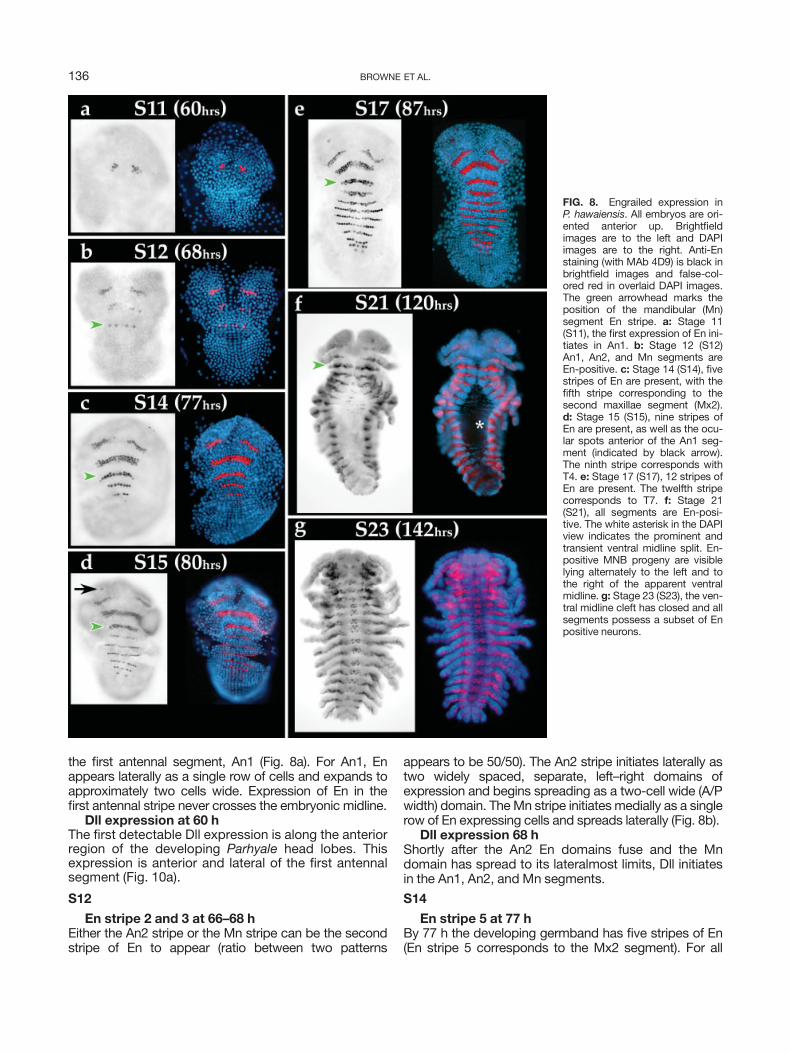
the first antennal segment, An1 (Fig. 8a). For An1, Enappears laterally as a single row of cells and expands toapproximately two cells wide. Expression of En in thefirst antennal stripe never crosses the embryonic midline.
Dll expression at 60 hThe first detectable Dll expression is along the anteriorregion of the developing Parhyale head lobes. Thisexpression is anterior and lateral of the first antennalsegment (Fig. 10a).
S12
En stripe 2 and 3 at 66–68 hEither the An2 stripe or the Mn stripe can be the secondstripe of En to appear (ratio between two patterns
appears to be 50/50). The An2 stripe initiates laterally astwo widely spaced, separate, left–right domains ofexpression and begins spreading as a two-cell wide (A/Pwidth) domain. TheMn stripe initiates medially as a singlerow of En expressing cells and spreads laterally (Fig. 8b).
Dll expression 68 hShortly after the An2 En domains fuse and the Mndomain has spread to its lateralmost limits, Dll initiatesin the An1, An2, and Mn segments.
S14
En stripe 5 at 77 hBy 77 h the developing germband has five stripes of En(En stripe 5 corresponds to the Mx2 segment). For all
FIG. 8. Engrailed expression inP. hawaiensis. All embryos are ori-ented anterior up. Brightfieldimages are to the left and DAPIimages are to the right. Anti-Enstaining (with MAb 4D9) is black inbrightfield images and false-col-ored red in overlaid DAPI images.The green arrowhead marks theposition of the mandibular (Mn)segment En stripe. a: Stage 11(S11), the first expression of En ini-tiates in An1. b: Stage 12 (S12)An1, An2, and Mn segments areEn-positive. c: Stage 14 (S14), fivestripes of En are present, with thefifth stripe corresponding to thesecond maxillae segment (Mx2).d: Stage 15 (S15), nine stripes ofEn are present, as well as the ocu-lar spots anterior of the An1 seg-ment (indicated by black arrow).The ninth stripe corresponds withT4. e: Stage 17 (S17), 12 stripes ofEn are present. The twelfth stripecorresponds to T7. f: Stage 21(S21), all segments are En-posi-tive. The white asterisk in the DAPIview indicates the prominent andtransient ventral midline split. En-positive MNB progeny are visiblelying alternately to the left and tothe right of the apparent ventralmidline. g: Stage 23 (S23), the ven-tral midline cleft has closed and allsegments possess a subset of Enpositive neurons.
136 BROWNE ET AL.

segments that will be incorporated into the cephalon (T1and anterior), the lateral extent of the En stripe is contig-uous with the edge of the developing appendage bud.However, for segmental En domains including T2 andposterior, the En stripe continues laterally and, ulti-mately, dorsally along the future body wall. En stripe 2(An2) spreads both medially and laterally and expandsto 3–4 cell widths (A/P) after the two separate domainsconnect along the midline. All En stripes posterior of themandibular segment arise in a strictly A-P temporal pro-gression (Fig. 7).
Dll at 77 hBy the time the fifth En stripe appears Dll is detectable inAn1, An2, and theMn segments. These Dll domains ariseas bilaterally symmetric clusters of cells in which the pos-terior half of each Dll domain lies within the En-express-ing domain and the anterior half of each Dll domain liesjust anterior to the En expression domain (e.g., Fig. 10b).Dll protein is detected strongly throughout the develop-ing Mn appendage fields during and after the initial for-mation of the proximal–distal (P-D) axis. This is in con-trast to the insect Mn, in which ectodermal Dll expressionis not seen during development of theMn appendage.
S15
En stripe 9 plus ocular spots at 80 hThe developing germband has nine stripes of En, aswell as the bilateral ocular En domains in the develop-ing head (Fig. 8d).
Dll at 80 hThe Mx1 Dll domain lies more medial than any of theother Dll domains and also enlarges at a slower ratethan the other Dll domains. All Dll domains from Mx2posterior initiate in the same way—Dll protein begins toaccumulate in cell d4 as the posteriorly abutting cellsa4, a5, and then a6 accumulate En protein. After Dllexpression begins in d4, the next cell to begin toexpress Dll is d5 (Fig. 6b). Dll is detected strongly in thelabrum, anterior and medial of the An1 domain (Fig.10b).
S19
En in the CNS at 96 hThe first thoracic segment, T1, has a number of En-positive neuroblasts as well as En-positive GMCs (Fig.9a,b).
Dll at 96 hThe labral Dll domain is medial and slightly anterior ofthe En and Dll ocular expression domains. Initiationand early development of the labrum occurs in thepreantennal segment (Fig. 10b–d).
S21
En staining in all segmentsAt 117 h (slightly before S21), En expression is seen inall segments. A subset of the MNB daughter cells areEn positive and lie to the right and the left of the mid-line along the ventral thoracic cleft (Fig. 8f).
FIG. 9. Identification of Engrailed-positive CNS cells in T1. In all panels anterior is up. a,b: Stage 19 (S19). At �96 h the T1 segment hasgenerated a series of En-positive neuroblasts (NBs) and ganglion mother cells (GMCs). a: Ventralmost focal plane shows several En-positiveneuroblasts (arrows). b: Slightly more dorsal focal plane from embryo shown in a. The black arrowheads in b indicate the position of thesmaller En-positive GMCs directly above the parent NBs shown by arrows in a. c,d: Stage 23 (23). At �140 h T1 has generated a number ofpositionally identifiable En positive neurons in the CNS. c: Ventralmost plane of En-positive staining. Black arrows indicate IC neurons. Themedial cluster of En-positive cells are presumably MNB progeny cells. d: Slightly more dorsal focal plane from embryo shown in c. At thelevel of the connective and commissural axon tracts, black arrows indicate En-positive LE neurons, triangles indicate En-positive ECa neu-rons, and arrowheads indicate En-positive ECp neurons.
137STAGES OF DEVELOPMENT P. HAWAIENSIS

S22
En staining is widely present in the developingembryonic CNS at 139 h (Fig. 9c,d)The thoracic ventral midline split has closed. The T1segment is positive for a number of identifiable neu-rons: ECa, ECp, LE, and IC (Figs. 8g, 9c,d) (Duman-Scheel and Patel, 1999).
S24
En staining persistsEn expression remains in the posterior compartment of de-veloping segments and dorsal body wall as well as in thedevelopingCNS. The expression pattern remains fairly sta-tic until cuticle deposition (S28, at 216 h) precludes detec-tion via standard antibody-based histochemical protocols.
FIG. 10. Distalless and Engrailed Expression in P. hawaiensis. All embryos are oriented anterior up. For each panel brightfield image is onthe left and DAPI image is on the right. In a–c brightfield images, Dll is in black and En is in brown. In panel d brightfield image, Dll is in brownand En is in black. In DAPI images of panels a–d Dll is false-colored yellow. In c,d DAPI images, En is false-colored red. Because of the man-ner in which the channel overlay was done in d, red indicates either En or EnþDll. a: Stage 11 (S11), 60-h germband. Dll initiates in two arcsin the optic lobes anterior of the An1 segment (arrow). En is just beginning to be expressed in the An1 segment and can be seen as a singlebrown cell on the left side of the brightfield embryo. The DAPI field shows the Mn segment just beginning to organize into transverse cellrows. b: Stage 15 (S15), 80-h germband labeled for Dll only. The optic lobes remain Dll-positive (arrow). The medial labrum is beginning toexpress Dll anterior of the An1 appendage field (asterisk). The An1, An2, and Mn appendage fields are Dll-positive. The Mx1, Mx2, and T1appendage fields are just beginning to initiate Dll expression (red arrowheads). c: Stage 17 (S17), 87-h germband. En has just initiatedexpression in T6 (out of focal plane). Dll has just initiated expression in the T5 appendage field. Dll expression in Mx1 and Mx2 appendagefields remains at low levels. The Mn is still expressing Dll broadly throughout. The asterisk marks Dll expression in the developing labrumanterior of An1. d: Stage 19 (S19), 96-h embryo. Upper image shows embryo before it was dissected flat. En has initiated in A4 and is visiblein the posterior compartment of developing antennal, gnathal, and thoracic appendage fields. The transient midline cleft has begun to formin the posterior thoracic region. The forming stomodeum is clearly visible and is positioned medial of An1 (green arrow). Lateral esophagealprojections are visible forming at the interior lateral edges of the stomodeum (green arrowhead). Neither of these structures (stomodeum norlateral esophageal projections) are Dll- or En-positive. The Dll-positive labrum is visible just anterior of the developing stomodeum (asterisk).The En-positive staining in the developing optic lobes remains visible (black arrowhead in brightfield image, white arrowhead in DAPIimage).
138 BROWNE ET AL.

S11 Stage 11 (60 h; 24%)
Formation of the germband: initial ectodermaland mesodermal cell rows form
Engrailed expression: An1In vivo. Progressive formation of ectodermal and mes-
odermal cell rows along the A-P axis begins at 60 h of
development and continues to S21 (120 h of develop-
ment). Also at this time, the midgut anlagen are becom-
ing visible as lateral disc-like structures flanking and just
posterior of the head lobes (Fig. 5d).
Fixed + DAPI. At 60 h of development the organiza-tion of the ectoderm into grid-like rows and columns isespecially striking in DAPI-stained preparations (Figs. 5d,8a, 10a). Several ‘‘numeral rows’’ (parasegment precur-sor rows) have organized by this time, with the anterior-most transverse row forming parasegment 1. The firstmediolateral mitotic wave (forming the a/b and c/d prog-eny) has not yet begun. After the formation of the firsttwo anterior transverse rows, cells forming the embry-onic ventral midline begin to organize into a single A-Pcolumn. As development proceeds, additional organized
FIG. 11. P. hawaiensis append-age development. a: Photos ofAn2, T1, T3, and T5 appendagesfrom 84–240 h of development.All appendages are from the leftside of the animal. b: An amphi-pod pereopod schematic is onthe left. Dark gray shading indi-cates the basal elements formingthe coxopodite; coxa (C), basis(B), and ischium (I). Light grayshading indicates the distal ele-ments forming the telopodite;merus (M), carpus (C), propodus(P), and dactyl (D). Two dorsalbranches initiate from the pereo-pod coxa. The outer branch is thecoxal plate. The inner, epipod,branch initiates at the distal pos-terior region of the coxa fromwhich it articulates both posteri-orly and medially. It forms a later-ally flattened and highly vascular-ized sheet that can perform respi-ratory functions (gill). To the rightis pereopod T5 stained with En(marking the posterior compart-ment) in black. The appendage isoriented dorsal up. The wide flatdorsal branch initiating across thecoxa is the coxal plate. The dorsalbranch initiating from the poste-rior distal margin is the gill. The gillis a modified epipod.
139STAGES OF DEVELOPMENT P. HAWAIENSIS

‘‘numeral’’ rows will form in an anterior to posterior pro-gression. Also at this stage, the nuclei defining the ringshaped walls of the midgut anlagen discs are welldefined and appear relatively closely packed (Fig. 5d).
S12 Stage 12 (68 h; 27.2%)
Initial germband elongation stageEngrailed expression: An1, An2, MnIn vivo. The bi-lobed head is multilayered at this time
and appears as a distinct clearing at the anterior ventralend of the egg. The laterally positioned midgut anlagenhave become very prominent disc-shaped structures.The dorsal organ is clearly visible as a thick ring and liesalong the dorsal anterior midline of the developingembryo. The region of the germband from Mx1 and pos-terior appears as a relatively optically clear region ven-tral and posterior of the developing head segments andgut anlagen.
Fixed + DAPI. In DAPI-stained embryos the germbandregion of the embryo has the highest density of nucleiand the ectoderm posterior to the Mn is clearly organizedinto mediolateral columns and A-P rows. At 68 h of devel-opment the first mediolateral mitotic wave can be foundmoving across parasegment precursor row 3 (Fig. 8b).Nuclei found within the disc-shaped gut anlagen appearto be dispersed and occur in several layers; however, thenuclei positioned along the edges of the discs appear tobe more closely arranged. The ectodermal cells of theanterior head segments have begun to undergo asymmet-ric cleavages, and at least some of the progeny cells arelikely to be neuroblasts (e.g., Fig. 6a).
Appendage Development
For the purposes of providing additional staging crite-ria, we have chosen to describe the development offour appendages, the second antenna (An2), the firstthoracic appendage (maxilliped; T1), the third thoracicappendage (gnathopod; T3), and the fifth thoracicappendage (pereopod; T5) in some detail. The appen-dages were dissected intact from developing embryosto facilitate description. Each of the four appendagesbears a distinct morphology and thus brief descrip-tions at various time points can provide landmarks toeasily gauge the relative age of the embryo underexamination (Fig. 11).
The antennae are uniquely derived appendages. Thefive proximal-most elements of An2 comprise thepeduncle and each individual element is rather stout inappearance. The remaining distal elements are termedflagella and they progressively narrow along the P-Daxis of An2. The maxilliped on T1 is strongly modified toserve as the outermost feeding appendage of the buc-cal mass (posteriormost feeding appendage along theA-P axis, see Fig. 1). The maxilliped possesses twoventral limb branches (endites). The T3 gnathopod andT5 pereopod appendages serve grasping and locomo-tory functions, respectively. Both T3 and T5 possess acoxal plate and a modified epipod. The P-D axis of the
endopod (the primary appendage branch) of T1, T3,and T5 is composed of the coxa, basis, ischium, merus,carpus, propodus, and dactyl elements (Fig. 11).
S13 Stage 13 (72 h; 28.8%)
Germ cell cluster visibleEngrailed expression: An1, An2, MnIn vivo. Bilateral germ cell cluster migration: During
the development of the embryonic germband the germcells become clearly visible underneath the developingmaxillae 1 (Mx1) and maxillae 2 (Mx2) segments. Ini-tially, they are present as a single medial cluster of whit-ish cells underneath the ectoderm at �72 h of develop-ment. A similar position of the germ cells during germ-band development in a related amphipod, Gammarus
pulex, was described by Weygoldt (1958) based on fixedmaterial. Additional descriptions of germ cell develop-ment in Parhyale can be found in Gerberding et al.(2002) and Extavour (2005).
Appendage buds begin to visibly project from the ven-tral ectodermal surface in an anterior-to-posterior pro-gression at 72 h of development beginning with the firstantennae (An1).
Fixed + DAPI. In DAPI preparations the progressionof appendage development can be clearly tracked by theappearance of dense aggregations of nuclei along the P-D axis of each appendage. The An1 and An2 appendagesare beginning to extend along their respective P-D axes.
S14 Stage 14 (77 h; 30.8%)
Germ cell cluster bi-lobedEngrailed expression: An1, An2, Mn, Mx1, Mx2In vivo. At 77 h of development the ventral germband
has expanded posteriorly the full length of the egg. Thebilateral midgut anlagen have become two well-definedcircular structures flanking the gnathal segments (Fig.12a). The germ cells are beginning to separate into twobilaterally opposed whitish clusters. The anterior cepha-lon continues to thicken and is becoming optically clear(Fig. 12a).
Fixed + DAPI. In DAPI-stained embryos the ectodermgrid is clearly visible and extends the full length of theventral surface of the egg. Nuclei associated with themidgut anlagen occur in several layers (Fig. 12a). Differen-tial and asymmetric cleavages begin to spread throughoutthe ectodermal grid (Fig. 12a; for further informationabout differential cleavages, see section on ectoderm andmesoderm segmentation). Some of the progeny of thesedifferential, asymmetric cleavages are likely to be neuro-nal stem cells (neuroblasts) by both cell position and cellcleavage criterion (Dohle, 1976; Scholtz, 1990, 1992).
S15 Stage 15 (80 h; 32%)
Germ cell cluster splitsEngrailed expression: Ocular spots, An1, An2,
Mn, Mx1, Mx2, T1, T2, T3, T4In vivo. The cluster of germ cells located ventrome-
dially between segments Mx1 and Mx2 has completelysplit into two bilaterally opposed whitish clusters that
140 BROWNE ET AL.

flank the embryonic midline. Each cluster of germ cellsthen begins to move laterally away from the embryonicmidline. The An1, An2, and Mn appendages are clearlyvisible as proximal distal outgrowths from the develop-ing embryonic head.
S16 Stage 16 (85 h; 34%)
Bilateral germ cell cluster migrationEngrailed expression: Ocular spots, An1, An2,
Mn, Mx1, Mx2, T1, T2, T3, T4, T5, T6In vivo. The two germ cell clusters have begun to
migrate laterally and posteriorly. This migration is visiblefrom Stage 16 to Stage 23 (Figs. 12, 13).
Fixed + DAPI. The An2 appendage primordium hasbegun to elongate and is clearly composed of two P-D
elements. The T1 appendage primordium has begun toelongate and is composed of a single element. The T3 andT5 appendage primordia are visible as buds (Fig. 11a).
S17 Stage 17 (87 h; 34.8%)
Posterior (ventral) flexure visibleEngrailed expression: Ocular spots, An1, An2,
Mn, Mx1, Mx2, T1, T2, T3, T4, T5, T6, T7In vivo. As the germband elongates it begins to fold
inwards toward the egg interior at segment T5 (Fig. 12b).As elongation proceeds, this posterior (ventral) flexureexpands to include more anterior and more posterior seg-ments. This flexure is visible as a clear furrow in the germ-band when viewed from the side (Figs. 12, 13).
FIG. 12. P. hawaiensis light microscopy and DAPI part II. Live brightfield and matching postfixation DAPI images showing lateral(upper image pairs) and ventral (lower image pairs) views. a: Stage 14 (S14), the white arrow on the lateral view (upper image pair),shows the ovoid shaped midgut anlagen. The white arrowhead at the midline on ventral view (lower image), shows the position ofthe germ cell clusters as they are separating from one another. Organization of the ectoderm into transverse cell rows ventrally isclearly visible in the DAPI view. b: Stage 17 (S17), ventral flexure visible. White triangles on the lateral view (upper), indicates theposition at which the posterior ventral flexure initiates (T4/T5). White arrow shows the position of the midgut anlagen. White arrow-heads flanking the midline on the ventral view (lower), show the position of the migrating germ cell clusters. The ventral flexure isclearly visible in the DAPI images. c: Stage 18 (S18). White arrow on lateral view (upper) shows the position of the midgut anlagen.The posterior, ventral edge of the anlagen is coincident with T2. The deepening ventral flexure is clearly visible in the posteriorquadrant of the egg. White arrowheads on ventral view (lower) show the position of the migrating germ cell clusters. The ventralDAPI field shows the formation of appendage fields along the embryo. d: Stage 19 (S19), paddle-shaped posterior quadrant. Whitearrow on lateral view (upper) shows the position of the ovoid shaped midgut anlagen. The posterior region of the embryo hasexpanded laterally and posteriorly forming a paddle shaped posterior quadrant. White arrowheads on ventral view (lower) show theposition of the migrating germ cell clusters just lateral of the Mx1 and Mx2 appendage fields.
141STAGES OF DEVELOPMENT P. HAWAIENSIS

Fixed + DAPI. In DAPI preparations at 87 h the devel-oping ventral surface has the highest density of nuclei andis organized into clear segmental units (Fig. 12b). In com-parison, the dorsal and lateral surfaces of the developingembryo have relatively sparse cell populations and tempo-rally lag behind in terms of segmental organization(Fig. 12b). The midgut anlagen have increased in size dra-matically and meet one another at their ventralmost mar-gin and begin to fuse (Fig. 12b). The two germ clusters
are clearly visible sandwiched between the ectoderm andthe midgut anlagen (arrowheads, Fig. 12b).
S18 Stage 18 (90 h; 36%)
Ventral flexure of embryoEngrailed expression: Ocular spots, An1, An2,
Mn, Mx1, Mx2, T1, T2, T3, T4, T5, T6, T7, T8, A1In vivo. At 90 h ventral flexure of the embryo is appa-
rent (Fig. 12c). The infolded region includes the entire
FIG. 13. P. hawaiensis light microscopy and DAPI part III. Live brightfield and matching postfixation DAPI images showing lateral (upperimage pairs) and ventral (lower image pairs) views in a and b and just lateral views in c–f. a: Stage 20 (S20), posterior quadrant narrows, thesecond mitotic wave has passed through all parasegments of the ectoderm, and the telson appears. Larger white arrowhead on lateral view(upper) shows position of the migrating germ cell clusters. The posterior quadrant has significantly narrowed. A wide area of clearing is visi-ble dorsally. The highest concentration of nuclei is in the developing appendages and cephalon. In lower, ventral view, the telson is apparentand projects forward to T1. The labrum (white arrow) is visible medially projecting posterior towards the stomodeum from between theantennal segments. The lower edge of the stomodeum (smaller white arrowhead) is visible medially and just posterior of the labrum. Justposterior of the developing stomodeum, the paragnaths can be seen projecting medially from the ventral base of the mandibles. b: Stage21 (S21) hindgut proctodeum becomes visible. White arrowhead on lateral view (upper) shows position of the germ cell clusters. In ventralview (lower) the hindgut proctodeum (white arrow) is visible as a wedge-shaped ridge of cells at the posterior end of the embryo. The labrumhas extended significantly. Just posterior and ventral of the labrum are the prominent mandibles that are flanked medially by the paragnaths.c: Stage 23 (S23), digestive cecae begins to extend. The white arrow indicates the initial posterior projection of the digestive cecum. Thewhite arrowhead shows the position of the germ cell clusters at the shoulder of the digestive cecae. The germ cells will come to occupy aposition on the medial surface of the extending cecae, facing the midgut. DAPI staining clearly shows body wall segmental boundariesextending dorsally. d: Stage 24 (S24), midpoint of digestive cecae extension. The white arrow shows the posterior extent of extendingcecum (�T7). Yolk granules have cleared from the cephalon. Body tergites and coxal plates are visible. e: Stage 26 (S26), white-eyes andbeating heart. The white arrow indicates the position of the developing rhabdomeres. The embryo is beginning to visibly accumulate cuticle,and the appendages are achieving their final hatching morphology. f: Stage 28 (S28), yellow cuticle. As cuticle continues to accumulate theembryo acquires a yellow-gold appearance. The eye rhabdomeres fields appear intensely red, the limbs have acquired their final morphol-ogy, and the digestive cecae are significantly depleted of yolk stores.
142 BROWNE ET AL.

ventral region of the abdomen and a large part of theposterior thoracic region. The An1, An2, and Mn seg-ments are beginning to expand dorsally (Fig. 12c). Ven-trally, the germ cell clusters have migrated to positionsjust lateral of the Mx1 and Mx2 appendage fields andappear as two gray-white masses (Fig. 12c).
Fixed + DAPI. Dorsally, nuclei still appear rather dis-persed and unorganized, with the notable exception ofthe anterior dorsal surface where the dorsal organ islocated and the posterior dorsal surface of the eggwhere a portion of the posterior end of the embryo isnow visible. At the dorsal organ, nuclei are arranged ina ring. The nuclei of the midgut anlagen are visible astwo medially fused, rhomboid-shaped aggregations withthe posterior ventral margin projecting to T2 (Fig. 12c).Ventrally, nuclei are dense and clearly ordered into seg-mental units along the A-P axis of the embryo. Ventralnuclei are particularly dense in the developing append-age buds of An1, An2, Mn, Mx1, Mx2, T1, and T2(Fig. 12c).
The length of the An2 second element has increasedrelative to the proximal first element. The T1 appendageis composed of two elements of which the proximal ele-ment begins to bulge medially. The T3 and T5 appen-dages have begun to elongate (Fig. 11a).
S19 Stage 19 (96 h; 38.4%)
Posterior widensEngrailed expression: Ocular spots, An1, An2,
Mn, Mx1, Mx2, T1, T2, T3, T4, T5, T6, T7, T8, A1, A2,A3, A4
In vivo. At 96 h the region enclosed by posterior flex-ure has broadened laterally, extended posteriorly, andappears paddle-shaped (Fig. 12d). The germ cells havemaintained their position lateral to Mx1 and Mx2 appen-dages; however, they have become slightly more opaquein appearance. The head region has moved dorsally tooccupy the anterior face of the egg. The midgut anlagenhave increased in size and appear to be ovoid. Cellsacross the face of the midgut anlagen are large andclearly organized into a hexagonal array (Fig. 12d).
Fixed + DAPI. The extreme dorsal region of theembryo still bears relatively few nuclei, but the lateralthoracic ectoderm now shows organization of nucleiinto rows. The posterior paddle retains an organizationof aligned cell rows. The appendage fields are increasingin size, number of nuclei, and bulge prominently fromthe ventral surface of the segmenting ectoderm. Thefused midgut anlagen have begun to spread dorsally andposteriorly under the ectoderm (Fig. 12d).
A split in the ventral midline begins to appear at thisstage (Fig. 10d). As ventral flexure continues, the splitextends anteriorly and is similar in appearance to thesplit ventral midline observed in Orchestia cavimana
(Gerberding and Scholtz, 1999, 2001). The developingstomodeum and two lateral esophageal projections arevisible medially at the level of An1 (Fig. 10d). The para-
gnaths can be seen developing at the medial edges ofthe mandible primordia (Fig. 10d).
At 96 h, An2 is composed of three elements. The twovisible elements of T1 have continued to extend. T3 iscomposed of three elements, and T5 is composed oftwo elements (Fig. 11a).
S20 Stage 20 (112 h; 44.8%)
Posterior narrows, morphologically distinct tel-son appears
Engrailed expression: Ocular spots, An1, An2,Mn, Mx1, Mx2, T1, T2, T3, T4, T5, T6, T7, T8, A1, A2,A3, A4, A5, A6, telson
In vivo. At 112 h the posterior paddle has signifi-cantly narrowed and limb buds are visible on the abdo-men (Fig. 13a). The developing telson is visible at theposterior end of the embryo. Due to the flexure of theembryo and continued growth, the telson is orientedanteriorly and projects towards the first thoracic seg-ment (Fig. 13a). The head segments have returned to amore ventral aspect. An area of clearing is visible in thedorsal part of the dorsal organ (Fig. 13a). The germ cellclusters are visible in the lower hemisphere of themidgut anlagen and are beginning to migrate dorsally inthe embryo (Fig. 13a). The developing midgut can beseen as an organized epithelial sheet spreading over theyolk.
Fixed + DAPI. In DAPI preparations at 112 h theantennae arc prominently anteriorly and dorsallytowards the dorsal organ. The labrum is visible projec-ting medially and posteriorly between the antennae (Fig.13a). Just posterior of the labrum the lower edge of thestomodeum is visible as a raised medial ridge. The firstand second maxillae appendage fields appear to makecontact with the ventral surface of the egg membranes.The proximal base of each of the anterior thoracicappendage fields have begun to branch (T2–T6 coxalplates). The dorsal edges of the midgut anlagen fuse andposterior envelopment of the yolk continues to progress(the leading edge can be seen just above T4 in Fig. 13a).
By 112 h, An2 has undergone a rapid elongation and iscomposed of five proximal elements forming thepeduncle and one distal flagella element. An2 growsalong a dorsal vector across the developing head. T1 andT5 are composed of three visible endopodal elements.The basis of T1 has begun to generate a new P-D axismedially. T3 is composed of four visible endopodal ele-ments (Fig. 11a).
S21 Stage 21 (120 h; 48%)
Hindgut proctodeum becomes visibleIn vivo. At 120 h, all abdominal segments possess visi-
ble limb buds. The telson now projects forward to theMn segment. The hindgut proctodeum is just becomingvisible at the posterior terminus (Fig. 13b). The clearingat the position of the dorsal organ appears to increase insize (Fig. 13b). Laterally, the midgut appears as a wedge
143STAGES OF DEVELOPMENT P. HAWAIENSIS

with a broad dorsal aspect that progressively narrowsventrally (Fig. 13b). The germ cell clusters continue tobe visible in the lower portion of the midgut wedge(Fig. 13b).
Fixed + DAPI. In DAPI preparations the labrum hasnearly completed its posterior extension and is visiblejust anterior to the paragnaths (Fig. 13b). The forminghindgut proctodeum is clearly visible as a wedge-shapedridge of nuclei just anterior to the embryo’s posteriorterminus (Fig. 13b). The split midline is most prominentalong the thoracic ventral midline at this stage (Fig. 8f).After this stage the split ventral midline will close as theventral ganglia begin to mature and fuse.
At 120–132 h, An2 continues to extend in length asindividual elements continue to enlarge. T1 is composedof four elements. In addition, two nascent P-D axes form-ing the endites are being generated ventrally, one fromthe proximalmost element and one from the second ele-ment of the endopodal appendage axis (Fig. 11a). T3 iscomposed of seven elements along the endopodal P-Daxis: coxa, basis, ischium, merus, carpus, propodus, anddactyl. The coxa possesses two nascent P-D axes, bothgenerating dorsal appendage branches (proximally thecoxal plate and distally the epipod gill). The ischium iswedge-shaped with the narrow end oriented anteriorly,thus giving the T3 appendage its characteristic anteriororientation (also a diagnostic feature for T2). In addition,the propodus has become expanded laterally (also adiagnostic feature for T2). All seven endopodal elementsof T5 are visible and the coxa begins to bud two nascentP-D axes (Fig. 11a).
S22 Stage 22 (132 h; 52.8%)
Limb bud elongation stageIn vivo. The midgut completely envelops the remain-
ing yolk.Fixed + DAPI. The ventral midline is no longer split,
as the left and right halves have closed back together bythis stage.
At 132 h the fourth element of the peduncle of An2undergoes a distinct change in shape, narrowing at itsproximal end and expanding at its distal end. The twoendites of T1 are beginning to elongate and the principalP-D axis of T1 is now composed of five elements. T3 iscontinuing to elongate and both dorsal appendagebranches are increasing in size. The endopod of T5 nowpossesses a complete complement of seven elements:coxa, basis, ischium, merus, carpus, propodus, and dac-tyl and both dorsal appendage branches (coxal plate andepipod gill) are well formed (Fig. 11a).
S23 Stage 23 (144 h; 57.6%)
Digestive cecae begin to extend, yolk granulesclear from the head, tergites and coxal plates visible
In vivo. At �144 h the developing body wall tergitesare visible through the eggshell. Dorsal closure of theectoderm over the gut appears to be nearly complete.The coxal plate (dorsalmost branch) of the thoracicappendages (T2–T8) becomes visible laterally. The ceph-
alon is completely clear of yolk granules (Fig. 13c). Theantennae now arc posteriorly along the ventral marginof the head capsule. The clearing associated with thedorsal organ projects posteriorly as a narrow strip alongthe dorsalmost aspect of the embryo (Fig. 13c). Thegerm cell clusters have taken positions on the anteriormedial-lateral and anterior dorsal region of each of thebilateral paired cecae rudiments that are beginning toproject posteriorly (Fig. 13c).
The digestive cecae are major secretory organs con-sisting of a pair of blind tubes that open directly into theanterior end of the medial midgut. They are thought toplay a role in nutrient adsorption and storage (reviewedin Schmitz, 1992). At 144 h the anterior medial cecumrudiment begins to project anteriorly and dorsally intothe medial portion of the head capsule. The two bilater-ally positioned cecae rudiments simultaneously begin toproject posteriorly from the ventral posterior aspect ofthe wedge-shaped midgut anlagen (Fig. 13c). The exten-sion of the cecum rudiments occurs in unison and ischaracterized by a series of peristaltic, muscular contrac-tions that initiate alternately from the anterior and poste-rior ends of the rudiment. Each contractile waveincreases the overall length and girth of the cecum. Ulti-mately, the two cecae extend to very near the posteriorlimit of the embryo. The initial peristaltic wavefronts areof short duration (traveling the length of a small, shortrudiment), whereas the terminal wavefront durationscan be several minutes (traveling the length of a fullyextended cecum). This process of extension requires�20 h to reach completion.
Fixed + DAPI. In DAPI preparations at 144 h the den-sity of nuclei along the dorsal aspect of the embryo isbeginning to increase and resolve into distinct segmentalbands.
At 144 h, An2 begins to conform to its final cylindricaluniramous shape. The T1, T3, and T5 appendages arecontinuing to increase in size (Fig. 11a).
S24 Stage 24 (155 h; 62%)
Midpoint of digestive cecae extensionIn vivo. The cephalon has acquired a rounded shape.
The abdomen is well formed. The lateral and dorsal bodywall tergites are clearly visible. The bilateral digestivececae have progressed approximately midway and arenearing the abdomen (Fig. 13d). The yolk depositswithin the digestive cecum visibly change color as thegranules are broken down (Fig. 13d).
Fixed + DAPI. The cephalon has the highest densityof visible nuclei with the notable exception of the form-ing eye fields (Fig. 13d). Segmentation is apparentthroughout the lateral and dorsal body wall and thethoracic coxal plates are clearly visible (Fig. 13d).
S25 Stage 25 (168 h; 67.2%)
Digestive cecae extension completeIn vivo. At the end of the digestive cecae extension
process, most of the remaining yolk has been transferred
144 BROWNE ET AL.

from the developing medial midgut to the digestivececae. The midgut remains visible medially along thedorsal midline between the digestive cecae sacs as a thinyolk colored strip.
Fixed + DAPI. At 168 h, An2 has taken on a cylindri-cal shape. The An2 elements appear to decrease in sizealong the P-D axis (Fig. 11a). T1 has acquired its com-plete complement of seven elements (coxa, basis,ischium, merus, carpus, propodus, and dactyl) along theprincipal P-D axis and has begun to acquire its character-istic shape. Both ventral appendage branches of T1 con-tinue to increase in size. T3 begins to acquire its finalshape in which the anterior bend in the ischium is pro-nounced and the laterally expanded face of the propo-dus is enhanced. The T3 dactyl begins to develop its pos-terior orientation, which will align with the expandeddistal ridge of the propodus. The dorsalmost branch, thecoxal plate, becomes expanded laterally. The remainingdorsal branch, the epipod gill, becomes laterallyexpanded and oriented both posteriorly and ventrally,thus occupying a position ventral to the endopod. T5also begins to acquire its final shape in which the coxalplate becomes laterally expanded and the gill takes on asimilar orientation as in T3 (Fig. 11a).
S26 Stage 26 (180 h; 72%)
White eyes and beating heartIn vivo. The eye fields become distinctly visible as
two bilaterally symmetric, whitish hued clusters of rhab-domeres (Fig. 13e). The tubular heart of Parhyale
hawaiensis is located along the dorsal thoracic regionfrom T2–T7 and begins to visibly beat. It is a bilaterallysymmetric, single-chambered structure positioned alongthe midline of the embryo, dorsal to the midgut (Diva-karan, 1975). Yolk stores in the digestive cecum begin todiminish.
Fixed + DAPI. The appendages appear to becrowded into the curled embryo and begin to narrowsignificantly. Secretion of cuticle is visually detectable atthis stage. The spatial organization of cells comprisingthe coxal plates and tergites is clearly visible (Fig. 13e).
S27 Stage 27 (192 h; 76.8%)
Red eyesIn vivo. Pigment cells that surround the whitish rhab-
domeres begin to accumulate a deep red color at 192 hof development.
Fixed + DAPI. At 192 h, An2 has begun to visiblycuticularize and the diameter of the peduncular and flag-ellar elements continues to narrow as An2 achieves itsfinal morphology. The T1, T3, and T5 appendages con-tinue to increase in size.
S28 Stage 28 (216 h; 86.4%)
Yellow cuticleIn vivo. At 216 h the embryo cuticle acquires a yel-
low-gold appearance. Detection of gene expressionbecomes problematic as the cuticle thickens. The diges-
tive cecae are significantly depleted of yolk stores (Fig.13f). The heart is beating strongly.
Fixed + DAPI. Most DAPI preparations at this stage,and later, burst through the eggshell during processing(Fig. 13f). The appendages have largely acquired theirunique morphologies; for example, T1 has been incorpo-rated into the buccal mass (Fig. 13f).
At 216 h, An2 is becoming heavily cuticularized. TheT1, T3, and T5 appendages have acquired their final mor-phology and are also becoming heavily cuticularized.
S29 Stage 29 (230 h; 92%)
Muscular twitchingIn vivo. Muscular twitching of appendages and body
are readily visible through the egg shell.Fixed + DAPI. Although An2 has acquired its final
shape at S28, the appendage has continued to grow. TheT1 appendage appears to have acquired both its finalhatching morphology and hatching setae. The proximalendite of T1 has formed a plate-like structure, the medialendite has formed a toothed comb, and the distal por-tion of the endopodal branch (palp) is strongly curvedmedially and terminates with a hook-like structure (seeFig. 11a, �240 h). The T3 and T5 appendages haveacquired hatching setation and morphology.
S30 Stage 30 (250 h; 100%)
HatchingIn vivo. Hatching occurs at �250 h of development.
Immediately prior to hatching the embryo is in a con-stant state of coordinated motion. In addition, theembryo increases substantially in size immediately priorto and during hatching, most likely via active uptake ofwater. Soon after breaking free of the eggshell, hatch-lings will undergo a molt and revert to a smaller size.
DISCUSSION
Embryonic staging systems, including the most recentarthropod systems, have historically taken two principaldescriptive forms: 1) age-dependent staging using abso-lute time or percent of development, or 2) event-dependent staging (Campos-Ortega and Hartenstein,1985; Hartenstein, 1993; Olesen and Walossek, 2000;Bentley et al., 1979; Sandeman and Sandeman, 1991).We have attempted to combine both staging forms tofacilitate the use of Parhyale hawaiensis for a variety ofdevelopmental and comparative studies (Figs. 2, 7).Strict adherence to either an age or percentage stagingsystem bearing a linear relationship to time would, bynecessity, deemphasize the occurrence of important sin-gular events during embryogenesis. For example, cleav-age events that occur during the first 5% of developmentin P. hawaiensis establish the primary axes of the devel-oping embryo (Figs. 2–4) (Gerberding et al., 2002). Bythe same token, a strict adherence to event stagingwould hamper comparative studies between relatedorganisms in which synonymous, analogous, or homolo-
145STAGES OF DEVELOPMENT P. HAWAIENSIS

gous developmental events have a shifted temporal rela-tionship with one another (Jeffery et al., 2002). A com-prehensive approach comprising a descriptive accountof developmental events within an age-dependent frame-work, upon which the percentage of total developmen-tal time has been mapped, will allow the maximumdegree of flexibility and accuracy when planning andimplementing developmental and comparative studies.
A particularly distinctive aspect of Parhyale develop-ment is the holoblastic cell cleavage pattern seen duringearly development. The eight-cell stage (S4) of P. hawai-
ensis is described here (Figs. 3, 4) and the associated fatemapping of each of the eight blastomeres is described inGerberding et al. (2002). The early establishment of dis-tinct cell lineages in P. hawaiensis allows for a widerange of experimental manipulations of both specificcell lineages and germ layers via microinjection andother perturbation techniques (i.e., Extavour, 2005). Inaddition, some variation in cleavage patterns (both intime and pattern) is observed in Orchestia cavimana
(Wolff and Scholtz, 2002) and other amphipods(C. Babbitt, M. Modrell, and N.H. Patel, unpubl. data).Early holoblastic cleavage appears to be a general featureof amphipod embryogenesis (see Scholtz and Wolff,2002), and thus future studies of these variations in tim-ing and pattern of cell cleavage, and their resulting line-ages, may perhaps be informative with regard to evolu-tionary patterns within the Amphipoda.
The current widespread use of several pan-specificantibodies makes the inclusion of the associated expres-sion patterns highly desirable in staging systems. Themonoclonal antibody to Engrailed (MAb4D9; Patel et al.,1989b) allows for finer resolution of several patterningevents along the A-P axis (Figs. 7, 8), as well as later pat-terning within the developing CNS (Duman-Scheel andPatel, 1999; Fig. 9). Similarly, the antibody to Distallessallows for a more detailed examination of the formationof appendage fields (Fig. 10). For example, Dll initiationin the d4 cell of developing Parhyale hawaiensis para-segments (parasegment 3 and posterior; Figs. 6d, 10b,c)is slightly different than that observed in a related amphi-pod, Orchestia cavimana, and the isopod Porcellio
scaber (Hejnol and Scholtz, 2004). Notably, the temporaldynamics of Dll expression in the mandibular segmentof P. hawaiensis (Fig. 10b,c) supports the hypothesisthat Dll is required for patterning the crustacean mandib-ular primordia (Browne and Patel, 2000). Thus, theabsence of Dll in the insect mandible is likely an autopo-morphy of the insect lineage (Scholtz et al., 1998).
The segmental organization of the arthropod head hasbeen a formidable problem for biologists. Recently, theorigin and segmental affiliation of the arthropod labrumhas been reexamined (Haas et al., 2001a,b; Boyan et al.,2002; Eriksson et al., 2003). An appendicular origin hasbeen proposed based on homeotic transformationresults in the flour beetle, Tribolium (Haas et al.,2001b). The segmental affiliation of the labrum has beensuggested to be on the intercalary segment based oninnervation data in both Tribolium and the grasshopper,
Schistocerca (Haas et al., 2001a; Boyan et al., 2002). Inthis scenario the stomodeum would have its origin at theintercalary/mandibular segment boundary (Haas et al.,2001a). Eriksson et al. (2003), however, suggest supportfor an ancestral anterior terminal position for the stomo-deum based on development in the OnychophoranEuperipatoides, a basal arthropod. In Euperipatoidesthe stomodeum appears first at an anterodorsal positionand migrates ventroposteriorly during development.Weygoldt (1958) also describes a ventroposterior migra-tion of both the labrum and the stomodeum in theamphipod Gammarus pulex.
The origin, segmental affiliation, and morphogenesisof both the labrum and stomodeum were followed in P.
hawaiensis (Figs. 10, 13). The labrum expresses Dllstrongly at S15 (Fig. 10b), supporting an appendicularorigin. However, the position of the labrum first appearsanterior and medial of the En and Dll ocular expressiondomains. Thus, the positional origin and segmental affili-ation of the labrum is anterior of the first antennal seg-ment, contrary to the model suggested by Haas et al.(2001a,b). The ectodermal origin of the labrum precedesCNS innervation and thus calls into question the use oftritocerebral innervation patterns as the sole means todetermine the positional origin and segmental affiliationof the labrum (Boyan et al., 2002). The stomodeum isreadily visible via DAPI staining at S19 posterior of thelabrum (Fig. 10d). At this stage the anterior half of thestomodeum is in the medial region of the An1 segmentand the posterior half of the stomodeum is in the ante-rior medial region of the An2 segment (Fig. 10d). Asdevelopment continues, both the labrum and the stomo-deum move posteriorly. The labrum will continue toproject ventral and posterior serving as the ‘‘upper lip’’of the stomodeum. The entrance of the stomodeum laterin development will largely be at the level of the mandi-bles, which will flank the esophageal opening.
As illustrated in Figure 11, amphipods such as P. hawai-ensis display remarkable morphological variation in theappendages along their anterior/posterior axis. Theappendage staging data provided here will be valuable forfuture studies of appendage development and evolution.For example, either in studies designed to understandpossible roles of homeotic genes in crustacean append-age evolution or investigations into molecular mecha-nisms used to generate branched appendages (e.g.,Giorgianni and Patel, 2004; Hejnol and Scholtz, 2004).
In summary, we have described a number of aspects ofthe life history and development of Parhyale hawaiensis
that make it particularly attractive for developmental andevolutionary studies. It is a widespread, common, circum-tropical species that breeds year-round. The adults arerelatively small and are easy to maintain in dense labora-tory cultures. Individual broods of synchronous eggs canbe easily removed from females and maintained in smallvolumes of seawater until hatching. Complete embryo-genesis of P. hawaiensis at 268C requires 10 days, andthe egg-to-egg generation time is 7–8 weeks when main-tained at 26–288C. While the yolk remains opaque, it
146 BROWNE ET AL.

quickly becomes sequestered towards the center of theegg and eventually into the developing midgut; theembryo itself is optically clear. The embryos are robustand highly resistant to environmental variations in tem-perature and salinity and are suitable for a wide range ofexperimental manipulations. Additionally, the embryosare direct developers and the hatchlings are morphologi-cally similar to adults. The staging system presented hereshould be beneficial to all further developmental studiesof P. hawaiensis and related amphipods.
MATERIALS AND METHODS
Amphipod Culture
The laboratory population of Parhyale hawaiensis origi-nated from the John G. Shedd Aquarium (Chicago, IL) in1997 as an isolate from their marine filtration system,and thus was ‘‘preselected’’ as a crustacean species thatrequires minimal care. Laboratory breeding colonies aremaintained in shallow plastic trays (47 � 33 � 12 cmcontaining 16 L of saltwater) at �268C. Individual traysare covered with clear Plexiglas and illumination is pro-vided from ambient room illumination and fluorescentstrip lighting with a day/night cycle of �12/12 h. Wateris circulated within trays by submersible pumps (40–150US gal/h). Crushed coral substrate is provided as a thinbottom layer (1.5–2.5 cm) in each tray. Phosphate-absorbing resin (Phosguard, Seachem) is used to controlaccumulation of free phosphates. Either artificial salt(HW Marinemix) premixed in a 55 US gal container to atrue specific gravity of 1.018–1.022 or filtered naturalseawater is used. Each tray receives a 50% water changeevery week. Temperature is maintained by heating theroom in which the tanks are located. Animals are main-tained on a diet of algae wafers (Hikari), kelp powder/flakes (NOW), and a 1:1:1 liquid mixture of fatty acids(Selcon), plankton (MarineSnow), and vitamins (Multi-Vit, Hawaiian Marine).
For experimental manipulation of embryos, sexuallymature pairs in amplexus are collected and placed insmall dishes (13 � 18 � 8.5 cm containing 1 L of freshseawater) overnight at 188C. The following day, most ofthe males and females in amplexus have separated. Theseparation of the male and females occurs as the femalesmolt (shed their old cuticle). Once the molt is complete,mature oocytes are released through the oviducts, fertil-ized, and deposited in the female’s ventral brood pouch.At this time single-cell embryos are collected by immobi-lizing the adult female and opening the ventral broodpouch with forceps. In order to establish fairly precisetime points for our staging system, synchronousembryos from single brood pouches were then placed infilter sterilized seawater (0.22 �m Millipore filter, Bed-ford, MA) in 35-mm Petri dishes and maintained in a highhumidity chamber at 268C until they reached specifictime points for manipulation, dissection, and/or fixation.
Light Microscopy
All light micrographs were taken on either a Zeiss StemiDRC dissecting microscope or a Zeiss Axiophot with aKontron 3012 digital camera. For light microscopy, indi-vidual embryos were placed in shallow well slides withseawater and illuminated with intense side lighting.
DAPI (4,6-Diamidino-2-phenylindole) StainingAlone
After live light microscopy and photography, individualembryos were fixed by boiling in 1� PBS (1.86 mMNaH2PO4; 8.41 mM Na2HPO4; 175 mM NaCl; pH 7.4) forseveral minutes (<7 min), then washed with methanoland incubated in 50% (v:v) glycerol/1� PBS containing1 mg/ml DAPI overnight at 48C for visualization of nucleiby fluorescence microscopy. The individual embryoswere then rephotographed to match the live photos.Thus, for Figures 5, 12, and 13 image pairs are a lightmicroscope picture of a living embryo and the matchingDAPI image of the same embryo after fixation (relativeorientation matched as closely as possible). This proce-dure was followed because light microscope images ofchemically or heat-fixed embryos look quite differentfrom live embryos (individual cells and structures aremore clearly visible in live preparations than in fixedembryos with just light microscopy), and it is importantto be able to stage living embryos for various experimen-tal procedures.
Antibody Staining
Embryos that had been allowed to develop for specificperiods of time were dissected and fixed for 20 min in a9:1 (v:v) mixture of Hepes fixation buffer (100 mMHepes, pH 6.9, 2 mM MgSO4, and 1 mM EGTA) and 37%formaldehyde. Tungsten wire needles were used duringfixation to remove extracellular membranes. Fixedembryos were then washed with PT (1� PBS, pH 7.4,and 0.1% Triton X-100) and stored in 100% methanol at�208C. Antibody detection was performed as describedin Patel (1994). All antibody incubations were done over-night at 48C at the following concentrations: mouseMAb 4D9 anti-Engrailed (Patel et al., 1989b) 1:1, rabbitanti-Distalless (Panganiban et al., 1995) 1:100, HRP con-jugated goat anti-mouse (Jackson ImmunoResearch,West Grove, PA) 1:300, HRP conjugated goat anti-rabbit(Jackson ImmunoResearch) 1:500. After histochemicalDAB reactions, embryos were washed and incubatedovernight at 48C in 50% glycerol þ 1 mg/ml DAPI solu-tion. Embryos were then stored in 70% glycerol at�208C.
ACKNOWLEDGMENTS
We thank Mark Q. Martindale, Danielle Liubicich,Melinda Modrell, Patricia M. Lee, Amy Maxmen, and twoanonymous reviewers for providing critical commentsthat significantly improved this communication. Wethank the John G. Shedd Aquarium, Chicago, IL, for
147STAGES OF DEVELOPMENT P. HAWAIENSIS

allowing us to collect Parhyale hawaiensis from theirmarine filtration system; these collected animals are thefounding population of all our cultures. NHP is an Inves-tigator of the Howard Hughes Medical Institute.
LITERATURE CITED
Barnard JL. 1965. Marine Amphipoda of atolls in Micronesia. Proc U SNatl Mus 117:459–551.
Barnard JL, Karaman GS. 1991. The families and genera of marine gam-maridean Amphipoda (except marine gammaroids). Rec Aust MusSuppl 13:1–866.
Bentley D, Keshishian H, Shankland M, Toroian-Raymond A. 1979.Quantitative staging of embryonic development of the grasshop-per, Schistocerca nitens. J Embryol Exp Morphol 54:47–74.
Boore JL, Collins TM, Stanton D, Daehler LL, Brown WM. 1995. Deduc-ing the pattern of arthropod phylogeny from mitochondrial DNArearrangements. Nature 376:163–165.
Boore JL, Lavrov DV, Brown WM. 1998. Gene translocation links insectsand crustaceans. Nature 392:667–668.
Boyan GS, Williams JLD, Posser S, Braunig P. 2002. Morphological andmolecular data argue for the labrum being non-apical, articulated,and the appendage of the intercalary segment in the locust.Arthropod Struct Dev 31:65–76.
Browne WE, Patel NH. 2000. Molecular genetics of crustacean feedingappendage development and diversification. Semin Cell Dev Biol11:427–435.
Campos-Ortega JA, Hartenstein V. 1985. The embryonic developmentof Drosophila melanogaster. Berlin: Springer.
Dana JD. 1853. Crustacea. Part II. United States Exploring Expedition14:689–1618.
Divakaran O, Pillai NK. 1975. The vascular system of Parhyale hawaien-sis (Amphipoda). Acta Zool 56:129–139.
Divakaran O, Pillai NK. 1981. Functional morphology of the respiratorysystem of Parhyale hawaiensis Dana (Crustacea: Amphipoda). ProcIndian Nat Sci Acad B47:664–669.
Dohle W. 1970. Die Bildung und Differenzierung des postnauplialenKeimstreifs von Diastylis rathkei (Crustacea, Cumacea). Zeit Mor-phol Tiere 67:307–392.
Dohle W. 1976. Die Bildung und Differenzierung des postnauplialenKeimstreifs von Diastylis rathkei (Crustacea, Cumacea). Zoomor-phologie235–277.
Dohle W, Scholtz G. 1988. Clonal analysis of the crustacean segment:the discordance between genealogical and segmental borders.Development 104(Suppl):147–160.
Dohle W, Scholtz G. 1997. How far does cell lineage influence cell fatespecification in crustacean embryos? Semin Cell Dev Biol 8:379–390.
Duman-Scheel M, Patel NH. 1999. Analysis of molecular marker expres-sion reveals neuronal homology in distantly related arthropods.Development 126:2327–2334.
Eernisse DJ. 1997. Arthropod and annelid relationships re-examined. In:Fortey RA, Thomas RH, editors. Arthropod relationships. London:Chapman and Hall. p 43–56.
Eriksson BJ, Tait NN, Budd GE. 2003. Head development in the Ony-chophoran Euperipatoides kanangrensis with particular referenceto the central nervous system. J Morphol 255:1–23.
Extavour CG. 2005. The fate of isolated blastomeres with respect togerm cell formation in the amphipod crustacean Parhyale hawaien-sis. Dev Biol 277:387–402.
Friedrich M, Tautz D. 1995. Ribosomal DNA phylogeny of the majorextant arthropod classes and the evolution of myriapods. Nature376:165–167.
Gasca R, Haddock SHD. 2004. Associations between gelatinous zoo-plankton and hyperiid amphipods (Crustacea: Peracarida) in theGulf of California. Hydrobiologia 530/531:529–535.
Gerberding M, Scholtz G. 1999. Cell lineage of the midline cells in theamphipod crustacean Orchestia cavimana (Crustacea, Malacos-traca) during formation and seperation of the germ band. DevGenes Evol 209:91–102.
Gerberding M, Scholtz G. 2001. Neurons and glia in the midline of thehigher crustacean Orchestia cavimana are generated via an invari-ant cell lineage that comprises a median neuroblast and glial pro-genitors. Dev Biol 235:397–409.
Gerberding M, Browne WE, Patel NH. 2002. Cell lineage analysis of theamphipod crustacean Parhyale hawaiensis reveals an early restric-tion of cell fates. Development 129:5789–5801.
Giorgianni MW, Patel NH. 2004. Patterning of the branched headappendages in Schistocerca americana and Tribolium castaneum.Evol Dev 6:402–410.
Giribet G, Edgecombe GD, Wheeler WC. 2001. Arthropod phylogenybased on eight molecular loci and morphology. Nature 413:157–161.
Gonzalez-Crespo S, Morata G. 1996. Genetic evidence for the subdivi-sion of the arthropod limb into coxopodite and telopodite. Devel-opment 122:3921–3928.
Haas MS, Brown SJ, Beeman RW. 2001a. Pondering the procephalon:the segmental origin of the labrum. Dev Genes Evol 211:89–95.
Haas MS, Brown SJ, Beeman RW. 2001b. Homeotic evidence for theappendicular origin of the labrum in Tribolium castaneum. DevGenes Evol 211:96–102.
Hartenstein V. 1993. Atlas of Drosophila development. Cold SpringHarbor, NY: Cold Spring Harbor Laboratory Press.
Hejnol A, Scholtz G. 2004. Clonal analysis of Distal-less and engrailedexpression patterns during early morphogenesis of uniramousand biramous crustacean limbs. Dev Genes Evol 214:473–485.
Hwang UW, Friedrich M, Tautz D, Park CJ, Kim W. 2001. Mitochondrialprotein phylogeny joins myriapods with chelicerates. Nature413:154–157.
Jeffery JE, Richardson MK, Coates MI, Bininda-Emonds ORP. 2002. Ana-lyzing developmental sequences within a phylogenetic frame-work. Syst Biol 51:478–491.
Kamaltynov RM. 1999. On the evolution of Lake Baikal Amphipods.Crustaceana 72:921–931.
Langenbeck C. 1898. Formation of the germ layers in the amphipodMicrodeutopus gryllotalpa costa. J Morphol 14:301–336.
Lindeman D. 1991. Natural history of the terrestrial amphipod Cer-rorchestia hyloraina Lindeman (Crustacea: Amphipoda; Talitridae)in a Costa Rican cloud forest. J Nat Hist 25:623–638.
Meschenmoser M. 1989. Ultrastructure of the embryonic dorsal organof Orchestia cavimama (Crustacea, Amphipoda); with a note onlocalization of chloride and on the change in calcium-depositionbefore the embryonic moult. Tissue Cell 21:431–442.
Myers AA. 1985. Shallow-water, coral reef and mangrove Amphipoda(Gammaridea) of Fiji. Rec Aust Mus Suppl 5:1–143.
Olesen J, Walossek D. 2000. Limb ontogeny and trunk segmentation inNebalia species (Crustacea, Malacostraca, Leptostraca). Zoomor-phology 120:47–64.
Panganiban G, Sebring A, Nagy L, Carroll S. 1995. The development ofcrustacean limbs and the evolution of arthropods. Science 270:1363–1366.
Panganiban G, Irvine SM, Lowe C, Roehl H, Corley LS, Sherbon B, Gre-nier JK, Fallon JF, Kimble J, Walker M, Wray GA, Swalla BJ, Martin-dale MQ, Carroll SB. 1997. The origin and evolution of animalappendages. Proc Natl Acad Sci U S A 94:5162–5166.
Patel N. 1994. Imaging neuronal subsets and other cell types in wholemount Drosophila embryos and larvae using antibody probes.Methods Cell Biol 44:445–487.
Patel NH, Kornberg TB, Goodman CS. 1989a. Expression of engrailedduring segmentation in grasshopper and crayfish. Development107:201–212.
Patel NH, Martin-Blanco E, Coleman KG, Poole SJ, Ellis MC, KornbergTB, Goodman CS. 1989b. Expression of engrailed proteins inarthropods, annelids, and chordates. Cell 58:955–968.
Poltermann M, Hop H, Falk-Peterson S. 2000. Life under Arctic seaice — reproduction strategies of two sympagic (ice-associated)amphipod species, Gammarus wilkitzkii and Apherusa glacialis.Mar Biol 136:913–920.
Poovachiranon S, Boto K, Duke N. 1986. Food preference studies andingestion rate measurements of the mangrove amphipod Parhyalehawaiensis (Dana). J Exp Mar Biol Ecol 98:129–140.
148 BROWNE ET AL.
Regier JC, Shultz JW. 1997. Molecular phylogeny of the major arthro-pod groups indicates polyphyly of crustaceans and a new hypothe-sis for the origin of hexapods. Mol Biol Evol 14:902–913.
Sandeman R, Sandeman D. 1991. Stages in the development of theembryo of the fresh-water crayfish Cherax destructor. Roux’s ArchDev Biol 200:27–37.
Schmitz EH. 1992. Amphipoda. In: Harrison FW, editor. Microscopicanatomy of invertebrates. New York: Wiley-Liss. p 443–528.
Scholtz G. 1990. The formation, differentiation and segmentation ofthe post-naupliar germ band of the amphipod Gammarus pulex L.Proc R Soc Lond B 239:163–211.
Scholtz G. 1992. Cell lineage studies in the crayfish Cherax destructor(Crustacea, Decapoda): germ band formation, segmentation, andearly neurogenesis. Roux’s Arch Dev Biol 202:36–48.
Scholtz G, Dohle W. 1996. Cell lineage and cell fate in crustaceanembryos — a comparative approach. Int J Dev Biol 40:211–220.
Scholtz G, Wolff C. 2002. Cleavage, gastrulation, and germ disc forma-tion of the amphipod Orchestia cavimana (Crustacea, Malacos-traca, Peracarida). Contrib Zool 71:9–28.
Scholtz G, Dohle W, Sandeman RE, Richter S. 1993. Expression ofengrailed can be lost and regained in cells of one clone in crusta-cean embryos. Int J Dev Biol 37:299–304.
Scholtz G, Patel NH, Dohle W. 1994. Serially homologous engrailedstripes are generated via different cell lineages in the germ band ofamphipod crustaceans (Malacostraca, Peracarida). Int J Dev Biol38:471–478.
Scholtz G, Mittmann B, Gerberding M. 1998. The pattern of Distal-lessexpression in the mouthparts of crustaceans, myriapods andinsects: new evidence for a gnathobasic mandible and the com-mon origin of Mandibulata. Int J Dev Biol 42:801–810.
Schram FR. 1986. Crustacea. Oxford: Oxford University Press.Sheader M, Van Dover CL, Shank TM. 2000. Structure and function
of Halice hesmonectes (Amphipoda: Pardaliscidae) swarms fromhydrothermal vents in the eastern Pacific. Mar Biol 136:901–911.
Sherbakov DY, Kamaltynov RM, Ogarkov OB, Vainola R, Vainio JK,Verheyen E. 1999. On the phylogeny of Lake Baikal amphipods inthe light of mitochondrial and nuclear DNA sequence data. Crusta-ceana 72:911–919.
Shoemaker CR. 1956. Observations on the amphipod genus Parhyale.Proc U S Natl Mus 106:345–358.
Vainola R, Kamaltynov RM. 1999. Species diversity and speciation inthe endemic amphipods of Lake Baikal: molecular evidence. Crus-taceana 72:945–956.
Vinogradov ME, Volkov AF, Semenova TN. 1996. Hyperiid amphipods(Amphipoda, Hyperiidea) of the world oceans. Washington, DC:Smithsonian Institution Libraries.
Weygoldt P. 1958. Die Embryonalentwicklung des Amphipoden Gam-marus pulex pulex (L). Zool Jb Anat 77:51–110.
Wolff C, Scholtz G. 2002. Cell lineage, axis formation, and the origin ofgerm layers in the amphipod crustacean Orchestia cavimana. DevBiol 250:44–58.
149STAGES OF DEVELOPMENT P. HAWAIENSIS