Embed Size (px)
Citation preview

Squamous Cell Carcinoma of the Head and Neck (SCCHN)
PD-L1 IHC 28-8 pharmDx Interpretation Manual
E D U C AT I O N
PD-L1 IHC 28-8 pharmDx is FDA-approved for In Vitro Diagnostic Use
Table of Contents
Introduction...........................................................................................................................................................................................5
Intended Use in Squamous Cell Carcinoma of the Head and Neck......... ...................................................................................................5
How to Use the PD-L1 IHC 28-8 pharmDx Interpretation Manual.............................................................................................................5
The Role of PD-1/PD-L1 Pathway in Cancer...................................................................................................................................6
The Clinical Value of PD-L1 IHC 28-8 pharmDx Expression in Head and Neck Cancer............................................................7
Study Data for PD-L1 IHC 28-8 pharmDx in SCCHN Patients.......................................................................................................8
PD-L1 IHC 28-8 pharmDx Overview................................................................................................................................................10
Technical Considerations for Optimal Performance of PD-L1 IHC 28-8 pharmDx...................................................................12
Specimen Collection and Processing.......................................................................................................................................................12
Control Tissue..................................................................................................................................................................................12
Tissue Processing................... ..........................................................................................................................................................12
PD-L1 IHC 28-8 pharmDx Staining Procedure............. ............................................................................................................................12
Reagent Storage. ............................................................................................................................................................................12
Reagent Preparation................... .....................................................................................................................................................12
Controls to Assess Staining Quality ..................................................................................................................................................13
Staining Protocol .............................................................................................................................................................................13
Deparaffinization, Rehydration and Target Retrieval.. .......................................................................................................................13
Staining and Counterstaining...........................................................................................................................................................13
Mounting...............................................................................................................................................................................................13
PD-L1 IHC 28-8 pharmDx Technical Checklist..............................................................................................................................14
Guidelines for Scoring PD-L1 IHC 28-8 pharmDx.........................................................................................................................15
Recommended Slide Order for Interpretation of PD-L1 IHC 28-8 pharmDx..............................................................................16
Recommendations for Interpretation of PD-L1 IHC 28-8 pharmDx in SCCHN .........................................................................18
Patient Specimen Stained with H&E .......................................................................................................................................................18
PD-L1 IHC 28-8 pharmDx Control Slide ..................................................................................................................................................18
Positive Control Tissue Slides...... ............................................................................................................................................................19
Negative Control Tissue Slides................................................................................................................................................................19
Patient Specimen Stained with Negative Control Reagent ......................................................................................................................19
Patient Specimen Stained with Primary Antibody.... ................................................................................................................................19
Tips and Considerations............ ..............................................................................................................................................................19
Non-evaluable Specimens...............................................................................................................................................................19
Indeterminate Specimen........................................................................................................................................................19
Reporting Results...............................................................................................................................................................................20
PD-L1 IHC 28-8 pharmDx Immunostaining Examples in SCCHN................................................................................................21
Challenging Cases for SCCHN.........................................................................................................................................................26
Troubleshooting Guide for PD-L1 IHC 28-8 pharmDx...................................................................................................................30
Bibliography........................................................................................................................................................................................31

PD-L1 IHC 28-8 pharmDx Interpretation Manual - US Version4 5PD-L1 IHC 28-8 pharmDx Interpretation Manual - US Version
Intended Use in Squamous Cell Carcinoma of the Head and Neck FDA-approved for in vitro diagnostic use.
PD-L1 IHC 28-8 pharmDx is a qualitative immunohistochemical assay using Monoclonal Rabbit Anti-PD-L1, Clone 28-8 intended for use in the detection of PD-L1 protein in formalin-fixed, paraffin-embedded (FFPE) non-squamous non-small cell lung cancer (NSCLC), squamous cell carcinoma of the head and neck (SCCHN), urothelial carcinoma (UC) and melanoma tissues using EnVision FLEX visualization system on Autostainer Link 48. PD-L1 protein expression is defined as the percentage of evaluable tumor cells exhibiting partial or complete membrane staining at any intensity. Tumor PD-L1 status is defined by indication specific staining interpretation.
How to Use the PD-L1 IHC 28-8 pharmDxInterpretation Manual This PD-L1 IHC 28-8 pharmDx Interpretation Manual is provided as a tool to help guide pathologists and laboratory technicians to achieve correct and reproducible results. The goal of this manual is to familiarize you with the requirements for scoring SCCHN specimens stained with PD-L1 IHC 28-8 pharmDx. Photomicrographs of example cases are provided for reference.
PD-L1 IHC 28-8 pharmDx instructions for use (IFU) contain guidelines and technical tips for ensuring high-quality staining in your laboratory.
Review of this PD-L1 IHC 28-8 pharmDx Interpretation Manual will provide a solid foundation for evaluating SCCHN specimens stained with PD-L1 IHC 28-8 pharmDx. For more details, please refer to the current version of PD-L1 IHC 28-8 pharmDx IFU provided or visit www.agilent.com.
PD-L1 expression as detected by PD-L1 IHC 28-8 pharmDx in SCCHN may be associated with enhanced survival from OPDIVO (nivolumab).
Introduction
The included photomicrographs are SCCHN unless otherwise noted. OPDIVO is a registered trademark of Bristol-Myers Squibb Company.
Tumor Indication* Intended Use
PD-L1 Expression
Clinical Cut off
nsNSCLCPD-L1 expression as detected by PD-L1 IHC 28-8 pharmDx in non-squamous NSCLC and SCCHN may be associated with enhanced survival from OPDIVO®(nivolumab).
≥1%, ≥5%, ≥10%
SCCHN ≥1%
UC
PD-L1 expression as detected by PD-L1 IHC 28-8 pharmDx in UC may be associated withenhanced response rate from OPDIVO®.
≥1%
Melanoma
Positive PD-L1 status as determined by PD-L1 IHC 28-8 pharmDx in melanoma is correlated with the magnitude of the treatment effect on progression-free survival from OPDIVO®.
≥1%
*For details on staining interpretation, refer to section 13 of the product insert and the indication specific PD-L1 IHC 28-8 pharmDx Interpretation Manual.
PD-L1 IHC 28-8 pharmDx Interpretation Manual - US Version6 7PD-L1 IHC 28-8 pharmDx Interpretation Manual - US Version
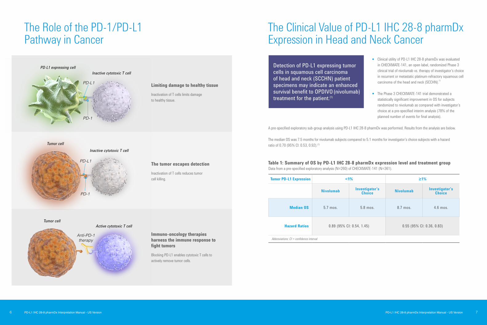
PD-1
PD-1
Inactive cytotoxic T cell
Inactive cytotoxic T cell
Active cytotoxic T cell
PD-L1 expressing cell
Tumor cell
Tumor cell
PD-L1
PD-L1
Anti-PD-1 therapy
Limiting damage to healthy tissue Inactivation of T cells limits damage to healthy tissue.
The tumor escapes detection Inactivation of T cells reduces tumor cell killing.
Immuno-oncology therapies harness the immune response to fight tumors
Blocking PD-L1 enables cytotoxic T cells to actively remove tumor cells.
Clinical utility of PD-L1 IHC 28-8 pharmDx was evaluated in CHECKMATE-141, an open label, randomized Phase 3 clinical trial of nivolumab vs. therapy of investigator’s choice in recurrent or metastatic platinum-refractory squamous cell carcinoma of the head and neck (SCCHN).(1)
The Phase 3 CHECKMATE-141 trial demonstrated a statistically significant improvement in OS for subjects randomized to nivolumab as compared with investigator’s choice at a pre-specified interim analysis (78% of the planned number of events for final analysis).
A pre-specified exploratory sub-group analysis using PD-L1 IHC 28-8 pharmDx was performed. Results from the analysis are below.
The median OS was 7.5 months for nivolumab subjects compared to 5.1 months for investigator’s choice subjects with a hazard ratio of 0.70 (95% CI: 0.53, 0.92).(1)
Detection of PD-L1 expressing tumor cells in squamous cell carcinoma of head and neck (SCCHN) patient specimens may indicate an enhanced survival benefit to OPDIVO (nivolumab) treatment for the patient.(1)
Tumor PD-L1 Expression <1% ≥1%
Nivolumab Investigator’s Choice Nivolumab Investigator’s
Choice
Median OS 5.7 mos. 5.8 mos. 8.7 mos. 4.6 mos.
Hazard Ratios 0.89 (95% CI: 0.54, 1.45) 0.55 (95% CI: 0.36, 0.83)
Abbreviations: CI = confidence interval
Table 1: Summary of OS by PD-L1 IHC 28-8 pharmDx expression level and treatment groupData from a pre-specified exploratory analysis (N=260) of CHECKMATE-141 (N=361).
The Clinical Value of PD-L1 IHC 28-8 pharmDxExpression in Head and Neck Cancer
The Role of the PD-1/PD-L1Pathway in Cancer
PD-L1 IHC 28-8 pharmDx Interpretation Manual - US Version8 9PD-L1 IHC 28-8 pharmDx Interpretation Manual - US Version
PD-L1 expression, as determined by PD-L1 IHC 28-8 pharmDx in SCCHN, may be associated with enhanced survival from OPDIVO (nivolumab).
Head and neck carcinomas are the sixth most common cancer globally, accounting for approximately 550,000 new cases and approximately 300,000 deaths each year. While only a small minority of patients present with metastatic disease initially (~10%; Stage IV-C), approximately 50% of the population initially treated in a locally advanced setting will eventually develop recurrent or refractory disease.
Recurrent or metastatic SCCHN remains an area of high unmet medical need, since patients who progress after platinum-based treatment (platinum-refractory or resistant disease) have poor prognosis, with a median OS of approximately 4 to 6 months. There is no effective standard of care to provide survival benefits in platinum-refractory recurrent or metastatic SCCHN.
Clinical utility of PD-L1 IHC 28-8 pharmDx in SCCHN was evaluated using specimens from patients enrolled in clinical trial CHECKMATE-141, a randomized Phase 3 clinical trial of OPDIVO (nivolumab) vs. therapy of investigator’s choice in recurrent or metastatic platinum-refractory squamous cell carcinoma of the head and neck (SCCHN).
Baseline (pre-study) tumor tissue specimens were collected prior to randomization.
Primary Study Objective:
To compare OS of nivolumab to investigator’s choice in patients with metastatic or recurrent SCCHN who had experienced disease progression during or within 6 months of receiving platinum-based therapy administered in either the adjuvant, neo-adjuvant, primary (unresectable locally advanced) or metastatic setting.
Secondary Study Objectives:
To compare Progression Free Survival (PFS) of nivolumab to investigator’s choice.
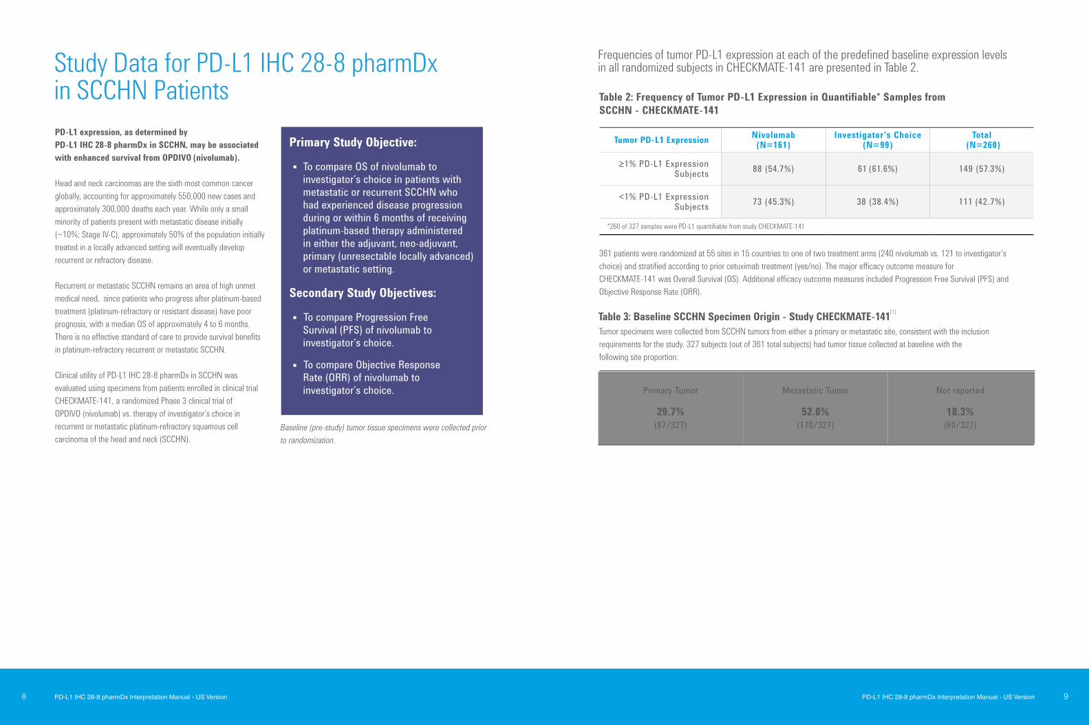
To compare Objective Response Rate (ORR) of nivolumab to investigator’s choice. Primary Tumor
29.7%
(97/327)
Metastatic Tumor
52.0% (170/327)
Not reported
18.3% (60/327)
Table 3: Baseline SCCHN Specimen Origin - Study CHECKMATE-141[1]
Frequencies of tumor PD-L1 expression at each of the predefined baseline expression levels in all randomized subjects in CHECKMATE-141 are presented in Table 2.
Table 2: Frequency of Tumor PD-L1 Expression in Quantifiable* Samples from SCCHN - CHECKMATE-141
Tumor specimens were collected from SCCHN tumors from either a primary or metastatic site, consistent with the inclusion requirements for the study. 327 subjects (out of 361 total subjects) had tumor tissue collected at baseline with the following site proportion:
361 patients were randomized at 55 sites in 15 countries to one of two treatment arms (240 nivolumab vs. 121 to investigator’s choice) and stratified according to prior cetuximab treatment (yes/no). The major efficacy outcome measure for CHECKMATE-141 was Overall Survival (OS). Additional efficacy outcome measures included Progression Free Survival (PFS) and Objective Response Rate (ORR).
Tumor PD-L1 Expression Nivolumab(N=161)
Investigator’s Choice (N=99)
Total (N=260)
≥1% PD-L1 Expression Subjects 88 (54.7%) 61 (61.6%) 149 (57.3%)
<1% PD-L1 Expression Subjects 73 (45.3%) 38 (38.4%) 111 (42 .7%)
*260 of 327 samples were PD-L1 quantifiable from study CHECKMATE-141
Study Data for PD-L1 IHC 28-8 pharmDxin SCCHN Patients
PD-L1 IHC 28-8 pharmDx Interpretation Manual - US Version10 11PD-L1 IHC 28-8 pharmDx Interpretation Manual - US Version
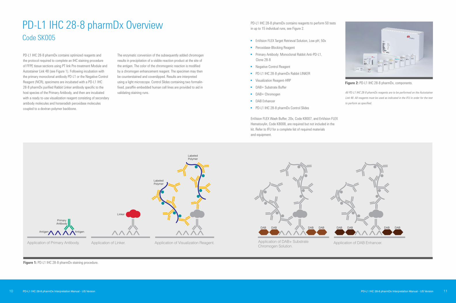
PD-L1 IHC 28-8 pharmDx contains optimized reagents and the protocol required to complete an IHC staining procedure of FFPE tissue sections using PT link Pre-treatment Module and Autostainer Link 48 (see Figure 1). Following incubation with the primary monoclonal antibody PD-L1 or the Negative Control Reagent (NCR), specimens are incubated with a PD-L1 IHC 28-8 pharmDx purified Rabbit Linker antibody specific to the host species of the Primary Antibody, and then are incubated with a ready to-use visualization reagent consisting of secondary antibody molecules and horseradish peroxidase molecules coupled to a dextran polymer backbone.
The enzymatic conversion of the subsequently added chromogen results in precipitation of a visible reaction product at the site of the antigen. The color of the chromogenic reaction is modified by a chromogen enhancement reagent. The specimen may then be counterstained and coverslipped. Results are interpreted using a light microscope. Control Slides containing two formalin-fixed, paraffin-embedded human cell lines are provided to aid in validating staining runs.
Antigen
PrimaryAntibody
Linker
LabeledPolymer
LabeledPolymer
DABDAB DABDABAntigen
Application of Primary Antibody.
Application of Visualization Reagent.
Application of DAB+ Substrate Chromogen Solution.
Application of Linker.
PD-L1
DABDAB DABDAB
Application of DAB Enhancer.
Application of DAB+ Substrate Chromogen Solution.
Application of DAB Enhancer.Application of Primary Antibody. Application of Linker. Application of Visualization Reagent.
Antigen
PrimaryAntibody
Linker
LabeledPolymer
LabeledPolymer
DABDAB DABDABAntigen
Application of Primary Antibody.
Application of Visualization Reagent.
Application of DAB+ Substrate Chromogen Solution.
Application of Linker.
PD-L1
DABDAB DABDAB
Application of DAB Enhancer.
PD-L1 IHC 28-8 pharmDx contains reagents to perform 50 tests in up to 15 individual runs, see Figure 2.
EnVision FLEX Target Retrieval Solution, Low pH, 50x
Peroxidase-Blocking Reagent
Primary Antibody: Monoclonal Rabbit Anti-PD-L1, Clone 28-8
Negative Control Reagent
PD-L1 IHC 28-8 pharmDx Rabbit LINKER
Visualization Reagent-HRP
DAB+ Substrate Buffer
DAB+ Chromogen
DAB Enhancer
PD-L1 IHC 28-8 pharmDx Control Slides
EnVision FLEX Wash Buffer, 20x, Code K8007, and EnVision FLEX Hematoxylin, Code K8008, are required but not included in the kit. Refer to IFU for a complete list of required materials and equipment.
All PD-L1 IHC 28-8 pharmDx reagents are to be performed on the Autostainer
Link 48. All reagents must be used as indicated in the IFU in order for the test
to perform as specified.
Figure 2: PD-L1 IHC 28-8 pharmDx, components.
Figure 1: PD-L1 IHC 28-8 pharmDx staining procedure.
PD-L1 IHC 28-8 pharmDx OverviewCode SK005
PD-L1 IHC 28-8 pharmDx Interpretation Manual - US Version12 13PD-L1 IHC 28-8 pharmDx Interpretation Manual - US Version
Optimal staining performance is achieved by adhering to the PD-L1 IHC 28-8 pharmDx protocol. The following are tips for optimizing staining performance. Technical problems related to the performance of PD-L1 IHC 28-8 pharmDx may arise; those involving specimen collection, specimen preparation prior to performing the test and problems involving the actual performance of the test itself. Technical problems of the test can be minimized with a thorough understanding of the product instructions by the user.
Specimen Collection and ProcessingSpecimens must be handled in a way which preserves the tissue for immunohistochemical staining. Tissue should be stained and interpreted as close to time of biopsy as possible. Stability ofPD-L1 immunoreactivity in tissue blocks has not been assessed. Tissue may be susceptible to loss of PD-L1 immunoreactivity with age. Confirm appropriate intact tumor morphology and the presence of sufficient tumor cells for evaluation. Use recommended methods of tissue processing for all specimens.
Control TissueDifferences in processing and embedding in the user’s laboratory may produce significant variability in results. Include positive and negative control tissue in each staining run, in addition to PD-L1 IHC 28-8 pharmDx Control Slides.
Select positive and negative control tissue from fresh SCCHN specimens. Fix, process, and embed the control tissue in the same manner as patient specimens. Control tissue processed differently from the patient specimen validates reagent performance only and does not verify tissue preparation. The ideal positive control tissue shows weak to moderate PD-L1 expression. The variety of different cell types present in most tissue sections offers internal negative control sites; this should be verified by the user. A suggested SCCHN-negative control tissue is one that shows no staining in tumor cells but possesses stained immune cells such as macrophages and lymphocytes.
Tissue ProcessingFormalin-fixed, paraffin-embedded tissues are suitable for use. Block specimens into a thickness of 3 mm or 4 mm, fix in 10% Neutral Buffered Formalin (NBF), and dehydrate and clear in a series of alcohols and xylene, followed by infiltration with melted paraffin. An ischemia time from excision to fixation start time
of less than 30 minutes followed by immersion in 10% neutral buffered formalin for 24-48 hours is recommended. The paraffin temperature should not exceed 60 °C. The use of PD-L1 IHC 28-8 pharmDx on decalcified tissues has not been validated and is not recommended. Cut tissue specimens into sections of 4-5 μm. After sectioning, mount tissues on FLEX IHC microscope slides, Code K8020, or Fisherbrand Superfrost Plus charged slides and place in a 58 ± 2 °C oven for 1 hour. To preserve antigenicity, tissue sections, once mounted on slides, should be stored in the dark at 2-8 °C, or room temperature up to 25 °C, and stained within 4 months of sectioning. Slide storage and handling conditions should not exceed 25 °C at any point post-mounting to ensure tissue integrity and antigenicity.
PD-L1 IHC 28-8 pharmDx Staining ProcedurePD-L1 IHC 28-8 pharmDx reagents and instructions have been designed for optimal performance. Further dilution of the reagents, alteration of incubation times, temperatures, or materials may give erroneous results.
Reagent Storage Store all components of PD-L1 IHC 28-8 pharmDx, including Control Slides, in the dark at 2-8 °C when not in use on Autostainer Link 48.
Reagent Preparation Equilibrate all components to room temperature (20-25 °C) prior to immunostaining. Do not use after the expiration date printed on the outside package. EnVision FLEX Target Retrieval Solution, Low pH Dilute EnVision FLEX Target Retrieval Solution, Low pH (50x) 1:50 using distilled or deionized water (reagent-quality water). One 30 mL bottle of concentrate provides 1.5 L of working solution which is sufficient to fill one PT Link Pretreatment Module tank and will treat up to 24 slides per use. The pH of the working solution should be 6.1 ± 0.2. Discard Low pH working solution after three uses. Do not use after 5 days following dilution.
EnVision FLEX Wash Buffer, Code K8007 Dilute EnVision FLEX Wash Buffer (20x) 1:20 using distilled or deionized water (reagent-quality water). Store unused working solution at 2-8 °C for no more than one month. Wash buffer can also be stored for up to 7 days at 25 °C. Discard if cloudy in appearance.
DAB+ Substrate-Chromogen Solution Add 1 drop of DAB+ Chromogen per mL of DAB+ Substrate Buffer and mix. Prepared DAB+ Substrate-Chromogen Solution is stable for 5 days if stored in the dark at 2-8 °C. Mix thoroughly prior to use. Any precipitate developing in the solution does not affect staining quality. Add 9 drops of DAB+ Chromogen to a full bottle of DAB+
Substrate Buffer. Although the DAB+ Substrate Buffer label states 7. 2 mL, this is the usable volume and does not account for the “dead” volume of DAB+ Substrate Buffer in the bottle.
The color of the DAB+ Chromogen may vary from clear to lavender brown. This will not affect the performance of the product. Dilute per the guidelines above. Adding excess DAB+ Chromogen to the DAB+ Substrate Buffer results in deterioration of the positive signal.
Controls to Assess Staining QualityControl slides are recommended to determine that PD-L1 IHC 28-8 pharmDx results (generated by the system containing reagents, instrument hardware and software) are valid and the reagents are functioning properly. For each staining run include the following control slides:
One PD-L1 IHC 28-8 pharmDx Control Slide stained with the Primary Antibody in each staining run.
Two positive control tissue slides (one stained with Primary Antibody and the other stained with Negative Control
Reagent) for each set of test conditions.
Two negative control tissue slides (one stained with Primary Antibody and the other stained with Negative Control Reagent).
Lastly, for each patient specimen stained with Primary Antibody, include a sequential section of patient specimen
stained with Negative Control Reagent.
Staining ProtocolProgram slides by selecting PD-L1 IHC 28-8 pharmDx staining protocol from the options in the DakoLink drop down menu. All of the required steps and incubation times for staining are preprogrammed in the DakoLink software. Print and attach slide labels to each slide.
Deparaffinization, Rehydration and Target Retrieval Use PT Link Pretreatment Module to perform a deparaffinization, rehydration and target retrieval 3-in-1 procedure.
Set Preheat and Cool to 65 °C, and set Heat to 97 °C for 20 minutes.
Fill PT Link tanks with 1.5 L per tank of prepared EnVision FLEX Target Retrieval Solution, Low pH, working solution to cover the tissue sections.
Preheat the Target Retrieval Solution, Low pH to 65 °C.
Immerse Autostainer racks containing mounted, FFPE tissue sections into the pre-heated Target Retrieval Solution, Low pH in PT Link tank. Start the PT Link program and incubate for 20 minutes at 97 °C.
When target retrieval incubation has been completed and the temperature has cooled to 65 °C, remove each Autostainer slide rack with slides from the PT Link tank and immediately place the rack with slides into a tank (e.g., PT Link Rinse Station, Code PT109) containing room temperature EnVision FLEX Wash Buffer working solution.
Leave Autostainer rack with slides in room temperature EnVision FLEX Wash Buffer for 5 minutes.
Staining and CounterstainingPlace the Autostainer rack with slides on the Autostainer Link 48. Ensure slides remain wet with buffer while loading and prior to initiating the run. Dried tissue sections may display increased non-specific staining.
Start the Autostainer to run the PD-L1 IHC 28-8 pharmDx protocol. The instrument performs the staining and counterstaining procedures by applying the appropriate reagent, monitoring the incubation time and rinsing slides between reagents. Counterstain slides for 7 minutes using EnVision FLEX Hematoxylin. Do not allow slides to dry prior to mounting.
MountingUse non-aqueous permanent mounting media. To minimize fading, store slides in the dark at room temperature (20-25 °C).
Example of Adequate Dehydration Procedure
95% EtOH (1 min)95% EtOH (1 min)100% EtOH (1 min)100% EtOH (1 min) Xylene (3 min) Xylene (3 min)
Ensure that slides do not dry between the end of the Autostainer run and
mounting procedure. Xylene may be substituted with Histoclear solution.
Technical Considerations for OptimalPerformance of PD-L1 IHC 28-8 pharmDx
PD-L1 IHC 28-8 pharmDx Interpretation Manual - US Version14 15PD-L1 IHC 28-8 pharmDx Interpretation Manual - US Version
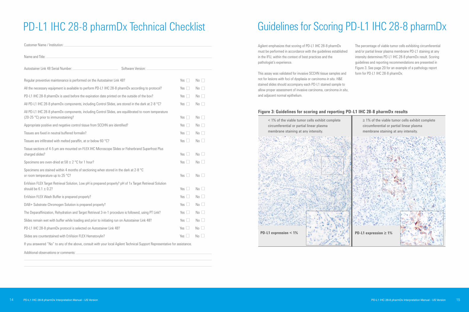
Agilent emphasizes that scoring of PD-L1 IHC 28-8 pharmDx must be performed in accordance with the guidelines established in the IFU, within the context of best practices and the pathologist’s experience. This assay was validated for invasive SCCHN tissue samples and not for lesions with foci of dysplasia or carcinoma in situ. H&E stained slides should accompany each PD-L1 stained sample to allow proper assessment of invasive carcinoma, carcinoma in situ, and adjacent normal epithelium.
The percentage of viable tumor cells exhibiting circumferential and/or partial linear plasma membrane PD-L1 staining at any intensity determines PD-L1 IHC 28-8 pharmDx result. Scoring guidelines and reporting recommendations are presented in Figure 3. See page 20 for an example of a pathology report form for PD-L1 IHC 28-8 pharmDx.
Customer Name / Institution: Name and Title:
Autostainer Link 48 Serial Number: Software Version:
Regular preventive maintenance is performed on the Autostainer Link 48? Yes No
All the necessary equipment is available to perform PD-L1 IHC 28-8 pharmDx according to protocol? Yes No
PD-L1 IHC 28-8 pharmDx is used before the expiration date printed on the outside of the box? Yes No
All PD-L1 IHC 28-8 pharmDx components, including Control Slides, are stored in the dark at 2-8 °C? Yes No
All PD-L1 IHC 28-8 pharmDx components, including Control Slides, are equilibrated to room temperature (20-25 °C) prior to immunostaining? Yes No
Appropriate positive and negative control tissue from SCCHN are identified? Yes No Tissues are fixed in neutral buffered formalin? Yes No
Tissues are infiltrated with melted paraffin, at or below 60 °C? Yes No
Tissue sections of 4-5 µm are mounted on FLEX IHC Microscope Slides or Fisherbrand Superfrost Plus charged slides? Yes No
Specimens are oven-dried at 58 ± 2 °C for 1 hour? Yes No
Specimens are stained within 4 months of sectioning when stored in the dark at 2-8 °C or room temperature up to 25 °C? Yes No
EnVision FLEX Target Retrieval Solution, Low pH is prepared properly? pH of 1x Target Retrieval Solution should be 6.1 ± 0.2? Yes No
EnVision FLEX Wash Buffer is prepared properly? Yes No
DAB+ Substrate-Chromogen Solution is prepared properly? Yes No
The Deparaffinization, Rehydration and Target Retrieval 3-in-1 procedure is followed, using PT Link? Yes No
Slides remain wet with buffer while loading and prior to initiating run on Autostainer Link 48? Yes No
PD-L1 IHC 28-8 pharmDx protocol is selected on Autostainer Link 48? Yes No
Slides are counterstained with EnVision FLEX Hematoxylin? Yes No
If you answered “No” to any of the above, consult with your local Agilent Technical Support Representative for assistance.
Additional observations or comments:
Guidelines for Scoring PD-L1 IHC 28-8 pharmDxPD-L1 IHC 28-8 pharmDx Technical Checklist
Figure 3: Guidelines for scoring and reporting PD-L1 IHC 28-8 pharmDx results
< 1% of the viable tumor cells exhibit complete circumferential or partial linear plasma membrane staining at any intensity.
≥ 1% of the viable tumor cells exhibit complete circumferential or partial linear plasma membrane staining at any intensity.
PD-L1 expression < 1% PD-L1 expression ≥ 1%
PD-L1 IHC 28-8 pharmDx Interpretation Manual - US Version16 17PD-L1 IHC 28-8 pharmDx Interpretation Manual - US Version
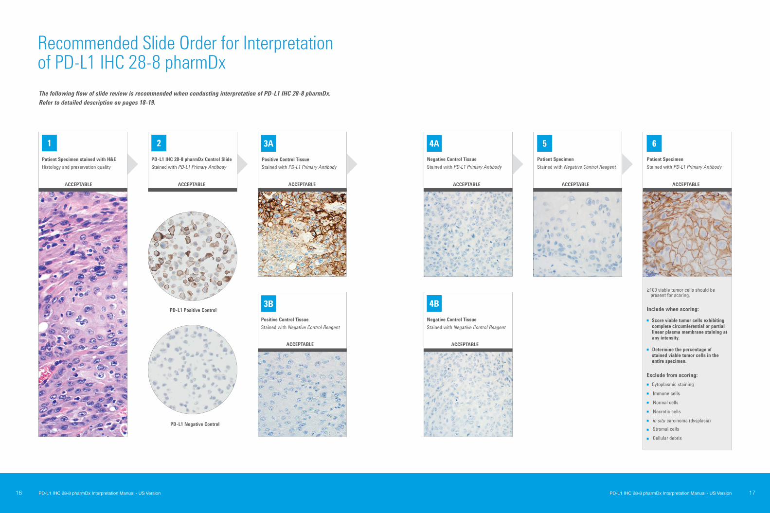
The following flow of slide review is recommended when conducting interpretation of PD-L1 IHC 28-8 pharmDx. Refer to detailed description on pages 18-19.
1 2 3A 4A 5 6
3B 4B
Patient Specimen stained with H&E Histology and preservation quality
PD-L1 IHC 28-8 pharmDx Control SlideStained with PD-L1 Primary Antibody
Positive Control TissueStained with PD-L1 Primary Antibody
Negative Control TissueStained with PD-L1 Primary Antibody
Patient Specimen Stained with Negative Control Reagent
Patient SpecimenStained with PD-L1 Primary Antibody
Positive Control TissueStained with Negative Control Reagent
Negative Control TissueStained with Negative Control Reagent
≥100 viable tumor cells should be present for scoring.
Include when scoring:
Score viable tumor cells exhibiting complete circumferential or partial linear plasma membrane staining at any intensity.
Determine the percentage of stained viable tumor cells in the entire specimen.
Exclude from scoring: Cytoplasmic staining Immune cells Normal cells Necrotic cells in situ carcinoma (dysplasia) Stromal cells
Cellular debris
ACCEPTABLE ACCEPTABLE ACCEPTABLE
PD-L1 Positive Control
PD-L1 Negative Control
ACCEPTABLE ACCEPTABLE ACCEPTABLE
ACCEPTABLEACCEPTABLE
Recommended Slide Order for Interpretation of PD-L1 IHC 28-8 pharmDx

PD-L1 IHC 28-8 pharmDx Interpretation Manual - US Version18 19PD-L1 IHC 28-8 pharmDx Interpretation Manual - US Version
3. Positive Control Tissue Slides Examine the positive SCCHN control tissue slides (one stained with Primary Antibody and the other with Negative Control Reagent) to ascertain if tissues are correctly prepared and reagents are functioning properly. Any background staining should be of ≤ 1+ staining intensity. Exclude necrotic or degenerated cells from evaluation. If staining of positive control tissues is not satisfactory, all results with the patient specimens should be considered invalid. Do not use control tissue as an aid in interpretation of patient results.
4. Negative Control Tissue Slides Examine the negative SCCHN control tissue slides (one stained with Primary Antibody and the other with Negative Control Reagent) to confirm that there is no unintended staining. Any background staining should be of ≤ 1+ staining intensity. If unwanted specific plasma membrane staining of malignant cells occurs in the negative control tissue, all results with the patient specimens should be considered invalid. Do not use control tissue as an aid in interpretation of patient results.
5. Patient Specimen Stained with Negative Control Reagent The Negative Control Reagent indicates non-specific background staining and allows better interpretation of patient specimenstained with the Primary Antibody. Examine the patient specimen stained with Negative Control Reagent to identify non-specific background staining. Staining by the Negative Control Reagent
must not show positive membrane staining and non-specific background should be ≤ 1+. If staining is not satisfactory, results with the patient specimen should be considered invalid.
6. Patient Specimen Stained with Primary Antibody Staining should be assessed within the context of any non-specific background staining of the patient specimen stained with Negative Control Reagent. A minimum of 100 viable tumor cells should be present in the PD-L1 stained patient specimen slide to determine the percentage of stained cells.
Tips and Special Considerations Include the entire specimen for evaluation of PD-L1 expression Use higher magnifications to confirm cell types and areas
absent of staining Be careful not to overlook weak 1+ staining, which can be
missed at 4x and 10x
Non-evaluable specimens: The specimen should be considered non-evaluable if there are fewer than 100 viable tumor cells. A different section from the same block or another block from the same patient may be required to present a sufficient quantity of viable tumor cells for PD-L1 IHC 28-8 pharmDx evaluation.
Indeterminate specimen: The tumor cell membrane has been hampered for reasons attributed to the biology of the tumor tissue sample rather than improper sample preparation. For example, high cytoplasmic staining of the tumor cells can hamper scoring of the membrane staining. An additional cut section or section from another block of the same patient may be required for PD-L1 IHC 28-8 pharmDx evaluation.
PD-L1 IHC 28-8 pharmDx evaluation must be performed by a pathologist using a bright field microscope. Before examining the patient specimen for PD-L1 staining, it is important to examine the hematoxylin and eosin (H&E) and controls first to assess staining quality. Examine a serial section of the patient specimen stained with H&E for histology and preservation quality. Then, examine PD-L1 IHC 28-8 pharmDx Control Slide, followed by the positive and negative control tissue slides, stained with Negative Control Reagent and Primary Antibody for each set of test conditions. Lastly, examine the patient specimen stained with Negative Control Reagent and Primary Antibody to assess the percentage staining of viable tumor cells.
PD-L1 staining is defined as complete circumferential and/or partial linear plasma membrane staining of tumor cells at any intensity. Only the PD-L1 IHC 28-8 pharmDx Control Slide is provided in the PD-L1 IHC 28-8 pharmDx kit. Positive control tissue slides and negative control tissue slides should be supplied by the laboratory. Laboratory provided positive and negative control tissue may be included on the same slide as the patient specimen.
1. Patient Specimen Stained with H&E An H&E stained section is required for the evaluation of histology and preservation quality. PD-L1 IHC 28-8 pharmDx and the H&E staining should be performed on serial sections from the same paraffin block of the specimen.
Foci of dysplasia and carcinoma in situ are excluded from scoring. An accompanying H&E section allows for the proper assessment of invasive carcinoma, carcinoma in situ, and adjacent normal epithelium.
2. PD-L1 IHC 28-8 pharmDx Control Slide Examine the PD-L1 IHC 28-8 pharmDx Control Slide to ascertain that reagents are functioning properly. Each slide contains sections of cell pellets with positive and negative PD-L1 expression, see Figure 4. If any staining of the Control Slide is not satisfactory, all results with the patient specimens should be considered invalid. Do not use the Control Slide as an aid in interpretation of patient results.
For the PD-L1 positive cell pellet on the Control Slide, the following staining is acceptable, see Figure 5:
At least 80% of the cells contain plasma membrane staining of at least 2+ average staining intensity
Any background staining is of less than 1+ staining intensity
For the PD-L1 negative cell pellet on the Control Slide, the following staining is acceptable, see Figure 6: No plasma membrane staining
Any background staining is of less than 1+ staining intensity
Staining of a few cells in the negative pellet on the Control Slide may occasionally be observed. The presence of 10 or fewer cells with distinct plasma membrane staining, or cytoplasmic staining with ≥ 1+ intensity within the boundaries of the negative cell pellet are acceptable.
Figure 4: Each Control Slide contains sections of cell pellets with positive and negative PD-L1 expression.
Figure 5: Acceptable Positive PD-L1 Control.
Figure 6: Acceptable Negative PD-L1 Control.
Assess the percentage of cells with plasma membrane staining and the staining intensity. Evaluate the overall staining intensity using the following guide:
PD-L1 positive
PD-L1 negative
PD-L1 IHC
28-8
xxxxx 0 No staining
1+ Weak staining
2+ Moderate staining
3+ Strong staining
1
2
3
At 4x objective magnification, carefully examine the tumor areas of the entire specimen. All areas with viable tumor cells on the specimen should be evaluated. Exclude cytoplasmic staining from scoring. Exclude immune cells, necrotic cells, normal cells, and in situ carcinoma from scoring.
Use the 10-20x objective magnifications to determine the percentage of viable tumor cells expressing PD-L1 membranous staining. The 40x objective can be used for confirmation if needed. Tumor cells are considered to be PD-L1 positive if they exhibit either partial linear or complete circumferential staining of the plasma membrane at any intensity.
Record if the specimen has tumor PD-L1 expression < 1% or ≥ 1%. When determining the percentage of stained tumor cells in the entire specimen, the numerator is the number of stained viable tumor cells and the denominator is the total number of viable tumor cells in the specimen.
Recommendations for Interpretation of PD-L1 IHC 28-8 pharmDx in SCCHN
% PD-L1 positive = x100
Number of tumor cells expressing PD-L1 positive membrane staining
Total number of viable tumor cells present in the section

PD-L1 IHC 28-8 pharmDx Interpretation Manual - US Version20 21PD-L1 IHC 28-8 pharmDx Interpretation Manual - US Version
The following images present examples of SCCHN tumor samples stained with PD-L1 IHC 28-8 pharmDx.
Figure 7: An example of a squamous cell carcinoma of the tonsil stained with PD-L1 IHC 28-8 pharmDx assay. The staining shows a range of PD-L1 expression. This specimen would be appropriate to use as a positive control specimen for detection of subtle changes in assay sensitivity. Note the partial linear (red arrow) and complete circumferential (black arrow) plasma membrane staining. 20x magnification.
Note: PD-L1 IHC 28-8 pharmDx was validated for invasive SCCHN tissue samples and not for lesions with foci of dysplasia or carcinoma in situ. An H&E stained slide should accompany each PD-L1 stained sample to allow proper assessment of invasive carcinoma, carcinoma in situ, and adjacent normal epithelium.
Suggested information to include when reporting results with PD-L1 IHC 28-8 pharmDx in SCCHN
PD-L1 IHC 28-8 pharmDx, Code SK005 Summary of Sample Tested:
Date of Run: PD-L1 IHC 28-8 pharmDx Lot:
Staining Run Log ID: Specimen ID:
Patient Identifier: Type of service: IHC Stain with Manual Interpretation
Other:
Type of Tissue: Additional Tests Performed with PD-L1 IHC 28-8 pharmDx:
PD-L1 IHC 28-8 pharmDx Controls Results:
PD-L1 IHC 28-8 Control Slides: Pass Fail Positive Control Tissue Slides: Pass Fail Negative Control Tissue Slides: Pass Fail Patient Specimen, Negative Control Reagent: Pass Fail
PD-L1 Results: Detection of PD-L1 expressing tumor cells in SCCHN patient specimens may indicate an enhanced survival benefit to OPDIVO (nivolumab) treatment for the patient.(1)
Viable Tumor Cells Present ≥ 100 cells Not Evaluable
PD-L1 expression < 1%: Percent of SCCHN cells with complete circumferential and/or partial linear membrane PD-L1 staining is < 1%
PD-L1 expression ≥ 1%: Percent of SCCHN cells with complete circumferential and/or partial linear membrane PD-L1 staining is ≥ 1%.
Percent Expression PD-L1 Tumor Cells: % Other Comments to Treating Physician:
Reporting Results PD-L1 IHC 28-8 pharmDx ImmunostainingExamples in SCCHN
PD-L1 IHC 28-8 pharmDx Interpretation Manual - US Version22 23PD-L1 IHC 28-8 pharmDx Interpretation Manual - US Version
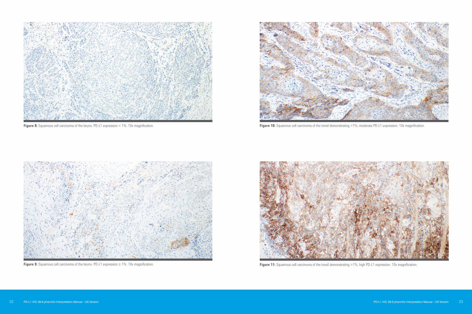
Figure 8: Squamous cell carcinoma of the larynx. PD-L1 expression < 1%. 10x magnification.
Figure 9: Squamous cell carcinoma of the larynx. PD-L1 expression ≥ 1%. 10x magnification. Figure 11: Squamous cell carcinoma of the tonsil demonstrating >1%, high PD-L1 expression. 10x magnification.
Figure 10: Squamous cell carcinoma of the tonsil demonstrating >1%, moderate PD-L1 expression. 10x magnification.
PD-L1 IHC 28-8 pharmDx Interpretation Manual - US Version24 25PD-L1 IHC 28-8 pharmDx Interpretation Manual - US Version
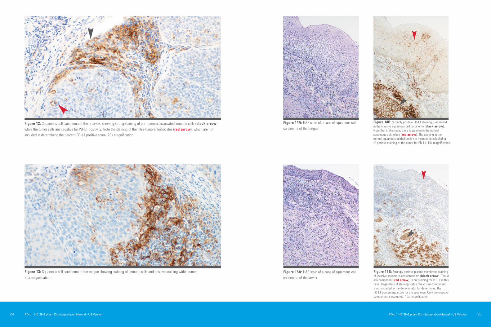
Figure 15A: H&E stain of a case of squamous cell carcinoma of the larynx.
Figure 15B: Strongly positive plasma membrane staining of invasive squamous cell carcinoma (black arrow). The in situ component (red arrow), is not staining for PD-L1 in this case. Regardless of staining status, the in situ component is not included in the denominator for determining the PD-L1 percentage score for the specimen. Only the invasive component is evaluated. 10x magnification.
Figure 13: Squamous cell carcinoma of the tongue showing staining of immune cells and positive staining within tumor. 20x magnification.
Figure 14A: H&E stain of a case of squamous cell carcinoma of the tongue.
Figure 14B: Strongly positive PD-L1 staining is observed in the invasive squamous cell carcinoma (black arrow). Note that in this case, there is staining in the normal squamous epithelium (red arrow). The staining in the normal squamous epithelium is not included in calculating % positive staining of the tumor for PD-L1. 10x magnification.
Figure 12: Squamous cell carcinoma of the pharynx, showing strong staining of peri-tumoral associated immune cells (black arrow), while the tumor cells are negative for PD-L1 positivity. Note the staining of the intra-tumoral histiocytes (red arrow), which are not included in determining the percent PD-L1 positive score. 20x magnification.
PD-L1 IHC 28-8 pharmDx Interpretation Manual - US Version26 27PD-L1 IHC 28-8 pharmDx Interpretation Manual - US Version
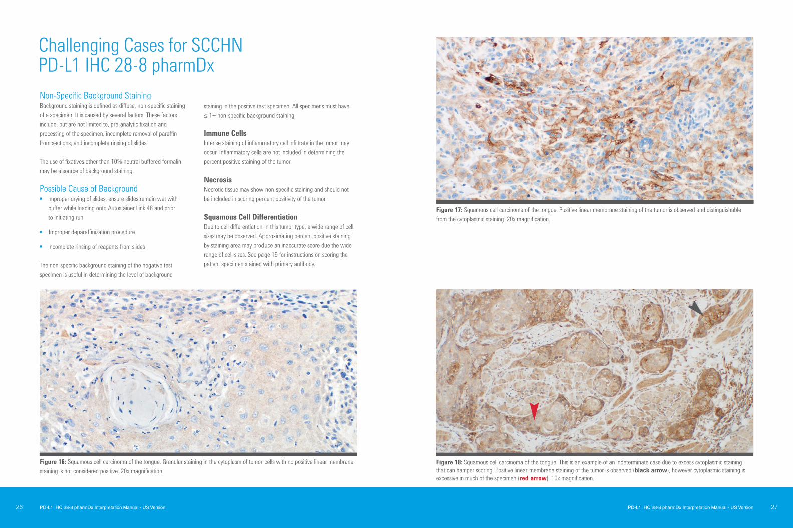
Figure 17: Squamous cell carcinoma of the tongue. Positive linear membrane staining of the tumor is observed and distinguishable from the cytoplasmic staining. 20x magnification.
Figure 16: Squamous cell carcinoma of the tongue. Granular staining in the cytoplasm of tumor cells with no positive linear membrane staining is not considered positive. 20x magnification.
Non-Specific Background Staining Background staining is defined as diffuse, non-specific staining of a specimen. It is caused by several factors. These factors include, but are not limited to, pre-analytic fixation and processing of the specimen, incomplete removal of paraffin from sections, and incomplete rinsing of slides. The use of fixatives other than 10% neutral buffered formalin may be a source of background staining.
Possible Cause of Background Improper drying of slides; ensure slides remain wet with
buffer while loading onto Autostainer Link 48 and prior to initiating run
Improper deparaffinization procedure
Incomplete rinsing of reagents from slides
The non-specific background staining of the negative testspecimen is useful in determining the level of background
staining in the positive test specimen. All specimens must have≤ 1+ non-specific background staining.
Immune CellsIntense staining of inflammatory cell infiltrate in the tumor may occur. Inflammatory cells are not included in determining the percent positive staining of the tumor.
NecrosisNecrotic tissue may show non-specific staining and should not be included in scoring percent positivity of the tumor.
Squamous Cell DifferentiationDue to cell differentiation in this tumor type, a wide range of cell sizes may be observed. Approximating percent positive staining by staining area may produce an inaccurate score due the wide range of cell sizes. See page 19 for instructions on scoring the patient specimen stained with primary antibody.
Figure 18: Squamous cell carcinoma of the tongue. This is an example of an indeterminate case due to excess cytoplasmic staining that can hamper scoring. Positive linear membrane staining of the tumor is observed (black arrow), however cytoplasmic staining is excessive in much of the specimen (red arrow). 10x magnification.
Challenging Cases for SCCHN PD-L1 IHC 28-8 pharmDx
PD-L1 IHC 28-8 pharmDx Interpretation Manual - US Version28 29PD-L1 IHC 28-8 pharmDx Interpretation Manual - US Version
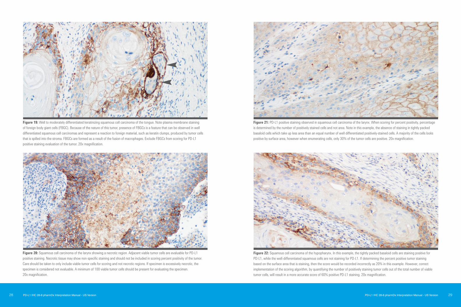
Figure 20: Squamous cell carcinoma of the larynx showing a necrotic region. Adjacent viable tumor cells are evaluable for PD-L1 positive staining. Necrotic tissue may show non-specific staining and should not be included in scoring percent positivity of the tumor. Care should be taken to only include viable tumor cells for scoring and not necrotic regions. If specimen is excessively necrotic, the specimen is considered not evaluable. A minimum of 100 viable tumor cells should be present for evaluating the specimen. 20x magnification.
Figure 22: Squamous cell carcinoma of the hypopharynx. In this example, the tightly packed basaloid cells are staining positive for PD-L1, while the well-differentiated squamous cells are not staining for PD-L1. If determining the percent positive tumor staining based on the surface area that is staining, then the score would be recorded incorrectly as 20% in this example. However, correct implementation of the scoring algorithm, by quantifying the number of positively staining tumor cells out of the total number of viable tumor cells, will result in a more accurate score of 60% positive PD-L1 staining. 20x magnification.
Figure 19: Well to moderately differentiated keratinizing squamous cell carcinoma of the tongue. Note plasma membrane staining of foreign body giant cells (FBGC). Because of the nature of this tumor, presence of FBGCs is a feature that can be observed in well differentiated squamous cell carcinomas and represent a reaction to foreign material, such as keratin clumps, produced by tumor cells that is spilled into the stroma. FBGCs are formed as a result of the fusion of macrophages. Exclude FBGCs from scoring for PD-L1 positive staining evaluation of the tumor. 20x magnification.
Figure 21: PD-L1 positive staining observed in squamous cell carcinoma of the larynx. When scoring for percent positivity, percentage is determined by the number of positively stained cells and not area. Note in this example, the absence of staining in tightly packed basaloid cells which take up less area than an equal number of well differentiated positively-stained cells. A majority of the cells looks positive by surface area, however when enumerating cells, only 30% of the tumor cells are positive. 20x magnification.
PD-L1 IHC 28-8 pharmDx Interpretation Manual - US Version30 31PD-L1 IHC 28-8 pharmDx Interpretation Manual - US Version
Argiris A, Karamouzis MV, Raben D, et al. Head and neck cancer. Lancet 2008; 371:1695-709.
Clinical and Laboratory Standards Institute (formerly NCCLS). Protection of Laboratory Workers From Occupationally Acquired Infections; Approved Guideline – Fourth Edition. CLSI document M29-A4 [ISBN 1-56238-962-9]. Clinical and Laboratory Standards Institute, 950 West Valley Road, Suite 2500, Wayne, Pennsylvania 19087 – 1898 USA, 2014.
Clinical and Laboratory Standards Institute (formerly NCCCLS). Quality assurance for Design Control and Implementation of Immunohistochemistry Assays; Approved guideline. CLSI document I/LA28-A2; Vol. 31 No. 4 (ISBN 1-56238-745-6) CLSI, 940 West Valley Road, Suite 1400, Wayne, Pennsylvania 19087 USA; 2011.
Colevas AD. Systemic therapy for metastatic or recurrent squamous cell carcinoma of the head and neck. J Natl Compr Canc Netw 2015;13: e37-e48.
Department of Health, Education and Welfare, National Institutes for Occupational Safety and Health, Rockville, MD.
“Procedures for the decontamination of plumbing systems containing copper and/or lead azides.” DHHS (NIOSH) Publ. No. 78-127, Current 13. August 16, 1976.
Ferris RL, Blumenschein G, Fayette J, et. al. Nivolumab for Recurrent Squamous-Cell Carcinoma of the Head and Neck, N Engl J Med. 2016: DOI: 10.1056/NEJMoa1602252.
Omata M, Liew C-T, Ashcavai M, Peters RL. Nonimmunologic binding of horseradish peroxidase to hepatitis B surface antigen: a possible source of error in immunohistochemistry. Am J Clin Path 1980; 73:626.
OPDIVO package insert.
Phelps RM, et al. NCI-navy medical oncology branch cell line data base. J. Cell. Biochem. 1996; 63: 32-91.
Phillips T, Simmons P, Inzunza HD, et al. Development of an automated PD-L1 immunohistochemistry (IHC) assay for non-small cell lung cancer. Appl Immuno Molec Morph 2015; 23(8):541-9.
Pignon JP, Bourhis J, Domenge C, et al. Chemotherapy added to locoregional treatment for head and neck squamous-cell carcinoma: three meta-analyses of updated individual data. MACH-NC Collaborative Group. Meta-Analysis of Chemotherapy on Head and Neck Cancer. Lancet 2000; 355:949-55.
Siegel RL, Miller KD, Jemal A. Cancer Statistics 2016. Cancer J Clin 2016; 7-30.
Taylor CR and Rudbeck L. Education Guide: Immunohistochemical Staining Methods. Sixth Edition. Dako, Carpinteria, California; 2013.
Topalian SL, Drake CG, Pardoll DM. Targeting the PD-1/B7-H1 (PD-L1) pathway to activate anti-tumor immunity. Curr Opin Immunol 2012; 24(2):207-212.
Topalian SL, Hodi FS, Brahmer JR, et. al. Safety, Activity, and Immune Correlates of Anti-PD-1 Antibody in Cancer. New Eng. J. Med. 2012; 366(26):2455-2465.
Wang C, Thudium KB, Han M, et al. In vitro characterization of the anti-PD-1 antibody nivolumab, BMS-936558, and in vivo toxicology in non-human primates. Cancer Immunol Res 2014; 2(9):846-56.
Problem Probable Cause Suggested Action
1 No staining of control or specimen slides
1a Programming error Verify that the SK005 PD-L1 IHC 28-8 pharmDx protocol was selected for programming of slides
1b Lack of reaction with DAB+ Substrate-Chromogen Solution
Verify that DAB+ Substrate-Chromogen Solution was prepared properly
1c Sodium azide in wash buffer Use only Dako Wash Buffer, Code K8007
1d Degradation of Control Slide Check kit expiration date and kit storage conditions on outside of package
2 Weak staining of specimen slides
2a Inappropriate fixation method used Ensure that only approved fixatives and fixation methods are used
2b Insufficient reagent volume applied Check size of tissue section and reagent volume applied
2c Inappropriate wash buffer used Use only Dako Wash Buffer, Code K8007
2d Reagents used when cold Allow reagents to come to room temperature (20-25 °C) prior to staining
3Weak staining of specimen slides or of the positive cell line on the Dako-provided Control Slide
3a Inadequate target retrieval Verify that the 3-in-1 pre-treatment procedure was correctly performed
3b Inappropriate wash buffer used Use only Dako Wash Buffer, Code K8007
4 Excessive background staining of slides
4a Paraffin incompletely removed Verify that the 3-in-1 pre-treatment procedure was correctly performed
4b Slides dried while loading onto the Autostainer Link 48 Ensure slides remain wet with buffer while loading and prior to initiating run
4c Non-specific binding of reagents to tissue section
Check for proper fixation of the specimen and/or the presence of necrosis
4d Inappropriate fixation method used Ensure that only neutral buffered formalin fixative and approved fixation methods are used
5 Tissue detached from slides5a Use of incorrect microscope slides Use Dako FLEX IHC Microscope Slides (Code K8020), or Fisherbrand
Superfrost Plus slides
5b Inadequate preparation of specimens Cut sections should be placed in a 58 ± 2 °C oven for one hour prior to staining
6 Excessively strong specific staining
6a Inappropriate fixation method used Ensure that only approved fixatives and fixation methods are used
6b Inappropriate wash buffer used Use only Dako Wash Buffer, Code K8007
6c Temperature of staining reagents too high It is recommended that reagents are equilibrated to 20-25 °C prior to staining
7Target Retrieval Solution is cloudy in appearance when heated
7 When heated the Target Retrieval Solution turns cloudy in appearance This is normal and does not influence staining
8 Artifacts in specimen giving a bubbling staining appearance 8 Inadequate slide dehydration procedure prior to
slide mountingEnsure adequate slide dehydration procedure was used. An example of dehydration procedure is given in this manual
References
[1] PD-L1 IHC 28-8 pharmDx Instructions for Use.
BibliographyTroubleshooting Guide for PD-L1 IHC 28-8 pharmDx
PD-L1 IHC 22C3 pharmDx Interpretation Manual – US Version32
Trusted Answers. Together.
2918
6 20
17 S
EP 1
5
Australia +61 1800 802 402
Canada +1 800 387 8257
France +33 1 64 53 57 77
Japan +81 3 5232 9970
Poland +48 58 661 1879
United Kingdom +44 (0)1 353 66 99 11
Austria +43 (0) 800 0800 7153
China +800 820 3278
Germany +49 40 69 69 470
Korea +82 2 402 6775
Spain +34 93 344 57 77
United States of America +1 800 235 5763
Belgium +32 16 930 030
Denmark +45 44 85 97 56
Ireland +353 1 479 0568
The Netherlands +31 20 547 2026
Sweden +46 8 556 20 600
Brazil +55 0800 728 1405
Finland +358 9 50 991
Italy +39 02 58 078 213
Norway +47 23 14 05 40
Switzerland +41 41 760 11 66
www.agilent.comRepresented in more than 100 countries
PRINT SPECIFICATIONS
DIGITAL SPECIFICATIONS
SPECIAL INSTRUCTIONS
No special instructions
NOTES & COMMENTS
Trim Size: 210mm x 280mm Live Area: Bleed: 3mm Colors: 4/4 Media:
Size: Max Weight: Host Subload: Platform: File Format:
URL:
PROJECT NAME:
PDL-1 IHC 28-8 Interpretation Manual_US version
CLIENT NAME:
AgilentJOB NUMBER:
159-1145
REVISION DATE:
09/15/17DUE DATE:
2107 Sawtelle Blvd, Los Angeles, CA 90025
ROUND/REVISION
V11CLIENT CONTACT:
Martha
CONTACT:
Mathieu Fischer [email protected]


![ARID1A prevents squamous cell carcinoma initiation and ...SCCs include the skin, head and neck, esophagus, lung, and cervix [2]. Cutaneous squamous cell carcinoma (cSCC) is a nonmelanoma](https://img.dokumen.tips/doc/110x75/6012df67f7a82c062d6f1b92/arid1a-prevents-squamous-cell-carcinoma-initiation-and-sccs-include-the-skin.jpg)