Embed Size (px)
Citation preview

396 BRIT. J. SURG., 1970, Vol. 57, No. 5, MAY
In this type of liposarcoma varying degrees of differ- entiation and pleomorphism may be found, and these factors may influence the clinical behaviour of a parti- cular tumour.
Using this classification, the tumour of the forearm was assigned to the well-differentiated myxoid type, so well differentiated in fact that little myxoid was demonstrable either in background matrix or as a substance stainable with Alcian blue in the developing lipoblasts.
On the other hand, the omental growth showed both large amounts of background myxomatous matrix material and also marked cellular pleomor- phism with numbers of multivacuolated giant-cell forms.
The clinical behaviour of these two liposarcomas contrasted remarkably. Excision of the subcutaneous liposarcoma resulted in an apparent cure for 8 years, but removal of the liposarcoma of the omentum was followed by local recurrence within 4 weeks and death within 4 months. In Enzinger and Winslow’s (1962) series of 103 cases a myxoid or round-cell lipo- sarcoma of the lower extremity was followed by a tumour of similar structure in the retroperitoneum, mesentery, or omentum in 11 patients. The average interval between the onset of liposarcoma in the lower extremity and that in the abdomen was 4’7 years. The opposite, namely a tumour of the abdo- men followed by a similar lesion in an extremity, was not encountered.
Dissemination of metastases from a liposarcoma is usually blood-borne, spread via lymphatics rarely being encountered. Enzinger and Winslow (1962), for example, reported only I case of regional lymph- node involvement out of 24 examined. The lungs, liver, bone-marrow, and central nervous system are the usual sites of secondary deposits, but extension of the tumour also occurs by local infiltration along fascia1 planes and over serosal membranes. In the case reported here, there was no local recurrence at the left forearm nor any clinical evidence of spread to axillary or the other lymph-nodes. Radiographs of the lungs were normal but unfortunately no post- mortem could be performed to determine the exact extent of spread.
The difficulty of deciding the question of multi- centricity or metastases occurs in certain cases of most large series of liposarcomas, and is important as it has a bearing on therapy (Stout, 1944).
From a review of the literature and consideration of this case, it is still undecided whether multiple liposarcomas can be regarded as a condition in which adipose tissue is predisposed to malignant change at multiple sites.
In Enzinger and Winslow’s (1962) series of 103 cases only I tumour arose in the subcutaneous fat and they concluded, after considering the literature, that liposarcoma arising in this situation is exceeding- ly rare. The subcutaneous liposarcoma of the fore- arm described above thus adds another such case.
Acknowledgements.-I wish to thank Mr. Owen Daniel and Mr. Ivor Lewis for permission to publish the case. I am grateful to Dr. Alban Lloyd for the pathological reports; to Dr. Hilton Jones for help with the preparation of the paper; to Mr. G. Tushingham for the photography; and to Miss Iona Williams for the secretarial work.
BIBLIOGRAPHY ATIK, M., and WHITTLESEY, R. H. (1957)~ Ann. Surg.,
DEWEERD, J. H., and DOCKERTY, M. B. (1952), Am. 3. ENTERLINE, H. T., CULBERSON, J. D., ROCHLIN, D. B.,
ENZINGER, F. M., and WINSLOW, D. J. (1962), Virchows
EVANS, R. W. (1966), Histological Appearances of Tumours,
EWING, J. (rg35), Archs Surg., Chicago, 31, 507. EWING, M. R., and HARRISON, C. V. (1g57), Br. 3. Surg.,
HOLTZ, F. (1999, Cancer, Philad., 6, 1103. MACFARLANE, A. (I957), Br. 3. Surg., 45, 106. MORGAN, J. 0. (1950),J. int. Coil. Surg., 14, 491. PACK, G. T., and PIERSON, J. C. (1954)~ Surgery, St Louis,
146, 837.
Surg., 84, 397.
and BRADY, L. W. (1960), Cancer, Philad., 13, 932.
Arch. path. Anat. Physiol., 335, 367.
2nd ed., p, 76. London: Livingstone.
44, 408.
36. 687. - - > - - I -
STOUT, A. P. (rg44), Ann. Surg., 119, 86. WILLIAMS, J. P., and SAVAGE, P. T. (1993, Br. 3. Surg.,
46, 225.- ~
p. 678. London: Butterworths. WILLIS, R. A, (1967), Pathology of Tumours, 4th ed.,
SPONTANEOUS RUPTURE OF THE SPLEEN IN INFECTIOUS MONONUCLEOSIS
BY G. B. RAWSTHORNE, T. P. COLE, AND J. KYLE WOODEND GENERAL HOSPITAL, ABERDEEN
SPONTANEOUS rupture of the spleen in infectious mononucleosis was first recognized by King in 1941. The complication is still diagnosed rarely, although it remains the commonest cause of death in this other- wise benign disease. York, in 1962, reviewed the 45 cases that had been reported up to that date. Hedrick and Lettner (1965) had the unique experience of treating 3 patients with infectious mononucleosis complicated by splenic rupture. In the patient described by Wetherill and Oldfield (1963) there was
a marked thrombocytopenia as well as mononucleosis. One example of an accessory spleen rupturing at the same time as the spleen itself has been recorded (Huebner and Reservitz, 1966). Four reports of single cases of splenic rupture in infectious mono- nucleosis (Soucy, 1963; Brown, Sass, and Cheng, 1964; Sakulsky, Wallace, Silverstein, and Dockerty, 1967; Beck, Blundell, and Meban, 1968) brought the total number of cases to 54 at the end of 1968. The case described here was the only one detected in a

RAWSTHORNE E T AL. : SPONTANEOUS R U P T U R E OF SPLEEN 397
population of 440,000 during the 15-year period
CASE REPORT A 31-year-old woman was admitted as an emergency
with a history of 6 hours’ severe left shoulder and abdominal pain.
HIsToRY.-For 10 days before admission she had been in bed at home because of a ’flu-like illness with fever, rigors, tiredness, and weakness. On the day of admission, whilst lying in bed, she had suddenly experienced severe
1954-68. positive at 1/256. Before absorption the titre was I/I792, while after absorption in guinea-pig kidney it was 1/896, and after absorption on ox cells less than r/7.
PATHOLOGICAL FEATURES.-Grossly, the spleen weighed 450 g. and was soft in consistency. A laceration 8 cm. in length was noted at the upper pole. Histologically, atypical lymphocytes were seen distributed diffusely throughout the pulp and clumped together in sinusoids (Fig. I). The lymphoid follicles were reduced in size and number. There was infiltration and oedema of the capsule. The liver-biopsy specimen on histological examination
FIG. spleen, showing diffuse infiltration of the splenic pulp by atypical mononuclear cells. In the centre of the field are two large reticulum cells. H. and E. ( x 260.)
FIG. 2.-Liver, showing notable pleomorphic periportal infiltration. H. and E. ( Y 63.)
left shoulder-tip pain which had rapidly spread to the left side of her abdomen. Shortly afterwards, her ab- dominal pain had become generalized and the pain had also spread to her right shoulder tip. The pain was associated with faintness and shivering. There was no previous menstrual irregularity or dyspepsia. In particular, despite the most careful questioning, no history of any trauma, however trivial, could be elicited.
ON EXAMINATION the patient was pale and shocked and obviously in severe pain. Her pulse-rate was 120 per minute, weak, and thready; blood-pressure was 60/30 mm. Hg. She was apyrexial. The abdomen was rigid with marked generalized tenderness. Bowel-sounds were absent. There was no visible evidence of trauma and no lymphadenopathy. It was not possible to palpate liver or spleen. A provisional diagnosis of splenic rupture was made and replacement of blood-volume was started.
OPERATION.-At laparotomy the peritoneal cavity was found to contain 1600 ml. of fresh blood and clots. The spleen was three times its normal size with a tear at its upper pole beside the attachment of the vasa brevia. The liver was enlarged and firm but there was no evidence of mesenteric lymphadenopathy. Splenectomy and liver biopsy were performed. Postoperatively the patient made a good recovery. A respiratory infection was treated with ampicillin (250 mg. every 6 hours). Forty-eight hours after commencing this drug, she developed a widespread papular skin rash, most marked on the trunk and forearms. The rash settled rapidly on stopping the ampicillin and giving systemic antihistamines.
SUBSEQUENT INVEsT1GATIoNs.-These confirmed the diagnosis of infectious mononucleosis. Examination of her blood showed a haemoglobin of 10.5 g. per roo ml.; the white-cell count was 16,100 per c.mm., 45 per cent of the cells being atypical mononuclears. Tests of liver function showed some derangement: serum glutamic oxalic transaminase, 42 units per litre; serum glutamic pyruvate transaminase, 40 units per litre ; alkaline phosphatase, 32 K.A. units per litre. The Paul Bunnell test was
showed a notable periportal infiltration (Fig. 2 ) by round cells, plasma cells, and other leucocytes and there was also a marked increase of leucocytes in the sinusoids. Focal vacuolation and/or pyknosis of liver parenchyma cells were noted. These changes were considered to be con- sistent with the clinical diagnosis of infectious mono- nucleosis.
LATER PRoGRESS.-The patient was discharged home well on the twelfth postoperative day. Two months later she was symptom-free. The differential white blood-count had returned to normal, and all liver-function tests were also normal. Titres in the Paul Bunnell test had decreased, but the test was still positive at 1/112 before absorption.
DISCUSSION Although a well-documented entity, spontaneous
rupture of the spleen is extremely rare. T h e diagnosis can be difficult because of the absence of symptoms before rupture and their variable nature afterwards. Shoulder-tip pain often is the most outstanding feature of splenic rupture (Sargison, Cole, and Kyle, 1968) and therefore it is not surprising that in the present patient, when there had been no injury, pain should first have been noticed in the shoulder.
I f the spleen ruptures in infectious mononucleosis, i t does so 10-21 days after the onset of the disease, when the pathological changes in the spleen are greatest. I t is important to recognize this condition as, when undiagnosed, the mortality is very high, whereas the mortality following splenectomy in the diagnosed condition is negligible (Wetherill and Oldfield,
Whether rupture of the spleen is due to intrinsic ‘weakness’ because of lymphocytic infiltration of the splenic trabeculae and capsule, or whether the pathological enlargement predisposes to rupture of
1963).
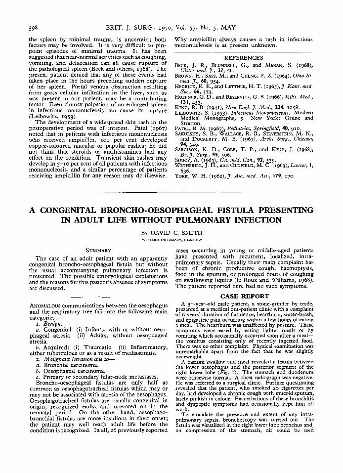
398 BRIT. J. SURG., 1970, Vol. 57, No. 5, MAY
the spleen by minimal trauma, is uncertain; both factors may be involved. It is very difficult to pin- point episodes of minimal trauma. It has been suggested that near-normal activities such as coughing, vomiting, and defaecation can all cause rupture of the pathological spleen (Beck and others, 1968). The present patient denied that any of these events had taken place in the hours preceding sudden rupture of her spleen. Portal venous obstruction resulting from gross cellular infiltration in the liver, such as was present in our patient, may be a contributing factor. Even clumsy palpation of an enlarged spleen in infectious mononucleosis can cause its rupture (Leibowitz, 1953).
The development of a widespread skin rash in the postoperative period was of interest. Pate1 (1967) noted that in patients with infectious mononucleosis who received ampicillin, IOO per cent developed copper-coloured macular or papular rashes; he did not think that steroids or antihistamines had any effect on the condition. Transient skin rashes may develop in 5-10 per cent of all patients with infectious mononucleosis, and a similar percentage of patients receiving ampicillin for any reason may do likewise.
Why ampicillin always causes a rash in infectious mononucleosis is at present unknown.
REFERENCES BECK, J. R., BLUNDELL, G., and MEBAN, S. (1968),
BROWN, H., SASS, M., and CHENG, P. Z . (1964)~ Ohio St . Ulster med. 3., 37, 56.
nied. J., 60, 954.
soc.. 66. 174. HEDRICK, K. E., and LETTNER, H. T. (1965),J. Kans. tired.
HUEBNER, G:D., and RESERVITZ, G. B. (1966), Milit. Med.,
KING, R. B. (1941)~ New Engl. 3. Med., 224, 1058. LEIBOWITZ, S . (1953), Infectious Mononucleosis. Modern
Medical Monographs, 5 . New York: Grune and Stratton.
PATEL, B. M. (1967)~ Pediatrics, Springfield, 40, 910. SAKULSKY, S. B., WALLACE, R. B., SILVERSTEIN, M. N.,
and DOCKERTY, M. B. (1967), Archs Surg., Chicago,
131,453.
947 349. SARGISON, K. D., COLE, T. P., and KYLE, J. (1968),
SOUCY, A. (1963), Un. mid. Can., 92, 539. WETHERILL, J. H., and OLDFIELD, M. C. (1963)~ Lancet, I,
Br.J . Surg., 55, 506.
636. YORK, W. H. (1962),3. Am. rned. Ass., 179, 170.
A CONGENITAL BRONCHO-OESOPHAGEAL FISTULA PRESENTING IN ADULT LIFE WITHOUT PULMONARY INFECTION
BY DAVID C. SMITH WESTERN INFIRMARY, GLASGOW
SUMMARY The case of an adult patient with an apparently
congenital broncho-oesophageal fistula but without the usual accompanying pulmonary infection is presented. The possible embryological explanations and the reasons for this patient’s absence of symptoms are discussed.
ANOMALOUS communications between the oesophagus and the respiratory tree fall into the following main categories :-
I .- Benign.- a . Congenital: (i) Infants. with or without oeso-
\ ,
phageal Gresia. (ii) Adults, without oesophageal atresia.
either tuberculous or as a result of mediastinitis. b. Acquired: (i) Traumatic. (ii) Inflammatory,
2. Malignant Invasion due to- a. Bronchial carcinoma. b. Oesophageal carcinoma. c. Primary or secondary hilar-node metastases. Broncho-oesophageal fistulas are only half as
common as oesophagotracheal fistulas which may or may not be associated with atresia of the oesophagus. Oesophagotracheal fistulas are usually congenital in origin, recognized early, and operated on in the neonatal period. On the other hand, oesophago- bronchial fistulas are more insidious in their onset; the patient may well reach adult life before the condition is recognized. In all, 26 previously reported
cases occurring in young or middle-aged patients have presented with recurrent, localized, intra- pulmonary sepsis. Usually their main complaint has been of chronic productive cough, haemoptysis, food in the sputum, or prolonged bouts of coughing on swallowing liquids (le Roux and Williams, 1968). The patient reported here had no such symptoms.
CASE REPORT A 31-year-old male patient, a stone-grinder by trade,
presented at a medical out-patient clinic with a complaint of 6 years’ duration of flatulence, heartburn, water-brash, and epigastric pain occurring within a few hours of eating a meal. The heartburn was unaffected by posture. These symptoms were eased by eating lighter meals or by vomiting which occasionally occurred soon after a meal- the vomitus consisting only of recently ingested food. There was no other complaint. Physical examination was unremarkable apart from the fact that he was slightly overweight.
A barium swallow and meal revealed a fistula between the lower oesophagus and the posterior segment of the right lower lobe (Fig. I). The stomach and duodenum were otherwise normal. A chest radiograph was negative. He was referred to a surgical clinic. Further questioning revealed that the patient, who smoked t o cigarettes per day, had developed a chronic cough with mucoid sputum, lately pinkish in colour. Exacerbations of these bronchitic and dyspeptic symptoms had occasionally kept him off work.
To elucidate the presence and extent of any intra- pulmonary sepsis, bronchoscopy was carried out. The fistula was visualized in the right lower lobe bronchus and, on compression of the stomach, air could be seen