Embed Size (px)
Citation preview
Spine Surgery Basics
Vikas V. PatelAlpesh PatelJames S. HarropEvalina BurgerEditors
123
Spine Surgery Basics

Vikas V. Patel • Alpesh Patel James S. Harrop • Evalina Burger Editors
Spine Surgery Basics
ISBN 978-3-642-34125-0 ISBN 978-3-642-34126-7 (eBook) DOI 10.1007/978-3-642-34126-7 Springer Heidelberg New York Dordrecht London
Library of Congress Control Number: 2013946428
© Springer-Verlag Berlin Heidelberg 2014 This work is subject to copyright. All rights are reserved by the Publisher, whether the whole or part of the material is concerned, speci fi cally the rights of translation, reprinting, reuse of illustrations, recitation, broadcasting, reproduction on micro fi lms or in any other physical way, and transmission or information storage and retrieval, electronic adaptation, computer software, or by similar or dissimilar methodology now known or hereafter developed. Exempted from this legal reservation are brief excerpts in connection with reviews or scholarly analysis or material supplied speci fi cally for the purpose of being entered and executed on a computer system, for exclusive use by the purchaser of the work. Duplication of this publication or parts thereof is permitted only under the provisions of the Copyright Law of the Publisher’s location, in its current version, and permission for use must always be obtained from Springer. Permissions for use may be obtained through RightsLink at the Copyright Clearance Center. Violations are liable to prosecution under the respective Copyright Law. The use of general descriptive names, registered names, trademarks, service marks, etc. in this publication does not imply, even in the absence of a speci fi c statement, that such names are exempt from the relevant protective laws and regulations and therefore free for general use. While the advice and information in this book are believed to be true and accurate at the date of publication, neither the authors nor the editors nor the publisher can accept any legal responsibility for any errors or omissions that may be made. The publisher makes no warranty, express or implied, with respect to the material contained herein.
Printed on acid-free paper
Springer is part of Springer Science+Business Media (www.springer.com)
Editors Vikas V. Patel Department of Orthopaedics University of Colorado, Denver Aurora, CO USA
Alpesh Patel Department of Orthopaedic Surgery Northwestern University Chicago, IL USA
James S. Harrop Department of Neurosurgery Jefferson Medical College Philadelphia, PA USA
Evalina Burger Department of Orthopaedics University of Colorado, Denver Aurora, CO USA

To our families for all the support and to Heidi Armbruster for all the organization to make this project possible.

vii
Preface
The stronger the foundation, the grander the possibilities!
The purpose of this textbook is to lay the foundation of spine education for medical students, residents, fellows, and junior attendings. It provides a con-cise and accurate methodology to understanding and diagnosing different spine conditions followed by the basics of treatment. This textbook will, thus, provide the link between education, optimized clinical evaluation, and evi-dence-based decision making.
This is especially relevant today as with an enlarging and aging popula-tion, spinal diseases will become more prevalent. Already back pain is the second most common complaint for which patients seek care after the com-mon cold. Concurrently, the past two decades have seen an explosion of tech-nology utilization in patient care. This has led to students and naive practioners diagnosing by exclusion-based testing as opposed to using sound clinical judgement. As healthcare funding becomes limited, this current trend in uti-lization of special high-end testing and imaging will be unsustainable. The skills to obtain an accurate, real-time clinical diagnosis and treatment plan will be in high demand. In the ever growing plethora of diagnoses, adhering to basics will provide the knowledge and framework necessary to make the correct decision for the patient.
Aurora, CO Vikas V. Patel Chicago, IL Alpesh Patel Philadelphia, PA James S. Harrop Aurora, CO Evalina Burger

ix
Contents
Part I General
1 Anatomy ........................................................................................ 3Christopher M.J. Cain
2 Physical Examination of the Cervical Spine .............................. 13Virgilio Matheus and Edward C. Benzel
3 Examination of Back Pain ........................................................... 23Jennifer Malone, Ravi Madineni, Niti Cooper, Angud Mehdi, and Ashwini Sharan
4 Radiologic Imaging of the Spine ................................................. 37Angela Valladares-Otero, Brian Christenson, and Brian D. Petersen
5 Diagnostic Evaluations ................................................................ 75Richard Kendall and Zach Beresford
6 General Considerations for Spine Surgery Including Consent and Preparation. General Surgical Principles, Guidelines for Informed Consent, Patient Positioning for Surgery, Equipment Needed, and Postoperative Considerations .............................................................................. 83Evalina Burger
7 Surgical Approaches .................................................................... 91Michael C. Gerling and Sheeraz A. Qureshi
8 Cervical and Cervicothoracic Instrumentation ........................ 99Adam Pearson and Todd Albert
9 Lumbosacral Instrumentation .................................................... 113Joanne Elston and Nelson Saldua
10 Bone Graft and Bone Substitute Biology ................................... 147Harshpal Singh and Allan D. Levi
11 Neurological Monitoring in Orthopedic Spine Surgery ........... 153Tod B. Sloan, Leslie Jameson, Daniel Janik, and Paul Mongan
x Contents
Part II Degenerative Spine
12 Cervical Disk Herniation and Radiculopathy ........................... 177Selvon F. St. Clair and John M. Rhee
13 Cervical Spondylotic Myelopathy (CSM) .................................. 185Prokopis Annis and Alpesh A. Patel
14 Thoracic Disc Herniation ............................................................ 193Michael Fernandez and Sandeep N. Gidvani
15 Lumbar Disc Herniation ............................................................. 203William Ryan Spiker and Brandon D. Lawrence
16 Lumbar Stenosis........................................................................... 215Paul D. Kim and Hyun Bae
17 Lumbar Degenerative Spondylolisthesis .................................... 221Loukas Koyonos and Jeffrey A. Rihn
18 Adult Spondylolysis and Isthmic Spondylolisthesis .................. 229William D. Long III and Peter G. Whang
19 Degenerative Disk Disease with and Without Facet Arthritis .............................................................................. 239Kern Singh, Jonathan A. Hoskins, Steven J. Fineberg, and Matthew Oglesby
20 Adult Degenerative Scoliosis ....................................................... 247Joshua Ellwitz and Munish Gupta
Part III Pediatric Spine
21 Scoliosis ........................................................................................ 261Prerana Patel and Andrew G.S. King
22 Pediatric Kyphosis ....................................................................... 287Michael J. Kramarz, Steven W. Hwang, Amer F. Samdani, and Phillip B. Storm
23 Congenital ..................................................................................... 301William D. Long III and Jonathan N. Grauer
24 Spondylolysis and Isthmic Spondylolisthesis............................. 311Sumeet Garg and Mark Erickson
25 Spine Trauma: Occipital and Upper Cervical Spine ................ 325Xuan Lo, Raymond W. Hwang, Harvey E. Smith, David Gendelberg, and Alexander R. Vaccaro
26 Spine Trauma: Subaxial Cervical Spine (C3–C7) ..................... 339Kelli L. Crabtree, Paul M. Arnold, and Karen K. Anderson
27 Thoracolumbar Spine (T1–L2) ................................................... 359Yasutsugu Yukawa
xiContents
28 Spine Trauma: Low Lumbar Spine (L3–5) ............................... 373Vu H. Le and Nitin Bhatia
29 Sacral Spine, Pelvis, and Pelvic Ring ......................................... 387Megan Brady, Stephen Tolhurst, and Timothy Moore
30 Acute Traumatic Spinal Cord Injury: Epidemiology, Evaluation, and Management ..................................................... 399Jefferson R. Wilson, Newton Cho, and Michael G. Fehlings
Part IV Tumor, Infection, Inflammatory and Metabolic Conditions
31 Primary Spine Tumors ................................................................ 413Marco Ferrone and Joseph Schwab
32 Metastatic Spine Tumors ............................................................. 423Byung C. Yoon, Camilo Molina, and Daniel M. Sciubba
33 Primary Spinal Infections ........................................................... 433David B. Bumpass and Jacob M. Buchowski
34 Intradural Spinal Cord Tumors ................................................. 453Ricky R. Kalra and Andrew T. Dailey
35 Rheumatoid Arthritis .................................................................. 465Scott D. Daffner and Colleen M. Watkins
36 Ankylosing Spondylitis and Diffuse Idiopathic Skeletal Hyperostosis ................................................................... 475Xuan Luo, Harvey E. Smith, Raymond Hwang, and Scott D. Daffner
37 Osteoporosis and the Aging Spine .............................................. 491Jacques Hacquebord and Michael J. Lee
Part V Complications
38 Postoperative Spinal Infections ................................................... 501Michael Murray and Wellington Hsu
39 Management of Dural Tears in Spinal Surgery ........................ 509Sheeraz A. Qureshi, Steven M. Koehler, and Michael C. Gerling
40 Complications: Neurological Injury ........................................... 521Shannon Hann, Nelson Saldua, and James S. Harrop
41 Complications: Pseudoarthrosis/Nonunion ............................... 533Raj Kullar, Eric Klineberg, and Munish Gupta
42 Medical Complications ................................................................ 541Rachid Assina and Robert F. Heary
Index ...................................................................................................... 567

Part I
General
3V.V. Patel et al. (eds.), Spine Surgery Basics, DOI 10.1007/978-3-642-34126-7_1, © Springer-Verlag Berlin Heidelberg 2014
1
1.1 Overview
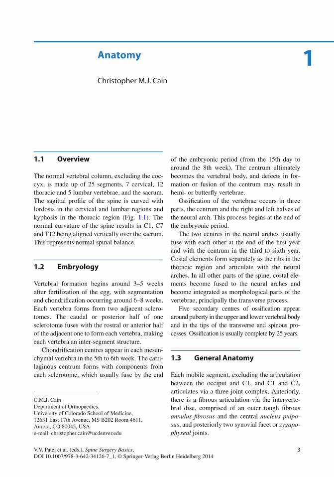
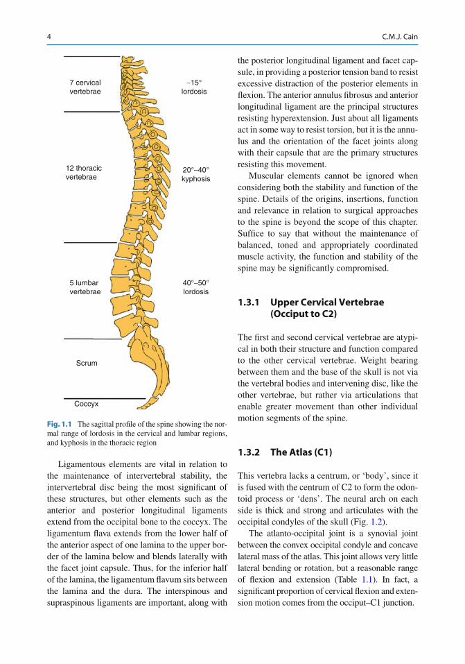
The normal vertebral column, excluding the coc-cyx, is made up of 25 segments, 7 cervical, 12 thoracic and 5 lumbar vertebrae, and the sacrum. The sagittal pro fi le of the spine is curved with lordosis in the cervical and lumbar regions and kyphosis in the thoracic region (Fig. 1.1 ). The normal curvature of the spine results in C1, C7 and T12 being aligned vertically over the sacrum. This represents normal spinal balance.
1.2 Embryology
Vertebral formation begins around 3–5 weeks after fertilization of the egg, with segmentation and chondri fi cation occurring around 6–8 weeks. Each vertebra forms from two adjacent sclero-tomes. The caudal or posterior half of one sclerotome fuses with the rostral or anterior half of the adjacent one to form each vertebra, making each vertebra an inter-segment structure.
Chondri fi cation centres appear in each mesen-chymal vertebra in the 5th to 6th week. The carti-laginous centrum forms with components from each sclerotome, which usually fuse by the end
of the embryonic period (from the 15th day to around the 8th week). The centrum ultimately becomes the vertebral body, and defects in for-mation or fusion of the centrum may result in hemi- or butter fl y vertebrae.
Ossi fi cation of the vertebrae occurs in three parts, the centrum and the right and left halves of the neural arch. This process begins at the end of the embryonic period.
The two centres in the neural arches usually fuse with each other at the end of the fi rst year and with the centrum in the third to sixth year. Costal elements form separately as the ribs in the thoracic region and articulate with the neural arches. In all other parts of the spine, costal ele-ments become fused to the neural arches and become integrated as morphological parts of the vertebrae, principally the transverse process.
Five secondary centres of ossi fi cation appear around puberty in the upper and lower vertebral body and in the tips of the transverse and spinous pro-cesses. Ossi fi cation is usually complete by 25 years.
1.3 General Anatomy
Each mobile segment, excluding the articulation between the occiput and C1, and C1 and C2, articulates via a three-joint complex. Anteriorly, there is a fi brous articulation via the interverte-bral disc, comprised of an outer tough fi brous annulus fi brosus and the central nucleus pulpo-sus , and posteriorly two synovial facet or zygapo-physeal joints.
C. M. J. Cain Department of Orthopaedics , University of Colorado School of Medicine , 12631 East 17th Avenue, MS B202 Room 4611 , Aurora , CO 80045 , USA e-mail: [email protected]
Anatomy
Christopher M. J. Cain
4 C.M.J. Cain
Ligamentous elements are vital in relation to the maintenance of intervertebral stability, the intervertebral disc being the most signi fi cant of these structures, but other elements such as the anterior and posterior longitudinal ligaments extend from the occipital bone to the coccyx. The ligamentum fl ava extends from the lower half of the anterior aspect of one lamina to the upper bor-der of the lamina below and blends laterally with the facet joint capsule. Thus, for the inferior half of the lamina, the ligamentum fl avum sits between the lamina and the dura. The interspinous and supraspinous ligaments are important, along with
the posterior longitudinal ligament and facet cap-sule, in providing a posterior tension band to resist excessive distraction of the posterior elements in fl exion. The anterior annulus fi brosus and anterior longitudinal ligament are the principal structures resisting hyperextension. Just about all ligaments act in some way to resist torsion, but it is the annu-lus and the orientation of the facet joints along with their capsule that are the primary structures resisting this movement.
Muscular elements cannot be ignored when considering both the stability and function of the spine. Details of the origins, insertions, function and relevance in relation to surgical approaches to the spine is beyond the scope of this chapter. Suf fi ce to say that without the maintenance of balanced, toned and appropriately coordinated muscle activity, the function and stability of the spine may be signi fi cantly compromised.
1.3.1 Upper Cervical Vertebrae (Occiput to C2)
The fi rst and second cervical vertebrae are atypi-cal in both their structure and function compared to the other cervical vertebrae. Weight bearing between them and the base of the skull is not via the vertebral bodies and intervening disc, like the other vertebrae, but rather via articulations that enable greater movement than other individual motion segments of the spine.
1.3.2 The Atlas (C1)
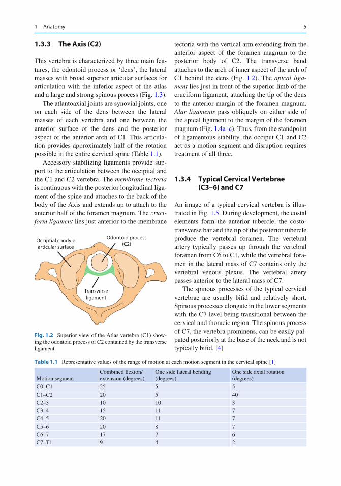
This vertebra lacks a centrum, or ‘body’, since it is fused with the centrum of C2 to form the odon-toid process or ‘dens’. The neural arch on each side is thick and strong and articulates with the occipital condyles of the skull (Fig. 1.2 ).
The atlanto-occipital joint is a synovial joint between the convex occipital condyle and concave lateral mass of the atlas. This joint allows very little lateral bending or rotation, but a reasonable range of fl exion and extension (Table 1.1 ). In fact, a signi fi cant proportion of cervical fl exion and exten-sion motion comes from the occiput–C1 junction.
7 cervicalvertebrae
12 thoracicvertebrae
5 lumbarvertebrae
Scrum
Coccyx
∼15°lordosis
20°−40°kyphosis
40°−50°lordosis
Fig. 1.1 The sagittal pro fi le of the spine showing the nor-mal range of lordosis in the cervical and lumbar regions, and kyphosis in the thoracic region
51 Anatomy
1.3.3 The Axis (C2)
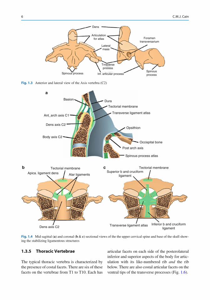
This vertebra is characterized by three main fea-tures, the odontoid process or ‘dens’, the lateral masses with broad superior articular surfaces for articulation with the inferior aspect of the atlas and a large and strong spinous process (Fig. 1.3 ).
The atlantoaxial joints are synovial joints, one on each side of the dens between the lateral masses of each vertebra and one between the anterior surface of the dens and the posterior aspect of the anterior arch of C1. This articula-tion provides approximately half of the rotation possible in the entire cervical spine (Table 1.1 ).
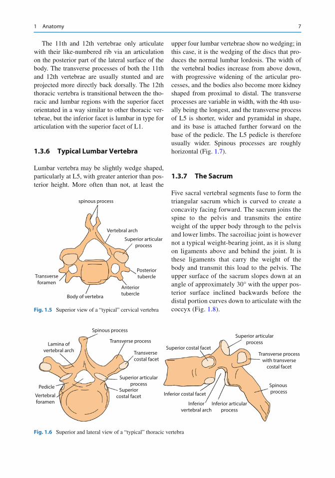
Accessory stabilizing ligaments provide sup-port to the articulation between the occipital and the C1 and C2 vertebra. The membrane tectoria is continuous with the posterior longitudinal liga-ment of the spine and attaches to the back of the body of the Axis and extends up to attach to the anterior half of the foramen magnum. The cruci-form ligament lies just anterior to the membrane
tectoria with the vertical arm extending from the anterior aspect of the foramen magnum to the posterior body of C2. The transverse band attaches to the arch of inner aspect of the arch of C1 behind the dens (Fig. 1.2 ). The apical liga-ment lies just in front of the superior limb of the cruciform ligament, attaching the tip of the dens to the anterior margin of the foramen magnum. Alar ligaments pass obliquely on either side of the apical ligament to the margin of the foramen magnum (Fig. 1.4a–c ). Thus, from the standpoint of ligamentous stability, the occiput C1 and C2 act as a motion segment and disruption requires treatment of all three.
1.3.4 Typical Cervical Vertebrae (C3–6) and C7
An image of a typical cervical vertebra is illus-trated in Fig. 1.5 . During development, the costal elements form the anterior tubercle, the costo-transverse bar and the tip of the posterior tubercle produce the vertebral foramen. The vertebral artery typically passes up through the vertebral foramen from C6 to C1, while the vertebral fora-men in the lateral mass of C7 contains only the vertebral venous plexus. The vertebral artery passes anterior to the lateral mass of C7.
The spinous processes of the typical cervical vertebrae are usually bi fi d and relatively short. Spinous processes elongate in the lower segments with the C7 level being transitional between the cervical and thoracic region. The spinous process of C7, the vertebra prominens, can be easily pal-pated posteriorly at the base of the neck and is not typically bi fi d. [ 4 ]
Occiptial condylearticular surface
Transverseligament
Odontoid process(C2)
Fig. 1.2 Superior view of the Atlas vertebra (C1) show-ing the odontoid process of C2 contained by the transverse ligament
Table 1.1 Representative values of the range of motion at each motion segment in the cervical spine [ 1 ]
Motion segment Combined fl exion/extension (degrees)
One side lateral bending (degrees)
One side axial rotation (degrees)
C0–C1 25 5 5 C1–C2 20 5 40 C2–3 10 10 3 C3–4 15 11 7 C4–5 20 11 7 C5–6 20 8 7 C6–7 17 7 6 C7–T1 9 4 2
6 C.M.J. Cain
1.3.5 Thoracic Vertebrae
The typical thoracic vertebra is characterized by the presence of costal facets. There are six of these facets on the vertebrae from T1 to T10. Each has
articular facets on each side of the posterolateral inferior and superior aspects of the body for artic-ulation with its like-numbered rib and the rib below. There are also costal articular facets on the ventral tips of the transverse processes (Fig. 1.6 ).
Dens
Articulationfor atlas
Lateralmass
Foramentransversarium
Spinousprocess
Trnsverseprocess
Inf. articular processSpinous process
Fig. 1.3 Anterior and lateral view of the Axis vertebra (C2)
Basion Dura
Tectorial membrane
Transverse ligament atlas
Opsithion
Occispital bone
Post arch axis
Spinous process atlas
Ant_arch axis C1
Dens axis C2
Body axis C2
Tectorial membrane
a
b c
Alar ligamentsApica. ligament dens
Dens axis C2
Tectorial membrane
Inferior b and cruciformligament
Transverse ligament atlas
Superior b and cruciformligament
Fig. 1.4 Mid sagittal ( a ) and coronal ( b & c ) sectional views of the the upper cervical spine and base of the skull show-ing the stabilizing ligamentous structures
71 Anatomy
The 11th and 12th vertebrae only articulate with their like-numbered rib via an articulation on the posterior part of the lateral surface of the body. The transverse processes of both the 11th and 12th vertebrae are usually stunted and are projected more directly back dorsally. The 12th thoracic vertebra is transitional between the tho-racic and lumbar regions with the superior facet orientated in a way similar to other thoracic ver-tebrae, but the inferior facet is lumbar in type for articulation with the superior facet of L1.
1.3.6 Typical Lumbar Vertebra
Lumbar vertebra may be slightly wedge shaped, particularly at L5, with greater anterior than pos-terior height. More often than not, at least the
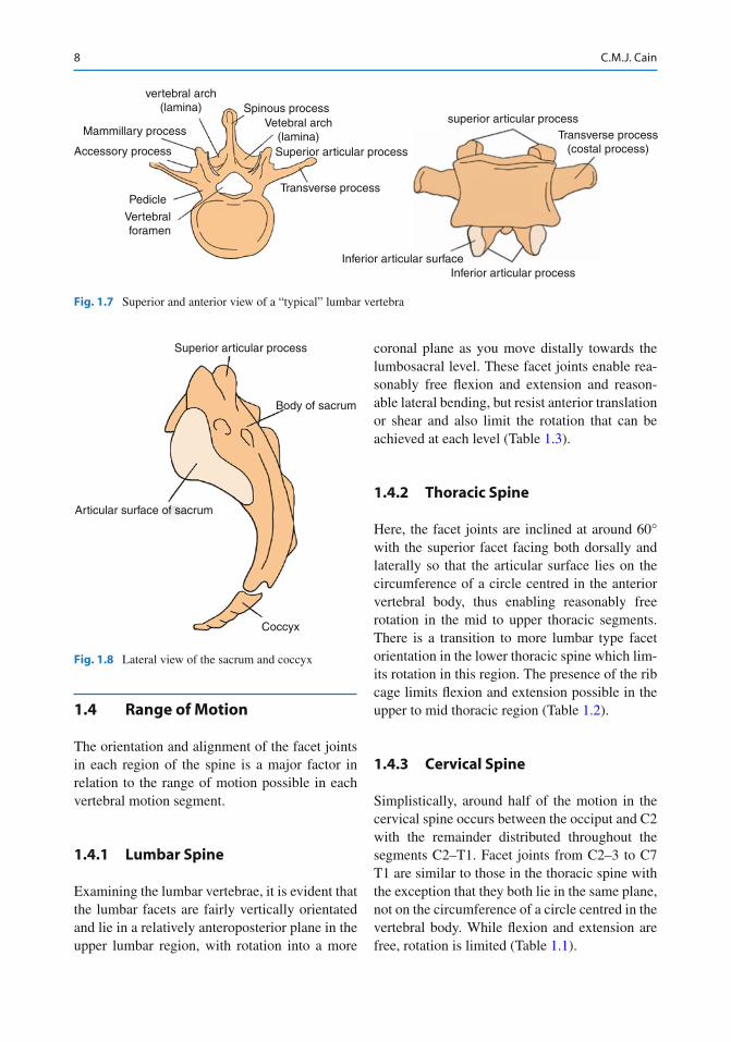
upper four lumbar vertebrae show no wedging; in this case, it is the wedging of the discs that pro-duces the normal lumbar lordosis. The width of the vertebral bodies increase from above down, with progressive widening of the articular pro-cesses, and the bodies also become more kidney shaped from proximal to distal. The transverse processes are variable in width, with the 4th usu-ally being the longest, and the transverse process of L5 is shorter, wider and pyramidal in shape, and its base is attached further forward on the base of the pedicle. The L5 pedicle is therefore usually wider. Spinous processes are roughly horizontal (Fig. 1.7 ).
1.3.7 The Sacrum
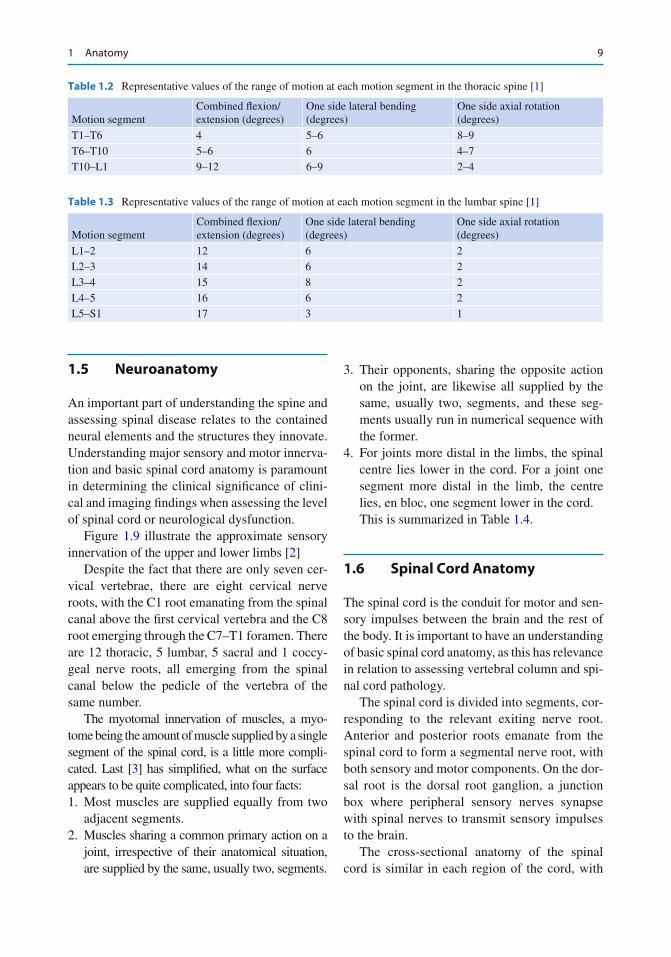
Five sacral vertebral segments fuse to form the triangular sacrum which is curved to create a concavity facing forward. The sacrum joins the spine to the pelvis and transmits the entire weight of the upper body through to the pelvis and lower limbs. The sacroiliac joint is however not a typical weight-bearing joint, as it is slung on ligaments above and behind the joint. It is these ligaments that carry the weight of the body and transmit this load to the pelvis. The upper surface of the sacrum slopes down at an angle of approximately 30° with the upper pos-terior surface inclined backwards before the distal portion curves down to articulate with the coccyx (Fig. 1.8 ).
spinous process
Vertebral arch
Superior articularprocess
Posteriortubercle
AnteriortubercleBody of vertebra
Transverseforamen
Fig. 1.5 Superior view of a “typical” cervical vertebra
Spinous process
Transverse processSuperior costal facet
Superior articularprocess
Transverse processwith transverse
costal facet
Spinous processInferior costal facet
Inferiorvertebral arch
Inferior articularprocess
Transversecostal facet
Superior articularprocess
Superiorcostal facetVertebral
foramen
Pedicle
Lamina ofvertebral arch
Fig. 1.6 Superior and lateral view of a “typical” thoracic vertebra
8 C.M.J. Cain
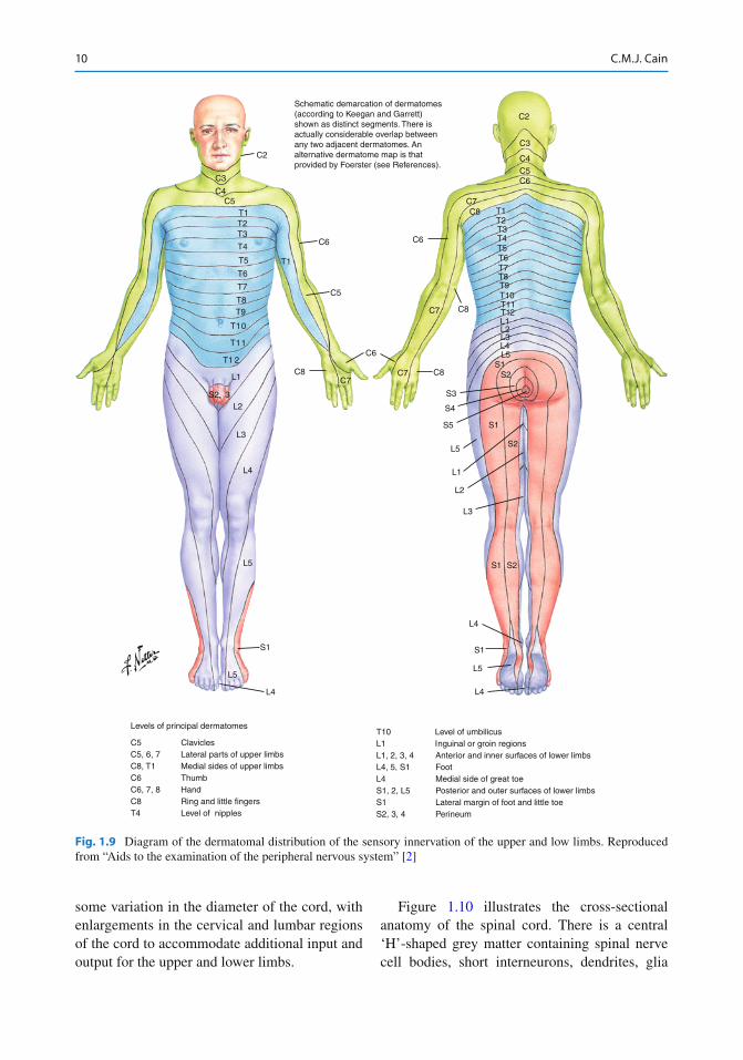
1.4 Range of Motion
The orientation and alignment of the facet joints in each region of the spine is a major factor in relation to the range of motion possible in each vertebral motion segment.
1.4.1 Lumbar Spine
Examining the lumbar vertebrae, it is evident that the lumbar facets are fairly vertically orientated and lie in a relatively anteroposterior plane in the upper lumbar region, with rotation into a more
coronal plane as you move distally towards the lumbosacral level. These facet joints enable rea-sonably free fl exion and extension and reason-able lateral bending, but resist anterior translation or shear and also limit the rotation that can be achieved at each level (Table 1.3 ).
1.4.2 Thoracic Spine
Here, the facet joints are inclined at around 60° with the superior facet facing both dorsally and laterally so that the articular surface lies on the circumference of a circle centred in the anterior vertebral body, thus enabling reasonably free rotation in the mid to upper thoracic segments. There is a transition to more lumbar type facet orientation in the lower thoracic spine which lim-its rotation in this region. The presence of the rib cage limits fl exion and extension possible in the upper to mid thoracic region (Table 1.2 ).
1.4.3 Cervical Spine
Simplistically, around half of the motion in the cervical spine occurs between the occiput and C2 with the remainder distributed throughout the segments C2–T1. Facet joints from C2–3 to C7 T1 are similar to those in the thoracic spine with the exception that they both lie in the same plane, not on the circumference of a circle centred in the vertebral body. While fl exion and extension are free, rotation is limited (Table 1.1 ).
superior articular processTransverse process
(costal process)
Inferior articular processInferior articular surface
vertebral arch(lamina)
Mammillary process
Spinous processVetebral arch
(lamina)Superior articular process
Transverse process
Accessory process
Pedicle
Vertebralforamen
Fig. 1.7 Superior and anterior view of a “typical” lumbar vertebra
Superior articular process
Body of sacrum
Articular surface of sacrum
Coccyx
Fig. 1.8 Lateral view of the sacrum and coccyx
91 Anatomy
Table 1.3 Representative values of the range of motion at each motion segment in the lumbar spine [ 1 ]
Motion segment Combined fl exion/extension (degrees)
One side lateral bending (degrees)
One side axial rotation (degrees)
L1–2 12 6 2 L2–3 14 6 2 L3–4 15 8 2 L4–5 16 6 2 L5–S1 17 3 1
Table 1.2 Representative values of the range of motion at each motion segment in the thoracic spine [ 1 ]
Motion segment Combined fl exion/extension (degrees)
One side lateral bending (degrees)
One side axial rotation (degrees)
T1–T6 4 5–6 8–9 T6–T10 5–6 6 4–7 T10–L1 9–12 6–9 2–4
1.5 Neuroanatomy
An important part of understanding the spine and assessing spinal disease relates to the contained neural elements and the structures they innovate. Understanding major sensory and motor innerva-tion and basic spinal cord anatomy is paramount in determining the clinical signi fi cance of clini-cal and imaging fi ndings when assessing the level of spinal cord or neurological dysfunction.
Figure 1.9 illustrate the approximate sensory innervation of the upper and lower limbs [ 2 ]
Despite the fact that there are only seven cer-vical vertebrae, there are eight cervical nerve roots, with the C1 root emanating from the spinal canal above the fi rst cervical vertebra and the C8 root emerging through the C7–T1 foramen. There are 12 thoracic, 5 lumbar, 5 sacral and 1 coccy-geal nerve roots, all emerging from the spinal canal below the pedicle of the vertebra of the same number.
The myotomal innervation of muscles, a myo-tome being the amount of muscle supplied by a single segment of the spinal cord, is a little more compli-cated. Last [ 3 ] has simpli fi ed, what on the surface appears to be quite complicated, into four facts: 1. Most muscles are supplied equally from two
adjacent segments. 2. Muscles sharing a common primary action on a
joint, irrespective of their anatomical situation, are supplied by the same, usually two, segments.
3. Their opponents, sharing the opposite action on the joint, are likewise all supplied by the same, usually two, segments, and these seg-ments usually run in numerical sequence with the former.
4. For joints more distal in the limbs, the spinal centre lies lower in the cord. For a joint one segment more distal in the limb, the centre lies, en bloc, one segment lower in the cord. This is summarized in Table 1.4 .
1.6 Spinal Cord Anatomy
The spinal cord is the conduit for motor and sen-sory impulses between the brain and the rest of the body. It is important to have an understanding of basic spinal cord anatomy, as this has relevance in relation to assessing vertebral column and spi-nal cord pathology.
The spinal cord is divided into segments, cor-responding to the relevant exiting nerve root. Anterior and posterior roots emanate from the spinal cord to form a segmental nerve root, with both sensory and motor components. On the dor-sal root is the dorsal root ganglion, a junction box where peripheral sensory nerves synapse with spinal nerves to transmit sensory impulses to the brain.
The cross-sectional anatomy of the spinal cord is similar in each region of the cord, with
10 C.M.J. Cain
Fig. 1.9 Diagram of the dermatomal distribution of the sensory innervation of the upper and low limbs. Reproduced from “Aids to the examination of the peripheral nervous system” [ 2 ]
Levels of principal dermatomes
C5 ClaviclesC5, 6, 7 Lateral parts of upper limbsC8, T1 Medial sides of upper limbsC6 ThumbC6, 7, 8 HandC8 Ring and little fingersT4 Level of nipples
T10 Level of umbilicusL1 Inguinal or groin regionsL1, 2, 3, 4 Anterior and inner surfaces of lower limbsL4, 5, S1 FootL4 Medial side of great toeS1, 2, L5 Posterior and outer surfaces of lower limbsS1 Lateral margin of foot and little toeS2, 3, 4 Perineum
Schematic demarcation of dermatomes(according to Keegan and Garrett)shown as distinct segments. There isactually considerable overlap betweenany two adjacent dermatomes. Analternative dermatome map is thatprovided by Foerster (see References).
C2
C3
C4C5
T1T2T3
T4
T5
T6
T7
T8T9
T10
T11
T1 2
L1
L3
L4
L5
S1
L5
L4
C2
C3
C4
C5C6
C7C8 T1
T2T3T4T5T6T7T8T9T10T11T12L1
L3
L2
L1
S1 S2
L4
S1
L5
L4
S2
S1
L5
L2L3L4L5
S1S2
S3
S4
S5
C6
T1
C5
C6
C8
C6
C7C7 C8
C7 C8
S2, 3L2
some variation in the diameter of the cord, with enlargements in the cervical and lumbar regions of the cord to accommodate additional input and output for the upper and lower limbs.
Figure 1.10 illustrates the cross-sectional anatomy of the spinal cord. There is a central ‘H’-shaped grey matter containing spinal nerve cell bodies, short interneurons, dendrites, glia
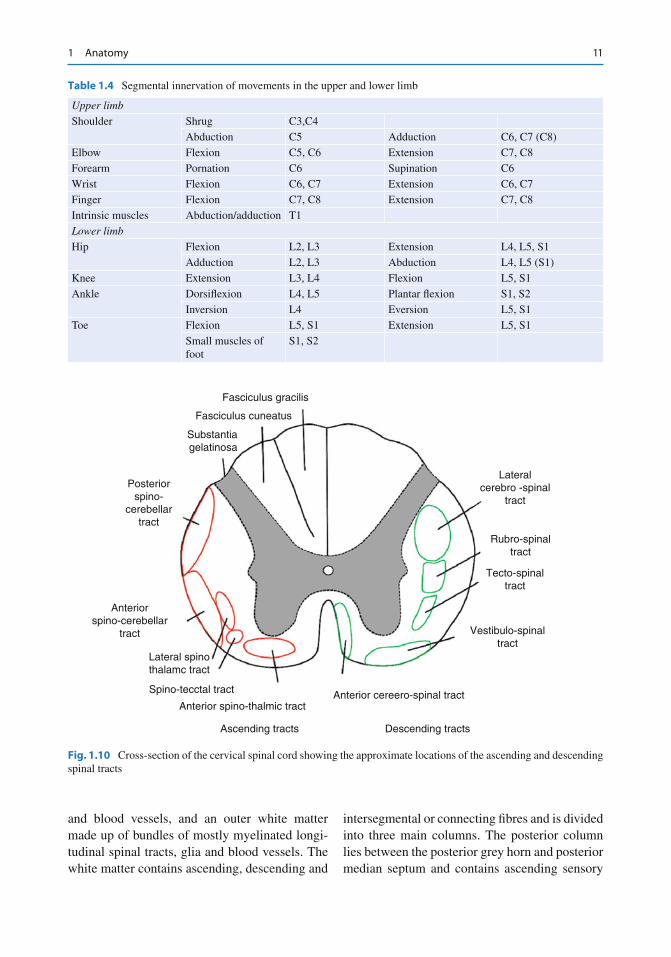
111 Anatomy
and blood vessels, and an outer white matter made up of bundles of mostly myelinated longi-tudinal spinal tracts, glia and blood vessels. The white matter contains ascending, descending and
intersegmental or connecting fi bres and is divided into three main columns. The posterior column lies between the posterior grey horn and posterior median septum and contains ascending sensory
Table 1.4 Segmental innervation of movements in the upper and lower limb
Upper limb Shoulder Shrug C3,C4
Abduction C5 Adduction C6, C7 (C8) Elbow Flexion C5, C6 Extension C7, C8 Forearm Pornation C6 Supination C6 Wrist Flexion C6, C7 Extension C6, C7 Finger Flexion C7, C8 Extension C7, C8 Intrinsic muscles Abduction/adduction T1 Lower limb Hip Flexion L2, L3 Extension L4, L5, S1
Adduction L2, L3 Abduction L4, L5 (S1) Knee Extension L3, L4 Flexion L5, S1 Ankle Dorsi fl exion L4, L5 Plantar fl exion S1, S2
Inversion L4 Eversion L5, S1 Toe Flexion L5, S1 Extension L5, S1
Small muscles of foot
S1, S2
Fasciculus gracilis
Fasciculus cuneatus
Substantiagelatinosa
Posteriorspino-
cerebellartract
Anteriorspino-cerebellar
tract
Lateral spinothalamc tract
Spino-tecctal tract
Anterior spino-thalmic tract
Ascending tracts Descending tracts
Anterior cereero-spinal tract
Lateralcerebro -spinal
tract
Rubro-spinaltract
Tecto-spinaltract
Vestibulo-spinaltract
Fig. 1.10 Cross-section of the cervical spinal cord showing the approximate locations of the ascending and descending spinal tracts
12 C.M.J. Cain
fi bres. The lateral column lies between the ante-rior and posterior grey horns and contains pre-dominantly descending, but also some ascending tracts, and the anterior column which lies between the anterior grey horn and the anterior median fi ssure contains descending motor tracts.
The posterior white columns convey normal sensation, warmth and coolness and joint posi-tion or proprioception. Their cell bodies lie in the dorsal root ganglia of the spinal nerves and more distal fi bres, from the sacrum, lie medially, with fi bres from the lumbar, thoracic and cervical regions layered more laterally. Sensory fi bres synapse in the nucleus gracilis and cuneatus near the base of the fourth ventricle in the medulla oblongata and cross to the opposite side of the brain via the sensory decussation.
Anterior white columns contain uncrossed pyramidal fi bres whose cell bodies lie in the brainstem near the fl oor of the fourth ventricle. Motor fi bres from the cerebral cortex cross in the motor or pyramidal decussation, also in the medulla oblongata.
Pain and temperature fi bres entering the cord via the posterior spinal roots enter the dorsal horn of the grey matter synapse and cross the spinal cord to the lateral spinothalamic tract on the oppo-site side. As a result of this, hemisection of the spi-nal cord results in a dissociated sensory loss, with loss of joint position and light touch sensation, along with motor function, on the same side as the cord injury, with loss of pain and temperature on the opposite side of the body below the lesion.
Neurons are also layered in the various tracts, with sensory fi bres entering the cord fi rst, dis-tally, lying closest to the midline, and those entering last, in the cervical region, lying more laterally. The same is also true for motor tracts, with those leaving the cord fi rst, cervical fi bres, lying more laterally. This arrangement leads to the typical features of a central cord lesion that may result from stenosis and a hyperextension injury, conditions such as a syringomyelia and spinal cord tumours, where motor tracts are affected more than sensory, the upper limb more than the lower limb, and distal parts of the limb more than proximal.
Anterior spinal cord pathology such as ante-rior spinal artery occlusion, compression due to a kyphotic deformity or a central disc protrusion will result in anterior cord syndrome where there is loss of motor function and pain and tempera-ture below the lesion with preserved posterior column function.
References
1. Panjabi M, White A (1990) Clinical biomechanics of the spine, 2nd edn. J.B Lippincott Company, Philadelphia
2. Medical Research Council (1976) Aids to the examina-tion of the peripheral nervous system. Her Majesty’s Stationery Of fi ce, London
3. Last R (1984) Anatomy, regional and applied, 7th edn. Churchill Livingstone, Edinburgh
4. Clemente C (1976) Anatomy: a regional atlas of the human body. Urban & Schwarzenberg, Baltimore
13V.V. Patel et al. (eds.), Spine Surgery Basics, DOI 10.1007/978-3-642-34126-7_2, © Springer-Verlag Berlin Heidelberg 2014
2
2.1 Anatomy of the Cervical Spine
The cervical spine is gifted with the capacity to provide a wide range of motions which facilitate head movements. Based on its functions, the cer-vical spine can be divided into two segments: an upper portion, which involves the occipitoatlan-toaxial complex, and a lower portion, consisting of C3–C7.
Due to the unique anatomical features associ-ated with the atlas and the axis, the upper seg-ment of the cervical spine forms an extremely versatile and complex articulation that allows for a wide range of head and neck movements.
The atlas is formed by a ring of bone, which can be divided into a ventral and dorsal arch. It lacks a central vertebral body but displays large lateral masses. The latter serve to accommodate the occipital condyles and form the only weight-bearing articulation between the skull and the spine. A small fl attening at the rostral border of the dorsal arch represents the trough through which the vertebral artery passes over C1 on its trajectory toward the intradural space.
The axis resembles the typical cervical vertebra with the peculiarity of having a ventral bony pro-cess projecting rostrally from its rudimentary
vertebral body known as the odontoid process or dens. This process serves as an anchoring point for several ligaments that provide support between the atlas, axis, and condyles. This ligamentous complex is referred to as the cruciate ligament complex. Added stability is provided by the ante-rior and posterior longitudinal ligaments, which run ventral to and dorsal to the vertebral bodies, up to the skull base. The C1–C2 segment lacks an intervertebral disc. The most rostral disc is, hence, located between the axis and C3. Usually the spinous processes of C2 through C6 display a bi fi d appearance. In the majority of the cases, the verte-bral artery enters a bony ring on the lateral aspect of C6 known as the transverse foramen. The artery follows this path rostrally until exiting the foramen of the axis and curving over the arch of the atlas - to fi nally pass between the atlas and the condyle as it passes through the foramen magnum.
In addition to the ligamentous support, the cer-vical spine relies on muscular support for both support and mobility. A combination of unique features exhibited by these muscles and the cervi-cal spine permits extreme fl exion, extension, and tilting of the head, without adverse consequences.
2.2 Palpation
The ventral and lateral aspects of the cervical spine are covered by surrounding structures that can lead or suggest potential underlying patholo-gies, which in some cases might show no relation-ship with cervical spine pathology. Prior to laying
V. Matheus • E. C. Benzel , M.D. (*) Department of Neurosurgery , Neurological Institute, Cleveland Clinic , 9500 Euclid Avenue, S4 , Cleveland , OH 44195 , USA e-mail: [email protected] ; [email protected]
Physical Examination of the Cervical Spine
Virgilio Matheus and Edward C. Benzel
14 V. Matheus and E.C. Benzel
hands on the patient, the examiner must look for points of skin erythema or diaphoresis, which could represent painful areas that must be approached carefully to avoid unnecessary pain [ 1 ] . Careful palpation of the sternocleidomastoid muscle and all of the triangles it forms is per-formed next. One should begin by instructing the patient to turn his head toward one side, after which the examiner proceeds to “travel” with his hand along the full extent of the ipsilateral mus-cle. The examiner should look for masses and ten-der points. He should repeat this maneuver on the opposite side. It is important to compare muscle bulk and appearance. Next, the examiner should proceed with palpation of the carotid pulse, using the second and third digits. It is normally located medial to the sternocleidomastoid muscle. The examiner should pay attention to strength and symmetry, and to not forget to auscultate the underlying structures, seeking bruits, etc. It is also important to assess anatomical landmarks relevant for surgical approaches such as the thyroid and cricoid cartilages, hyoid bone, and trachea as well as, if possible, palpate the carotid tubercle which usually is palpable at the C6 level. The examiner should palpate, with the same fi ngers, the supra-clavicular region. Abnormal masses or tenderness may represent lymphadenopathy, apical lung masses, or even clavicular fractures. If the palpa-ble structure seems to be bony, suspect an acces-sory cervical rib.
When palpating the dorsal aspect of the cervi-cal spine, one must remember that the cervical spine is covered by large muscles, most notably the splenius and trapezius muscles, which have insertions on the suboccipital, scapular, and shoulder regions. It, therefore, is important to begin palpating from the occiput down to the cer-vicothoracic region as well as lateral over the scapula. It is useful to use a systematic approach, fi rst examining the soft tissue and subsequently the bony structures or vice versa. In fl ammation and tenderness can be due to muscle spasm. Potential etiologies include trauma, muscle fi brosis, and fi bromyalgia. Reproducible focal points of tenderness, with palpation over the scapula or shoulder joint, may indicate ligamen-tous damage due to overuse.
Finally, one should proceed to palpating the spinous processes. The patient should sit up and perform gentle fl exion of the neck. Palpation of spinous process in the midline is appropriate. Tenderness, masses, absence of processes, or any other abnormalities should be noted. One should pay close attention to alignment and tenderness, so underlying fractures and/or luxations are not missed.
2.3 Range of Motion
The most important step prior to performing a range of motion examination is to obtain a thorough his-tory to assess for instability. If the patient is aware of speci fi c painful movements, elicitation of such movements should be reserved for the latter portion of the examination in order to avoid muscle spasm and the carrying of the pain through the remaining steps of the examination. Flexion, extension, lateral fl exion, and head rotation are performed in order to seek sources of pain. One should begin with active movements and follow them with passive move-ments. Compare among them for differences. While performing these movements, the examiner should assess for resistive isometric muscle testing (resis-tive strength). Careful attention should be paid to pain associated with speci fi c muscles, as well as weakness and/or atrophy - both of which may indi-cate a muscle strain or a neurological injury [ 2 ] .
Flexion : Instruct the patient to bring the chin down to the sternum without fl exing the chest.
Extension : Instruct the patient to bring the head back without extending his chest. The mouth can be kept open to avoid traction over the ante-rior neck structures.
Lateral rotation : Instruct the patient to rotate his head to each side at the time. The chin should be above the shoulder joint at the point of maximal rotation (approx. 80–90°). Asymmetry on rotation should raise concern for an underlying problem.
Lateral bending : Instruct the patient to bend his neck sideways, without performing any neck rotation or raising the ipsilateral shoulder. The ear should touch or almost reach the shoulder joint. Pay careful attention to asymmetry while performing the maneuver.
152 Physical Examination of the Cervical Spine
2.4 Fractures
Fractures can be classi fi ed as stable vs. unstable, with or without compromise of the spinal canal. If suspicion of a fracture is present, imaging of the spine should be obtained prior to performing any manipulation. Signs of underlying fracture include pain, muscle spasm, limited range of motion, neurological dysfunction, and any obvi-ous deformity. One should pay close attention to the mannerisms of the patient; with severe inju-ries, it is not uncommon to observe a patient holding his head with his hands, in an attempt to provide extra support and self-limit the range of motion.
Following observation of the neck by the examiner, gentle percussion of the spine can be performed with the patient in the sitting position (assuming that the spine has been otherwise cleared) with his head gently tilted forward. During this test, the development of pain and/or neurological symptoms can represent an under-lying fractured vertebra. This test is very nonspeci fi c and may be positive in cases of a ligamentous sprain or strain. Paraspinal muscle percussion can elicit pain in many cases of muscle strain.
2.5 Instability
Similar to fractures, instability usually arises as consequence of an underlying trauma or a degen-erative or infection-related process. Instability may be occult or obvious. It, nevertheless, is imperative that imaging tests be performed prior to attempting the maneuvers that are presented here [ 3 ] .
2.6 Vascular Assessment
2.6.1 Vertebrobasilar Circulation
It is imperative to assess for normal posterior cir-culation in a patient with whom cervical traction or manipulation is planned. The posterior circula-tion is most vulnerable with rotation of C1 over
C2. Under normal circumstances, the vertebral artery can be compromised with rotation from 30 to 45°, thus collapsing the contralateral vertebral artery. Provocative or functional testing can com-press the circulation at several points between the foramen magnum and the transverse process of C6. This compression can be due to rotation itself but also may be due to underlying spondylotic alterations of the uncinate joints. Auscultation for bruits and palpation for pulses are an integral part of the examination.
After performing any test, it is important to provide an examination free time interval in order to prevent confusing any latent symp-toms with symptoms elicited by performing maneuver. Signs and symptoms of posterior circulation insuf fi ciency include vertigo, light-headedness, diplopia, dysarthria, dysphagia, gait ataxia, nausea, and paresthesias. Many dif-ferent stress-inducing maneuvers are described, most of them involving head rotation with extension.
2.6.2 Subclavian Artery
Both subclavian arteries, after branching off the aorta, eventually give rise to the vertebral arteries in most people. Compromise of this vessel results in symptoms in the upper extremities that may mimic cervical lesions as well as symptoms of posterior circulation insuf fi ciency. Compression of the subclavian artery may arise from hypertro-phy or spasm of the anterior scalene muscle, ath-erosclerotic plaque, and apical lung masses. Symptoms include arm pain, cold limb, supra-clavicular region pain, and paresthesias [ 4 ] .
With the patient in the seated position, the blood pressure is taken in both arms. There should be no more than 10 mmHg difference between them. If the difference is greater than 10 mm and the radial pulse is weak, subclavian artery com-promise should be considered. One should also auscultate the supraclavicular area in search of a bruit. If the index of suspicion for pathology is elevated, one may proceed to imaging of the chest (X-rays, CT, MRI) and/or vascular imaging (US, CTA, MRA).
16 V. Matheus and E.C. Benzel
2.7 Neurologic Assessment
2.7.1 C1–C4
A lesion at this level will compromise the inner-vations to the diaphragm, often resulting in the need for ventilator support.
2.7.1.1 Motor Scapular elevation (C3–C4). To assess its integ-rity, the examiner stands behind the patient and instructs him to shrug his shoulders. He then places his hands over the shoulders – pushing them downward. In normal conditions, one should not be able to force the shoulders down-ward. One should also pay careful attention to asymmetry during the elevation phase.
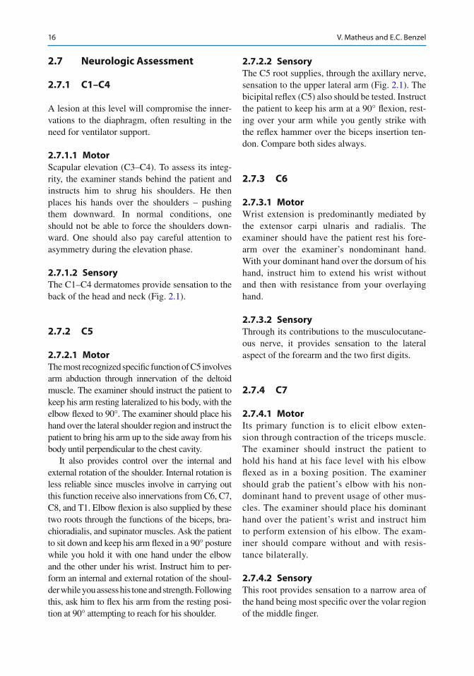
2.7.1.2 Sensory The C1–C4 dermatomes provide sensation to the back of the head and neck (Fig. 2.1 ).
2.7.2 C5
2.7.2.1 Motor The most recognized speci fi c function of C5 involves arm abduction through innervation of the deltoid muscle. The examiner should instruct the patient to keep his arm resting lateralized to his body, with the elbow fl exed to 90°. The examiner should place his hand over the lateral shoulder region and instruct the patient to bring his arm up to the side away from his body until perpendicular to the chest cavity.
It also provides control over the internal and external rotation of the shoulder. Internal rotation is less reliable since muscles involve in carrying out this function receive also innervations from C6, C7, C8, and T1. Elbow fl exion is also supplied by these two roots through the functions of the biceps, bra-chioradialis, and supinator muscles. Ask the patient to sit down and keep his arm fl exed in a 90° posture while you hold it with one hand under the elbow and the other under his wrist. Instruct him to per-form an internal and external rotation of the shoul-der while you assess his tone and strength. Following this, ask him to fl ex his arm from the resting posi-tion at 90° attempting to reach for his shoulder.
2.7.2.2 Sensory The C5 root supplies, through the axillary nerve, sensation to the upper lateral arm (Fig. 2.1 ). The bicipital re fl ex (C5) also should be tested. Instruct the patient to keep his arm at a 90° fl exion, rest-ing over your arm while you gently strike with the re fl ex hammer over the biceps insertion ten-don. Compare both sides always.
2.7.3 C6
2.7.3.1 Motor Wrist extension is predominantly mediated by the extensor carpi ulnaris and radialis. The examiner should have the patient rest his fore-arm over the examiner’s nondominant hand. With your dominant hand over the dorsum of his hand, instruct him to extend his wrist without and then with resistance from your overlaying hand.
2.7.3.2 Sensory Through its contributions to the musculocutane-ous nerve, it provides sensation to the lateral aspect of the forearm and the two fi rst digits.
2.7.4 C7
2.7.4.1 Motor Its primary function is to elicit elbow exten-sion through contraction of the triceps muscle. The examiner should instruct the patient to hold his hand at his face level with his elbow fl exed as in a boxing position. The examiner should grab the patient’s elbow with his non-dominant hand to prevent usage of other mus-cles. The examiner should place his dominant hand over the patient’s wrist and instruct him to perform extension of his elbow. The exam-iner should compare without and with resis-tance bilaterally.
2.7.4.2 Sensory This root provides sensation to a narrow area of the hand being most speci fi c over the volar region of the middle fi nger.
172 Physical Examination of the Cervical Spine
2.7.5 C8
2.7.5.1 Motor C8 function refers to the fl exion of the fi ngers. This function is mediated through the fl exor digitorum and lumbrical muscles. The examiner should instruct the patient to fl ex his fi ngers. Then, the patient should be asked to attempt extension, with and without resistance from the examiner’s fi ngers.
2.7.5.2 Sensory
The dermatome to this root is localized over the fi fth digit and lateral aspect of the fourth digit (Fig. 2.1 ).
2.7.6 T1
2.7.6.1 Motor T1 controls abduction of the fi ngers through innervations of the dorsal interossei and adduc-tion through innervations of the palmar interos-sei. To test for abduction, the patient is instructed to spread apart his fi ngers. The examiner should pinch together every set of fi ngers to try to force
them together. To test for adduction, the exam-iner should instruct the patient to keep his fi ngers together on extension after the examiner places a piece of paper between them and pulls it out.
2.7.6.2 Sensory Sensation over the medial aspect of the forearm (Fig. 2.1 ).
2.8 Miscellaneous
“Space-occupying para- and intraspinal lesions” can present in many ways, including neurological de fi cits. Speci fi c tests can be performed during the physical examination to exacerbate these symptoms and con fi rm the presence of one of these lesions. Unspeci fi c symptoms patients can complain of include neck pain and paresthesias of the upper and lower extremities.
2.8.1 Valsalva Maneuver
With the patient in sitting position, instruct the patient to hold his breath and bear down as if defecating. Inquire about worsening symptoms. This maneuver will raise the intrathecal pressure and possibly exac-erbate any symptoms caused by the compressive intraspinal, particularly intradural, lesion [ 5 ] .
It is important to evaluate the patient’s swallow-ing function during the physical examination. Patients may complain of dysphagia or odynophagia that could be due to an expansive cervical spine mass compressing the esophagus. These and other pathological fi ndings that are observed during swallowing could be manifestations of cranial nerve compression.
2.8.2 Cervical Neural Compression
Both spinal cord and nerve root compression can lead to neurologic compromise. Such may be the case with herniated discs, osteophytes, fractures, luxations, or tumors. Patients with neural com-pression and/or irritation may complain of cervi-calgia, radicular pain, paresthesias, weakness,
Fig. 2.1 Dermatomes of the cervical and brachial plexus
18 V. Matheus and E.C. Benzel
and myelopathy. It goes without saying that when suspecting high-grade neural compression, one should complement the history and physical examination with the pertinent imaging studies. The following tests can help clinically localize the offending pathology.
2.8.3 Foraminal Compression Test
With the patient in the sitting position and the head in a neutral position, the application of strong downward pressure with both hands for a few seconds can elicit radicular symptoms. Repeating these steps with the patient’s head rotated to each side can increase sensitivity.
By applying axial loading, the intervertebral disc is compressed, the foraminal cross-sectional area should decrease, and pressure will hence be exerted upon the apophyseal joints. If the patient develops symptoms or worsening of the preexisting symptoms, the dermatome should be relatively identi fi able based on classical dermato-mal distributions.
2.8.4 Extension Compression Test
With the patient in the sitting position, he is asked to extend his neck. The examiner then applies his hands on the forehead and applies downward pressure. Such axial loading in an extended spine results in compres-sion of the dorsal apophyseal joints and thus results in the worsening of existing or the development of localized pain related to joint disease. Simultaneously, it decreases the cross-sectional area of the foraminal space which may result in radicular pain.
2.8.5 Flexion Compression Test
The examiner asks the patient to fl ex his head while in the sitting position. He then applies downward pressure on the cranial vertex. With the head fl exed and with axial loading, the compres-sion of the ventral aspect of the disc induces dor-
sal displacement of a bulging disc into the central canal, thus potentially causing symptoms related to compression of the spinal cord. At the same time, pressure is taken off the dorsal apophyseal joints. Hence, preexisting facet origin pain may improve.
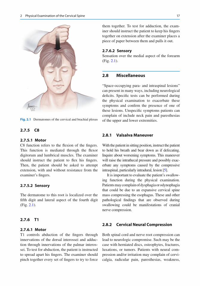
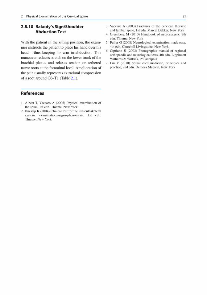
2.8.6 Spurling’s Test
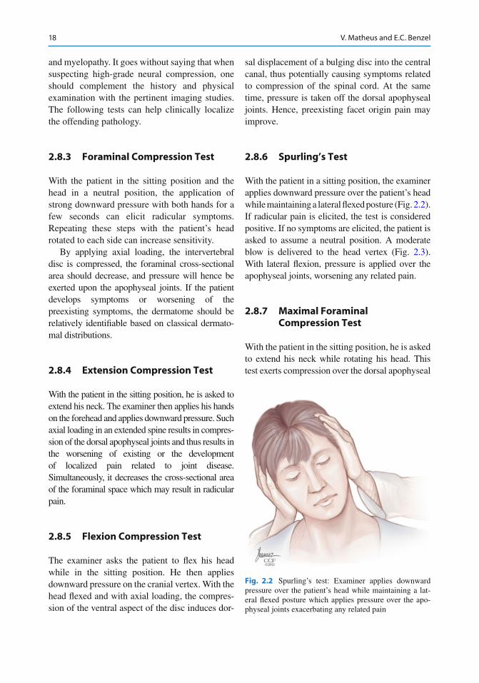
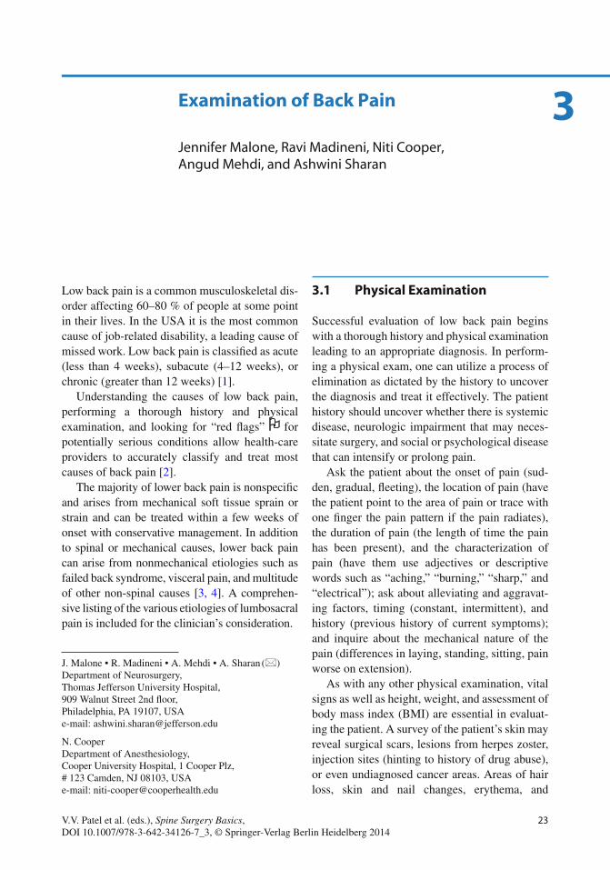
With the patient in a sitting position, the examiner applies downward pressure over the patient’s head while maintaining a lateral fl exed posture (Fig. 2.2 ). If radicular pain is elicited, the test is considered positive. If no symptoms are elicited, the patient is asked to assume a neutral position. A moderate blow is delivered to the head vertex (Fig. 2.3 ). With lateral fl exion, pressure is applied over the apophyseal joints, worsening any related pain.
2.8.7 Maximal Foraminal Compression Test
With the patient in the sitting position, he is asked to extend his neck while rotating his head. This test exerts compression over the dorsal apophyseal
Fig. 2.2 Spurling’s test: Examiner applies downward pressure over the patient’s head while maintaining a lat-eral fl exed posture which applies pressure over the apo-physeal joints exacerbating any related pain
192 Physical Examination of the Cervical Spine
joints and compresses the foraminal spaces, thus exacerbating pain related to nerve root encroach-ment [ 6 ] .
2.8.8 L’hermitte’s Phenomenon
With the patient in a relaxed seated position, he is asked to perform head fl exion. This results in stressing of the dorsal ligaments and elements of the spine plus compression of the ventral segment of the intervertebral disc. This in turn displaces dorsal disc bulges into the central canal, while the fl exion stretches the spinal cord over the ventral compressive masses (sagittal bowstring effect). A positive test involves the development of sud-den electrical tingling or shocks down the spine and/or extremities. Such a fi nding is consistent with signi fi cant stenosis and is a sign of myelopa-thy. Local cervical pain during the test could represent muscle sprain, meningeal irritation from an underlying in fl ammatory process, apo-physeal joint disease, or radiculopathy [ 7 ] .
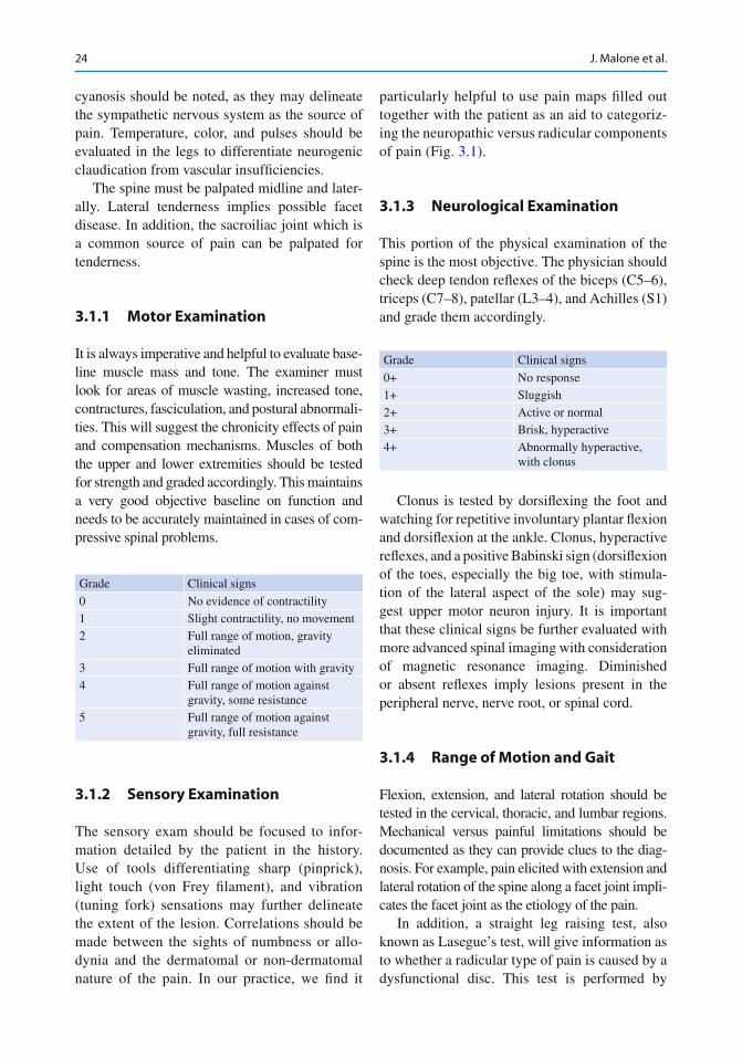
2.8.9 Distraction Test
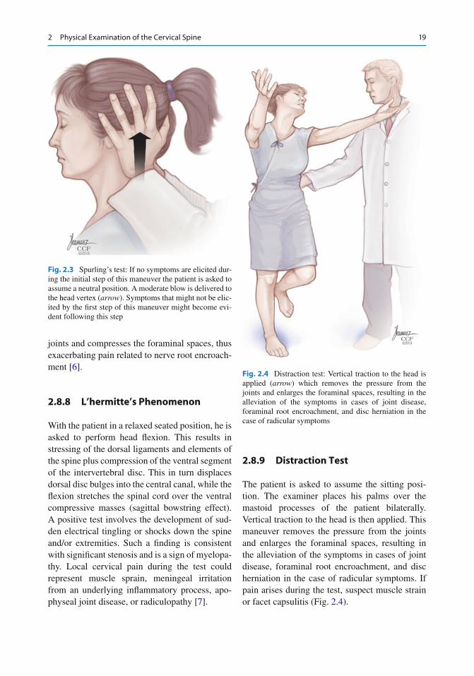
The patient is asked to assume the sitting posi-tion. The examiner places his palms over the mastoid processes of the patient bilaterally. Vertical traction to the head is then applied. This maneuver removes the pressure from the joints and enlarges the foraminal spaces, resulting in the alleviation of the symptoms in cases of joint disease, foraminal root encroachment, and disc herniation in the case of radicular symptoms. If pain arises during the test, suspect muscle strain or facet capsulitis (Fig. 2.4 ).
Fig. 2.3 Spurling’s test: If no symptoms are elicited dur-ing the initial step of this maneuver the patient is asked to assume a neutral position. A moderate blow is delivered to the head vertex ( arrow ). Symptoms that might not be elic-ited by the fi rst step of this maneuver might become evi-dent following this step
Fig. 2.4 Distraction test: Vertical traction to the head is applied ( arrow ) which removes the pressure from the joints and enlarges the foraminal spaces, resulting in the alleviation of the symptoms in cases of joint disease, foraminal root encroachment, and disc herniation in the case of radicular symptoms
20 V. Matheus and E.C. Benzel
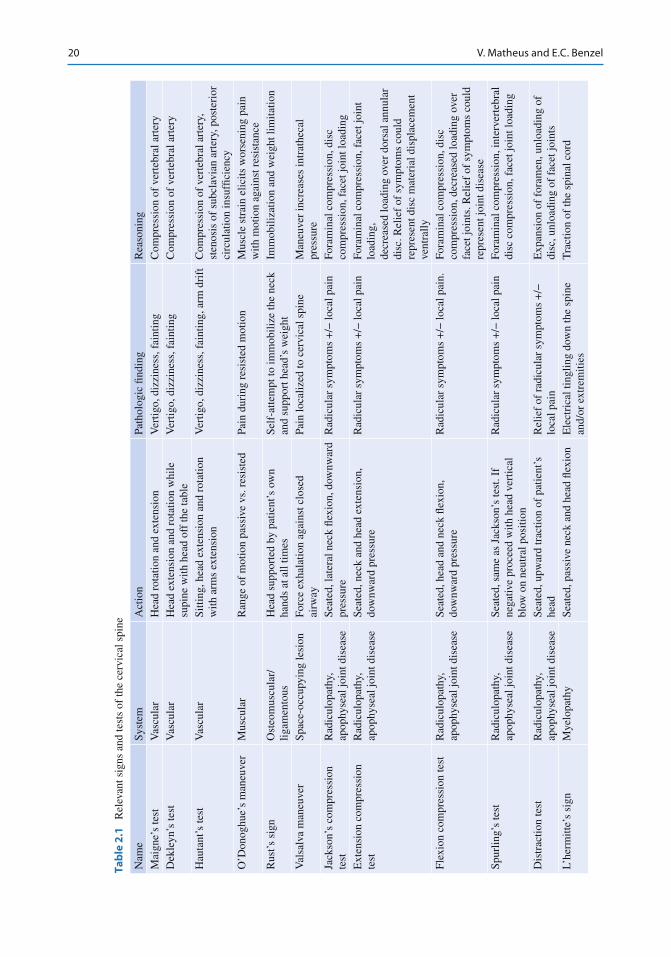
Tab
le 2
.1
Rel
evan
t sig
ns a
nd te
sts
of th
e ce
rvic
al s
pine
Nam
e Sy
stem
A
ctio
n Pa
thol
ogic
fi nd
ing
Rea
soni
ng
Mai
gne’
s te
st
Vas
cula
r H
ead
rota
tion
and
exte
nsio
n V
ertig
o, d
izzi
ness
, fai
ntin
g C
ompr
essi
on o
f ve
rteb
ral a
rter
y D
ekle
yn’s
test
V
ascu
lar
Hea
d ex
tens
ion
and
rota
tion
whi
le
supi
ne w
ith h
ead
off
the
tabl
e V
ertig
o, d
izzi
ness
, fai
ntin
g C
ompr
essi
on o
f ve
rteb
ral a
rter
y
Hau
tant
’s te
st
Vas
cula
r Si
tting
, hea
d ex
tens
ion
and
rota
tion
with
arm
s ex
tens
ion
Ver
tigo,
diz
zine
ss, f
aint
ing,
arm
dri
ft
Com
pres
sion
of
vert
ebra
l art
ery,
st
enos
is o
f su
bcla
vian
art
ery,
pos
teri
or
circ
ulat
ion
insu
f fi ci
ency
O
’Don
oghu
e’s
man
euve
r M
uscu
lar
Ran
ge o
f m
otio
n pa
ssiv
e vs
. res
iste
d Pa
in d
urin
g re
sist
ed m
otio
n M
uscl
e st
rain
elic
its w
orse
ning
pai
n w
ith m
otio
n ag
ains
t res
ista
nce
Rus
t’s s
ign
Ost
eom
uscu
lar/
ligam
ento
us
Hea
d su
ppor
ted
by p
atie
nt’s
ow
n ha
nds
at a
ll tim
es
Self
-atte
mpt
to im
mob
ilize
the
neck
an
d su
ppor
t hea
d’s
wei
ght
Imm
obili
zatio
n an
d w
eigh
t lim
itatio
n
Val
salv
a m
aneu
ver
Spac
e-oc
cupy
ing
lesi
on
Forc
e ex
hala
tion
agai
nst c
lose
d ai
rway
Pa
in lo
caliz
ed to
cer
vica
l spi
ne
Man
euve
r in
crea
ses
intr
athe
cal
pres
sure
Ja
ckso
n’s
com
pres
sion
te
st
Rad
icul
opat
hy,
apop
hyse
al jo
int d
isea
se
Seat
ed, l
ater
al n
eck
fl exi
on, d
ownw
ard
pres
sure
R
adic
ular
sym
ptom
s +
/− lo
cal p
ain
Fora
min
al c
ompr
essi
on, d
isc
com
pres
sion
, fac
et jo
int l
oadi
ng
Ext
ensi
on c
ompr
essi
on
test
R
adic
ulop
athy
, ap
ophy
seal
join
t dis
ease
Se
ated
, nec
k an
d he
ad e
xten
sion
, do
wnw
ard
pres
sure
R
adic
ular
sym
ptom
s +
/− lo
cal p
ain
Fora
min
al c
ompr
essi
on, f
acet
join
t lo
adin
g,
decr
ease
d lo
adin
g ov
er d
orsa
l ann
ular
di
sc. R
elie
f of
sym
ptom
s co
uld
repr
esen
t dis
c m
ater
ial d
ispl
acem
ent
vent
rally
Fl
exio
n co
mpr
essi
on te
st
Rad
icul
opat
hy,
apop
hyse
al jo
int d
isea
se
Seat
ed, h
ead
and
neck
fl ex
ion,
do
wnw
ard
pres
sure
R
adic
ular
sym
ptom
s +
/− lo
cal p
ain.
Fo
ram
inal
com
pres
sion
, dis
c co
mpr
essi
on, d
ecre
ased
load
ing
over
fa
cet j
oint
s. R
elie
f of
sym
ptom
s co
uld
repr
esen
t joi
nt d
isea
se
Spur
ling’
s te
st
Rad
icul
opat
hy,
apop
hyse
al jo
int d
isea
se
Seat
ed, s
ame
as J
acks
on’s
test
. If
nega
tive
proc
eed
with
hea
d ve
rtic
al
blow
on
neut
ral p
ositi
on
Rad
icul
ar s
ympt
oms
+/−
loca
l pai
n Fo
ram
inal
com
pres
sion
, int
erve
rteb
ral
disc
com
pres
sion
, fac
et jo
int l
oadi
ng
Dis
trac
tion
test
R
adic
ulop
athy
, ap
ophy
seal
join
t dis
ease
Se
ated
, upw
ard
trac
tion
of p
atie
nt’s
he
ad
Rel
ief
of r
adic
ular
sym
ptom
s +
/−
loca
l pai
n E
xpan
sion
of
fora
men
, unl
oadi
ng o
f di
sc, u
nloa
ding
of
face
t joi
nts
L’he
rmitt
e’s
sign
M
yelo
path
y Se
ated
, pas
sive
nec
k an
d he
ad fl
exio
n E
lect
rica
l tin
glin
g do
wn
the
spin
e an
d/or
ext
rem
ities
T
ract
ion
of th
e sp
inal
cor
d
212 Physical Examination of the Cervical Spine
2.8.10 Bakody’s Sign/Shoulder Abduction Test
With the patient in the sitting position, the exam-iner instructs the patient to place his hand over his head – thus keeping his arm in abduction. This maneuver reduces stretch on the lower trunk of the brachial plexus and relaxes tension on tethered nerve roots at the foraminal level. Amelioration of the pain usually represents extradural compression of a root around C6–T1 (Table 2.1 ).
References
1. Albert T, Vaccaro A (2005) Physical examination of the spine, 1st edn. Thieme, New York
2. Buckup K (2004) Clinical test for the musculoskeletal system: examinations-signs-phenomena, 1st edn. Thieme, New York
3. Vaccaro A (2003) Fractures of the cervical, thoracic and lumbar spine, 1st edn. Marcel Dekker, New York
4. Greenberg M (2010) Handbook of neurosurgery, 7th edn. Thieme, New York
5. Fuller G (2008) Neurological examination made easy, 4th edn. Churchill Livingstone, New York
6. Cipriano JJ (2003) Photographic manual of regional orthopaedic and neurological tests, 4th edn. Lippincott Williams & Wilkins, Philadelphia
7. Lin V (2010) Spinal cord medicine, principles and practice, 2nd edn. Demoes Medical, New York
23V.V. Patel et al. (eds.), Spine Surgery Basics, DOI 10.1007/978-3-642-34126-7_3, © Springer-Verlag Berlin Heidelberg 2014
3
Low back pain is a common musculoskeletal dis-order affecting 60–80 % of people at some point in their lives. In the USA it is the most common cause of job-related disability, a leading cause of missed work. Low back pain is classi fi ed as acute (less than 4 weeks), subacute (4–12 weeks), or chronic (greater than 12 weeks) [ 1 ] .
Understanding the causes of low back pain, performing a thorough history and physical examination, and looking for “red fl ags” for potentially serious conditions allow health-care providers to accurately classify and treat most causes of back pain [ 2 ] .
The majority of lower back pain is nonspeci fi c and arises from mechanical soft tissue sprain or strain and can be treated within a few weeks of onset with conservative management. In addition to spinal or mechanical causes, lower back pain can arise from nonmechanical etiologies such as failed back syndrome, visceral pain, and multitude of other non-spinal causes [ 3, 4 ] . A comprehen-sive listing of the various etiologies of lumbosacral pain is included for the clinician’s consideration.
3.1 Physical Examination
Successful evaluation of low back pain begins with a thorough history and physical examination leading to an appropriate diagnosis. In perform-ing a physical exam, one can utilize a process of elimination as dictated by the history to uncover the diagnosis and treat it effectively. The patient history should uncover whether there is systemic disease, neurologic impairment that may neces-sitate surgery, and social or psychological disease that can intensify or prolong pain.
Ask the patient about the onset of pain (sud-den, gradual, fl eeting), the location of pain (have the patient point to the area of pain or trace with one fi nger the pain pattern if the pain radiates), the duration of pain (the length of time the pain has been present), and the characterization of pain (have them use adjectives or descriptive words such as “aching,” “burning,” “sharp,” and “electrical”); ask about alleviating and aggravat-ing factors, timing (constant, intermittent), and history (previous history of current symptoms); and inquire about the mechanical nature of the pain (differences in laying, standing, sitting, pain worse on extension).
As with any other physical examination, vital signs as well as height, weight, and assessment of body mass index (BMI) are essential in evaluat-ing the patient. A survey of the patient’s skin may reveal surgical scars, lesions from herpes zoster, injection sites (hinting to history of drug abuse), or even undiagnosed cancer areas. Areas of hair loss, skin and nail changes, erythema, and
J. Malone • R. Madineni • A. Mehdi • A. Sharan (*) Department of Neurosurgery , Thomas Jefferson University Hospital , 909 Walnut Street 2nd fl oor , Philadelphia , PA 19107 , USA e-mail: [email protected]
N. Cooper Department of Anesthesiology , Cooper University Hospital , 1 Cooper Plz, # 123 Camden , NJ 08103, USA e-mail: [email protected]
Examination of Back Pain
Jennifer Malone , Ravi Madineni , Niti Cooper , Angud Mehdi , and Ashwini Sharan
24 J. Malone et al.
cyanosis should be noted, as they may delineate the sympathetic nervous system as the source of pain. Temperature, color, and pulses should be evaluated in the legs to differentiate neurogenic claudication from vascular insuf fi ciencies.
The spine must be palpated midline and later-ally. Lateral tenderness implies possible facet disease. In addition, the sacroiliac joint which is a common source of pain can be palpated for tenderness.
3.1.1 Motor Examination
It is always imperative and helpful to evaluate base-line muscle mass and tone. The examiner must look for areas of muscle wasting, increased tone, contractures, fasciculation, and postural abnormali-ties. This will suggest the chronicity effects of pain and compensation mechanisms. Muscles of both the upper and lower extremities should be tested for strength and graded accordingly. This maintains a very good objective baseline on function and needs to be accurately maintained in cases of com-pressive spinal problems.
Grade Clinical signs
0 No evidence of contractility 1 Slight contractility, no movement 2 Full range of motion, gravity
eliminated 3 Full range of motion with gravity 4 Full range of motion against
gravity, some resistance 5 Full range of motion against
gravity, full resistance
3.1.2 Sensory Examination
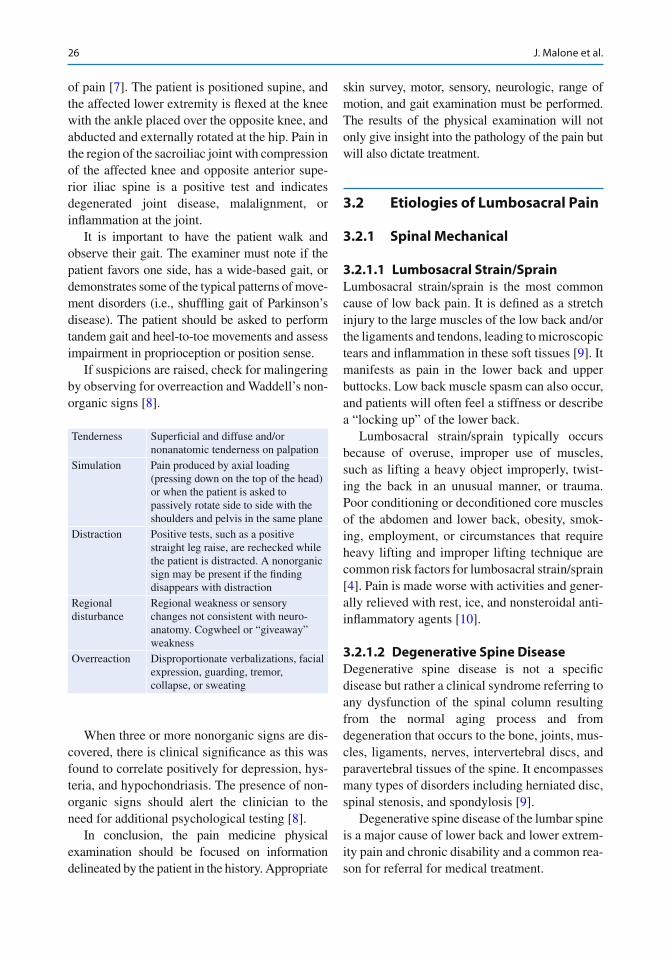
The sensory exam should be focused to infor-mation detailed by the patient in the history. Use of tools differentiating sharp (pinprick), light touch (von Frey fi lament), and vibration (tuning fork) sensations may further delineate the extent of the lesion. Correlations should be made between the sights of numbness or allo-dynia and the dermatomal or non-dermatomal nature of the pain . In our practice, we fi nd it
particularly helpful to use pain maps fi lled out together with the patient as an aid to categoriz-ing the neuropathic versus radicular components of pain (Fig. 3.1 ).
3.1.3 Neurological Examination
This portion of the physical examination of the spine is the most objective. The physician should check deep tendon re fl exes of the biceps (C5–6), triceps (C7–8), patellar (L3–4), and Achilles (S1) and grade them accordingly.
Grade Clinical signs
0+ No response 1+ Sluggish 2+ Active or normal 3+ Brisk, hyperactive 4+ Abnormally hyperactive,
with clonus
Clonus is tested by dorsi fl exing the foot and watching for repetitive involuntary plantar fl exion and dorsi fl exion at the ankle. Clonus, hyperactive re fl exes, and a positive Babinski sign (dorsi fl exion of the toes, especially the big toe, with stimula-tion of the lateral aspect of the sole) may sug-gest upper motor neuron injury. It is important that these clinical signs be further evaluated with more advanced spinal imaging with consideration of magnetic resonance imaging. Diminished or absent re fl exes imply lesions present in the peripheral nerve, nerve root, or spinal cord.
3.1.4 Range of Motion and Gait
Flexion, extension, and lateral rotation should be tested in the cervical, thoracic, and lumbar regions. Mechanical versus painful limitations should be documented as they can provide clues to the diag-nosis. For example, pain elicited with extension and lateral rotation of the spine along a facet joint impli-cates the facet joint as the etiology of the pain.
In addition, a straight leg raising test, also known as Lasegue’s test, will give information as to whether a radicular type of pain is caused by a dysfunctional disc. This test is performed by

253 Examination of Back Pain
having the examiner passively raise the patient’s leg as the patient is lying supine. A positive test is when the pain is reproduced as the leg is raised between 30° and 70° [ 5 ] . Similarly, a Spurling’s test can be performed for the cervical spine.
A positive test occurs when radicular pain is felt with extension, lateral rotation, and compression of the head [ 6 ] .
A positive FABER test, also known as Patrick’s test, will lead to the sacroiliac joint as the cause
Levels of principal dermatomesC5 ClaviclesC5, 6, 7 Lateral parts of upper limbs C8, T1 Medial sides of upper limbsC6 ThumbC6, 7, 8 HandC8 Ring and little fingersT4 Level of nipples
T10 Level of umbilicusL1 Inguinal or groin regionsL1, 2, 3, 4 Anterior and inner surfaces of lower limbsL4, 5, S1 FootL4 Medial side of great toeS1, 2, L5 Posterior and outer surfaces of lower limbsS1 Lateral margin of foot and little toe S2, 3, 4 Perineum
Schematic demarcation of dermatomes(according to Keegan and Garrett)shown as distinct segments. There isactually considerable overlap betweenany two adjacent dermatomes. Analternative dermatome map is thatprovided by Foerster (see References).
C2
C3C4
C5T1T2T3T4T5T6T7T8T9
T10
T11
T12
L1
L3
L4
L5
S1
L5
L4
C2
C3
C4C5C6
C7C8 T1
T2T3T4T5T6T7T8T9T10T11T12L1
L3
L2
L1
S1 S2
L4
S1
L5
L4
S2
S1
L5
L2L3L4L5
S1S2
S3S4
S5
C6
T1
C5
C6
C8
C6
C7C7 C8
C7 C8
S2, 3L2
Fig. 3.1 Diagram of the dermatomal distribution of the sensory innervation of the upper and low limbs. Reproduced from “Aids to the examination of the peripheral nervous system”
26 J. Malone et al.
of pain [ 7 ] . The patient is positioned supine, and the affected lower extremity is fl exed at the knee with the ankle placed over the opposite knee, and abducted and externally rotated at the hip. Pain in the region of the sacroiliac joint with compression of the affected knee and opposite anterior supe-rior iliac spine is a positive test and indicates degenerated joint disease, malalignment, or in fl ammation at the joint.
It is important to have the patient walk and observe their gait. The examiner must note if the patient favors one side, has a wide-based gait, or demonstrates some of the typical patterns of move-ment disorders (i.e., shuf fl ing gait of Parkinson’s disease). The patient should be asked to perform tandem gait and heel-to-toe movements and assess impairment in proprioception or position sense.
If suspicions are raised, check for malingering by observing for overreaction and Waddell’s non-organic signs [ 8 ] .
When three or more nonorganic signs are dis-covered, there is clinical signi fi cance as this was found to correlate positively for depression, hys-teria, and hypochondriasis. The presence of non-organic signs should alert the clinician to the need for additional psychological testing [ 8 ] .
In conclusion, the pain medicine physical examination should be focused on information delineated by the patient in the history. Appro priate
skin survey, motor, sensory, neurologic, range of motion, and gait examination must be performed. The results of the physical examination will not only give insight into the pathology of the pain but will also dictate treatment.
3.2 Etiologies of Lumbosacral Pain
3.2.1 Spinal Mechanical
3.2.1.1 Lumbosacral Strain/Sprain Lumbosacral strain/sprain is the most common cause of low back pain. It is de fi ned as a stretch injury to the large muscles of the low back and/or the ligaments and tendons, leading to microscopic tears and in fl ammation in these soft tissues [ 9 ] . It manifests as pain in the lower back and upper buttocks. Low back muscle spasm can also occur, and patients will often feel a stiffness or describe a “locking up” of the lower back.
Lumbosacral strain/sprain typically occurs because of overuse, improper use of muscles, such as lifting a heavy object improperly, twist-ing the back in an unusual manner, or trauma. Poor conditioning or deconditioned core muscles of the abdomen and lower back, obesity, smok-ing, employment, or circumstances that require heavy lifting and improper lifting technique are common risk factors for lumbosacral strain/sprain [ 4 ] . Pain is made worse with activities and gener-ally relieved with rest, ice, and nonsteroidal anti-in fl ammatory agents [ 10 ] .
3.2.1.2 Degenerative Spine Disease Degenerative spine disease is not a speci fi c disease but rather a clinical syndrome referring to any dysfunction of the spinal column resulting from the normal aging process and from degeneration that occurs to the bone, joints, mus-cles, ligaments, nerves, intervertebral discs, and paravertebral tissues of the spine. It encompasses many types of disorders including herniated disc, spinal stenosis, and spondylosis [ 9 ] .
Degenerative spine disease of the lumbar spine is a major cause of lower back and lower extrem-ity pain and chronic disability and a common rea-son for referral for medical treatment.
Tenderness Super fi cial and diffuse and/or nonanatomic tenderness on palpation
Simulation Pain produced by axial loading (pressing down on the top of the head) or when the patient is asked to passively rotate side to side with the shoulders and pelvis in the same plane
Distraction Positive tests, such as a positive straight leg raise, are rechecked while the patient is distracted. A nonorganic sign may be present if the fi nding disappears with distraction
Regional disturbance
Regional weakness or sensory changes not consistent with neuro-anatomy. Cogwheel or “giveaway” weakness
Overreaction Disproportionate verbalizations, facial expression, guarding, tremor, collapse, or sweating
273 Examination of Back Pain
3.2.1.3 Herniated Disc A herniated disc refers to localized displacement of the nucleus pulposus through a tear in the annu-lus fi brosus beyond the limits of the intravertebral disc space [ 11 ] . The tear in the annulus fi brosus may result in the release of in fl ammatory media-tors which may cause severe pain even without nerve root compression. Only 1–3 % of people presenting with low back pain have lumbar disc herniation and only 1 % will have a nerve root symptom. Most common level for lumbar disc her-niations occurs between L4–5 and L5–S1 [ 12 ] .
Risk factors for herniated lumbar disc include age (between 30 and 50), male gender, lifting a heavy object especially when using a twisting motion of the spine, occupation in a physically demanding job, cigarette smoking, and obesity [ 13 ] .
Symptoms of lumbar disc herniation often include acute lower back pain and muscle spasm, followed by sudden or gradual radicular leg pain, then typically a reduction in the degree of lower back pain. Bladder symptoms such as urinary urgency, frequency, and hesitancy may be present in up to 18 % of patients with acute disc hernia-tions without cauda equina syndrome [ 14 ] . Outright urinary retention or over fl ow inconti-nence may however be seen in cauda equina syn-drome (see below).
When lumbar disc herniation is associated with heavy lifting, pulling, pushing, or twisting, some patients state that they hear and/or feel a “pop” in their back. They may describe the initial pain as “searing” or “hot” [ 14 ] . Patients with radicular leg pain may fi nd relief fl exing the knee or the thigh of the affected leg because full exten-sion creates nerve root tension. Patients will gen-erally avoid excessive activity, and yet they cannot remain in one position (sitting, standing, lying) for too long either, prompting them to fre-quent position changes. Valsalva maneuvers (coughing, sneezing, and straining at the bowel or bladder) may also worsen radicular pain.
3.2.1.4 Cauda Equina Syndrome Cauda equina syndrome, CES, is a serious neuro-logic condition involving impairment of the nerves of the lumbar plexus from compression of
the cauda equina. CES frequently necessitates emergent or urgent surgical decompression to prevent permanent de fi cits and/or incontinence.
CES is prevalent in only 0.04 % of all patients with low back pain and only 1–2 % of all patients with lumbar disc herniations [ 14 ] . CES is caused by any compressive lesion causing pressure on the nerve roots in the lumbar spinal canal below the conus medullaris; the most common cause of this problem however is a central disc herniation. Other causes include metastatic disease; intrinsic spinal tumors; burst fractures; direct trauma from anesthetic, diagnostic, or therapeutic lumbar puncture; spinal epidural hematoma; penetrating trauma such as knife or bullet injuries; compres-sive abscess; and spinal stenosis [ 12 ] .
Spontaneous low back and radiating lower extremity pain with severe or progressive weak-ness usually involving more than one nerve root may be the most prominent symptom in CES. Pain in a radicular distribution is more prominent than back pain in these cases. Saddle anesthesia is the most common sensory de fi cit in CES with a distri-bution involving some or most of the anus, geni-tals, perineum, buttocks, and posterior- superior thighs. This can be unilateral and asymmetric as well. A patient with CES or developing CES will often describe symptoms of sphincter disturbance; urinary retention is the most common, but other symptoms include bladder frequency, urgency, over fl ow incontinence, fecal incontinence, and diminished anal sphincter tone. Retention can be evaluated and documented by performing post-void residual measurements either by catheteriza-tion or ultrasound measuring residual. Diminished or absent patellar and Achilles re fl ex may be found [ 2, 12 ] . No single sign or symptom de fi nes a CES, but it is a constellation of fi ndings instead.
3.2.1.5 Spinal Stenosis Spinal stenosis refers to a condition of narrowing in and around the spinal canal, causing compres-sion on the neural elements. It is classi fi ed as cen-tral canal stenosis, foraminal stenosis, or lateral recess stenosis. Central canal stenosis refers to narrowing of the anteroposterior dimension of the spinal canal, causing compression of the spi-nal cord or cauda equina. Foraminal stenosis
28 J. Malone et al.
refers to narrowing of the neural foramen, caus-ing compression on spinal nerves, and lateral recess stenosis, a type of lumbar spinal stenosis, arises from hypertrophy of the superior articular aspect of the facet joint. In the lumbar spine, lat-eral recess stenosis most commonly affects the L4–5 facet [ 15 ] .
The most common cause of stenosis is related to degeneration and the aging process, osteoar-thritis, disc degeneration, and thickened spinal ligaments. Other causes include spinal trauma, previous spinal surgery, spinal tumors, Paget’s disease, and having a congenitally small central canal as seen in achondroplasia [ 12 ] .
Symptoms of spinal stenosis depend on the location of the narrowing and resultant impinge-ment or compression. Any one or combination of the following symptoms of stenosis can be pres-ent: low back pain; numbness, paresthesias, cramping, weakness, and pain in the buttocks, legs, and feet; radiating leg pain; and bowel and/or bladder dysfunction.
Symptomatic lumbar stenosis is most com-mon at L4–5 then L3–4 followed by L2–3 and then L5–S1. Lumbar stenosis is classi fi ed as stable, facet hypertrophy, thickening of the ligamentum fl avum, and disc degeneration. Unstable stenosis is marked by the addition of degenerative spondylolisthesis or degenerative scoliosis [ 16 ] .
Neurogenic claudication is a common symp-tom of lumbar spinal stenosis. It can be unilateral or bilateral buttock, hip, leg, or foot discomfort, pain, or weakness that is aggravated by standing and walking and alleviated by sitting or lying. Patients with neurogenic claudication may develop “anthropoid posture,” an exaggerated fl exion at the waist which possibly creates a reduced lumbar lordosis and opens the facet joints [ 12 ] . The patient may reveal that they are also more comfortable leaning forward, for example, on a counter at home or, classically, a grocery cart in the supermarket.
3.2.1.6 Fracture Most fractures of the spine occur in the thoracic and lumbar spines and most commonly at the thoracolumbar junction, T12–L1. These fractures
are typically caused by major trauma such as motor vehicle accidents, falls, or sports accidents, but even minor trauma in a compromised spine can lead to fracture. Persons with bone weakened by osteoporosis, long-term corticosteroid use, substance abuse, or systemic disease and spinal tumors can suffer a nontraumatic fracture during normal daily activities [ 2, 4 ] .
Types of spinal fractures include compression, burst, fl exion/distraction, seatbelt, or Chance fracture, transverse process, fracture dislocation, pathologic fracture from infection or tumor, and osteoporotic fracture [ 12 ] .
Plain radiography is recommended in patients with persistent pain, history of trauma, fever, unexplained weight loss, cancer, substance abuse, and age greater than 50. Computed tomography (CT) and magnetic resonance imaging (MRI) are more useful for the detection of infection and fracture caused by cancer [ 4 ] .
Nonsurgical treatment includes 6–8 weeks of bracing, activity modi fi cation, analgesics, physical therapy and a gradual return to normal physical activity, and treatment of the underlying systemic disease if present. Surgery is typically reserved for unstable and comminuted fractures, the presence of neurologic de fi cit, progressive spinal deformity, and pain refractory to nonsurgical management.
3.2.1.7 Spondylolisthesis Spondylolisthesis is slippage of the superior ver-tebra over inferior vertebra. This condition most commonly affects the lumbar spine and is less common in the cervical spine. There are fi ve types of lumbar spondylolisthesis: 1. Dysplastic spondylolisthesis – this is caused
by a congenital defect in the facet that allows the vertebra to slip forward.
2. Isthmic spondylolisthesis – this results from a defect in the part of vertebra called pars inter-articularis. This defect is thought to be caused by repetitive trauma and is more common in athletes due to hyperextension motion.
3. Degenerative spondylolisthesis (DS) – this type occurs due to arthritic changes in the facet joints of vertebra. It is more common in older patients and represents the most com-mon form of spondylolisthesis.
293 Examination of Back Pain
4. Traumatic spondylolisthesis – this occurs sec-ondary to direct trauma and can include a frac-ture of the pedicle, lamina, or facet joints
5. Pathological spondylolisthesis – this type is caused by bony defect due to tumor which causes bone to be abnormal. Spondylolisthesis is graded based on extent
of slippage of lateral radiograph by Meyerding. This measurement is distance from the poste-rior edge of upper vertebra to posterior edge of lower vertebra and is reported as a percentage of total upper vertebral body. There are fi ve grades of slippage: grade1, 0–25 %; grade 2, 25–50 %; grade 3, 50–75 %; grade 4, 75–100 %; and grade 5, spondyloptosis when upper verte-bra is completely fallen off in relation to lower vertebra.
Among these fi ve types of spondylolisthesis, DS is most commonly seen in patients over 50 years and a common cause of low back pain (LBP). It also commonly involves L4–L5 level and to lesser extent L5–S1. DS is approximately four to fi ve times more common in females than in males, due to greater ligamentous laxity and hormonal effects [ 17, 18 ] .
LBP is the most common presentation in patients with DS; however, some of them may be asymptomatic. LBP may be mechanical type to pain and relieved with rest. This condition is also associated with neurogenic claudication. Leg pain can be radicular or diffuse and involving dermatomal distribution of L4, L5, and S1 nerve roots, although single nerve root, most commonly L5, involvement may also be seen. These symp-toms are seen in 42–82 % of patients who see a spine surgeon for help. Bladder and bowel dys-functions due to DS can occur but less profound than cauda equina syndrome from disc hernia-tion. This can be seen in severe stenosis in 3 % of patients [ 19 ] .
Patients with mechanical type of LBP and neurogenic claudication should be investigated with standing lumbar spine x-rays including fl exion and extension fi lms. Supine fi lms may not demonstrate the slippage. CT scan and MRI of lumbar spine add to diagnosing this condition accurately with degree of slippage and extent of stenosis causing neural compression.
3.2.1.8 Kyphosis Kyphosis is a term to describe the natural forward curve of the thoracic and lumbosacral spines where the lumbar and cervical spines have a natu-ral lordosis or lordotic curve. When kyphosis is used to describe a spinal deformity, it refers to an exaggeration of the forward curve in a portion of any part of the spine, also called a kyphotic defor-mity. Kyphotic deformities can create symptoms that vary from pain and neurologic de fi cit to com-pensatory and cosmetic deformities [ 13 ] .
Kyphotic deformity has a multitude of causes including degenerative (osteoporotic compres-sion fracture, Paget’s disease), traumatic, devel-opmental (scoliosis), iatrogenic (following spinal decompressive laminectomy or radiation to the spine), neoplastic (primary spinal tumor or metastatic disease), congenital (achondroplasia), infectious (Pott’s disease, osteomyelitis), in fl a-mmatory (ankylosing spondylitis, rheumatoid arthritis), and neuromuscular (cerebral palsy) or from Scheuermann’s disease [ 13 ] .
A careful history and exam will reveal pres-ence of deformity, underlying or contributing conditions, neurologic impairment (from spinal cord or spinal nerve compression), and the devel-opment of compensatory deformities. Standing or upright x-rays of the entire spine in one view, commonly referred to as “scoliosis x-rays” with anterior-posterior and lateral views, are used to evaluate the structure of the spine and measure the degree of kyphosis or other abnormal curves. When neurologic de fi cits are discovered, worsen-ing pain or spinal instability is suspected; further imaging with CT and/or MRI is warranted.
3.2.1.9 Scoliosis The term scoliosis originates from Greek word skoliosis meaning obliquity or bending. Adult scoliosis is a de novo development of curved spinal architecture after completion of skeletal maturity. It is also seen in children and adoles-cents; however, adult scoliosis differs from child or adolescent scoliosis in terms of curve types and patterns, rate of deformity progres-sion, rigidity of deformity, patient comorbidi-ties, and clinical symptoms and presentation [ 20 ] . Some of adolescent scoliosis can be
30 J. Malone et al.
asymptomatic and get detected during adult life due to progression of curvature. Other patients may develop scoliosis after spine surgery for disc degeneration or spinal fusion surgery as adjacent-level degeneration with scoliosis.
The prevalence of adult scoliosis is probably on the rise due to increasing life expectancies. The most common types of scoliosis encountered in adults are idiopathic and degenerative scoliosis. The former condition starts in childhood or adoles-cence and progresses over a period of time with added degeneration of disc and facets. Degenerative scoliosis is a de novo development of scoliosis sec-ondary to asymmetric involvement of disc degen-eration, facet arthrosis, and disc collapse [ 21 ] .
Patients with scoliosis may present to spine surgeons with symptoms of back pain due to spine deformity or symptoms of neural compres-sion unrelated to deformity. Adult patients with scoliosis present with axial low back pain, neurogenic pain, as well as changes in gait and posture. Physical examination of the back while palpating spine will reveal abnormal curvature of spine and asymmetry of the pelvic crests. Patients with stooped posture may have sagittal imbal-ance forcing them to walk with walker or cane. Imaging studies begin with plane x-rays of the spine standing, and scoliosis fi lms which deter-mine the severity of sagittal and coronal imbal-ance and pelvic tilt.
3.2.2 Spinal Nonmechanical
3.2.2.1 Neoplasia: Intradural or Vertebral Tumors/Pathologic Fracture
Diagnosis of spinal neoplasia begins with the his-tory. Patients who complain of subacute back pain that is worse at night or with rest should raise the clinician’s suspicion for a possible neoplastic process especially if the patient reports unintentional weight loss and general malaise.
3.2.2.2 Infections: Osteomyelitis, Discitis, and Epidural Abscesses
Spinal infections arise from bacteria carried through the bloodstream to the spine from a site of infection elsewhere in the body, urinary and
respiratory tract infection, soft tissue (infections on the skin), dental fl ora, or through intravenous drug use, surgery, injection treatments, or as a result of direct trauma. Infections are most fre-quently seen in the lumbar spine, followed by thoracic, cervical, and sacrum.
Back pain may be a result of an infection in the bone (osteomyelitis), in the disc (discitis), or on the spinal cord (epidural abscess). Vertebral body collapse with kyphotic deformity is com-mon. Necrotic bone and disc fragments as well as abscess formation can cause spinal cord or cauda equina compression [ 12 ] . Presenting symptoms can be as generalized as back pain, low-grade temperatures, malaise, and anorexia. Unexplained back pain following a recent infec-tion or iatrogenic procedure with a strong mechanical component should be considered for a spinal infection.
3.2.3 In fl ammatory Arthropathies
3.2.3.1 Ankylosing Spondylitis Ankylosing spondylitis (AS) is a chronic, sys-temic in fl ammatory disease of the joints and the axial skeleton characterized by back and neck pain and progressive stiffening, or ankylosing, of the spine. Pain and stiffness is typical in the tho-racic spine or sometimes the entire spine, with referred pain to the buttocks and hamstrings. Pain is often present in the morning, is severe at rest, and improves with physical activity. Sacroiliitis is present in greater than 95 % of persons with AS.
The onset is gradual, typically beginning in late adolescence and early adulthood, and is slightly more common in men than in women in whom the disease evolves more slowly. Approximately 90 % of AS patients express the HLA-B27 genotype, meaning there is a strong genetic association. However, only 5 % of individuals with the HLA-B27 genotype contract the disease [ 13, 22 ] .
3.2.3.2 Rheumatoid Arthritis Rheumatoid arthritis (RA) is a systemic in fl ammatory autoimmune disease which is chronic and progressive in nature. RA affects multiple tissues and organs but predominantly attacks synovial joints, leading to destruction of
313 Examination of Back Pain
articular cartilage and ankylosis. It occurs in 0.3–1.5 % of the population, with women affected 2–3 times more often than men. Onset occurs most frequently between the ages of 40 and 50, but people of any age can be affected [ 22 ] . Often a disabling and painful condition, it has an insidi-ous onset marked by fatigue, anorexia, weight loss, and generalized aching and stiffness (espe-cially morning stiffness). RA can lead to substan-tial loss of function and mobility if not adequately treated.
3.2.3.3 Reiter’s Syndrome A reactive arthritis usually occurring 1–3 weeks following certain bacterial infections (commonly Chlamydia, Shigella, Salmonella, Yersinia, and Campylobacter) with involvement of at least one other non-joint area, speci fi cally urethritis, uveitis/conjunctivitis, skin lesions, and mucosal ulcerations. Between 75 and 90 % of patients are also HLA-B27 positive [ 13 ] .
3.2.3.4 Paget’s Disease Paget’s disease (PD) is a metabolic disorder with abnormal bone remodeling, causing spinal steno-sis and facet arthropathy. In this disorder, there is excessive breakdown of bone and formation of weak bone, causing pain, fracture, and joint arthritis. Etiology of Paget’s disease remains unclear, and it has been thought to be caused by viral infection [ 23 ] . Paget’s disease also can be inherited as autosomal dominant trait with high penetrance [ 24 ] . Reported incidence of back pain in PD patients ranges from 11 to 42 % [ 25 ] . Several mechanisms have been described in neu-ral symptoms in patients with PD: (1) compres-sion of neural elements by pagetic process, (2) neural ischemia, and (3) pagetic sacromatous degeneration [ 26 ] . Diagnosis of PD is by x-rays, bone scan, and CT scan and MRI. Once PD is con fi rmed as underlying pathology, treatment is initiated with biphosphonates with goal of reliev-ing bone pain and arrest progression of disease. In addition to biphosphonates, other drugs used are calcitonin and mithramycin.
3.2.3.5 Sacroiliitis In fl ammation of the sacroiliac joint, sacroiliitis is a frequent initial manifestation of one of the
seronegative spondyloarthropathies. Sacroiliitis most commonly presents in young people who are HLA-B27 positive and/or have ankylosing spon-dylitis, psoriatic arthritis, or Reiter’s disease. The pain of sacroiliitis most commonly occurs in the lower back and tops of the buttocks, but it can also radiate to incorporate the groin, legs, and feet.
3.2.3.6 Scheuermann’s Kyphosis Scheuermann’s kyphosis most commonly affects the vertebral bodies of the thoracic spine but can also occur at the thoracolumbar junction. It is best described as a growth abnormality of one or more vertebral bodies where the anterior portion of the vertebrae stops growing, yet the posterior portion continues to grow, creating an abnormal amount of kyphosis at the apex of the deformity. It is most often seen in males and typically occurs in the fi nal growth spurt of adolescence. Pain and discomfort are common along the site of the kyphotic deformity [ 13 ] .
3.2.4 Arachnoiditis
Arachnoiditis is a neuropathic disease caused by in fl ammation of the arachnoid membrane of the spinal cord and spinal nerves. It can be caused by chemical irritation, infection, injury, previous spinal surgery, or other invasive spinal proce-dures. Arachnoiditis can lead to adhesions, caus-ing the spinal nerves to “clump.” The presenting symptoms of arachnoiditis may include constant chronic low back pain unrelated to and unrelieved by speci fi c positions, radiating leg pain, perineal pain, and pseudoclaudication. Treatment is insuf fi cient with the main focus on medication and intrathecal steroid injections [ 9 ] .
3.2.5 Failed Back Syndrome
Failed back surgery syndrome (FBSS) and post-laminectomy syndrome are terms used to describe a type of a chronic and persistent back and/or leg pain condition that occurs following lumbar spi-nal surgery. FBSS does not include persistent lower extremity weakness, sensory changes, or re fl ex abnormalities [ 13 ] .
32 J. Malone et al.
Patients with FBS often demonstrate frustra-tion and anger and often have an accompanying diagnosis of depression. These patients frequently require long-term pain management, speci fi cally with narcotic agents. The clinician should be astute to the possibility of malingering for a vari-ety of secondary gain issues which can be found to coexist in patients with FBS.
The causes of FBS include incorrect initial diagnosis; improper preoperative patient selection; continued nerve root compression caused by recur-rent disc herniation, scar tissue, pseudomeningo-cele, hematoma, spinal instability, or stenosis at the surgical or junctional level; permanent nerve injury; technical error during surgery; adhesive arachnoiditis; infection (discitis, osteomyelitis); spondylosis; other non-spinal causes of back pain; and nonanatomic factors/malingering [ 12 ] .
3.3 Visceral Causes of Back Pain
Occasionally, back pain is the single presenting symptom of a variety of serious medical condi-tions or possible emergency. Clinicians must be astute to possible medical or visceral causes of back pain when the patient describes severe, refractory, or atypical pain. Examination of the abdomen with palpation of the visceral organs is a necessary step of diagnosis. This chapter will not cover the atypical presentations but will just report a laundry list of differential diagnoses which also may present with back pain: dissecting aortic aneurysm, urinary tract infection, pyelonephritis, prostatitis, pelvic in fl ammatory disease (PID), endometriosis, ovulation, pregnancy, ectopic preg-nancy, acute pancreatitis, duodenal ulcer, chole-cystitis, nephrolithiasis, and visceral cancers.
3.4 Non-spinal
3.4.1 Piriformis Syndrome
Piriformis syndrome refers to sciatic symptoms (low back pain radiating in the buttock, thigh, calf, and foot) that do not originate from the lum-bosacral plexus and/or from disc herniation but
rather by pressure from the piriformis muscle on the sciatic nerve [ 9 ] . Pain is made worse with activity such as prolonged sitting and walking.
The piriformis muscle originates at the antero-lateral aspect of the sacroiliac region, transverses the sciatic nerve, and inserts on the greater tro-chanter of the femur. It is innervated by the ventral rami of S1 and S2 and abducts and laterally rotates the femur. The sciatic nerve passes between the two bellies of the piriformis [ 27 ] . Trauma or over-use can lead to irritation, in fl ammation, and spasm of the piriformis, which can compress the trans-versing sciatic nerve and mimic a herniated lum-bar disc or spinal nerve compression.
Performing a straight leg raise (SLR) test can differentiate between piriformis syndrome and sci-atica caused by lumbosacral nerve compression. In piriformis syndrome the SLR is only mildly posi-tive or negative unless the examiner fl exes, adducts, and internally rotates the proximal leg. Adding these maneuvers places tension on the irritated piri-formis and stretches the in fl amed nerve root [ 9 ] .
3.4.2 Bursitis
Trochanteric bursitis and ischiogluteal bursitis are types of pelvic pain that is often misdiagnosed as herniated lumbar disc or sciatica from the lum-bosacral spine especially in the elderly patient population. The symptoms of trochanteric bursi-tis are pain in the hip region with activity and point tenderness over the greater trochanter. Patients will often report that they are unable to lie on the affected side. With ischiogluteal bursi-tis, pain is localized deep in the center of the buttock. Patients will describe the pain as “unre-lenting,” aggravated by sitting or walking, and accompanied by radicular leg pain that is unre-lieved by rest. Treatments include rest, anti-in fl ammatory agents, physical therapy, cortisone injections, or in extreme cases bursectomy [ 28 ] .
3.4.3 Fibromyalgia
Fibromyalgia is a chronic pain syndrome charac-terized by widespread musculoskeletal pain,
333 Examination of Back Pain
fatigue and heightened tenderness to tactile pressure, general fatigue, and sleep disturbance. The most common sites of pain include “tender points” of the neck, back, shoulders, bony pelvis, and hands. Over 6 million Americans are diag-nosed yearly, 90 % of which are women between the ages of 20 and 55 years old. Pain is described as a deep ache, sometimes shooting, and burning.
3.4.4 Spasticity
Spasticity is often found in people with cerebral palsy, traumatic brain injury, stroke, multiple sclerosis, and spinal cord injury. Spasticity results from upper motor neuron lesions creating an imbalance of inhibitory in fl uence on alpha and gamma motor neurons. Spasticity is clinically manifested as a hypertonic state of muscles with clonus and involuntary movement. Often painful and debilitating, spasticity creates challenges with self-care, hygiene, posture, and balance. Back pain is a cardinal feature of spasticity [ 12 ] .
3.4.5 Degenerative Joint Disease (DJD) of the Hip
DJD of the hip is most often caused by osteoar-thritis. It is characterized by pain and stiffness from the breakdown of joint surface cartilage. DJD of the hip creates ipsilateral groin and medial thigh pain. At times the pain can radiate to the knee on the same side, creating confusion as to whether the problem is from the hip, from the knee, or from a lumbar radiculopathy. Hip dys-plasia and avascular necrosis of the hip can cause these symptoms as well.
When pain originates from the hip, walking and prolonged activity worsen the pain and motion in the joint is limited, often realized when trying to go from sitting or lying to a standing position. Initially, pain is relieved by rest, but once the DJD progresses, minimal activity such as slight movements in bed can worsen the pain. Treatment includes rest, ice, anti-in fl ammatory agents, cortisone injections, and, if necessary, joint replacement surgery [ 29 ] .
3.4.6 Leg Length Discrepancy/Pelvic Level/Gait Abnormality
A leg length discrepancy can be due to a mild variation between two sides of the body and is a normal variation when the difference is 3/5 of an inch or less. There are a variety of causes for leg length discrepancy including previous injury or fracture to the leg (especially in children who are fractured at the growth plate), bony diseases, in fl ammation and osteoarthritis, and neurologic conditions.
The correlation between patients with a clini-cally signi fi cant leg length discrepancy and the incidence of low back pain is controversial. Leg length discrepancy can lead to a pelvic obliquity which changes the coronal balance of the sacrum, leading to a segmental scoliosis and potential for increased low back pain [ 9 ] .
Leg length discrepancy can lead to gait abnor-malities requiring patients to exert more effort with ambulation and hasten degenerative joint disease. Treatment includes orthotics and surgery for inhib-iting growth, lengthening, and shortening.
3.4.7 Posture
The position of the body in both the sitting and standing positions can have considerable effect on the development or prevention of lower back pain. Swayback posture (lumbar hyperlordosis) in standing and a slouched or slumped (thoracic hyperkyphosis) posture in sitting can impact the health and function of the muscles of the abdo-men and lower back. When daily activities require prolonged sitting or standing and a balanced seated or standing posture is not achieved, the stabilizing muscles of the spine fatigue and lead to pain and stiffness [ 9 ] .
3.4.8 Obesity
More than half of the adult population of Americans is categorized as being overweight or obese. Overweight and obesity is a contributing factor to back pain and a signi fi cant cause for
34 J. Malone et al.
seeking medical care. Being overweight or obese can contribute to the symptoms of osteoporosis, osteoarthritis, degenerative spine disease, and spondylolisthesis. The addition of excess weight and a deconditioned core translate extra strain and stress to the spine, speci fi cally the lower back [ 30 ] .
3.4.9 Mood Disorders
Pain, anxiety, and depression are commonly expe-rienced together. In patients with chronic and disabling pain syndromes such as fi bromyalgia, low back pain, and nerve pain, this is particularly evident. Patients with diagnosed anxiety and depression report higher incidences and greater severity of pain in addition to increased disability and dysfunction due to pain compared to patients without depression and anxiety [ 31 ] . Treatment of both pain and mood disorder is challenging but can be accomplished with a comprehensive plan including cognitive behavioral therapy, relaxation techniques, hypnosis, exercise, antidepressants, and mood stabilizers.
3.4.10 Secondary Gain/Malingering
Secondary gain is an external psychological motivator that may drive a patient to report cer-tain symptoms. If a patient’s pain or illness allows them to miss work or gain extra sympathy and attention, these would be examples of secondary gain. With secondary gain, a patient is uncon-sciously seeking these “rewards.” If he or she is deliberately exaggerating symptoms for personal gain, that is, to win a legal dispute, then he or she is malingering [ 32 ] . In either case, it is in the patient’s best interest to remain debilitated or in pain in order to continue to receive the rewards.
References
1. National Institute of Neurological Disorders and Stroke; National Institute of Health. Low back pain sheet. Updated 15 June 2011. http://ninds.nih.gov/ disorders/backpain/detail_backpain.htm
2. Bigos S, Bowyer O, Braen G et al (1994) Acute low back problems in adults, vol Clinical practice guide-line no.14. AHCPR publication no. 95-0642. Agency for Health Care Policy and Research PHS, U.S Department of Health and Human Services, Rockville
3. Hart LG, Deyo RA, Cherkin DC (1995) Physician of fi ce visits for low back pain. Frequency, clinical evaluation, and treatment patterns from a U.S. National survey. Spine (Phila Pa 1976) 20(1):11–19
4. Deyo RA, Weinstein JN (2001) Low back pain. N Engl J Med 344(5):363–370
5. Speed C (2004) Low back pain. BMJ 328(7448):1119–1121
6. Tong HC, Haig AJ, Yamakawa K (2002) The spurling test and cervical radiculopathy. Spine (Phila Pa 1976) 27(2):156–159
7. Ross MD, Nordeen MH, Barido M (2003) Test-retest reliability of Patrick’s hip range of motion test in healthy college-aged men. J Strength Cond Res 17(1):156–161
8. Scalzitti DA (1997) Screening for psychological fac-tors in patients with low back problems: Waddell’s nonorganic signs. Phys Ther 77(3):306–312
9. Cailliet R (1988) Low back pain syndrome, 4th edn. FA Davis, Philadelphia
10. Malanga G, Dunn K (2010) Low back pain manage-ment: approaches to treatment. J Musculoskel Med 27:305–315
11. Fardon DF (2001) Nomenclature and classi fi cation of lumbar disc pathology. Spine (Phila Pa 1976) 26(5):461–462
12. Greenberg MS, Arredondo N (2006) Handbook of neurosurgery, 6th edn. Thieme Medical Publishers, New York
13. Vaccaro AR (2005) Spine: core knowledge in ortho-paedics. Mosby, St. Louis
14. Deyo RA, Rainville J, Kent DL (1992) What can the history and physical examination tell us about low back pain? JAMA 268(6):760–765
15. Epstein NE, Maldonado VC, Cusick JF (1998) Symptomatic lumbar spinal stenosis. Surg Neurol 50(1):3–10
16. Duggal N, Sonntag V, Dickman CA (2001) Fusion option and indications in the lumbosacral spine. Contemp Neurosurg 23(1):1–8
17. Rosenberg NJ (1976) Degenerative spondylolisthesis: surgical treatment. Clin Orthop Relat Res 117:112–120
18. Bird H, Eastmond C, Hudson A, Wright V (1980) Is generalized joint laxity a factor in spondylolisthesis? Scand J Rheumatol 9(4):203–205
19. Kostuik JP, Harrington I, Alexander D, Rand W, Evans D (1986) Cauda equina syndrome and lumbar disc her-niation. J Bone Joint Surg Am 68(3):386–391
20. Herkowitz HN, Rothman RH, Simeone FA (2011) Rothman-simeone, the spine, 6th edn. Saunders Elsevier, Philadelphia
21. Simmons ED Jr, Simmons EH (1992) Spinal stenosis with scoliosis. Spine (Phila Pa 1976) 17(6 Suppl):S117–S120
22. Klippel J (2001) Primer of rheumatic disease, 12th edn. Arthritis Foundation, Atlanta

353 Examination of Back Pain
23. Mee AP (1999) Paramyxoviruses and Paget’s disease: the af fi rmative view. Bone 24(5 Suppl):19S–21S
24. Hocking L, Slee F, Haslam SI et al (2000) Familial Paget’s disease of bone: patterns of inheritance and frequency of linkage to chromosome 18q. Bone 26(6):577–580
25. Rosenkrantz JA, Wolf J, Kaicher JJ (1952) Paget’s disease (osteitis deformans); review of one hundred eleven cases. AMA Arch Intern Med 90(5):610–633
26. Herkowitz HN (2004) The lumbar spine, 3rd edn. Lippincott Williams & Wilkins, Philadelphia
27. Saladin K (1998) Anatomy & physiology: the unity of form and function, 1st edn. McGraw-Hill, WCB
28. Dougherty CD, Dougherty JJ (2008) Evaluating hip pathology in trochanteric pain syndrome. J Muscu-loskel Med 9:428–436
29. Hoaglund FT, Steinbach LS (2001) Primary osteoar-thritis of the hip: etiology and epidemiology. J Am Acad Orthop Surg 9(5):320–327
30. Shiri R, Karppinen J, Leino-Arjas P, Solovieva S, Viikari-Juntura E (2010) The association between obesity and low back pain: a meta-analysis. Am J Epidemiol 171(2):135–154
31. Means-Christensen A, Roy-Byrne P, Sherourne C, Craske M, Stein M (2008) Relationships among pain, anxiety, and depression in primary care. Depress Anxiety 25:593–600
32. McDermott BE, Feldman MD (2007) Malingering in the medical setting. Psychiatr Clin North Am 30(4):645–662
37V.V. Patel et al. (eds.), Spine Surgery Basics, DOI 10.1007/978-3-642-34126-7_4, © Springer-Verlag Berlin Heidelberg 2014
4
4.1 Imaging Option Overview
4.1.1 Radiography
4.1.1.1 Physics Radiography is the fi rst-line technique in the evaluation of the spine. It is relatively inexpen-sive and widely available. X-rays are produced by rapidly moving streams of electrons from the cathode to the anode. The produced ionizing radiation (photons) penetrates an object and the differences in X-ray attenuation, which depends on differences in tissue density, are registered on a sensor plate. Bones absorb the radiation more than soft tissues; denser tissues attenuate the X-ray beam to a greater degree, lessening the number of X-ray photons sensed on the detector plate. The greater the number of X-ray beams to reach the detector, the darker the image. This is
why bones are bright and lungs are dark on a con-ventional radiograph.
4.1.1.2 Radiography as a Screening Tool in Spine Trauma
The standard method of screening the cervical spine is a conventional radiographic series, which typically includes lateral, anteroposterior, and odontoid views. Other views include swimmers lat-eral (to clear the shoulders and allow visualization of the cervical thoracic junction), oblique views, and fl exion/extension views in the lateral projec-tion. Particular dif fi culty in positioning high-mech-anism, poly-trauma patients may lead to a large number of inadequate radiographic examinations.
Cervical spine radiography, although rela-tively cheap, adds substantially to health-care costs because of the high volume of its use. Emergency departments annually treat millions of patients with trauma who are at risk for cervi-cal spine injury, and the total cost of cervical spine radiography is therefore substantial and judicious use of cervical spine radiography in the emergency trauma is necessary.
Several decision algorithms have been devel-oped. The Canadian C-spine Rule (CCR) [ 1 ] and the National Emergency X-Radiography Utilization Study (NEXUS) low-risk criteria (NLC) [ 2 ] were developed independently. Both rules are sensitive for detecting acute C-spine injury which allows the emergency department physicians to be more selective in the use of radi-ography in alert and stable trauma patients [ 3 ] .
A. Valladares-Otero , M.D. • B. Christenson , M.D. Department of Radiology , University of Colorado School of Medicine , 12401 East 17th Avenue, Room 1618 , Aurora , CO 80045 , USA
B.D. Petersen , M.D. (*) Associate Professor of Radiology and Orthopaedics,Chief of Musculoskeletal Radiology , University of Colorado , Mail Stop L954, 12401 East 17th Avenue, Room 5-006 , Aurora , CO 80045 , USA e-mail: [email protected]
Radiologic Imaging of the Spine
Angela Valladares-Otero , Brian Christenson , and Brian D. Petersen
38 A. Valladares-Otero et al.
4.1.1.3 Normal Radiographic Anatomy of the Spine
When evaluating spinal radiographs, a fi rm understanding of normal anatomy is necessary to allow one to detect pathology.
Cervical Spine Radiographic assessment of the cervical spine requires adequately exposed images to allow visualization of the bone trabeculae, as well as adequate patient positioning. On a true lateral radiograph of the cervical spine, the facet joints are superimposed on each other. A fron-tal view of the cervical spine should have simi-lar coverage as a lateral view. On a true frontal view, the spinous processes are midline. Open-mouth odontoid view should be centered on the C1–C2 articulation and the teeth and occiput should not be superimposed over the area of interest.
Lateral View (Fig. 4.1 ) The lateral view (Fig. 4.1a ) is the most important view in the routine trauma series. The lateral view must include all seven cervical vertebrae, as well as the C7–T1 intervertebral space. Several methods have been used to include the vertebral bodies and posterior elements of the cervical thoracic junction. On a swimmers view (Fig. 4.1b ), one of the arms is raised above the head to avoid superimposition of the shoulders over the cervical thoracic junction. Disadvantages to the swimmers view include higher radiation dose, high scatter, and dif fi cult positioning. Although bony details are usually suboptimal on the swimmer’s lateral view, gross alignment can be con fi rmed.
Lateral view of the cervical spine allows eval-uation of the vertebral bodies and intervertebral disks height, as well as the cervical spine align-ment which physiologically has a gentle lordotic
a b
Fig. 4.1 Normal lateral ( a ) and swimmer’s lateral ( b ) projections of the cervical spine with labeled pertinent anatomy. Anterior spinal line ( ASL ), posterior spinal line ( PSL ), spinolaminar line ( SLL ), and spinous process line ( SPL )
394 Radiologic Imaging of the Spine
curve. Vertebral bodies should be rectangular in shape and similar in size to the adjacent ones.
The articular facets connect the posterolateral aspect of vertebral bodies and combine to form the facet joints. On the lateral view, the lateral masses appear as rhomboid-shaped structures projecting posteroinferiorly.
The anterior longitudinal ligament (ALL) and the posterior longitudinal ligament (PLL) are major stabilizers of the intervertebral joints, help-ing to maintain the vertebral body alignment [ 4 ] . When assessing spinal alignment, it is helpful to evaluate the integrity of the anterior spinal line (Fig. 4.1a , ASL) which extends along the anterior margin of the vertebral bodies; the posterior spi-nal line (Fig. 4.1a , PSL) along the posterior mar-gin of the vertebral bodies; the spinolaminar line (Fig. 4.1a , SLL) along the posterior margin of spinal canal; and a line connecting the tips of C2–C7 spinous processes, the spinous process line (Fig. 4.1a , SPL). Misalignment of ASL, PSL, SLL, or SPL can suggest ligamentous injury or
occult fracture. Although any spinal offset should be scrutinized, there are common nonpathologic reasons for minimal offset including pseudosub-luxation of C2 in the pediatric population [ 5 ] and the slight offset of the SLL between the posterior elements of C1 and C2.
Radiographic assessment of the craniocervical and atlantoaxial articulations is dif fi cult but cru-cial (Figs. 4.2 , 4.3 , and 4.4 ). Craniocervical and atlantoaxial biomechanical continuity depends on the integrity of the skull base (occipital condyles), atlas (C1), and axis (C2) and their stabilizing liga-ments. On lateral view, the craniocervical rela-tionship can be assessed visually by several methods. The basion is the caudal tip of the clivus and can be a critical bony landmark for assess-ment of the craniocervical relationship. Harris et al. [ 6, 7 ] described a simple and reliable method that used the basion-axial interval (BAI) and basion-dens interval (BDI) for accurate assess-ment of occipitovertebral relationships on initial lateral radiographs in the supine position.
a b
Fig. 4.2 Sagittal CT reconstruction ( a ) and lateral cervi-cal spine radiograph ( b ) demonstrating the basion-axial interval ( BAI ). Basion-axial interval is the distance
between basion ( black arrows ) and a line extending supe-riorly, tangential to the posterior cortex of the C2 vertebral body, the posterior axial line ( PAL )
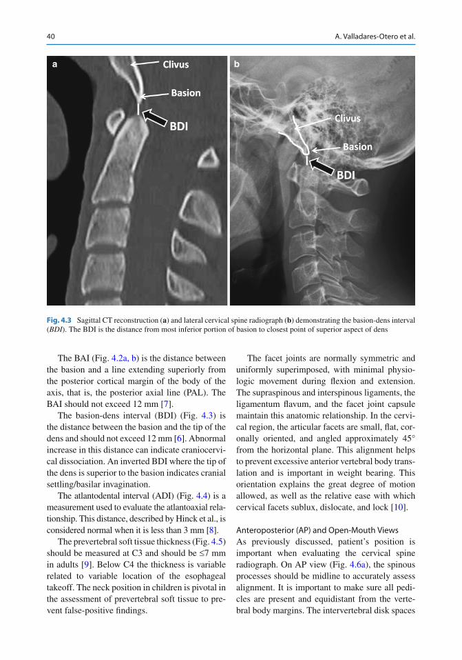
40 A. Valladares-Otero et al.
The BAI (Fig. 4.2a, b ) is the distance between the basion and a line extending superiorly from the posterior cortical margin of the body of the axis, that is, the posterior axial line (PAL). The BAI should not exceed 12 mm [ 7 ] .
The basion-dens interval (BDI) (Fig. 4.3 ) is the distance between the basion and the tip of the dens and should not exceed 12 mm [ 6 ] . Abnormal increase in this distance can indicate craniocervi-cal dissociation. An inverted BDI where the tip of the dens is superior to the basion indicates cranial settling/basilar invagination.
The atlantodental interval (ADI) (Fig. 4.4 ) is a measurement used to evaluate the atlantoaxial rela-tionship. This distance, described by Hinck et al., is considered normal when it is less than 3 mm [ 8 ] .
The prevertebral soft tissue thickness (Fig. 4.5 ) should be measured at C3 and should be £ 7 mm in adults [ 9 ] . Below C4 the thickness is variable related to variable location of the esophageal takeoff. The neck position in children is pivotal in the assessment of prevertebral soft tissue to pre-vent false-positive fi ndings.
The facet joints are normally symmetric and uniformly superimposed, with minimal physio-logic movement during fl exion and extension. The supraspinous and interspinous ligaments, the ligamentum fl avum, and the facet joint capsule maintain this anatomic relationship. In the cervi-cal region, the articular facets are small, fl at, cor-onally oriented, and angled approximately 45° from the horizontal plane. This alignment helps to prevent excessive anterior vertebral body trans-lation and is important in weight bearing. This orientation explains the great degree of motion allowed, as well as the relative ease with which cervical facets sublux, dislocate, and lock [ 10 ] .
Anteroposterior (AP) and Open-Mouth Views As previously discussed, patient’s position is important when evaluating the cervical spine radiograph. On AP view (Fig. 4.6a ), the spinous processes should be midline to accurately assess alignment. It is important to make sure all pedi-cles are present and equidistant from the verte-bral body margins. The intervertebral disk spaces
a b
Fig. 4.3 Sagittal CT reconstruction ( a ) and lateral cervical spine radiograph ( b ) demonstrating the basion-dens interval ( BDI ). The BDI is the distance from most inferior portion of basion to closest point of superior aspect of dens
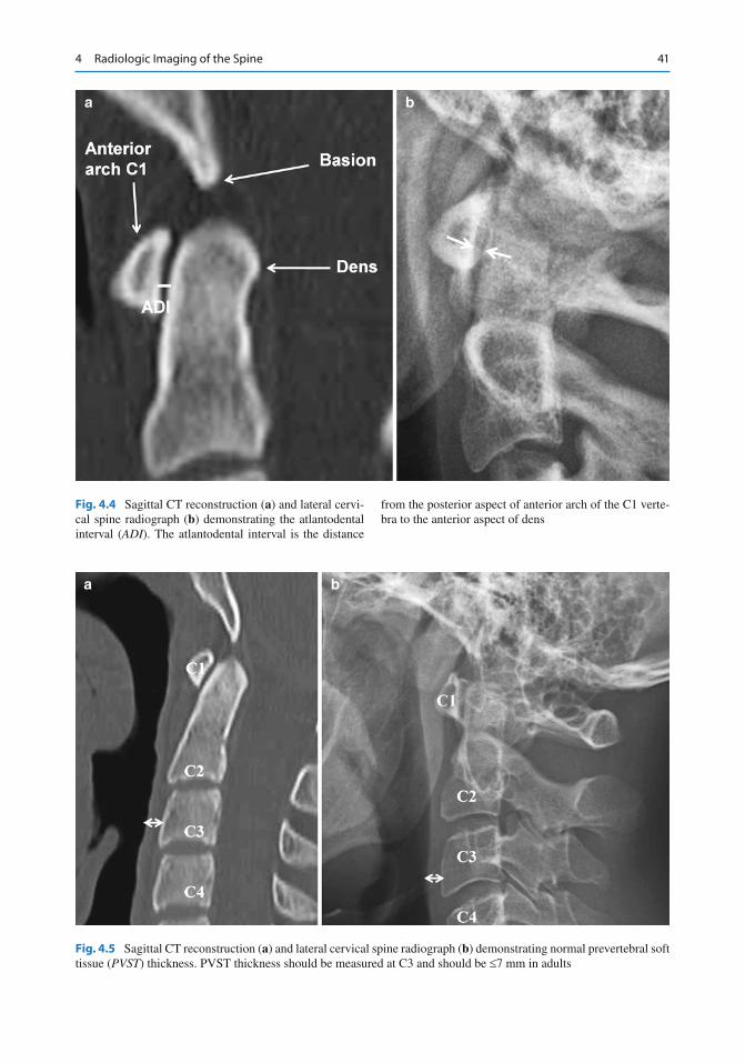
414 Radiologic Imaging of the Spine
a b
Fig. 4.4 Sagittal CT reconstruction ( a ) and lateral cervi-cal spine radiograph ( b ) demonstrating the atlantodental interval ( ADI ). The atlantodental interval is the distance
from the posterior aspect of anterior arch of the C1 verte-bra to the anterior aspect of dens
a b
Fig. 4.5 Sagittal CT reconstruction ( a ) and lateral cervical spine radiograph ( b ) demonstrating normal prevertebral soft tissue ( PVST ) thickness. PVST thickness should be measured at C3 and should be £ 7 mm in adults
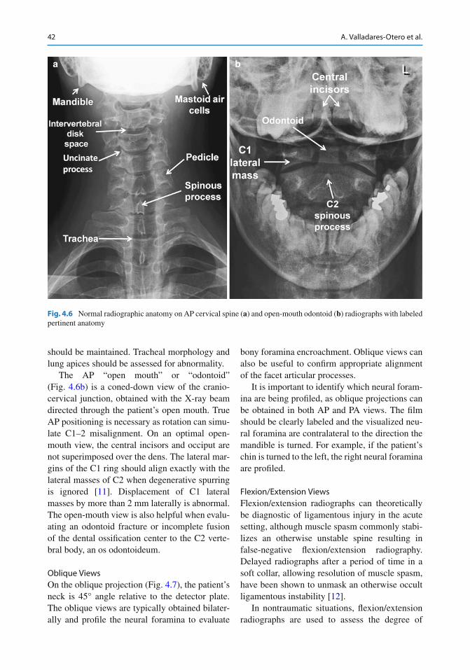
42 A. Valladares-Otero et al.
should be maintained. Tracheal morphology and lung apices should be assessed for abnormality.
The AP “open mouth” or “odontoid” (Fig. 4.6b ) is a coned-down view of the cranio-cervical junction, obtained with the X-ray beam directed through the patient’s open mouth. True AP positioning is necessary as rotation can simu-late C1–2 misalignment. On an optimal open-mouth view, the central incisors and occiput are not superimposed over the dens. The lateral mar-gins of the C1 ring should align exactly with the lateral masses of C2 when degenerative spurring is ignored [ 11 ] . Displacement of C1 lateral masses by more than 2 mm laterally is abnormal. The open-mouth view is also helpful when evalu-ating an odontoid fracture or incomplete fusion of the dental ossi fi cation center to the C2 verte-bral body, an os odontoideum.
Oblique Views On the oblique projection (Fig. 4.7 ), the patient’s neck is 45° angle relative to the detector plate. The oblique views are typically obtained bilater-ally and pro fi le the neural foramina to evaluate
bony foramina encroachment. Oblique views can also be useful to con fi rm appropriate alignment of the facet articular processes.
It is important to identify which neural foram-ina are being pro fi led, as oblique projections can be obtained in both AP and PA views. The fi lm should be clearly labeled and the visualized neu-ral foramina are contralateral to the direction the mandible is turned. For example, if the patient’s chin is turned to the left, the right neural foramina are pro fi led.
Flexion/Extension Views Flexion/extension radiographs can theoretically be diagnostic of ligamentous injury in the acute setting, although muscle spasm commonly stabi-lizes an otherwise unstable spine resulting in false-negative fl exion/extension radiography. Delayed radiographs after a period of time in a soft collar, allowing resolution of muscle spasm, have been shown to unmask an otherwise occult ligamentous instability [ 12 ] .
In nontraumatic situations, fl exion/extension radiographs are used to assess the degree of
a b
Fig. 4.6 Normal radiographic anatomy on AP cervical spine ( a ) and open-mouth odontoid ( b ) radiographs with labeled pertinent anatomy
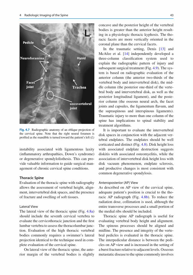
434 Radiologic Imaging of the Spine
instability associated with ligamentous laxity (in fl ammatory arthropathies, Down’s syndrome) or degenerative spondylolisthesis. This can pro-vide valuable information to guide surgical man-agement of chronic cervical spine conditions.
Thoracic Spine Evaluation of the thoracic spine with radiography allows the assessment of vertebral height, align-ment, intervertebral disk spaces, and the presence of fracture and swelling of soft tissues.
Lateral View The lateral view of the thoracic spine (Fig. 4.8a ) should include the seventh cervical vertebra to evaluate the cervicothoracic junction and the fi rst lumbar vertebra to assess the thoracolumbar junc-tion. Evaluation of the high thoracic vertebral bodies commonly requires a swimmer’s lateral projection identical to the technique used in com-plete evaluation of the cervical spine.
On lateral view of the thoracic spine, the ante-rior margin of the vertebral bodies is slightly
concave and the posterior height of the vertebral bodies is greater than the anterior height result-ing in a physiologic thoracic kyphosis. The tho-racic facets are more vertically oriented in the coronal plane than the cervical facets.
In the traumatic setting, Denis [ 13 ] and McAfee et al. [ 14 ] independently developed a three-column classi fi cation system used to explain the radiographic pattern of injury and subsequent surgical treatment (Fig. 4.9 ). The sys-tem is based on radiographic evaluation of the anterior column (the anterior two-thirds of the vertebral body and intervertebral disk), the mid-dle column (the posterior one-third of the verte-bral body and intervertebral disk, as well as the posterior longitudinal ligament), and the poste-rior column (the osseous neural arch, the facet joints and capsules, the ligamentum fl avum, and the supraspinous and interspinous ligaments). Traumatic injury to more than one column of the spine has implications to spinal stability and treatment algorithms.
It is important to evaluate the intervertebral disk spaces in conjunction with the adjacent ver-tebral endplates. The endplates should be well corticated and distinct (Fig. 4.8 ). Disk height loss with associated endplate destruction suggests diskitis with associated osteomyelitis, while the association of intervertebral disk height loss with disk vacuum phenomenon, endplate sclerosis, and productive changes is most consistent with common degenerative spondylosis.
Anteroposterior (AP) View As described on AP view of the cervical spine, adequate patient’s position is crucial to the tho-racic AP radiograph (Fig. 4.8b ). To reduce the radiation dose, collimation is used, although the entire transverse processes and a small portion of the medial ribs should be included.
Thoracic spine AP radiograph is useful for evaluating vertebral body height and alignment. The spinous processes should be aligned and midline. The presence and integrity of the verte-bral pedicles is evaluated in the thoracic spine. The interpedicular distance is between the pedi-cles on AP view and is increased in the setting of a burst fracture relative to adjacent levels. Osseous metastatic disease to the spine commonly involves
Fig. 4.7 Radiographic anatomy of an oblique projection of the cervical spine. Note that the right neural foramen is pro fi led as the mandible is turned toward the patient’s left ( L )
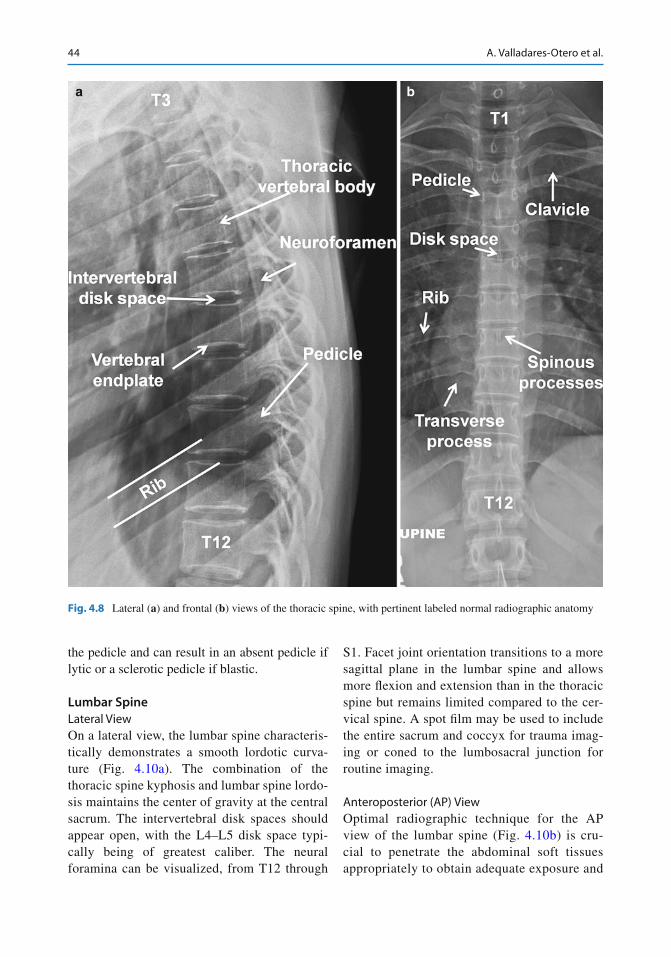
44 A. Valladares-Otero et al.
the pedicle and can result in an absent pedicle if lytic or a sclerotic pedicle if blastic.
Lumbar Spine Lateral View On a lateral view, the lumbar spine characteris-tically demonstrates a smooth lordotic curva-ture (Fig. 4.10a ). The combination of the thoracic spine kyphosis and lumbar spine lordo-sis maintains the center of gravity at the central sacrum. The intervertebral disk spaces should appear open, with the L4–L5 disk space typi-cally being of greatest caliber. The neural foramina can be visualized, from T12 through
S1. Facet joint orientation transitions to a more sagittal plane in the lumbar spine and allows more fl exion and extension than in the thoracic spine but remains limited compared to the cer-vical spine. A spot fi lm may be used to include the entire sacrum and coccyx for trauma imag-ing or coned to the lumbosacral junction for routine imaging.
Anteroposterior (AP) View Optimal radiographic technique for the AP view of the lumbar spine (Fig. 4.10b ) is cru-cial to penetrate the abdominal soft tissues appropriately to obtain adequate exposure and
a b
Fig. 4.8 Lateral ( a ) and frontal ( b ) views of the thoracic spine, with pertinent labeled normal radiographic anatomy
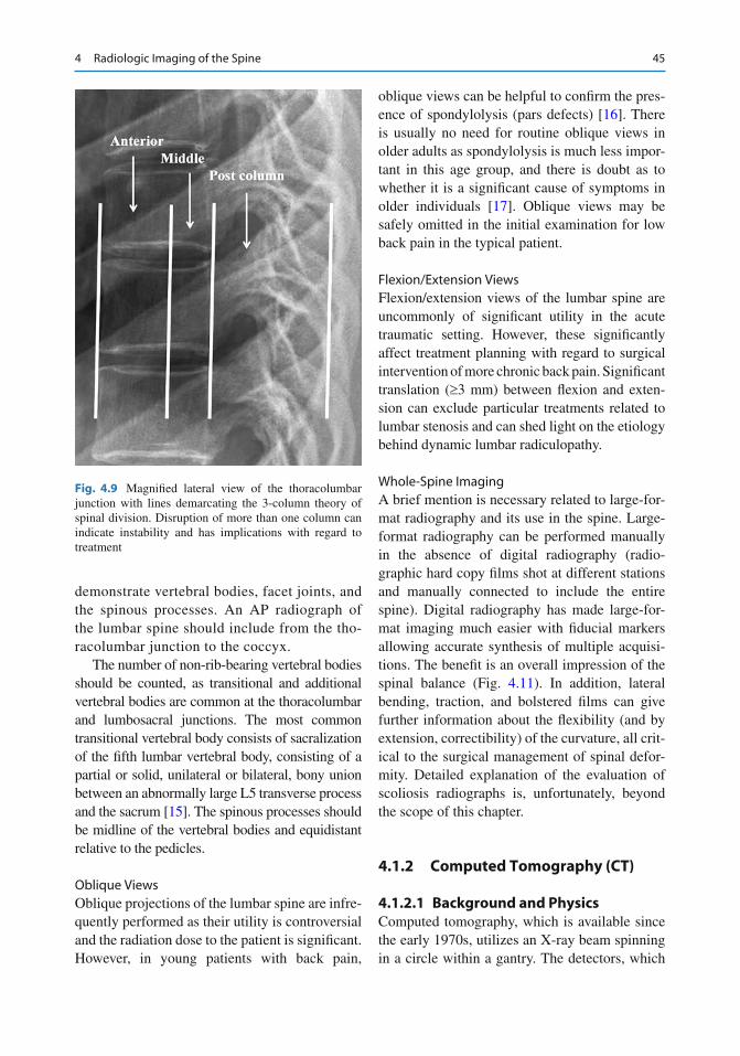
454 Radiologic Imaging of the Spine
demonstrate vertebral bodies, facet joints, and the spinous processes. An AP radiograph of the lumbar spine should include from the tho-racolumbar junction to the coccyx.
The number of non-rib-bearing vertebral bodies should be counted, as transitional and additional vertebral bodies are common at the thoracolumbar and lumbosacral junctions. The most common transitional vertebral body consists of sacralization of the fi fth lumbar vertebral body, consisting of a partial or solid, unilateral or bilateral, bony union between an abnormally large L5 transverse process and the sacrum [ 15 ] . The spinous processes should be midline of the vertebral bodies and equidistant relative to the pedicles.
Oblique Views Oblique projections of the lumbar spine are infre-quently performed as their utility is controversial and the radiation dose to the patient is signi fi cant. However, in young patients with back pain,
oblique views can be helpful to con fi rm the pres-ence of spondylolysis (pars defects) [ 16 ] . There is usually no need for routine oblique views in older adults as spondylolysis is much less impor-tant in this age group, and there is doubt as to whether it is a signi fi cant cause of symptoms in older individuals [ 17 ] . Oblique views may be safely omitted in the initial examination for low back pain in the typical patient.
Flexion/Extension Views Flexion/extension views of the lumbar spine are uncommonly of signi fi cant utility in the acute traumatic setting. However, these signi fi cantly affect treatment planning with regard to surgical intervention of more chronic back pain. Signi fi cant translation ( ³ 3 mm) between fl exion and exten-sion can exclude particular treatments related to lumbar stenosis and can shed light on the etiology behind dynamic lumbar radiculopathy.
Whole-Spine Imaging A brief mention is necessary related to large-for-mat radiography and its use in the spine. Large-format radiography can be performed manually in the absence of digital radiography (radio-graphic hard copy fi lms shot at different stations and manually connected to include the entire spine). Digital radiography has made large-for-mat imaging much easier with fi ducial markers allowing accurate synthesis of multiple acquisi-tions. The bene fi t is an overall impression of the spinal balance (Fig. 4.11 ). In addition, lateral bending, traction, and bolstered fi lms can give further information about the fl exibility (and by extension, correctibility) of the curvature, all crit-ical to the surgical management of spinal defor-mity. Detailed explanation of the evaluation of scoliosis radiographs is, unfortunately, beyond the scope of this chapter.
4.1.2 Computed Tomography (CT)
4.1.2.1 Background and Physics Computed tomography, which is available since the early 1970s, utilizes an X-ray beam spinning in a circle within a gantry. The detectors, which
Fig. 4.9 Magni fi ed lateral view of the thoracolumbar junction with lines demarcating the 3-column theory of spinal division. Disruption of more than one column can indicate instability and has implications with regard to treatment
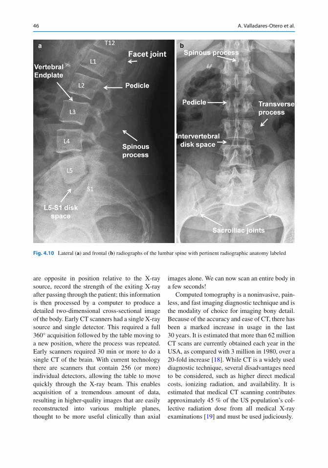
46 A. Valladares-Otero et al.
are opposite in position relative to the X-ray source, record the strength of the exiting X-ray after passing through the patient; this information is then processed by a computer to produce a detailed two-dimensional cross-sectional image of the body. Early CT scanners had a single X-ray source and single detector. This required a full 360° acquisition followed by the table moving to a new position, where the process was repeated. Early scanners required 30 min or more to do a single CT of the brain. With current technology there are scanners that contain 256 (or more) individual detectors, allowing the table to move quickly through the X-ray beam. This enables acquisition of a tremendous amount of data, resulting in higher-quality images that are easily reconstructed into various multiple planes, thought to be more useful clinically than axial
images alone. We can now scan an entire body in a few seconds!
Computed tomography is a noninvasive, pain-less, and fast imaging diagnostic technique and is the modality of choice for imaging bony detail. Because of the accuracy and ease of CT, there has been a marked increase in usage in the last 30 years. It is estimated that more than 62 million CT scans are currently obtained each year in the USA, as compared with 3 million in 1980, over a 20-fold increase [ 18 ] . While CT is a widely used diagnostic technique, several disadvantages need to be considered, such as higher direct medical costs, ionizing radiation, and availability. It is estimated that medical CT scanning contributes approximately 45 % of the US population’s col-lective radiation dose from all medical X-ray examinations [ 19 ] and must be used judiciously.
a b
Fig. 4.10 Lateral ( a ) and frontal ( b ) radiographs of the lumbar spine with pertinent radiographic anatomy labeled
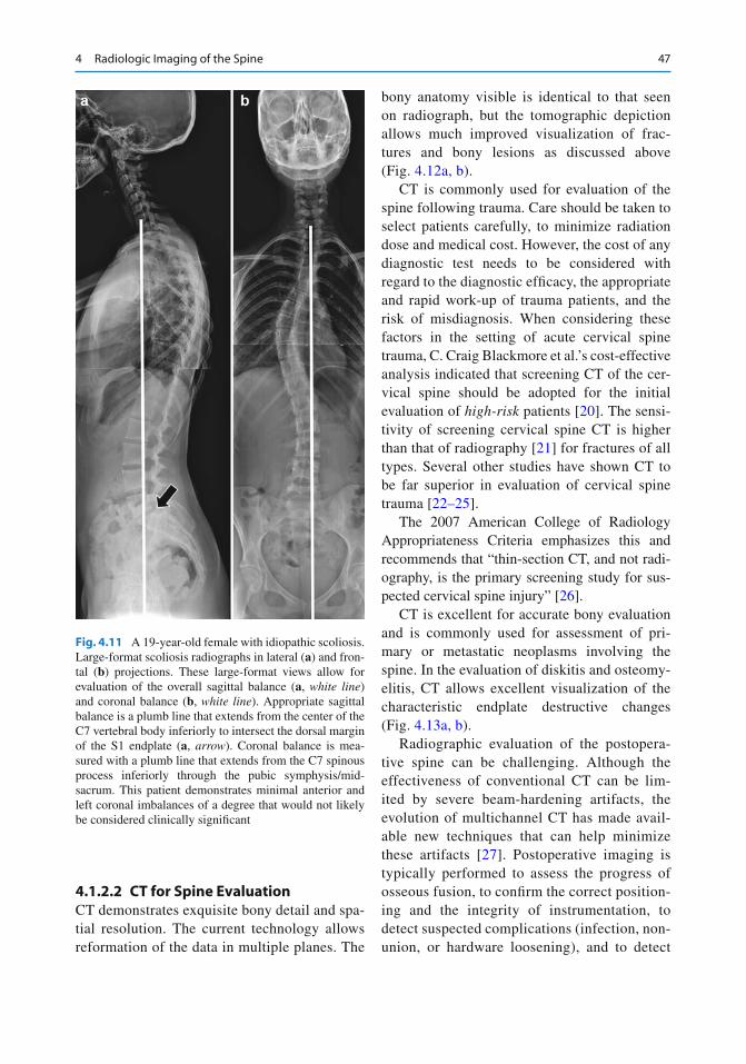
474 Radiologic Imaging of the Spine
4.1.2.2 CT for Spine Evaluation CT demonstrates exquisite bony detail and spa-tial resolution. The current technology allows reformation of the data in multiple planes. The
bony anatomy visible is identical to that seen on radiograph, but the tomographic depiction allows much improved visualization of frac-tures and bony lesions as discussed above (Fig. 4.12a, b ).
CT is commonly used for evaluation of the spine following trauma. Care should be taken to select patients carefully, to minimize radiation dose and medical cost. However, the cost of any diagnostic test needs to be considered with regard to the diagnostic ef fi cacy, the appropriate and rapid work-up of trauma patients, and the risk of misdiagnosis. When considering these factors in the setting of acute cervical spine trauma, C. Craig Blackmore et al.’s cost-effective analysis indicated that screening CT of the cer-vical spine should be adopted for the initial evaluation of high-risk patients [ 20 ] . The sensi-tivity of screening cervical spine CT is higher than that of radiography [ 21 ] for fractures of all types. Several other studies have shown CT to be far superior in evaluation of cervical spine trauma [ 22– 25 ] .
The 2007 American College of Radiology Appropriateness Criteria emphasizes this and recommends that “thin-section CT, and not radi-ography, is the primary screening study for sus-pected cervical spine injury” [ 26 ] .
CT is excellent for accurate bony evaluation and is commonly used for assessment of pri-mary or metastatic neoplasms involving the spine. In the evaluation of diskitis and osteomy-elitis, CT allows excellent visualization of the characteristic endplate destructive changes (Fig. 4.13a, b ).
Radiographic evaluation of the postopera-tive spine can be challenging. Although the effectiveness of conventional CT can be lim-ited by severe beam-hardening artifacts, the evolution of multichannel CT has made avail-able new techniques that can help minimize these artifacts [ 27 ] . Postoperative imaging is typically performed to assess the progress of osseous fusion, to con fi rm the correct position-ing and the integrity of instrumentation, to detect suspected complications (infection, non-union, or hardware loosening), and to detect
Fig. 4.11 A 19-year-old female with idiopathic scoliosis. Large-format scoliosis radiographs in lateral ( a ) and fron-tal ( b ) projections. These large-format views allow for evaluation of the overall sagittal balance ( a , white line ) and coronal balance ( b , white line ). Appropriate sagittal balance is a plumb line that extends from the center of the C7 vertebral body inferiorly to intersect the dorsal margin of the S1 endplate ( a , arrow ). Coronal balance is mea-sured with a plumb line that extends from the C7 spinous process inferiorly through the pubic symphysis/mid-sacrum. This patient demonstrates minimal anterior and left coronal imbalances of a degree that would not likely be considered clinically signi fi cant
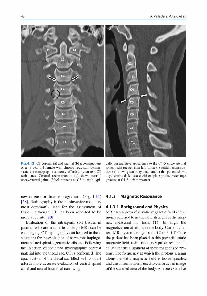
48 A. Valladares-Otero et al.
new disease or disease progression (Fig. 4.14 ) [ 28 ] . Radiography is the noninvasive modality most commonly used for the assessment of fusion, although CT has been reported to be more accurate [ 29 ] .
Evaluation of the intraspinal soft tissues in patients who are unable to undergo MRI can be challenging. CT myelography can be used in these situations for the evaluation of nerve root impinge-ment-related spinal degenerative disease. Following the injection of iodinated myelographic contrast material into the thecal sac, CT is performed. The opaci fi cation of the thecal sac fi lled with contrast affords more accurate evaluation of central spinal canal and neural foraminal narrowing.
4.1.3 Magnetic Resonance
4.1.3.1 Background and Physics MR uses a powerful static magnetic fi eld (com-monly referred to as the fi eld strength of the mag-net, measured in Tesla (T)) to align the magnetization of atoms in the body. Current clin-ical MRI systems range from 0.2 to 3.0 T. Once the patient has been placed in this powerful static magnetic fi eld, radio-frequency pulses systemati-cally alter the alignment of these magnetized pro-tons. The frequency at which the protons realign along the static magnetic fi eld is tissue speci fi c, and this information is used to construct an image of the scanned area of the body. A more extensive
a b
Fig. 4.12 CT coronal ( a ) and sagittal ( b ) reconstructions of a 43-year-old female with chronic neck pain demon-strate the tomographic anatomy afforded by current CT techniques. Coronal reconstruction ( a ) shows normal uncovertebral joints ( black arrows ) at C3–4, with typi-
cally degenerative appearance to the C4–5 uncovertebral joints, right greater than left ( circle ). Sagittal reconstruc-tion ( b ) shows great bony detail and in this patient shows degenerative disk disease with endplate productive change greatest at C4–5 ( white arrows )