Embed Size (px)
Citation preview


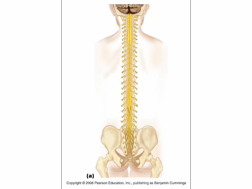

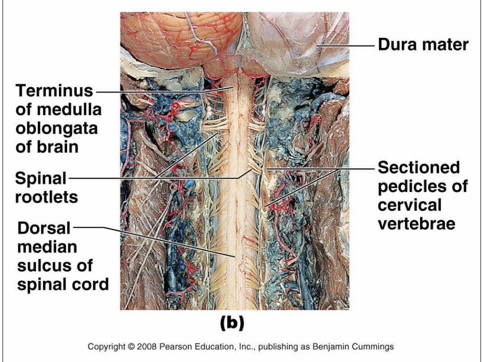
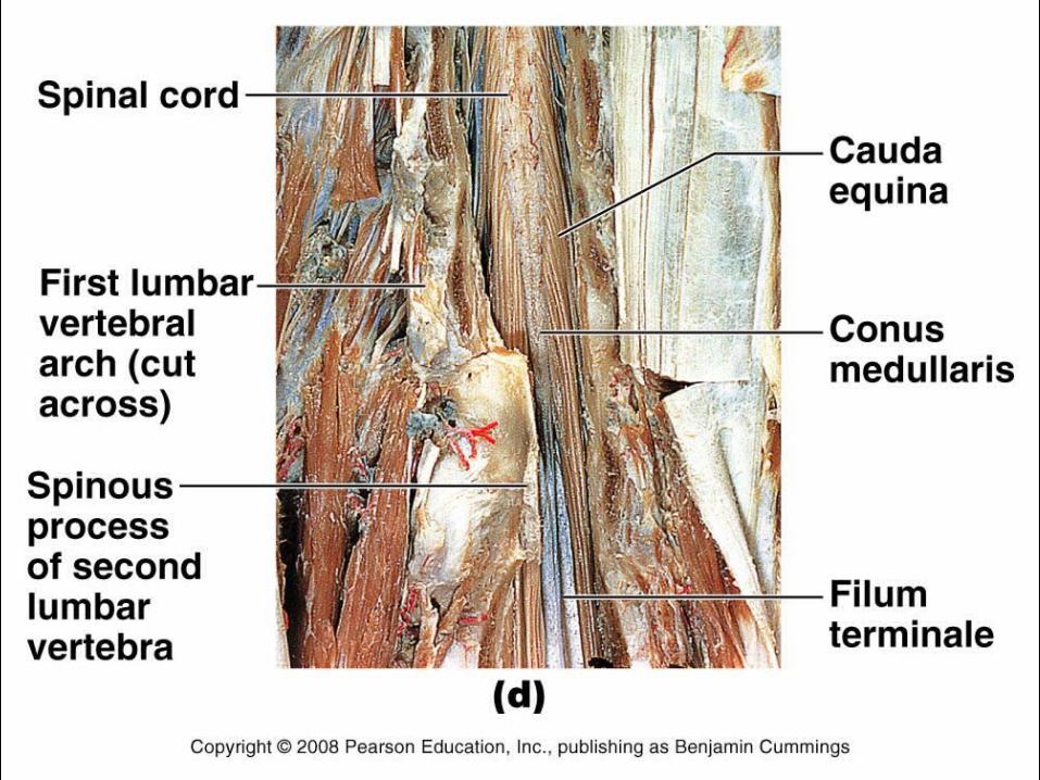
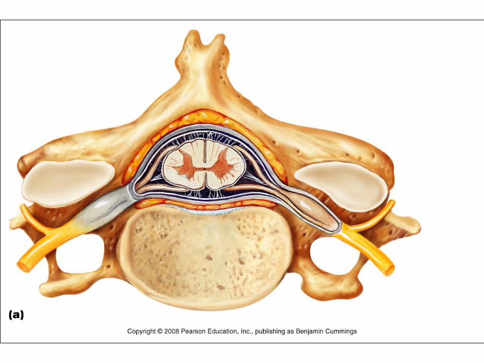
Spinal nerves• Part of the peripheral nervous system• 31 pairs attach through dorsal and ventral nerve roots• Lie in intervertebral foramina
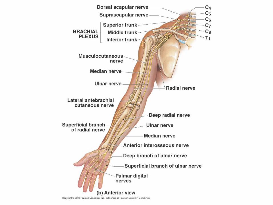

Musculo-cutaneous
Median
Ulnar
Axillary
Radial
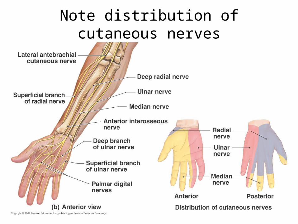
Note distribution of cutaneous nerves

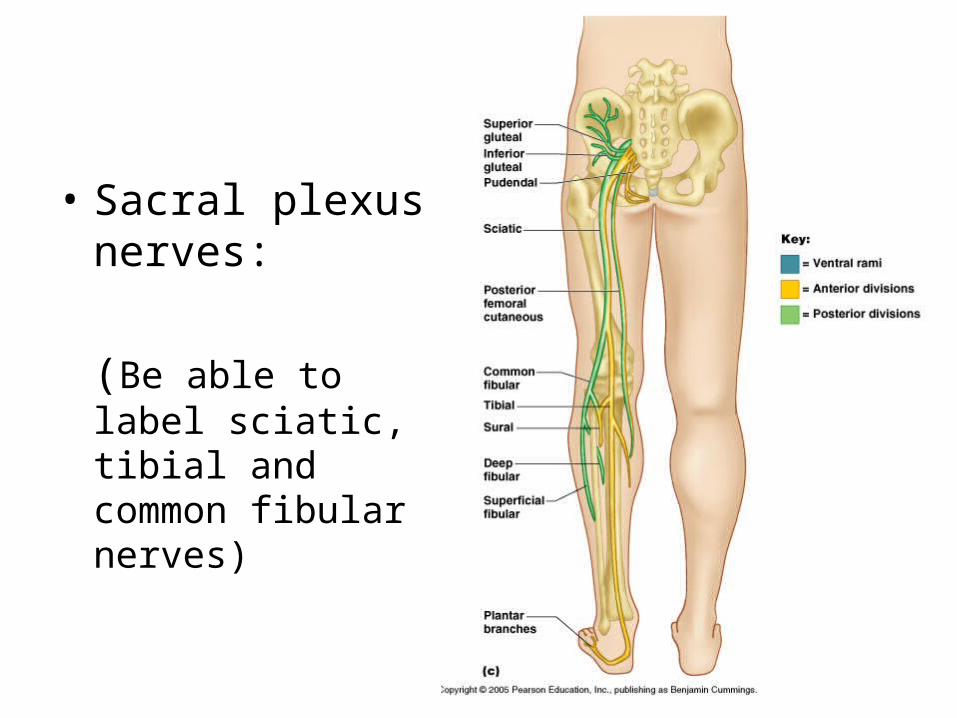
• Sacral plexus nerves:
(Be able to label sciatic, tibial and common fibular nerves)

Dermatomes (innervation of skin) Dermatomes
(area of skin innervated by the cutaneous branches from a single
spinal nerve is called a dermatome)
Reveal sites of damage to spinal
nerves or spinal cord


The Respiratory Organs
Conducting zone– Respiratory passages
that carry air to the site of gas exchange
– Filters, humidifies and warms air
Respiratory zone– Site of gas exchange– Composed of
• Respiratory bronchioles• Alveolar ducts• Alveolar sacs
Conducting zone labeled

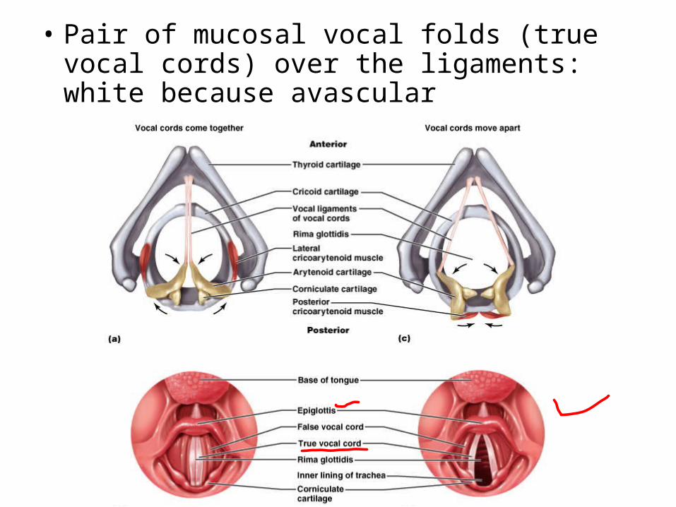
• Pair of mucosal vocal folds (true vocal cords) over the ligaments: white because avascular

Microscopic detail of alveoli• Alveoli surrounded by fine elastic fibers• Alveoli interconnect via alveolar pores• Alveolar macrophages – free floating “dust cells”• Note type I and type II cells and joint membrane


BOWMAN’S CAPSULE
• Look at the left side of the slide and notice the visceral and parietal layer (in the form of podocytes) of Bowman’s Capsule. Also look at the capillaries within the capsule.
• The other white areas are most likely proximal convoluted tubules.

Interlobar a.
Arcuate a.
Interlobular a.
Afferent a.
AFFERENT AND EFFERENT ARTERIOLES
• The upper portion of the slide where the arcuate arteries are and you may notice interlobar arteries coming up (on the right), then efferents.
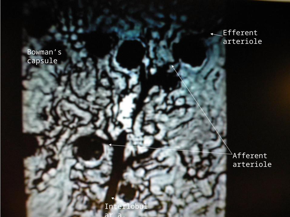
Bowman’s capsule
Interlobular a.
Afferent arteriole
Efferent arteriole

GLOMERULI
• This slide is stained with India ink.
• Notice the glomeruli and collecting ducts

Parietal layer of Bowman’s cap. Made of simple squamous epithelium
Visceral layer (podocytes)
filtrate
Capillary bed

BOWMAN’S CAPSULE
• A very close shot of the visceral and parietal layers . You can see the capillaries and the space where the filtrate is squeezed out, and then moving to the proximal convoluted tubules.
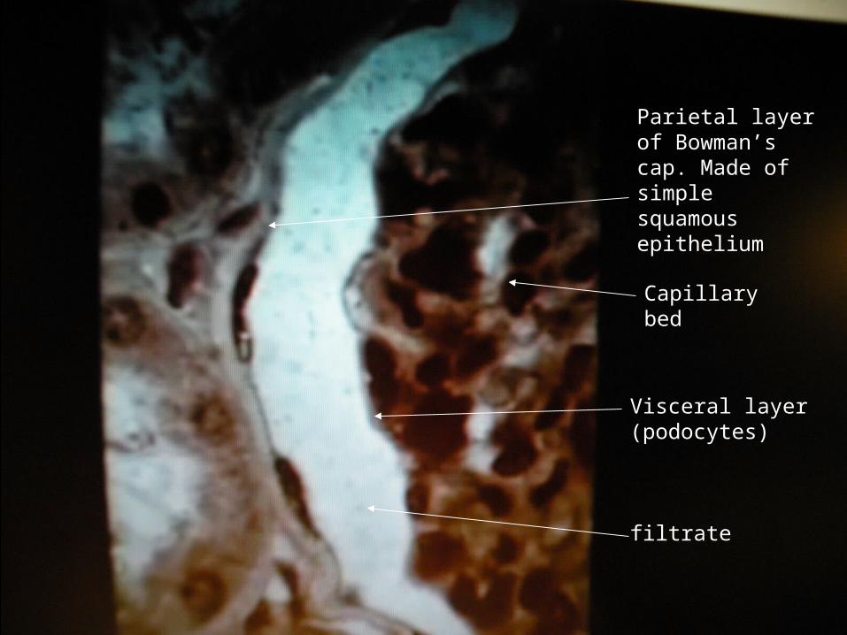
Parietal layer of Bowman’s cap. Made of simple squamous epithelium
Visceral layer (podocytes)
filtrate
Capillary bed
Afferent arteriole
Efferent arteriole
Proximal convoluted tubule


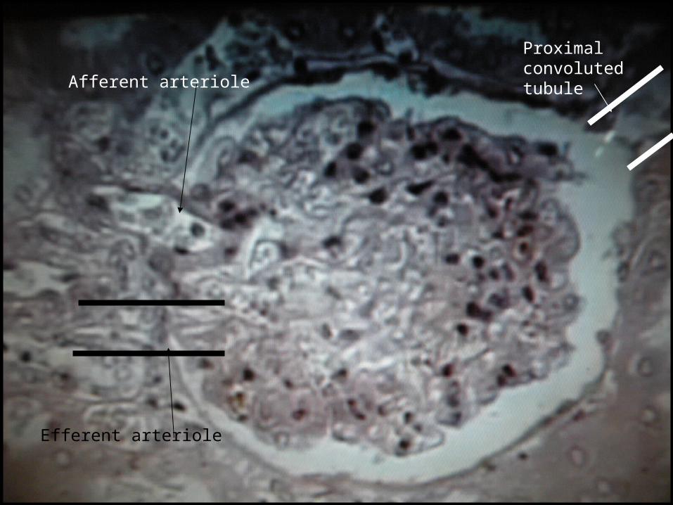
BOWMAN’S CAPSULE

Afferent arteriole
Efferent arteriole
Proximal convoluted tubule

AFFERENT AND EFFERENT ARTERIOLES
• On the left is either an afferent or efferent arteriole.
• It is very rare to find a good slide like this


PCT
• Notice the “brush border” on the PCT
• Brush border is a name given prior to advanced microscopy. They are now known to be microvilli
• 100ml of water needs to be reabsorbed here per minute! That’s why the surface area needs to be increased


PCT
• Closer view


LOOP OF HENLE
• Beautiful and rare longitudinal view of the loop of Henle


COLLECTING DUCT


CROSS SECTION OF COLLECTING DUCT