Embed Size (px)
Citation preview
CASE REPORTS
Sp~na~ Dural Arteriovenous Cause of yelo thy
cdformation : A
W K Ng, MRCP*, S A Samad, FRCR**, C T Tan, FRCP*, * Division of Neurology, lDepartmentof Medicine, Faculty of Medicine, Universiti Malaya, Kuala Lumpur, ** Department of Radiology, Faculty of Medicine, Universiti Kebangsaan Malaysia, Kuala Lumpur
Spinal vascular malformation causing myelopathy is often underestimated. According to McCormick's classificatIon of spinal vascular malformation, spinal dural arteriovenous fistulas (SDAF) account for 75% of these abnormalities l . We report two cases of SDAF documented on spinal angiogram.
Case 1
A 47-year-old Chinese man presented with progressive spastic paraparesis associated with sphincteric loss of his bladder over three years. A year after the onset of illness, he was confined to his home. The impairment was associated with frequent episodes of painful flexor spasms.
Neurological examination showed spastic paraparesis of grade 4 power associated with hyperreflexia and cloIlUs in both lower limbs. There was loss of sensation to
all modalities from T8 dermatomes downwards. Examination of the upper limbs, cranial nerves and other systems was normal. Baseline blood
Med J Malaysia Vol 51 No 1 March 1996
investigations, plain X-rays of the thoraco-lumbar spine were normal. Cerebrospinal fluid microscopy and IgG index were normal.
The supine myelogram showed a tortuous dilated serpenginous filling defect at the thoraco-lumbar junction suggestive of a spinal vascular malformation. Spinal angiogram showed a single feeding vessel to the SDAF from the right T9 intercostal artery (Fig. 1).
Embolisation of the SDAF was done in Singapore. Although the patient continued to have spastic paraparesis at 9 months of follow-up, there was no further progression of the myelopathy following embolisation.
C(1J£S :2
A 60-year-old Sikh man with longstanding hypertension and diabetes mellitus presented with several episodes of transient paraparesis lasting from one to twelve hours duration over a two-month -period. This was associated with low back pain and urinary retention.
Examination during one such episode showed
151
CASE REPORTS
paraparesis with sensory loss at T9 downwards. There was hyperreflexia in the lower limbs and bilateral extensor plantar responses.
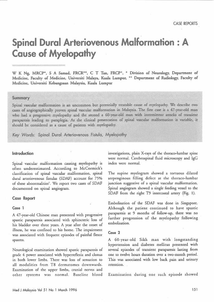
Routine blood investigations, cerebrospinal fluid studies and gadolinium enhanced MRI of the thoraco-Iumbar spine were normal. Supine myelogram showed serpenginous filling defects at the thoraco-lumbar region. Spinal angiogram showed a dural fistula at T8 intercostal artery consistent with a SDAF (Fig. 2).
This patient declined any further treatment and eventually developed paraplegia two months later.
(ljngiogrom showing ((ljtheteriso~ion T9 intercosiil:d ar~e!"y supplying ~he SDAF
152
Spinal dural arteriovenous fistulas (SDAF) is a relatively new pathological entity first described by Kendall and Logue in 19772 • It has been recognised as the most common type of spinal vascular malformation in adults, occurring most commonly in the thoracolumbar region. It is more common in men than women, with a male to female ratio of 8:1. The males affected are usually middle-aged or elderlyl.
We describe two cases of angiographically' proven spinal dural arteriovenous fistula at the thoracolumbar region. Both patients presented with symptoms of myelopathy with differing temporal sequence. The former case had a progressive spastic paraparesis and the latter recurrent episodes of transient paraparesis.
The variability of the presentation of SDAF is well recognised : this may be acute, subacute, episodic or progressive myelopathyl. The two cases described were consistent with the known demographic and clinical manifestation of SDAF.
The pathological features of SDAF are due to an acquired dural fistula communicating between a radicular artery ~nd vein located on the dural nerve sleeve. The varied clinical presentation is believed to be due to a combination of chronic venous hypertension and associated dural sinus thrombosis l . However, a more recent paper on a proven case of SDAF by angiography and pathology supported increased venous pressure as a mechanism of neurologic dysfunction3•
Supine myelograms performed on both patients showed serpenginous filling defects in the thoraco-Iumbar regions. However, in both patients the MRI with gadolinium enhancement failed to demonstrate these lesions. This is most likely related to the magnetic strength of the MRI machine which was 0.5 Tesla in both patients. SDAF was reported to be visible in only 25-63% of cases2 .
Both supine myelogram and MRI with gadolinium enhancement are usually performed to seek colloborative evidence prior to spinal angiography. Spinal angiography is a tedious procedure and has
Med J Malaysia Vol 51 No 1 March 1 996
significant morbidity with selective cannulation of the intercostal arteries, lumbar arteries and the vertebral arteries.
Interventional neuroradiology has an important role in the treatment of SDAF. The first case was treated with endovascular embolisation which halted the progression
1. Mc Corinick Pc. Spinal vascular malformations. Sem Neuro 1993;13 : 349-58.
2. Bradac BG, Daniele D, Riva A, Bracchi M, Stura G, Riccio A, Pagni CA. Spinal dural arteriovenous fistulas: an underestimated cause of myelopathy. Eur Neurol 1993;34 : 87-94.
CASE REPORTS
of the paraparesis. Unfortunately, the second patient declined treatment.
In conclusion, spinal dural arteriovenous fistula is a potentially treatable cause of myelopathy. Spinal angiography remains the gold standard for confirming or refuting the presence of SDAF.
3. Hurst RW, Kenyon LC, Lavi E, Raps EC, Marcotte P. Spinal dural arteriovenous fistula: The pathology of venous hypertensive myelopathy. Neurology 1995;45 : 1309-13.
Psychiatric Presentation of Huntington's Disease in a Malaysian Family
C N Chin, MRCPsych*, K H S'ng, FRCP**, G Philip, MBBS*, R Rosdinom, MMed(Psych)*, A Wahidah, MPath***, * Department of Psychiatry, Faculty of Medicine, Universiti Kebangsaan Malaysia, Jalan Raja Muda Abdul Aziz, 50300 Kuala Lurnpur, ** Department of Neurology, Kuala Lurnpur Hospital; Jalan Pahang, 50586 Kuala Lurnpur, ***Department of Pathology, Kuala Lumpur Hospital, Jalan Pahang, 50586 Kuala Lurnpur
Med J Malaysia Vol 51 No 1 March 1996 153