Embed Size (px)
Citation preview
Spi-1/PU.1 Oncoprotein Affects Splicing Decisions in aPromoter Binding-dependent Manner*
Received for publication, November 8, 2005, and in revised form, May 11, 2006 Published, JBC Papers in Press, May 11, 2006, DOI 10.1074/jbc.M512049200
Christel Guillouf1, Isabelle Gallais, and Francoise Moreau-Gachelin2
From the Institut Curie, INSERM U528, Paris 75248, France
The expression of the Spi-1/PU.1 transcription factor istightly regulated as a function of the hematopoeitic lineage. It isrequired for myeloid and B lymphoid differentiation. Whenoverexpressed inmice, Spi-1 is associatedwith the emergence oftransformed proerythroblasts unable to differentiate. In thecourse of a project undertaken to characterize the oncogenicfunction of Spi-1, we found that Spi-1 interacts with proteins ofthe spliceosome in Spi-1-transformedproerythroblasts andpar-ticipates in alternative splice site selection. Because Spi-1 is atranscription factor, it could be hypothesized that these twofunctions are coordinated. Here, we have developed a systemallowing the characterization of transcription and splicing froma single target. It is shown that Spi-1 is able to regulate alterna-tive splicing of a pre-mRNA for a gene whose transcription itregulates. Using a combination of Spi-1 mutants and Spi-1-de-pendent promoters, we demonstrate that Spi-1 must bind andtransactivate a givenpromoter to favor theuse of theproximal 5�
alternative site. This establishes that Spi-1 affects splicing deci-sions in a promoter binding-dependent manner. These resultsprovide new insight into how Spi-1 may act in the blockage ofdifferentiation by demonstrating that it can deregulate geneexpression and alsomodify the nature of the products generatedfrom target genes.
The transcription factor Spi-1/PU.1 plays an important rolein the coordination of hematopoiesis. It is required for thedevelopment of both myeloid and B lymphoid lineages (1, 2).The level of its expression is accurately regulated as a functionof hematopoietic development (3, 4). The relevance of the dif-ferential and tight regulation of spi-1 was demonstrated by theconsequences of its deregulation in adultmice. Indeed, reducedSpi-1 expression can be associated with the development ofacutemyeloid leukemia (5–7). In contrast, spi-1 transcriptionalactivation exhibits an oncogenic activity in the erythroid line-age in which it is not normally expressed (8). To better under-stand the role of spi-1 overexpression in the development oferythroleukemia, spi-1 transgenic mice were generated (9). In
these animals, forced Spi-1 activity blocks the differentiation ofproerythroblasts, leading to thedevelopmentof severeanemiaandhepatosplenomegaly. Subsequently, proerythroblasts acquire anabnormal proliferation ability; resulting in the development of anerythroleukemia. Spi-1 belongs to the ETS family of transcrip-tion factors. Its ETS domain recognizes DNA on a 5�-(A/G)GAA-3� core (10). In addition, Spi-1 contains an amino-ter-minal transactivation domain and a central PEST region (11).Spi-1 controls primarily the transcription of myeloid andlymphoid genes. Additionally, fli-1 is a direct target gene ofSpi-1 in the erythroid tissue (12). The transcriptional activity ofSpi-1 depends on its combinatorial association within multi-protein complexes. Some of these proteins are ubiquitous fac-tors such as the basal transcription factor TFIID (13) and theco-activator/integrator CREB-binding protein (cAMP-re-sponse element-binding protein) (14). Other Spi-1-interactingpartners are tissue-specific, such as the B lymphoid factorNF-EM5/Pip (15, 16), the myeloid regulators c-JUN (17),AML1 and C/EBP� (18), MafB (19), and the erythroid tran-scription factor GATA-1 (20, 21).Spi-1 has also been identified as a partner of proteins of the
spliceosome. It participates in the choice of alternative splice sitesby favoring the selection of the proximal 5�-splice site of E1Apre-mRNA (22). Translocated in liposarcoma (TLS)3 is one of theSpi-1 partners that is able to recognize RNA and to act in RNAsplicing (22, 23). Spi-1 counterbalances the effect of TLS in theselection of alternative 5�-splice sites in erythroid cells. Its func-tion in splicing as well as its interference with TLS requires theDNA binding domain (DBD) associated with the transactiva-tion domain or the PEST region (24). The function of somesplicing factors requires an ability to identify intronic or exonicRNA elements within pre-mRNAs. Although the DBD of Spi-1is able to interact with poly(A)� RNAs and homoribonucle-otide poly(G) polymers (25), Spi-1 does not exhibit RNA recog-nition specificity (from a SELEX strategy).4 Thus, it is unlikelythat the role of Spi-1 in splicing proceeds via recognition of aspecific RNA sequence.Transcription and splicing are coordinated events (26). This
coordination appears to be mediated, in part, by the COOH-terminal domain of RNA polymerase II, which recruits splicingfactors to transcription sites. Furthermore, it has been demon-strated that splicing of a transcript can be modified by changes
* This work was supported by the Institut National de la Sante et de la Recher-che Medicale (INSERM) and Institut Curie, the Ligue contre le Cancer(Comite de Paris), and the Association pour la Recherche contre le Cancer.The costs of publication of this article were defrayed in part by the pay-ment of page charges. This article must therefore be hereby marked“advertisement” in accordance with 18 U.S.C. Section 1734 solely to indi-cate this fact.
1 To whom correspondence may be addressed: 26 rue d’Ulm, 75248 ParisCedex 05, France. Tel.: 33-1-42-34-66-48; Fax: 33-1-42-34-66-50; E-mail:[email protected].
2 To whom correspondence may be addressed. E-mail: [email protected].
3 The abbreviations used are: TLS, translocated in liposarcoma; DBD, DNAbinding domain; CMV, cytomegalovirus; MT, Myc tag; RT, reverse tran-scriptase; EMSA, electrophoretic mobility shift assay; IRES, internal ribo-some entry site; SR protein; Ser/Arg-rich protein; wt, wild-type.
4 A. Lerga and F. Moreau-Gachelin, unpublished data.
THE JOURNAL OF BIOLOGICAL CHEMISTRY VOL. 281, NO. 28, pp. 19145–19155, July 14, 2006© 2006 by The American Society for Biochemistry and Molecular Biology, Inc. Printed in the U.S.A.
JULY 14, 2006 • VOLUME 281 • NUMBER 28 JOURNAL OF BIOLOGICAL CHEMISTRY 19145
by guest on March 22, 2018
http://ww
w.jbc.org/
Dow
nloaded from

in the promoter-driven transcription (27, 28). This led to theidea that transcription factors could regulate subsequent proc-essing events. In fact, this notion has been further supported bythe observation that several transcription factors interact withproteins of the spliceosome and/or display dual functions insplicing and transcription. Among these factors are p54nrb (29),YB-1 (30), and p52 (31) (for additional listing of candidateslinking transcription and splicing, see Ref. 26). It is onlyrecently, however, that a direct role for a factor in the co-regu-lation of splicing and transcription through the promoter hasbeen described (32). Nuclear hormone receptors have also beenshown to affect splicing decisions in a promoter-dependentmanner (33). Indeed, alternatively spliced variants are selectedaccording to the nature of co-activators recruited to the pro-moters by nuclear hormone receptors (34, 35). Because Spi-1 isalso a transcription factor, it can be hypothesized that if the twoSpi-1 functions are coordinated, the action of Spi-1 in splicingmay proceed via DNA recognition. To examine this possibility,we investigated whether Spi-1 modifies the splicing of apre-mRNA for a gene whose transcription it controls.Here, we provide evidence that the Spi-1 protein is able to
modify alternative splicing of the E1A pre-mRNA expressedfrom Spi-1-dependent promoters. Using a combination ofSpi-1mutants and Spi-1-dependent promoters, it is shown thatSpi-1 modifies splicing as a function of its ability to bind DNA.Indeed, Spi-1 must bind and transactivate a given promoter tofavor the use of the proximal 5� alternative site. Moreover, it isdemonstrated that this effect is not due to the modulation oftarget mRNA transcription levels but depends on qualitativeeffect of Spi-1 in splicing. These results establish that Spi-1affects splicing decisions in a promoter binding-dependentmanner.
EXPERIMENTAL PROCEDURES
Cell Lines and Cell Culture—HeLa cells were maintained inDulbecco’s modified Eagle’s medium (Invitrogen) supple-mented with 10% fetal calf serum, penicillin/streptomycin, andL-glutamine (Invitrogen).Plasmid Constructs—pCS3-MT, pCS3-MT-Spi-1, and pCS3-
E1Awere previously described (22, 23). The CMVpromoter con-tained in the pCS3 vector corresponds to the CMVEI94sequence. pCS3-MT-DBD, pCS3-MT-�101, and pCS3-MT-�Cter were generated by PCR amplification of the appropriateregions from the spi-1 cDNA and insertion in the pCS3-MTvector. The pCS3-MT-��4mutant, with amino acids 250–254deleted in the pCS3-MT expression vector, was obtained bymutagenesis of the wild-type spi-1 cDNA using theQuikChange site-directed mutagenesis system (Stratagene)according to the manufacturer’s recommendations. The fol-lowingmutagenic oligonucleotides were used for PCR: forwardprimer, 5�-GAAGAAAGTCAAGAAGAAGAGCGGCGAG-GTGCTG-3�; reverse primer, 5�-CAGCACCTCGCCGCTCT-TCTTCTTGACTTTCTTC-3�. In all constructs, a nuclearlocalization signal and 6 copies of a Myc epitope (MT) wereadded to theNH2-terminal part of the proteins asmentioned inRef. 22. The fes binding sites and the fli-1 (�270/�41) promot-ers were previously described (12, 36). In the case of the fespromoter, a thymidine kinase minimal promoter was added
downstream from the fes target element. These sequencesreplaced the CMV promoter in the pCS3-E1A vector. The neo-mycin resistance sequence from pIRESNeo (Clontech) wasreplaced by the firefly luciferase sequence derived from thepGL2 basic luciferase reporter (Promega). All clonings wereverified by sequencing. Detailed cloning procedures are avail-able on request.Transfections—Cells (0.5 � 106) were transfected with Lipo-
fectamine reagent (Invitrogen) according to themanufacturer’sinstructions. The Lipofectamine Plus/DNAmixture was left oncells for 5 h. The plasmid DNA quantities used were as follows:100 ng for the pE1A-IRES-LucF, 10 ng for the CMVE1A vec-tors, and 20–500 ng for the different Spi-1 expression vectors.When transcriptional or splicing effects of different amounts ofreporter vectors or spi-1 expression vectorswere compared, thetotal quantities of DNA were equalized using pBluescript(Stratagene) or pCS3-MT plasmids, respectively. Transfectionefficiencies were normalized by co-transfection of a pCMV-Renilla luciferase (Promega) reporter vector (10 ng). The CMVpromoter of the pCMV-Renilla luciferase vector does not con-tain any characterized or putative Spi-1 binding sites. Cellswere harvested 24 h post-transfection. Transfected cells wereseparated into three parts: 1/5 to measure luciferase activity,1/10 to analyze protein expression byWestern blotting, and therest was used to extract total RNA.Luciferase Activity Measurement—Luciferase activity reflects
the accumulation of RNA following both the transcription anddegradation of RNA. Because the various spliced forms of E1AmRNA appear not to differ in terms of degradation (Ref. 37 andthe comparison of RNAaccumulation usingmonocistronic andbicistronic vectors), “transcriptional activity” in the text standsfor RNA accumulation. Twenty-four hours post-transfection,1/5 of the cell pellets were lysed and the firefly (LucF) andRenilla (LucR) luciferase activities weremeasuredwith the dualluciferase kit (Promega) according to the manufacturer’sinstructions. The -fold induction of LucF was calculated afternormalization to LucR activities.RNA Purification and RT-PCR Analysis—Total RNA was
prepared and treated with DNase I (Qiagen) as previouslydescribed (22). RNA was reverse transcribed with Moloneymurine leukemia virus Superscript II reverse transcriptase(Invitrogen) in the presence of 50�MdNTP and 2 pmol of 3�RTE1A primer. PCR to study the splicing profile was performedwith Taq DNA polymerase (PerkinElmer Life Sciences) in thepresence of a 5� E1A primer that was 5� end-labeled with T4polynucleotide kinase (Roche) and [�-32P]ATP (AmershamBiosciences), as described elsewhere (22). The number of PCRcycles was kept to a minimum (18–22 cycles) to detect signalswithin the linear range of the assay. Control RT-PCR containeda RNA template that had not undergone reverse transcription.E1A RT-PCR products were resolved on 6% polyacrylamide-urea gels, autoradiographed, and quantified with ImageQuanton a GE Healthcare PhosphorImager. All transfection experi-ments were repeated at least three times. Semi-quantitativeRT-PCR was performed with two independent dilutions ofRNA that had been reverse-transcribed using primers amplify-ing all E1A forms (Fig. 1A, black arrows). The amount ofreverse-transcribed RNA used for PCR was calculated as a
Coordination of Splicing and Transcription by Spi-1/PU.1
19146 JOURNAL OF BIOLOGICAL CHEMISTRY VOLUME 281 • NUMBER 28 • JULY 14, 2006
by guest on March 22, 2018
http://ww
w.jbc.org/
Dow
nloaded from

function of transfection efficiencies measured using LucRactivity. The two RNA dilutions and the number of PCR cycles(30) used were within a proportional range of the assay. Thesequences of the primers are: forward primer, 5�-TCAGCTG-GTCCAAAAGACTG-3� and reverse primer, 5�-CAAGCTT-GATTTAGGTGA-3�. RT-PCR products were resolved on 1%agarose gels and stained with ethidium bromide.Immunoblotting—Proteins were boiled in sample loading
buffer (62 mM Tris, pH 6.8, 2% SDS, 10% glycerol, 0.1% brom-phenol blue, 100 mM dithiothreitol), then separated by 10%SDS-PAGE and electrotransferred onto Hybond nitrocellulosemembranes (Amersham Biosciences). Membranes wereblocked with 5% nonfat drymilk in PBS, 0.1% Tween 20 (PBST)before incubation with the monoclonal 9E10 antibody directedagainst theMyc epitope (Santa Cruz Biochemicals, Santa Cruz,CA) in 5% nonfat dry milk/PBST. After three washes in PBST,the membrane was incubated with horseradish peroxidase-conjugated secondary antibody and finally washed in PBS/Tween. The proteins were visualizedwith the enhanced chemi-luminescence Western blotting detection system (AmershamBiosciences).Nuclear Extracts and Electrophoretic Mobility Shift Assay
(EMSA)—For the DNA binding assay, the various Spi-1 pro-teins were translated in vitro from CS3 vectors using TNT-cou-pled reticulocyte lysates (Promega). Nuclear extracts were pre-pared from 6 � 106 transfected HeLa cells. Nuclei werecollected by centrifugation after incubation for 20min on ice in200 �l of buffer containing 20 mM HEPES (pH 7.6), 20% glyc-erol, 10 mM NaCl, 1.5 mM MgCl2, 0.2 mM EDTA (pH 7.5), 0.1%Triton X-100, 1 mM dithiothreitol, and protease inhibitor mix-ture tablets (Roche). The nuclei were resuspended in 80 �l ofthe same buffer containing 500 mMNaCl and incubated for 1 hon ice. The sampleswere centrifuged at 45,000� g for 15min at4 °C to recover the supernatants corresponding to nuclearextracts.The sequences of the wt fes (fes-wt) and mutated fes (fes-mut)
probes were the following: fes-wt, 5�-GAGGAAGCGCGGAA-TCAGGAACTGGCCGGGGC-3�, and fes-mut, 5�-GAGGAAG-CGCGGAATCACCAACTGGCCGGGGC-3�. The sequences ofthe CMV probes were: seqA, 5�-TATAGTATTTCCATATAT-GGGTTTTCCTATTGACG; seqB, 5�-CCATATATGGGGCT-TCCTAATACCGCCCATA; and seqC, 5�-TATATATGGT-CTTTCCTATTGACGTCAT. The nucleotides recognized bySpi-1 are in bold characters. The probes were 5�-labeled withT4 polynucleotide kinase (Roche) and [�-32P]ATP (5000Ci/mmol). EMSAwere performed as previously described (36).For the supershift assay, the monoclonal 9E10 antibody (SantaCruz Biochemicals) was added to the binding reaction beforeaddition of the probes. The DNA-protein complexes were sub-mitted to electrophoresis on native 6% polyacrylamide gels in0.5� Tris borate-EDTA (TBE) buffer and autoradiographed.
RESULTS
Spi-1 Is Able to Control Splicing of a Pre-mRNA for a GeneWhose Transcription It Regulates—To determine whetherSpi-1 controls the splicing of a pre-mRNA for a gene whosetranscription it controls, we developed a model system using avector that expresses a bicistronic pre-mRNA encoding the
E1A minigene and a luciferase gene. This vector (pfes-E1A-IRES-LucF or pfli-1-E1A-IRES-LucF) allows the characteriza-tion of transcription and RNA maturation simultaneously forthe same gene. Two types of spi-1 target promoters were usedto control themRNA expression; one derived from themyeloidfes gene (36) and the other from the hematopoietic fli-1 gene(12). The fes box consists of 3 Spi-1 DNA binding sites plus theminimal thymidine kinase promoter (Fig. 1A). The fli-1sequence includes the minimal promoter of the fli-1 gene con-taining 2 Spi-1 DNA binding sites (Fig. 2A). The pfes or pfli-E1A-IRES-LucF construct was transfected together withincreasing amounts of Spi-1 expression vector in HeLa cells,which do not express the genomic spi-1 gene (data not shownand Fig. 3). For each sample, the luciferase activity was meas-ured, the Spi-1 expression was analyzed by Western blot, andthe E1A isoforms were amplified by radioactive RT-PCR, sepa-rated by electrophoresis, and quantified by phosphorimaging.First, we controlled that the luciferase activity, encoded by
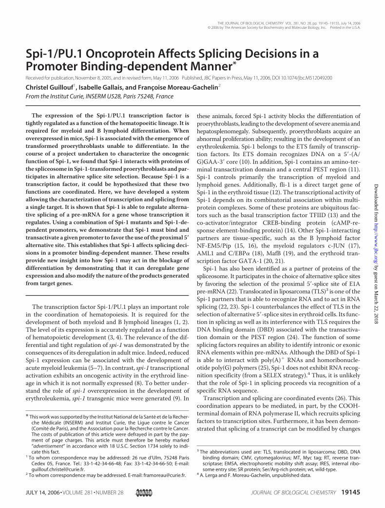
the samemRNAmolecule as the E1A, reflects the level of RNAtranscribed. E1A transcription was analyzed by semi-quantita-tive RT-PCR of all E1A RNA isoforms, spliced or not (see theposition of the primers in Fig. 1A), and comparedwith the lucif-erase activity. In all experiments, the luciferase activity and thequantity of RNA used for semi-quantitative RT-PCR were nor-malized to the transfection efficiency. The results obtainedwiththe fes reporter vector are shown in Fig. 1B. An increase in E1Apre-mRNA transcription by Spi-1 as revealed by semi-quanti-tative RT-PCR (compare the band intensities for control cells toSpi-1-transfected cells) was correlated with an increase in lucif-erase activity (9-fold), establishing that the luciferase activitycorrectly reflects the amount of RNA transcribed. Thus, weused this system to further investigate Spi-1 function in tran-scription and splicing. As shown in Fig. 1B, Spi-1 enhancedtranscription of the minigene driven by the fes box in a dose-dependent manner as measured by luciferase activities. Thetranscription was increased 9-, 13-, or 15-fold according to theamount of Spi-1 expression vector transfected. The presence ofthree alternative 5�-splice sites in the first exon of the E1Apre-mRNAgives rise to threemajormRNA isoforms, 13 S, 12 S,and 9 S, transcribed from theE1Aminigene (Fig. 1A). TwoRNAisoforms (11 S and 10 S)were also generated thatwere not takeninto account because they arise from double splicing events(38). Fig. 1C presents the splicing profiles of the E1A mRNA.The histograms represent the relative proportion of 13 S, 12 S,and 9 S E1A isoforms. The decrease in the proportion of the 9 Sisoform in Spi-1-transfected HeLa cells relative to thatobserved in HeLa cells transfected with the empty vector wereindicated under each histogram as % 9 S decrease versus con-trol. The increase of E1A minigene transcription by Spi-1 wasassociated with a decrease in the proportion of the E1A 9 Sisoform (up to 70% compared with control cells), whereas theproportion of the 13 S and 12 S isoforms increased (Fig. 1C).These results revealed that Spi-1 promotes the use of the prox-imal 5�-splice site of an E1A pre-mRNA whose transcription isdriven by a Spi-1-responsive promoter. Similar experimentswere performed with the vector containing the fli-1 promoter(Fig. 2). Once again, E1A transcription was enhanced in Spi-1-transfectedHeLa cells up to 4.7-fold compared with cells trans-
Coordination of Splicing and Transcription by Spi-1/PU.1
JULY 14, 2006 • VOLUME 281 • NUMBER 28 JOURNAL OF BIOLOGICAL CHEMISTRY 19147
by guest on March 22, 2018
http://ww
w.jbc.org/
Dow
nloaded from
fected with the empty vector (Fig. 2B). With regard to E1Asplicing, as seen for the fes promoter, the relative proportion ofthe 9 S isoform decreased as a function of the Spi-1 expressionlevel, up to 54% of the 9 S proportion observed in control cells(Fig. 2C). These experiments were also performed in co-trans-fection assays using vectors carrying a monocistronicpre-mRNA encoding either the luciferase gene or the E1Aminigene under control of the fes or fli-1 promoters (data notshown). The variations in both luciferase activity andE1A splic-ing relative to Spi-1 expression levels were similar to thoseobserved in the assay using the bicistronic vectors, demonstrat-ing that the IRES did not interfere with the role of Spi-1 intranscription and splicing. Our results demonstrate that Spi-1is able to regulate the alternative splicing of a gene whose tran-scription it regulates.Splicing Effect of Spi-1 Is Determined by Its Ability to Bind to
the Promoter Driving the Transcription of the Pre-mRNA—Thefact that Spi-1modulated the splicing of a pre-mRNA for a genewhose transcription it controls raised the possibility that Spi-1binding to DNA was necessary to affect splicing decisions.Thus, to examine this question, we set up a strategy based onthe swapping of promoters driving the transcription of thebicistronic pre-mRNA encoding the E1A minigene and theluciferase gene. Because we did not find mutations that com-pletely abolished the binding of Spi-1 to the fli-1 promoter, wedecided to focus on the fes promoter.We have previously described the minimal fes recognition
sequence of Spi-1 as 5�-AGGAA-3�, and shown that thereplacement of the two Gly by two Cys abolished binding ofSpi-1 (36). We performed EMSA using the fes-wt and fes-mutprobes and nuclear extracts from transiently transfected HeLacellswith the Spi-1 expression vector (Fig. 3). As a control, Spi-1protein generated in reticulocyte lysates was used. As seen inFig. 3, Spi-1 that was contained in nuclear extract from HeLacells transfected with an Spi-1 expression vector induced a shiftof the fes-wt probe but not the fes-mut probe in agreement withthe fact that Spi-1 did not bind to the mutated fes sequence,5�-ACCAA-3�. Thus, a bicistronic expression vector containingthree mutated fes oligonucleotides in the promoter was con-structed (pfes-mut-E1A-IRES-LucF). It was used in transactiva-tion and splicing assays to determinewhether the effect of Spi-1on splicing was different when it could no longer bind to thepromoter.The transcriptional activity of Spi-1 was measured in HeLa
cells transfected with a Spi-1 expression vector and either thepfes-wt-E1A-IRES-LucF or pfes-mut-E1A-IRES-LucF targetvector. As reported above, transcription levels were evaluatedby semi-quantitative RT-PCR of E1A RNA and by luciferaseactivity. Spi-1 expressionwas determined in transfected cells byWestern blotting. It can be seen in Fig. 4A that the luciferase
FIGURE 1. Transcriptional and splicing activities of the wild-type Spi-1protein on the E1A minigene whose transcription is driven by Spi-1responsive elements of the fes gene promoter. HeLa cells were co-trans-fected with increasing amounts of Spi-1 expression vector as indicated, 100ng of the vector carrying the bicistronic pre-mRNA (pfes-wt-E1A-IRES-LucF),and 10 ng of the pCMV-LucR normalization vector. A, the diagram shows thestructure of the bicistronic vector driven by fes sequences. Three majormRNAs are generated from E1A cDNA by alternative 5�-splice site selection.The dotted line represents the intron. Black arrows indicate the primers usedfor semi-quantitative RT-PCR for the amplification of all E1A RNA forms. Thegray arrows indicate the primers used to distinguish the different splicedforms of E1A RNAs by their size. B, effect of Spi-1 protein overexpression ontranscription as measured by the dual luciferase assay. The histograms givethe relative light units normalized to transfection efficiency. The -fold induc-tion is relative to the luciferase activity obtained in control cells transfectedwith empty expression vector. Semi-quantitative RT-PCR of total E1A RNAmigrated on the agarose gel is shown. Expression of transfected Spi-1 pro-teins analyzed by immunoblotting with the anti-Myc epitope monoclonalantibody (�MT ) is shown. ND, not determined. C, effect of Spi-1 proteinexpression on the choice of 5� E1A alternative splice sites. Positions of the
unspliced E1A pre-mRNA (us), 13 S, 12 S, 11 S, 10 S, and 9 S RNAs are indicated.Autoradiograms of the radiolabeled PCR products obtained in a representa-tive experiment are shown. Histograms represent the percentage of 13 S, 12S, and 9 S mRNA isoforms. The % 9 S decrease is calculated from the propor-tion of 9 S isoform in the indicated Spi-1-transfected cells relative to the pro-portion of the 9 S isoform in cells transfected with the empty vector. Mean �S.D. were calculated from three independent experiments with duplicatesamples.
Coordination of Splicing and Transcription by Spi-1/PU.1
19148 JOURNAL OF BIOLOGICAL CHEMISTRY VOLUME 281 • NUMBER 28 • JULY 14, 2006
by guest on March 22, 2018
http://ww
w.jbc.org/
Dow
nloaded from

activity was enhanced 7-fold by Spi-1 when the transcriptionwas driven by the fes-wt promoter, whereas it was similar in theabsence and presence of Spi-1 when transcription of luciferasewas driven by the fes-mut promoter (Fig. 4A). Similarly, semi-quantitative RT-PCR of E1A RNA showed that Spi-1 increasedE1A RNA expression from pfes-wt-E1A-IRES but not from thepfes-mut-E1A-IRESLucF vector (Fig. 4A). Similar results wereobtained with the monocistronic vectors (data not shown).Next, we investigated the splicing effect of Spi-1 on E1A in thesame transfected samples. All the experiments presented usedthe bicistronic E1A vector. As described in Fig. 1, when the E1Aminigene was transcribed from the fes-wt promoter, Spi-1modified the splicing pattern, resulting in a decrease in the pro-portion of the 9 S isoform (up to 40% of the 9 S proportionfound in control cells), and an increase in the 13 S RNA iso-forms (Fig. 4B, left part). When transcription of the E1A mini-gene was driven by the fes-mut promoter, Spi-1 did not favorthe use of themost proximal 5� alternative site but slightly rein-forced the use of the distal 5� splice site (Fig. 4B, right part), asdeduced from the 20% increase in the proportion of the 9 Sisoform compared with the control. These results demonstratethat Spi-1 affects splicing decisions as a function of its ability tobind to the promoter.It should be noted that the effects of Spi-1 on the splicing of
the E1A target whose transcription was driven by the fes andfli-1 promoters was qualitatively similar to those previously
FIGURE 2. Transcriptional and splicing activities of the wild-type Spi-1protein on an E1A minigene whose transcription is driven by the fli-1gene promoter. HeLa cells were co-transfected with increasing amounts ofSpi-1 expression vector as indicated, 100 ng of the vector carrying the bicis-tronic pre-mRNA (pfli-wt-E1A-IRES-LucF) and 10 ng of the pCMV-LucR nor-malization vector. A, the diagram shows the structure of the bicistronic vectordriven by the fli-1 promoter. The gray arrows indicate the primers used todistinguish the different spliced forms of E1A RNAs by their size. B, effect ofSpi-1 protein overexpression on transcription as measured by the dual lucif-erase assay. The histograms give the relative light units normalized to thetransfection efficiency. The -fold induction is relative to the luciferase activityobtained in control cells transfected with the empty expression vector.Expression of transfected Spi-1 proteins analyzed by immunoblotting withthe anti-Myc epitope monoclonal antibody is shown. C, effect of Spi-1 proteinexpression on the choice of the 5� E1A alternative splice sites. Positions of theunspliced E1A pre-mRNA (us), 13 S, 12 S, 11 S, 10 S, and 9 S RNAs are indicated.Autoradiograms of the radiolabeled PCR products obtained in a representa-tive experiment are shown. Histograms represent the percentage of 13 S, 12S, and 9 S mRNA isoforms. The % 9 S decrease versus control cells are indicatedunder each histogram. Mean � S.D. were calculated from three independentexperiments with duplicate samples.
FIGURE 3. DNA binding analysis of the Spi-1 protein to the Spi-1 wt andmutated binding site of the fes promoter in HeLa-transfected cells. HeLacells were transfected with 20 ng of Spi-1 expression vector and the nuclearprotein extracts were prepared from spi-1-transfected cells. The sequences ofthe oligonucleotide probes used and the mutated nucleotides are describedunder “Experimental Procedures.” DNA-protein complexes were separatedby EMSA and analyzed by autoradiography. The shift obtained with the invitro produced Spi-1 was shown as a control (TNT Spi-1). Nuclear extract (NE)CS3 corresponds to the nuclear proteins extracted from HeLa cells transfectedwith the CS3 empty vector. NE Spi-1 are nuclear protein extracts from HeLacells transfected with vector expressing wt-Spi-1. The lane corresponding tothe competition with the anti-Myc epitope antibody is noted Antimyc comp.
Coordination of Splicing and Transcription by Spi-1/PU.1
JULY 14, 2006 • VOLUME 281 • NUMBER 28 JOURNAL OF BIOLOGICAL CHEMISTRY 19149
by guest on March 22, 2018
http://ww
w.jbc.org/
Dow
nloaded from

observed for the E1A target whose transcription was driven byCMV (24). In view of the different Spi-1 splicing effectsdescribed here, it was necessary to further characterize the
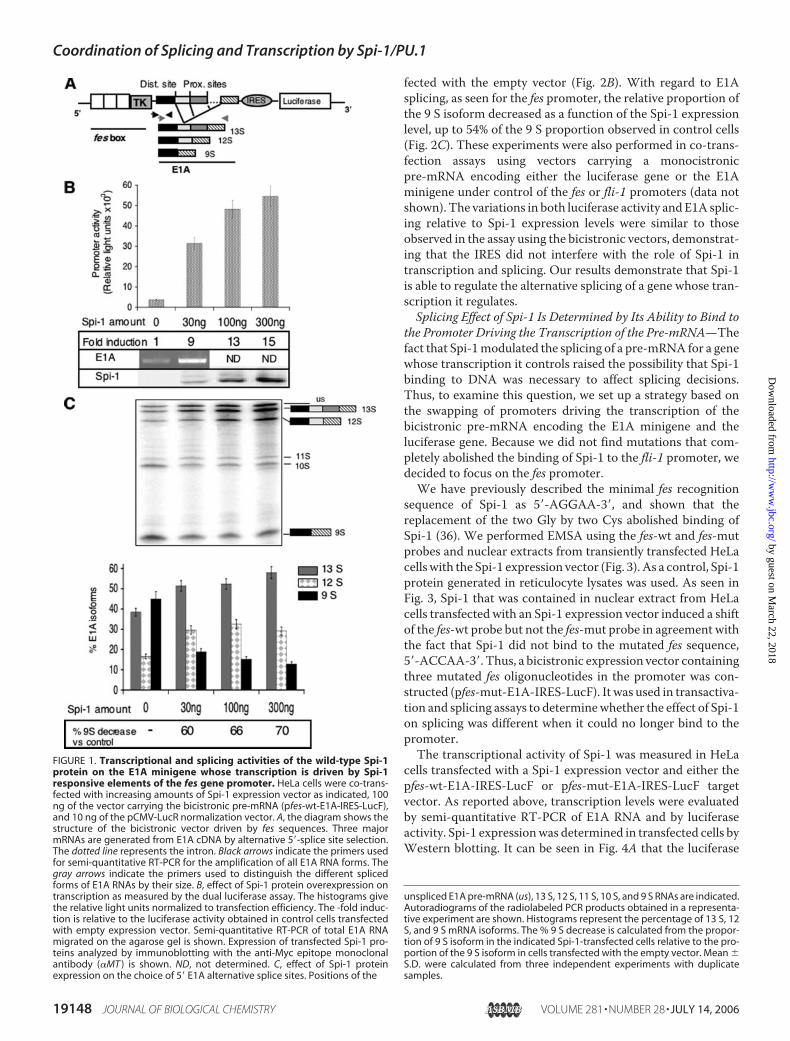
Spi-1 splicing effect on CMVE1A pre-mRNA. First, we ana-lyzed whether Spi-1 was able to bind to the CMV promoter byEMSA. Eight putative Spi-1 binding sites, 5�-(A/G)GAA-3�,were identified in the CMV promoter. EMSA was performedusing 3 different probes containing a total of 4 putative Spi-1binding sites. As seen in Fig. 5A, Spi-1 induced a shift of two ofthe three probes tested. HeLa cells were co-transfected with 10ng of the CMVE1A vector and 500 ng of the Spi-1 expressionvector or an empty vector. Then, the accumulation of RNA inthe presence of Spi-1 was measured by semi-quantitative RT-PCR of E1A pre-mRNA as described above (Fig. 5B). A 2-foldincrease in E1A RNA accumulation was observed. As previ-ously described (24), Spi-1 modified the splicing pattern result-ing in a decrease in the proportion of the 9 S isoform (Fig. 5C,30% of the 9 S proportion found in the control cells). Conse-quently, these results are consistent with our demonstration ofthe need for Spi-1 to bind DNA to favor the use of the proximal5� alternative splice site. The increase of transcription as well asthe splicing effect of Spi-1 on E1A RNA transcribed from theCMV promoter was less significant than the effects on E1ARNA whose transcription was driven by the fes promoter(9-fold transactivation and 60% of the 9 S proportion found inthe control cells, Fig. 1, B and C). We decided not to pursue theexperiments using the CMV promoter because multiple tran-scription factors bind to the CMVpromoter andmost probablyattenuate the Spi-1 effects, impeding a proper interpretation ofthe data.In conclusion, when the Spi-1 protein recognizes responsive
elements in the transcriptional promoter and transactivates, itfavors the use of the proximal 5� alternative splice site. Con-versely, when Spi-1 cannot bindDNA responsive elements, thisqualitative splicing effect is lost.The Effect of Spi-1 on Splicing Is Independent of Transcription
Levels—The experiments presented above suggest that the roleplayed by Spi-1 in transcription and splicing are co-regulated.So, it was of interest to determine whether the modifications ofsplicing profiles in the presence of Spi-1 resulted from differ-ences in the abundance of pre-mRNA. To examine this ques-tion, we wondered whether the number of transcriptional unitsaffects the processing of the pre-mRNAs by Spi-1.First, we compared the splicing patterns of E1A pre-mRNA
transcribed from the same quantity of vector carrying promot-ers of different strengths (Figs. 1 and 2). Similar patterns of E1Asplicing isoformswere obtainedwith the fes and fli-1 promotersin the absence of Spi-1 even though the basal level of the E1Atranscript was 3 times higher when controlled by the fes pro-moter compared with the fli-1 promoter (3,700 relative lightunits for the fes promoter, in the absence of Spi-1, Fig. 1B; and1,300 relative light units for the fli-1 promoter in the absence ofSpi-1, Fig. 2B). Second, 300ng of Spi-1 protein induced a level oftranscription 9-fold higher when driven from the fes promoterthan from the fli-1 promoter as illustrated by the differentialluciferase activities (54,500 relative light units for the fes pro-moter, Fig. 1B; and 6,300 relative light units for the fli-1 pro-moter, Fig. 2B). Nevertheless, Spi-1 favored the use of the 5�proximal site when the E1A pre-mRNA was expressed down-stream from both the fes and fli-1 promoters (Figs. 1C and 2C).
FIGURE 4. Spi-1 affects the splicing decisions as a consequence of promoterusage. HeLa cells were co-transfected with 20 ng of Spi-1 expression vector, 100ng of pfes-wt-E1A-IRES-LucF (left) or pfes-mut-E1A-IRES-LucF (right), and 10 ng ofthe pCMVLucR normalization vector. A, effect of Spi-1 protein expression on tran-scription as measured by dual luciferase assay. The histograms represent therelative light units normalized to the transfection efficiency. The -fold induction isrelative to the luciferase activity obtained in control (c) cells transfected withempty expression vector. The semi-quantitative RT-PCR of total E1A RNAmigrated on the agarose gel is shown. Expression of transfected Spi-1 proteinsanalyzed by immunoblotting with the anti-Myc epitope antibody is shown. B,effect of Spi-1 protein expression on the choice of the 5� E1A alternative splicesites. Autoradiograms of the radiolabeled PCR products obtained in a represent-ative experiment are shown. The histograms represent the percentage of 13 S, 12S, and 9 S mRNA isoforms detected after E1A PCR amplification. The % 9 Sdecrease indicated is calculated from the proportion of the 9 S isoform in theindicated Spi-1-transfected cells relative to the proportion of 9 S isoform in cellstransfected with empty vector. The mean�S.D. were calculated from three inde-pendent experiments with duplicate samples.
Coordination of Splicing and Transcription by Spi-1/PU.1
19150 JOURNAL OF BIOLOGICAL CHEMISTRY VOLUME 281 • NUMBER 28 • JULY 14, 2006
by guest on March 22, 2018
http://ww
w.jbc.org/
Dow
nloaded from
Subsequently, the splicing patterns for two different quanti-ties of E1A pre-mRNA transcribed from pfes-wt-E1A-IRES-LucF were analyzed. To be comparable, the total quantities ofDNA transfected in the cells have been equalized using thepBluescript plasmid. As shown in Fig. 6, 12.5 and 100 ng ofpfes-wt-E1A-IRES-LucF transfected in the cells generated dif-ferent amounts of E1A pre-mRNA as determined from theluciferase activities (in the absence as well as in the presence ofSpi-1). Despite the differences in the quantities of pre-mRNAproduced from the two quantities of reporter vectors, the mat-uration of the E1A pre-mRNA was similar regardless of thepresence or absence of Spi-1 (Fig. 6).These results show that the decrease in the relative propor-
tion of 9 S by Spi-1 is independent of the amount of targetpre-mRNA expressed or the amount of reporter vector trans-fected. This result suggests that the augmentation of the num-ber of transcriptional units by Spi-1 does not saturate the splic-ing machinery. In conclusion, these data are in agreement withthe fact that the splicing effects of Spi-1 are not a consequenceof differences in the synthesis level of RNA but depend on aqualitative effect of Spi-1 in splicing.Analysis of the Effect of Spi-1 Mutants on Splicing of E1A
Pre-mRNA—To further investigate the relationship betweenthe splicing and transcription functions of Spi-1, we examinedthe correlation between the ability of Spi-1 to bind DNA, totransactivate and to modulate alternative splicing of E1A usingvarious Spi-1 mutants (Fig. 7A). The Spi-1 mutants were syn-thesized in reticulocyte lysates and their DNA binding abilitywas analyzed by an EMSA using the fes DNA binding site asprobe (Fig. 7B). �-Pest-Spi-1, devoid of the PEST domain, andDBD-Spi, lacking the transactivation and PEST domains,induced a shift in probemigration (Fig. 7B), consistent with thepresence of the DBD in these two mutants. �-Cter-Spi-1 lacksthe 27 carboxyl-terminal amino acids and �-�4-Spi-1 containsdeletions of 5 amino acids in the �4 region (39). No protein-DNA complex was detected in EMSA using these mutants,showing that they were deficient in DNA binding (Fig. 7B).Similar results were obtained with the fli-1 probe (data notshown).Fig. 8 presents the transcriptional activity of themutants evalu-
ated in transactivation experiments performed inHeLa cells usingpfes-wt-E1A-IRES-LucF and 20 ng of Spi-1 expression vectors(Fig. 8A) orpfli-wt-E1A-IRES-LucFand300ngofSpi-1expressionvectors (Fig. 8B).�-Pest-Spi-1, as the wild-type Spi-1 protein (wt-Spi-1), stimulated luciferase activity as comparedwith the control,indicating an increased transcription (Fig. 8, upper histograms).
FIGURE 5. DNA binding, transcriptional, and splicing activities of thewild-type Spi-1 protein on a E1A minigene whose transcription is drivenby the CMVIE94 promoter. A, DNA binding analysis of Spi-1 protein to theCMVIE94 promoter. The sequences of the three oligonucleotide probes usedare described under “Experimental Procedures.” In vitro translated Spi-1 wasincubated with a radiolabeled oligonucleotide. DNA-protein complexes wereseparated by EMSA and analyzed by autoradiography. c corresponds to theproduct derived from in vitro translation of the CS3 empty vector. B, effect ofSpi-1 protein overexpression on transcription as measured by semi-quantita-tive RT-PCR. HeLa cells were co-transfected with 500 ng of Spi-1 expressionvector or empty vector (c) as indicated, 10 ng of the vector carrying thepre-mRNA (CMV-E1A), and 10 ng of the pCMV-LucR normalization vector. Theprimers used for semi-quantitative RT-PCR for the amplification
of all E1A RNA forms are as indicated by the black arrows in Fig. 1A. Semi-quantitative RT-PCR of total E1A RNA migrated on a agarose gel is shown. The-fold induction is relative to the expression obtained in control cells trans-fected with the empty expression vector (c). Expression of transfected Spi-1proteins analyzed by immunoblotting with the anti-Myc epitope monoclonalantibody is shown. C, effect of Spi-1 protein expression on the choice of 5� E1Aalternative splice sites. Positions of the unspliced E1A pre-mRNA (us), 13 S, 12S, 11 S, 10 S, and 9 S RNAs are indicated. Autoradiograms of the radiolabeledPCR products obtained in a representative experiment are shown. Histo-grams represent the percentage of 13 S, 12 S, and 9 S mRNA isoforms. The %9 S decrease versus control cells are indicated under each histogram. Mean �S.D. were calculated from three independent experiments with duplicatesamples.
Coordination of Splicing and Transcription by Spi-1/PU.1
JULY 14, 2006 • VOLUME 281 • NUMBER 28 JOURNAL OF BIOLOGICAL CHEMISTRY 19151
by guest on March 22, 2018
http://ww
w.jbc.org/
Dow
nloaded from

Although able to bindDNA,DBD-Spi did not significantly induceluciferaseactivity, consistentwith the lackof anactivationdomain.Similarly,�-Cter-Spi-1 and�-�4-Spi-1 did not activate transcrip-tion from fes and fli-1 promoters as deduced from the absence ofincreased luciferase activity. In terms of splicing activity, �-Pest-Spi-1decreased9SRNAproductionasdid the intactSpi-1protein(Fig. 8, lower histograms). Interestingly, the mutants that did nottransactivate whether they bound (DBD-Spi-1) or not (�-Cter-Spi-1 and �-�4-Spi-1) to the Spi-1 sensitive promoters were notable to exert the splicing activity of the wt-Spi-1 protein. The rel-ative proportion of 13 S, 12 S, and 9 S E1A isoforms were eithersimilar to that observed in cells transfected with the empty vector(DBD-Spi-1) or displayed an increase in the 9 S isoform that wascounterbalanced by a reduction of the 13 S isoform (�-Cter-Spi-1and �-�4-Spi-1; 26 and 31% or 42 and 48% of 9 S increase com-pared with control cells for fes promoter and fli-1 promoter,respectively). So, in the absence of binding to DNA and transacti-vation, such as observed for �-Cter-Spi-1 and �-�4-Spi-1, thesplicing patterns revealed a shift toward a processing using thedistal 5� site. This splicing profile was reminiscent of the effect ofwt-Spi-1 on E1A transcribed from the fes-mut promoter. It isinteresting to note that the mutant effects of Spi-1 on pre-mRNAprocessing were similar when transcription was conducted fromfes or fli-1 promoters. These results are again consistent with theidea that Spi-1 favors the use of the proximal 5� alternative splicesite only when bound to DNA and able to transactivate.
DISCUSSION
We have previously shown that the oncogenic protein Spi-1/PU.1, initially described as a transcription factor, also affectssplicing (22, 23, 25). The splicing function requires the DNAbinding domain containing the ETS region (24). This led to thehypothesis that Spi-1 may affect the splicing of a gene whosetranscription it regulates. Thus, we developed a model systemallowing the characterization of pre-mRNA transcription andmaturation from a single minigene whose transcription isdriven by Spi-1-dependent promoters. Here, we demonstratethat co-regulation of transcription and splicing can take placethrough the promoter-specific loading of Spi-1. Furthermore,our results establish that Spi-1 splicing activity is dependent onits DNA binding ability, which is associated to its transactiva-tion effect. The absence of a correlation between promoterstrength or number of transcripts and splicingmodulation indi-cates that the function of Spi-1 in splicing is not simply due toits modulation of transcription but is related to the intrinsicproperties of the Spi-1 protein. This is consistent with previ-ously published data (27, 28, 33, 34, 40).The splicing activity of Spi-1 has been previously character-
ized using E1A pre-mRNA whose transcription was driven bythe CMV promoter (22, 23, 25). Spi-1 inhibited the 9 S E1Aproduction as observed for the fes-wt and fli-1-wt promoters.The data presented here show that Spi-1 is able to bind andtransactivate the CMV promoter. Altogether, these data dem-onstrate that Spi-1 exhibits a coordinated action in transcrip-tion and splicing.The observation that promoter structure contributes to
alternative splicing constituted one of the firstmechanistic datatoward the understanding of how alternative splicing and tran-scription are co-regulated (27). Since then, several studies have
FIGURE 6. Splicing effect of Spi-1 is not a consequence of a change in thetranscriptional level. HeLa cells were co-transfected with 20 ng of Spi-1vector, 12.5 or 100 ng of pfes-wt-E1A-IRES-LucF, and 10 ng of the pCMV-LucRnormalization vector. The mean � S.D. of three independent experimentswith duplicate samples are shown. A, effect of Spi-1 protein expression ontranscription as a function of the different quantities of E1A expression plas-mid used. The histograms represent the relative light units normalized to thetransfection efficiency; the exact value of the relative light units is indicatedabove each histogram. B, histograms representing the relative percentage of13 S, 12 S, and 9 S mRNA isoforms detected are shown and the % 9 S decreasecompared with control cells is indicated.
FIGURE 7. DNA binding activity of Spi-1 mutant proteins. A, schematicrepresentation of the Spi-1 mutants. The top diagram represents the wild-type 272-amino acid protein. The open box corresponds to the transactivationdomain, the hatched box to the PEST region, and the gray box to the DNAbinding domain. B, DNA binding activity of Spi-1 and Spi-1 mutants. In vitrotranslated Spi-1 and Spi-1 mutants were incubated with the radiolabeledfes-wt oligonucleotide as a probe (sequences under “Experimental Proce-dures”). DNA-protein complexes were separated by EMSA and analyzed byautoradiography. Expression of transfected Spi-1 proteins was analyzed byimmunoblotting using the anti-Myc epitope antibody (�MT ). WB, Westernblot.
Coordination of Splicing and Transcription by Spi-1/PU.1
19152 JOURNAL OF BIOLOGICAL CHEMISTRY VOLUME 281 • NUMBER 28 • JULY 14, 2006
by guest on March 22, 2018
http://ww
w.jbc.org/
Dow
nloaded from

shown that this process is complex and involves cell-specificpromoter occupancy associated with splicing factors activity(for review, Ref. 26). Actually, proteins of the spliceosome inter-acting physicallywith the transcriptionalmachinery or proteinsplaying a role directly in both mechanisms have been proposedto be candidates linking transcription and splicing (26, 30–35,41). According to the cell-specific promoter occupationmodel,Spi-1, by recruiting splicing proteins to specific promoters,would play the role of scaffold to bring splicing proteins to thenascent transcribed RNA. Currently, no splicing factor hasbeen identified that could be recruited by Spi-1 and be a candi-date for mediating the Spi-1 splicing effect on a transcriptionaltarget RNA. Indeed, Spi-1 interacts with several proteinsinvolved in RNA processing, such as p54nrb, polypyrimidinetract-binding protein-associated splicing factor (25), TLS (22),and the heterogeneous nuclear ribonucleoproteins hnRNPA1.Nevertheless, the effects of each one of these factors has beenshown to be opposite to the splicing effect of Spi-1 on E1Apre-mRNA maturation, i.e. increase of the distal 5� splice siteuse of E1A pre-mRNA (22, 24, 25, 42). Furthermore, Spi-1 pre-vents TLS (24) and p54nrb (25) from binding to RNA and is
also able to interfere with thesplicing effect of TLS (22). The SR(Ser/Arg-rich) proteins ASF/SR2and SC35 activate the proximalalternative 5� splice site on E1ARNA as does Spi-1 (43, 44). There-fore, it will be interesting to exam-ine whether Spi-1 acts togetherwith SR proteins to favor selectionof the proximal alternative5�-splice site.When Spi-1 was unable to bind
DNA, such as for wt-Spi-1 onmutated promoters or�-Cter-Spi-1and �-�4-Spi-1 on wt promoters, itwas associated with a splicing pat-tern opposite to the splicing effect ofSpi-1 bound to DNA. Indeed, theproportion of the 9 S mRNA iso-form was augmented due to anincreased use of the distal 5� splicesite. These results may be explainedby the fact that Spi-1, not bound toDNA, traps and modifies the equi-librium of available splicing factorsacting on E1A pre-mRNA pro-cesses. In this respect, a high level ofSpi-1 expression may modify thealternative splicing of a gene whosetranscription it does not controland, in this case, it would act as aninterfering protein. Again, even ifthe Spi-1 interference on TLS,p54nrb, and the polypyrimidinetract-binding protein-associatedsplicing factor could take place inthe absence of Spi-1 binding to the
promoter, it would result in an inhibition of the distal alterna-tive 5� splice site, which is contrary to the Spi-1 effect seen on agene whose transcription it does not control. Proteins interact-ing with Spi-1 that would modulate RNA splicing may not bethe same whether Spi-1 does or does not bind DNA. Currently,we have no information about the nature of putative splicingpartnersmediating Spi-1 splicing effectswhen it is not bound totranscriptional promoters.An alternate hypothesis may involve control of the elongation
rate of transcription. It has been shown that transcription can alsocontrol splicing through regulation of RNApolymerase II proces-sivity and elongation rates (40, 45–47). Notably, the Brm subunitof the SWI/SNF complex, involved in chromatin remodeling onpromoters, has been recently demonstrated to contribute tocross-talk between transcription and alternative splicing bydecreasing the RNA polymerase II elongation rate (48). Inter-estingly, a high RNA polymerase II elongation rate favors theuse of the proximal 5� splice site of the adenovirus E1A mini-gene, generating increased amounts of the 13 S isoform anddecreasing the 9 S isoform. This would be compatible with thecombined activities of Spi-1 in transcription and splicing.A role
FIGURE 8. Transcriptional and splicing activities of Spi-1 mutant proteins on the E1A minigene whose tran-scription is driven by Spi-1 responsive elements of the fes or fli-1 gene promoters. HeLa cells were co-trans-fected with the E1A reporter gene driven by fes-wt and 20 ng of the control empty vector (c) or the Spi-1 mutantexpression vectors (A), or fli-1-wt promoters and 300 ng of the control empty vector (c) or the Spi-1 mutantexpression vectors (B). Mean � S.D. were calculated from five independent experiments with duplicate sam-ples. Upper panel, effect of Spi-1 proteins on transcription measured by dual luciferase assay. Luciferase activitywas measured from cellular extracts according to the dual luciferase assay instructions. The histograms repre-sent the relative light units normalized to transfection efficiency (LucF/LucR). The -fold induction is relative tothe luciferase activity obtained in control cells transfected with the empty expression vector. Lower panel,effect of Spi-1 mutant protein expression on the choice of 5� E1A alternative splice sites. Histograms give therelative percentage of 13 S, 12 S, and 9 S mRNA isoforms detected after E1A radioactive PCR amplification. The% 9 S decrease indicated is calculated from the proportion of the 9 S isoform in the indicated Spi-1-transfectedcells relative to the proportion of 9 S isoform in cells transfected with the empty vector. Expression of trans-fected Spi-1 proteins analyzed by immunoblotting with the anti-Myc epitope antibody (�MT ) is shown. Molec-ular masses are indicated on the left (kDa).
Coordination of Splicing and Transcription by Spi-1/PU.1
JULY 14, 2006 • VOLUME 281 • NUMBER 28 JOURNAL OF BIOLOGICAL CHEMISTRY 19153
by guest on March 22, 2018
http://ww
w.jbc.org/
Dow
nloaded from

for Spi-1 in regulating pol II elongation has not so far beendescribed. Consequently, more studies are required to drawdefinitive conclusions.Differences in splicing have been noted between normal and
tumor cells. These differences are often due to deregulation ofsplicing factors in tumor cells (49). A subset of aberrant RNA-spliced isoforms may confer a selective advantage to cancercells, even if some of the modifications of splicing may be asso-ciated with stress induced by the disease (49). The high expres-sion of Spi-1 in mice is associated with the development oferythroleukemia due to the blockage of erythroid differentia-tion. We have recently demonstrated using the E1A minigenemodel that in leukemic proerythroblasts, the overexpression ofspi-1 affects splicing (24), suggesting that this function maydirectly contribute to oncogenic activity of Spi-1. The resultsdescribed in this article were obtained using HeLa epithelialcells. Even if Spi-1 exerts an oncogenic activity only in theerythroid lineage, its splicing activity is not restricted to eryth-roid cells but was also detected in myeloid, T and B lymphoid,and epithelial cells (Ref. 24 and this study). So, if the interfer-ence of Spi-1 in splicing is involved in the blockage of erythroiddifferentiation, one can envision at least two possibilities. Onepossibility is that the Spi-1 splicing activity modifies the proc-essing of ubiquitously expressed genes. Because Spi-1 regulatestranscription of genes from themyeloid and lymphoid lineages,the splicing effect of Spi-1 in this case would be independent ofits activity as a transcription factor. The spliced isoforms spe-cifically associated with the presence of a strong Spi-1 expres-sion would code for a protein modifying only the erythroiddifferentiation program.Another possibility is that Spi-1 affectsgenes for which splicing is tightly regulated in a differentiationstage-specific manner. Ample evidence exists that alternativesplicing is important for erythroid differentiation (50). A recentpublication described the existence of an erythroid cell-specificsplice variant of the CP2 transcription factor (51). The protein4.1R, a vital component of red blood cell membrane cytoskele-ton, is also encoded by a gene whose splicing is highly regulatedduring erythroid differentiation (52, 53). Interestingly, Spi-1has been recently shown to modify erythroid-specific alterna-tive splicing of 4.1R in murine erythroleukemia cells (54). TheSR protein SF2/ASF provokes a splicing modification of the4.1R RNA that is opposite to the one observed in cells overex-pressing Spi-1 (54, 55). Whether Spi-1 competes with SF2/ASFto modulate 4.1R maturation remains to be determined.In conclusion, we had previously shown that transcription
factor Spi-1 is involved in the choice of alternative splice sitesused on a pre-mRNA. We have now established that Spi-1 isable to display a specific splicing activity on transcriptional tar-gets. Until now, the involvement of the splicing function ofSpi-1 in the blockage of erythroid differentiation and its onco-genic activity has not been established. Our results suggest thatif the splicing activity of Spi-1 is involved in blockage of differ-entiation, it could do so by acting on genes whose transcriptionit regulates. Nevertheless, the oncogenic function of Spi-1 isdirectly related to its overexpression. So, overexpressed Spi-1might act as a titrating factor, as described here, and conse-quently modify indirectly the processing of transcriptionallyindependent genes. Strategies are currently being developed to
unravel the Spi-1 target genes for splicing and their contribu-tion to oncogenesis.
Acknowledgments—We thank Didier Auboeuf, Jean de Gunzburg,Olivier Kosmider, Richard Monni, Pauline Rimmele, and FilippoRosselli for advice and comments and Julianna Smith for criticalreading the manuscript. We are grateful to N. Denis and N. Brandonfor technical assistance.
REFERENCES1. McKercher, S. R., Torbett, B. E., Anderson, K. L., Henkel, G. W., Vestal,
D. J., Baribault, H., Klemsz, M., Feeney, A. J., Wu, G. E., Paige, C. J., andMaki, R. A. (1996) EMBO J. 15, 5647–5658
2. Scott, E. W., Fisher, R. C., Olson, M. C., Kehrli, E. W., Simon, M. C., andSingh, H. (1997) Immunity 6, 437–447
3. Back, J., Allman, D., Chan, S., and Kastner, P. (2005) Exp. Hematol. 33,395–402
4. Nutt, S. L., Metcalf, D., D’Amico, A., Polli, M., and Wu, L. (2005) J. Exp.Med. 201, 221–231
5. Rosenbauer, F., Wagner, K., Kutok, J. L., Iwasaki, H., Le Beau, M. M.,Okuno, Y., Akashi, K., Fiering, S., and Tenen, D. G. (2004)Nat. Genet. 36,624–630
6. Cook, W. D., McCaw, B. J., Herring, C., John, D. L., Foote, S. J., Nutt, S. L.,and Adams, J. M. (2004) Blood 104, 3437–3444
7. Suraweera,N.,Meijne, E.,Moody, J., Carvajal-Carmona, L. G., Yoshida, K.,Pollard, P., Fitzgibbon, J., Riches, A., van Laar, T.,Huiskamp, R., Rowan,A.,Tomlinson, I. P., and Silver, A. (2005) Oncogene 24, 3678–3683
8. Moreau-Gachelin, F., Tavitian, A., and Tambourin, P. (1988)Nature 331,277–280
9. Moreau-Gachelin, F., Wendling, F., Molina, T., Denis, N., Titeux, M.,Grimber, G., Briand, P., Vainchenker, W., and Tavitian, A. (1996) Mol.Cell. Biol. 16, 2453–2463
10. Klemsz, M. J., McKercher, S. R., Celada, A., Van Beveren, C., and Maki,R. A. (1990) Cell 61, 113–124
11. Pongubala, J. M., Vanbeveren, C., Nagulapalli, S., Klemsz, M. J.,McKercher, S. R., Maki, R. A., and Atchison, M. L. (1993) Science 259,1622–1625
12. Starck, J., Doubeikovski, A., Sarrazin, S., Gonnet, C., Rao, G., Skoultchi, A.,Godet, J., Dusanter-Fourt, I., and Morle, F. (1999) Mol. Cell. Biol. 19,121–135
13. Hagemeier, C., Bannister, A. J., Cook, A., and Kouzarides, T. (1993) Proc.Natl. Acad. Sci. U. S. A. 90, 1580–1584
14. Yamamoto, H., Kihara-Negishi, F., Yamada, T., Hashimoto, Y., andOikawa, T. (1999) Oncogene 18, 1495–1501
15. Perkel, J. M., and Atchison, M. L. (1998) J. Immunol. 160, 241–25216. Brass, A. L., Zhu, A. Q., and Singh, H. (1999) EMBO J. 18, 977–99117. Behre, G., Whitmarsh, A. J., Coghlan, M. P., Hoang, T., Carpenter, C. L.,
Zhang, D. E., Davis, R. J., and Tenen, D. G. (1999) J. Biol. Chem. 274,4939–4946
18. Petrovick, H. S., Hiebert, S. W., Friedman, A. D., Hetherington, C. J.,Tenen, D. G., and Zhang, D. E. (1998)Mol. Cell. Biol. 18, 3915–3925
19. Bakri, Y., Sarrazin, S., Mayer, U. P., Tillmanns, S., Nerlov, C., Boned, A.,and Sieweke, M. H. (2005) Blood 105, 2707–2716
20. Zhang, P., Behre, G., Pan, J., Iwama, A., Wara-Aswapati, N., Radomska,H. S., Auron, P. E., Tenen, D. G., and Sun, Z. (1999) Proc. Natl. Acad. Sci.U. S. A. 96, 8705–8710
21. Rekhtman, N., Radparvar, F., Evans, T., and Skoultchi, A. I. (1999) GenesDev. 13, 1398–1411
22. Hallier, M., Lerga, A., Barnache, S., Tavitian, A., andMoreau-Gachelin, F.(1998) J. Biol. Chem. 273, 4838–4842
23. Lerga, A., Hallier, M., Delva, L., Orvain, C., Gallais, I., Marie, J., andMoreau-Gachelin, F. (2001) J. Biol. Chem. 276, 6807–6816
24. Delva, L., Gallais, I., Guillouf, C., Denis, N., Orvain, C., andMoreau-Gachelin, F. (2004) Oncogene 23, 4389–4399
25. Hallier, M., Tavitian, A., and Moreau-Gachelin, F. (1996) J. Biol. Chem.271, 11177–11181
Coordination of Splicing and Transcription by Spi-1/PU.1
19154 JOURNAL OF BIOLOGICAL CHEMISTRY VOLUME 281 • NUMBER 28 • JULY 14, 2006
by guest on March 22, 2018
http://ww
w.jbc.org/
Dow
nloaded from

26. Kornblihtt, A. R., de la Mata, M., Fededa, J. P., Munoz, M. J., and Nogues,G. (2004) RNA 10, 1489–1498
27. Cramer, P., Pesce, C. G., Baralle, F. E., and Kornblihtt, A. R. (1997) Proc.Natl. Acad. Sci. U. S. A. 94, 11456–11460
28. Cramer, P., Caceres, J. F., Cazalla, D., Kadener, S.,Muro,A. F., Baralle, F. E.,and Kornblihtt, A. R. (1999)Mol. Cell 4, 251–258
29. Kameoka, S., Duque, P., and Konarska, M. M. (2004) EMBO J. 23,1782–1791
30. Chansky, H. A., Hu, M., Hickstein, D. D., and Yang, L. (2001) Cancer Res.61, 3586–3590
31. Ge, H., Si, Y., and Volffe, A. P. (1998)Mol. Cell 2, 751–75932. Monsalve, M., Wu, Z., Adelmant, G., Puigserver, P., Fan, M., and
Spiegelman, B. M. (2000)Mol. Cell 6, 307–31633. Auboeuf, D., Honig, A., Berget, S. M., and O’Malley, B. W. (2002) Science
298, 416–41934. Auboeuf, D., Dowhan, D. H., Kang, Y. K., Larkin, K., Lee, J. W., Berget,
S. M., and O’Malley, B. W. (2004) Proc. Natl. Acad. Sci. U. S. A. 101,2270–2274
35. Auboeuf, D., Dowhan, D. H., Li, X., Larkin, K., Ko, L., Berget, S. M., andO’Malley, B. W. (2004)Mol. Cell. Biol. 24, 442–453
36. Ray-Gallet, D., Mao, C., Tavitian, A., and Moreau-Gachelin, F. (1995)Oncogene 11, 303–313
37. Gattoni, R., Chebli, K., Himmelspach, M., and Stevenin, J. (1991) GenesDev. 5, 1847–1858
38. Caceres, J. F., Stamm, S., Helfman, D.M., andKrainer, A. D. (1994) Science265, 1706–1709
39. Kodandapani, R., Pio, F., Ni, C. Z., Piccialli, G., Klemsz, M., McKercher,S. R., Maki, R. A., and Ely, K. R. (1996) Nature 380, 456–460
40. Kadener, S., Fededa, J. P., Rosbash, M., and Kornblihtt, A. R. (2002) Proc.
Natl. Acad. Sci. U. S. A. 99, 8185–819041. Knoop, L. L., and Baker, S. J. (2001) J. Biol. Chem. 276, 22317–2232242. Zhang, W. J., and Wu, J. Y. (1996)Mol. Cell. Biol. 16, 5400–540843. Cowper, A. E., Caceres, J. F., Mayeda, A., and Screaton, G. R. (2001) J. Biol.
Chem. 276, 48908–4891444. Krainer, A. R., Conway, G. C., and Kozak, D. (1990) Cell 62, 35–4245. Kadener, S., Cramer, P., Nogues, G., Cazalla, D., de la Mata, M., Fededa,
J. P., Werbajh, S. E., Srebrow, A., and Kornblihtt, A. R. (2001) EMBO J. 20,5759–5768
46. de la Mata, M., Alonso, C. R., Kadener, S., Fededa, J. P., Blaustein, M.,Pelisch, F., Cramer, P., Bentley, D., and Kornblihtt, A. R. (2003)Mol. Cell12, 525–532
47. Nogues, G., Munoz, M. J., and Kornblihtt, A. R. (2003) J. Biol. Chem. 278,52166–52171
48. Batsche, E., Yaniv, M., andMuchardt, C. (2006)Nat. Struct. Mol. Biol. 13,22–29
49. Venables, J. P. (2004) Cancer Res. 64, 7647–765450. Hou, V. C., and Conboy, J. G. (2001) Curr. Opin. Hematol. 8, 74–7951. Kang, H. C., Chae, J. H., Lee, Y. H., Park, M. A., Shin, J. H., Kim, S. H., Ye,
S. K., Cho, Y. S., Fiering, S., and Kim, C. G. (2005) Mol. Cell. Biol. 25,6005–6020
52. Chasis, J. A., Coulombel, L., Conboy, J., McGee, S., Andrews, K., Kan,Y. W., and Mohandas, N. (1993) J. Clin. Investig. 91, 329–338
53. Baklouti, F., Huang, S. C., Tang, T. K., Delaunay, J., Marchesi, V. T., andBenz, E. J., Jr. (1996) Blood 87, 3934–3941
54. Theoleyre, O., Deguillien,M., Moriniere, M., Starck, J., Moreau-Gachelin,F., Morle, F., and Baklouti, F. (2004) Oncogene 23, 920–927
55. Yang, G., Huang, S. C., Wu, J. Y., and Benz, E. J., Jr. (2005) Blood 105,2146–2153
Coordination of Splicing and Transcription by Spi-1/PU.1
JULY 14, 2006 • VOLUME 281 • NUMBER 28 JOURNAL OF BIOLOGICAL CHEMISTRY 19155
by guest on March 22, 2018
http://ww
w.jbc.org/
Dow
nloaded from

Christel Guillouf, Isabelle Gallais and Françoise Moreau-GachelinBinding-dependent Manner
Spi-1/PU.1 Oncoprotein Affects Splicing Decisions in a Promoter
doi: 10.1074/jbc.M512049200 originally published online May 11, 20062006, 281:19145-19155.J. Biol. Chem.
10.1074/jbc.M512049200Access the most updated version of this article at doi:
Alerts:
When a correction for this article is posted•
When this article is cited•
to choose from all of JBC's e-mail alertsClick here
http://www.jbc.org/content/281/28/19145.full.html#ref-list-1
This article cites 55 references, 35 of which can be accessed free at
by guest on March 22, 2018
http://ww
w.jbc.org/
Dow
nloaded from


![Human Papillomavirus Type 16 E7 Oncoprotein-induced ... · [CANCER RESEARCH 61, 2356–2360, March 15, 2001] Advances in Brief Human Papillomavirus Type 16 E7 Oncoprotein-induced](https://img.dokumen.tips/doc/110x75/605dd1c1b72c9c6f905bfd49/human-papillomavirus-type-16-e7-oncoprotein-induced-cancer-research-61-2356a2360.jpg)