Embed Size (px)
Citation preview

19
Specific Immunotherapy and Central Immune System
Celso Pereira, Graça Loureiro, Beatriz Tavares and Filomena Botelho Immunoallergology Department, Coimbra University Hospital,
Portugal
1. Introduction
Despite the current knowledge, the mechanisms by which the specific immunotherapy (SIT) achieves clinical improvement remains unclear. However, it is now clear that the immune tolerance is one of the major targets of this kind of treatment. Immune tolerance depends on different mechanisms, including T-cell anergy, T-cell depletion by apoptosis, and active immune suppression (Akdis & Akdis, 2011). One of the goals of SIT is the induction of tolerance to allergens to which the patient is sensitized. IL-10 is probably a relevant cytokine induced by this treatment and is associated to regulatory T cells (T-regs) that actively control or suppress the function of other cells, generally in an inhibitory pattern (Frew, 2010). The changes in microenvironment due to the decrease in histamine and PGE2 release by mast
cell, and the IL-10 and TGF- release by dendritic cells (DC) could switch the T-cell population into T-regs. These alterations will then lead to tolerance (Schmidt-Weber & Blaser 2005). Much of the knowledge about SIT has been based on studies using subcutaneous route of administration (SCIT), but increasing data is now available based on studies using sublingual immunotherapy (SLIT). The sublingual mucosa, where the deposition of the extract occurs, has very particular characteristics, quite different from the cellular subcutaneous tissue. The dendritic cells present in the buccal region are distinct from the Langerhans cells present in the skin. These
cells present, constitutively, receptors with high affinity for IgE, FcRI+, MHC class I and II molecules, as well as co-stimulation molecules, namely CD40, CD80/B7.1 and CD86/B7.2 (Allam et al., 2006). There is also expression of CD14, a lipopolysaccharide (LPS) receptor, which is relevant for the modulation of Th2 and Th1. In SLIT, the allergen is captured by the DC cell by C-lectin endocytosis receptors and/or by ligation to the IgE on the surface. After the internalization, the migration to the regional lymphoid nodules occurs, and it is then presented to the T cells (Geijtenbeek, 2006). A study that compares the DC population in the oral and nasal mucosa was able to demonstrate that only the DC myeloid type is profusely present in the oral region, in contrast with the nasal mucosa where both populations are present (Allam et al., 2006). Another very important
difference is the high expression levels of FcRI in the oral mucosa, which is almost absent in the skin’s Langerhans cells (Allam et al., 2003). Furthermore, the expression levels of MHC class I and II molecules, CD40, CD80 and CD86 is significantly higher in oral DC than in the skin.
www.intechopen.com

Allergic Diseases – Highlights in the Clinic, Mechanisms and Treatment
376
The integrity of the lamina propria in the oral mucosa, along with the highly reduced population of mast cells, eosinophils and basophils, are the factors that limit the contact of the allergen with submucosal areas or with circulating blood cells. All of these together constitute the excellent security profile characteristic of the SLIT (Moingeon et al., 2006). The DC seems to be crucial in the induction of the tolerance profile due to the production of IL-
10 and TGF- after activation, as well as an increase in indoleamine dioxygenase type 2 (IDO), necessary to the decrease in T cells proliferation. The allergen absorption through the intact mucosa and the interaction with local dendritic cells could switch on the process leading to immune-tolerance. In addition to the local effect, the swallowed allergen could induce a supplementary outcome based on a GALT-related mechanism (Akdis et al., 2001). In fact, the first method of sublingual administration of SIT, in which the allergen was spat out, was significantly less effective. Most of the immunological changes and mechanisms that occur with sublingual administration of high allergen dosage have been recently demonstrated (Moingeon et al., 2006). The aqueous extracts are highly effective, but induce more side-effects (local and systemic) than depot and modified vaccines (Larché et al., 2006). These have been developed in an attempt to reduce or remove allergenicity, while preserving or increasing the immunogenicity (Casanovas et al., 2005). Aqueous subcutaneous SIT is now the gold standard treatment for hymenoptera venom allergy and depot aeroallergen extracts for respiratory allergy (Wheeler & Woroniecki, 2004). The use of chemically modified allergens is not consensual. Although recent papers demonstrate a clinical benefit with allergoid therapy (Ibarrola et al., 2004), double blind placebo controlled studies comparing both extracts are still missing. On the other hand, the sublingual-swallowed administration of SIT in higher dosages had a clinical efficacy entirely demonstrated with similar immunological effects, namely the increase in allergen-blocking IgG, the induction of IgE-modulating CD8+ T cells, the reduction of mast cells and eosinophils on the target mucosa, the decrease in inflammatory mediators, and a modulation on the inflammatory trafficking cells by reduced expression of adhesion molecules (Bousquet, 2006).
2. Kinetics of immunotherapy mechanism
The studies on the dynamics of SIT are of extreme relevance, because they represent the only approach available to define, on a biological point of view, the mechanism by which the therapeutics works. Despite the current advances contributing to the wide knowledge of the modulator effect, these studies are extremely rare, as a result of several ethical, technical and economical boundaries. Furthermore, there are also several difficulties in transposing the results from experimental studies using laboratory animals to Humans. In a study using mice, the systemic activity of a modified aeroallergen, an allergoid administrated sublingually, was shown (Mistrello, 1994). However, before the study of an Italian group from Genoa in 1997, the kinetics and dynamics of the allergen in Humans was not known (Bagnasco, 1997). This study had the merit of assessing the response of the sublingual administration of a radioactively labeled allergen in a group of 9 healthy individuals (Bagnasco, 1997). Par j 1, the major Parietaria judaica allergen, was labeled with 123I. In respect to the sublingual route, the application and deposition of the labeled allergen in 3 individuals was monitored for a period of 30 minutes before deglutition, by the acquisition of static images in the following 1, 2, 3, 5, 24 and 48 hours. In parallel, blood draws were collected for the determination of the radioactive counts at 5, 10, 20, 30 and 60
www.intechopen.com

Specific Immunotherapy and Central Immune System
377
minutes, and then at 2, 3, 5 and 24 hours after the administration. A quantification of the same parameters in the urine was also performed. This study was able to demonstrate that the allergen was not degraded by the saliva and interestingly that there was no absorption by the sublingual mucosa. The plasma radioactive activity only occurs after the deglutition of the allergen. Another
surprising fact is that an enormous radioactive activity of the labeled allergen in the sublingual
region is still detected, even after 48hours and despite the deglutition. Furthermore, the study
also compared the same methodology either with nasal and oral (immediate deglutition)
administration. The results from the application of the allergen in a nasal pulverization
demonstrated activity in the superior region of the pharynx and proximal esophagus, and
persistence of the labeled allergen until 36hours, with no bronchial deposition, but having
activity in the plasma since the beginning. The oral route is simultaneous to the detection of
immediate activity in the plasma, characterized by a pick at 2hours, followed by a decline, but
with no identification of the labeled allergen. Probably there is protein degradation and only
small peptides are absorbed at the level on the intestinal mucosa.
These results, done afterwards by the same group and obtained from healthy individuals,
were crucial for the evaluation of the kinetics and dynamics of the allergen administration in 9
allergic patients with rhinitis (Bagnasco, 2001). The study compared the different alternatives
of sublingual administration of the Par j 1 allergen labeled with 123I. Namely non-modified
allergen solution (drops), non-modified allergen in pills and modified allergen (allergoid) also
in pills. The patients were monitored with dynamic acquisition in the first 20 minutes and
static acquisitions of 5 minutes every hour until the 16th hour. Serial blood draws were also
collected in order to quantify the radioactive plasmatic activity. The results confirmed
persistent radioactive activity at the local of the administration of the allergen. For all the
extracts, the plasmatic activity only occurs after deglutition. With the allergoid, the activity is
higher, probably dependent of a lower enzymatic degradation and consequent lower
possibility of intestinal absorption. The acquisitions in a gamma-rays camera only visualizes
the activity after deglutition (1-5 minutes later), in the pharyngeal and esophagic region.
The same group of investigators studied the kinetics of the intranasal administration of Par j
1 labeled with 123I in a group of 3 patients with allergic rhinitis, using a similar methodology
although with distinct time points (Passalacqua, 2005). The radioactive activity in the nasal
region disappears rapidly in allergic patients when compared to healthy individuals. The
clinic inflammation controlled by the administration of the therapeutic allergen in these
patients may condition an increase in the depuration or clearance of the mucosa. Plasmatic
activity is also observed after an effective deglutition of the allergen. Despite the reduced
number of patients used to conducted this study, the results are significantly distinct from
the results observed when using the sublingual route, in which the radioactive activity
persist at the local of administration, long after the deglutition of the therapeutic extract
(Bagnasco, 2001).
The same Italian group responsible for the study with allergens administrated by non-injectable routes prompt the question of whether the results obtained up until that moment were restricted to specific characteristics of Parietaria judaica. Following this, the group published another study using a major allergen from Dermatophagoides pteronyssinus, the Der p 2 (Bagnasco, 2005). For the 7 patients with allergic rhinitis, the results were analogous to the ones mentioned above. This lead to the conclusion that the persistence of the radioactive activity at the local of administration of the allergen and the systemic absorption after the
www.intechopen.com

Allergic Diseases – Highlights in the Clinic, Mechanisms and Treatment
378
deglutition is independent of the allergen used in the treatment and the chemical modification used. The studies regarding the kinetics and dynamics of immune-therapy administrated by non-injectable routes were pioneer, even though its implementation was substantially more recent than conventional route, and interestingly they included the nasal route, which had no scientific validation for its efficacy (Bousquet, 1998; Alvarez-Cuesta, 2006). In respect to the subcutaneous route, our group has published in 2004 the first in vivo results regarding the dynamics of SIT in patients with allergic disease (Pereira, 2004).
3. Dynamics of the immunotherapy mechanism
The radioactive labeling of an allergen from the therapeutic extract is possible, but it would be restricted, exclusively, to one protein only. This could have been a strategy to evaluate the dynamics of the biological response to SIT. However, for patients under a maintenance treatment, the exclusion of some allergenic epitopes present in the therapeutic extracts would condition the interpretation of the results. Furthermore, it could, eventually, change the immunogenic structure of that epitope and as a consequence, the immune response that was induced. In these studies, the administration of the allergen extract, according to the maintenance scheme in place for each patient, would occur simultaneously with the reinjection of leucocytes labeled with 99mTc-HMPAO. The labeling of leucocytes with 99mTc-HMPAO is a technique that, on the theoretical level, has affinity for all the cellular elements of the white series (mononuclear and polymorphonuclear) and all of these have a specific contribution in the inflammatory process (Kumar, 2005; Peters, 1986). The therapeutic allergenic extract injections always induce an inflammatory reaction at the local of the administration, detectable clinically by the presence of typical local signals. In the maintenance treatment, these symptoms have variable intensity and depend not only on the type of extract but also on the severity of the basal clinical features (Alvarez-Cuesta et al., 2006). The modified extracts and the extracts administrated sublingually are the ones that induce minor local secondary effects, as mentioned before. Therefore, although in a limited and controlled way, the SIT leads to an effective inflammatory allergic reaction in the area where the extract is applied, despite all the relevant immune-modulator mechanisms, which at the moment are well established for this therapeutic. This local inflammatory allergic reaction will be, presumably, responsible by the dynamics of a response highly similar to the one observed in the allergic response in general (Pereira, 2009). Therefore, it can be admitted that the autologous reinjection of leucocytes will have a migration at that level by mechanisms dependent on the IgE-mediated mast cells activation (Bousquet, 1998; Alvarez-Cuesta et al., 2006).
3.1 Patients studied Seventeen adult volunteer allergic patients were selected, 15 of them under maintenance therapeutic with specific immunotherapy. All were under a regular and programmed follow-up at the Immunoallergology department for the treatment of allergic pathology: respiratory allergy (bronchial asthma and rhinitis) and anaphylaxis (latex or hymenoptera venom). Until the date of selection of the patients, this treatment scheme has demonstrated clinical efficacy, translated by the complete remission of the symptoms, absent regular and preventive anti-allergic medication, no need of symptomatic medication in periods of worsening of the symptoms. Along with these criteria, it was also taken into account the
www.intechopen.com

Specific Immunotherapy and Central Immune System
379
favorable evolution of the laboratory parameters, namely reduction of the cutaneous reactivity to the responsible allergen by allergic diathesis in cutaneous prick tests using standardized extracts, and the reduction in the levels of the serum specific IgE, relatively to the beginning of the treatment. For the study of the dynamics of this therapeutic, samples from patients using different routes of administration of SIT and different allergenic extracts were also included. Patients were selected according to the type of allergen extract and route of administration of the SIT (Table 1).
Subcutaneous Administration Route
Aqueous subcutaneous extract (anaphylaxis)
4 patients: latex extract
2 patients: Apis mellifera extract
Subcutaneous depot extract (respiratory allergy)
2 patients: D pteronyssinus extract
2 patients: pollen from Poaceae extract
Subcutaneous modified allergen extract (respiratory allergy)
1 patient: pollen from Poaceae extract
1 patient: pollen from Parietaria judaica extract
Sublingual Administration Route (respiratory allergy)
2 patients: D pteronyssinus extract
1 patient: pollen from Parietaria judaica extract
Table 1. Patients under a specific immunotherapy treatment (extract, route of administration and allergic disease)
The control group included 2 patients allergic to Dermatophagoides pteronyssinus, with asthma and rhinitis, non-submitted to SIT and controlled with daily inhaled corticotherapy (fluticasone furoate). None of the selected patients presented any other concomitant pathology, namely inflammatory, which could interfere with the interpretation of the results. Furthermore, neither of the patients was under any other medication, besides the ones mentioned. All female patients were submitted to a quick urinary test to exclude pregnancy. All the studies were conducted under strict hospital vigilance, and the day of the immunotherapy administration was exactly the one previously defined in the maintenance therapeutic scheme. This study was, obviously, submitted for the approval of the Ethics Committee of the Institution, according to the “Declaration of Helsinki” of the World Medical Association (Helsinki 1964; Tokyo 1975; Venice 1983; Hong-Kong 1989).
3.2 Methodology for this study A standard technique was used for the labeling of circulating leucocytes with 99mTc-HMPAO (Peters, 1986). The allergen extracts used in the patients with subcutaneous immunotherapy (aqueous and depot) or submitted to sublingual Dermatophagoides pteronyssinus treatment were produced by the ALK-Abelló Laboratory (Madrid, Spain). For another patient in SLIT, the Parietaria judaica extract was provided by the Stallergenes Laboratory (Paris-France). In respect to the SCIT with modified extracts (allergoid), the glutaraldehyde-modified extract from pollens of
www.intechopen.com

Allergic Diseases – Highlights in the Clinic, Mechanisms and Treatment
380
Poaceae was supplied by BIAL-Aristegui Laboratory (Bilbao, Spain) and the Parietaria judaica extract modified by depigmentation was provided by the Leti Laboratory (Madrid, Spain). The maintenance dosage to which the patients were submitted on the day of the study corresponded to the one established on their treatment scheme, and was administrated on the pre-defined schedule. Two controls non-submitted to SIT with respiratory allergy to Dermatophagoides pteronyssinus were also studied. They were submitted, respectively to: -0,5cc of bacterial aqueous extract by the subcutaneous route, immune stimulant inductor of a IgG response (Ribomunyl ®, Pierre Fabre Médicament, France), containing ribosomal fractions of Klebsiella pneumoniae, Streptococcus pneumoniae, Streptococcus pyogenes, Haemophilus influenzae and membrane fractions of Klebsiella pneumoniae. -0,5cc of phenolated aqueous solution administrated by the subcutaneous route, diluting the solution from the ALK-Abelló Laboratory (Madrid, Spain). Patients were positioned in dorsal decubitus, under a gamma camera. The puncture of the cubital vein with a caliber 14 catheter allowed the reinjection of the autologous leucocytes labeled with a radio-labeled pharmaceutical compound in fast bolus, followed by a venous wash with physiological serum. Simultaneously, the allergenic extract was administrated subcutaneously, according to the usual technique (Bousquet et al., 1998) in the external surface of the arm contralateral to the administration of the labeled cells. Both injections occurred at the same time, simultaneously to the beginning of the scintigraphic acquisition. The 3 allergic patients submitted to SLIT were placed in dorsal decubitus under a gamma-rays camera, and drops containing the allergenic extract were administrated at the same time that the reinjection of radio labeled leucocytes in the cubital vein was performed. Three minutes later the allergen was swallowed, according to the usual protocol. For the control group, the subcutaneous administration of the saline solution and the bacterial extract were performed the same way as for the patients in the active group. In all the studies, the residual activity of the syringe containing the autologous cells that were reinjected was assessed, in order to quantify the radioactive dosage effectively administrated. The scintigraphic studies were performed under a gamma rays camera (GE XR, Milwaukee, USA) using a low energy and parallel cavities collimator coupled to a Camstar acquisition unity and to a eNTEGRA processing unity. The dynamic acquisition was obtained in an anterior side view of the head and neck to 64x64 resolution matrixes during 60 minutes (120 images x 30 seconds), followed by a static study at 60, 120, 180, 240, 300 and 360 minutes after the administration of the allergen and leucocytes labeled with 99mTc-HMPAO, during 5 minutes for each acquisition (256 x 256 resolution elements). The static images were obtained in anterior and posterior side views for the following projections: head and neck, thorax and abdomen. During the acquisitions, patients were asked to remain at rest, despite the induction of nasal symptoms, in order to maintain the geometry of the projections, and at the same time minimizing the distance to the detector. A qualitative evaluation of the images obtained in the dynamic acquisition was then performed, either by sequential analysis or using a video of 120 images. The moment in which the activity in the nasal region started was determined, as well as the local of the administration and the subsequent focalizations of the inflammatory activity that was induced. In the same way, a qualitative evaluation of the focalized inflammatory activity on defined timings for the static acquisition was performed. For the quantitative results, a different approach was chosen, in respect to the type of image in study. For the dynamic images obtained from the head and neck, the regions of interest were drawn (ROIs, region of interest) at the local of the administration of the allergen extract and /or controls, the background area (muscle) and also the following areas: oropharynx, cranial calotte and
www.intechopen.com

Specific Immunotherapy and Central Immune System
381
cervical lateral-external region of the neck. The dynamic acquisition was obtained in anterior view of the thorax and neck for matrixes with a resolution of 64 x 64 elements, during 60 minutes (120 images x 30 seconds), followed by a static study at 60, 120, 180, 240, 300 and 360 minutes for 5 minutes each acquisition (256 x 256 elements of resolution), after the administration of leucocytes labeled with 99mTc-HMPAO and the therapeutic allergen. The static images were obtained in anterior and posterior view for the thoracic projection and in anterior view for the abdominal projection. A qualitative study of the images obtained in dynamic acquisition was then conducted by a sequential analysis or in a video of 120 images. The moment of the beginning of the activity at the local of the administration of the extract and in subsequent focalizations of the inflammatory activity that was induced was then determined. In the same way, a qualitative evaluation of the inflammatory activity focalized at defined timings for the static acquisition was performed. For the quantitative analysis a different approach was taken, regarding the type of image in study. For the dynamic thoracic images, ROIs were draw at the local of administration of the allergen and/or controls, as well as in the background area (muscle) and in possible focalizations (cervical, armpit and thoracic). For each ROI the total values, the average per pixel and the maximum values were determined. The uptake coefficient (UC, uptake coefficient ROI) was then calculated as the ratio between the maximum measurements of each ROI and the average value in the background area. For a best accuracy of the results it was also calculated the corrected uptake coefficient (UCC, corrected uptake coefficient ROI) by subtracting the uptake coefficient ROI in the background from the uptake coefficient ROI for each of the areas analyzed.
3.3 Results The qualitative and quantitative results from patients submitted to specific treatment will be presented.
3.3.1 Qualitative results All images from patients submitted to SIT were interpreted by analyzing the images with the same color scale, Figure 1. These crescent colors (from black to white) translate the inflammatory activity induced by the therapeutic action.
Fig. 1. Color scale from minimum activity (black) to maximum activity (white).
The studies regarding each type of SIT in analysis, namely regarding the type of extract and route of administration, are presented here (Figures 2 – 7). Besides the static images for the acquisitions of the thoracic and abdominal side view of each of the exemplified patients, an image from the dynamic sequence corresponding to the time point in which the beginning of the anti-inflammatory activity is observed at the site of administration of the extract will be presented. Furthermore, images obtained from two control patients will also be presented, namely with administration of a saline phenolated solution (SS) and a bacterial extract (BE). In all the studies the hepatosplenic region should be considered as an image of subtraction that should not be taken into account or interpreted. The spleen since it sequesters labeled erythrocytes, even though at residual levels, but always present in the pellet of isolated cells; the liver because it represents the preferential location for metabolization of the radio labeled pharmaceutical compound.
www.intechopen.com
Allergic Diseases – Highlights in the Clinic, Mechanisms and Treatment
382
Fig. 2. Scintigraphic study of patient C1, control, with respiratory allergy to Dermatophagoides pteronyssinus, submitted to subcutaneous injection with bacterial extracts. Panel A shows the beginning of the activity at the external side of the arm after 45 minutes. Panels B and C show, respectively, thoracic and abdominal static acquisitions, in an anterior view.
For patient C1, submitted to subcutaneous injection with bacterial extract (BE), at 45 minutes
we can observe inflammatory activity focalized at the site of administration. Furthermore,
we observe focalizations, in the static studies, with inflammatory activity in bone structures
(humeral head and iliac crest), as well as in the suprasternal region. The activity observed in
the sternum and spinal cord (in anterior side view, not shown) should be interpreted with
caution since there are superimposed structures with potential involvement. We do not
observe intrapulmonary or intra-abdominal (intestinal) focalizations.
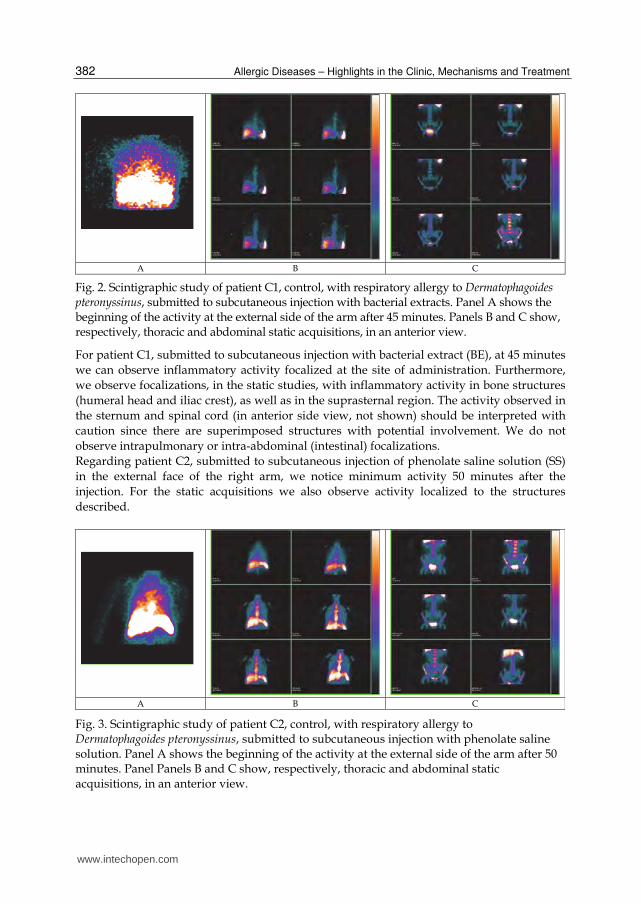
Regarding patient C2, submitted to subcutaneous injection of phenolate saline solution (SS)
in the external face of the right arm, we notice minimum activity 50 minutes after the
injection. For the static acquisitions we also observe activity localized to the structures
described.
Fig. 3. Scintigraphic study of patient C2, control, with respiratory allergy to Dermatophagoides pteronyssinus, submitted to subcutaneous injection with phenolate saline solution. Panel A shows the beginning of the activity at the external side of the arm after 50 minutes. Panel Panels B and C show, respectively, thoracic and abdominal static acquisitions, in an anterior view.
www.intechopen.com

Specific Immunotherapy and Central Immune System
383
Fig. 4. Scintigraphic study of patient 6 (anaphylaxis to Apis mellifera), submitted to subcutaneous injection with aqueous extract. Panel A shows the beginning of the activity at the external side of the arm after 35 minutes. Panel Panels B and C show, respectively, thoracic and abdominal static acquisitions, in an anterior view.
For the patient (Figure 4), under treatment for anaphylaxis to hymenoptera venom, we observe inflammatory activity earlier at the local of administration of the therapeutic allergen extract, but the systemic involvement is also observed in all of the posterior scintigraphic images. Higher activity was detected in the bone structures, when compared to patient 3 with anaphylaxis to latex. We did not detect intra-abdominal (intestinal) focalizations in this patient. For these patients, as well as for all the other studies, the sternum bone structures and spinal cord will not be interpreted because of all the reasons already discussed. For the patient allergic to pollens (Figure 5), the beginning of the inflammatory activity was detected at 35 minutes on the site of administration of the therapeutic extract. A systemic involvement in all the acquisitions was observed. In the armpit homolateral to the injection of the extract, we detect activity that persists throughout the study, similar to what happened in the cervical-lateral region of the neck. In the intestinal area we only observe focalizations at 60 and 120 minutes.
Fig. 5. Scintigraphic study of patient 8 with respiratory allergy (asthma and rhinitis) to grass pollen submitted to subcutaneous injection with depot extract. Panel A shows the beginning of the activity at the external side of the arm after 35 minutes. Panel Panels B and C show, respectively, thoracic and abdominal static acquisitions, in an anterior view.
www.intechopen.com

Allergic Diseases – Highlights in the Clinic, Mechanisms and Treatment
384
For patient on figure 6, the inflammatory activity at the site of administration of the allergoid extract is highly reduced, when compared to the patients previously described. The systemic activity persists in all the structures considered, including the intra-abdominal area, from minute 60 to 120.
Fig. 6. Scintigraphic study of patient 11 with respiratory allergy (asthma and rhinitis) to grass pollens, submitted to subcutaneous injection of a glutaraldehyde modified extract. Panel A shows the beginning of the activity at the external side of the arm after 50 minutes. Panel Panels B and C show, respectively, thoracic and abdominal static acquisitions, in an anterior view.
The SLIT administration induced a very precocious beginning of activity when compared to the subcutaneous route. Patient from Figure 7, submitted to treatment with D pt extract, the inflammatory activity in the oropharynx occurred 3 minutes after the administration of the drop extract. We also observed a more relevant involvement of the cervical-lateral structures of the neck, which persisted throughout the trial. The systemic involvement was also present in this patient in different structures: intrapulmonary, bones and suprasternal region.
Fig. 7. Scintigraphic study of patient 13, with respiratory allergy (asthma and rhinitis) to Dermatophagoides pteronyssinus, submitted to sublingual administration. Panel A shows the beginning of the activity in the oral region after 3 minutes. Panel Panels B and C show, respectively, thoracic and abdominal static acquisitions, in an anterior view.
www.intechopen.com

Specific Immunotherapy and Central Immune System
385
3.3.2 Quantitative analysis All the scintigraphic studies were submitted to a quantitative evaluation in order to
determine the corrected uptake coefficients. ROIs were draw in the areas with focalized
activity, which included: local of administration of the allergenic extract, suprasternal
region, humerus head and iliac crest. Besides this, every time intra-abdominal (intestinal)
focalizations were present, the respective UCCs were calculated. Naturally, for each
projection a ROI in the muscle area was draw, representing the background area.
Fig. 8. Corrected uptake coefficients in the region of the administration of the allergenic therapeutic extract for the subcutaneous injectable treatments (1. Dynamic; 2. Static) throughout the study (expressed in minutes). A: aqueous extracts; B: depot extracts; C: chemically modified extracts (allergoid).
The beginning of the inflammatory activity for patients submitted to SCIT was different
between the groups of patients in this study. The aqueous extracts for the treatment of
anaphylaxis were the ones that induced higher UCCs, both in the dynamic and static
evaluations. However, for the same type of extract, it seems that there are no relevant
differences in respect to the type of allergen in the treatment, Figure 8.
Patients under treatment with the depot extracts showed intermediate levels between
patients with aqueous extracts and allergoid. However, we should emphasize that a patient
under depot extracts to dust mites showed a highly reduced UCC at the spot of the
therapeutic injection, as well as a patient under treatment with pollinic extract modified
with glutaraldehyde. The control patient submitted to BE, showed very significant UCCs at
the local of administration in the evaluations made after the first 60 minutes. On the other
side, for the patient submitted to SS, the quantification of the activity at the local of injection
persisted always in much reduced levels.
www.intechopen.com

Allergic Diseases – Highlights in the Clinic, Mechanisms and Treatment
386
For patients submitted to treatment with SLIT, the inflammatory activity is detected very early in the process, and persists throughout time at relatively constant levels, Figure 9. The patients allergic to dust mites showed higher levels when compared to the patient allergic to Parietaria judaica. However, for this patient, the study took place during the pollinic period, and therefore a reduced therapeutic dosage was administrated, which can justify, eventually, the results obtained.
Fig. 9. Corrected uptake coefficients in the region of the administration of the sublingual allergenic therapeutic extract throughout the study (expressed in minutes). 1. Dynamic; 2. Static
The area located above the sternal furcula presented an early inflammatory activity, even
during the dynamic studies. Since this region includes an extremely rich set of vascular
structures, the effect of leukocyte recirculation maintained in the beginning of the study
cannot be excluded. Therefore, it was decided to only evaluate the coefficients acquired with
the static studies, Figure 10. Globally, the results are substantially higher than the control
patient submitted to SS injection. Furthermore, it was also observed a slow increase during
the experiment.
Fig. 10. Corrected uptake coefficients, taken at different time points during the experiment (minutes), obtained in the suprasternal region after the administration of the therapeutic extract. A: aqueous extracts; B: depot extracts; C: chemically modified extracts (allergoid); D: sublingual extracts
www.intechopen.com

Specific Immunotherapy and Central Immune System
387
The evaluation of the UCCs in the bone structures localized in the humeral head and iliac crest was then performed, exclusively for static acquisitions, Figure 11.
Fig. 11. Corrected uptake coefficients, taken at different time points during the experiment (minutes), obtained in the iliac crest after the administration of the therapeutic extract. A: aqueous extracts; B: depot extracts; C; chemically modified extracts (allergoid); D: sublingual extracts
The coefficients obtained in the two bone structures were similar and, globally, there was an
apparent progressive increase throughout the experiment. However, regarding the type of
extract and the method of administration, there wasn’t a characteristic profile of the
inflammatory activity in these structures. For the motives previously expressed, no other
bone focalizations were considered.
Fig. 12. Comparative results of the UCC obtained for the suprasternal of patients submitted to aqueous extracts (SCIT1) for the treatment of anaphylaxis; depot and allergoid extracts (SCIT2) for the treatment of respiratory allergy and sublingual aqueous extracts (SLIT), also for the treatment of respiratory allergy. The results obtained for control patients submitted to injection of bacterial extract (BE) or phenolated saline solution (SS). For all patients, samples were collected at 60, 180 and 360 minutes.
www.intechopen.com

Allergic Diseases – Highlights in the Clinic, Mechanisms and Treatment
388
Fig. 13. Comparative results of the UCC obtained for the iliac crest of patients submitted to aqueous extracts (SCIT1) for the treatment of anaphylaxis; depot and alergoids extracts (SCIT2) for the treatment of respiratory allergy and sublingual aqueous extracts (SLIT), also for the treatment of respiratory allergy. The results obtained for control patients submitted to injection of bacterial extract (BE) or phenolated saline solution (SS). For all patients, samples were collected at 60, 180 and 360 minutes.
In the inflammatory focalization dependent of the ROI from the suprasternal region, no prevalence for a type of extract or method of administration of the treatment seems to exist, Figure 12. Similarly, it can be observed an increase in UCCs throughout the experiment. The same considerations can be applied to the inflammatory activity induced in the iliac crest, Figure 13.
3.4 Discussion The administration of an allergenic therapeutical extract induces, always, an inflammatory reaction at the administration site that is characterized by the presence of clinically validated local symptoms. The modified extracts and the extracts delivered sublingually are the ones that induced lower secondary effects, as previously described. Although the main immunomodulator mechanism is already well established in SIT, it can dictate, in a limited and controlled approach, the effective inflammatory allergic reaction at the administration site. Therefore, we can admit that the autologous reinjection of leucocytes will have a migration dependent of the mast cell activation mediated by IgE (Togias, 2004; Kelly, 2007). This local inflammatory allergic reaction is, presumably, responsible for the dynamics of a response similar to allergy. Regarding the lymphocyte population, the circulatory migration to the tissue where the specific allergic reaction took place, is mediated by the expression of specific adhesion molecules that specify the type of cell implicated in the migration to the tissue (Togias, 2004). The migration of Th2 cells is associated to the expression of VCAM-1 and P-selectin, under effect of IL-4 and IL-13, that depends on the activation of lymphocyte transduction signals by transcription factors similar to STAT6 (Lukacs, 2000). On the other hand, the migration of Th1 cells seems to be dependent on the expression of STAT4, due to the effect of IL-12, and associated to the RANTES and ICAM-1 mechanism. In an interesting study with intradermic injection of allergenic peptides without the ability to induce an IgE response, it was possible to observe lymphocyte trafficking from the vascular department (Haselden, 1999). Also, the intervention of T-reg cell in the mechanisms of immunomodulation of SIT are well established either by inducing the population of Th1 cells or reducing the number of Th2 (Woodfolk, 2007; Francis, 2003). Nevertheless, in these cells, it is not clear which is the specificity towards the allergen or what is the eligibility of the method of the induction (systemic or inhaled). Furthermore, it
www.intechopen.com

Specific Immunotherapy and Central Immune System
389
was also not clear what is the dosage required for induction or what type of regulatory cells are induced (natural T-reg produced by the thymus and with an effect dependent of cellular contact or adaptive T-reg cells induced on the periphery and with the effect dependent of IL-10 or TGF-┚) (Woodfolk, 2007). At the same time, other lymphocytes, such as CD8+ e NKT cells, have the ability to respond to the allergen and represent other vectors involved in the SIT mechanisms (Woodfolk, 2007, Agea, 2005)). As previously mentioned, DCs are considered central cells in inflammation and immune
tolerance often reported in the SIT mechanisms. These cells represent the antigen presenting
cells (APC) that, after migrating to the regional lymph node, induce an effective and
differentiated immune response. Nevertheless, this is a very heterogeneous and diversified
population, ubiquitously present, although in a reduced number when compared to other
resident or circulation populations (von Bubnoff, 2002; Novak, 2004). Furthermore, it is
known that myeloid DC cells can migrate from the blood to the tissues in order to capture
antigens (Novak, 2004; Schmit-Weber, 2002, Allam, 2003). In specific situations, and under
appropriated stimulus, the peripheral monocytes can differentiate into myeloid DC (von
Bubnoff, 2002). The skin and sublingual mucosa are, naturally, distinct. Therefore the mechanisms derived from the administration of the therapeutical allergenic extract should reflect this difference. The Langerhans cell (LC) is a DC myeloid cell (DC1) and are the most representative APCs in the skin. After a correct local stimulus, the monocyte chemotactic protein (MCP) determines the recruitment of LC progenitors from the bone marrow to the skin and the subsequent migration to the peripheral lymph nodes (Novak, 2004). The DC in the oral mucosa presents differences relatively to the skin, namely it expresses a higher number of MHCI and MCHII molecules, as well as the co-stimulation molecules CD40, CD80 and CD86 and also more Fc┛RIII (CD16), Fc┛RI (CD64) and FcεRI receptors (Allam, 2003). Several lymphoid structures are present in the oropharynx: tonsils, lymphatic ganglions and diffuse lymphoid tissue (Bienestock, 2005). It is well established that plasmacytoid DCs (DC2) are the APC most abundant in the Waldeyer’s tonsillar ring and in the ganglion areas dependent on T cells, but that these cells are unable to effectively internalize the antigen (Novak, 2004). This is surely, a biologic characteristic of this region with consequences on the immune response on this area. The subcutaneous tissue is, undoubtedly, composed by a population of resident cells that
are biologically less active that the ones present in the oral mucosa. Nevertheless, the
therapeutical administration of allergenic extracts by both ways has proven to be effective
and able to induce an immunomodulator response in patients of IgE mediated allergy. This
project did not intended to validate the efficacy of this therapy, broadly documented, but to
study the dynamic of the resulting mechanism due to the administration of the therapeutical
extract in patients maintained in the previously determined individual dosage.
Theoretically, it would be extremely relevant to have monitored, under the same
methodology, this treatment in different periods throughout the year, but the radioactive
dosage accumulated would be ethically unacceptable.
In patients under subcutaneous injectable treatment, the anaphylaxis treatment with aqueous extracts induced the fastest signs of inflammatory activity in the administration site. The activity became visible 35 minutes after the administration, while with depot extracts, the activity was only visible between 40 and 45 minutes after the administration. In all patients, an increase in the intensity of the activities was observed throughout the
www.intechopen.com

Allergic Diseases – Highlights in the Clinic, Mechanisms and Treatment
390
observation period. The higher activity induced by the aqueous extracts did not translate into more exuberant clinical signs. The patients in SCIT to latex presented local clinical signs more pronounced than two patients allergic to Apis. In these two patients, the inflammatory activity was bigger and deeper, which can be explained by a higher amount of protein and the volume of extract administered. In patients with respiratory allergy submitted to SCIT with depot extracts there were no evident differences connected to the extract composition, either mites or pollens. Regarding the allergoid extracts, a reduced inflammatory activity was observed at the
administration site, similarly to what was previously reported in the literature and
demonstrated in daily clinical practice (Casanovas et al., 2005, 2007; Ariano et al., 1999). The
modification of the allergenic with glutaraldehyde reduced the local inflammatory activity
to levels similar to the ones observed in the control patients submitted to SS. In patient C1,
asthmatic allergic to dust mites submitted to subcutaneous injection with BE, a local
inflammatory activity was observed 45 minutes after the administration. This is an extract
with immunomodulator activity, which induces the response of an IgG polyclonal antibody
well documented in the literature (Clot, 1997). Although the local demonstration of activity
was expected, the time necessary to observe activity was also extremely reduced.
The patient submitted to a phenolated solution presented a minimal local inflammatory
activity only seen 60 minutes after the subcutaneous injection. Similarly to the patients
studied in the previous chapter, it is considered that the irritation potential of this
preservative can justify this result (Spiller et al., 1993).
In this study it was possible to assess and confirm the dynamics of the inflammation
induced by the subcutaneous application of an allergenic extract. These results, although not
surprising, were not unequivocally demonstrated, namely in terms of extension, depth of
the adjacent region or evolution of the reaction along the course of the experiment. One
advantage of the depot extracts when compared to the aqueous extracts, besides the safety
profile, was the slower release of the allergenic protein (Wheeler & Woroniecki, 2004). In our
patients we clearly observed a lower activity of these extracts, despite the fact that the
patients with the aqueous extracts presented a worse level of basal allergic disease. The
lymphocyte traffic seemed to be more intense and persistent with these extracts since the
soluble allergenic is more readily available. In all patients submitted to SCIT, it is possible to
observe systemic focalizations before the locally induced inflammatory activity. In the
dynamic acquisition during the first 60 minutes, the anterior thoracic projection only
permits to infer the potential activity in this view. In the intramedullary area of the humeral
head is evident a significant intensity, surely dependent on the recirculation of cells
representing all leukocyte cell populations.
In patients submitted to SCIT, the activity in bone structures and in thymic tissue presented
a very intense activity that increased throughout the experiment. It should be noticed that
these structures anatomically overlap to other organs from the central immune systems, but
that in the peripheral immune lymphoid system, such as regional ganglion, and presumably
in the region of the mediastinum, there is also evidence of activity. Furthermore, the mucosa
immune system seems to be involved namely the pulmonary and less consistently the
intestinal. The early involvement of the central lymphoid structures was an unexpected
event, but was very consistent in all patients.
Obviously, with the methodology used it is not possible to determine the cell type and profile that is responsible by the activity induced by the migration from the circulation to a
www.intechopen.com

Specific Immunotherapy and Central Immune System
391
specific focalization. Nevertheless, given the actual knowledge regarding the mechanism that modulate this therapeutic, it is possible to admit that a diversified set of cells with different activation profiles and with divergent implications in the different structures. The subcutaneous administration of the allergenic extract, particularly with aqueous
extracts, defines an evident local inflammatory process. To this level, we admit that some
conjunctive mast cells can be activated, although with limited intensity. As described in the
literature, we also admit that the presence of conjunctive DCs, with IgE receptors, can favor
this type of reaction. However, the most likely mechanism can depend on an effective
presentation of the allergen to presenting cells and posterior migration to local-regional
structures. This mechanism could explain the ganglion activity observed in the homolateral
arm, with the involvement of other ganglion structures more distant, such as the ones
present in the cervical lateral-external area.
On the other hand, we admit the influx from the circulation of DC or monocytes into the site of
administration of the allergenic extract, since it is well known the ability of these cells to
maturate into DCs or phagocytes. This dual mechanism, that simultaneously induce a
biological defense from the aggression and favors an effective immune response, are probable
local mechanisms. Therefore, the local administration of therapeutical allergens limits the local
response that rapidly seems to have local-regional and systemic consequences. Probably, not
only the biologic mediator produced in situ, as well as the following cell migration,
presumably DCs, and the cellular limitations in venular structures in the proximity of the
allergenic, allow the amplification of the systemic effect by mechanisms, possibly similar to the
ones verified in the allergic reaction (Togias, 2004; Kelly, 2007).
The qualitative analysis of our SCIT patients corroborates, although indirectly, the
complexity of the tolerance mechanisms, well described in the literature. The anatomic
involvement of structures implicated with organs of the central immune system seems to
sustain these effects. The activity observed in the bone marrow areas allow us to speculate
that it could limit the modification of cellular profiles, namely lymphocyte, that has been
documented in these patients (Denburg et al., 2000). Also, the demonstration of the
contribution of regulatory cells can be sustained by our results given the involvement of the
anatomic region reported to the thymic tissue, whose function persists throughout life
(Arellano et al., 2006). Nevertheless, the pace of this process and the systemic involvement,
evidenced by the focalization of the inflammatory activity, is clearly higher than expected,
given the present knowledge. It should be noticed that this dynamic study evaluates, in a
certain way, the biologic response in the whole organism. This contrasts with the usage of
small biologic samples (such as tissues or humors), a procedure that is very common in most
clinical and experimental studies.
We admit that the lungs are involved in the influx of leukocytes to the BALT, but it should
be noticed that this is a preponderant location for the clinical symptoms that lead to
treatment in patients with respiratory allergy.
In patients in SLIT, the beginning of the inflammatory activity in the site of administration
was substantially earlier than the observed in patients in SCIT. In the two patients allergic to
mites and in the patient allergic to Parietaria judaica, the activity was noticed 3 and 5 minutes
after the respective induction. Despite this early onset of activity, no substantial differences
were registered when the final results were compared, namely in the effects of regional or
distant structures.
www.intechopen.com

Allergic Diseases – Highlights in the Clinic, Mechanisms and Treatment
392
In fact, it seems evident that since the administration of the extract, a continuous increase of the local inflammatory activity as well as in cervical structures, particularly in the cervical lateral-external region. As described previously for SCIT patients, the venous recirculation at very early stages does not allow an effective validation of this effect, but the assessment in later periods, both in the dynamic or static acquisitions, there is an evident focalization in these cervical structures. For these patients, and with exception of the patient injected with extract modified with glutaraldehyde, and using the color scale, the intensity of the oropharynx is clearly inferior than the one verified in the injection site. Other clear difference, is the absence of a progressive increase of the inflammatory response focalized at the administration site verified in subsequent static evaluations. However, the systemic effect is very similar to the observed in patients under injectable treatment. We observe pulmonary focalizations as well as in regions reported to the organs from the immune central system. In these regions, the areas reported to the bones and in suprasternal, the images from the static studies support an increase of activity throughout the experiment. In patients under SLIT, the extract was swallowed 3 minutes after the administration. It would be expected an intra-abdominal involvement, with intestinal focalizations. However, in none of these 3 patients that effect was observed. We admit that the extract can be completely degraded in the stomach, which would prevent an effect in the intestinal mucosa. Our results seem to corroborate the therapeutic inefficiency of the oral immunotherapy, which is not scientifically validated by the literature (Bousquet et al., 1998). It is not possible to define and characterize the migrating population that focalizes in the distinct structures in patients under SLIT. Despite this, the available scientific knowledge allows us to assume that the allergenic persistence in the sublingual mucosa, as previously demonstrated (Bagnasco et al., 2005), allows the induction of the following immune response. As previously described, abundant lymphoid structures with specific biologic characteristics are present in this mucosa. The DC permits a fast transport to the tonsils and lymphatic ganglions in a magnitude that cannot be compared to the subcutaneous tissue. However, it is well established that the majority of DCs present in the tonsils are DC-2. These cells do not have the capacity to effectively internalize the allergenic protein and efficiently present it to the T-cell. Therefore, and although in smaller numbers, the DC-1 cells probably have a preponderant function. As a result, we admit that there might be an influx from the circulation of DC and monocytes, and that these, once in the mucosa, will differentiate and maturate. The early start of the activity will, presumably, result from the presence and availability of a biologically more active and diversified resident population. The reduced population of mast cells in the oral mucosa limits the local inflammatory response when compared to the subcutaneous administration. Therefore, and in theory, we can predict that the resulting mechanism derives from the activation of DCs and the presentation to lymphocytes in structures in the oral mucosa and that are dependent from the immune system. Thus, the therapeutical extract produces a powerful stimulus that initiates a local and systemic immune response, surely by regulating biologic mediators of cytokines and chemokines, as well as by limiting the entry of circulating cells from the vascular-lymphoid structures present in the oral mucosa. The systemic effects in areas of the central immune system, similarly to what was observed for the injectable extracts, can corroborate the tolerance and immunomodulation, presently well documented in the therapeutical intervention. In patients under injectable therapy, the UCCs results obtained in the homolateral armpit are higher that was expected by the exclusive visualization of the scintigraphic images. We
www.intechopen.com

Specific Immunotherapy and Central Immune System
393
admit that the results obtained in this focalization are, eventually, due to the migration of lymphatic cells from the administration site of the therapeutical extract to the ganglion structures present in the armpit region that allow a therapeutical amplification with natural systemic effect. In fact, functional studies in melanoma using a radio-labeled pharmaceutical compound administered intradermally evidenced the high rate of lymphatic migration to distant ganglion structures (Alazraki et al., 2002). Since the most important ganglion structures of the subcutaneous cellular tissue of the arm are located in the armpit, that the effective presentation of the allergenic protein to the T-cell in the lymph node is mediated by the DC and that the intralymphatic migration is very fast, the UCCs obtained in our patients are, probably, justified. The eventual lymphatic migration proportions, are in fact, an amplification of the immune mechanism with characteristics that clearly favors tolerance. Therefore, it could lead not only to an effective diffusion of the response throughout the immune-lymphoid system, as well as a recirculation of cells from the allergenic administration site to distant central structures and organs that can allow the synergistic effects that concur with the immunomodulation of this therapy. Therefore, the presumed ascending drainage justifies the precocious and persistent UCCs induced by the subcutaneous administration of the allergen in other vascular-lymphatic structures, anatomically located in the lateral-external region of the neck. Eventually, this process allows an amplification of the response to cervical structures with high biological activity. To this level, we do not observe significant differences of UCCs due to usage of different injectable extracts. Although we verify focalization in the pulmonary region throughout the dynamic acquisition, we only calculated the coefficients in the static acquisition. The intense venous recirculation of leukocytes after the reinjection could falsify the results. However, all our patients presented intrapulmonary activity. Particularly in patients undergoing treatment for respiratory allergy, the influx of these cells to the targeted organ is compatible to the methods sensitivity. This way, the recirculation of leukocytes between the vascular compartment and the mucosa of the different organs of the immune system allow the convenient organic homeostasis that justifies the reduced numbers of pulmonary UCCs verified in control patients. Given the results obtained in the patient submitted to BE, we presume that the unspecific bacterial immunomodulators have, besides favoring an IgG polyclonal response, a more central and selective regulator effect than the one presumed until now, but for which the clinically efficacy has been shown (Bousquet & Fiocchi, 2006). Regarding the patient allergic to dust mites submitted to SS injection, the reduced UCCs values were consistent throughout the experiment, and we presume that are derived from the organic recirculation of leukocytes, and correspond to the basal function of this tissue The activity observed in the bone structures was confirmed by the UCCs calculated in the two considered ROIs. There were no differences in the results obtained in the medullary area of the humeral head or the iliac crest. Our results suggest that either the bone marrow or the functional thymic tissue, organs from the central immune system, are preferential targets of the induction mechanism of SCIT, regardless of the type of allergenic extract. It should be noticed that in patients treated with modified extracts, allergoid, the dosage of allergenic used was substantially higher than the dosages in the depot extracts, and that the UCCs obtained in the regions associated with bone marrow were also higher. However, the size of the sample does not allow us to
www.intechopen.com

Allergic Diseases – Highlights in the Clinic, Mechanisms and Treatment
394
conclude if this treatment is more selective or effective. Nevertheless, we presume that given the previously described mechanisms, the bone marrow may participate in the SIT mechanism since earlier stages, right after the injection of the extracts. In the suprasternal region, the UCCs were very similar in the different groups under active treatment and significantly distinct from the ones obtained in the patient submitted to SS injection. This data corroborates the eventual physiological dynamic of the leukocytes, possibly dependent of the persistent limitation of the medulla, even in cells apparently mature that were collected from the venous compartment. In the intra-abdominal region, the UCCs obtained in the intestinal focalizations, observed occasionally in the scintigraphic images, confirmed the involvement of this mucosa. The calculations were only performed in focalizations areas during the first 4 hours, in order to minimize interferences due to faecal elimination. For the three patients under sublingual treatment we do not have a control that we can compare to. Despite this, the values obtained in the asthma and rhinitis patients allergic to dust mites submitted to BE or SS in the nasal region (see previous chapter), are similar to the type of response verified in SLIT patients. The capture and the inflammatory activity on the site of the administration of the
therapeutic extract evidenced a very precocious activity that subdue in relatively high
values throughout the experiment. These results are consistent with previously described
dynamic studies (Bagnasco et al., 2005). The quantitative analysis does not evidence an
increase of the local activity as seen in the majority of patients submitted to injectable
extracts. In this mucosa, the presence of mast cells is extremely reduced which, despite the
persistence of the extract (even after it was swallowed), does not induce a broader influx of
circulating leukocytes due to this mechanism. Therefore, we presume that the much more
limited activity observed can be due to circulating DCs and mast cells that have a
recognized ability to mature and differentiate in the offended tissues. The readily
availability of immune-lymphoid tissue in this localization will allow a fast amplification
and dissemination of cells with the ability to induce an immunomodulator mechanism.
In patient 15, submitted to sublingual treatment with Parietaria judaica extracts, the UCCs
obtained throughout the observation period were the lowest in these three patients. In fact,
since the test was conducted during the pollinic period, we established a reduction of the
allergenic dosage during the pollination period of this herb. It is a clinical procedure well
established in the therapeutic with injectable extracts, although not consensual in sublingual
treatment (Bousquet, 2006; Bousquet et al., 1998). Despite this, and since this patient lives in
a geographic area with high levels of aeropalinologics, it was decided to reduce the
maintenance dosage to 2/3 (10:15 drops). Eventually, this procedure had implications in the
type of response that was evidenced, not only in the local activity as well as in the distant
focalizations.
In patients under SLIT treatment, the UCCs obtained in the structures localized in the lateral-external region of the neck confirmed the analytic results from the scintigraphic images. The apparent descendent drainage in these vascular-lymphatic systems corroborates our proposal, particularly in the intrapulmonary capture. In the anatomic regions reported to the organs of the central immune system we observed UCCs with similar behavior than those verified in the patients under injectable treatment. Therefore, we presume that in SLIT there is also an early participation of the bone marrow and the functional thymic tissue.
www.intechopen.com
Specific Immunotherapy and Central Immune System
395
4. Involvement of the organs from the immune system
In SCIT, we admit that the APC migration to ganglion structures is determinant to obtain a systemic effect and to acquire an immunomodulator and/or tolerance state. In patients submitted to SLIT, the inflammatory activity was observed in the first minutes, much earlier but with clearly lower gradients throughout the experiment. As previously described, the morphological characteristics of the conjunctive subcutaneous tissue and the oral, in particular from the sublingual region, are completely distinct. Therefore, the local availability of the whole array of the adjacent immuno-lymphatic-ganglion structures allows the effectiveness of the early stages of the mechanism. Paradoxically, the characteristics of the DCs present in the oral mucosa, mainly from the plasmacytoid type, are responsible for a less effective response (Novak et al., 2004). Previous work evidenced the presence of the allergenic extract in the sublingual mucosa long after it was swallowed, but it was extremely reduced 24 hours after that administration (Bagnasco et al., 2005). This difference, relatively to the subcutaneous administration, has natural implications in the periodicity of the therapeutical application. The amplification of the systemic effect seems to result from a descending involvement of structures presumably vascular-lymphatic in the lateral-external of the neck by a mechanism similar to SCIT. Given the differences between the two administration methods and the type of therapeutic extract, the results obtained suggest a potentially selective influx of cells from the mononuclear-phagocyte system to the deposition site, similarly to the allergic reaction, which was documented in an interesting study done in ten patients submitted to an inhaled aggression (Upham et al., 2002). Naturally, the mediators produced locally will have a potential modulator effect in the adjacent vascular endothelium, which allows a possible immunomodulator effect in circulating leukocytes that allows a systemic involvement. The recirculation and following focalization of intrapulmonary leukocytes was observed in
all patients. The presence of the bronchus-associated lymphoid tissue (BALT) supports these
results, in particular since this mucosa is a potential target in patients undergoing treatment
of respiratory allergy. However, regarding the involvement of the intestinal mucosa, where
the gut associated lymphoid tissue (GALT) is located, focalization of the inflammatory
activity was occasionally observed. The SLIT with allergenic deglutition three minutes after
the deposition did not produced any additional gain. In fact, we admit that the protein
degradation can occur as result of the gastric enzymatic degradation as proposed in the
literature (Bousquet et al., 1998). The lymphocyte cellular modulation from a mainly Th2 profile into a lymphocyte profile mostly Th1, as well as the induction of T-lymphocytes with regulatory characteristics, are the most studied and documented mechanisms in the literature (Larché et al., 2006; Till et al., 2004; Canonica & Passalacqua, 2003). The complexity of the processes involved allows a constant effect throughout the treatment, but its dynamic mechanism is not well established. The need to extend in time the specific treatment translates the need to promote an immunologic memory status. The patients studied in this project were well documented, both clinically and laboratorial, and all were in maintenance treatment. Although we know several mechanisms involved, it is not known the dynamics and the regions implicated after the administration of the extract, nor in the induction stages or in maintenance periods. It seems obvious the involvement of organs from the central immune system in the SIT action mechanisms. It also appears, given the results obtained in allergic patients submitted to specific provocation, that the participation of these organs could have a dynamic and
www.intechopen.com

Allergic Diseases – Highlights in the Clinic, Mechanisms and Treatment
396
pace with similar magnitude. These results look as redundant or expectable, but we had no previous knowledge about the location and timing of the immunomodulation process that has been so well characterized in the laboratory. The induction of medullar cellular clones with phenotypes that favor the secretion type Th1 or clones precursors of DCs with tolerogenic characteristics are events that sustain the activity of the bone marrow, and is present at an early stage in patients undergoing treatment (van Helvoort et al., 2004; Moingeon et al., 2006). The essential function of several regulatory T-cells, well placed and with central function in the SIT mechanism, is an element that supports not only the intervention of bone marrow as well as the thymic activity (Schmidt-Weber & Blaser, 2005; Moingeon et al., 2006). Despite the anatomic involution, the organic function persists in adulthood (Hakim et al., 2005; Harris et al., 2005; Arellano et al., 2006). In this regard, we should notice that in one patient we had the opportunity to acquire a scintigraphic 21 hours after the simultaneous administration of leukocytes labeled with 99mTc-HMPAO and allergenic aqueous latex extract. In this patient we observe the persistence of the inflammatory activity in both the suprasternal and bone medullar areas, Figure 14. It should be noticed that the half-life of the radio-labeled compound is 6 hours, and therefore the acquisition occurred at a time well past its half life. Nevertheless, the activity persisted at the administration site, allowing the therapeutic effect to continue, as well as activity in intra-bone areas and in the suprasternal region, which we judge representative the functional thymic tissue. The study in this patient was done occasionally and was, in fact, a surprise, revealing
activity that we supposed it was not possible due the anatomic physical conditionings. We
proceeded with the quantitative evaluation that unequivocally contributed the qualitative
assessment done in this patient. In Figure 15 we show the UCCs obtained for this patient
after 6 hours (last programmed observation that coincides with 99mTc half-life) and 21 hours.
Although the UCCs are lower after 21 hours when compared to the values obtained after 6
hours, it is evident that, despite after nearly four half-lives, the activity reported is very
significant. We should highlight that the UCC in the ROI draw in the homolateral armpit
maintains constant values of activity, which supports a continuous involvement. The study in this patient, since it is not likely a continuous recirculation of leukocytes without focalization and influx to this organ, allows us to deduce that we were able to obtain an image of the functional thymic tissue. Our results support the research developed since 1985 by a Canadian group that sustains the involvement of the central immune system’s organs, mainly bone marrow, in the pathophysiology of the allergic inflammation (Denburg et al., 1998). The results obtained in experimental models and in humans with allergy are essential, regarding the participation of hematopoietic mechanisms during the response to an allergenic (Denburg et al., 1998; Denburg & van Eeden, 2006, Sehmi et al., 1996; Cyr & Denburg, 2001). In a comparative study with 153 patients with pollinic rhinoconjunctivitis, the effect of the administration of SIT either subcutaneously or intralymphatic was compared (Senti et al., 2008). In the later group, the administration of the pollinic allergen directly in a inguinal lymphatic ganglion using ultrasound as a control, with only three injections separated by 4 weeks, in the response to a specific nasal induction 4 months after the last administration, required a dosage ten times higher than the one required in patients under SCIT treatment for three years and with a cumulative dosage drastically higher and with multiple allergenic injections. In addition to the clinical efficacy, the adverse effects were neglectable and the lymphatic intraganglionic injection was very well tolerated. This promising and innovative
www.intechopen.com

Specific Immunotherapy and Central Immune System
397
study can induce a complete reversion on the treatment of IgE mediated allergy. It should be noted that these results were obtained with conventional allergens. However we consider that the future use of highly selective peptides directly administered in immune structures can induce an even more elective therapeutical effect.
Fig. 14. Scintigraphic study in patient with anaphylaxis to latex, submitted to subcutaneous aqueous extract. Static acquisitions, anterior thoracic (A) and abdominal (B) views, 21 hours after the beginning of the study.
Fig. 15. Corrected uptake coefficient obtained in static acquisitions after 6 and 21 hours in different locations in patient with the diagnosis of latex anaphylaxis submitted to subcutaneous aqueous extract treatment.
5. Final remarks
SCIT limits the visualization of the inflammatory activity in systemic focalizations earlier than at the site of the extract administration. Generically, the local inflammatory activity
0
10
20
30
40
50
60
70
6 hour 21 hour
Local
Armpit
Suprasternal area
Iliac
www.intechopen.com
Allergic Diseases – Highlights in the Clinic, Mechanisms and Treatment
398
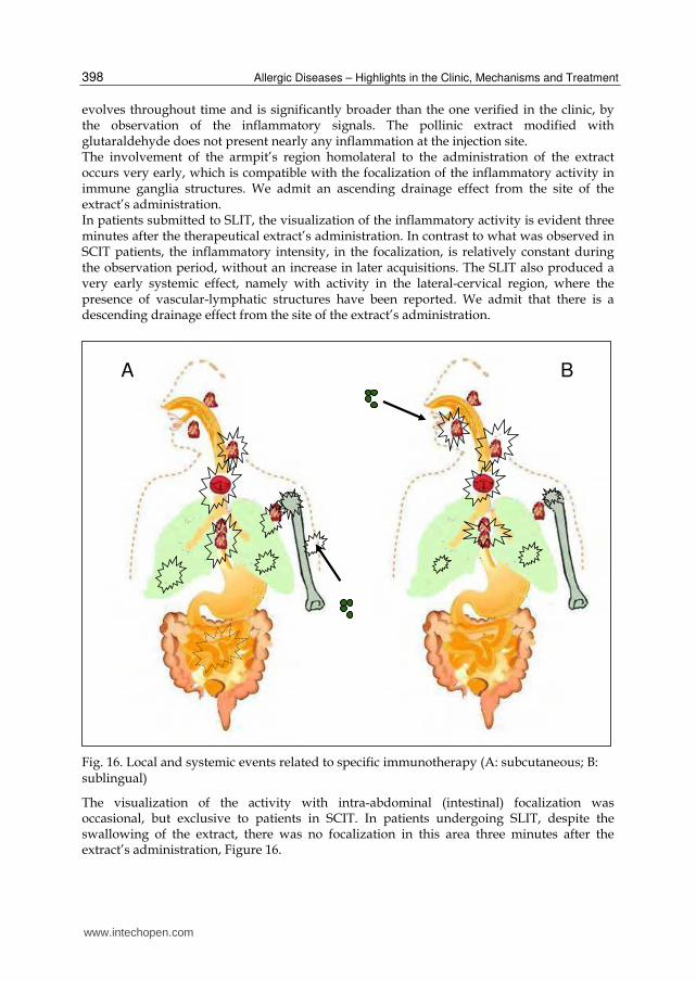
evolves throughout time and is significantly broader than the one verified in the clinic, by the observation of the inflammatory signals. The pollinic extract modified with glutaraldehyde does not present nearly any inflammation at the injection site. The involvement of the armpit’s region homolateral to the administration of the extract occurs very early, which is compatible with the focalization of the inflammatory activity in immune ganglia structures. We admit an ascending drainage effect from the site of the extract’s administration. In patients submitted to SLIT, the visualization of the inflammatory activity is evident three minutes after the therapeutical extract’s administration. In contrast to what was observed in SCIT patients, the inflammatory intensity, in the focalization, is relatively constant during the observation period, without an increase in later acquisitions. The SLIT also produced a very early systemic effect, namely with activity in the lateral-cervical region, where the presence of vascular-lymphatic structures have been reported. We admit that there is a descending drainage effect from the site of the extract’s administration.
Fig. 16. Local and systemic events related to specific immunotherapy (A: subcutaneous; B: sublingual)
The visualization of the activity with intra-abdominal (intestinal) focalization was occasional, but exclusive to patients in SCIT. In patients undergoing SLIT, despite the swallowing of the extract, there was no focalization in this area three minutes after the extract’s administration, Figure 16.
A B
www.intechopen.com
Specific Immunotherapy and Central Immune System
399
The anatomic regions reported to the central immune system (bone marrow and suprasternal area) evidenced a constant growth in UCCs during the experiment. The SIT triggers an early and continuous influx of leukocytes, from the circulation into the central immune organs, with potential implications in the immunomodulatory mechanism. Regarding the response’s magnitude in the bone marrow and functional thymic tissue, there were no apparent differences regarding the type of extract and the administration type during the first six hours after the administration of the therapeutical extract. The persistence of the therapeutical extract administered subcutaneously indicates an inflammatory activity and a continuous influx of inflammatory cells. In the sublingual administration, the influx and local activity are lower, without any evidence of an increase in the inflammatory activity throughout the experiment. Therefore, patients under SLIT treatment will require administrations more frequent and with shorter breaks between administration in order to obtain a persistent therapeutic effect. Our results reinforce the need for new and elective pharmacologic investigation strategies, focusing on the mainstay function of the central immune organs in the treatment of systemic inflammatory disease such as allergy. In fact, in face of the current knowledge of the immunological effects induced by SIT, namely the effect on T-regs with a long-lasting biological effect, this would only be possible if a central immune cellular modulation had occurred.
6. Acknowledgment
We would like to thank Dr Margarida Abrantes (IBILI, Medicine Faculty of Coimbra University. Portugal), Dr Daniel Machado (Immunoallergology Department, Coimbra University Hospital. Coimbra. Portugal), and Prof J Pedroso Lima (Nuclear Medicine Department, Coimbra University Hospital. Portugal) for all the performance and commitment to this project. The authors thank Dr Joana Branco and Dr André Faustino for the precious English writing assistance.
7. References
Agea, E.; et al. (2005). Human CD1-restricted T cell recognition of lipids from pollens. The Journal Experimental Medicine,.Vol.202, No.2, (July 2005), pp. 295-308, ISSN 0022-1007
Akdis, C.A. & Akdis, M. (2011) Mechanisms of allergen-specific immunotherapy. The Journal of Allergy and Clinical Immunology,.Vol.127, No.1, (January 2011), pp. 18-27, ISSN 0091-6749
Akdis, C.; et al. (2001). Mechanisms of IL10 induced T cell inactivation in allergic inflammation and normal response to allergens. International Archives of Allergy and Immunology, Vol.124, No.1-3, (January-March 2001), pp. 124: 180-182, ISSN 1018-2438
Allam, J.P.; et al. (2006). Comparative analysis of nasal and oral mucosa dendritic cells. European journal of allergy and clinical immunology (Allergy), Vol.61, No.2, (February 2006), pp. 166-172, ISSN 0105-4538
Allam, J.P.; et al. (2003). Characterization of dendritic cells from human oral mucosa: a new Langerhans_ cell type with high constitutive FcepsilonRI expression. The Journal of Allergy and Clinical Immunology, Vol. 112, No.1, (July 2003), pp. 141-148, ISSN 0091-6749
Alvarez-Cuesta E.; et al, (2006). Standards for pratical allergen- specific immunotherapy. Allergy, 2006; Vol. 61, Suppl.82, (October 2006), pp. 1-20, ISSN 0105-4538
www.intechopen.com

Allergic Diseases – Highlights in the Clinic, Mechanisms and Treatment
400
Alazraki, N.; et al. (2002). Procedure guideline for lymphoscintigraphy and the use of intraoperative gamma probe for sentinel lymph node localization in melanoma of intermediate thickness. Journal Nuclear Medicine, Vol. 43, No.43, (October 2002), pp. 1414-1418, ISSN 0161-5505
Ariano, R., et al. (1999). Long-term treatment with allergoid immunotherapy with Parietaria. Clinical and immunologic effects in a randomized, controlled trial. European journal of allergy and clinical immunology (Allergy), Vol.54, No.4, (April 1999), pp. 313-319, ISSN 0105-4538
Arellano, M.V.; et al. (2006). Thymic Function-Related Markers Within the Thymus and Peripheral Blood: Are They Comparable? Journal of Clinical Immunolology, Vol.26, No.1, (January 2006), pp. 96-100, ISSN 0271-9142
Bagnasco, M.; et al. (2005). Pharmacokinetics of Der p 2 allergen and derived monomeric allergoid in allergic volunteers. International Archives of Allergy and Immunology, Vol.138, No.3, (November 2005), pp. 197–202, ISSN 1018-2438
Bagnasco, M.; et al. (1997). Absortion and distribution kinetics of the major Parietaria judaica (Par j 1) administered by noninjectable routes in healthy human beings. The Journal of Allergy and Clinical Immunology, Vol.100, No.1, (July 1997), pp. 122-129, ISSN 0091-6749
Bagnasco, M.; et al. (2001). Pharmacokinetics of an allergen and a monomeric allergoid for oromucosal immunotherapy in allergic volunteers. Clinical and Experimental Allergy, Vol.31, No.1, (January 2001), pp. 54-60, ISSN 0954-7894
Bienestock, J. & McDermott, M.R.. (2005). Bronchus- and nasal-associated lymphoid tissues. Immunological Reviews. Vol.206, (August 2005), pp. 22-31, ISSN 0105-2896
Bousquet, J.. (2006). Sublingual immunotherapy: validated! Allergy, Vol.61, Suppl.81, (July 2006), pp. 5-31, ISSN 0105-4538
Bousquet, J. & Fiocchi, A. (2006). Prevention of recurrent respiratory tract infections in children using a ribosomal immunotherapeutic agent: a clinical review. Paediatric Drugs, Vol.8, No.4, pp. 235-243, ISSN 1174-5878
Bousquet, J.; et al. (1998). Allergen immunotherapy: therapeutic vaccines for allergic diseases. A WHO position paper. The Journal of Allergy and Clinical Immunology, Vol.102, No.4, (October 1998), pp. 558-562, ISSN 0091-6749
Canónica, G.W. & Passalacqua, G. (2003). Noninjection routes for immunotherapy. Journal of Allergy and Clinical Immunology, Vol.111, No.3, (March 2003), pp. 437-448, ISSN 0091-6749
Casanovas, M.; et al. (2005). Comparative Study of Tolerance between Unmodified and High Doses of Chemically Modified Allergen Vaccines of Dermatophagoides pteronyssinus. International Archives of Allergy and Immunology, Vol.137, No.3, (July 2005), pp. 211–218, ISSN 1018-2438
Casanovas, M.; et al. (2007). Safety of immunotherapy with therapeutic vaccines containing depigmented and polymerized allergen extracts. Clinical and Experimental Allergy, Vol. 37, No.3, (March 2007), pp. 434-440, ISSN 0954-7894
Clot, J. (1997). Pharmacology of ribosomal immunotherapy. Drugs, Vol. 54, Suppl.1, pp. 33-36, ISSN 0012-6667
Cyr, M.M. & Denburg, J.A.. (2001). Systemic aspects of allergic disease: the role of the bone marrow. Current Opinion in Immunology, Vol.13), No.6, (December 2001), pp. 727-732, ISSN 0952-7915
www.intechopen.com

Specific Immunotherapy and Central Immune System
401
Denburg, J.A.; et al.. (1998). Hemopoietic mechanisms in allergic airway inflammation. International Archives of Allergy and Immunology, Vol.117, No.3, (November 1998), pp. 155-159, ISSN 1018-2438
Denburg, J.A.; et al.. (2000). Systemic aspects of allergic disease: Bone marrow responses. The Journal of Allergy and Clinical Immunology, Vol.106, Suppl.5.2, (November 2000), pp. S242-6, ISSN 0091-6749
Denburg, J.A. & van Eeden, S.F.. (2006). Bone marrow progenitors in inflammation and repair: new vistas in respiratory biology and pathophysiology. The European Respiratory Journal, Vol. 27, No.3, (March 2006), pp. 441-445, ISSN 0903-1936
Francis, J.N.; et al.. (2003). Induction of IL-101CD41CD251 T cells by grass pollen immunotherapy. The Journal of Allergy and Clinical Immunology, Vol.111, No.6, (June 2003), pp 1255-1261, ISSN 0091-6749
Frew, A.J.. (2010). Allergen immunotherapy. The Journal of Allergy and Clinical Immunology, Vol.125, No.2 (Suppl 2), (February 2010), pp. S306-133, ISSN 0091-6749
Geijtenbeek, T.B. et al.. (2002). Engering A, Van KY. DC-SIGN, a C-type lectin on dendritic cells that unveils many aspects of dendritic cell biology. Journal of Leukocyte Biology, Vol. 71, No.6, (June 2002), pp. 921–931, ISSN 0741-5400
Hakim, F.T.; et al.. (2005). Age-dependent incidence, time course, and consequences of thymic renewal in adults.The Journal of Clinical Investigation, Vol.115, No.4, (April 2005), pp. 930-939, ISSN 0021-9738
Harris, J.M.; et al.. (2005). Multiparameter evaluation of human thymic function: interpretations and caveats. Clinical Immunology, Vol. 115, No.2, (May 2005), pp. 138-146, ISSN 1521-6616
Haselden, B.M.; et al.. (1999). Immunoglobulin E-independent major histocompatibility complex-restricted T cell peptide epitope-induced late asthmatic reactions. The Journal of Experimental Medicine, Vol.189, No.12, (June 1999), pp. 1885-1894, ISSN 0022-1007
Ibarrola, I.; et al.. (2004). Biological characterization of glutaraldehyde-modified Parietaria judaica pollen extracts. Clinical Experimental Allergy, Vol. 34, No.2, (February 2004), pp. 303–309, ISSN 0954-7894
Kelly, M.; et al.. (2007). Modulating leukocyte recruitment in inflammation. The Journal of Allergy and Clinical Immunology, Vol.120, No.1, (July 2007), pp. 3-10, ISSN 0091-6749
Kumar V.. (2005) Radiolabeled white blood cells and direct targeting of micro-organisms for infection imaging. The Quarterly Journal of Nuclear Medicine and Molecular Imaging, Vol.49, No.4. (December 2005), pp. 325-338, ISSN 1824-4785
Lambrecht, B.N. & Hammad, H.. (2002) Myeloid dendritic cells make it to the top. Clinical Experimental Allergy, Vol.32, No.6, (June 2002), pp. 805-810, ISSN 0954-7894
Larché, M.; et al.. (2006). Immunological mechanisms of allergen-specific immunotherapy. Nature Reviews Immunology, Vol. 6, No.10, (October 2006), pp. 761-71, ISSN 1474-1733
Lukacs, N.. (2000). Migration of helper T-lymphocyte subsets into inflamed tissues. The Journal of Allergy and Clinical Immunology, Vol.106, No.5(Part 2), (November 2000), pp. S264-269, ISSN 0091-6749
Mistrello, G.; et al.. (1994). Modified Par j I allergen from P judaica pollen and its rate of absorption in rats. Immunology Letter,s Vol. 40, No.1, (April 1994), pp 31-36, ISSN 0165-2478
Moingeon, P.; et al.. (2006). Immune mechanisms of allergen-specific sublingual immunotherapy. Allergy, Vol. 61, No.2, (February 2006), pp. 151-165, ISSN 0105-4538
www.intechopen.com
Allergic Diseases – Highlights in the Clinic, Mechanisms and Treatment
402
Novak, N.; et al.. (2004). The role of antigen presenting cells at distinct anatomic sites: they accelarate and they slow down allergies. Allergy, Vol. 59, No.1, (January 2004), pp. 5-14, ISSN 0105-4538
Passalacqua, G.; et al.. (2005). Pharmacokinetics of radiolabelled Par j 1 administered intranasally to allergic and healthy subjects. Clinical and Experimental Allergy, Vol. 35, No.7, (July 2005), pp. 880–883, ISSN 0954-7894
Pereira C. Dinâmica da inflamação alérgica e da imunoterapia específica. Contribuição para o seu estudo in vivo. PhD Thesis. Medicine Faculty from the University of Coimbra. 2009 (pp:1-516); University of Coimbra, Portugal.
Pereira, C.; et al.. (2004). Kinetics and dynamic evaluation of specific immunotherapy. European annals of allergy and clinical immunology, Vol.36, No.10, (December 2004), pp. 375-386. ISSN 1764-1489
Peters, A.M.; et al.. (1986). Clinical experience with 99m Tc-hexamethyl propylene-amine oxime for labelling leucocytes and imaging inflammation. Lancet, Vol. 25, No.2(8513), (October 1986), pp. 946-949, ISSN 0140-6736
Sehmi, R.; et al.. (1996). Increased levels of CD34+ hemopoietic progenitor cells in atopic subjects. American Journal of Respiratory Cell Molecular Biology, Vol.15, No.5, (November 1996), pp. 645-654, ISSN 1044-1549
Senti, G.; et al.. (2008). Intralymphatic allergen administration renders specific immunotherapy faster and safer: a randomized controlled trial. Proceedings of the National Academy of Sciences of the United States of America, Vol.105, No.46, (November 2008), pp. 17908-17912, ISSN 0027-8424
Schmit-Weber, C.B. & Blaser, K.. (2002). T-cell tolerance in allergic response. Allergy, Vol. 57, No.9, (September 2002), pp. 762-768, ISSN 0105-4538
Schmit-Weber, C.B. & Blaser, K.. (2005). New insights into the mechanisms of allergen-specific immunotherapy. Current Opinion in Allergy Clinical Immunology, Vol. 5, No.6, (December 2005), pp. 525-530, ISSN 1528-4050
Spiller, H.A.; et al.. (1993), A five year evaluation of acute exposures to phenol disinfectant (26%). Journal of toxicology. Clinical toxicology, Vol.31, No.2, pp. 307-313, ISSN 0731-3810
Till, S.J.; et al.. (2004). Mechanisms of immunotherapy. The Journal of Allergy and Clinical Immunology, Vol.113, No.6, (June 2004), pp. 1025-1034, ISSN 0091-6749
Togias, A.. (2004). Systemic effects of local allergic disease The Journal of Allergy and Clinical Immunology, Vol.113, No.1(Suppl 1), (January 2004), pp. S8-14, ISSN 0091-6749
Upham, J.W.; et al. (2002). Rapid response of circulating myeloid dendritic cells to inhaled allergen in asthmatic subjects. Clinical Experimental Allergy, Vol. 32, No.6, (June 2002), pp. 818-823, ISSN 0954-7894
von Bubnoff, D.; et al.. (2002). Antigen-presenting cells and tolerance induction. Allergy, Vol. 57, No.1, (January 2002), pp. 2-8, ISSN 0105-4538
van Helvoort, J.M.; et al.. (2004). Preferential expression of IgG2b in nose draining cervical lymph nodes and its putative role in mucosal tolerance induction. Allergy, Vol. 59, No.11, (November 2004), pp. 1211–1218, ISSN 0105-4538
Wheeler, A.W. & Woroniecki, S.R..(2004). Allergy vaccines- new approaches to an old concept. Expert opinion on biological therapy, Vol. 4, No.9, (September 2004), pp. 1473–11481, ISSN 1471-2598
Woodfolk, J.A.. (2007). T-cell responses to allergens. The Journal of Allergy and Clinical Immunology, Vol.119, No.2, (February 2007), pp. 280-294, ISSN 0091-6749
www.intechopen.com

Allergic Diseases - Highlights in the Clinic, Mechanisms andTreatmentEdited by Prof. Celso Pereira
ISBN 978-953-51-0227-4Hard cover, 554 pagesPublisher InTechPublished online 14, March, 2012Published in print edition March, 2012
InTech EuropeUniversity Campus STeP Ri Slavka Krautzeka 83/A 51000 Rijeka, Croatia Phone: +385 (51) 770 447 Fax: +385 (51) 686 166www.intechopen.com
InTech ChinaUnit 405, Office Block, Hotel Equatorial Shanghai No.65, Yan An Road (West), Shanghai, 200040, China
Phone: +86-21-62489820 Fax: +86-21-62489821
The present Edition "Allergic diseases - highlights in the clinic, mechanisms and treatment" aims to presentsome recent aspects related to one of the most prevalent daily clinical expression disease. The effort of agroup of outstanding experts from many countries reflects a set of scientific studies very promising for a betterclinical care and also to the treatment and control of the allergy. This book provides a valuable reference textin several topics of the clinical allergy and basic issues related to the immune system response. Theinflammatory reaction understanding in allergic disease is clearly evidenced, as well as new strategies forfurther researches.
How to referenceIn order to correctly reference this scholarly work, feel free to copy and paste the following:
Celso Pereira, Graça Loureiro, Beatriz Tavares and Filomena Botelho (2012). Specific Immunotherapy andCentral Immune System, Allergic Diseases - Highlights in the Clinic, Mechanisms and Treatment, Prof. CelsoPereira (Ed.), ISBN: 978-953-51-0227-4, InTech, Available from: http://www.intechopen.com/books/allergic-diseases-highlights-in-the-clinic-mechanisms-and-treatment/specific-immunotherapy-and-central-immune-system-

© 2012 The Author(s). Licensee IntechOpen. This is an open access articledistributed under the terms of the Creative Commons Attribution 3.0License, which permits unrestricted use, distribution, and reproduction inany medium, provided the original work is properly cited.