Embed Size (px)
Citation preview

Specific Dopaminergic Neuro
Current Biology 20, 1445–1451, August 24, 2010 ª2010 Elsevier Ltd All rights reserved DOI 10.1016/j.cub.2010.06.048
Reportns for the
Formation of Labile Aversive Memory
Yoshinori Aso,1 Igor Siwanowicz,1 Lasse Bracker,1 Kei Ito,2
Toshihiro Kitamoto,3 and Hiromu Tanimoto1,*1Max-Planck-Institut fur Neurobiologie, Am Klopferspitz 18,D-82152 Martinsried, Germany2Institute of Molecular and Cellular Biosciences,The University of Tokyo, Yayoi, Bunkyo-ku, Tokyo 113-0032,Japan3Department of Anesthesia and Interdisciplinary Programsin Genetics and Neuroscience, University of Iowa,51 Newton Road, Iowa City, IA 52242, USA
Summary
A paired presentation of an odor and electric shock induces
aversive odor memory in Drosophila melanogaster [1, 2].Electric shock reinforcement is mediated by dopaminergic
neurons [3–5], and it converges with the odor signal in themushroom body (MB) [2, 6–8]. Dopamine is synthesized in
w280 neurons that form distinct cell clusters [9–11] and isinvolved in a variety of brain functions [9, 12–20]. Recently,
one of the dopaminergic clusters (PPL1) that includes MB-projecting neurons was shown to signal reinforcement for
aversive odor memory [21]. As each dopaminergic clustercontainsmultiple types of neuronswith different projections
and physiological characteristics [11, 20], functional under-standing of the circuit for aversive memory requires cellular
identification. Here, we show that MB-M3, a specific type of
dopaminergic neurons in the PAM cluster, is preferentiallyrequired for the formation of labile memory. Strikingly, flies
formed significant aversive odor memory without electricshock when MB-M3 was selectively stimulated together
with odor presentation. In addition, we identified anothertype of dopaminergic neurons in the PPL1 cluster, MB-
MP1, which can induce aversive odor memory. As MB-M3andMB-MP1 target the distinct subdomains of the MB, these
reinforcement circuits might induce different forms of aver-sive memory in spatially segregated synapses in the MB.
Results and Discussion
Dopaminergic Neurons that Project to the Mushroom Body
To functionally manipulate restricted neurons for the inductionof aversive odor memory, we searched for GAL4 expressiondrivers that label specific subsets of the mushroom body(MB)-projecting dopaminergic neurons. A recent anatomicalstudy using GAL4 drivers systematically described theneurons connecting the MB and other brain regions [22]. Byimmunolabeling of tyrosine hydroxylase (TH) [23], an enzymerequired for dopamine biosynthesis, we found at least fivedifferent types of MB-projecting dopaminergic neurons dis-tributed to two clusters: MB-M3 and MB-MVP1 in the PAMcluster, and MB-V1, MB-MV1, and MB-MP1 in the PPL1cluster (see Table S2, available online, for the summary). Theirterminals in the MB are restricted in distinct subdomains. As
*Correspondence: [email protected]
different types of the major MB-intrinsic neurons (Kenyoncells) have their own roles in dynamics of short- [24–26] andlong-lasting [25, 27] odor memories, these dopaminergicneurons may signal different forms of reinforcement. Incontrast, MB-M4, MB-V2, MB-V3, MB-V4, MB-CP1, MB-MV2, and MB-MVP2 were not labeled by the TH antibody(data not shown). On the basis of the specificity of GAL4drivers, we started our behavioral analysis with a specifictype of dopaminergic neurons: MB-M3 (Figure 1A).Two independent drivers, NP1528 and NP5272, selectively
label three dopaminergic MB-M3 neurons per brain hemi-sphere, on average (Figures 1A–1D). They are also labeled inthe TH-GAL4 driver (Figure 1C), which covers many moredopaminergic neurons (total ca. 130 cells in the brain),including at least six and two types of neurons projecting tothe lobes and the calyx of the MB, respectively (Figure 1D)[9, 11]. Presynaptic sites of MB-M3 are preferentially localizedin the distal tip of the b lobe (bs2) and sparsely in the limitedregion of the distal b0 lobe (Figure 1B; see [22] for nomencla-ture), suggesting that they receive input from the anteriorand middle inferior medial protocerebrum and give output inthese subdomains of the MB lobes.
Requirement of MB-M3 Output for Shock-Induced Memory
To address the role of the three MB-M3 neurons in aversivereinforcement via electric shock, we blocked output of theseneurons by expressing Shits1, a dominant-negative, tempera-ture-sensitive variant of Dynamin that blocks synaptic vesicleendocytosis at high temperature [28]. Blocking not only manytypes of dopaminergic neurons but alsoMB-M3 neurons aloneimpaired aversive memory tested at 30 min after conditioning(Figure 1E). Notably, the effect of blocking MB-M3 on aversiveodor memory was significant, but less pronounced than thatobserved with TH-GAL4. Consistent with the previous report[3], blocking MB-M3 and other dopaminergic neurons did notsignificantly affect reflexive avoidance of the electric shock(Table S1). We then contrasted aversive and appetitivememory with the same odorants, because the requirement ofthe reinforcement circuit should be selective. As expected,appetitive odor memory induced by sugar was not disturbed(Figure 1F), suggesting that these flies’ odor discriminationand locomotion required for the task should not be affectedsignificantly under the blockade of GAL4-expressing cells.We next asked whether the output of the MB-M3 neurons is
required specifically during memory formation by blockingthem only transiently during training. We found significantimpairment of 30 min memory by the transient block of MB-M3 (Figure 1G). By contrast, the block after the training period(i.e., during the retention interval and the test period) and theexperiment at continuous permissive temperature did notsignificantly impair odor memory (Figure S1). These resultstogether indicate that the phenotype is attributed to theimpairment of memory formation rather than memory reten-tion, retrieval, or the effect of the genetic background.
Aversive Reinforcement for a Distinct Memory Component
Given the partial requirement of MB-M3 for 30 min memory(Figures 1E and 1G), we hypothesized that the output of

MBA B
aimpr
mimpr
'2
'2
NP5272 (MB-M3) 2
mCD8::GFP Syt::HA
NP5272 (MB-M3)2
TH-GAL4
NP5272
or NP1528
PAM
PPL1
C
f la
be
lle
d
AM
clu
ster
10121416
1: NP5272
2: NP1528
3: NP5272+NP1528
D
Training TestºC Training TestºC Training TestºC
aimprasmpr
fan-shapedbody
Nu
mb
er o
ce
lls
in
P
A
02468
1 2 3 4 5 6
3: NP5272+NP1528
4: NP5272+TH-GAL4
5: NP1528+TH-GAL4
6: TH-GAL4 areas surroudingthe vertical lobes
5060
5060
5060
*
E F
-30 0 30
G
n s n s
min-30
3325
0 30
Training Test
min-30
3325
0 30
Training Test
min
3325
Training Test
Sugar Electric shockElectric shock
y (P
I)
y (P
I)
y (P
I)
010203040
010203040
010203040
***
** n.s.n.s. n.s.
***
****
1 2 3 4 5 6 7
30
min
me
mo
ry
TH-GAL4/+1: NP1528/+5:NP5272/+3: +/UAS-shits17:
30
min
me
mo
ry
30
min
me
mo
ry
1 2 3 4 5 6 7 1 2 3 4 5 6 7 TH GAL4/+1:TH-GAL4/UAS-shi
ts12:NP1528/+5:NP1528/UAS-shi
ts16:NP5272/+3:NP5272/UAS-shi
ts14:+/UAS shi7:
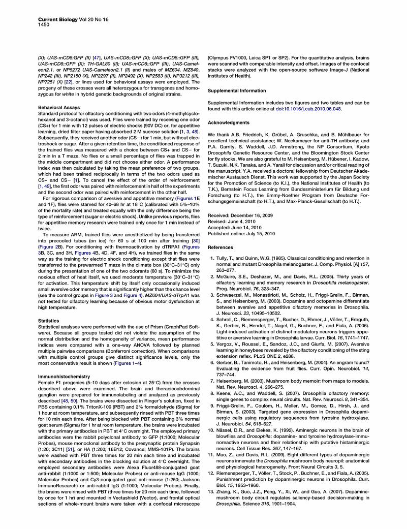
Figure 1. Dopaminergic Inputs into the Mush-
room Body via MB-M3 Neurons
(A) Projection of MB-M3 neurons visualized in
NP5272 UAS-mCD8::GFP. In the MB (light green
overlay based on the Synapsin counterstaining),
MB-M3 neurons arbor in the medial tip of the
b lobe (b2; arrowhead). Sparse terminals are
also detected in the b0 lobe (b02; small arrowhead).
(B) In UAS-Syt::HA; NP5272 UAS-mCD8::GFP,
the arborizations in the anterior andmiddle inferi-
ormedial protocerebrum (aimpr-mimpr; arrow)
are only weakly labeled by presynaptic marker
Syt::HA (magenta), if at all, relative to mCD8::GFP
(green), implying their dendritic nature. Scale bar
represents 20 mm.
(C) The GAL4-expressing cells in the PAM cluster
are visualized with UAS-Cameleon2.1 and
counted. No significant difference is observed
between NP5272, NP1528, and the combination
of the two. Also, combining NP5272 and
NP1528 does not significantly increase the
number of the labeled cells as compared to TH-
GAL4 alone.
(D) The diagram illustrates the terminal areas of
GAL4-expressing cells of TH-GAL4 (green) and
NP5272/NP1528 (orange) in the MB lobes, based
on cell counting and single-cell analysis [11]. The
MB lobes are shown as an outline. See Table S2
as well.
(E) Aversive odor memory tested at 30 min after
training. The respective dopaminergic neurons
are blocked with Shits1 driven by TH-GAL4,
NP5272, or NP1528. Block with these drivers
significantly impairs aversive memory. n = 17–22.
(F) 30 min appetitive memory of the same geno-
types. Learning indices of all the experimental
groups (TH-GAL4/UAS- shits1, NP1528/UAS-
shits1, and NP5272/UAS- shits1) are not signifi-
cantly different from corresponding control
groups (p > 0.05; one-way ANOVA). n = 13–16.
(G) Transient block only during aversive training. The results are essentially the same as in (E). n = 20–22.
Bars and error bars represent themean and SEM, respectively. Unless otherwise stated, themost conservative statistical result ofmultiple pairwise compar-
isons is shown. *p < 0.05; **p < 0.01; ***p < 0.001; n.s., not significant.
Current Biology Vol 20 No 161446
MB-M3 may be responsible for a specificmemory component.We blocked MB-M3 or neurons labeled in TH-GAL4 duringtraining and examined memory retention up to 9 hr. Blockingwith TH-GAL4 significantly impaired aversive odor memory atall time points (Figure 2A). Intriguingly, the effect of the MB-M3 block was most pronounced in the middle-term memory(2 hr after training); memory tested immediately and 9 hr aftertraining was only slightly impaired, if at all. The dynamics ofmemory decay was different from that of the block with TH-GAL4 and control groups (Figure 2A). This result is consistentwith the previous report showing that immediate memory isnot affected by blockingmany of the PAMcluster neurons [21].
What type of memory is impaired by blocking MB-M3?Initially, labile odor memory in Drosophila is consolidatedgradually and becomes resistant to retrograde amnesia[29, 30]. At 2 hr after training, labile anesthesia-sensitivememory (ASM) and consolidated anesthesia-resistantmemory(ARM) coexist [29, 30]. ARM can be measured by erasing ASMwith short cold anesthesia of flies. Manipulation of varioussignaling molecules, such as Amnesiac, AKAP, DC0, Rac, orNMDAR [1, 31–35] (but see [36] for the role of DC0 in ARM),affects ASM and causes memory dynamics similar to thatcaused by the MB-M3 block (Figure 2A).
To address whether the MB-M3 neurons are required selec-tively for ASM, we trained flies with their MB-M3 neurons
blocked and, 2 hr later, measured their total memory andARM. The output of MB-M3 during training was required forthe total 2 hr memory, whereas ARM was not significantlyaffected (Figure 2B). This suggests that the MB-M3 neuronspreferentially contribute to the formation of ASM. In contrast,the block with TH-GAL4 significantly impaired both totalmemory and ARM (Figure 2B). Although the scores of ARMwere small, subtle differences in ARM were detectable,because unpaired conditioning resulted in significantly lowermemory in every genotype (Figure 2B). Taken together, theseresults imply that multiple types of reinforcement neuronsare recruited for the formation of different forms of memory.
Aversive Odor Memory Formed by the Activation of MB-M3We then examinedwhether selective stimulation of theMB-M3neurons can induce aversive odor memory without electricshock. Drosophila heat-activated cation channel dTRPA1(also known as dANKTM1) allows transient depolarization oftargeted neurons by raising temperature [37, 38]. For selectiveactivation of the corresponding dopaminergic neurons, fliesthat express dTrpA1 by the above-described GAL4 driverswere trained with odor presentation and a concomitant tem-perature shift instead of electric shock (Figure 3A). Tominimizethe noxious effect of heat itself, we used moderate tempera-ture (30�C) for activation. Upon examination immediately

304050607080A TH-GAL4/+
TH-GAL4/UAS-shits1
NP5272/+
NP5272/UAS-shits1
+/UAS-shits1
mo
ry (P
I)
0102030
0 2 9 Time(hour)
Me
60B
NP5272/+3:NP5272/UAS-shi
ts14:+/UAS-shi
ts15:
0102030405060
****
** n.s.
2h
m
em
ory (P
I)
Electric shock TH-GAL4/+1:TH-GAL4/UAS-shi
ts12:
-100
ARM+ASM(Cold anesthesia-)
1 2 3 4 5ARM
(Cold anesthesia+) Unpaired
1 2 3 4 51 2 3 4 5
Figure 2. Preferential Requirement of MB-M3 for Inducing Labile Middle-
Term Memory
(A) Requirement of the MB-M3 neurons for different memory phases. Flies
were trained and tested immediately at restrictive temperature or kept for
2 or 9 hr and tested at permissive temperature. Memory is significantly
impaired at all three retention intervals by TH-GAL4. Blocking MB-M3
slightly affects immediate and 9 hr memory, but only 2 hr memory is signif-
icantly impaired. n = 14–20.
(B) Total memory (ASM+ARM; the same data set as in [A]), a consolidated
memory component (ARM), and memory induced by unpaired presentation
of odors and electric shock tested at 2 hr after training (n = 12–18). Although
the block with TH-GAL4 impairs both total memory and ARM, only total
memory is significantly impaired when MB-M3 neurons are blocked during
training. The requirement of the MB-M3 neurons for the total memory and
ARM is differential (p < 0.05; significant interaction [genotype 3 cold shock
treatment] in two-way ANOVA).
Bars and error bars represent the mean and SEM, respectively. Unless
otherwise stated, the most conservative statistical result of multiple pair-
wise comparisons is shown. *p < 0.05; **p < 0.01; ***p < 0.001; n.s., not
significant.
Dopamine Neurons for Aversive Reinforcement1447
(approximately 2 min) after training, robust aversive memorywas formed by the activation with TH-GAL4 (Figure 3B). Strik-ingly, selective activation of MB-M3 also caused significantaversive odor memory (Figure 3B). Unpaired presentation ofan odor and dTRPA1-dependent activation did not causesignificant associative memory (Figure 3C), indicating theimportance of stimulus contingency.
Although the drivers for MB-M3 have a selective expressionpattern, NP5272 and NP1528 have additional faint labeling ofnerves in the abdominal ganglion and, only occasionally, otherneurons in the brain (Figure 3F and Figure S2A, available on-line). To confirm that the activation of MB-M3 neurons wasthe cause of dTRPA1-induced odor memory, we expresseda GAL4 inhibitor, GAL80, in dopaminergic neurons using TH-GAL80 [39]. Indeed, TH-GAL80 suppressed reporter expres-sion in dopaminergic neurons in NP5272 and TH-GAL4(Figures 3D–3G) and dTRPA1-induced odor memory to thecontrol level (Figure 3H). These data suggest that selectiveactivation of MB-M3 can induce immediate aversive memory,whereas blocking of MB-M3 has a limited effect on immediateshock-induced memory (Figure 2A). This may suggest that thecontribution of MB-M3 is redundant with other dopaminergicneurons in shock-induced immediate memory. Alternatively,
the activation of MB-M3 by dTRPA1 might not fully recapitu-late that in electric shock conditioning in terms of a temporalpattern and intensity.We also measured dTRPA1-induced memory at 2 hr after
training. With TH-GAL4, aversive memory was still significant,whereas the memory induced with MB-M3 activation dimin-ished by 2 hr, indicating the labile nature of the memory(Figure 3H). Given the selective requirement of MB-M3(Figure 2A), the contribution of MB-M3 to 2 hr memory mightbe interdependent with other dopaminerigic neurons. A similarinteraction has been shown at the level of different subsets ofKenyon cells [24, 26, 27, 34, 40] and might thus be a potentialconsequence of the synergistic action of dopaminergicneurons.
Other Individual Dopaminergic Neurons for AversiveReinforcement
To explore the function of other types of MB-projectingdopaminergic neurons for aversive memory formation, weindividually stimulated four different cell types (MB-MP1,MB-V1, MB-MVP1, and an unnamed type that projects to theb0 lobe), using selective GAL4 driver lines (NP2758, c061;MB-GAL80, MZ840, and NP6510) (Figure 4 and Figure S2; seeTable S2 for the summary of labeled neurons).c061;MB-GAL80 labels three dopaminergic neurons in the
PPL1 cluster, including one MB-MP1 neuron that is alsolabeled in NP2758 (Figures 4A and 4C, Figures S2B and S2E,Table S2) [20]. Activation with c061;MB-GAL80 induced robustimmediate memory (Figure 4B). Furthermore, we found thatCha3.3kb-GAL80 strongly silenced reporter expression in twoof three PPL1 neurons in c061;MB-GAL80 and the effect onaversive memory to the control level (Figure 4B, Figures S2Band S2C) [41]. Given that one remaining dopaminergic neuronprojects to the anterior inferior medial protocerebrum, butnot to the MB (Figure S2C) [11], MB-MP1 is more likely tobe responsible for the formation of aversive memory. Thissuppression of transgene expression in dopaminergic neuronsmight be due to the incomplete recapitulation of the Cha3.3kb
enhancer (Figures S2B and S2C).The addition of TH-GAL80also suppressed the effect of c061;MB-GAL80 to the controllevel (Figure 4B). Consistently, significant memory wasinduced with NP2758 (Figures 4C and 4D), although thesuppression by TH-GAL80was partial (Figure 4D), presumablythrough expression in nondopaminergic neurons in NP2758 orincomplete suppression of dTRPA1 expression (Figures S2Eand S2F). The formed memory with c061; MB-GAL80 decayedsignificantly but lasted for 2 hr (Figure 4B). Taken together,these results revealed that the specific cell type within thePPL1 cluster, MB-MP1, can mediate aversive reinforcement.Intriguingly, the recent work using the same driver reportedthat MB-MP1 has another important role for suppressing theretrieval of appetitivememory depending on the feeding states[20]. Given that the output of MB-MP1 is dispensable for 3 hrmemory induced by electric shock [20], MB-MP1might mainlyinduce short-lasting odor memory. Alternatively, MB-MP1neurons might be recruited to mediate aversive reinforcementother than electric shock.MZ840 and NP6510 label the single PPL1 neuron (MB-V1)
and 15 PAM neurons (MB-MVP1 and an unnamed cell type),respectively (Figures 4E and 4G, Figures S2G–S2J). Ther-moactivation with these drivers did not induce significantmemory (Figures 4E–4H, Figures S2G–S2J). This may indicatethat the PAM and PPL1 clusters are functionally heteroge-neous in terms of aversive reinforcement signals. Consistently,

BA C
102030405060
***
*** **
Activation (paired)
0102030405060
n.s. n.s. n.s.
Activation (unpaired)
in
me
mo
ry
(P
I)
in
me
mo
ry
(P
I)
Training Test
vs
60s
OCT MCH
30ºC
OCT
MCH
-100
-100
wTH-GAL4/+1:TH-GAL4/UAS-dTrpA12:
2 m
2 m
NP1528/+5:NP1528/UAS-dTrpA16:+/UAS-dTrpA17:
NP5272/+3:NP5272/UAS-dTrpA14:
1 2 3 4 5 6 7 1 2 3 4 5 6 7
D
vsMCH OCT
30ºC
OCT
MCH
H
TH-GAL4
TH-GAL80TH-GAL4
E
1 2 3 4 5 6 7 8 -100
102030405060 Activation (paired)
Me
mo
ry
(P
I)
1 2 3 4 5 6 7 8
***** **
n.s.
TH-GAL4/+1:TH-GAL4/UAS-dTrpA12:TH-GAL4/TH-GAL80 UAS-dTrpA13:NP5272/+4:NP5272/UAS-dTrpA15:NP5272/TH-GAL80 UAS-dTrpA16:+/UAS-dTrpA17:NP5272
TH GAL80(MB
F G2 min 2 hour
+/TH-GAL80 UAS-dTrpA18:TH-GAL80NP5272 -M3)
Figure 3. dTRPA1-Dependent Activation of MB-M3 Can Induce Aversive Odor Memory
(A) The conditioning protocol for dTRPA1-induced memory (see Experimental Procedures for detail).
(B) Immediate aversive odor memory formed by odor presentation and simultaneous thermoactivation of the subsets of dopaminergic neurons expressing
dTrpA1. n = 18–22.
(C) Thermoactivation did not induce aversive odor memory when it was applied 60–120 s prior to the presentation of the odor. n = 10–12.
(D–G) Expression pattern of TH-GAL4 (D) and NP5272 (F) in the brain (left panels; frontal view, dorsal up) and thoracicoabdominal ganglion (right panels;
dorsal view, anterior up). TH-GAL80 silences mCD8::GFP expression in MB-M3 (G) and most of the cells labeled by TH-GAL4 (E). The remaining cells
are presumably nondopaminergic cells, judging from their size and position [11]. Scale bars represent 20 mm.
(H) Immediate aversive odor memory induced by dTRPA1-dependent activation is significantly suppressed by TH-GAL80, indicating that the corresponding
dopaminergic neurons are responsible. n = 15–17.
Bars and error bars represent themean and SEM, respectively. Unless otherwise stated, themost conservative statistical result ofmultiple pairwise compar-
isons is shown. **p < 0.01; ***p < 0.001; n.s., not significant.
Current Biology Vol 20 No 161448
each type of PPL1 neuron differentially responds to odors andelectric shock [11]. It is noteworthy that MB-MVP1 synapseonto the restricted subdomains adjacent to the terminals ofMB-M3. Thus, the activation of specific sets of dopaminergicneurons rather than the total amount of dopamine input inthe MB may be critical for memory formation. Despite theparticular importance of the dopamine signal in the verticallobes of the MB [11, 42, 43], we could not examine them,except for MB-V1, because of the lack of reasonably specificGAL4 drivers.
Parallel Reinforcement Input to the Mushroom BodyIn a current circuit model of aversive odor memory, associa-tive plasticity is generated in the output site of the MB (i.e.,the presynaptic terminals of Kenyon cells) upon internal
convergence of neuronal signals of odor and electric shock[2, 6, 7]. Type I adenylyl cyclase, Rutabaga, is an underlyingmolecular coincidence detector that forms a memory trace[6, 26, 43, 44]. Rutabaga in different types of Kenyon cells(e.g., g and a/b neurons) together acts to form complete aver-sivememory [24, 26, 27,]. Thus, local, but spatially segregated,Rutabaga stimulation through multiple dopaminergic path-ways may induce distinct memory traces [11, 43–45].We have shown the selective requirement of MB-M3 for
middle-term ASM, whereas blocking of many more dopami-nergic neurons impaired all memory phases examined in thisstudy (Figure 2). Therefore, electric shock recruits a set ofdistinct dopaminergic neurons that forms a parallel reinforce-ment circuit in the subdomains of the MB. Compartmentalizedsynaptic organization along the trajectory of Kenyon cell axons

6070 c061;MB-GAL80/+1:
c061;MB GAL80/ UAS dTrpA12:A BMB
Activation (paired)
***
c061;MB-GAL80 (MB-MP1) -100
102030405060
1 2 3 4 5 6 72 min 2 hour
Mem
ory (P
I)
1 2 5
+/UAS-dTrpA15:c061;MB-GAL80 /Cha
3.3kb-GAL80 UAS-dTrpA14:
+/TH-GAL80UAS-dTrpA16:+/Cha
3.3kb-GAL80UAS-dTrpA17:
c061;MB-GAL80/ UAS-dTrpA12:c061;MB-GAL80 /TH-GAL80 UAS-dTrpA13:
***
1
ped( / )
Mem
ory (P
I)
+//TH-GAL80 UAS-dTrpA15:+/UAS-dTrpA14:
010203040506070
1 2 3 4 5 1 2 3 4 5
NP2758/+1:NP2758/UAS-dTrpA12:NP2758/TH-GAL80 UAS-dTrpA13:
D*
n.s.
MBC
E
NP2758 (MB-MP1)
1
ped( / )
10203040506070
n.s.
Me
mo
ry (P
I)
-10 1 2 3 4 5 2 min 2 hour
1 2 3 4 5
MZ840/+1:MZ840/UAS-dTrpA12:+/UAS-dTrpA13:
FMB
2/ '
2
NP2758 (MB-MP1) ped( / )
-100
10
G MB
'2
M
NP6510/+1:NP6510/UAS-dTrpA12:+/ UAS-dTrpA13:
2 min
203040506070
1 2 3
mo
ry (P
I)
H
MZ840 (MB-V1)
1NP6510 (MB-MVP1)
/ UAS dTrpA1n.s.
-100
1020
2 min
1 2 3
Me
m
Figure 4. dTrpA1-Induced Memory by Other Dopaminergic Neurons
(A, C, E, and G) Projection of brain regions including the MB (light green outline; frontal view, dorsal up). Various dopaminergic neurons projecting to the MB
are visualized with mCD8::GFP (arrowheads) driven by c061;MB-GAL80 (A), NP2758 (C), MZ840 (E), and NP6510 (G). Scale bar represents 20 mm. see also
Figure S2 and Table S2.
(B, D, F, and H) Memory induced by dTRPA1-dependent activation of the various types of dopaminergic neurons.
(E) With c061;MB-GAL80, robust immediate and 2 hr memory are formed and significantly suppressed by TH-GAL80 and Cha3.3kb-GAL80. n = 18–22.
(F) Activation of dTrpA1-expressing cells in NP2758 induces robust aversive odor memory, which is significantly suppressed by TH-GAL80.
(G) Despite the tendency of conditioned avoidance, aversive memory with MZ840 is not significant.
(H) With NP6510, the learning index of NP6510/UAS-dTrpA1 is different than that of NP6510/+ but not +/UAS-dTrpA1. n = 15-18.
Bars and error bars represent themean and SEM, respectively. Unless otherwise stated, themost conservative statistical result ofmultiple pairwise compar-
isons is shown. *p < 0.05; ***p < 0.001; n.s., not significant.
Dopamine Neurons for Aversive Reinforcement1449
may well explain how the MB as a single brain structure cansupport ‘‘pleiotropic’’ behavioral functions [12, 25, 45].
Experimental Procedures
Fly Strains
All flies were raised on standard medium. For behavioral assay, F1 proge-
nies of the crosses between females of UAS-shits1 (X, III) [28], UAS-
dTrpA1(II) [37], UAS-dTrpA1 TH-GAL80 (II) [39], UAS-dTrpA1;Cha3.3kb-
GAL80 (II, III) [41], or white and males of NP5272 (II) [22], NP1528 (II) / CyO
[22], NP2758 (X) [22], NP6510 (III) [22], MZ840 (III), or TH-GAL4 (III) [9]
were employed. For the experiments with c061;MB-GAL80 (X and III), the
female of this strain was used for crosses. After measurement, flies without
a GAL4 driver (i.e., those with the balancer or male of NP2758 crosses) were
excluded from calculation. Accordingly, for experiments with NP2758 (Fig-
ure 4D), only the performance indices of females were compared. For exper-
iments withUAS-shits1 andUAS-dTrpA1, flies were raised at 18�C and 25�C,60% relative humidity, and were used during 8–14 and 7–12 days after eclo-
sion, respectively, to allow sufficient accumulation of effecter genes without
age-related memory impairment. For anatomical assay, F1 progenies of the
crosses between females of UAS-mCD8::GFP (X, II, III) [46], UAS-Syt::HA
Current Biology Vol 20 No 161450
(X); UAS-mCD8:GFP (II) [47], UAS-mCD8::GFP (X); UAS-mCD8::GFP (III),
UAS-mCD8::GFP (X); TH-GAL80 (II); UAS-mCD8::GFP (III), UAS-Camel-
eon2.1, or NP5272 UAS-Cameleon2.1 (II) and males of MZ604, MZ840,
NP242 (III), NP2150 (X), NP2297 (II), NP2492 (X), NP2583 (II), NP3212 (III),
NP7251 (X) [22], or lines used for behavioral assays were employed. The
progeny of these crosses were all heterozygous for transgenes and homo-
zygous for white in hybrid genetic backgrounds of original strains.
Behavioral Assays
Standard protocol for olfactory conditioning with two odors (4-methylcyclo-
hexanol and 3-octanol) was used. Flies were trained by receiving one odor
(CS+) for 1 min with 12 pulses of electric shocks (90V DC) or, for appetitive
learning, dried filter paper having absorbed 2 M sucrose solution [1, 3, 48].
Subsequently, they received another odor (CS2) for 1 min, but without elec-
troshock or sugar. After a given retention time, the conditioned response of
the trained flies was measured with a choice between CS+ and CS2 for
2 min in a T maze. No flies or a small percentage of flies was trapped in
the middle compartment and did not choose either odor. A performance
index was then calculated by taking the mean preference of two groups,
which had been trained reciprocally in terms of the two odors used as
CS+ and CS2 [1]. To cancel the effect of the order of reinforcement
[1, 49], the first odor was pairedwith reinforcement in half of the experiments
and the second odor was paired with reinforcement in the other half.
For rigorous comparison of aversive and appetitive memory (Figures 1E
and 1F), flies were starved for 40–68 hr at 18�C (calibrated with 5%–10%
of the mortality rate) and treated equally with the only difference being the
type of reinforcement (sugar or electric shock). Unlike previous reports, flies
for appetitive memory research were trained only once for 1 min instead of
twice.
To measure ARM, trained flies were anesthetized by being transferred
into precooled tubes (on ice) for 60 s at 100 min after training [30]
(Figure 2B). For conditioning with thermoactivation by dTRPA1 (Figures
3B, 3C, and 3H, Figures 4B, 4D, 4F, and 4H), we trained flies in the same
way as the training for electric shock conditioning except that flies were
transferred to the prewarmed T maze in the climate box (30�C–31�C) onlyduring the presentation of one of the two odorants (60 s). To minimize the
noxious effect of heat itself, we used moderate temperature (30�C–31�C)for activation. This temperature shift by itself only occasionally induced
small aversive odor memory that is significantly higher than the chance level
(see the control groups in Figure 3 and Figure 4). MZ604/UAS-dTrpA1 was
not tested for olfactory learning because of obvious motor dysfunction at
high temperature.
Statistics
Statistical analyses were performed with the use of Prism (GraphPad Soft-
ware). Because all groups tested did not violate the assumption of the
normal distribution and the homogeneity of variance, mean performance
indices were compared with a one-way ANOVA followed by planned
multiple pairwise comparisons (Bonferroni correction). When comparisons
with multiple control groups give distinct significance levels, only the
most conservative result is shown (Figures 1–4).
Immunohistochemistry
Female F1 progenies (5–10 days after eclosion at 25�C) from the crosses
described above were examined. The brain and thoracicoabdominal
ganglion were prepared for immunolabeling and analyzed as previously
described [48, 50]. The brains were dissected in Ringer’s solution, fixed in
PBS containing 0.1% TritonX-100 (PBT) and 2% formaldehyde (Sigma) for
1 hour at room temperature, and subsequently rinsed with PBT three times
for 10 min each time. After being blocked with PBT containing 3% normal
goat serum (Sigma) for 1 hr at room temperature, the brains were incubated
with the primary antibodies in PBT at 4�C overnight. The employed primary
antibodies were the rabbit polyclonal antibody to GFP (1:1000; Molecular
Probes), mouse monoclonal antibody to the presynaptic protein Synapsin
(1:20; 3C11) [51], or HA (1:200; 16B12; Covance; MMS-101P). The brains
were washed with PBT three times for 20 min each time and incubated
with secondary antibodies in the blocking solution at 4�C overnight. The
employed secondary antibodies were Alexa Fluor488-conjugated goat
anti-rabbit (1:1000 or 1:500; Molecular Probes) or anti-mouse IgG (1000;
Molecular Probes) and Cy3-conjugated goat anti-mouse (1:250; Jackson
ImmunoResearch) or anti-rabbit IgG (1:1000; Molecular Probes). Finally,
the brains were rinsed with PBT (three times for 20 min each time, followed
by once for 1 hr) and mounted in Vectashield (Vector), and frontal optical
sections of whole-mount brains were taken with a confocal microscope
(Olympus FV1000, Leica SP1 or SP2). For the quantitative analysis, brains
were scanned with comparable intensity and offset. Images of the confocal
stacks were analyzed with the open-source software Image-J (National
Institutes of Health).
Supplemental Information
Supplemental Information includes two figures and two tables and can be
found with this article online at doi:10.1016/j.cub.2010.06.048.
Acknowledgments
We thank A.B. Friedrich, K. Grubel, A. Gruschka, and B. Muhlbauer for
excellent technical assistance; W. Neckameyer for anti-TH antibody; and
P.A. Garrity, S. Waddell, J.D. Armstrong, the NP Consortium, Kyoto
Drosophila Genetic Resource Center, and the Bloomington Stock Center
for fly stocks. We are also grateful to M. Heisenberg, M. Hubener, I. Kadow,
T. Suzuki, N.K. Tanaka, and A. Yarali for discussion and/or critical reading of
the manuscript. Y.A. received a doctoral fellowship from Deutscher Akade-
mischer Austausch Dienst. This work was supported by the Japan Society
for the Promotion of Science (to K.I.), the National Institutes of Health (to
T.K.), Bernstein Focus Learning from Bundesministerium fur Bildung und
Forschung (to H.T.), the Emmy-Noether Program from Deutsche For-
schungsgemeinschaft (to H.T.), and Max-Planck-Gesellschaft (to H.T.).
Received: December 16, 2009
Revised: June 4, 2010
Accepted: June 14, 2010
Published online: July 15, 2010
References
1. Tully, T., and Quinn, W.G. (1985). Classical conditioning and retention in
normal and mutant Drosophila melanogaster. J. Comp. Physiol. [A] 157,
263–277.
2. McGuire, S.E., Deshazer, M., and Davis, R.L. (2005). Thirty years of
olfactory learning and memory research in Drosophila melanogaster.
Prog. Neurobiol. 76, 328–347.
3. Schwaerzel, M., Monastirioti, M., Scholz, H., Friggi-Grelin, F., Birman,
S., and Heisenberg, M. (2003). Dopamine and octopamine differentiate
between aversive and appetitive olfactory memories in Drosophila.
J. Neurosci. 23, 10495–10502.
4. Schroll, C., Riemensperger, T., Bucher, D., Ehmer, J., Voller, T., Erbguth,
K., Gerber, B., Hendel, T., Nagel, G., Buchner, E., and Fiala, A. (2006).
Light-induced activation of distinct modulatory neurons triggers appe-
titive or aversive learning in Drosophila larvae. Curr. Biol. 16, 1741–1747.
5. Vergoz, V., Roussel, E., Sandoz, J.C., and Giurfa, M. (2007). Aversive
learning in honeybees revealed by the olfactory conditioning of the sting
extension reflex. PLoS ONE 2, e288.
6. Gerber, B., Tanimoto, H., and Heisenberg, M. (2004). An engram found?
Evaluating the evidence from fruit flies. Curr. Opin. Neurobiol. 14,
737–744.
7. Heisenberg, M. (2003). Mushroom body memoir: from maps to models.
Nat. Rev. Neurosci. 4, 266–275.
8. Keene, A.C., and Waddell, S. (2007). Drosophila olfactory memory:
single genes to complex neural circuits. Nat. Rev. Neurosci. 8, 341–354.
9. Friggi-Grelin, F., Coulom, H., Meller, M., Gomez, D., Hirsh, J., and
Birman, S. (2003). Targeted gene expression in Drosophila dopami-
nergic cells using regulatory sequences from tyrosine hydroxylase.
J. Neurobiol. 54, 618–627.
10. Nassel, D.R., and Elekes, K. (1992). Aminergic neurons in the brain of
blowflies and Drosophila: dopamine- and tyrosine hydroxylase-immu-
noreactive neurons and their relationship with putative histaminergic
neurons. Cell Tissue Res. 267, 147–167.
11. Mao, Z., and Davis, R.L. (2009). Eight different types of dopaminergic
neurons innervate the Drosophilamushroom body neuropil: anatomical
and physiological heterogeneity. Front Neural Circuits 3, 5.
12. Riemensperger, T., Voller, T., Stock, P., Buchner, E., and Fiala, A. (2005).
Punishment prediction by dopaminergic neurons in Drosophila. Curr.
Biol. 15, 1953–1960.
13. Zhang, K., Guo, J.Z., Peng, Y., Xi, W., and Guo, A. (2007). Dopamine-
mushroom body circuit regulates saliency-based decision-making in
Drosophila. Science 316, 1901–1904.

Dopamine Neurons for Aversive Reinforcement1451
14. Seugnet, L., Suzuki, Y., Vine, L., Gottschalk, L., and Shaw, P.J. (2008).
D1 receptor activation in the mushroom bodies rescues sleep-loss-
induced learning impairments in Drosophila. Curr. Biol. 18, 1110–1117.
15. Zhang, S., Yin, Y., Lu, H., and Guo, A. (2008). Increased dopaminergic
signaling impairs aversive olfactory memory retention in Drosophila.
Biochem. Biophys. Res. Commun. 370, 82–86.
16. Andretic, R., Kim, Y.C., Jones, F.S., Han, K.A., and Greenspan, R.J.
(2008). Drosophila D1 dopamine receptor mediates caffeine-induced
arousal. Proc. Natl. Acad. Sci. USA 105, 20392–20397.
17. Selcho, M., Pauls, D., Han, K.A., Stocker, R.F., and Thum, A.S. (2009).
The role of dopamine in Drosophila larval classical olfactory condi-
tioning. PLoS ONE 4, e5897.
18. Lebestky, T., Chang, J.S., Dankert, H., Zelnik, L., Kim, Y.C., Han, K.A.,
Wolf, F.W., Perona, P., and Anderson, D.J. (2009). Two different forms
of arousal in Drosophila are oppositely regulated by the dopamine D1
receptor ortholog DopR via distinct neural circuits. Neuron 64, 522–536.
19. Liu, T., Dartevelle, L., Yuan, C.,Wei, H.,Wang, Y., Ferveur, J.F., andGuo,
A. (2009). Reduction of dopamine level enhances the attractiveness of
male Drosophila to other males. PLoS ONE 4, e4574.
20. Krashes, M.J., DasGupta, S., Vreede, A., White, B., Armstrong, J.D., and
Waddell, S. (2009). A neural circuit mechanism integrating motivational
state with memory expression in Drosophila. Cell 139, 416–427.
21. Claridge-Chang, A., Roorda, R.D., Vrontou, E., Sjulson, L., Li, H., Hirsh,
J., and Miesenbock, G. (2009). Writing memories with light-addressable
reinforcement circuitry. Cell 139, 405–415.
22. Tanaka, N.K., Tanimoto, H., and Ito, K. (2008). Neuronal assemblies of
the Drosophila mushroom body. J. Comp. Neurol. 508, 711–755.
23. Neckameyer, W.S., Woodrome, S., Holt, B., and Mayer, A. (2000). Dopa-
mine and senescence in Drosophila melanogaster. Neurobiol. Aging 21,
145–152.
24. Akalal, D.B., Wilson, C.F., Zong, L., Tanaka, N.K., Ito, K., and Davis, R.L.
(2006). Roles for Drosophila mushroom body neurons in olfactory
learning and memory. Learn. Mem. 13, 659–668.
25. Isabel, G., Pascual, A., and Preat, T. (2004). Exclusive consolidated
memory phases in Drosophila. Science 304, 1024–1027.
26. Zars, T., Fischer, M., Schulz, R., and Heisenberg, M. (2000). Localization
of a short-term memory in Drosophila. Science 288, 672–675.
27. Blum, A.L., Li, W., Cressy, M., and Dubnau, J. (2009). Short- and long-
term memory in Drosophila require cAMP signaling in distinct neuron
types. Curr. Biol. 19, 1341–1350.
28. Kitamoto, T. (2001). Conditional modification of behavior in Drosophila
by targeted expression of a temperature-sensitive shibire allele in
defined neurons. J. Neurobiol. 47, 81–92.
29. Folkers, E., Drain, P., and Quinn, W.G. (1993). Radish, a Drosophila
mutant deficient in consolidated memory. Proc. Natl. Acad. Sci. USA
90, 8123–8127.
30. Quinn,W.G., andDudai, Y. (1976). Memory phases in Drosophila. Nature
262, 576–577.
31. Dubnau, J., and Tully, T. (1998). Gene discovery in Drosophila: new
insights for learning and memory. Annu. Rev. Neurosci. 21, 407–444.
32. Schwaerzel, M., Jaeckel, A., and Mueller, U. (2007). Signaling at
A-kinase anchoring proteins organizes anesthesia-sensitive memory
in Drosophila. J. Neurosci. 27, 1229–1233.
33. Wu, C.L., Xia, S., Fu, T.F., Wang, H., Chen, Y.H., Leong, D., Chiang, A.S.,
and Tully, T. (2007). Specific requirement of NMDA receptors for long-
termmemory consolidation in Drosophila ellipsoid body. Nat. Neurosci.
10, 1578–1586.
34. Shuai, Y., Lu, B., Hu, Y., Wang, L., Sun, K., and Zhong, Y. (2010). Forget-
ting is regulated through Rac activity in Drosophila. Cell 140, 579–589.
35. Li, W., Tully, T., and Kalderon, D. (1996). Effects of a conditional
Drosophila PKA mutant on olfactory learning and memory. Learn.
Mem. 2, 320–333.
36. Horiuchi, J., Yamazaki, D., Naganos, S., Aigaki, T., and Saitoe, M. (2008).
Protein kinase A inhibits a consolidated form of memory in Drosophila.
Proc. Natl. Acad. Sci. USA 105, 20976–20981.
37. Hamada, F.N., Rosenzweig, M., Kang, K., Pulver, S.R., Ghezzi, A., Jegla,
T.J., and Garrity, P.A. (2008). An internal thermal sensor controlling
temperature preference in Drosophila. Nature 454, 217–220.
38. Viswanath, V., Story, G.M., Peier, A.M., Petrus, M.J., Lee, V.M., Hwang,
S.W., Patapoutian, A., and Jegla, T. (2003). Opposite thermosensor in
fruitfly and mouse. Nature 423, 822–823.
39. Sitaraman, D., Zars, M., Laferriere, H., Chen, Y.C., Sable-Smith, A.,
Kitamoto, T., Rottinghaus, G.E., and Zars, T. (2008). Serotonin is
necessary for place memory in Drosophila. Proc. Natl. Acad. Sci. USA
105, 5579–5584.
40. Krashes, M.J., Keene, A.C., Leung, B., Armstrong, J.D., and Waddell, S.
(2007). Sequential use of mushroom body neuron subsets during
Drosophila odor memory processing. Neuron 53, 103–115.
41. Kitamoto, T. (2002). Conditional disruption of synaptic transmission
induces male-male courtship behavior in Drosophila. Proc. Natl. Acad.
Sci. USA 99, 13232–13237.
42. Tomchik, S.M., and Davis, R.L. (2009). Dynamics of learning-related
cAMP signaling and stimulus integration in the Drosophila olfactory
pathway. Neuron 64, 510–521.
43. Gervasi, N., Tchenio, P., and Preat, T. (2010). PKA dynamics in
a Drosophila learning center: coincidence detection by Rutabaga
adenylyl cyclase and spatial regulation by dunce phosphodiesterase.
Neuron 65, 516–529.
44. Tomchik, S.M., and Davis, R.L. (2009). Dynamics of learning-related
cAMP signaling and stimulus integration in the Drosophila olfactory
pathway. Neuron 64, 510–521.
45. Yu, D., Akalal, D.B., and Davis, R.L. (2006).Drosophila alpha/betamush-
room body neurons form a branch-specific, long-term cellular memory
trace after spaced olfactory conditioning. Neuron 52, 845–855.
46. Lee, T., and Luo, L. (1999). Mosaic analysis with a repressible cell marker
for studies of gene function in neuronal morphogenesis. Neuron 22,
451–461.
47. Robinson, I.M., Ranjan, R., and Schwarz, T.L. (2002). Synaptotagmins I
and IV promote transmitter release independently of Ca(2+) binding in
the C(2)A domain. Nature 418, 336–340.
48. Thum, A.S., Jenett, A., Ito, K., Heisenberg, M., and Tanimoto, H. (2007).
Multiple memory traces for olfactory reward learning in Drosophila.
J. Neurosci. 27, 11132–11138.
49. Kim, Y.C., Lee, H.G., and Han, K.A. (2007). D1 dopamine receptor dDA1
is required in the mushroom body neurons for aversive and appetitive
learning in Drosophila. J. Neurosci. 27, 7640–7647.
50. Aso, Y., Grubel, K., Busch, S., Friedrich, A.B., Siwanowicz, I., and
Tanimoto, H. (2009). Themushroom body of adultDrosophila character-
ized by GAL4 drivers. J. Neurogenet. 23, 156–172.
51. Klagges, B.R., Heimbeck, G., Godenschwege, T.A., Hofbauer, A.,
Pflugfelder, G.O., Reifegerste, R., Reisch, D., Schaupp, M., Buchner,
S., and Buchner, E. (1996). Invertebrate synapsins: a single gene codes
for several isoforms in Drosophila. J. Neurosci. 16, 3154–3165.