Embed Size (px)
DESCRIPTION
Special Senses. Chapter 17. The Special Senses. Smell, taste, vision, hearing and equilibrium Housed in complex sensory organs Ophthalmology is science of the eye Otolaryngology is science of the ear. Olfactory Epithelium and Receptors. Physiology of Olfaction. - PowerPoint PPT Presentation
Citation preview

Special Senses
Chapter 17

The Special Senses
• Smell, taste, vision, hearing and equilibrium
• Housed in complex sensory organs
• Ophthalmology is science of the eye
• Otolaryngology is science of the ear
Olfactory Epithelium and Receptors
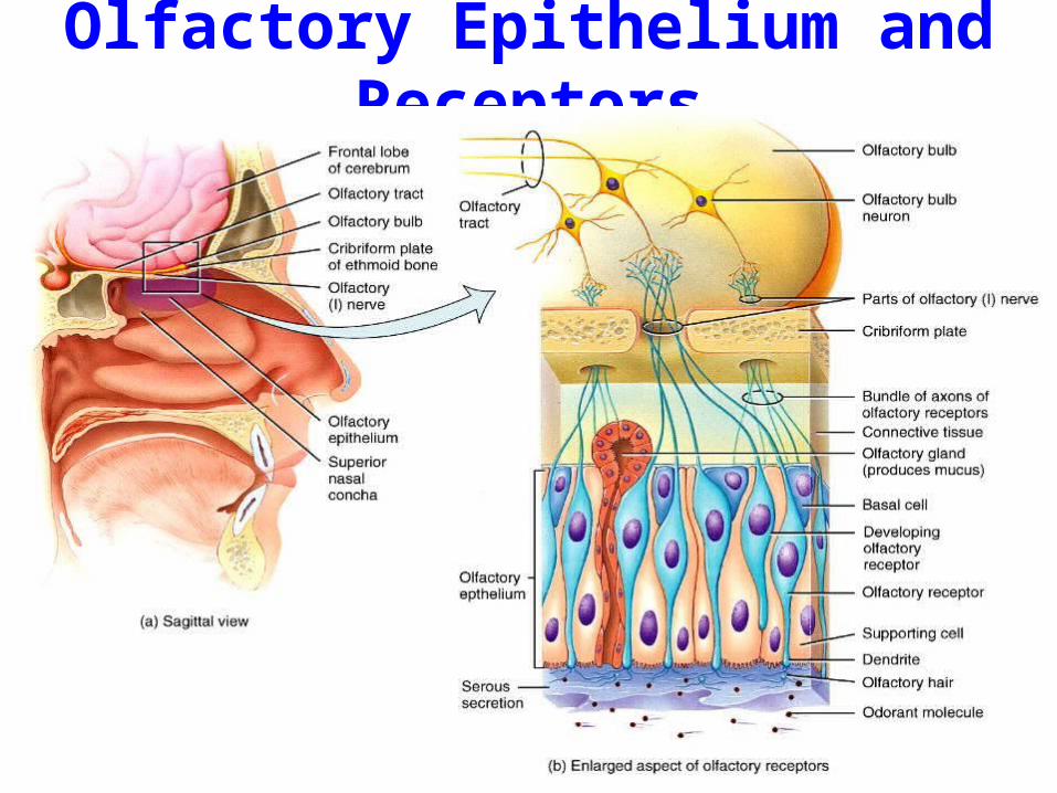
Physiology of Olfaction
Many different combinations of receptors produces the possibility for thousands of different odor sensations.
Low threshold, only few molecules needed.
Adaptation - rapid
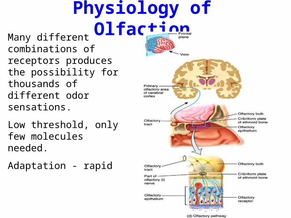
Physiology of Olfaction

Olfactory Pathway
Olfactory receptors
Olfactory (I) nerves
Olfactory tract
Temporal lobe (primary olfactory area)
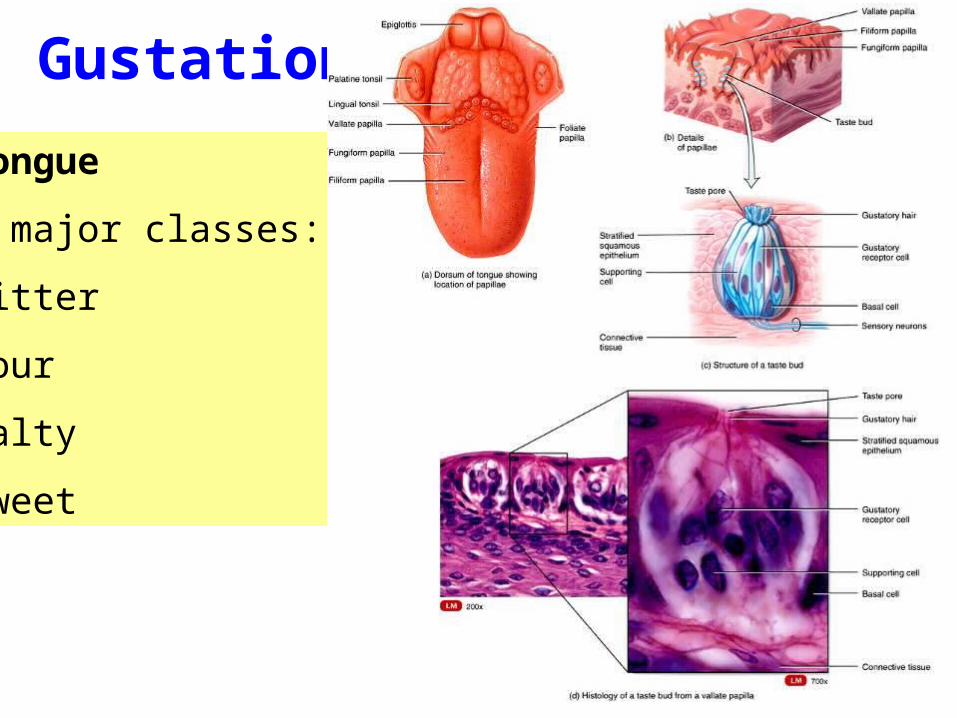
Tongue
4 major classes:
bitter
sour
salty
sweet
Gustation
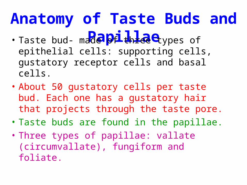
Anatomy of Taste Buds and Papillae• Taste bud- made of three types of epithelial
cells: supporting cells, gustatory receptor cells and basal cells.
• About 50 gustatory cells per taste bud. Each one has a gustatory hair that projects through the taste pore.
• Taste buds are found in the papillae.
• Three types of papillae: vallate (circumvallate), fungiform and foliate.
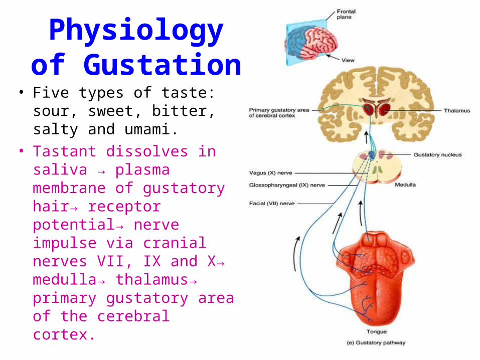
Physiology of Gustation
• Five types of taste: sour, sweet, bitter, salty and umami.
• Tastant dissolves in saliva → plasma membrane of gustatory hair→ receptor potential→ nerve impulse via cranial nerves VII, IX and X→ medulla→ thalamus→ primary gustatory area of the cerebral cortex.

Vision

Accessory Structures of Eye
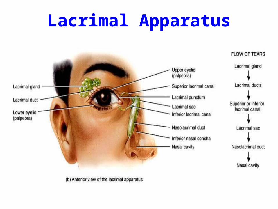
Lacrimal Apparatus

Anatomy of Eye
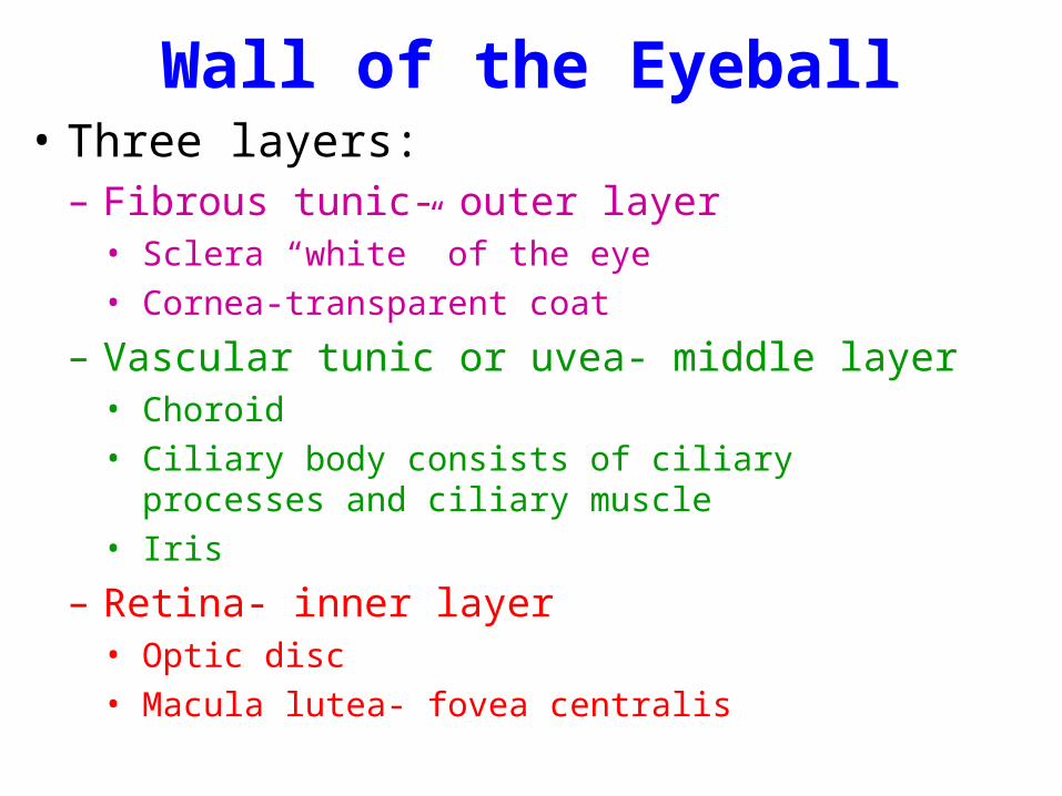
Wall of the Eyeball• Three layers:
– Fibrous tunic- outer layer• Sclera “white” of the eye
• Cornea-transparent coat
– Vascular tunic or uvea- middle layer• Choroid
• Ciliary body consists of ciliary processes and ciliary muscle
• Iris
– Retina- inner layer• Optic disc
• Macula lutea- fovea centralis
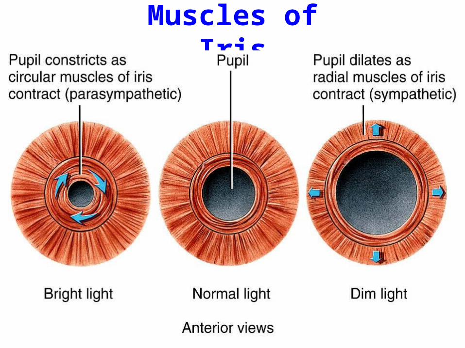
Muscles of Iris
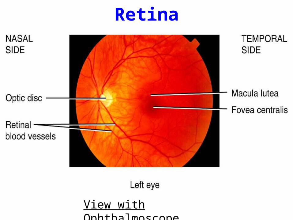
Retina
View with Ophthalmoscope

Retina

Interior of the Eyeball• Lens
– lack blood vessels, consists of a capsule with proteins (crystallins) in layers; transparent.
– divides the eyeball into two cavities: anterior and posterior.
- Anterior cavity- further divided into anterior and posterior chambers. Both are filled with aqueous humor.
- Posterior cavity (vitreous chamber)-filled with vitreous body.
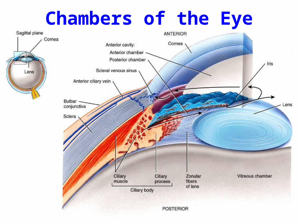
Chambers of the Eye

Cavities of the Interior of Eyeball• Anterior cavity (anterior to lens)
– filled with aqueous humor• produced by ciliary body• continually drained• replaced every 90 minutes
– 2 chambers• anterior chamber between cornea and iris• posterior chamber between iris and lens
• Posterior cavity (posterior to lens)– filled with vitreous body (jellylike)– formed once during embryonic life– floaters are debris in vitreous

Eye Structure Summary
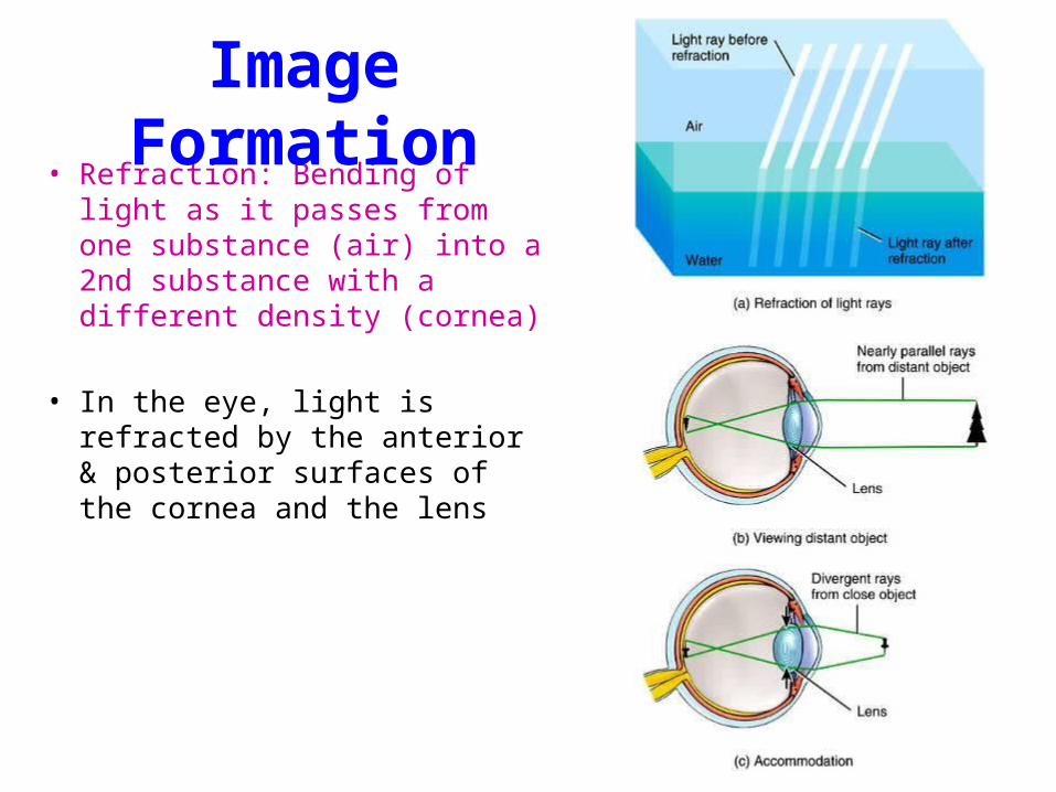
• Refraction: Bending of light as it passes from one substance (air) into a 2nd substance with a different density (cornea)
• In the eye, light is refracted by the anterior & posterior surfaces of the cornea and the lens
Image Formation

Vision Correction
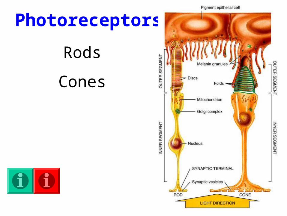
Photoreceptors
Rods
Cones
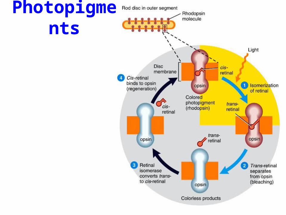
Photopigments
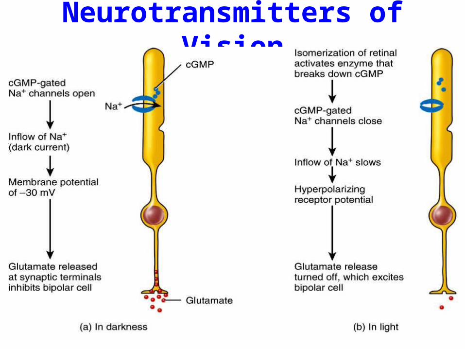
Neurotransmitters of Vision
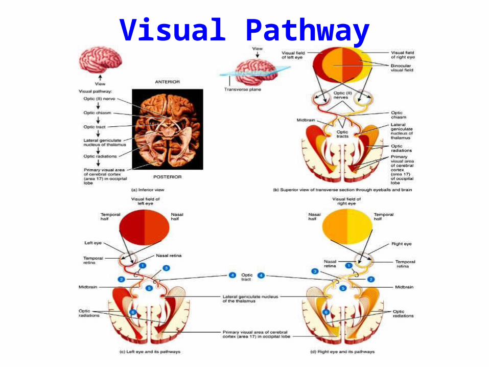
Visual Pathway

Anatomy of Ear

Middle Ear

Inner Ear

Cochlea and
Cranial Nerve VIII
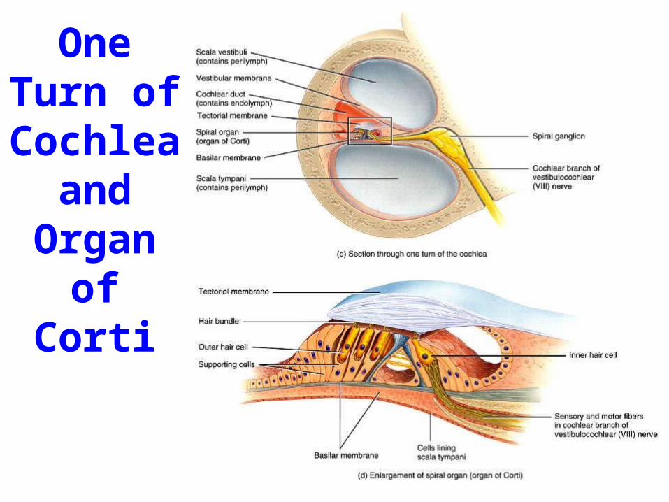
One Turn of
Cochlea and
Organ of Corti

Physiology of Hearing
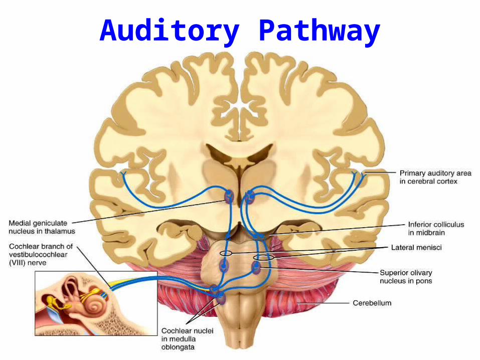
Auditory Pathway
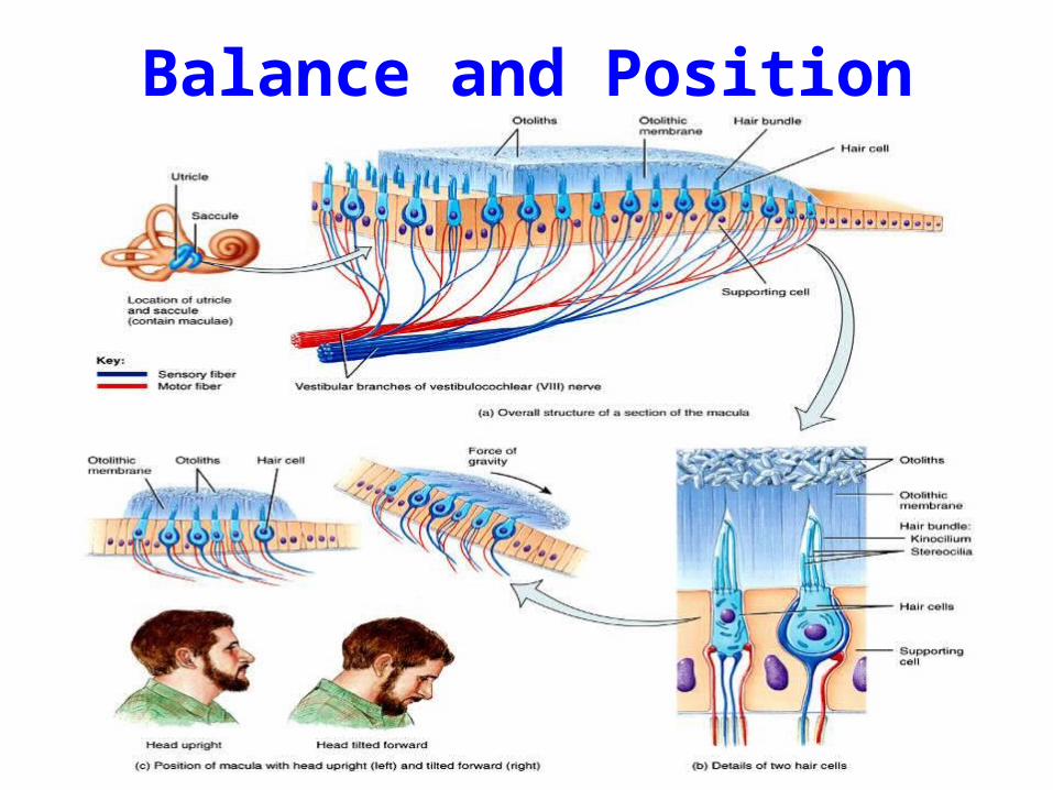
Balance and Position
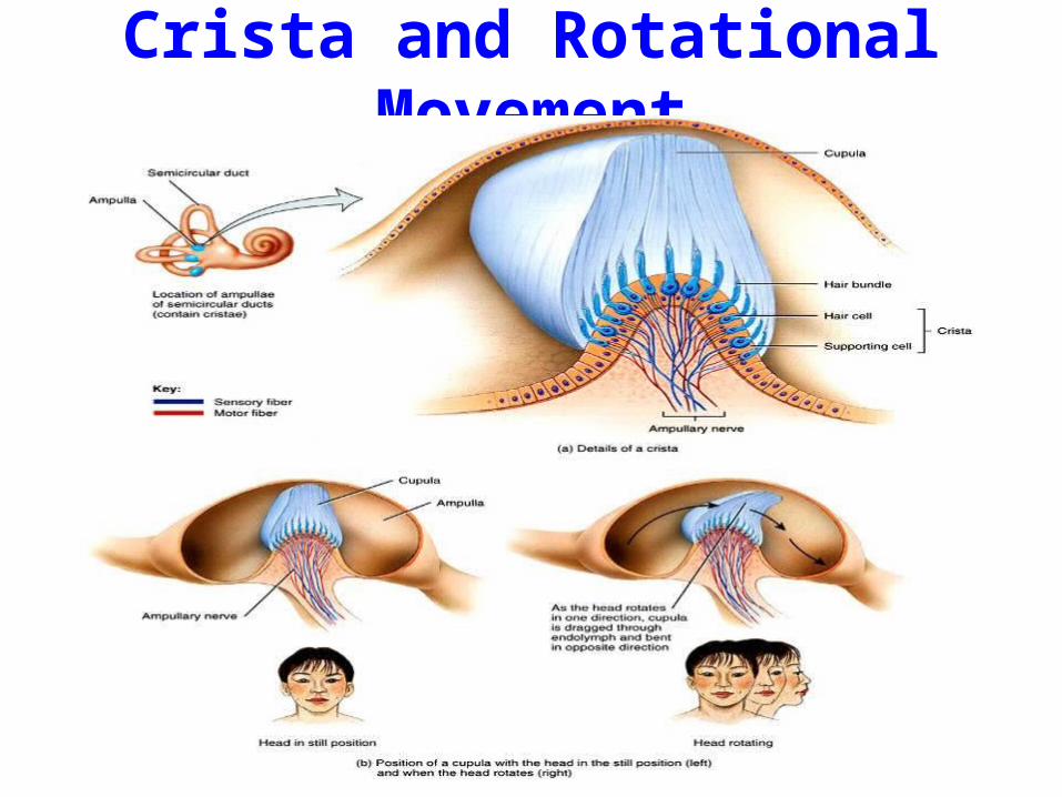
Crista and Rotational Movement

Ear Structure Summary