Embed Size (px)
Citation preview
Spatiotemporal Characterization of
Stochastic Bacterial Growth in Biofilm Environment
Sung-Ho Paek
Dissertation submitted to the faculty of the Virginia Polytechnic Institute and State University in
partial fulfillment of the requirements for the degree of
Doctor of Philosophy
In
Biological Systems Engineering
Warren C. Ruder, Chair
Yi-Heng Percival Zhang
Justin Barone
Pablo Sobrado
April 25th 2017
Blacksburg, Virginia
Keywords: synthetic biology, biofilms, stochastic gene expression, stochastic phenotypic
expression, microfluidics, quorum sensing, genetic toggle switch, droplet-microfluidics,
biomimetic

Spatiotemporal Characterization of
Stochastic Bacterial Growth in Biofilm Environment
Sung-Ho Paek
ACADEMIC ABSTRACT
Research on bacteria in their biofilm form is limited by the ability to artificially culture
bacterial biofilms in a system that permits the visualization of individual cells. The experiments
comprising this thesis research are on-going investigations of bacterial culture systems engineered
to provide an environment that mimics biofilms while enabling real-time microscopy. Specifically,
the microfluidic systems developed and assessed as part of this thesis permit the visualization of
individual bacteria cells within consortia growing within a narrow space provided by a
microfluidic device. This research demonstrates the versatility of these microfluidic systems across
potentially high-throughput microbiological experiments utilizing genetically engineered
Escherichia coli.
Before demonstrating the efficacy of these systems, the development of the field of
synthetic biology over the past half century is reviewed, focusing on synthetic genetic circuits and
their applications (Chapter 2). The first and main microfluidic device explored in this research was
developed to mimic the nutrient-deficient conditions within biofilms by forcing media to enter the
culture area through a narrow, torturous channel. The microfluidic channel was thin enough (0.97
µm) to prevent the motility of 1-µm-wide E. coli cells, enabling visualization of individual cells.
The bacteria cultured in the device contained either a simple Plux-driven quorum sensing receiver
(Chapters 3 and 5) or a LacI- and TetR-driven genetic toggle switch (Chapter 4). Under the culture
conditions, the quorum sensing reporter signal was detected even without addition of the signaling
molecule (Chapter 3). The genetic toggle switch was stable when the system began in the high-
LacI expression state, but after 5 days of culture, >5% of high-TetR expression cells began to
consistently express the high-LacI state (Chapter 4). This system was also employed to track
lineages of cells using real-time microscopy, which successfully characterized the inheritance of
aberrant, enlarged cell phenotypes under stress (Chapter 5).

Another microfluidic device, a droplet bioreactor, was also developed to culture small
numbers of cells in an aqueous bubble suspended in oil (Chapter 6). Quorum sensing receiver cells
were cultured in this device, demonstrating that it is well suited for testing the effects of
compounds on biofilms within water-in-oil droplets.
Spatiotemporal Characterization of
Stochastic Bacterial Growth in Biofilm Environment
Sung-Ho Paek
GENERAL AUDIENCE ABSTRACT
Bacteria are the most abundant organisms globally, yet relatively little is understood about
the basic biology of biofilms, one of the most common natural states of bacteria. Biofilms are
ubiquitous consortia of individual microbial cells that send and received chemical signals from
one another to carry out group behaviors such as quorum sensing. The impacts of biofilms range
from the contamination of food processing equipment to antibiotic resistant bacterial infections.
The vast majority of microbiological research has been conducted on bacteria in their planktonic
state as individual cells cultured in a liquid medium. This form of culture does not permit the types
of research that can help address the impacts of biofilms on human health and economic activities,
never mind examine the biological mechanism of random gene and morphological expression
within bacterial biofilm.
This thesis presents research utilizing two microfluidic devices that will enable further
large-scale studies to unravel the mechanisms that create biofilms as well as permit high-
throughput testing of chemical compounds to control the growth and development of biofilms.
Moreover, these devices permit the use of real-time microscopy to track cells and their growth
over time. The first microfluidic device utilized in this research mimics the nutrient-limiting
conditions of biofilms. This biofilm-mimicking device was used to culture a common research
bacteria, Escherichia coli, with one of two engineered genetic circuits (reviewed in Chapter 2): a
quorum sensing receiver (Chapters 3 and 5) or genetic toggle switch (Chapter 4). Both of these
genetic circuits demonstrated stochasticity in their gene expression states under the culture
conditions in the biofilm-mimicking device. The second microfluidic device successfully
permitted the culture of small numbers of isolated cells within a small bubble of bacterial media
suspended in oil (Chapter 6). Additionally, this device enabled the addition of chemical
compounds to influence the growth and metabolism of the trapped cells. Collectively, these
microfluidic devices provide the ability to effectively study both the mechanisms underlying

random gene expression within biofilms as well as explore the chemical factors that can be used
to control and mitigate biofilm formation and growth.

vi
ACKNOWLEDGEMENTS
The authors gratefully acknowledge support from Virginia Tech Institute for Critical
Technology and Applied Science’s Graduate Research Scholarship Program. The authors
additionally acknowledge support from award FA9550-13-1-0108 from the Air Force Office of
Scientific Research of the USA and also awards N00014-17-12306 and N00014-15-1-2502 (to
W.C.R.) from the Office of Naval Research of the USA.
vii
TABLE OF CONTENTS
CHAPTER 1 ................................................................................................................................... 1
INTRODUCTION .......................................................................................................................... 1
1.1. Organization of the Thesis ................................................................................................... 1
1.2. Objective and Significance .................................................................................................. 3
1.3. Specific Goals ...................................................................................................................... 5
1.4. Experimental Design ............................................................................................................ 7
1.4.1. Biomimetic Microfluidic-channel Approach ................................................................ 9
1.4.2. Micro-droplet Bioreactor ............................................................................................ 11
CHAPTER 2 ................................................................................................................................. 13
OVERVIEW OF SYNTHETIC CIRCUITS IN SYNTHETIC BIOLOGY ................................. 13
2.1. The Initial Development of Synthetic Biology .................................................................. 13
2.2. Synthetic Circuits ............................................................................................................... 15
2.2.1. Engineering Synthetic Gene Circuits .......................................................................... 16
2.2.2. Engineering Synthetic Protein Circuits ....................................................................... 19
2.2.3. Deploying Circuits in Mammalian Cells .................................................................... 20
2.3. Utilities of Engineered Bacteria ......................................................................................... 23
2.3.1. Cell Therapy ................................................................................................................ 24
2.3.2. Cellular Signal Transduction ...................................................................................... 27
CHAPTER 3. ................................................................................................................................ 30
STOCHASTICALLY INDUCED QUORUM SENSING ............................................................ 30
3.1. Abstract .............................................................................................................................. 30
3.2 Introduction ......................................................................................................................... 30
3.3 Materials and Methods ........................................................................................................ 33
3.3.1. Manufacture of microfluidic device ............................................................................ 33
3.3.2. Construction and characterization of the quorum sensing receiver circuit ................. 34
3.3.3. Maintenance and monitoring of cell growth in microfluidic channels ....................... 35
3.4. Results and Discussion ...................................................................................................... 36
3.4.1. Validation of a quorum sensing receiver cell line ...................................................... 36
3.4.2. Biomimetic biofilm ..................................................................................................... 39
3.4.3. Stochastically expressed quorum sensing GFP reporter in batch culture ................... 41
3.4.4. Stochastic quorum sensing expression in biomimetic biofilm channels .................... 43
3.5. Conclusion ......................................................................................................................... 45
CHAPTER 4. ................................................................................................................................ 48
TOGGLE SWITCH STABILITY IN BIOFILM STRUCTURES ............................................... 48
4.1. Abstract .............................................................................................................................. 48
4.2. Introduction ........................................................................................................................ 48
4.3. Materials and Methods ....................................................................................................... 50
4.3.1. Manufacture of microfluidic device ............................................................................ 51
4.3.2. Characterization of the toggle ..................................................................................... 51
4.3.3. Maintenance and monitoring of cell growth in microfluidic channels ....................... 52
4.4. Results and Discussion ...................................................................................................... 52
4.4.1. Evaluation of stochastic gene expression in microenvironments utilizing the toggle
switch .................................................................................................................................... 53
4.4.2. Evaluating the toggle under exponential batch culture ............................................... 54

viii
4.4.3. Evaluating the toggle under stationary batch culture .................................................. 55
4.4.4. Characterization of the toggle under biomimetic biofilm channel culture ................. 57
4.5. Conclusion ......................................................................................................................... 60
CHAPTER 5. ................................................................................................................................ 62
STOCHASTIC PHENOTYPIC EXPRESSION OF SINGLE BACTERIAL CELLS CONFINED
IN A MICROFLUIDIC CHANNEL ............................................................................................ 62
5.1. Abstract .............................................................................................................................. 62
5.2. Introduction ........................................................................................................................ 63
5.3. Materials and Methods ....................................................................................................... 65
5.3.1. Manufacture of microfluidic device ............................................................................ 65
5.3.2. Maintenance and monitoring of cell growth in microfluidic channels ....................... 66
5.4. Results and Discussion ...................................................................................................... 66
5.4.1. Stochastic growth of single cells under spatial limitation .......................................... 66
5.4.2. Microfluidic environment for individual cell growth ................................................. 69
5.4.3. Morphological changes induced by stochastic growth ............................................... 71
5.4.3. Distinct aging characteristics of cells under stochastic growth .................................. 73
5.4.4. Stochastic growth of single cells that exhibit typical gene expression patterns ......... 75
5.5. Conclusion ......................................................................................................................... 77
CHAPTER 6. ................................................................................................................................ 79
BACTERIAL BIOFILM FORMATION AT THE INTERFACE OF WATER-IN-OIL
DROPLETS SPATIALLY SEGREGATED WITHIN A MICROFLUIDIC DEVICE ............... 79
6.1. Abstract .............................................................................................................................. 79
6.2. Introduction ........................................................................................................................ 80
6.3. Materials and Methods ....................................................................................................... 82
6.3.1. Manufacture of microfluidic device ............................................................................ 82
6.3.2. Construction and characterization of the quorum sensing receiver circuit ................. 83
6.3.3. Maintenance and monitoring of cell growth in microfluidic channels ....................... 84
6.4. Results and Discussion ...................................................................................................... 84
6.4.1. Utilization of quorum sensing bacteria for monitoring biofilm formation in droplets 84
6.4.2. Producing droplets that retain the receiver cells ......................................................... 86
6.4.3. Bacterial cell culture in a single-droplet microfluidic bioreactor ............................... 88
6.4.4. Formation of a bacterial biofilm at the water–oil interface ........................................ 90
6.5. Conclusion ......................................................................................................................... 92
CHAPTER 7. ................................................................................................................................ 94
CONCLUSIONS........................................................................................................................... 94
REFERENCES ............................................................................................................................. 97

ix
LIST OF FIGURES AND TABLES
Figure 1.1. Organizational chart summarizing the thesis structure .................................................2
Figure 1.2. Biofilm behavioral mechanisms that exert protective effects .......................................4
Figure 1.3. Experimental design for tests of the three hypotheses. .................................................8
Figure 1.4. Summary of the biomimetic microfluidic channel approach. .....................................10
Figure 1.5. Microfluidic device for the maintenance of a micro-droplet bioreactor in a spatially
segregated manner. ........................................................................................................................12
Figure 2.1. Creating new cell therapies with synthetic circuits. ....................................................15
Figure 2.2. A broad range of synthetic components and control structures exist. .........................18
Figure 2.3. Synthetic circuits for the control of mammalian cells. ................................................22
Figure 2.4. The gut microbiome can be synthetically reprogrammed by adding new members to
the gut ecosystem. ..........................................................................................................................25
Figure 2.5. Bacteria can be synthetically programmed to invade cancer. .....................................27
Figure 3.1. The receiver circuit (pSP035) used in this study .........................................................35
Table 3.1. Primers used in this study .............................................................................................36
Figure 3.2. Understanding quorum sensing for biofilm consortia. ................................................38
Figure 3.3. Design of the microfluidic device that mimics biofilm structures. ............................40
Figure 3.4. Biofilm developmental process and the design of a biofilm-like structure using
microfluidic channels. ....................................................................................................................41
Figure 3.5. Comparison of gene expression between exponential and stationary phase cultures of
quorum sensing receiver cells in batch cultures. ...........................................................................43
Figure 3.6. Motility and doubling time of the quorum sensing cells within the biomimetic biofilm
channel. ..........................................................................................................................................44
Figure 3.7. Stochastically expressed quorum signaling within the biomimetic biofilm channel. .45
Figure 4.1. Schematic of the toggle switch circuit. ........................................................................53
Figure 4.2. Schematic of a balanced toggle under exponential batch culture, the traditional
approach for evaluating a toggle circuit. ........................................................................................54

x
Figure 4.3. The toggle-containing E. coli cells cultured in stationary phase in batch cultures over
a 5-day period. ...............................................................................................................................56
Figure 4.4. Cells with the toggle circuit cultured inside a biomimetic biofilm channel after
induction to either toggle state. ......................................................................................................58
Figure 5.1. A diagram of the device’s structure, which mimics a typical biofilm structure, and the
interconnected fluid paths from which bacterial colonies are able to grow. .................................68
Figure 5.2. Growth kinetics of the quorum-sensing cells within the biomimetic biofilm
channel. ..........................................................................................................................................69
Figure 5.3. Structural changes of single cells maintained within the biomimetic biofilm channel
over a long-term period of 72 h. ....................................................................................................70
Figure 5.4. Monitoring of the morphology of single cells grown in the microfluidic chamber at a
fine timescale. ................................................................................................................................71
Figure 5.5. Image time series showing the cell life span under stochastic growth within the spatially
confined chamber of the microfluidic device. ...............................................................................74
Figure 5.6. Stochastic phenotypic expression of the quorum-sensing signal of bacterial cells and
cell division. ...................................................................................................................................77
Figure 6.1. Response of a quorum sensing receiver bacterium to the presence of an inducer that
diffuses from the main medium. ....................................................................................................85
Figure 6.2. Manufacture of homogeneous, water-in-oil droplets using a microfluidic device. .....86
Figure 6.3. Characterization of the receiver system in E. coli cells. ..............................................87
Figure 6.4. Spatial segregation of the droplet-based bacterial culture system using a microfluidic
device. ............................................................................................................................................89
Figure 6.5. Maintenance of the droplet-based bioreactor for cultivating engineered E. coli cells in
a semi-continuous manner. ............................................................................................................90
Figure 6.6. Long-term culture of engineered receiver E. coli cells in a spatially isolated droplet
bioreactor used to monitor biofilm formation. ...............................................................................91
xi
LIST OF ABBREVIATIONS
autoinducer 1 (CAI-1)
autoinducer 2 (AI-2)
anhydrotetracycline (aTc)
carbenicillin (Cb)
cholera toxin (CT)
differential interference contrast (DIC)
Escherichia coli (E. coli)
extracellular polymeric substances (EPS)
green fluorescence protein (GFP)
invasin (inv)
isopropyl ß-D-1-thiogalactopyranoside (IPTG)
listeriolysin (LLO)
Luria–Bertani (LB)
mitogen activated protein kinase (MAPK)
N-acyl homoserine lactone (AHL)
phosphate-buffered saline (PBS)
polydimethylsiloxane (PDMS)
secreted alkaline phosphatase (SEAP)
short hairpin RNA (shRNA)
1
CHAPTER 1
INTRODUCTION
1.1. Organization of the Thesis
This doctoral thesis comprises seven chapters, consisting of one review chapter and four
specific research results, each of which was published, submitted to a journal, or has been prepared
for submission.
The overall organization of this thesis is presented in Figure 1.1. The main structure for
this investigation of bacterial biofilm characterization systems can be divided into two parts: (1)
application of a biofilm-mimicking microfluidic channel as an approach utilizing a well-defined
component to characterize the behavior of complex microbial populations (Chapters 3 to 5) and
(2) application of a water-in-oil droplet bioreactor for a multiplexed biofilm assay, as a useful
approach for saving time and labor in, for example, chemical screening (Chapters 6). These
bioengineering studies utilized synthetic biology-based signal transduction technology (e.g., gene
circuits; see an overview in Chapter 2). These tools were used to translate individual cellular
responses to a local environmental change to visible, quantifiable signals (e.g., fluorescence). The
signals indicated the current biochemical and physiological statuses of the bacterial cells grown
within the bioreactor, which enabled the evaluation and optimization of the microfluidic system.

2
Figure 1.1. Organizational chart summarizing the thesis structure.
3
1.2. Objective and Significance
Studies in this thesis are part of on-going investigations for the engineering of bacterial
culture systems that provide conditions similar to natural environments suitable for biofilm
formation, thus enabling the characterization of a bacterial consortium under defined conditions.
The main research objective is to investigate microfluidic channel devices that simplify the
complexity of biofilm developmental and stress response processes in combination with synthetic
biology signaling techniques for manipulating and observing cell signaling. We expect that the
application of these systems will further our understanding of the protective mechanisms within
biofilms against chemical substances, such as antibiotics. We hope to contribute to the eventual
identification of new methods for targeting biofilm colonies and, furthermore, for the disruption
of their functions.
Many challenges related to biofilms remain, e.g., the pervasive effects on human health,
water quality, corrosion, power generation efficiency, deterioration of dental surfaces, and
contamination in the food processing industry 1-4, owing to a lack of information pertaining to the
hundreds of genes that are differentially expressed during biofilm development, including stress-
associated genes 5. It is well known that gene expression differs between biofilm bacteria and
planktonic bacteria 6. These differences in gene expression have several implications. One example
is related to the development of antibiotics 7. These drugs have traditionally been developed to kill
planktonic bacteria. We now know that planktonic bacteria are more susceptible to antimicrobial
agents than biofilm bacteria. Traditional antibiotics target bacterial cells in a relatively unprotected
state 8. Moving forward, it will be necessary to develop new classes of antibiotics that target
bacteria within the biofilm structure.
Figure 1.2 illustrates some of the hypothesized mechanisms of biofilm protection against
antimicrobial agents. First, as shown in panel a, free-floating cells utilize nutrients in the
surrounding environment, but lack sufficient metabolic activity to utilize all substrates in their
vicinity. In contrast, the collective metabolic activity of cells in a biofilm leads to concentration
gradients in the chemical substrate as well as localized chemical microenvironments. As a result,
reduced metabolic activity may result in a lower susceptibility to antimicrobials. Second, as

4
depicted in panel b, free-floating cells harbor genes involved in many protective stress responses.
Planktonic cells, however, are overwhelmed by strong antimicrobial challenges and die before
stress responses can be activated. In contrast, stress responses are effectively implemented in some
cells within a biofilm at the expense of other cells. Last, as shown in panel c, free-floating cells
generate protected persister cells. However, under abundant growth conditions in planktonic
cultures, persisters rapidly revert to a susceptible state. In contrast, persister cells accumulate in
biofilms because they revert less readily and are physically retained within the biofilm matrix 9.
Figure 1.2. Biofilm behavioral mechanisms that exert protective effects: (a) chemical utilization;
green dots represent chemicals, such as antibiotics, in the vicinity of the biofilm. (b) stress
response; planktonic cells are overwhelmed by antimicrobial challenges and die before activating
stress responses, and (c) persister cells are physically retained within the biofilm structure.
To better understand the nuances of biofilm protective effects, we proposed to utilize
recombinant gene circuits, i.e., a quorum sensing circuit and a toggle switch, as synthetic biology
tools to translate microbial responses to physically measurable signals. We intend to simplify the
complexity of the biofilm developmental and stress response processes by utilizing well-
characterized components and further deploying cells within the biomimetic biofilm channel.
5
We describe our hypotheses and specific aims, including possible approaches, in the next
section. Furthermore, experimental designs for microfluidic systems and synthetic biological
methodologies used in this study are presented.
1.3. Specific Goals
Despite the pervasive effects of biofilms in various fields, including human health, research
is limited by a lack of information on the complex gene networks associated with biofilm formation
and the individual responses of single cells to local environmental changes. To simplify the
complexity of biofilm-associated processes and response mechanisms, in this thesis, we developed
sophisticated engineered microfluidic devices mimicking or carrying biofilm structures and
cultivated cells within the devices to analyze the microbial community. We further propose to
utilize recombinant gene circuits as synthetic biology tools for the translation of microbial
responses to local environmental changes to quantifiable signals. This integrative approach
combining synthetic biological tools with microfluidic channel technology may facilitate progress
in various areas of biofilm research, for example, the nuances of the biofilm protective effect.
As described in Figure 1.1, we have investigated novel approaches with (1) a biomimetic
system that can provide information on unique biofilm characteristics, such as resistance to
chemicals, and (2) a micro-droplet bioreactor suitable for multiplexed biofilm assays, unlike
conventional methods. We proposed and eventually obtained empirical support for the following
three hypotheses.
HYPOTHESIS 1:
The microfluidic device designed to allow a mass transport gradient from the main flow
channel to the inner regions of the channel can provide an environment for bacterial cell growth
that mimics natural biofilms.
HYPOTHESIS 2:
6
If the first hypothesis is true, cells residing in remote areas of the channel device will show
a heterogeneous stress response which will be observable at the single-cell level.
HYPOTHESIS 3:
A segregated water-in-oil droplet used for the cultivation of bacterial cells in the aqueous
core can be maintained to form a biofilm at the liquid–liquid interface.
We have established specific goals corresponding to the overall objective to obtain
experimental support for the proposed hypotheses as follows (refer to Figure 1.1):
1. Investigate indicators of bacterial responses that can be translated to measurable signals
through a literature survey; develop signal transduction technologies and design gene circuits (e.g.,
a quorum sensing circuit and toggle switch) that produce fluorescence as an indicator of bacterial
responses to environmental changes.
2. Design and construct a microfluidic channel device mimicking a biofilm structure, which
results in nutrient shortage within the deeper regions of the channel, and monitor the growth status
of bacterial cells within the device using a synthetic gene circuit.
3. Introduce a toggle switch to test a higher degree of complexity by the evaluation of gene
induction and repression and analyses of the toggle response to stochastic environments within the
artificial biofilm.
4. Select a phenotypic factor that is distinguishable in single cells using a microscope;
specifically, introduce an elastic bacteria (e.g., Escherichia coli, [E. coli]) in the culture and
selectively capture cells with morphological changes (e.g., swollen cells) at predetermined sites in
the device.
5. Develop a microfluidic device for micro-droplet formation, droplet segregation and the
maintenance of cells in stationary phase for long-term monitoring utilizing quorum sensing
signaling.
7
1.4. Experimental Design
The experimental design used to test the three hypotheses is summarized in Figure 1.3. As
mentioned above, we have developed two different systems that can be used to characterize biofilm
microbial populations: a biomimetic microchannel and micro-droplet bioreactor. The biomimetic
system is a simplified, artificial version of biofilm structures comprising complex extracellular
polymeric substances. It was constructed using microfluidic technology to show a mass transport
gradient for nutrient supply from the main flow to peripheral regions. The droplet bioreactor uses
a water-in-oil configuration and bacterial cells are cultured in the aqueous core, providing a high
potential for biofilm formation at the hydrophobic interface.
We have tested the first two hypotheses using the artificial biofilm, and the droplet bioreactor was
required to test the third hypothesis. We selected the following procedure for the design and
execution of the experiments to investigate the systems: (1) introduce two gene circuits, a quorum
sensing circuit and toggle switch as stress indicators separately into the bacterial genome, (2)
validate the performance of the biomimetic microfluidic device by culturing the recombinant cells
within the device (hypothesis 1), (3) obtain additional stochastic information for biofilm
characterization, such as morphological changes, at the single-cell level (hypothesis 2), and (4)
develop a water-in-oil droplet bioreactor and culture the encapsulated cells in stationary phase for
biofilm formation (hypothesis 3).

8
Figure 1.3. Experimental design for tests of the three hypotheses.

9
1.4.1. Biomimetic Microfluidic-channel Approach
The biomimetic microfluidic strategy using a nutrient transport-limiting model mimicking
the biofilm structure is described in Figure 1.4. An E. coli strain will be engineered to contain
recombinant gene circuits producing signals in response to a local environmental change (Figure
1.4, top) and further seeded into the biomimetic biofilm channel (Figure 1.4, middle). We will
characterize the circuit upon stress induction, such as nutrient shortage and/or space scarcity, to
observe adaptive responses within the biofilm community (Figure 1.4, bottom).
Engineering of an artificial biofilm. The biofilm structure consisting of different
extracellular polymeric matrices is extremely complex and difficult to control, limiting the ability
to obtain reproducible responses from the microbial community to environmental changes (refer
to Chapter 3 for details). Therefore, the use of an engineered biomimetic system is highly valuable
for the simplification of conditions to investigate the mechanism underlying, for example, the
biofilm protective effect. We here designed an artificial biofilm model in a geometry that offers a
medium gradient that determines the nutrient transfer rate from the main flow channel to peripheral
tortuous locations (Figure 1.4, middle). Furthermore, such well-characterized components enable
us to analyze biofilm developmental and stress response processes by integrating the system with
recombinant cells, which are able to generate signals, from synthetic biology (see the next
subsection).
To this end, we designed and constructed a microfluidic channel device mimicking
biofilms (Goal 2) by (a) simplifying the complexity of the biofilm structure, (b) manufacturing a
microfluidic device in a geometry offering medium transfer gradient, and (c) analyzing the growth
status of bacterial cells within the device by monitoring signals emitted from gene circuits (see
below; Chapter 3).
Bacterial response via gene circuits. The responses of E. coli cells grown within the
microfluidic device can be quantified using synthetic gene networks that express green fluorescent
protein in the presence of an inducer, e.g., the quorum signal. This technology is capable of
promoting the growth of cells that stochastically expresses the quorum sensing reporter gene,

10
without the addition of inducer. Thus, this engineered gene construct used in our biomimetic
biofilm device allows us to study gene expression dynamics for quorum sensing (Chapters 3 and
6).
We first investigated a quorum sensing indicator to translate bacterial responses to
measurable signals (Goal 1) through (a) a literature survey of signal transduction technologies and
(b) the design of gene circuits producing fluorescence as an indicator of bacterial responses to
environmental change (Chapter 3). The introduction of a toggle switch was then examined to test
a higher degree of complexity in the biofilm (Goal 3) by (a) evaluating the toggle with respect to
gene induction and repression, (b) deploying recombinant cells in the biomimetic system, and (c)
interpreting the toggle response with respect to stochastic activity (Chapter 4).
Figure 1.4. Summary of the biomimetic microfluidic channel approach.
Heterogeneous stochastic activity. An elastic bacteria (e.g., E. coli) cultivated in the
biofilm-mimicking microfluidic device may provide insights into single-cell growth and division

11
under stochastic growth conditions. We monitored cellular behavior based on morphological
changes of bacterial cells by microscopy to minimize the intrinsic noise in biological processes.
Genetic variability of single cells may result in cell size expansion as an indicator of clonal
adaptation to a stressful environment.
We selected a phenotypic factor distinguishable in single cells using a microscope (Goal
4) by the following procedure: (a) introducing an elastic bacteria (e.g., E. coli) to the culture, (b)
cultivating the cells in the microfluidic device, selectively confining swollen cells, (c)
microscopically observing target cells with respect to growth and division, and (d) determining
whether the observed morphologic changes reflect epigenetic mechanisms (Chapter 5).
1.4.2. Micro-droplet Bioreactor
A water-in-oil micro-droplet bioreactor that carries bacterial cells in the aqueous core can
be maintained to allow biofilm formation at the hydrophobic interface. As shown in Figure 1.5, a
microfluidic channel device can be used to run the droplet-based culture in a spatially segregated
manner, readily providing the necessary settings for biofilm formation (Figure 1.5, right). Under
optimal conditions, the cells retaining the quorum-sensing gene circuit (Figure 1.5, left) are grown
in the stationary phase, which allows biofilm formation to be monitored by microscopy. As the
microfluidic device is manufactured with multiple channels, biofilm characterization can be
conducted in parallel during long-term cultivation.

12
Figure 1.5. Microfluidic device for the maintenance of a micro-droplet bioreactor in a spatially
segregated manner.
To this end, we investigated microfluidic culture conditions necessary to maintain the
bacterial population in a droplet at the stationary phase. The approach can be summarized as
follows: (a) develop microfluidic technologies for micro-droplet formation and droplet
segregation, (b) establish bacterial population monitoring techniques, and (c) maintain biofilm
cells for long periods.
13
CHAPTER 2
OVERVIEW OF SYNTHETIC CIRCUITS IN
SYNTHETIC BIOLOGY
2.1. The Initial Development of Synthetic Biology
Approaches that change the cellular composition of the body through the introduction of
new cellular constituents have proved to be dramatically successful as medical interventions over
the past century. Ranging from the transplant of bone marrow stem cells to treat leukemia
beginning in the 1950’s to the transplant of gut flora to fight Clostridium difficile infection in
recent decades, the manipulation of the body’s cellular composition through cell therapy has
uniquely complemented pharmaceutical and surgical approaches to disease treatment. As a result,
the underlying biological signaling pathways and cellular interactions that control successful cell
therapies are of significant interest. In particular, it would be ideal if these pathways and
interactions could be synthetically tuned to enhance cellular therapy 10. Fortunately, the years since
2000 have been marked by the development of synthetic biology as a field that specializes in
reengineering cellular behaviors with synthetic networks 11. Borrowing inspiration from electrical
engineering, synthetic biologists have developed engineered gene and protein circuits in microbial
and mammalian cells. Furthermore, they have harnessed and reengineered novel phenotypes from
across the phylogenic kingdoms to provide cells with new capabilities. Together, these initial novel
circuits and capabilities have allowed biomedical scientists and engineers to create enhanced
cellular therapies. For example, synthetic circuits and capabilities have been used to develop
bacteria that invade cancer as well as bacteria that disrupt cholera infection 12-13. At the same time,
new synthetic circuits have been developed in mammalian cells that allow the regulation of blood
glucose and the proliferation of T-cells 14-15. Ultimately, these efforts to combine synthetic biology
with cellular therapy are poised to make significant breakthroughs in the coming decade.

14
As a first step toward exploring the current and future impact of synthetic biology in
cellular therapy, it will be important to review developments in each field that are particularly
amenable to integration. Given the engineering undercurrent of synthetic biology, this definition
will include both research thrusts where living cells are reengineered, as well as efforts to construct
cell-like encapsulations of biological networks and functions. Synthetic networks can be
constructed to control functions in both living cells and liposomal pseudo-cells. Furthermore,
although many synthetic networks can be constructed in vivo, cell-free synthetic biology has an
important role in the optimization of synthetic pathways for cell therapy. The genome-scale
engineering tools are certain to be useful in expanding these initial, synthetically enabled therapies
in the future. However, initial efforts in synthetic biology in general, and specifically in cell
therapy, have focused on the development of individual, fundamental control modules that create
new phenotypes. Ideally, at some future point, genome-scale techniques will allow diverse
combinations of these synthetic circuits to be deployed in enhanced therapies.
As a result, this chapter will focus on the synthetic components and circuits that have thrust
synthetic biology to the forefront of new bioengineering disciplines as well as how they can
enhance cell therapy. Over a decade ago, the creation of two engineered gene networks – a toggle
switch 16 and an oscillator 17 – began the rapid advance of synthetic biology as a field. In the years
that followed, increasingly complex synthetic circuits were developed that enabled fundamental
engineering control processes in cellular networks. These synthetic biological devices were
inspired by electrical circuits as well as natural biomolecular networks, and include timers,
counters, clocks, logic processors, pattern detectors, and intercellular communication modules 18-
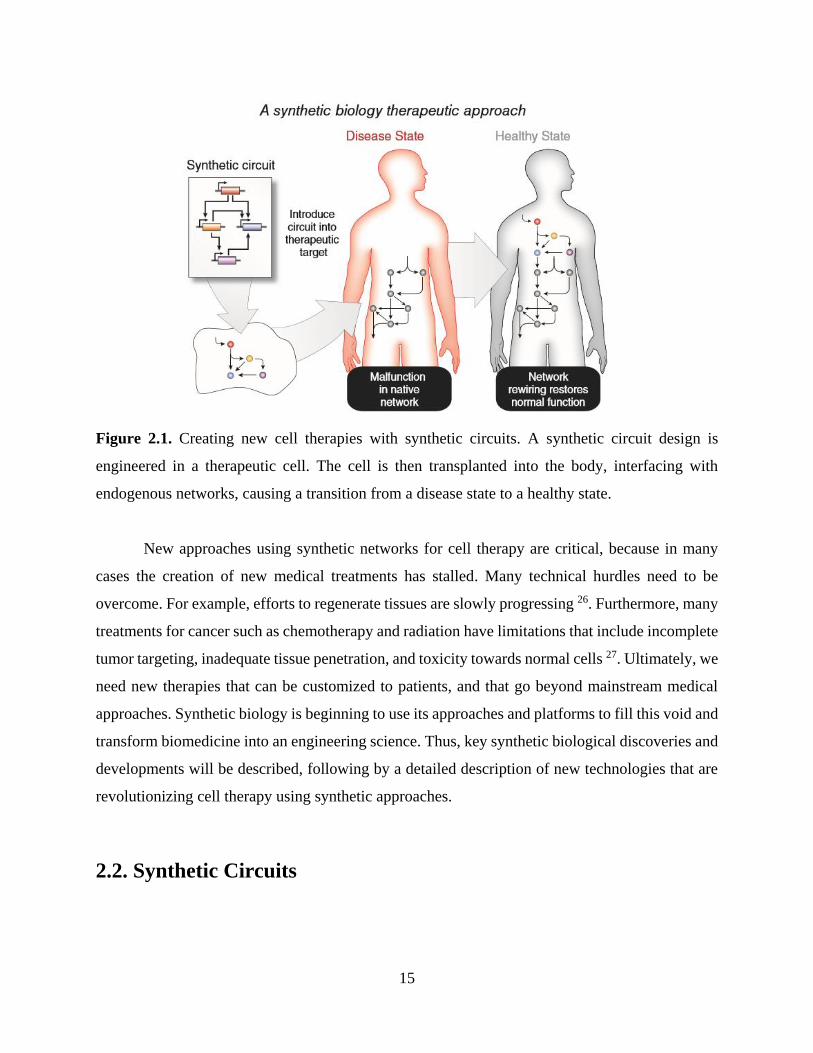
25. As shown in Figure 2.1, these DNA-encoded synthetic circuits are typically uploaded into cells,
enhancing them with new abilities that have been externally programmed by the circuit’s designer.
In the case of cell therapies, upon infusion or implantation, the programmed networks can then
interface with endogenous physiological networks to correct aberrant conditions in vivo.
15
Figure 2.1. Creating new cell therapies with synthetic circuits. A synthetic circuit design is
engineered in a therapeutic cell. The cell is then transplanted into the body, interfacing with
endogenous networks, causing a transition from a disease state to a healthy state.
New approaches using synthetic networks for cell therapy are critical, because in many
cases the creation of new medical treatments has stalled. Many technical hurdles need to be
overcome. For example, efforts to regenerate tissues are slowly progressing 26. Furthermore, many
treatments for cancer such as chemotherapy and radiation have limitations that include incomplete
tumor targeting, inadequate tissue penetration, and toxicity towards normal cells 27. Ultimately, we
need new therapies that can be customized to patients, and that go beyond mainstream medical
approaches. Synthetic biology is beginning to use its approaches and platforms to fill this void and
transform biomedicine into an engineering science. Thus, key synthetic biological discoveries and
developments will be described, following by a detailed description of new technologies that are
revolutionizing cell therapy using synthetic approaches.
2.2. Synthetic Circuits

16
Synthetic circuits are one of the core technologies available in synthetic biology. They are
critical for reprogramming cellular phenotypes to enhance therapies. As noted previously, the
development of synthetic circuits has spurred the growth of synthetic biology as a field. The
discussion that follows will outline design approaches in synthetic circuit construction as well as
provide a review of robust gene and protein circuits that have been successfully constructed.
Synthetic biology and its revolutionizing approaches to cellular engineering have already shown
the potential to impact cell therapies by altering their internal signaling networks. Whenever
attempting to engineer cellular signaling cascades, care must be taken to ensure robust design and
predictable cell behavior, along with repeatable and precise expression of critical genetic outputs.
Biological systems naturally incorporate all of these requirements in their internal programming.
As previously mentioned, the development of two synthetic circuits, the toggle switch and
an oscillator deemed “the repressilator,” inspired the field of synthetic biology in 2000 16-17. Instead
of open-loop control of gene expression, these circuits used feedback to provide an additional level
of complexity in the precision control of phenotype. In the years following these developments,
new circuits have been developed that leverage components from a range of different organisms.
Synthetic circuit design has also expanded beyond these initial circuits that were based on DNA-
protein interactions and now includes circuits based on protein-protein interactions as well 28-29. In
both cases, new computational algorithms and software programs have been created to speed the
engineering of synthetic biological circuits 30-33. These research thrusts have built a foundation for
synthetic biology as a complete discipline.
2.2.1. Engineering Synthetic Gene Circuits
In order to develop the first synthetic circuits, several molecular biological components
and control structures had to be available. In the last decades of the 20th century, investigators
began to develop control elements for each step in the cascade of events between gene transcription
and protein translation. Each of these processes is regulated by biophysical interactions, such as
the docking of transcription factors and RNA polymerase to operator sites on promoters, or the
binding of ribosomes to messenger RNA (mRNA) transcripts. Synthetic biology components that
control and manipulate all of these interactions have been created. These biological components

17
can be combined to form modules for the design and construction of synthetic circuits. Biological
circuits consist of modules of interacting genes and proteins, and synthetic biology seeks to rewire
these modules to create new circuits and new phenotypes. Several other components have been
created that leverage the interactions between RNA molecules themselves 34-35, RNA molecules
and small chemical molecules 15, 36-39, and recombinases with DNA templates 20, 40.
For example, new engineered promoters 41 were critical in the design of networks like the
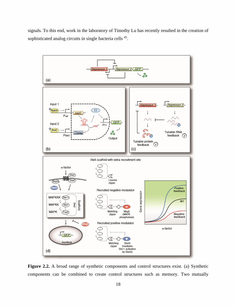
aforementioned toggle switch developed in the laboratory of James Collins 16. As shown in Figure
2.2a the toggle consists of two mutually repressible genetic operons, and when the strength of each
repression event is balanced, a bistable switch is formed. Over multiple generations of cell
division, the toggle will remain in either an ON or OFF state, regardless of the presence of a
corresponding inducer. In this network topology, the ability of each operon to repress the other
must be balanced, and these repressing interactions are governed by several factors, such as the
strength of each operon’s promoter as well as the strength of the ribosome binding sites (RBS)
associated with each repressor’s mRNA transcript. By carefully altering the nucleic acid bases in
each promoter and RBS, the switching properties of the toggle switch can be manipulated.
Other synthetic networks illustrated the potential for digital logic gate behavior in synthetic
circuits 22, 40, 42-44. In one of the first examples, Anderson et al. created a synthetic AND-gate in
bacteria as shown in Figure 2.2b. This AND-gate switching behavior, mimicking the gates enabled
by integrated circuits in Electrical Engineering, used an engineered viral RNA polymerase with a
slight defect (an amber – UAG – stop codon) in the middle of its transcript 44. As a result, when
one input (such as salicylate) was provided, only a partial polymerase was expressed. With the
addition of a second input, such as arabinose, transcription of a small RNA was initiated, and this
small molecule worked to suppress the defect. As a result, the fully translated viral polymerase
was able to activate its viral promoter. In another study, the Voigt group engineered communities
of bacteria to produce NOR-gate behavior 43. This is especially important as NOR-gates are
Boolean complete, and therefore can be combined to form any other type of logic gate. As a result,
this work suggests that with enough discrete bacterial colonies, significantly more complexity in
digital behavior computing could be possible. Alternately, other applications could require analog
18
signals. To this end, work in the laboratory of Timothy Lu has recently resulted in the creation of
sophisticated analog circuits in single bacteria cells 45.
Figure 2.2. A broad range of synthetic components and control structures exist. (a) Synthetic
components can be combined to create control structures such as memory. Two mutually

19
repressing operons can be used to create a bistable toggle switch; (b) Alternately, logic behavior
such as a digital AND-gate can be created. Two engineered, synthetically activated operons
alternately drive the expression of a faulty viral polymerase transcript or an RNA element that
corrects the resulting translation errors. When expressed together, the resulting translated viral
polymerase can activate the expression of an output gene; (c) Filter characteristics can be modified
by tuning the degradation of protein or RNA regulators; (d) A synthetic scaffold can be rewired to
control the mitogen activated protein kinase (MAPK) cascade in eukaryotes. By engineering
docking sites for positive and negative effector proteins using leucine zippers, synthetic circuits
can be created that control the MAPK cascade.
Other synthetic control behaviors are also possible, such as the genetic timers created by
Collins and colleagues. These timers, deployed in yeast, allowing flocculation to be precisely
timed 19, and therefore can control the fermentation process. In an another interesting study,
Stricker et al. showed that a minimal set of components could create an oscillator much more
robust than the repressilator 46. In fact, several oscillators have been developed since the original
repressilator in both bacterial and mammalian systems 17, 21, 46-49. These oscillator enhancements
mirror efforts to improve the efficiency of other basic control modules that have been built into
synthetic gene circuits. Other studies also have demonstrated that signal filtering can be created in
auto-regulated, negative-feedback circuits by tuning the degradation level of repressor elements as
shown in Figure 2.2c 50-53. More relevant to cell therapy, each could allow unique, precise types
of signaling behaviors to drive the therapeutic function of cellular therapeutics.
2.2.2. Engineering Synthetic Protein Circuits
Although engineering circuits that rely on DNA-protein interactions has been a major
thrust in synthetic biology, another key thrust that could be even more critical in leveraging
synthetic biology in cell therapy has been the development of protein-protein signaling circuits.
Just as genetic components allowed the development of diverse, tunable synthetic gene circuits,
engineered protein signaling components have allowed for new synthetic circuit designs as well.
These engineered proteins interactions have been important in two key ways. First, many of these
new components interface with the previously described genetic circuits providing enhanced

20
features. Second, other aspects of these components allow for robust, speedy protein-protein
interaction circuits. In fact, a key advantage of these protein-based circuits is the opportunity to
take advantage of the speed at which proteins interact. In comparison to gene expression, enzyme-
mediated single phosphorylation signals are evident in cellular phenotype on the order of seconds,
whereas gene expression events frequently require minutes or longer to emerge in a cell’s
phenotype.
Several exciting examples of these protein-protein components have been developed in the
past few years. For example, synthetic biology has significantly impacted the field of optogenetics.
In early work, a light sensor was created by fusing a cyanobacterial photoreceptor to an E. coli
intracellular histidine kinase domain to control gene expression 28. This work has now been
expanded to allow activation by multiple different wavelengths along with multiple applications
in a significant expansion the components available to synthetic biologists 14, 54-56. In a particularly
interesting advance, June Medford’s laboratory developed a plant signaling receptor that allows
plants to detect TNT. Using this synthetic component, her group engineered sensitive Arabidopsis
plants that turn white in the presence of TNT explosives 57. Beyond components, protein-protein
interactions have formed the basis of synthetic circuits as well. Some of the most interesting work
along these lines has come from the laboratory of Wendell Lim 29, 58-60. For example as shown in
Figure 2.2d, Lim and colleagues showed that positive and negative feedback loops could be
engineered by anchoring synthetic protein signaling components directly to an engineered protein
scaffold using leucine zippers 29.
2.2.3. Deploying Circuits in Mammalian Cells
The initial circuit modules developed in microbes like bacteria and yeast have been
expanded in higher order eukaryotes, and several synthetic circuits are now available that robustly
control gene expression in mammalian cells. This precise control of specific genes will be critical
for effective cell therapies. As an example of these mammalian circuits, Deans et al. developed a
tunable, modular mammalian genetic switch 61. This synthetic gene network coupled repressors
with an RNAi design involving shRNA. Gene expression was turned on by the addition of an
inducer, which controlled the transcription of a repressor, and simultaneously turned off the
21
generation of the RNAi component. Thus, this synthetic module allowed the transcript to be
retained and translated (Figure 2.3Aa). This construct offered >99% repression, along with an
ability to tune gene expression. This modular component can allow for the regulation of any gene,
and was validated in both mouse and human cells.
Another synthetic control module – the toggle switch – has also been developed in
mammalian cells as shown in Figure 2.3b. In multicellular systems such as the tissues of humans,
cell identity is regulated by epigenetic networks that determine which genes become part of each
cell’s transcriptome. By combining two repressors, which control each other’s expression, Kramer
et al. developed a mammalian epigenetic circuitry that exhibited genetic toggle behavior and could
be switched using two different drugs. These researchers used the toggle to regulate expression
profiles of a human glycoprotein in engineered Chinese hamster ovary cells, and these cells also
functioned after microencapsulation and implantation into mice. Just like previous bacterial
toggles, switching dynamics and expression could be predicted with mathematical models 62.
While these synthetic networks show the usefulness of synthetic control in mammalian
cells, the engineering of synthetic networks in mammalian cells is significantly more challenging
that developing circuits in bacteria or yeast. Bacteria and yeast are much more tractable as model
organisms for the design and tuning of synthetic circuits, as they are more easily genetically
engineered. Ideally, mammalian circuits could be first developed in simple eukaryotes like yeast
in a way that would allow their behavior in mammalian cells to be easily predicted upon transfer.
Thus, for cell therapy applications, once a clinician determines the mammalian cellular behavior
needed to treat a disease, the corresponding synthetic circuits could be rapidly developed in yeast.
Synthetic biologists could generate a diverse library of functional circuits (e.g., though approaches
such as site-directed mutagenesis), each with predictable function in human cells. This library
would then provide the clinician with a spectrum of synthetically engineered cell therapies to
prescribe.

22
Figure 2.3. Synthetic circuits for the control of mammalian cells. (a) Tight control of specific
genes will be critical for effective cell therapies. Here, a tunable, modular mammalian genetic
switch was created that couples repressor proteins with an RNAi design involving shRNA. The
switch is controlled by an inducer, which activates repressor expression, while simultaneously
turning off RNAi components, allowing the output gene to be translated; (b) Genetic toggle
switches can also be created in mammalian cells. By combining engineered streptogramin and

23
macrolide-inducible promoters to drive the expression of their respective engineered repressors,
the mammalian output gene secreted alkaline phosphatase (SEAP) can be stably toggled by pulsing
the system with erythromycin (E) or pristinamycin I (PI); (c) Synthetic circuits can be constructed
in yeast with predictable behavior upon transfer to mammalian cells. Here, a linearizer gene circuit
was created using the TetR repressor and an arbitrary target gene, both controlled by the same
promoter. In the absence of inducer, the TetR protein blocks transcription from both promoters.
When an inducer is added to cause de-repression, protein levels increase until TetR synthesis
exceeds inducer influx, at which point TetR blocks both promoters once again. By using analytical
and computational modeling, investigators were able to accurately predict the behavior of the
circuit in mammalian cells after recording its behavior in yeast.
As a step toward predictably transferring synthetic circuits from yeast to mammalian cells,
the group of Gabor Balazi constructed a mammalian version of a negative feedback-based
“linearizer” gene circuit (Figure 2.3c). The work version from yeast was at first non-functional in
mammalian cells. However, computational modeling suggested that function could be recovered
by improving nuances gene expression and protein localization. As a result, after rationally
developing and combining new synthetic parts, as suggested by the model, the circuit regained
function in human cells. Investigators were able to tune and target gene expression linearly and
precisely, just as they had been able to do in yeast. This approach should be relevant in transferring
many gene circuits of interest from yeast to mammalian cells and will be critical for quickly
developing therapies in the future 63.
2.3. Utilities of Engineered Bacteria
The above-described collection of engineered gene networks forms the foundation of a
large synthetic biology toolkit that is now available for the creation of new applications such as
cell therapies and cellular signal transduction. These synthetic components and circuits – which
have been developed in both microbial and mammalian cells – form the basis of several approaches
that have shown promise in regulating organismic physiology. A selection of these new approaches

24
towards synthetically enabled cell therapy and, in direct relevance to this thesis research, cellular
signal transduction is described in the following sections.
2.3.1. Cell Therapy
As discussed previously, the cells regulating human physiology include both the body’s
human cells as well as the collection of all the microorganisms associated with the body (i.e., the
microbiome). As these commensal organisms are well tolerated, they provide an ideal platform for
launching synthetic networks in the body. With this approach in mind, Duan and March recently
engineered a commensal strain of E. coli to prevent cholera infection by creating a synthetic
interaction between gut microbes 13. Cholera infection is marked by the secretion of virulence
factors by Vibrio cholerae. At low population density, these virulence factors include cholera toxin
(CT). In order to assess its own density, V. cholerae uses quorum sensing, a process in which it
secretes and detects autoinducer signaling molecules. Specifically, V. cholerae detects cholera
autoinducer 1 (CAI-1) and autoinducer 2 (AI-2). When both autoinducers are high, it ceases
expression of virulence factors. Duan and March leveraged this mechanism by engineering E. coli
– that already produced AI-2 – to also secrete CAI-1 (Figure 2.4). They found that when infant
mice ingested the engineered E. coli eight hours prior to V. cholerae ingestion, their survival rate
increased dramatically. Furthermore, cholera toxin intestinal binding was reduced by 80%.
25
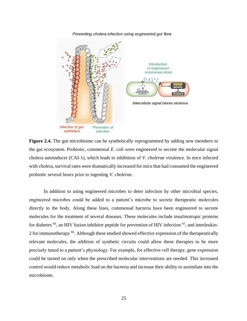
Figure 2.4. The gut microbiome can be synthetically reprogrammed by adding new members to
the gut ecosystem. Probiotic, commensal E. coli were engineered to secrete the molecular signal
cholera autoinducer (CAI-1), which leads to inhibition of V. cholerae virulence. In mice infected
with cholera, survival rates were dramatically increased for mice that had consumed the engineered
probiotic several hours prior to ingesting V. cholerae.
In addition to using engineered microbes to deter infection by other microbial species,
engineered microbes could be added to a patient’s microbe to secrete therapeutic molecules
directly to the body. Along these lines, commensal bacteria have been engineered to secrete
molecules for the treatment of several diseases. These molecules include insulinotropic proteins
for diabetes 64, an HIV fusion inhibitor peptide for prevention of HIV infection 65, and interleukin-
2 for immunotherapy 66. Although these studied showed effective expression of the therapeutically
relevant molecules, the addition of synthetic circuits could allow these therapies to be more
precisely tuned to a patient’s physiology. For example, for effective cell therapy, gene expression
could be turned on only when the prescribed molecular interventions are needed. This increased
control would reduce metabolic load on the bacteria and increase their ability to assimilate into the
microbiome.

26
Another approach toward using engineered bacteria to treat disease has been explored by
synthetic biologists who have created bacteria that seek and destroy cancer. Although it is not
entirely clear how these engineered microbes should come into contact with tumors occurring
distal to host locations where microbes normally reside, these engineered microbes use an
interesting approach to cancer treatment. In one of these studies, Voigt and colleagues created
bacteria that invaded cancer cells only in the hypoxic environments that frequently are indicative
of tumor tissue, as well as bacteria that used quorum sensing components from other species to
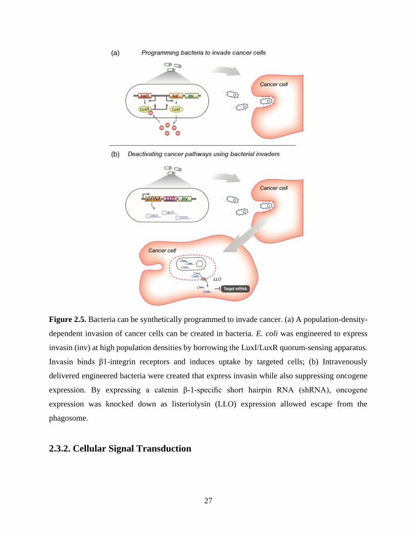
amplify their invasion response 12. As shown in Figure 2.5a, E. coli were engineered to invade by
causing them to express invasin (inv), an adhesion protein from Yersinia Pseudotuberculosis,
which tightly binds mammalian β1 integrin receptors and induces uptake. Invasin expression was
driven by the LuxI/LuxR quorum-sensing genes, allowing E. coli to produce an autoinducer that
amplified invasin expression as the colony grew. Of course, using bacteria to treat cancer could
come with a risk of infection from the bacteria themselves.
However, in another study, Li and colleagues intravenously delivered engineered, cancer-
invading bacteria to target a tumorigenic pathway in vivo 67. They used RNA interference (RNAi)
to create bacterial invaders that knocked down expression of CTNNB1 (encoding β-1 catenin), a
gene that initiates many colon cancers upon its overexpression or oncogenic mutation (Figure
2.5b). The engineered bacteria generated short hairpin RNA (shRNA) segments that bound to
CTNNB1 mRNA transcripts and induced mRNA cleavage. In addition to shRNA and invasin, the
synthetic system also produced lysteriolysin O (encoded by the hylA gene). Lysteriolysin enables
molecular transport out of vesicles potentially through entry vesicle disruption. When engineered
bacteria were intravenously delivered into immune-deficient deficient mice with subcutaneously
xenografted human colon cancer cells, the investigators observed significant knockdown of the
gene in tumor cells.
27
Figure 2.5. Bacteria can be synthetically programmed to invade cancer. (a) A population-density-
dependent invasion of cancer cells can be created in bacteria. E. coli was engineered to express
invasin (inv) at high population densities by borrowing the LuxI/LuxR quorum-sensing apparatus.
Invasin binds β1-integrin receptors and induces uptake by targeted cells; (b) Intravenously
delivered engineered bacteria were created that express invasin while also suppressing oncogene
expression. By expressing a catenin β-1-specific short hairpin RNA (shRNA), oncogene
expression was knocked down as listeriolysin (LLO) expression allowed escape from the
phagosome.
2.3.2. Cellular Signal Transduction

28
As introduced in the preceding section, although cell therapy is a widely used medical
intervention with many successes, it also has the potential for use as a tool for creating synthetic
biological constructs and circuits. While approaches that have leveraged synthetic biology to
engineer therapies have shown promise 68, many technical challenges remain. In the meantime, the
same technologies can be applied to the monitoring and control of microbial populations, in
particular, bacterial biofilms that have pervasive implications on, for example, human health as
well as water and food quality. Most of these challenges have not been successfully overcome
owing to a lack of information about the hundreds of genes that interact during the biofilm
development process, including stress-associated genes. The research presented in this thesis
utilized engineered bacteria with synthetic gene circuits (e.g., a quorum sensing receiver construct
and a toggle switch) for bacterial signal transduction in order to better understand the mechanisms
underlying genetic variation in biofilms.
Quorum sensing receiver cell line. The vast majority of microbiological studies have
examined bacteria in their planktonic state rather than their biofilm state. Bacterial biofilms
normally exhibit complex, heterogeneous structures, which vary widely in their mechanical
properties, chemical compositions, and morphologies. In general, the local cell densities of
biofilms are substantially higher than of planktonic bacterial cultures, as are the concentrations of
metabolic by-products, secondary metabolites, and other secretory microbial factors. Of these,
intercellular signaling and quorum sensing molecules are particularly important microbiological
factors, as they link individual bacterial cells to the formation or dissolution of the biofilm
community.
Environmentally sensitive bacterial cells, referred to as “receiver” cells - that generate
visible signals upon induction with N-acyl homoserine lactone (AHL) can be constructed to better
understand stochastic expression of quorum sensing signaling within biofilms (refer to Chapter 3
for details). The receiver contains a genetic construct with the luxR transcription factor and a green
fluorescence protein (GFP) gene encoded by a bidirectional Plux promoter. Quorum sensing is
activated by the production and release of AHL, thereby signaling the local cell density to
neighboring bacterial cells, which in turn express quorum signaling on their own. This type of
signal transduction to a visibly measurable signal can be used to monitor bacterial growth within

29
bioreactors as shown in Chapter 6. Furthermore, quorum sensing can also be triggered in model
bacteria systems like E. coli in the absence of AHL; bacteria with an identical gene circuit construct
was examined to monitor induction of stochastic quorum signaling within the biofilm-mimicking
microfluidic structure in the research presented in Chapters 3,5 and 6.
Toggle switch circuit-integrated bacteria. Bacteria endowed with a bistable toggle
switch have been fully characterized as described earlier and can be utilized to study stochasticity
in microenvironments, such as biofilms. A toggle switch is a circuit that can switch between two
stable states, ON and OFF. Consequently, such a switch possesses a form of memory. The toggle
switch consists of two transcriptional repressors, each repressing the other (refer to Chapter 4 for
further details). The switch uses two small molecule-responsive transcriptional repressors—TetR,
which is inhibited when it binds anhydrotetracycline (aTc), and LacI, which is inhibited when it
binds isopropyl β-D-1-thiogalactopyranoside (IPTG). The introduction of aTc into a bacterial
culture inactivates the ability of TetR to repress the PLtetO promoter, thereby switching the system
into a state with high expression of LacI, which then continually maintains a low concentration of
TetR. Conversely, addition of IPTG inactivates LacI from repressing the Ptrc-2 promoter, switching
the system into a high-TetR expression state, which then maintains a low concentration of LacI.
The function of the toggle switch depends on the relative expression levels of its component
transcriptional repressors. Additionally, this system can be utilized to drive the expression of
reporter molecules, such as GFP and mCherry, in order to visualize the toggle switch state within
a culture. We show in Chapter 4 that stochastic gene expression revealed in relatively simple
inducible genetic systems also appears in the bistable genetic toggle switch system. This approach
permits the study of stochastic gene expression with a higher degree of complexity.
30
CHAPTER 3.
STOCHASTICALLY INDUCED QUORUM
SENSING
3.1. Abstract
We have investigated a microfluidic environment that can be used to monitor stochastic
activities within a microbial consortium. The layout of microfluidic channels in a device was
designed to regulate the growth of bacterial communities by confining bacterial cells while
maintaining nutrient availability for survival, permitting cells to populate the region. Similar to
naturally occurring biofilms, such engineered environment can exhibit different local growth rates
as a function of distance from the bulk flow media. To demonstrate this phenomenon, we
developed a microfluidic bioreactor (a maximum 0.97-μm height) containing two distinct regions
for cell growth that differ in diffusion path length from the main fluid channel (12-μm height).
This reactor was then populated with Escherichia coli engineered to retain a quorum sensing (Plux-
driven) circuit. The cells that were 225-μm distance away from the bulk-flow channel exhibited a
strong GFP signal, our proxy for quorum, even in the absence of the quorum sensing signaling
molecule. This finding suggests quorum sensing pathways may be de-repressed as a stress
response in nutrient and space scarce environments. This new mode of quorum sensing activated
by a starvation-induced stress response can be used to study quorum sensing in the laboratory.
3.2 Introduction
Throughout its life cycle, bacteria can exist in both planktonic and sessile states. The latter
state is often associated with the formation of stationary, multicellular communities known as
biofilms. Nearly all microbes can occur as biofilms in aqueous environments. Under such

31
conditions, biofilms are formed by the attachment of bacteria to submerged macro-surfaces, micro-
particles, also to one another 69-71. Biofilms can form along a surprisingly wide variety of surfaces,
including metals, plastics, medical implants, and organic surfaces such as plant and animal tissues
72. While biofilm communities within laboratories are often comprised of a single bacterial strain,
naturally occurring biofilms can be composed of a broad array of bacterial, fungal, algal, and
protozoan species among other microorganisms 9, 73. For example, human dental plaque biofilms
can consist of over five hundred bacterial species 74. Furthermore, these communities are often
bound together with sugar-rich molecular strands, collectively referred to as extracellular
polymeric substances (EPS) 75. Taken together, even a simple biofilm can be composed of a
complex, heterogeneous network of spatial and genetic variations. Additionally, the biofilm
development process involves temporally varying regulated of many genes, a large portion of
which are stress-induced. The complex network of structure and responses makes it difficult for
scientists to recapitulate, and controllably observe and study, biofilms within a laboratory setting
76-78.
Biofilms are formed through bacterial aggregation and subsequent self-secretion of EPS.
Both of these processes are associated with a suite of phenotypic and physiological processes, such
as altered gene transcription 79, changes in nutrients accessibility and preference 80, and an
increased resistance to external stresses 81. The complexity of biofilm formation and the related
changes creates challenges in clinical and industrial settings, including human infections,
biofouling, clogs in flow systems and pipelines, and bactericide efficacy 71.
An enormous body of research has examined the planktonic state of bacteria, while
technological challenges have limited the number and scope of studies examining bacteria in their
sessile state (i.e., biofilm state) 82. These challenges arises from the complex, heterogeneous
structures of biofilms, which vary widely in mechanical properties, chemical composition, and
morphology 71, 83. In general, the local cell densities of biofilms are much higher than those of free-
floating planktonic cell cultures 84. Consequently, the concentrations of metabolic by-products,
secondary metabolites, and other secretory microbial factors are much higher within the interior
of biofilm structures 85. Of these, intercellular signaling and quorum sensing molecules are

32
particularly important factors in the biology of microbes, as they link individual bacterium to the
formation or dissolution of the biofilm community.
When planktonic bacteria cells encounter a submerged surface, they quickly adhere to the
surface 86. Once attached, the cells begin to produce EPS and colonize the surface 6. EPS
production allows the emerging biofilm community to develop a complex, three-dimensional
structure 87. These biofilm communities develop within hours and propagate to new surfaces over
time through detachment of small or large clumps of cells 88. Later stages of detachment allow
bacteria to propagate to wider surface areas or to a location downstream of the original community
79. Though the physiological process of attachment–growth–detachment process is well described
in the literature 89, the chemical signaling dynamics associated with biofilm quorum sensing has
not yet been fully investigated 90.
In addition to cell-produced signaling chemicals, biofilms are influenced by internal small-
molecule and nutrient gradients (Figure 1.2). These phenomena play a critical role in the cellular
physiology of pathogens 91. Near the fluid interface of biofilms, mass transport is high, providing
bacteria with the nutrients necessary for robust growth 92. In contrast, nutrient concentrations are
low within the biofilm interior. This lack of resources within the biofilm interior slows growth and
causes starvation-induced stress responses, potentially activating protective metabolic states 93.
These key features can be modeled using microfluidic channels, allowing the induction of a
biofilm-like structure to study stochastically expressed quorum sensing signals.
Biofilm quorum sensing mechanisms are difficult to study due to technical challenges in
examining bacterial aggregates over appropriate spatiotemporal scales. Additionally, the
mechanics of EPS and flow interactions pose a make modeling and studying quorum sensing
chemical transport non-trivial. However, microfluidic techniques have emerged as a promising
approach for many applications owing to their low cost and ease of use 94-97. Early research efforts
that utilized microfluidics to study biology focused on biochemical assays containing small
concentrations of DNA and proteins 94-95. Over the past fifteen years, microfluidic devices have
become significantly more complex. For example, Hasty et al. used microfluidic chambers to
observe and track individual cells within a single focal plane under various environmental

33
conditions 94, 98-99. Extensions of this work in an artificial biofilm structure could produce novel
insights made possible by controlled observations. For this reason, microfluidic devices have been
designed to mimic physical conditions that resemble natural cellular habitats, thus enabling the
investigation of complex physiological states such as biofilms 100.
However, researchers have yet to develop microfluidic systems that mimic the biophysics
of natural biofilm structures and while also enabling researchers to monitor cells in a planktonic
state. Here, we describe the development of a novel microfluidic device that enables monitoring
sessile, biofilm-like state cells and planktonic-like state cells together. This system allows
researchers to investigate cellular and community physiology while monitoring single-cell
phenotypic responses. Together, this allows researchers to study phenomena such as stochastic
gene regulation in a single cell, while observing the phenomena’s effect within a larger biofilm
community, all on a single microfluidic device. The microfluidic device developed herein, referred
to as a biomimetic biofilm, utilizes a unique design that mimics the key features of natural biofilm,
while allowing researchers to take advantage of a well-characterized, controllable environment.
This microfluidic tool can be used in combination with synthetic gene networks in E. coli that
express GFP upon induction with AHL, the quorum signal produced by Vibrio cholerae 101. Taken
together, this engineered gene construct and our biomimetic biofilm device allows us to study gene
expression dynamics for quorum sensing at a scale and time period not previously possible.
3.3 Materials and Methods
3.3.1. Manufacture of microfluidic device
Master mold fabrication. All master molds were fabricated in the Virginia Tech Micro &
Nano Fabrication Laboratory (MICRON). Photomasks were drawn in AutoCAD (Autodesk, San
Rafael, CA, USA), printed onto transparency film by CAD/Art Services, Inc. (Output City, Poway,
CA, USA), and mounted onto glass plates (McMaster-Carr, Los Angeles, CA, USA). The first
layer of the master mold was created by first spin-coating with SU-8 2000 negative photoresist
(MicroChem Corp., Newton, MA, USA) onto clean silicon wafers to 0.96 µm in height using a

34
WS-650-8B programmable spinner (Laurell Technologies Corp., North Wales, PA, USA) and then
using a MA/BA6 UV contact mask aligner (MicroTec, Garching, Germany) for subsequent UV
exposure. After being exposed to UV radiation, the unexposed photoresist was rinsed away using
developer (MicroChem Corp.). For the second layer, the SU-8 2010 negative photoresist was spin-
coated on top of the first layer to achieve a height of 10 µm. Again, the MA/BA6 UV contact mask
aligner was used for UV exposure, and the unexposed photoresist was rinsed away using the same
process described above.
PDMS device fabrication. Replica molds were created from master molds by mixing
polydimethylsiloxane (PDMS)/Sylgard 184 (Dow Corning, Midland, MI, USA) with a 10:1 ratio
of the elastomer base to curing agent, degassing in a vacuum desiccator for 1 h, and curing in an
oven at 90°C for 2 h. Following removal of the PDMS, devices were cut, holes were punched for
ports, and the cell was cleaned with filtered DI water and then permanently bonded to clean
coverslips (Thermo Fisher Scientific, Waltham, MA, USA) via exposure to O2 plasma for 1 min
in a PDC-32G plasma cleaner (Harrick Scientific, Ithaca, NY, USA).
3.3.2. Construction and characterization of the quorum sensing receiver circuit
Cloning details. The receiver circuit (pSP035) used in this study was constructed using six
components, pKE1-MCS as the vector, Plux, luxR T1T2, rbs027 and GFPmut3b as inserts (Figure
3.1). The desired restriction sites were added to the 5′ and 3′ ends of each construct using overhang
PCR (Table 3.1). For each insertion, both the insert construct and vector were uniquely double
digested using appropriate enzymes. Followed by gel purification, the fragments were then ligated
and transformed, and the resulting clones were screened against carbenicillin (Cb) resistance.
Induction conditions. To characterize the performance of the receiver circuit we
conducted a series of experiments. The cells were first grown overnight in Luria–Bertani (LB)
media with 100 g/ml Cb from a frozen stock. At the beginning of the experiment, overnight
grown cells were diluted 1:1000 in fresh LB media and once again grown and kept at exponential
growth density. The receiver cells were then induced with of AHL to yield final concentrations of
0.001, 0.01, 0.1 and 1 mM. The fluorescent intensity was measured at given times by diluting each

35
sample 1000-fold using phosphate-buffered saline (PBS) and collecting 10 000 events per replicate
using a BD Accuri™ C6 (BD Bioscience, Piscataway, NJ, USA). Cell were maintained at 37°C
before flow cytometry analysis.
3.3.3. Maintenance and monitoring of cell growth in microfluidic channels
Microfluidic cell culture. The recombinant E. coli cells were loaded into the trapping
channels of the microfluidic device by directing the flow in the direction from the cell port to the
waste port. Upon trapping a few cells in each region, the flow was reversed and slowed to steadily
supply the cells with fresh nutrients from the media port through a combination of diffusion and
advection. The cells were maintained at 22°C and grown to fill the chamber over a duration of 6–
10 h.
Cell microscopy. Cell images were acquired using an inverted epifluorescent microscope
(Ti-E; Nikon Instruments Inc., Tokyo, Japan). Differential interference contrast (DIC) and
fluorescent images were captured every 15 min, and focus was maintained automatically using
Nikon Elements software.
Figure 3.1. The receiver circuit (pSP035) used in this study was constructed using six components,
pKE1-MCS as the vector, Plux, luxR T1T2, rbs027 and GFPmut3b as inserts 102. Insert rbs027 was
first extended to GFPmut3b using overlap extension PCR and all other segments were amplified
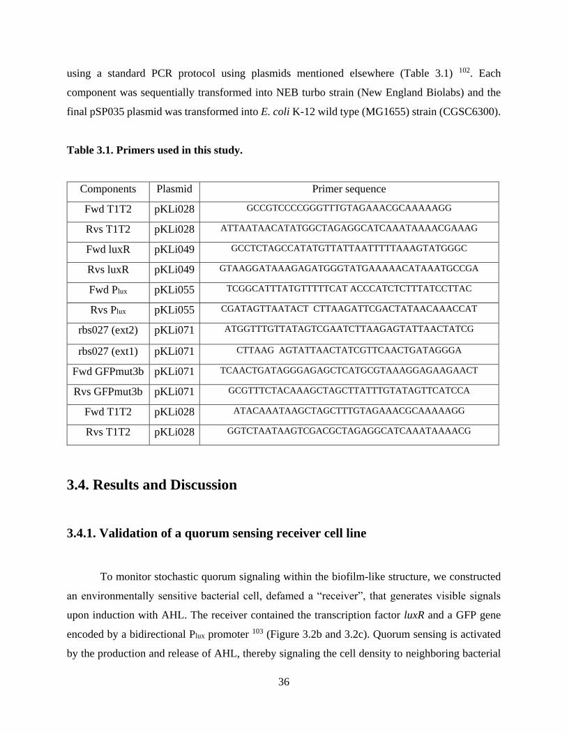
36
using a standard PCR protocol using plasmids mentioned elsewhere (Table 3.1) 102. Each
component was sequentially transformed into NEB turbo strain (New England Biolabs) and the
final pSP035 plasmid was transformed into E. coli K-12 wild type (MG1655) strain (CGSC6300).
Table 3.1. Primers used in this study.
Components Plasmid Primer sequence
Fwd T1T2 pKLi028 GCCGTCCCCGGGTTTGTAGAAACGCAAAAAGG
Rvs T1T2 pKLi028 ATTAATAACATATGGCTAGAGGCATCAAATAAAACGAAAG
Fwd luxR pKLi049 GCCTCTAGCCATATGTTATTAATTTTTAAAGTATGGGC
Rvs luxR pKLi049 GTAAGGATAAAGAGATGGGTATGAAAAACATAAATGCCGA
Fwd Plux pKLi055 TCGGCATTTATGTTTTTCAT ACCCATCTCTTTATCCTTAC
Rvs Plux pKLi055 CGATAGTTAATACT CTTAAGATTCGACTATAACAAACCAT
rbs027 (ext2) pKLi071 ATGGTTTGTTATAGTCGAATCTTAAGAGTATTAACTATCG
rbs027 (ext1) pKLi071 CTTAAG AGTATTAACTATCGTTCAACTGATAGGGA
Fwd GFPmut3b pKLi071 TCAACTGATAGGGAGAGCTCATGCGTAAAGGAGAAGAACT
Rvs GFPmut3b pKLi071 GCGTTTCTACAAAGCTAGCTTATTTGTATAGTTCATCCA
Fwd T1T2 pKLi028 ATACAAATAAGCTAGCTTTGTAGAAACGCAAAAAGG
Rvs T1T2 pKLi028 GGTCTAATAAGTCGACGCTAGAGGCATCAAATAAAACG
3.4. Results and Discussion
3.4.1. Validation of a quorum sensing receiver cell line
To monitor stochastic quorum signaling within the biofilm-like structure, we constructed
an environmentally sensitive bacterial cell, defamed a “receiver”, that generates visible signals
upon induction with AHL. The receiver contained the transcription factor luxR and a GFP gene
encoded by a bidirectional Plux promoter 103 (Figure 3.2b and 3.2c). Quorum sensing is activated
by the production and release of AHL, thereby signaling the cell density to neighboring bacterial

37
community members and in turn expressing the GFP reporter in the following experiments 104
(Figure 3.2d). However, quorum sensing can also be triggered in the absence of AHL (Figure
3.2e); in this situation, the Plux promoter is activated without the presence of AHL. This activation
could be the result of de-repression, a consequence of cell density or nutrient deprivation.
In order to validate the receiver circuitry that we used in subsequent experiments, we first
verified the GFP expression of the receiver cell when induced by AHL supplemented with LB
media containing 50 g/mL Cb. By varying the AHL concentrations over an induction period of
5 h, changes in fluorescence were observed using a flow cytometer. In the absence of AHL, GFP
was not expressed (Figure 3.2f). The GFP signal reached saturation at a 1 mM AHL concentration,
while intermediate concentrations produced intermediate expression (Figure 3.2f). A comparison
between the 0- and 1-mM AHL treatments clearly shows that the receiver cells were not induced
in the absence of AHL (Figure 3.2f).

38
Figure 3.2. Understanding quorum sensing for biofilm consortia. (a) Within a microbial
consortium, different states of bacterial cells exist. Part of these differences are a function of the
distance the bacteria cells are from the source of flow. (b) Normal quorum sensing system. Upon
reaching a certain threshold population density, the quorum sensing genes are induced, and the
cells GFP. (c) Stochastically induced quorum sensing system. Even without the signal molecule
(i.e., AHL), the quorum sensing gene circuit is triggered in certain cells within the microbial
population. This can occur from a shortage of LuxR production or an inability for LuxR to repress

39
the Plux promoter site. (d) Characterization of receiver cells using a flow cytometer. (d) The
receiver cells were exposed to different concentrations of AHL in the culture medium, and GFP
expression levels were measured continuously over 5 h. The fluorescence signal increased in
proportion to the inducer concentration between 0.001 and 1 mM over the 5-h time period. The
receiver cells were only responsive in the presence of AHL.
3.4.2. Biomimetic biofilm
To explore the spatiotemporal dynamics of quorum sensing within biofilms, we developed
a three-port microfluidic device that mimics the key transport phenomena in biofilm colonies. The
device consists of two regions on opposite side of the main flow channel in parallel: rectangular
chambers independent of one another and identical chamber regions connected via convolutions
resembling an inverted dichotomously branching tree of channels. Both chambers are open to the
flow of the medium. Along the direction of the flow, the convolutions converged into a single path
and ultimately came to a closed end (Figure 3.3a-c). The cell chamber region and convoluted
pathway both have a channel height of 0.97 μm. Since E. coli cells have a ~1 μm diameter, the
height of the trapping region allowed linearized observation of confined cells (Figure 3.4b). The
device was also designed to continuously discharge cells from the device through the main flow
channel (12 μm in height). In this biofilm-like structure, the PDMS structures of the microfluidic
device captured and accumulated the extracellular matrix within structures constrained to resemble
biofilms (Figure 3.4a and 3.4b). Additionally, we designed the device to have a well-characterized
ability to transport media throughout the biofilm-like structure.
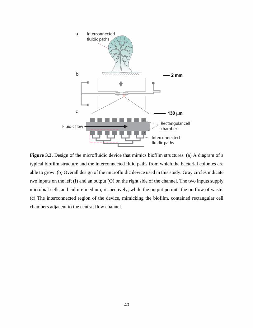
40
Figure 3.3. Design of the microfluidic device that mimics biofilm structures. (a) A diagram of a
typical biofilm structure and the interconnected fluid paths from which the bacterial colonies are
able to grow. (b) Overall design of the microfluidic device used in this study. Gray circles indicate
two inputs on the left (I) and an output (O) on the right side of the channel. The two inputs supply
microbial cells and culture medium, respectively, while the output permits the outflow of waste.
(c) The interconnected region of the device, mimicking the biofilm, contained rectangular cell
chambers adjacent to the central flow channel.

41
Figure 3.4. Biofilm developmental process and the design of a biofilm-like structure using
microfluidic channels. (a) Microbial cells are attached to solid surfaces and grow to form biofilm-
like structures. The outer cells are ultimately detached from the biofilm structure and return to
individual planktonic states. (b) To investigate spatiotemporal characteristics of quorum sensing
mechanisms within a microbial consortium, we have designed a device that mimics the key
features of a biofilm structure. The microfluidic device is constructed to retain microbial colonies
within interconnected fluidic paths. The device design limits growth areas in deeper regions of the
device by constraining space while imparting a nutrient gradient.
3.4.3. Stochastically expressed quorum sensing GFP reporter in batch culture
The effects of nutrient availability on the diversity of phenotypes in our tightly regulated
receiver colonies were also apparent in experiments lacking AHL within batch cultures in test
tubes. We conducted two experiments in parallel. The first maintained cells in the exponential

42
phase while the second maintained cells in the stationary phase over a 4-day duration. For the
exponential phase cultures, the media and cells were periodically diluted with a fresh batch of LB
supplemented with Cb in order to provide optimal nutrients and growth conditions. At each
observation point throughout the full duration of the experiment, the exponential phase cells
showed minimal GFP expression (Figure 3.5, Exponential, quadrant 2 [Q2] in each panel). On day
2, there was a slight overall shift (of only 0.27%) towards the high GFP expression state; however,
this increase was temporary, and the expression later returned to basal levels for the remainder of
the experiment (Figure 3.5, Exponential, days 3 and 4).
While exponentially maintained receiver cells did not exhibit an increase in stochastic
quorum signaling over time, we did observe an increase in stochastic behavior for receiver cells
held at stationary phase. For this trial, receiver cells were held in stationary phase, without being
supplemented with fresh nutrients after the inoculation. As above, we monitored for cells that were
expressing GFP stochastically without AHL induction. As shown in Figure 3.5, we were able to
observe a gradual increase of the cell population in the high GFP state, with day 4 showing the
most stochastic events (1.51%). We replotted our observations from the Q2 sections, respectively,
from the exponential and stationary phase cultured cells, in order to compare the course of
stochasticity over the 4-day period (Figure 3.5, far right panels). We were able to see a distinct
difference between the two culture phases; over time, the stationary phase receiver cells showed a
10-fold increase in cells transitioning to a high GFP expression state.
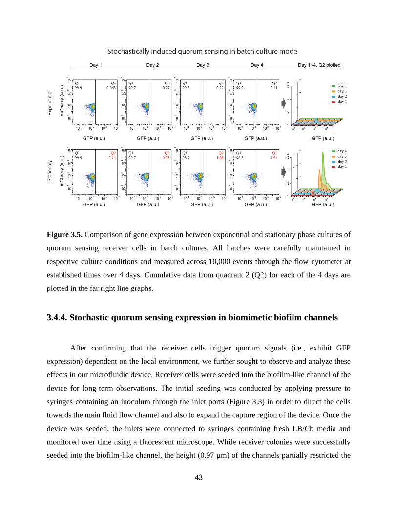
43
Figure 3.5. Comparison of gene expression between exponential and stationary phase cultures of
quorum sensing receiver cells in batch cultures. All batches were carefully maintained in
respective culture conditions and measured across 10,000 events through the flow cytometer at
established times over 4 days. Cumulative data from quadrant 2 (Q2) for each of the 4 days are
plotted in the far right line graphs.
3.4.4. Stochastic quorum sensing expression in biomimetic biofilm channels
After confirming that the receiver cells trigger quorum signals (i.e., exhibit GFP
expression) dependent on the local environment, we further sought to observe and analyze these
effects in our microfluidic device. Receiver cells were seeded into the biofilm-like channel of the
device for long-term observations. The initial seeding was conducted by applying pressure to
syringes containing an inoculum through the inlet ports (Figure 3.3) in order to direct the cells
towards the main fluid flow channel and also to expand the capture region of the device. Once the
device was seeded, the inlets were connected to syringes containing fresh LB/Cb media and
monitored over time using a fluorescent microscope. While receiver colonies were successfully
seeded into the biofilm-like channel, the height (0.97 µm) of the channels partially restricted the

44
ability of E. coli to swim, especially within deeper regions of the microfluidic structure where the
flow velocity is low. In these constricted environments, bacterial dispersal throughout the channel
is driven by growth and division of parental cells rather than motility Sequential images of a typical
experimental run are presented in Figure 3.7. Initially, a few isolated colonies began to grow and
subsequently merged into a large monolayer that filled the chamber (Figure 3.7, 20 h). Doubling
times are shown in Figure 3-6. Once the chamber areas of the channels were fully occupied by 20
h, a localized burst of GFP expression began to occur within the deeper regions of the convoluted
channel (in a localized area of stochastic bursting) as depicted in the red box in Figure 3.7. This
region of stochastic bursting, approximately 200–250 µm away from the flow main channel,
exhibits stochasticity as a result of several factors including a lack of nutrients and cell crowding.
By the conclusion of the full experiment over a duration of 60 h, these localized regions showed
an accumulation and intensification of quorum signaling cells.
Figure 3.6. Motility and doubling time of the quorum sensing cells within the biomimetic biofilm
channel. Red arrows are for tracking purposes. Single red arrows within a frame show the parental
cell. At approximately 45 min, the parental cell divides, generating two daughter cells. The second
cycle of division is shown in subsequent frames. The cell located on the top in the 45-min image
was tracked for an additional division cycle. All experiments were conducted at 22°C. Sequential
snapshots of a typical experiment are presented. Initially, we observed a few isolated receiver
colonies. Over time, the colonies subsequently merged into a larger monolayer that filled the
chamber.
We further compared these findings to colonies that were grown in independent
rectangular chambers (Figure 3.3). The rectangular chambers were located directly in parallel to
the biofilm-like channel (Movie 2). Owing to the difference in nutrient transport levels and growth

45
environments, stochastically triggered quorum sensing (i.e., GFP expression) was only observed
in the biofilm-like channels. These findings are also consistent with the results obtained previously
in batch culture modes (Figure 3.5).
Figure 3.7. Stochastically expressed quorum signaling within the biomimetic biofilm channel.
Quorum sensing colonies were seeded in the microfluidic channels, and GFP expression was
monitored over time. The red dotted line indicates localized areas of stochastic bursting of the
quorum sensing cell line. The yellow dotted line shows the path from the main fluid flow to the
center of the region of stochastic bursting. The total duration of the experiment was 60 h, with
time-lapse images obtained every 15 min. All scale bars indicate 25 µm.
3.5. Conclusion
In this work, we examine how quorum sensing can be triggered stochastically within
communities of E. coli cells. We developed an artificial biofilm-like structure that facilitates lateral
observations of microbial community structures. This enabled us to directly visualize phenomena
occurring within the core of these structures at the scale of individual cells. Moreover, the structure
induced expression of the GFP reporter, indicating the stochastic induction of quorum sensing.
The effects of quorum sensing within biofilms in the environmentally relevant context were made
possible by the design of our microfluidic device.

46
In nature, dense microbial groups, such as bacterial biofilms, commonly contain diverse
cell types and species due to local fitness advantages 105. Even within even isogenic cell
populations, some cells exhibit genetic mutations and stochastic gene expression 106-107, producing
fluctuations in transcription and translation as well as aberrant gene products. Our microfluidic
device is able to stochastically trigger quorum sensing within microbial communities that are
densely packed and lack sufficient nutrients, allowing us to study such mutations in a well-
controlled environment.
Recent research has shown that novel genotypes within biofilm structures can arise
through both mutation and recombination 107-108. These stress-induced genetic changes have been
primarily attributed to changes in the colony morphology following biofilm growth. In addition,
as shown in our study, subpopulations within biofilm colonies can exhibit distinct differences in
gene expression in a stochastic manner, leading to phenotypic heterogeneity. There are several
examples in which microenvironmental heterogeneity within biofilms leads to stochastic gene
expression and potential adaptive group behaviors. For example, a heterogeneous bacterial
population created as a result of stochastic gene expression may be able to adapt more efficiently
to sudden changes in environmental conditions, benefiting the entire biofilm structure 109.
The described microfluidics device provides a unique opportunity for the study of the
complex phenomenon of quorum sensing. This system vastly simplifies any E. coli assays that
require steady, reproducible populations of sessile cells in their biofilm state. There are three major
advantages over similar platforms. First, the microfluidics device induces quorum sensing
pathways without the use of signaling molecules such as AHL. In other words, the structure and
flow characteristics serve as appropriate environmental stimuli to induce quorum sensing in E.
coli, a major system for the study of biofilms. Second, the linearized channels of the microfluidics
device make it possible to visually isolate single E. coli cells. In this way, individual cells can be
visualized continuously. This capability may be combined with other visual reporter assays in
order to examine other genetic factors both downstream and upstream of the GFP reporter
construct as well as any small or environmental stimuli that can be applied through the apparatus.
Third, the design of the microfluidics device allows the parallel culture of cells exhibiting

47
stochastically induced quorum sensing in its convoluted chambers with cells cultured in the
independent rectangular chambers exhibiting no such phenotype. This parallel structure enables a
clear control treatment in any experimental design without any modification.
The microfluidic cell design is somewhat specific to E. coli cells; the channels in the
device were just sufficient in height (0.97 µm) to accommodate individual E. coli cells (1 µm).
However, similar systems could be designed to precisely accommodate other species of quorum
sensing bacteria. These organisms interact with many of the same environmental stresses in
biofilm formation 76-77, indicating the potential for similar induction, especially in an autoinducer
species capable of producing their own AHL.
The field of microbiology has long had a limited understanding of the mechanism by which
biofilms maintain these diverse cell populations and communities. This biomimetic biofilm
structure enables the visualization of phenotypic diversity within biofilm structures, thus providing
exciting new opportunities in the study of quorum sensing. Our results suggest that resource levels
as well as population density are keys to understanding heterogeneity in microbial communities.

48
CHAPTER 4.
TOGGLE SWITCH STABILITY IN BIOFILM
STRUCTURES
4.1. Abstract
Biofilm bacteria and planktonic bacteria exhibit substantially different gene expression
patterns; these differences are not only important for understanding biofilm biology, but also have
practical implications for antibiotic development and food processing technology, for example. To
better understand biofilm formation, we considered a toggle system in which inducers can be used
to switch a gene expression marker between two states. High LacI expression was induced by aTc
and indicated by high GFP levels, and high TetR expression was induced by IPTG and indicated
by high mCherry levels. We tracked expression under stressful biofilm-like conditions in our
previously developed microfluidic device to characterize the stochasticity of gene expression.
After induction with aTc, cells showed stable expression of GFP throughout the experiment. After
induction with IPTG, cells mainly expressed mCherry, but cells stochastically expressing GFP
increased over time. In a more detailed analysis, we demonstrated that this stochastic GFP
expression originated from deep regions within the channel and eventually expanded further into
the microfluidic device. These results demonstrate the utility of the toggle system and microfluidic
device to understand the dynamics of simple regulatory networks in biofilm environments.
4.2. Introduction
Biofilms, aggregated consortia of bacteria that inhabit a wide array of surfaces, have been
studied in many different contexts, including the characterization of their pervasive effects on
human health, water quality, corrosion, dental health, and contamination of food processing
49
equipment 1-4. Many of the challenges presented by biofilms in these contexts come from a lack of
information pertaining to the functions of the hundreds of genes that are involved in the biofilm
development process, including stress-associated genes in particular 5.
Bacteria comprising biofilms exhibit vastly different gene expression patterns compared to
those of planktonic bacteria of the same species. There are several implications of this difference
in gene expression, including the development of antibiotics 7. Most antibiotics have traditionally
been developed to kill planktonic bacteria. However, planktonic bacteria are more susceptible to
antimicrobial compounds than bacteria in biofilms, and traditional antibiotics have been developed
to target bacterial cells in their relatively unprotected planktonic state 8. Accordingly, modern
medicine will require new classes of antibiotics that target bacteria within biofilm structures.
Understanding the mechanism underlying the phenotypic variation of biofilms and the genetic
underpinnings of biofilm formation will help reveal new ways to target biofilm cells and further
disrupt their functions.
Microbiologists have also developed a comprehensive set of genetic engineering tools 110,
including genetic circuits such as genetic toggle switches for bacteria 111 that have potential
applications in synthetic regulatory networks for synthetic biology (McDaniel & Weiss 2005; 101,
112-113. However, these genetic toolboxes also may provide insight into the naturally occurring
complex genetic regulatory mechanisms that give rise to complex traits like biofilm formation 114-
115 and quorum sensing 116-117.
Gardner et al. (2000) developed the first synthetic genetic toggle switch as a simple, two-
element bistable gene-regulatory network in E. coli. This genetic toggle switch system utilized two
mutually inhibitory genetic elements in which the promoter of the first element regulated the
expression of a repressor of the second element, and vice versa. With the addition of a specific
signaling molecule that interferes with the function of either the first or second repressor protein,
the bacterial cell systems would continue to constitutively express the alternate repressor element,
thereby deterministically maintaining the cell in one of the two states until a new signal molecule
induces a change.

50
In this toggle switch system developed by Gardner et al. (2000), LacI binds to the promoter
of TetR, thereby repressing the expression of the TetR. Similarly, TetR binds to the promoter of
LacI, thereby repressing the expression of LacI (Figure 4.1). Each of these promoters can also be
used to drive the expression of reporter molecules—such as GFP or mCherry-in order to visualize
the state of the toggle switch. Most importantly, the bistability of the simple genetic feedback loop
can be initially overcome by the introduction of the small signaling molecules aTc and IPTG,
which inhibit the ability of TetR and LacI proteins, respectively, to bind to each other’s promoter.
Once aTc or IPTG is introduced into a cell culture, the feedback through the simple regulatory
network perpetuates the LacI or TetR expression state, respectively, as well as the expression state
of any of the proteins that share their promoters.
While the development of these toggle switches has typically assumed a deterministic
model of the toggle switch, stochasticity may alter the long-term state of these systems in
subpopulations of cells 118-119. Transitions to the alternate state under a genetic toggle system may
come both from the introduction of new stimuli as well as the inherent noise in all biological
systems 118. Moreover, theoretical models predict that cell metabolic parameters such as the rates
of protein synthesis and spontaneous unbinding of protein from DNA underlie the robustness of
genetic toggle switches 120. As cells endure stress and begin to exhibit stress responses associated
with biofilm formation, these basic metabolic factors may be influenced in such a way that the
toggle switch state stochastically switches to the alternate state.
The present study consisted of a series of experiments utilizing a genetic toggle switch to
better understand the nuances of biofilm formation. The expression of the toggle-determined
alternate GFP and mCherry reporter constructs under the stressful biofilm-like conditions in our
microfluidic device provide insight into the stochasticity of gene expression in complex systems.
The simplicity of this two-state toggle permits the examination of stochasticity in the simplest
regulatory network.
4.3. Materials and Methods

51
4.3.1. Manufacture of microfluidic device
Master mold fabrication. All master molds were fabricated in the MICRON facility.
Photomasks were drawn in AutoCAD (Autodesk, San Rafael, CA, USA), printed onto
transparency film by CAD/Art Services, Inc. (Output City, Poway, CA, USA), and mounted onto
glass plates (McMaster-Carr, Los Angeles, CA, USA). The first layer of the master mold was
created by first spin-coating with SU-8 2000 negative photoresist (MicroChem Corp., Newton,
MA, USA) onto clean silicon wafers to 0.96 µm in height using a WS-650-8B programmable
spinner (Laurell Technologies Corp., North Wales, PA, USA) and then using a MA/BA6 UV
contact mask aligner (MicroTec, Garching, Germany) for subsequent UV exposure. After being
exposed to UV radiation, the unexposed photoresist was rinsed away using developer (MicroChem
Corp.). For the second layer, the SU-8 2010 negative photoresist was spin-coated on top of the
first layer to achieve a height of 10 µm. Again, the MA/BA6 UV contact mask aligner was used
for UV exposure, and the unexposed photoresist was rinsed away using the same process described
above.
PDMS device fabrication. Replica molds were created from master molds by mixing
PDMS/Sylgard 184 (Dow Corning, Midland, MI, USA) with a 10:1 ratio of the elastomer base to
curing agent, degassing in a vacuum desiccator for 1 h, and curing in an oven at 90°C for 2 h.
Following removal of the PDMS, devices were cut, holes were punched for ports, and the cell was
cleaned with filtered DI water and then permanently bonded to clean coverslips (Thermo Fisher
Scientific, Waltham, MA, USA) via exposure to O2 plasma for 1 min in a PDC-32G plasma cleaner
(Harrick Scientific, Ithaca, NY, USA).
4.3.2. Characterization of the toggle
Exponential batch culture. To characterize the performance of the toggle circuit we
conducted a series of experiments. The cells were first grown overnight in LB media with 100
g/ml Cb from a frozen stock. At the beginning of the experiment, overnight grown cells were
diluted 1:200 in fresh M9 media with 100 g/ml Cb including aTc (final concentration of 10 ng/ml)
or IPTG (final concentration of 1 mM) and once again grown and kept at exponential growth. Once
52
the toggle was induced to either state, the inducers were removed by providing fresh media to the
culture after centrifugation and removal of the supernatant. The fluorescent intensity was measured
at given times by diluting each sample 1000-fold using phosphate-buffered saline (PBS) and
collecting 10 000 events per replicate using a BD Accuri™ C6 (BD Bioscience, Piscataway, NJ,
USA).
Stationary batch culture. All experiment conditions were identical to the exponential
batch culture procedure. However, in this case, the cells were maintained in stationary phase after
providing fresh media to the culture after centrifugation and removal of the supernatant. Cell were
also maintained at 37°C before flow cytometry analysis.
4.3.3. Maintenance and monitoring of cell growth in microfluidic channels
Microfluidic cell culture. The recombinant E. coli cells were loaded into the trapping
channels of the microfluidic device by directing the flow in the direction from the cell port to the
waste port. Upon trapping a few cells in each region, the flow was reversed and slowed to steadily
supply the cells with fresh nutrients from the media port through a combination of diffusion and
advection. The cells were maintained at 22°C and grown to fill the chamber over a duration of 20–
30 h.
Cell microscopy. Cell images were acquired using an inverted epifluorescent microscope
(Ti-E; Nikon Instruments Inc., Tokyo, Japan). DIC and fluorescent images were captured every
15 min, and focus was maintained automatically using Nikon Elements software.
4.4. Results and Discussion

53
4.4.1. Evaluation of stochastic gene expression in microenvironments utilizing
the toggle switch
Figure 4.1. Schematic of the toggle switch circuit. A genetic toggle switch was used to examine
stochastic gene expression in a system with a marginally higher degree of complexity. The toggle
consists of two different proteins with mutually repressive effects, LacI and TetR, driven by two
independent promoters, PLtetO and Ptrc-2, respectively. In this system, LacI and TetR repress each
other’s expression by binding to the other protein’s promoter and thereby inhibiting expression.
The toggle used in this study remains bistable when bacterial cultures are continually maintained
in the exponential phase.
Previously, we have reported stochastic gene expression in biofilm-like
microenvironments using bacterial cells harboring a gfp gene driven by a Plux promoter 68. We
showed that the receiver component of the quorum sensing circuit could be stochastically triggered
in the absence of the inducer chemical. In this research, we further discuss stochasticity in identical
microenvironments using the same microfluidic device described previously. However, in this
study, by utilizing bacteria containing a bistable switch (Figure 4.1), we were able to study
stochastic gene expression with a higher degree of complexity. The behavior of the toggle-
containing E. coli has been fully characterized in a previous study 102. A toggle switch is a circuit
that can switch between two stable states, ON and OFF (Figure 4.1). This toggle utilizes the TetR
and LacI transcriptional repressors, which bind aTc and IPTG, respectively; the toggle is activated
in different directions by the addition of aTc or IPTG. The addition of aTc inactivates TetR from
the PLtetO promoter and thus drives high levels of LacI expression, indicated by high GFP, thereby
maintaining low TetR protein concentrations. The toggle can also be flipped to the opposite side
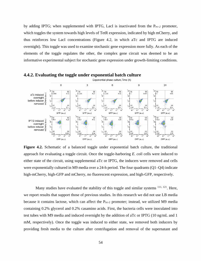
54
by adding IPTG; when supplemented with IPTG, LacI is inactivated from the Ptrc-2 promoter,
which toggles the system towards high levels of TetR expression, indicated by high mCherry, and
thus reinforces low LacI concentrations (Figure 4.2, in which aTc and IPTG are induced
overnight). This toggle was used to examine stochastic gene expression more fully. As each of the
elements of the toggle regulates the other, the complex gene circuit was deemed to be an
informative experimental subject for stochastic gene expression under growth-limiting conditions.
4.4.2. Evaluating the toggle under exponential batch culture
Figure 4.2. Schematic of a balanced toggle under exponential batch culture, the traditional
approach for evaluating a toggle circuit. Once the toggle-harboring E. coli cells were induced to
either state of the circuit, using supplemental aTc or IPTG, the inducers were removed and cells
were exponentially cultured in M9 media over a 24-h period. The four quadrants (Q1–Q4) indicate
high-mCherry, high-GFP and mCherry, no fluorescent expression, and high-GFP, respectively.
Many studies have evaluated the stability of this toggle and similar systems 111, 121. Here,
we report results that support those of previous studies. In this research we did not use LB media
because it contains lactose, which can affect the Ptrc-2 promoter; instead, we utilized M9 media
containing 0.2% glycerol and 0.2% casamino acids. First, the bacteria cells were inoculated into
test tubes with M9 media and induced overnight by the addition of aTc or IPTG (10 ng/mL and 1
mM, respectively). Once the toggle was induced to either state, we removed both inducers by
providing fresh media to the culture after centrifugation and removal of the supernatant and

55
fluorescent expression levels were recorded using a flow cytometer at a 1:1000 dilution. During
the 24-h culture, the cells were manually diluted with fresh M9 media and maintained in
exponential growth. The flow cytometry analysis revealed that the cells maintained their initial
state, depending on aTc or IPTG induction, throughout, as evidenced by cell counts in quadrant 4
or 1, representing high-GFP or high-mCherry expression states, respectively, consistent with
previous results 102 (Figure 4.2).
4.4.3. Evaluating the toggle under stationary batch culture
In stationary batch cultures, as stochastic quorum sensing has been shown in previous
studies utilizing the receiver circuit 68, we also expected to observe stochasticity within the
bacterial colonies influencing the toggle switch. Additionally, the toggle introduced a higher
degree of complexity, compared to the relatively simple receiver circuit, because this system
consisted of two proteins that regulate each other via their effects on the promoter upstream of
each protein. By utilizing different sets of proteins that repress functionality, we sought to further
evaluate the maintenance of protein functionality in a stressful environment, beyond the stochastic
expression of the protein itself.

56
Figure 4.3. The toggle-containing E. coli cells cultured in stationary phase in batch cultures over
a 5-day period. The cells were first induced to either side of the toggle prior to the experiment and
inducer compounds were removed on day 1. Cells that were induced with aTc showed stable
expression of the GFP state over the duration of the experiment (Q1; left panels). However, cells
that were induced with IPTG showed a relatively high degree of biased expression of mCherry

57
(Q1; right panels) to GFP (Q4). The four quadrants (Q1–Q4) indicate high-mCherry, high-GFP
and mCherry, no fluorescent expression, and high-GFP, respectively.
First, cells containing the toggle circuit were inoculated into M9 media containing either
aTc or IPTG. Once the cells reached exponential growth, the cells were removed from the inducer
via centrifugation and replacement of the supernatant with fresh M9 media containing antibiotics.
During the 5-day experiment, deionized water was added to account for evaporation and maintain
a constant final volume. The initial cell state was measured as described above. The cells that were
induced with aTc steadily expressed GFP throughout the 5-day experiment (Figure 4.3, left
panels). More than 85% of the population maintained the GFP expression state in Q4, with less
than 0.01% reverting to the alternate state indicated by mCherry expression in Q1. On the other
hand, cells that were induced with IPTG showed an incomplete reversal of expression towards the
opposite toggle state (i.e., the GFP expression state) throughout the 5-day experiment (Figure 4.3,
right panels). Initially, 85% of these cells expressed mCherry (Q1) on the first day of the
experiment. However, the number of cells that exhibited GFP expression (Q4) increased from
0.053% on day 1 to 5.86% on day 5, representing a dramatic increase in the biased gene expression
throughout the duration of the experiment. We thought that the increase in GFP expression could
be primarily explained by the stochastic bursting of the PLtetO promoter. As discussed in previous
reports, cells kept under stress conditions with minimum nutrients can exhibit changes in the
regulation of transcription and translation 68. We later discuss why only one of the promoters
exhibited this change over time.
4.4.4. Characterization of the toggle under biomimetic biofilm channel culture
After confirming that the toggle circuit cells had a bias towards the GFP-expression state
when grown in stationary cultures, we further sought to assess these effects in our biofilm-like
microfluidic device. As described using receiver cells in previous reports 68, the toggle circuit cells
were inoculated into the biofilm-like channel for long-term observations. The height of the
biofilm-like channel (0.97 µm) partially restricted cell motility and allowed the observation of the
state of individual cells over the course of the experiment. Stochastic burst-like expression of a
quorum-sensing gene without a signaling molecule had been previously observed based on GFP
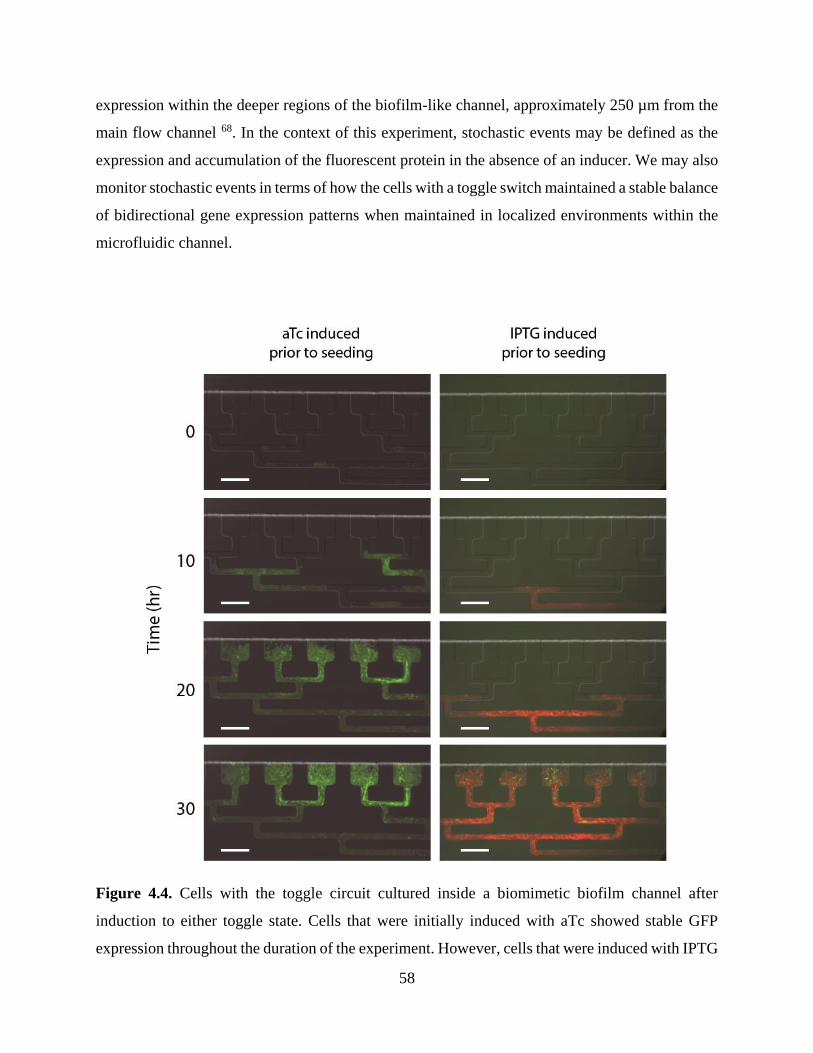
58
expression within the deeper regions of the biofilm-like channel, approximately 250 µm from the
main flow channel 68. In the context of this experiment, stochastic events may be defined as the
expression and accumulation of the fluorescent protein in the absence of an inducer. We may also
monitor stochastic events in terms of how the cells with a toggle switch maintained a stable balance
of bidirectional gene expression patterns when maintained in localized environments within the
microfluidic channel.
Figure 4.4. Cells with the toggle circuit cultured inside a biomimetic biofilm channel after
induction to either toggle state. Cells that were initially induced with aTc showed stable GFP
expression throughout the duration of the experiment. However, cells that were induced with IPTG

59
showed initially uniform mCherry expression and stochastic GFP expression originating from
deeper regions within the channel. The cells were observed over a 30-h time period. All scale bars
indicate 50 µm.
First, aTc-induced cells were inoculated into the microfluidic channel, and a few isolated
colonies began to grow, subsequently merging into a large monolayer that filled the entire
chamber. The entire chamber regions were fully occupied after 20 h, and higher levels of GFP
expression were observed approximately 120 µm from the main flow channel (Figure 4.4, 20 h in
left panels). When comparing these results to previous studies using receiver cells 68, the main
flow media used in each experiment must be fully considered. In this set of experiments, M9 media
was used, which contains considerably lower levels of nutrients, and thus may thus explain
discrepancies between the results of the two studies. For example, under nutrient-deficient
conditions, such as those occurring in cultures using M9 media, the heterogeneous stochastic burst-
like expression and changes in the toggle state may be more common within the biofilm-like
channel. However, higher levels of GFP expression were observed in localized regions of the
microfluidic device, and cells consistently maintained a high-LacI expression state, as
demonstrated by the visualization of GFP-expressing cells both proximately to the main flow
channel (Figure 4.4, 30 h in left panels, arrow).
In contrast, a portion of the IPTG-induced cells expressed the alternate toggle state, i.e.,
more than 5% of cells stochastically expressed GFP, rather than mCherry (Figure 4.4, IPTG
induced, 30 h). Though most of the chamber regions were dominated by mCherry-expressing cells,
not all of the cells stably maintained the high-TetR expression state, i.e., the mCherry expression
state. This heterogeneity may have been a consequence of the nutrient-deficient conditions, which
may have consequences on the stability of genetic toggle switches 120. We observed a small
population of cells that flipped towards the opposite toggle state and thus began to express GFP.
This phenomenon was initially detected in the deeper regions of the chambers (Figure 4.4, 20 h in
right panels) and gradually dispersed within the microfluidic channel as the subpopulation of
stochastically triggered GFP cells generated daughter cells, which maintained their altered
expression state (Figure 4.4, 30 h in right panels). These findings were consistent with previous
batch culture results. As reported previously 68, biofilm colonies can also have distinct differences

60
in gene expression owing to transport-dependent signaling states. In other words, there is high
nutrient availability and removal of cellular waste products at the biofilm–flow interface, which
provides bacteria with the appropriate conditions for robust growth and thus stabilizes gene
expression patterns. In contrast, within inner parts of the biofilm channel, where nutrient
concentrations are lowest, a lack of nutrients can cause starvation-induced stress responses, i.e.,
stochastic GFP expression in high-TetR state cells.
We also sought to understand the stochasticity of high-TetR-expressing cells. This requires
the consideration of additional factors, such as regulatory networks within E. coli that may
influence the state of the toggle. Our results suggest that the toggle is biased towards a high-LacI
state owing to the Ptrc-2 promoter originating from a chimeric lacUV5 and trp promoter, both of
which were isolated from E. coli 122, as well as the PLtetO promoter originated from an
Enterobacteria phage λ 123. Elements from the E. coli genome will have a regulatory effect on a
promoter isolated from E. coli; however, we hypothesize that E. coli regulatory networks will have
a minimal effect on a promoter originating from a viral source. In this way, stochasticity of high-
LacI-expressing cells will be triggered in conditions in which nutrient levels are low, causing
starvation-induced stress responses.
4.5. Conclusion
Stochastic gene expression revealed in relatively simple inducible genetic systems also
appears in the bistable genetic toggle switch system examined in the present study. Previous
research revealed that cell culture in the biofilm-mimicking microfluidic device could drive
expression of genes generally associated with biofilm formation, as demonstrated by patterns of
GFP expression in the device 68. In the current toggle switch system in the same culture device
(though with M9 medium instead of LB medium), stochasticity was also observed, though it was
asymmetric between the two toggle switch states. Specifically, induction of the genetic toggle with
aTc consistently drove GFP expression in the culture. Neither spurious expression of the mCherry
reporter nor stochastic switching of the genetic toggle switch was observed when cells were
induced with aTc. Less than 0.01% of GFP-expressing cells expressed mCherry. However, the
61
long-term effect of IPTG induction was not as consistent. When induced with IPTG, cells within
the microfluidic device overwhelmingly expressed mCherry on day 1, with only 0.053% of cells
expressing GFP. By day 5, many more cells (5.86% of those observed) expressed mCherry instead
of GFP. Moreover, this reverted expression state was inherited by daughter cells, as confirmed by
continuous microscopic observation, which was enabled by the design of the microfluidic device.
The current study differed from previous research using this biofilm-mimicking
microfluidic device in one key factor—the medium used. Here, M9 medium was used instead of
LB medium for the bacterial culture in order to ensure the genetic toggle switch functioned
properly. (LB medium can interfere with LacI, a repressor protein element of the toggle switch.)
Accordingly, previous experiments should be repeated using M9 medium instead of LB medium
to ensure that the observed differences were not caused by the difference in media. It is likely that
using M9 medium instead of the more nutrient-rich LB medium will result in an increased level
of stochastic burst-like expression observed in previous experiments. As stochasticity in toggle
switches may be an important factor in both synthetic biological applications as well as
understanding naturally occurring genetic regulatory networks, future research should examine
robustness of these systems by examining gradients of stress stimuli.

62
CHAPTER 5.
STOCHASTIC PHENOTYPIC EXPRESSION OF
SINGLE BACTERIAL CELLS CONFINED IN A
MICROFLUIDIC CHANNEL
5.1. Abstract
Bacterial growth in harsh environments, including environments with spatial and nutrient
limitations, has been a focus of research owing to the obvious and wide biological and human
health implications. However, it is difficult to distinguish noise (i.e., statistical and measurement
error) from individual among-cell variation under conventional culture methods. In order to
characterize individual cell properties, we studied the stochastic growth of E. coli cultured in a
biofilm-mimicking microfluidic device. Utilizing the narrow device aperture (determined by the
diameter of E. coli cells), target cells were selectively isolated from a mixed population, and their
morphological properties, growth, and fission were observed by microscopy at a fine timescale.
This system enabled the tracking of cells of interest (i.e., large cells) and the observation of
characteristic patterns of growth and aging, including an increased volume prior to cell division
and eventual apoptosis. Additionally, this behavior was associated with changes in cell wall
properties after a limited number of divisions, yielding daughter cells with relatively short
lifespans. Furthermore, we observed variation among target cells in morphology and survival (e.g.,
one cell survived the full study period). These findings demonstrate the suitability of the
microfluidic device for characterizing the responses of single cells and microbial populations to
various environmental conditions and stimuli.
63
5.2. Introduction
In clonal bacterial cultures, a small inoculum can multiply into hundreds of millions of
cells that are assumed to be essentially identical in general. Bacteria, however, often persistently
retain individual characteristics, as each cell may evolve from ancestors with distinct genetic
characteristics 124. This individualism is readily observed by measuring morphological changes in
single cells under microscopy in real time. Selection among such individual bacterial cells endows
populations with the ability to survive through adaptive evolution even within particularly hostile
environments, for example, under limitations in nutrition and space 68. Under nutritional
imbalance, a bacterial culture reaches stationary phase, at which the metabolism of cells can
become dormant, thus maintaining a constant or stationary cell density, which may further involve
genetic switches 68. In many natural habitats, bacteria live in spaces of less than a micrometer in
width; bacterial growth and motility under such restrictions are highly relevant to research in
diverse fields including soil microbiology, water purification, and many biomedical disciplines 125.
Although there is an essential need to understand the effects of bacterial growth under severe
constraints, such studies have been uncommon. Many basic questions about the law describing
bacterial growth under constraint, i.e., the function describing the size of an individual cell over
time, remain; it is also unknown whether cells specifically react to their own size, age, or particular
molecular features to initiate cell division 126.
Soft-photolithography is a technology with the potential to construct artificial bacterial
habitats that mimic the natural constrained habitats in which individual cells grow and divide 68.
The growth and division of individual cells are fundamental events underlying many biological
processes, including the development of organisms and tumors as well as pathogen–host
interactions 126. Some important aspects of microfluidic cell culture systems have previously been
reviewed in detail, including the effect of surface modification on cellular behavior, cell biology,
cell culture models, cellular analysis, cellular microenvironments, cell secretion, and more 127.
With microfluidic devices, researchers can closely mimic a cell's natural microenvironment, for
example, by continuous perfusion culture or by creating chemical gradients. Specifically, a
microfluidic device that mimics bacterial biofilm environments has been previously constructed
to observe bacterial growth under stochastic conditions 68. Such microfluidic techniques may

64
further facilitate studies of the characteristics of small numbers of cells or single cells at high
temporal and/or spatial resolution via automation, parallelization, on-chip analyses, or direct
coupling to downstream analytical chemistry platforms 127. At the same time, this approach offers
benefits including the reduced consumption of reagents, limited contamination risk, and efficient
high-throughput experimentation. Recently, the quantitative study of single cells under consistent
physiological conditions has been conducted through a unique combination of measurement and
analysis technologies together with mathematical modeling 126. Microfluidic technology may be
used to provide bioreactors that closely resemble natural habitats and thus reveal the interplay
between critical cell culture parameters and micro-environmental conditions 128.
Certain analytical methods may be necessary for accurately characterizing the behaviors
of single cells grown in microfluidic devices, including phenotypic changes and cell division.
Studies that require isolation of cells according to phenotype, for instance, to study cell aging, have
relied on laborious processes for sorting and manipulating cells. In one notable study, a
microfluidics-based cell culture device was used to facilitate cell isolation based on phenotypic
markers such as cell size, permitting real-time microscopic observation 126. In this technique, single
cells of different sizes, i.e., ranging from normal cells to expanded or swollen cells, were separately
confined within PDMS chambers to demonstrate that these morphological changes were not only
maintained across generations, but also had a unique role in the timing of the processes of aging
and cellular apoptosis. Another study used time-lapse fluorescent microscopy to show that bacteria
exhibit morphological adaptations within sub-micron-diameter pores and cavities that were
unexpectedly productive bacterial habitats 125. Adaptive potential was observed in E. coli cells
that, despite their considerable restriction, still grew and divided within the narrow channels. The
plasticity of these E. coli cells was substantially enabled by their thin cell walls, measuring only
approximately 3 nm in thickness 129.
We have studied the stochastic growth of an elastic bacteria (i.e., E. coli) cultivated in a
biofilm-mimicking microfluidic device that permits detailed analyses of single-cell growth and
division, which might otherwise be impractical owing to the intrinsic noise of biological processes.
This analytical limitation was overcome by monitoring cellular behavior and morphological
changes of bacterial cells using real-time microscopic observation. The ability of individual cells

65
to metabolically adapt and produce similarly adapted daughter cells may have many different
effects. For example, daughter cells may exhibit size expansions that indicate successful metabolic
adaptation to the stressful environments individual cells experience. The target cells were observed
by selectively isolating them from a mixed population and confining them in place under
conditions that allowed subsequent viability and potential clonal expansion. To achieve this end,
we developed a microfluidic device comprising cell chambers and convoluted paths with a limited
aperture that is slightly narrower than the diameter of E. coli cells (approximately 1 μm). As the
adapted single cells exhibited increased cytoplasmic volume, they were constrained between the
bottom and top walls of the channel with normal cells simultaneously exiting into the main
channel. This confinement technique enabled the real-time microscopic observation of
morphological changes of each individual cell, with images continuously captured to follow
growth and division at predetermined time points.
5.3. Materials and Methods
5.3.1. Manufacture of microfluidic device
Master mold fabrication. All master molds were fabricated in the MICRON facility.
Photomasks were drawn in AutoCAD (Autodesk, San Rafael, CA, USA), printed onto
transparency film by CAD/Art Services, Inc. (Output City, Poway, CA, USA), and mounted onto
glass plates (McMaster-Carr, Los Angeles, CA, USA). The first layer of the master mold was
created by first spin-coating with SU-8 2000 negative photoresist (MicroChem Corp., Newton,
MA, USA) onto clean silicon wafers to 0.96 µm in height using a WS-650-8B programmable
spinner (Laurell Technologies Corp., North Wales, PA, USA) and then using a MA/BA6 UV
contact mask aligner (MicroTec, Garching, Germany) for subsequent UV exposure. After being
exposed to UV radiation, the unexposed photoresist was rinsed away using developer (MicroChem
Corp.). For the second layer, the SU-8 2010 negative photoresist was spin-coated on top of the
first layer to achieve a height of 10 µm. Again, the MA/BA6 UV contact mask aligner was used
for UV exposure, and the unexposed photoresist was rinsed away using the same process described
above.

66
PDMS device fabrication. Replica molds were created from master molds by mixing
PDMS/Sylgard 184 (Dow Corning, Midland, MI, USA) with a 10:1 ratio of the elastomer base to
curing agent, degassing in a vacuum desiccator for 1 h, and curing in an oven at 90°C for 2 h.
Following removal of the PDMS, devices were cut, holes were punched for ports, and the cell was
cleaned with filtered DI water and then permanently bonded to clean coverslips (Thermo Fisher
Scientific, Waltham, MA, USA) via exposure to O2 plasma for 1 min in a PDC-32G plasma cleaner
(Harrick Scientific, Ithaca, NY, USA).
5.3.2. Maintenance and monitoring of cell growth in microfluidic channels
Microfluidic cell culture. The recombinant E. coli cells were loaded into the trapping
channels of the microfluidic device by directing the flow in the direction from the cell port to the
waste port. Upon trapping a few cells in each region, the flow was reversed and slowed to steadily
supply the cells with fresh nutrients from the media port through a combination of diffusion and
advection. The cells were maintained at 22°C and grown to fill the chamber over a duration of 20-
30 h.
Cell microscopy. Cell images were acquired using an inverted epifluorescent microscope
(Ti-E; Nikon Instruments Inc., Tokyo, Japan). DIC and fluorescent images were captured every
15 min, and focus was maintained automatically using Nikon Elements software.
5.4. Results and Discussion
5.4.1. Stochastic growth of single cells under spatial limitation
We manufactured a microfluidic device that mimics the type of biofilm structure that
typically forms during adaptation to hostile environments. The biomimetic device consisted of a
three-port microfluidic channel with two inlets and one outlet (Figure 5.1a). The main observation

67
region of the device contained two elements: a series of distinct rectangular cell chambers and a
convoluted path connecting the series of chambers (Figure 5.1b). The interior side of the chamber
elements opened to the main flow of the medium. The convoluted pathways, however,
progressively converged into a single channel, eventually leading to a closed end. The cell chamber
region and convoluted pathway both had a maximum height of 0.97 μm. Since E. coli cells have a
diameter of approximately 1 μm, the height of the trapping region permitted linearized
observations of confined cells. The device was also designed to discharge cells from the device
continuously through the main flow channel (12-μm height).
Since the convoluted pathways were constructed to maintain a limited height not exceeding
the dimensions of bacteria under cultivation, the conditions, i.e., the spatial limitation, could be
unfavorable to cells. Cell growth within the narrow channel may further generate a stressful
environment and eventually exhibit stochasticity caused by the spatial limitation and nutritional
imbalance. This was demonstrated in our previous study showing stochastic burst-like expression
of genes within deep regions of the channel using the same device 68. We further propose in this
study that such stochastic conditions can result in phenotypic variation, such as morphological
changes, as seen in individually confined bacterial cells 125. This can occur when individual cells
uniquely recognize the hostile surrounding environment, resulting in stochastic morphological
changes in single cells via genetic switches, epimutation, or even extraordinarily rare mutations
(Figure 5.1c). The shape may vary depending on the confined spatial geometry of a particular
culture device 130. Although cells may divide and grow in hostile surroundings, the culture may
not be sustainable compared to normal culture conditions.

68
Figure 5.1. A diagram of the device’s structure, which mimics a typical biofilm structure, and the
interconnected fluid paths from which bacterial colonies are able to grow. (a) The design of the
microfluidic device used in this study. Gray circles indicate two inputs on the left and an output
on the right side of the channel. The two inputs supply microbial cells and culture medium, while
the output permits the disposal of waste. (b) The interconnected region of the device, which mimics
biofilm structure, contains rectangular cell chambers adjacent to the central flow channel. (c) The
cell chambers are each limited to a height of 0.97 μm, which imposes limited growth conditions
on the bacteria (shown as oblongs).
To trigger the stochastic phenotypic change, we cultured quorum-sensing recombinant
cells within the biofilm-mimicking microfluidic device and then observed the morphological
changes of certain subpopulations in comparison with neighboring normal cells.

69
5.4.2. Microfluidic environment for individual cell growth
A synthetic receiver circuit (Plux-driven) has been previously designed to assess stochastic
quorum sensing 68. E. coli transformed with the receiver plasmid GFP upon chemical induction or
induction via environmental stimuli 68. The cells were inoculated in the biomimetic biofilm
channel device and maintained at 22°C. During the observation (Figure 5.2), the cell divisions of
parental cells were tracked, as indicated by the red arrow at 0 min. After 45 min, the cells divided
and generated two daughter cells. The second cycle of division was also detected within the
subsequent 45-min period, indicating a doubling time of the cells of approximately 45 min under
the culture conditions.
Figure 5.2. Growth kinetics of the quorum-sensing cells within the biomimetic biofilm channel.
Single red arrows within a frame show the parental cell. At approximately 45 min, the parental
cell divided into two daughter cells. The second cycle of division is shown in the subsequent
frames. The cell located on the top of the frame at 45 min was tracked for an additional division
cycle, shown occurring over the same time interval. All experiments were conducted at 22°C, and
sequential snapshots of a typical experiment are presented.
Long-term morphological changes of bacterial cells grown within the microfluidic device
were then monitored by microscopy (Figure 5.3). A few receiver cells as inoculum were detected
at the beginning of cultivation (0 h) and subsequently grew over time into a large mono-layer of
colonies that filled the microfluidic chamber (typically after 10 h). During the first 30 h, no
individual cells showed significant morphological changes. However, after 30 h, a cell population
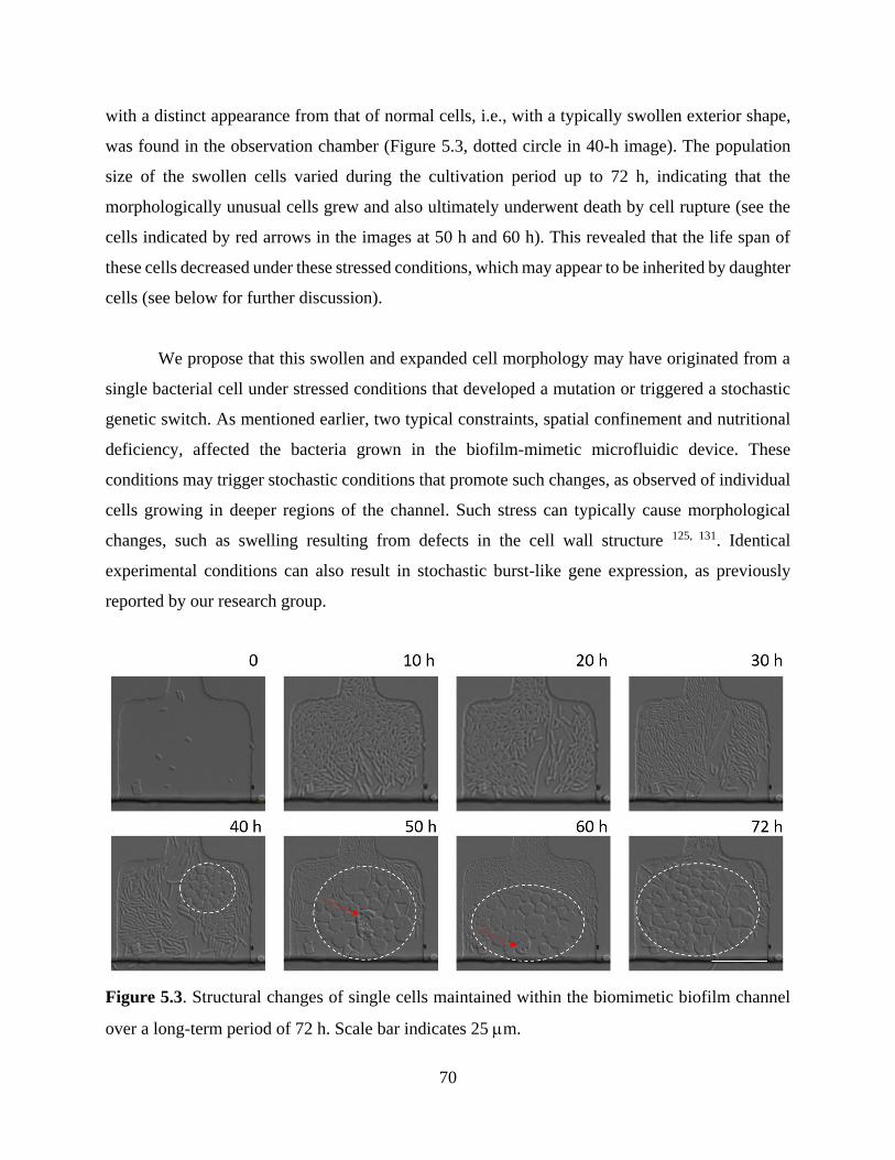
70
with a distinct appearance from that of normal cells, i.e., with a typically swollen exterior shape,
was found in the observation chamber (Figure 5.3, dotted circle in 40-h image). The population
size of the swollen cells varied during the cultivation period up to 72 h, indicating that the
morphologically unusual cells grew and also ultimately underwent death by cell rupture (see the
cells indicated by red arrows in the images at 50 h and 60 h). This revealed that the life span of
these cells decreased under these stressed conditions, which may appear to be inherited by daughter
cells (see below for further discussion).
We propose that this swollen and expanded cell morphology may have originated from a
single bacterial cell under stressed conditions that developed a mutation or triggered a stochastic
genetic switch. As mentioned earlier, two typical constraints, spatial confinement and nutritional
deficiency, affected the bacteria grown in the biofilm-mimetic microfluidic device. These
conditions may trigger stochastic conditions that promote such changes, as observed of individual
cells growing in deeper regions of the channel. Such stress can typically cause morphological
changes, such as swelling resulting from defects in the cell wall structure 125, 131. Identical
experimental conditions can also result in stochastic burst-like gene expression, as previously
reported by our research group.
Figure 5.3. Structural changes of single cells maintained within the biomimetic biofilm channel
over a long-term period of 72 h. Scale bar indicates 25 m.
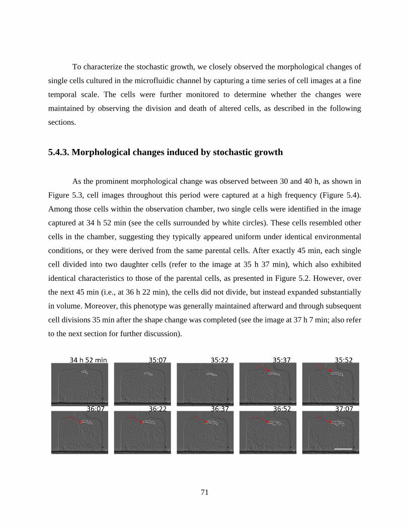
71
To characterize the stochastic growth, we closely observed the morphological changes of
single cells cultured in the microfluidic channel by capturing a time series of cell images at a fine
temporal scale. The cells were further monitored to determine whether the changes were
maintained by observing the division and death of altered cells, as described in the following
sections.
5.4.3. Morphological changes induced by stochastic growth
As the prominent morphological change was observed between 30 and 40 h, as shown in
Figure 5.3, cell images throughout this period were captured at a high frequency (Figure 5.4).
Among those cells within the observation chamber, two single cells were identified in the image
captured at 34 h 52 min (see the cells surrounded by white circles). These cells resembled other
cells in the chamber, suggesting they typically appeared uniform under identical environmental
conditions, or they were derived from the same parental cells. After exactly 45 min, each single
cell divided into two daughter cells (refer to the image at 35 h 37 min), which also exhibited
identical characteristics to those of the parental cells, as presented in Figure 5.2. However, over
the next 45 min (i.e., at 36 h 22 min), the cells did not divide, but instead expanded substantially
in volume. Moreover, this phenotype was generally maintained afterward and through subsequent
cell divisions 35 min after the shape change was completed (see the image at 37 h 7 min; also refer
to the next section for further discussion).
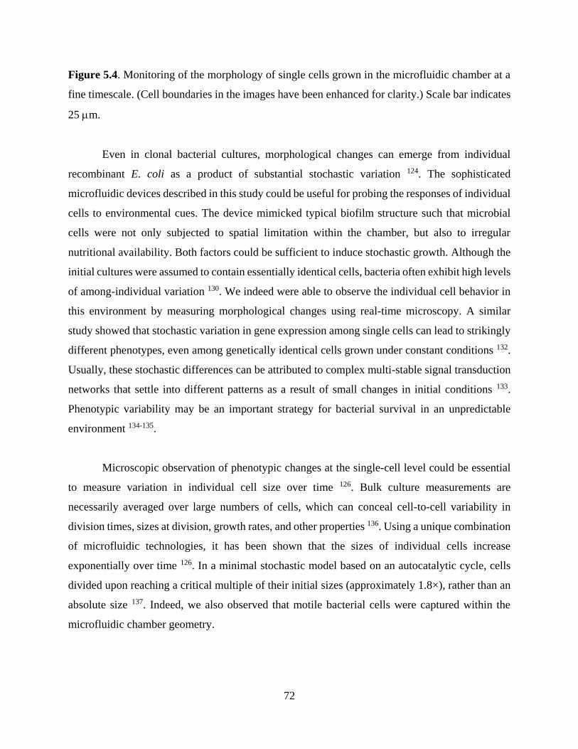
72
Figure 5.4. Monitoring of the morphology of single cells grown in the microfluidic chamber at a
fine timescale. (Cell boundaries in the images have been enhanced for clarity.) Scale bar indicates
25 m.
Even in clonal bacterial cultures, morphological changes can emerge from individual
recombinant E. coli as a product of substantial stochastic variation 124. The sophisticated
microfluidic devices described in this study could be useful for probing the responses of individual
cells to environmental cues. The device mimicked typical biofilm structure such that microbial
cells were not only subjected to spatial limitation within the chamber, but also to irregular
nutritional availability. Both factors could be sufficient to induce stochastic growth. Although the
initial cultures were assumed to contain essentially identical cells, bacteria often exhibit high levels
of among-individual variation 130. We indeed were able to observe the individual cell behavior in
this environment by measuring morphological changes using real-time microscopy. A similar
study showed that stochastic variation in gene expression among single cells can lead to strikingly
different phenotypes, even among genetically identical cells grown under constant conditions 132.
Usually, these stochastic differences can be attributed to complex multi-stable signal transduction
networks that settle into different patterns as a result of small changes in initial conditions 133.
Phenotypic variability may be an important strategy for bacterial survival in an unpredictable
environment 134-135.
Microscopic observation of phenotypic changes at the single-cell level could be essential
to measure variation in individual cell size over time 126. Bulk culture measurements are
necessarily averaged over large numbers of cells, which can conceal cell-to-cell variability in
division times, sizes at division, growth rates, and other properties 136. Using a unique combination
of microfluidic technologies, it has been shown that the sizes of individual cells increase
exponentially over time 126. In a minimal stochastic model based on an autocatalytic cycle, cells
divided upon reaching a critical multiple of their initial sizes (approximately 1.8×), rather than an
absolute size 137. Indeed, we also observed that motile bacterial cells were captured within the
microfluidic chamber geometry.

73
5.4.3. Distinct aging characteristics of cells under stochastic growth
After about 40 h from starting the culture, some individual bacterial cells appeared that
were substantially larger than the other, normal cells within the microfluidic chamber (see Figure
5.3). The cells of interest in the mixed populations were fixed within the biofilm-like structures by
the limited height of the chamber and were distinct from other cells based on their relative cell
size. We subsequently investigated cellular behavior across generations, particularly cell aging, by
continual microscope observations over time.
We extensively monitored the growth and death of cells with altered phenotypes by
analyzing images captured every 15 min over a defined time scale (Figure 5.5). Six cells exhibiting
morphological changes (indicated within the colored circles in Figure 5.5) were targeted for the
purpose of monitoring over a period of 3 h 45 min (Figure 5.5; 46 h 53 min after inoculation). Cell
#1 (Figure 5.5, in yellow), which was relatively larger than the other cells, had cell wall surfaces
that appeared punctured after 1 h from the beginning of the period of intensive observation. This
suggests earlier cell death compared with normal cells or necrosis by osmotic pressure (Figure 5.5,
double arrows, 47 h 53 min). Cell #2 (Figure 5.5, in green) had a wrinkle, which may have been
formed by compression within the observation chamber with restricted space, and divided 45 min
after monitoring began (Figure 5.5, single arrow, 47 h 38 min). However, their cell walls ruptured
shortly thereafter, similar to cell #1 (Figure 5, 48 h 8 min). Cell #3 (Figure 5.5, white) exhibited
similar properties to those of cell #2 with respect to doubling and puncture, although death was
delayed (Figure 5.5, 48 h 53 min). Cell #4 (Figure 5.5, red) exhibited significant growth, rather
than division, as the culture continued, reached a maximum size (Figure 5.5, 48 h 38 min), and its
cell wall eventually ruptured at 49 h 38 min. Cell #5 (Figure 5.5, blue) notably showed both size
expansion (Figure 5.5, maximum at 48 h 53 min) and division (Figure 5.5, 49 h 8 min), and died
30 min afterwards. It should be noted that several holes, an indicator of death, were detected on
the cellular surfaces prior to division (Figure 5.5, 48 h 38 min and 48 h 53 min). Cell #6 (Figure
5.5, orange), which uniquely survived throughout the monitoring period, underwent a volume
expansion and surface-hole formation in the middle of the observation period (Figure 5.5, 49 h 38
min).

74
Figure 5.5. Image time series showing the cell life span under stochastic growth within the
spatially confined chamber of the microfluidic device. Scale bar indicates 25 m.
The growth and death of relatively large cells targeted for observation revealed clonal
heterogeneity and aging under stochastic culture conditions. Observations of cell wall integrity, an
indicator of cell viability, showed that the majority of cells (five out of six) expressed phenotypes
consistent with apoptosis. They appeared expanded in volume prior to division, displayed a limited
number of divisions before apoptosis, and gave rise to daughter cells with short lifespans.
However, it was unclear whether the short lifespan was shared by other larger cells not observed
during the predetermined period. This phenotypic variation may have been induced by age
differences among the initially confined cells 126. Normal bacterial cells with much smaller sizes

75
can quickly reproduce and form dense populations. As these cells were not tightly confined within
the structures, they were able to flow out along the main medium flow into the microfluidic device,
and the density was consequently variable. These results were consistent with those of a previous
study revealing two distinct fates of yeast cells individually isolated within a microfluidic chamber
126, 131. In this previous study, a single isolated cell proliferated in a typical manner and, in contrast,
a confined larger cell exhibited limited cell divisions along with a substantial increase in cell size.
Viability tests showed that the majority of cells that divided into dense populations remained
viable, while the larger yeast cells displayed hallmarks of apoptosis.
Microscopic observations indicated that the apoptosis of larger cells was accompanied by
cell wall rupture, as described earlier, which could be explained by a high osmotic pressure in the
cytoplasm. Indeed, gram-negative E. coli cells have significantly thinner cell walls (only
approximately 3 nm; 129) than those of gram-positive bacteria (30–40 nm) 138. The relatively
thinner cell walls of E. coli do not allow the osmotic pressure to significantly exceed 2–3 atm 139.
This characteristic is associated with high plasticity, the ability to change in response to variable
environmental conditions, such as mechanical stress and nutrient availability 125. Thin cell walls,
however, are more vulnerable to damage and, thus, aberrant morphologies can result from defects
in cell walls 140. The cell-wall defects likely formed in the microfluidic channel where E. coli
underwent local, environmentally induced changes. Additional defects may have formed when the
cells were confined within the narrow chamber for an extended time period, leading to the
breakdown of peptidoglycan 125. The mechanical stress may also disrupt the functioning of
cytoskeletal proteins of E. coli in narrow channels, eventually resulting in cell death.
5.4.4. Stochastic growth of single cells that exhibit typical gene expression
patterns
At a late stage in the observation period, a single cell expressed GFP, indicating the
induction of the quorum-sensing gene circuit. As shown in the images captured after 59 h from the
culture onset in Figure 5.6, a single cell divided into two daughter cells with a doubling time of
about 45 min (Figure 5.6, between 59 h 7 min and 59 h 52 min). After 15 min, one of the
descendants emitted green fluorescence and divided again in approximately the same doubling

76
time (Figure 5.6, 60 h 52 min). Both daughter cells displayed fluorescence, and the intensity
increased gradually for the next 1.5 h (Figure 5.6, 60 h 52 min and 62 h 22 min). This phenotype
was accompanied by an increase in cell size, without division. Thereafter, the fluorescence faded
and was finally was lost after about 30 min. This may indicate stochastic quorum sensing as a
stress response occurred in the absence of the inducing molecule, as demonstrated in a previous
report 68.
Single cells exhibit phenotypic variation in response to local environmental conditions
under stressful environments, as mentioned earlier. Cells, like all organisms, continuously adapt
to varying conditions through the process of natural selection. Indeed, previous studies have
demonstrated a remarkable ability for wild-type E. coli to produce a very large and broad array of
irregular morphologies 131. Although the frequency of divisions decreased as cell size increased,
the accuracy of this type of division could remain essentially unaffected by the shape and size of
the cell. Asymmetrical inheritance of cell properties can also generate phenotypic diversity 124, 141.
For example, striking individual differences resulting from variation among the chemotactic
behaviors of individual E. coli cells 142 and among fluorescence signals from individual cells
expressing a fluorescent protein have been observed 106.

77
Figure 5.6. Stochastic phenotypic expression of the quorum-sensing signal of bacterial cells and
cell division. Scale bar indicates 25 m.
5.5. Conclusion
A microfluidic channel device that mimics bacterial biofilm structures was developed to
grow elastic E. coli cells, which eventually displayed stochastic phenotypic expression as a stress
response. Significant morphological changes were observed over the culture period, in addition to
the expression of a quorum-sensing GFP reporter, without the application of an inducer molecule.
The cell capture region for bacteria culture were specifically designed to have limited height of

78
<0.97 µm such that single cells with a swollen morphology were selectively confined within the
channel structure and to provide culture conditions similar to those of natural biofilms.
Microscopic observation of the isolated target cells revealed cellular expansion, low rates of cell
division, and early apoptosis, which are characteristic patterns of stochastic responses of single
cells to stressful conditions. In addition, stochastic quorum sensing in the absence of the AHL
signaling molecule was observed from single cells, consistent with previous results. We were able
to utilize the microfluidic device as an artificial biofilm model to further characterize the properties
of microbial populations, e.g., their chemical resistance to drugs such as antibiotics.

79
CHAPTER 6.
BACTERIAL BIOFILM FORMATION AT THE
INTERFACE OF WATER-IN-OIL DROPLETS
SPATIALLY SEGREGATED WITHIN A
MICROFLUIDIC DEVICE
6.1. Abstract
Biofilms are both a key research focus in fields spanning from microbial ecology to
industrial food processing. They are also an important public health issue owing to the ability of
biofilms to form even in harsh environments, including on medical equipment and in hospital
settings, and their resistance to conventional antibiotics. Accordingly, efficient and scalable
methods to screen biofilm control agents are of considerable practical value. To this end, we
developed a water-in-oil droplet-based bioreactor that encapsulates bacterial cells in an aqueous
core, with spatial segregation within microfluidic channels. The system was applied to the culture
of engineered Escherichia coli with quorum sensing genes using the signaling molecule N-acyl
homoserine lactone for visualizing biofilm consortia. We successfully observed initial bacterial
growth and subsequent biofilm formation at the hydrophobic interface based on green fluorescent
protein expression within the compartmentalized droplets. The droplet bioreactor developed in the
present study has broad applications in future research. This system facilitates the culture of small
aggregations of cells under controlled conditions such as the precise introduction of chemical
compounds (e.g., AHL and other cell inducing or inhibitory molecules) to assay their effects on
bacterial growth, biofilm formation, and visualize other fine-scale culture phenotypes. This system
further allows multiplexed assays for drug screening against the bacterial populations comprised
within biofilms.

80
6.2. Introduction
Many microorganisms are able to grow in various environments, which has serious human
health consequences, e.g., foods, medical instruments, and drinking water pipelines 143-144. Even
from an initially small inoculum, microorganisms exponentially grow into colonies that continue
unabated as long as permissive growth conditions are available. However, populations rapidly
approach saturation under various stresses, including nutrient depletion and the accumulation of
toxic metabolic compounds. Such conditions can cause epigenetic changes in microorganisms;
these modifications have the potential to promote survival and can result in adaptation to the
surrounding conditions through epigenetic mechanisms 143. Repression and induction of genetic
programs are activated by epigenetic switching mechanisms that facilitate long-term survival in
hostile environments. One typical example is the formation of bacterial biofilms, which are mostly
comprised of an extracellular matrix consisting of polysaccharides, cell debris, and nucleotides
occurring at interfaces (e.g., liquid–solid, liquid–air, and liquid–liquid interfaces) 145. As such,
consortia of microorganisms are most often metabolically dormant and nonculturable, and they are
notoriously difficult to control with traditional chemical compounds, such as antibiotics, which
enables cells to persist 146. Therefore, chemicals used to control bacterial growth in these unique
bacterial habitats require further investigation in order to validate their efficacy for controlling
biofilms. In order to screen appropriate effectors, conventional procedures based on microtiter
plates or test tubes are inefficient with respect to cost and labor 146. These conventional procedures
used to screen for compounds and surfaces with favorable properties include the alamarBlue assay
146, adenosine triphosphate-driven luminescent assays 146, the Calgary biofilm device 147, and the
crystal violet assay 148.
Conventional microbial tests have been conducted on the macro-scale, but these tests can
be improved through the utilization of microfluidic technology. A micro-bioreactor for microbial
culture can be produced that immobilizes or entraps cells in polymer matrices 149-150, e.g.,
polydimethylsiloxane. Immobilized cell technology has advanced to utilize cell encapsulation, in
particular, by entrapping cells in an alginate gel matrix 151, spray drying (commonly used in the

81
food industry) 152, and extrusion, which involves the projection of an emulsion core and coating
material via flow through a nozzle at high pressure 153. Recently, water-in-oil
compartmentalization has been introduced to retain microbial cells in the aqueous core for
cultivation 154-155. This cell capture method was accomplished using droplet microfluidic
technology. Briefly, a cell-containing aqueous medium is injected into the oil phase environment.
Nano- to micro-sized droplets form and are used to cultivate various bacterial cells. A similar
droplet-based platform was used to examine communication between quorum signaling
components located in spatially isolated droplets 154. Genetic constructs were designed using an
inducible Plux promoter attached to a reporter gene as the receiver and a constitutive PLtet0
promoter fused to the luxI gene as the signaling molecule. The cell-containing droplets were placed
within a micro-compartment under a moderate flow and maintained to observe quorum signaling
154. As microfluidic devices can be constructed to have parallel assay channels, similar experiments
can be conducted to test various chemical conditions, e.g., antibiotics, with a simultaneous control
156.
Bacteria primarily exist in surface-associated multicellular communities, and the term
biofilm describes a matrix-enclosed bacterial population of cells adherent to each other and/or
substrates 144, 157. Bacterial biofilms undergo several stages of development, from the initial
attachment of bacteria to the ultimate detachment and colonization of new interfaces 158. The
effectiveness of biofilm formation is dependent on cell-surface hydrophobicity, since
hydrophobicity is important in the initial attachment process of bacteria to hydrophobic surfaces.
Although the viscoelastic matrix of biofilms can be composed of extracellular DNA,
polysaccharides, and proteins, the precise structure, chemistry, and physiology depend on the
characteristics of the resident microbes and the local environment. Such phenotypic variability
may be an important strategy for bacterial survival in an unpredictable environment 124, 134-135. For
example, the stochastic development of persister cells enables lineages of pathogens to survive
antibiotic treatment and accordingly has serious clinical consequences 133. Furthermore, as
biofilms are adherent aggregates of bacterial cells that form on biotic and abiotic surfaces,
including human tissues, they also play a role in resistance to antibiotic treatment and bacterial
persistence in chronic infections 159.

82
To establish a multiplexed biofilm assay system, in this study, we developed a droplet-
based bioreactor in a spatially segregated configuration, which we inoculated with a bacterium
(i.e., E. coli) for culture. The droplet approach enabled relatively convenient control over the
culture and also allowed the spatial segregation of bacterial cells within the microfluidic channels,
providing the ability to perform multiple fine-scale assays in parallel. As hydrophobic surfaces are
preferable for primary cell adhesion, the cells were encapsulated in a water-in-oil droplet to
promote cell aggregate adherence at the interface 160. In this technique, droplets are captured and
maintained within a compartment of a microfluidic device through which oil continuously flows.
Cell aggregate formation was monitored using synthetic gene networks in E. coli that express green
fluorescent protein (GFP) upon induction with AHL, which was used to better visualize biofilm-
like structures. Bacterial growth was measured using a fluorescent microscope and images were
captured at preset time intervals.
6.3. Materials and Methods
6.3.1. Manufacture of microfluidic device
Master mold fabrication for water-in-oil bioreactor. All master molds were fabricated
in the Virginia Tech Micro & Nano Fabrication Laboratory (MICRON). Photomasks were drawn
in AutoCAD (Autodesk, San Rafael, CA, USA), printed onto transparency film by CAD/Art
Services, Inc. (Output City, Poway, CA, USA), and mounted onto glass plates (McMaster-Carr,
Los Angeles, CA, USA) maintaining the print side facing the silicon wafer. All silicon wafers were
cleaned prior to using by rinsing with acetone, isopropyl alcohol and deionized water and dried on
a hot plate (120°C) for 10 min. The master mold was created by first spin-coating with SU-8 2010
negative photoresist (MicroChem Corp., Newton, MA, USA) onto clean silicon wafers to 15 µm
in height using a WS-650-8B programmable spinner (Laurell Technologies Corp., North Wales,
PA, USA). The photoresist was first cured on a hot plate (120°C) for 2 min and exposed to UV
radiation using a MA/BA6 UV contact mask aligner (MicroTec, Garching, Germany). After being
exposed to UV radiation, the exposed photoresist was cured on a hot plate (120°C) for 3 min and

83
the unexposed photoresist was rinsed away using developer (MicroChem Corp.). The final master
mold was cured on a hot plate (120°C) for 1 hr.
Master mold fabrication for water-in-oil homogeneous droplet. All master molds were
fabricated using SU-8 2050 negative photoresist to yield 50 µm in height. Deposited photoresist
was cured on a hot plate for 3 and 5 min, prior and subsequent to UV exposure, respectively. All
other details follow the process mentioned above.
PDMS device fabrication. Replica molds were created from master molds by mixing
PDMS/Sylgard 184 (Dow Corning, Midland, MI, USA) with a 10:1 ratio of the elastomer base to
curing agent, degassing in a vacuum desiccator for 1 h, and curing in an oven at 90°C for 2 h.
Following removal of the PDMS, devices were cut, holes were punched for ports, and the cell was
cleaned with filtered DI water and then permanently bonded to clean coverslips (Thermo Fisher
Scientific, Waltham, MA, USA) via exposure to O2 plasma for 1 min in a PDC-32G plasma cleaner
(Harrick Scientific, Ithaca, NY, USA).
Fluidics. A syringe pump (PHD 2000, Harvard Apparatus, Holliston, MA, USA) was used
to generate and manoeuvre droplets along the microfluidic channel. The pump was used at a rate
of 5 l/ml to generate LB medium droplets that subsequently merged with the mother droplet and
HFE-7500 oil containing 0.0018% Krytox® 157 FSL (DuPont, Wilmington, DE, USA) as the
surfactant was gravity-driven in the main flow channel at a rate of 10 l/ml.
6.3.2. Construction and characterization of the quorum sensing receiver circuit
Cloning details. The receiver circuit (pSP035) used in this study was constructed using six
components, pKE1-MCS as the vector, Plux, luxR T1T2, rbs027 and GFPmut3b as inserts (Figure
3.1). The desired restriction sites were added to the 5′ and 3′ ends of each construct using overhang
PCR (Table 3.1). For each insertion, both the insert construct and vector were uniquely double
digested using appropriate enzymes. Followed by gel purification, the fragments were then ligated
and transformed, and the resulting clones were screened against carbenicillin (Cb) resistance.

84
Induction conditions. To characterize the performance of the receiver circuit we
conducted a series of experiments. The cells were first grown overnight in LB media with 100
g/ml Cb from a frozen stock. At the beginning of the experiment, overnight grown cells were
diluted 1:1000 in fresh LB media and once again grown and kept at exponential growth density.
The receiver cells were then either non-induced or induced with of AHL to yield final
concentrations 1 mM. The fluorescent intensity was measured after 5 hr of induction using a plate
reader (Biotek Instruments, Inc., Vermont, USA). Cell were maintained at 37°C before analysis.
6.3.3. Maintenance and monitoring of cell growth in microfluidic channels
Microfluidic cell culture. After filling the entire chamber with oil, the recombinant E. coli
cells suspended in LB media supplemented with 0.1 mM AHL were loaded into the trapping
channels of the microfluidic device by directing the flow in the direction from the inlet port to the
outlet port. Upon trapping a few cells in each region and creating a droplet, the oil flow was
controlled to a steady flow rate of 10 l/ml to prevent the loaded droplet from escaping the
chamber.
Cell microscopy. Cell images were acquired using an inverted epifluorescent microscope
(Ti-E; Nikon Instruments Inc., Tokyo, Japan). DIC and fluorescent images were captured every
15 min, and focus was maintained automatically using Nikon Elements software.
6.4. Results and Discussion
6.4.1. Utilization of quorum sensing bacteria for monitoring biofilm formation
in droplets
A synthetic receiver circuit was designed to test quorum sensing signals based on
communication between AHL and receiver cells 102; we used this system to visualize bacterial
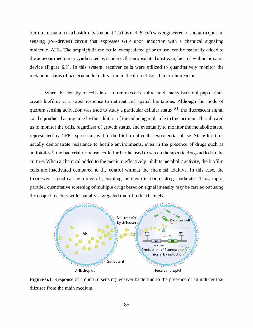
85
biofilm formation in a hostile environment. To this end, E. coli was engineered to contain a quorum
sensing (Plux-driven) circuit that expresses GFP upon induction with a chemical signaling
molecule, AHL. The amphiphilic molecule, encapsulated prior to use, can be manually added to
the aqueous medium or synthesized by sender cells encapsulated upstream, located within the same
device (Figure 6.1). In this system, receiver cells were utilized to quantitatively monitor the
metabolic status of bacteria under cultivation in the droplet-based micro-bioreactor.
When the density of cells in a culture exceeds a threshold, many bacterial populations
create biofilms as a stress response to nutrient and spatial limitations. Although the mode of
quorum sensing activation was used to study a particular cellular status 161, the fluorescent signal
can be produced at any time by the addition of the inducing molecule in the medium. This allowed
us to monitor the cells, regardless of growth status, and eventually to monitor the metabolic state,
represented by GFP expression, within the biofilm after the exponential phase. Since biofilms
usually demonstrate resistance to hostile environments, even in the presence of drugs such as
antibiotics 8, the bacterial response could further be used to screen therapeutic drugs added to the
culture. When a chemical added to the medium effectively inhibits metabolic activity, the biofilm
cells are inactivated compared to the control without the chemical additive. In this case, the
fluorescent signal can be turned off, enabling the identification of drug candidates. Thus, rapid,
parallel, quantitative screening of multiple drugs based on signal intensity may be carried out using
the droplet reactors with spatially segregated microfluidic channels.
Figure 6.1. Response of a quorum sensing receiver bacterium to the presence of an inducer that
diffuses from the main medium.
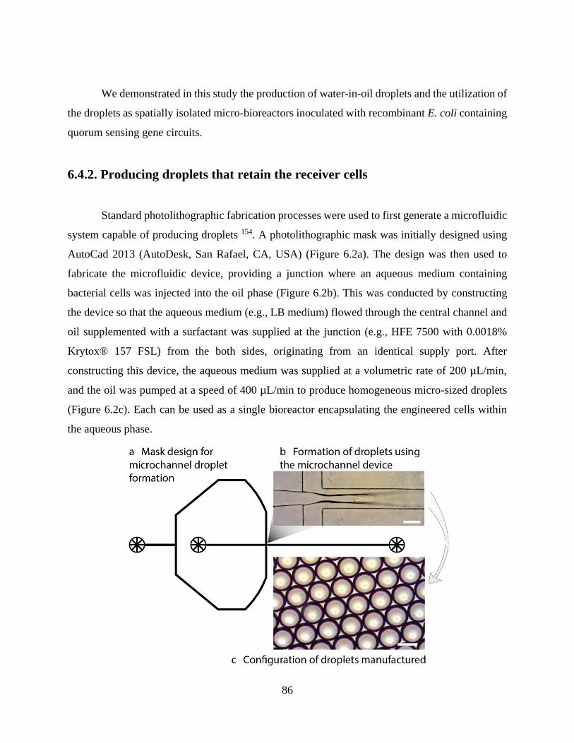
86
We demonstrated in this study the production of water-in-oil droplets and the utilization of
the droplets as spatially isolated micro-bioreactors inoculated with recombinant E. coli containing
quorum sensing gene circuits.
6.4.2. Producing droplets that retain the receiver cells
Standard photolithographic fabrication processes were used to first generate a microfluidic
system capable of producing droplets 154. A photolithographic mask was initially designed using
AutoCad 2013 (AutoDesk, San Rafael, CA, USA) (Figure 6.2a). The design was then used to
fabricate the microfluidic device, providing a junction where an aqueous medium containing
bacterial cells was injected into the oil phase (Figure 6.2b). This was conducted by constructing
the device so that the aqueous medium (e.g., LB medium) flowed through the central channel and
oil supplemented with a surfactant was supplied at the junction (e.g., HFE 7500 with 0.0018%
Krytox® 157 FSL) from the both sides, originating from an identical supply port. After
constructing this device, the aqueous medium was supplied at a volumetric rate of 200 µL/min,
and the oil was pumped at a speed of 400 µL/min to produce homogeneous micro-sized droplets
(Figure 6.2c). Each can be used as a single bioreactor encapsulating the engineered cells within
the aqueous phase.

87
Figure 6.2. Manufacture of homogeneous, micrometer-sized water-in-oil droplets using a
microfluidic device. The scale bar shown in b and c represents 100 µm.
To monitor quorum signaling, we constructed a synthetic bacterial cell line, referred to as
the ‘receiver’, which generated visible GFP signals upon induction with AHL, as mentioned
previously. The receiver contained the transcription factor luxR and a gfp gene with a bidirectional
Plux promoter (Figure 6.3a, left panel) 162. The gene construct for quorum sensing was tested by
activating the receiver system through the manual addition of AHL into receiver cultures in 96-
well plates, and monitoring the GFP signals via the plate reader (Figure 6.3a, left panel).
Subsequently, the induced receiver cells were encapsulated in droplets (average diameter = 100
m), as described in Figure 6.2, and transferred to an observation channel (2 × 4 mm) by simply
bridging the two ports extended from the outlet of the droplet-producing microfluidic device, for
further characterization (Figure 6.3b). The induced receiver cell-containing droplets where
cultured overnight for 12 h (average diameter = 100 m; Figure 6.3b, upper panel). Images of the
receiver cell fluorescence were captured at a predetermined time to observe the cell behavior and
viability (lower panel). Even after encapsulation of the recombinant cells, the receiver system
remained intact, permitting visualization of the quorum-induced cells. It should be noted that the
oil, HFE 7500, used as the ambient environment is Fluorinert™, i.e., an inert fluid normally used
to cool circuit boards 163.
88
Figure 6.3. Characterization of the receiver system in E. coli cells. The error bars in the graph
indicate standard deviation of triple measurements at each condition. The scale bar in b represents
50 m.
6.4.3. Bacterial cell culture in a single-droplet microfluidic bioreactor
Although colonies of the same engineered receiver cells contain the same gene circuit,
these cells may express a heterogeneous behavior in response to the inducer, complicating parallel
processing. Therefore, a microfluidic device allowing the spatial segregation of colonies was
developed in order to visualize induction of quorum sensing signals within micro-compartments.
First, a microcfluidic device was manufactured using standard microfabrication technology based
on soft-photolithography (Figure 6.4, left panel). The device comprised a single tortuous channel
for the flow of the fluorocarbon oil and a central compartment for maintaining a water-in-oil
droplet, i.e., a mother droplet, as a bioreactor for bacterial cultivation (Figure 6.4, right panel). The
compartment was constructed with one inlet and an outlet combined with gates to allow the
medium to flow through and a main physical support to hold the mother droplet within the
designated space. Subsequently, small aqueous droplets were maneuvered to flow in through a
single inlet (width = 70 m) and out through an outlet (width = 50 m), with significantly
decreased pressure towards the mother droplet, with the help of supporting pillars functioning as
a soft valve. This configuration can hold an aqueous droplet within the surrounding fluorocarbon
medium and further maintain the culture of the bacterial cells encapsulated within the mother
droplet.

89
Figure 6.4. Spatial segregation of the droplet-based bacterial culture system using a microfluidic
device. The scale bar represents 200 µm.
To further investigate the proper functionality of the device we maneuvered small droplets through
the channel (Figure 6.5a). Upon entering the inlet region of the holding compartment, the small
aqueous droplet subsequently went in contact with the mother droplet held in the in the chamber,
inducing coalescence caused by surface tension (Figure 6.5b). The droplet volume exceeding the
holding capacity of the chamber was drawn from the chamber by generating a daughter droplet in
the downstream channel of the device (Figure 6.5c). In further sections of our research, the
sequence of droplet maneuver, coalescence, and generation of daughter droplet will facilitate the
supply of nutrients to the cells cultivated within the mother droplet. The same approach can also
be used for the supply of the signaling molecule, AHL, and other chemical compounds, e.g.,
antibiotics, by its inclusion in the aqueous medium of the small droplets.
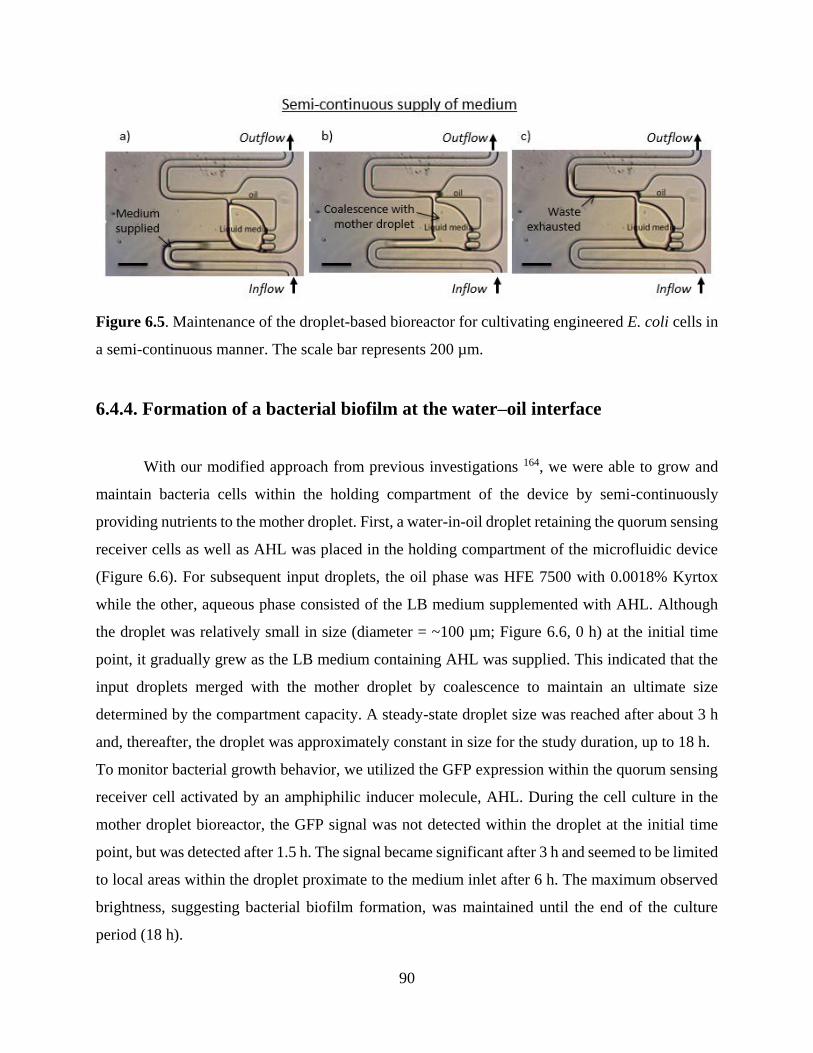
90
Figure 6.5. Maintenance of the droplet-based bioreactor for cultivating engineered E. coli cells in
a semi-continuous manner. The scale bar represents 200 µm.
6.4.4. Formation of a bacterial biofilm at the water–oil interface
With our modified approach from previous investigations 164, we were able to grow and
maintain bacteria cells within the holding compartment of the device by semi-continuously
providing nutrients to the mother droplet. First, a water-in-oil droplet retaining the quorum sensing
receiver cells as well as AHL was placed in the holding compartment of the microfluidic device
(Figure 6.6). For subsequent input droplets, the oil phase was HFE 7500 with 0.0018% Kyrtox
while the other, aqueous phase consisted of the LB medium supplemented with AHL. Although
the droplet was relatively small in size (diameter = ~100 µm; Figure 6.6, 0 h) at the initial time
point, it gradually grew as the LB medium containing AHL was supplied. This indicated that the
input droplets merged with the mother droplet by coalescence to maintain an ultimate size
determined by the compartment capacity. A steady-state droplet size was reached after about 3 h
and, thereafter, the droplet was approximately constant in size for the study duration, up to 18 h.
To monitor bacterial growth behavior, we utilized the GFP expression within the quorum sensing
receiver cell activated by an amphiphilic inducer molecule, AHL. During the cell culture in the
mother droplet bioreactor, the GFP signal was not detected within the droplet at the initial time
point, but was detected after 1.5 h. The signal became significant after 3 h and seemed to be limited
to local areas within the droplet proximate to the medium inlet after 6 h. The maximum observed
brightness, suggesting bacterial biofilm formation, was maintained until the end of the culture
period (18 h).

91
Figure 6.6. Long-term culture of engineered receiver E. coli cells in a spatially isolated droplet
bioreactor used to monitor biofilm formation. The scale bar represents 100 µm.
The rapid increase in signal intensity during the initial period, i.e., the first 3 h, could be
related to the growth in droplet size. As the size expansion indicated the immersion of fresh LB
medium nutrients into the mother droplet containing the cell inoculum, the cell culture was
provided with sufficient nutrients, fulfilling the conditions for growth and metabolism. Therefore,
cells within the mother droplet were able to achieve exponential growth, which was particularly
apparent in proximity to the entrance of the holding compartment. Since the engineered E. coli cell
maintained a doubling time of around 45 min 165, the cells can ideally expand up to 16 times in
density after 3 h and 256 times after 6 h. This may be sufficient to support growth of the bacterial
population beyond the stationary phase, eventually resulting in biofilm formation near the water–
oil interface after 6 h from the onset of cell cultivation. While this aspect of cellular community
was kept until the end of the culture, microscopic observations interestingly revealed the rotation
of biofilm formed within the mother droplet. Such cell aggregate rotation may result from the
fluorocarbon oil flow and can thus be controlled by manipulating the water–oil phase interface
according to the flow direction. Also, the E. coli cells grew within the droplet in three-dimensional
multicellular assemblies, rather than in a planar arrangement at the interface, consistent with
previous findings 158.
92
Bacterial adsorption at various interfaces (e.g., liquid–solid, liquid–air, and liquid–liquid)
is a key factor in biofilm formation 158. Bacteria can initially adhere to interfacial surfaces via
unspecific binding by means of electrostatic, short range Lewis acid–base, and hydration
interactions 166. The electrostatic interactions under most physiological conditions are repulsive,
as both the bacteria and most surfaces are net negatively charged. However, owing to the
hydrophobicity of the complex and the heterogeneity of the cell surface containing flagella,
fimbriae, pili, proteins, and surface polysaccharides, this repulsion is overcome. Numerous factors
influence bacterial adhesion, including pH, ionic strength, bacterial mobility, state of growth, and
growth factors in the medium 167. Therefore, biofilm formation and development can vary
according to bacterial species under different culture, nutritional, and environmental conditions.
To establish methods for the control of stable bacterial habitats, it may be critical to
construct a multiplex bacterial cell culture system permitting reproducible biofilm formation at
hydrophobic interfaces. In this study, bacterial attachment and biofilm growth over time at the
water–oil interface were monitored by microscopy. We were able to observe the initial bacterial
growth in segregated droplets using microfluidic channels and also biofilm formation at the liquid–
liquid interface. The recombinant E. coli cells were eventually allowed to adsorb at the water–oil
interface, particularly in their non-growing, stationary state. To minimize labor and time involved
in testing a number of potential control candidates, it is essential to reproducibly generate biofilms
in the small droplet spaces in a spatially segregated fashion. This could be advantageous for
constructing parallel, multiple experimental setups to accelerate the screening of substances that
efficiently inhibit bacterial metabolism in the biofilm structure. Biofilms are known to be difficult
to treat with antibiotics, to be associated with food poisoning outbreaks and clinical infections 167,
and to even clog industrial pipes 157, 168-170; accordingly, methods for the formation of biofilms in
spatially-segregated droplets have medical, industrial, and ecological relevance.
6.5. Conclusion

93
We demonstrated reproducible generation of biofilm structures in the small droplet spaces
in a spatially segregated fashion, which could be advantageous for constructing parallel, multiple
experimental setups to conduct biofilm assays in high throughput. A device such as this is essential
to minimizing the labor and time required to testing a large number of parameters that may
influence the formation and function of microbial consortia. One typical application of this system
is the screening of substances that efficiently inhibit bacterial metabolism in the biofilm structure.
In the future, this new technology can be extended to enable parallel assays using segregated
colonies within a single system. This system has broad potential applications across a range of
fields in which microbial cellular pattern formation and signal propagation are critical, such as
biophysics, developmental biology, and tissue engineering.

94
CHAPTER 7.
CONCLUSIONS
As a ubiquitous and naturally occurring form of bacteria, the most abundant type of
organism worldwide, biofilms are an essential target of microbiological research. Limitations to
the study of bacterial biofilms are one of the greatest obstacles to understanding this common
bacterial form as well as overcoming the challenges that biofilms present to human health and
economic activities. Accordingly, the microfluidic devices described in this thesis will be an
essential tool for exploring the biology of biofilms and devising control methods that can prevent
the negative impacts of biofilms. In conjunction with bacterial strains developed using genetic
engineering tools such as genetic circuits, these microfluidic devices provide a powerful approach
to dissecting the molecular mechanisms underlying biofilms.
The main microfluidic device utilized throughout this research is a flow cell instrument
that permits the culture of bacteria in artificial conditions that mimic the nutrient stress that bacteria
endure in their biofilm form. Experimental cultures have confirmed that this device has broad
potential for the study of bacterial biofilms based on several of its strengths. First and foremost,
the device PDMS device is small, relatively inexpensive, and designed to maximize the ability to
visualize individual cells using real-time epifluorescent microscopy under fluorescent. These
features enable high-throughput assays of chemical compounds with real-time imaging—the
former permitting large experiments with high statistical power and the latter permitting
confirmation of effects, especially in conjunction with genetic constructs that utilize fluorescent
reporter proteins such as GFP or mCherry.
The study of biofilms at the level of individual cells is impractical because cells are, by
definition, tightly aggregated within biofilms. For this reason, it is extremely difficult to visualize
a cell that is within a three-dimensional mass of neighboring cells. The biofilm-mimicking
microfluidic device eliminates this issue by only having physical space for a single layer of cells
to develop. The flow channels of the microfluidic device are just 0.97 µm in height, which is

95
slightly smaller than the typical 1-µm diameter of E. coli at their thinnest point. Importantly, this
flow cell can be redesigned to accommodate cell of sizes that are smaller or larger than E. coli. In
addition to trapping cells within a one-cell-thick plane, which enables the straightforward
visualization of individual cells, this restricted flow channel height also prevents cell motility. This
added benefit also facilitates the continuous tracking of cell lineages. As a mother cell divide into
two daughter cells, which in turn divide into four daughter cells as well, and so on, continuous
imaging of all cells divisions can be captured using microscopy. This enables viewing of the entire
time course of an experiment in great detail, especially when used in conjunction with E. coli cells
lines that contain reporter constructs.
In the first set of experiments utilizing this device, the ability of the microfluidic device to
induce biofilm-like stress was confirmed (proof of the first hypothesis presented in Chapter 1).
The E. coli cultured in this experiment were contained a simple genetic circuit—specifically, a
receiver construct containing the bidirectional Plux promoter and a GFP reporter regulated by a
luxR transcription factor. When induced with AHL, a quorum sensing signaling molecule, this cell
line expresses its GFP signal. However, under the stressful culture conditions of the microfluidic
device, the GFP signal was expressed even in the absence of AHL. This spontaneous expression
of the GFP quorum sensing reporter suggests that the biomimetic structures of the microfluidic
device impose conditions similar to those of natural biofilms, thus triggering stochastic gene
expression pathways.
Spontaneous expression of stress-induced responses in bacterial cells cultured in the
biofilm-mimicking device were also supported by a similar experiment utilizing an E. coli strain
containing a genetic toggle switch. The simple toggle switch constitutively expressed one of two
states determined by an initial stimulus, essentially acting as a simple ON/OFF switch. After
receiving the initial signal of aTc, nearly all cells remained in that expression state. However,
induction with IPTG, was stochastically reversed over the course of the 5-day culture period; by
the end of this time, more than 5% of cells had spontaneously reverted to the alternative toggle
switch state. This asymmetry was unexpected. However, the stochastic change in toggle switch
state indicates that the stressful conditions of the microfluidic cell overcame the bistability of this
system as predicted by previous theoretical 120 and empirical research 118.

96
The stress induction of the microfluidic system was further confirmed in a series of
experiments that utilized the facilitation of continuous cell tracking in the system (proof of the
second hypothesis). Specifically, an aberrant, enlarged cell morphology was observed within the
stressful environment of the microfluidic cell. Moreover, this phenotype was observed to be
inherited by daughter cells, suggesting that an epigenetic switch had been induced by the nutrient
deficient conditions.
The final series of experiments utilized an entirely different type of microfluidic cell that
may also be used to examine group phenotypes of small numbers of bacterial cells, such as may
occur within small portions of biofilms (proof of the third hypothesis). This microfluidic device
enabled the continuous culture of a small number of cells within a bubble of aqueous medium
suspended within a flow of oil. This water-in-oil droplet bioreactor was able to both continuous
culture a small population of cells as well as permit the precise introduction of chemical
compounds while generating biofilms. Under these culture conditions, AHL was successfully
introduced into the cultured cells, which then expressed their GFP quorum sensing reporter.
Both of the devices explored in this thesis will continue to be useful tools of the study
of E. coli cells within biofilms, and with minor design modifications, may be applied to other
microbial systems. Future research should be focused on the application of these devices to
large panels of chemical compounds for their inhibitory or cell-killing effects. This specific
line of research would be especially useful in identifying next-generation antibiotics that are
able to overcome the inherent defenses of bacterial biofilms.

97
REFERENCES
1. Mattila‐Sandholm, T.; Wirtanen, G., Biofilm formation in the industry: a review. Food
Reviews International 1992, 8 (4), 573-603.
2. Peters, B. M.; Jabra-Rizk, M. A.; Graeme, A.; Costerton, J. W.; Shirtliff, M. E.,
Polymicrobial interactions: impact on pathogenesis and human disease. Clinical microbiology
reviews 2012, 25 (1), 193-213.
3. Socransky, S. S.; Haffajee, A. D., Dental biofilms: difficult therapeutic targets.
Periodontology 2000 2002, 28 (1), 12-55.
4. Momba, M.; Kfir, R.; Venter, S. N.; Cloete, T. E., An overview of biofilm formation in
distribution systems and its impact on the deterioration of water quality. 2000.
5. Wang, X.; Wood, T. K., Toxin-antitoxin systems influence biofilm and persister cell
formation and the general stress response. Applied and environmental microbiology 2011, 77 (16),
5577-5583.
6. Costerton, J. W.; Stewart, P. S.; Greenberg, E. P., Bacterial biofilms: a common cause of
persistent infections. Science 1999, 284 (5418), 1318-1322.
7. Høiby, N.; Bjarnsholt, T.; Givskov, M.; Molin, S.; Ciofu, O., Antibiotic resistance of
bacterial biofilms. International journal of antimicrobial agents 2010, 35 (4), 322-332.
8. Stewart, P. S.; Costerton, J. W., Antibiotic resistance of bacteria in biofilms. The lancet
2001, 358 (9276), 135-138.
9. Stewart, P. S.; Franklin, M. J., Physiological heterogeneity in biofilms. Nature Reviews
Microbiology 2008, 6 (3), 199-210.
10. Ruder, W. C.; Lu, T.; Collins, J. J., Synthetic biology moving into the clinic. Science 2011,
333 (6047), 1248-52.
11. Khalil, A. S.; Collins, J. J., Synthetic biology: applications come of age. Nature reviews.
Genetics 2010, 11 (5), 367-79.
12. Anderson, J. C.; Clarke, E. J.; Arkin, A. P.; Voigt, C. A., Environmentally controlled
invasion of cancer cells by engineered bacteria. J Mol Biol 2006, 355 (4), 619-27.
13. Duan, F.; March, J. C., Engineered bacterial communication prevents Vibrio cholerae
virulence in an infant mouse model. Proc Natl Acad Sci U S A 2010, 107 (25), 11260-4.
14. Ye, H.; Daoud-El Baba, M.; Peng, R. W.; Fussenegger, M., A synthetic optogenetic
transcription device enhances blood-glucose homeostasis in mice. Science 2011, 332 (6037), 1565-
8.
15. Chen, Y. Y.; Jensen, M. C.; Smolke, C. D., Genetic control of mammalian T-cell
proliferation with synthetic RNA regulatory systems. Proc Natl Acad Sci U S A 2010, 107 (19),
8531-6.
16. Gardner, T. S.; Cantor, C. R.; Collins, J. J., Construction of a genetic toggle switch in
Escherichia coli. Nature 2000, 403 (6767), 339-42.
17. Elowitz, M. B.; Leibler, S., A synthetic oscillatory network of transcriptional regulators.
Nature 2000, 403 (6767), 335-8.
18. Danino, T.; Mondragon-Palomino, O.; Tsimring, L.; Hasty, J., A synchronized quorum of
genetic clocks. Nature 2010, 463 (7279), 326-30.
98
19. Ellis, T.; Wang, X.; Collins, J. J., Diversity-based, model-guided construction of synthetic
gene networks with predicted functions. Nat Biotechnol 2009, 27 (5), 465-71.
20. Friedland, A. E.; Lu, T. K.; Wang, X.; Shi, D.; Church, G.; Collins, J. J., Synthetic gene
networks that count. Science 2009, 324 (5931), 1199-202.
21. Fung, E.; Wong, W. W.; Suen, J. K.; Bulter, T.; Lee, S. G.; Liao, J. C., A synthetic gene-
metabolic oscillator. Nature 2005, 435 (7038), 118-22.
22. Rinaudo, K.; Bleris, L.; Maddamsetti, R.; Subramanian, S.; Weiss, R.; Benenson, Y., A
universal RNAi-based logic evaluator that operates in mammalian cells. Nat Biotechnol 2007, 25
(7), 795-801.
23. Tabor, J. J.; Salis, H. M.; Simpson, Z. B.; Chevalier, A. A.; Levskaya, A.; Marcotte, E. M.;
Voigt, C. A.; Ellington, A. D., A synthetic genetic edge detection program. Cell 2009, 137 (7),
1272-81.
24. You, L.; Cox, R. S., 3rd; Weiss, R.; Arnold, F. H., Programmed population control by cell-
cell communication and regulated killing. Nature 2004, 428 (6985), 868-71.
25. Qi, H.; Blanchard, A.; Lu, T., Engineered genetic information processing circuits. Wiley
Interdisciplinary Reviews: Systems Biology and Medicine 2013, 5 (3), 273-287.
26. Berthiaume, F.; Maguire, T. J.; Yarmush, M. L., Tissue Engineering and Regenerative
Medicine: History, Progress, and Challenges. Annual Review of Chemical and Biomolecular
Engineering 2011, 2 (1), 403-430.
27. Forbes, N. S., Engineering the perfect (bacterial) cancer therapy. Nat Rev Cancer 2010, 10
(11), 785-794.
28. Levskaya, A.; Chevalier, A. A.; Tabor, J. J.; Simpson, Z. B.; Lavery, L. A.; Levy, M.;
Davidson, E. A.; Scouras, A.; Ellington, A. D.; Marcotte, E. M.; Voigt, C. A., Synthetic biology:
engineering Escherichia coli to see light. Nature 2005, 438 (7067), 441-2.
29. Bashor, C. J.; Helman, N. C.; Yan, S.; Lim, W. A., Using engineered scaffold interactions
to reshape MAP kinase pathway signaling dynamics. Science 2008, 319 (5869), 1539-43.
30. Xia, B.; Bhatia, S.; Bubenheim, B.; Dadgar, M.; Densmore, D.; Anderson, J. C.,
Developer's and user's guide to Clotho v2.0 A software platform for the creation of synthetic
biological systems. Methods in enzymology 2011, 498, 97-135.
31. Bilitchenko, L.; Liu, A.; Cheung, S.; Weeding, E.; Xia, B.; Leguia, M.; Anderson, J. C.;
Densmore, D., Eugene--a domain specific language for specifying and constraining synthetic
biological parts, devices, and systems. PLoS One 2011, 6 (4), e18882.
32. Egbert, R. G.; Klavins, E., Fine-tuning gene networks using simple sequence repeats.
Proceedings of the National Academy of Sciences 2012, 109 (42), 16817-16822.
33. Salis, H. M.; Mirsky, E. A.; Voigt, C. A., Automated design of synthetic ribosome binding
sites to control protein expression. Nat Biotech 2009, 27 (10), 946-950.
34. Callura, J. M.; Dwyer, D. J.; Isaacs, F. J.; Cantor, C. R.; Collins, J. J., Tracking, tuning,
and terminating microbial physiology using synthetic riboregulators. Proc Natl Acad Sci U S A
2010, 107 (36), 15898-903.
35. Isaacs, F. J.; Dwyer, D. J.; Ding, C.; Pervouchine, D. D.; Cantor, C. R.; Collins, J. J.,
Engineered riboregulators enable post-transcriptional control of gene expression. Nat Biotechnol
2004, 22 (7), 841-7.
36. Gallivan, J. P., Toward reprogramming bacteria with small molecules and RNA. Current
opinion in chemical biology 2007, 11 (6), 612-9.
37. Topp, S.; Gallivan, J. P., Guiding bacteria with small molecules and RNA. Journal of the
American Chemical Society 2007, 129 (21), 6807-11.

99
38. Babiskin, A. H.; Smolke, C. D., A synthetic library of RNA control modules for predictable
tuning of gene expression in yeast. Mol Syst Biol 2011, 7, 471.
39. Win, M. N.; Smolke, C. D., Higher-order cellular information processing with synthetic
RNA devices. Science 2008, 322 (5900), 456-60.
40. Siuti, P.; Yazbek, J.; Lu, T. K., Synthetic circuits integrating logic and memory in living
cells. Nat Biotechnol 2013, 31 (5), 448-52.
41. Lutz, R.; Bujard, H., Independent and tight regulation of transcriptional units in
Escherichia coli via the LacR/O, the TetR/O and AraC/I1-I2 regulatory elements. Nucleic acids
research 1997, 25 (6), 1203-10.
42. Xie, Z.; Wroblewska, L.; Prochazka, L.; Weiss, R.; Benenson, Y., Multi-input RNAi-based
logic circuit for identification of specific cancer cells. Science 2011, 333 (6047), 1307-11.
43. Tamsir, A.; Tabor, J. J.; Voigt, C. A., Robust multicellular computing using genetically
encoded NOR gates and chemical 'wires'. Nature 2011, 469 (7329), 212-5.
44. Anderson, J. C.; Voigt, C. A.; Arkin, A. P., Environmental signal integration by a modular
AND gate. Mol Syst Biol 2007, 3, 133.
45. Daniel, R.; Rubens, J. R.; Sarpeshkar, R.; Lu, T. K., Synthetic analog computation in living
cells. Nature 2013, 497 (7451), 619-23.
46. Stricker, J.; Cookson, S.; Bennett, M. R.; Mather, W. H.; Tsimring, L. S.; Hasty, J., A fast,
robust and tunable synthetic gene oscillator. Nature 2008, 456 (7221), 516-9.
47. Wong, W. W.; Tsai, T. Y.; Liao, J. C., Single-cell zeroth-order protein degradation
enhances the robustness of synthetic oscillator. Mol Syst Biol 2007, 3, 130.
48. Tigges, M.; Marquez-Lago, T. T.; Stelling, J.; Fussenegger, M., A tunable synthetic
mammalian oscillator. Nature 2009, 457 (7227), 309-12.
49. Tigges, M.; Denervaud, N.; Greber, D.; Stelling, J.; Fussenegger, M., A synthetic low-
frequency mammalian oscillator. Nucleic acids research 2010, 38 (8), 2702-11.
50. Simpson, M. L.; Cox, C. D.; Sayler, G. S., Frequency domain analysis of noise in
autoregulated gene circuits. Proceedings of the National Academy of Sciences of the United States
of America 2003, 100, 4551-6.
51. Austin, D. W.; Allen, M. S.; McCollum, J. M.; Dar, R. D.; Wilgus, J. R.; Sayler, G. S.;
Samatova, N. F.; Cox, C. D.; Simpson, M. L., Gene network shaping of inherent noise spectra.
Nature 2006, 439, 608-11.
52. McGinness, K. E.; Baker, T. a.; Sauer, R. T., Engineering controllable protein degradation.
Molecular cell 2006, 22, 701-7.
53. Banaszynski, L. a.; Chen, L.-C.; Maynard-Smith, L. a.; Ooi, a. G. L.; Wandless, T. J., A
rapid, reversible, and tunable method to regulate protein function in living cells using synthetic
small molecules. Cell 2006, 126, 995-1004.
54. Auslander, D.; Fussenegger, M., Optogenetic therapeutic cell implants. Gastroenterology
2012, 143 (2), 301-6.
55. Bacchus, W.; Fussenegger, M., The use of light for engineered control and reprogramming
of cellular functions. Current opinion in biotechnology 2012, 23 (5), 695-702.
56. Tabor, J. J.; Levskaya, A.; Voigt, C. A., Multichromatic control of gene expression in
Escherichia coli. J Mol Biol 2011, 405 (2), 315-24.
57. Antunes, M. S.; Morey, K. J.; Smith, J. J.; Albrecht, K. D.; Bowen, T. A.; Zdunek, J. K.;
Troupe, J. F.; Cuneo, M. J.; Webb, C. T.; Hellinga, H. W.; Medford, J. I., Programmable ligand
detection system in plants through a synthetic signal transduction pathway. PLoS One 2011, 6 (1),
e16292.

100
58. Thompson, K. E.; Bashor, C. J.; Lim, W. A.; Keating, A. E., SYNZIP protein interaction
toolbox: in vitro and in vivo specifications of heterospecific coiled-coil interaction domains. ACS
synthetic biology 2012, 1 (4), 118-29.
59. Bashor, C. J.; Horwitz, A. A.; Peisajovich, S. G.; Lim, W. A., Rewiring cells: synthetic
biology as a tool to interrogate the organizational principles of living systems. Annual review of
biophysics 2010, 39, 515-37.
60. Bhattacharyya, R. P.; Remenyi, A.; Good, M. C.; Bashor, C. J.; Falick, A. M.; Lim, W. A.,
The Ste5 scaffold allosterically modulates signaling output of the yeast mating pathway. Science
2006, 311 (5762), 822-6.
61. Deans, T. L.; Cantor, C. R.; Collins, J. J., A tunable genetic switch based on RNAi and
repressor proteins for regulating gene expression in mammalian cells. Cell 2007, 130 (2), 363-72.
62. Kramer, B. P.; Viretta, A. U.; Daoud-El-Baba, M.; Aubel, D.; Weber, W.; Fussenegger,
M., An engineered epigenetic transgene switch in mammalian cells. Nat Biotechnol 2004, 22 (7),
867-70.
63. Nevozhay, D.; Zal, T.; Balazsi, G., Transferring a synthetic gene circuit from yeast to
mammalian cells. Nature communications 2013, 4, 1451.
64. Duan, F.; Curtis, K. L.; March, J. C., Secretion of insulinotropic proteins by commensal
bacteria: rewiring the gut to treat diabetes. Appl Environ Microbiol 2008, 74 (23), 7437-8.
65. Rao, S.; Hu, S.; McHugh, L.; Lueders, K.; Henry, K.; Zhao, Q.; Fekete, R. A.; Kar, S.;
Adhya, S.; Hamer, D. H., Toward a live microbial microbicide for HIV: commensal bacteria
secreting an HIV fusion inhibitor peptide. Proc Natl Acad Sci U S A 2005, 102 (34), 11993-8.
66. Farrar, M. D.; Whitehead, T. R.; Lan, J.; Dilger, P.; Thorpe, R.; Holland, K. T.; Carding,
S. R., Engineering of the gut commensal bacterium Bacteroides ovatus to produce and secrete
biologically active murine interleukin-2 in response to xylan. J Appl Microbiol 2005, 98 (5), 1191-
7.
67. Xiang, S.; Fruehauf, J.; Li, C. J., Short hairpin RNA-expressing bacteria elicit RNA
interference in mammals. Nat Biotechnol 2006, 24 (6), 697-702.
68. Paek, S. H.; Ruder, W. C., Stochastically induced quorum sensing. Submitted 2017.
69. Chmielewski, R.; Frank, J., Biofilm formation and control in food processing facilities.
Compr Rev Food Sci Food Saf 2003, 2 (1), 22-32.
70. Wood, T. K.; Hong, S. H.; Ma, Q., Engineering biofilm formation and dispersal. Trends
Biotechnol. 2011, 29 (2), 87-94.
71. Karimi, A.; Karig, D.; Kumar, A.; Ardekani, A., Interplay of physical mechanisms and
biofilm processes: review of microfluidic methods. Lab on a Chip 2015, 15 (1), 23-42.
72. Gao, G.; Lange, D.; Hilpert, K.; Kindrachuk, J.; Zou, Y.; Cheng, J. T.; Kazemzadeh-
Narbat, M.; Yu, K.; Wang, R.; Straus, S. K., The biocompatibility and biofilm resistance of implant
coatings based on hydrophilic polymer brushes conjugated with antimicrobial peptides.
Biomaterials 2011, 32 (16), 3899-3909.
73. Davey, M. E.; O'toole, G. A., Microbial biofilms: from ecology to molecular genetics.
Microbiol. Mol. Biol. Rev. 2000, 64 (4), 847-867.
74. Baehni, P.; Takeuchi, Y., Anti‐plaque agents in the prevention of biofilm‐associated oral
diseases. Oral Dis. 2003, 9 (s1), 23-29.
75. Wingender, J.; Neu, T. R.; Flemming, H.-C., Microbial extracellular polymeric
substances: characterization, structure and function. Springer Science & Business Media: 2012.

101
76. Weber, M. M.; French, C. L.; Barnes, M. B.; Siegele, D. A.; McLean, R. J., A previously
uncharacterized gene, yjfO (bsmA), influences Escherichia coli biofilm formation and stress
response. Microbiology 2010, 156 (1), 139-147.
77. Wen, Z. T.; Suntharaligham, P.; Cvitkovitch, D. G.; Burne, R. A., Trigger factor in
Streptococcus mutans is involved in stress tolerance, competence development, and biofilm
formation. Infection and immunity 2005, 73 (1), 219-225.
78. Rachid, S.; Ohlsen, K.; Witte, W.; Hacker, J.; Ziebuhr, W., Effect of subinhibitory
antibiotic concentrations on polysaccharide intercellular adhesin expression in biofilm-forming
Staphylococcus epidermidis. Antimicrob. Agents Chemother. 2000, 44 (12), 3357-3363.
79. O'Toole, G.; Kaplan, H. B.; Kolter, R., Biofilm formation as microbial development.
Annual Reviews in Microbiology 2000, 54 (1), 49-79.
80. Davies, D. G. In Physiological events in biofilm formation, Symposia-society for General
Microbiology, Cambridge; Cambridge University Press; 1999: 2000; pp 37-52.
81. Patel, R., Biofilms and antimicrobial resistance. Clin. Orthop. Relat. Res. 2005, 437, 41-
47.
82. Helloin, E.; Jänsch, L.; Phan‐Thanh, L., Carbon starvation survival of Listeria
monocytogenes in planktonic state and in biofilm: a proteomic study. Proteomics 2003, 3 (10),
2052-2064.
83. Webster, N. S.; Smith, L. D.; Heyward, A. J.; Watts, J. E.; Webb, R. I.; Blackall, L. L.;
Negri, A. P., Metamorphosis of a scleractinian coral in response to microbial biofilms. Appl.
Environ. Microbiol. 2004, 70 (2), 1213-1221.
84. Parsek, M. R.; Greenberg, E., Sociomicrobiology: the connections between quorum
sensing and biofilms. Trends Microbiol. 2005, 13 (1), 27-33.
85. Stewart, P. S., Diffusion in biofilms. J. Bacteriol. 2003, 185 (5), 1485-1491.
86. Dunne, W. M., Bacterial adhesion: seen any good biofilms lately? Clin. Microbiol. Rev.
2002, 15 (2), 155-166.
87. Di Bonaventura, G.; Pompilio, A.; Picciani, C.; Iezzi, M.; D'Antonio, D.; Piccolomini, R.,
Biofilm formation by the emerging fungal pathogen Trichosporon asahii: development,
architecture, and antifungal resistance. Antimicrob. Agents Chemother. 2006, 50 (10), 3269-3276.
88. Whitchurch, C. B.; Tolker-Nielsen, T.; Ragas, P. C.; Mattick, J. S., Extracellular DNA
required for bacterial biofilm formation. Science 2002, 295 (5559), 1487-1487.
89. Picioreanu, C.; Van Loosdrecht, M. C.; Heijnen, J. J., Two-dimensional model of biofilm
detachment caused by internal stress from liquid flow. Biotechnol. Bioeng. 2001, 72 (2), 205-218.
90. Donlan, R. M., Biofilms: microbial life on surfaces. Emerg. Infect. Dis. 2002, 8 (9).
91. De Beer, D.; Stoodley, P.; Roe, F.; Lewandowski, Z., Effects of biofilm structures on
oxygen distribution and mass transport. Biotechnol. Bioeng. 1994, 43 (11), 1131-1138.
92. Picioreanu, C.; Van Loosdrecht, M. C.; Heijnen, J. J., Effect of diffusive and convective
substrate transport on biofilm structure formation: a two-dimensional modeling study. Biotechnol.
Bioeng. 2000, 69 (5), 504-515.
93. Bishop, P. L.; Zhang, T. C.; Fu, Y.-C., Effects of biofilm structure, microbial distributions
and mass transport on biodegradation processes. Water Sci. Technol. 1995, 31 (1), 143-152.
94. Bennett, M. R.; Hasty, J., Microfluidic devices for measuring gene network dynamics in
single cells. Nature Rev Genet 2009, 10 (9), 628-638.
95. Gravesen, P.; Branebjerg, J.; Jensen, O. S., Microfluidics-a review. J. Micromech.
Microeng. 1993, 3 (4), 168.
102
96. Zheng, Y.; Nguyen, J.; Wei, Y.; Sun, Y., Recent advances in microfluidic techniques for
single-cell biophysical characterization. Lab on a Chip 2013, 13 (13), 2464-2483.
97. Zhu, Y.; Fang, Q., Analytical detection techniques for droplet microfluidics—A review.
Anal. Chim. Acta 2013, 787, 24-35.
98. Danino, T.; Mondragón-Palomino, O.; Tsimring, L.; Hasty, J., A synchronized quorum of
genetic clocks. Nature 2010, 463 (7279), 326-330.
99. Longo, D.; Hasty, J., Dynamics of single‐cell gene expression. Mol. Syst. Biol. 2006, 2 (1),
64.
100. Drescher, K.; Shen, Y.; Bassler, B. L.; Stone, H. A., Biofilm streamers cause catastrophic
disruption of flow with consequences for environmental and medical systems. Proc. Natl. Acad.
Sci. 2013, 110 (11), 4345-4350.
101. Khalil, A. S.; Collins, J. J., Synthetic biology: applications come of age. Nature Reviews
Genetics 2010, 11 (5), 367-379.
102. Litcofsky, K. D.; Afeyan, R. B.; Krom, R. J.; Khalil, A. S.; Collins, J. J., Iterative plug-
and-play methodology for constructing and modifying synthetic gene networks. Nature methods
2012, 9 (11), 1077-1080.
103. Shadel, G.; Baldwin, T., The Vibrio fischeri LuxR protein is capable of bidirectional
stimulation of transcription and both positive and negative regulation of the luxR gene. Journal of
bacteriology 1991, 173 (2), 568-574.
104. Waters, C. M.; Bassler, B. L., Quorum sensing: cell-to-cell communication in bacteria.
Annu. Rev. Cell Dev. Biol. 2005, 21, 319-346.
105. Lopez, D.; Vlamakis, H.; Kolter, R., Generation of multiple cell types in Bacillus subtilis.
FEMS microbiology reviews 2009, 33 (1), 152-163.
106. Raj, A.; van Oudenaarden, A., Nature, nurture, or chance: stochastic gene expression and
its consequences. Cell 2008, 135 (2), 216-226.
107. Bjedov, I.; Tenaillon, O.; Gerard, B.; Souza, V.; Denamur, E.; Radman, M.; Taddei, F.;
Matic, I., Stress-induced mutagenesis in bacteria. Science 2003, 300 (5624), 1404-1409.
108. Marks, L. R.; Reddinger, R. M.; Hakansson, A. P., High levels of genetic recombination
during nasopharyngeal carriage and biofilm formation in Streptococcus pneumoniae. MBio 2012,
3 (5), e00200-12.
109. Banas, J. A.; Miller, J. D.; Fuschino, M. E.; Hazlett, K. R.; Toyofuku, W.; Porter, K. A.;
Reutzel, S. B.; Florczyk, M. A.; McDonough, K. A.; Michalek, S. M., Evidence that accumulation
of mutants in a biofilm reflects natural selection rather than stress-induced adaptive mutation.
Applied and environmental microbiology 2007, 73 (1), 357-361.
110. Voigt, C. A., Genetic parts to program bacteria. Curr. Opin. Biotechnol. 2006, 17 (5), 548-
557.
111. Gardner, T. S.; Cantor, C. R.; Collins, J. J., Construction of a genetic toggle switch in
Escherichia coli. Nature 2000, 403 (6767), 339-342.
112. Ruder, W. C.; Lu, T.; Collins, J. J., Synthetic biology moving into the clinic. Science 2011,
333 (6047), 1248-1252.
113. McDaniel, R.; Weiss, R., Advances in synthetic biology: on the path from prototypes to
applications. Curr. Opin. Biotechnol. 2005, 16 (4), 476-483.
114. Prigent-Combaret, C.; Brombacher, E.; Vidal, O.; Ambert, A.; Lejeune, P.; Landini, P.;
Dorel, C., Complex regulatory network controls initial adhesion and biofilm formation in
Escherichia coli via regulation of thecsgD gene. J. Bacteriol. 2001, 183 (24), 7213-7223.
103
115. Fox, E. P.; Bui, C. K.; Nett, J. E.; Hartooni, N.; Mui, M. C.; Andes, D. R.; Nobile, C. J.;
Johnson, A. D., An expanded regulatory network temporally controls Candida albicans biofilm
formation. Mol. Microbiol. 2015, 96 (6), 1226-1239.
116. Schuster, M.; Greenberg, E. P., A network of networks: quorum-sensing gene regulation
in Pseudomonas aeruginosa. Int. J. Med. Microbiol. 2006, 296 (2), 73-81.
117. Ng, W.-L.; Bassler, B. L., Bacterial quorum-sensing network architectures. Annu. Rev.
Genet. 2009, 43, 197-222.
118. Wang, J.; Zhang, J.; Yuan, Z.; Zhou, T., Noise-induced switches in network systems of the
genetic toggle switch. BMC Syst. Biol. 2007, 1 (1), 50.
119. Bokes, P.; King, J. R.; Wood, A. T.; Loose, M., Transcriptional bursting diversifies the
behaviour of a toggle switch: hybrid simulation of stochastic gene expression. Bull. Math. Biol.
2013, 75 (2), 351-371.
120. Kim, K.-Y.; Wang, J., Potential energy landscape and robustness of a gene regulatory
network: toggle switch. PLoS Comput. Biol. 2007, 3 (3), e60.
121. Elowitz, M. B.; Leibler, S., A synthetic oscillatory network of transcriptional regulators.
Nature 2000, 403 (6767), 335-338.
122. Brosius, J.; Erfle, M.; Storella, J., Spacing of the-10 and-35 regions in the tac promoter.
Effect on its in vivo activity. Journal of biological chemistry 1985, 260 (6), 3539-3541.
123. Vogel, J.; Wagner, E. G. H., Target identification of small noncoding RNAs in bacteria.
Current opinion in microbiology 2007, 10 (3), 262-270.
124. Mullineaux, C. W., Bacteria in solitary confinement. J. Bacteriol. 2015, 197 (4), 670-671.
125. Männik, J.; Driessen, R.; Galajda, P.; Keymer, J. E.; Dekker, C., Bacterial growth and
motility in sub-micron constrictions. Proceedings of the National Academy of Sciences 2009, 106
(35), 14861-14866.
126. Iyer-Biswas, S.; Wright, C. S.; Henry, J. T.; Lo, K.; Burov, S.; Lin, Y.; Crooks, G. E.;
Crosson, S.; Dinner, A. R.; Scherer, N. F., Scaling laws governing stochastic growth and division
of single bacterial cells. Proceedings of the National Academy of Sciences 2014, 111 (45), 15912-
15917.
127. Halldorsson, S.; Lucumi, E.; Gómez-Sjöberg, R.; Fleming, R. M., Advantages and
challenges of microfluidic cell culture in polydimethylsiloxane devices. Biosens. Bioelectron.
2015, 63, 218-231.
128. Sackmann, E. K.; Fulton, A. L.; Beebe, D. J., The present and future role of microfluidics
in biomedical research. Nature 2014, 507 (7491), 181-189.
129. Yao, X.; Jericho, M.; Pink, D.; Beveridge, T., Thickness and elasticity of gram-negative
murein sacculi measured by atomic force microscopy. J. Bacteriol. 1999, 181 (22), 6865-6875.
130. Wu, F.; Van Schie, B. G.; Keymer, J. E.; Dekker, C., Symmetry and scale orient Min
protein patterns in shaped bacterial sculptures. Nature nanotechnology 2015, 10 (8), 719-726.
131. Männik, J.; Wu, F.; Hol, F. J.; Bisicchia, P.; Sherratt, D. J.; Keymer, J. E.; Dekker, C.,
Robustness and accuracy of cell division in Escherichia coli in diverse cell shapes. Proceedings
of the National Academy of Sciences 2012, 109 (18), 6957-6962.
132. Avery, S. V., Microbial cell individuality and the underlying sources of heterogeneity.
Nature Reviews Microbiology 2006, 4 (8), 577-587.
133. Davidson, C. J.; Surette, M. G., Individuality in bacteria. Annu. Rev. Genet. 2008, 42, 253-
268.
134. Veening, J.-W.; Smits, W. K.; Kuipers, O. P., Bistability, epigenetics, and bet-hedging in
bacteria. Annu. Rev. Microbiol. 2008, 62, 193-210.

104
135. Lidstrom, M. E.; Konopka, M. C., The role of physiological heterogeneity in microbial
population behavior. Nat. Chem. Biol. 2010, 6 (10), 705-712.
136. Maheshri, N.; O’Shea, E. K., Living with noisy genes: how cells function reliably with
inherent variability in gene expression. Annu. Rev. Biophys. Biomol. Struct. 2007, 36, 413-434.
137. Harper, J. C.; Brozik, S. M.; Brinker, C. J.; Kaehr, B., Biocompatible microfabrication of
3D isolation chambers for targeted confinement of individual cells and their progeny. Anal. Chem.
2012, 84 (21), 8985-8989.
138. Matias, V. R.; Beveridge, T. J., Cryo‐electron microscopy reveals native polymeric cell
wall structure in Bacillus subtilis 168 and the existence of a periplasmic space. Mol. Microbiol.
2005, 56 (1), 240-251.
139. Koch, A. L., The surface stress theory of microbial morphogenesis. Adv. Microb. Physiol.
1983, 24, 301-366.
140. Huang, K. C.; Mukhopadhyay, R.; Wen, B.; Gitai, Z.; Wingreen, N. S., Cell shape and cell-
wall organization in Gram-negative bacteria. Proceedings of the National Academy of Sciences
2008, 105 (49), 19282-19287.
141. Kulasekara, B. R.; Kamischke, C.; Kulasekara, H. D.; Christen, M.; Wiggins, P. A.; Miller,
S. I., c-di-GMP heterogeneity is generated by the chemotaxis machinery to regulate flagellar
motility. Elife 2013, 2, e01402.
142. Sourjik, V.; Wingreen, N. S., Responding to chemical gradients: bacterial chemotaxis.
Curr. Opin. Cell Biol. 2012, 24 (2), 262-268.
143. Garrett, T. R.; Bhakoo, M.; Zhang, Z., Bacterial adhesion and biofilms on surfaces. Prog.
Nat. Sci. 2008, 18 (9), 1049-1056.
144. Pratt, L. A.; Kolter, R., Genetic analysis of Escherichia coli biofilm formation: roles of
flagella, motility, chemotaxis and type I pili. Mol. Microbiol. 1998, 30 (2), 285-293.
145. Martí, S.; Rodríguez-Baño, J.; Catel-Ferreira, M.; Jouenne, T.; Vila, J.; Seifert, H.; Dé, E.,
Biofilm formation at the solid-liquid and air-liquid interfaces by Acinetobacter species. BMC Res.
Notes 2011, 4 (1), 5.
146. Trask Jr, O. J.; Davies, A.; Haney, S.; Peng, F.; Hoek, E. M.; Damoiseaux, R., High-content
screening for biofilm assays. Journal of biomolecular screening 2010, 15 (7), 748-754.
147. Olson, M. E.; Ceri, H.; Morck, D. W.; Buret, A. G.; Read, R. R., Biofilm bacteria:
formation and comparative susceptibility to antibiotics. Can. J. Vet. Res. 2002, 66 (2), 86-92.
148. Flick, D. A.; Gifford, G. E., Comparison of in vitro cell cytotoxic assays for tumor necrosis
factor. J. Immunol. Methods 1984, 68 (1-2), 167-175.
149. Stricker, J.; Cookson, S.; Bennett, M. R.; Mather, W. H.; Tsimring, L. S.; Hasty, J., A fast,
robust and tunable synthetic gene oscillator. Nature 2008, 456 (7221), 516-519.
150. Kailasapathy, K., Microencapsulation of probiotic bacteria: technology and potential
applications. Curr. Issues Intest. Microbiol. 2002, 3 (2), 39-48.
151. Champagne, C. P.; Lacroix, C.; Sodini-Gallot, I., Immobilized cell technologies for the
dairy industry. Crit. Rev. Biotechnol. 1994, 14 (2), 109-134.
152. F. Gibbs, S. K., Inteaz Alli, Catherine N. Mulligan, Bernard, Encapsulation in the food
industry: a review. Int. J. Food Sci. Nutr. 1999, 50 (3), 213-224.
153. Madene, A.; Jacquot, M.; Scher, J.; Desobry, S., Flavour encapsulation and controlled
release–a review. Int. J. Food Sci. Technol. 2006, 41 (1), 1-21.
154. Weitz, M.; Muckl, A.; Kapsner, K.; Berg, R.; Meyer, A.; Simmel, F. C., Communication
and computation by bacteria compartmentalized within microemulsion droplets. J. Am. Chem. Soc.
2013, 136 (1), 72-75.

105
155. Zhang, Y.; Ho, Y.-P.; Chiu, Y.-L.; Chan, H. F.; Chlebina, B.; Schuhmann, T.; You, L.;
Leong, K. W., A programmable microenvironment for cellular studies via microfluidics-generated
double emulsions. Biomaterials 2013, 34 (19), 4564-4572.
156. Groisman, A.; Lobo, C.; Cho, H.; Campbell, J. K.; Dufour, Y. S.; Stevens, A. M.;
Levchenko, A., A microfluidic chemostat for experiments with bacterial and yeast cells. Nature
methods 2005, 2 (9), 685-689.
157. Costerton, J. W.; Lewandowski, Z.; Caldwell, D. E.; Korber, D. R.; Lappin-Scott, H. M.,
Microbial biofilms. Annu. Rev. Microbiol. 1995, 49 (1), 711-745.
158. Rühs, P.; Böcker, L.; Inglis, R.; Fischer, P., Studying bacterial hydrophobicity and biofilm
formation at liquid–liquid interfaces through interfacial rheology and pendant drop tensiometry.
Colloids Surf., B. 2014, 117, 174-184.
159. Hoffman, L. R.; D'argenio, D. A.; MacCoss, M. J.; Zhang, Z.; Jones, R. A.; Miller, S. I.,
Aminoglycoside antibiotics induce bacterial biofilm formation. Nature 2005, 436 (7054), 1171-
1175.
160. Macedo, A. J.; Kuhlicke, U.; Neu, T. R.; Timmis, K. N.; Abraham, W.-R., Three stages of
a biofilm community developing at the liquid-liquid interface between polychlorinated biphenyls
and water. Appl. Environ. Microbiol. 2005, 71 (11), 7301-7309.
161. Miller, M. B.; Bassler, B. L., Quorum sensing in bacteria. Annu. Rev. Microbiol. 2001, 55
(1), 165-199.
162. Pearson, B.; Lau, K. H.; Allen, A.; Barron, J.; Cool, R.; Davis, K.; DeLoache, W.; Feeney,
E.; Gordon, A.; Igo, J., Bacterial hash function using DNA-based XOR logic reveals unexpected
behavior of the LuxR promoter. Interdisciplinary Bio Central 2011, 3 (3), 10.1-10.8.
163. Fabian, J.-H.; Hartmann, S.; Hamidi, A. In Analysis of insulation failure modes in high
power IGBT modules, Industry Applications Conference, 2005. Fourtieth IAS Annual Meeting.
Conference Record of the 2005, IEEE: 2005; pp 799-805.
164. Niu, X.; Gielen, F.; Edel, J. B., A microdroplet dilutor for high-throughput screening. Nat.
Chem. 2011, 3 (6), 437-442.
165. Hiraga, S.; Ichinose, C.; Niki, H.; Yamazoe, M., Cell Cycle–Dependent Duplication and
Bidirectional Migration of SeqA-Associated DNA–Protein Complexes in E. coli. Mol. Cell 1998,
1 (3), 381-387.
166. Poortinga, A. T.; Bos, R.; Norde, W.; Busscher, H. J., Electric double layer interactions in
bacterial adhesion to surfaces. Surf. Sci. Rep. 2002, 47 (1), 1-32.
167. Van Houdt, R.; Michiels, C. W., Biofilm formation and the food industry, a focus on the
bacterial outer surface. J. Appl. Microbiol. 2010, 109 (4), 1117-1131.
168. Hoyle, B. D.; Costerton, J. W., Bacterial resistance to antibiotics: the role of biofilms. In
Progress in Drug Research/Fortschritte der Arzneimittelforschung/Progrès des recherches
pharmaceutiques, Springer: 1991; pp 91-105.
169. Lawrence, J.; Korber, D.; Hoyle, B.; Costerton, J.; Caldwell, D., Optical sectioning of
microbial biofilms. J. Bacteriol. 1991, 173 (20), 6558-6567.
170. Finlay, B. B.; Falkow, S., Common themes in microbial pathogenicity revisited. Microbiol.
Mol. Biol. Rev. 1997, 61 (2), 136-169.