Embed Size (px)
Citation preview

J Investig Allergol Clin Immunol 2020; Vol. 30(4): 229-253© 2020 Esmon Publicidaddoi: 10.18176/jiaci.0480
GUIDELINES
Spanish Guidelines for Diagnosis, Management, Treatment, and Prevention of DRESS SyndromeCabañas R1,2,3,4, Ramírez E2,3,5, Sendagorta E3,6, Alamar R7, Barranco R3,8,9, Blanca-López N10, Doña I11,12, Fernández J13, Garcia-Nunez I14, García-Samaniego J15, Lopez-Rico R16, Marín-Serrano E17, Mérida C18, Moya M19, Ortega-Rodríguez NR20, Rivas Becerra B2,21, Rojas-Perez-Ezquerra P3,22, Sánchez-González MJ3,23, Vega-Cabrera C2,24, Vila-Albelda C25, Bellón T2,3,26
1Allergy Unit, Hospital Universitario La Paz, Madrid, Spain2Institute for Health Research (IdiPAZ), Madrid, Spain3PIELenRed Consortium4Centro de Investigación en Red de Enfermedades Raras (CIBERER, U754)5Pharmacology Department La Paz University Hospital, Madrid, Spain; PIELenRed Consortium6Dermatology Department La Paz University Hospital7Hospital Universitario y Politécnico la Fe de Valencia, Valencia, Spain8Allergy Unit, Hospital Universitario 12 de Octubre, Madrid, Spain 9ARADyAL10Allergy Service, Infanta Leonor University Hospital, Madrid, Spain11Allergy Research Group, Instituto de Investigación Biomédica de Málaga-IBIMA, Hospital Civil, Málaga, Spain 12Allergy Unit, Hospital Regional Universitario de Málaga, Málaga, Spain13Allergy Section, Alicante University Hospital, ISABIAL-UMH, Alicante, Spain14Allergy and Pneumology Department, Hospital Quironsalud Campo de Gibraltar, Los Barrios (Cádiz), Spain15Liver Unit, Hospital Universitario La Paz, CIBERehd, IdiPAZ, Madrid, Spain16Allergy Department, Hospital Universitario de A Coruña, A Coruña, Spain 17Gastroenterology Department La Paz University Hospital, Madrid, Spain18Allergy Unit, Hospital Clínico San Cecilio, Granada, Spain19Allergy Unit, Hospital Universitario Torrecárdenas, Almería, Spain20Allergy Unit, Hospital Universitario de Gran Canaria Dr. Negrín, Las Palmas de Gran Canaria, Spain21Nephrology Department, La Paz University Hospital, Madrid, Spain22Allergy Department, Hospital General Universitario Gregorio Marañón, Madrid, Spain23Allergy Department, Príncipe de Asturias University Hospital, Alcalá de Henares. Madrid, Spain24Nephrology Department, La Paz University Hospital, Madrid, Spain25Allergy Unit, "Severo Ochoa" University Hospital, Leganés, Madrid, Spain26Drug Hypersensitivity Laboratory, La Paz Hospital, Madrid, Spain
Abstract
Background: Drug reaction with eosinophilia and systemic symptoms (DRESS) syndrome is a complex multisystemic severe drug hypersensitivity reaction whose diagnosis and management are troublesome. DRESS syndrome requires management by various specialists. The correct identification of the culprit drug is essential to ensure safe future therapeutic options for the patient. There are no previous Spanish guidelines or consensus statements on DRESS syndrome. Aim: To draft a review and guidelines on the clinical diagnosis, allergy work-up, management, treatment, and prevention of DRESS syndrome in light of currently available scientific evidence and the experience of experts from multiple disciplines. Methods: These guidelines were drafted by a panel of allergy specialists from the Drug Allergy Committee of the Spanish Society of Allergy and Clinical Immunology (SEAIC), together with other medical specialists involved in the management of DRESS syndrome and researchers from the PIELenRed consortium. A review was conducted of scientific papers on DRESS syndrome, and the expert panel evaluated the quality of the evidence of the literature and provided grades of recommendation. Whenever evidence was lacking, a consensus was reached among the experts.Results: The first Spanish guidelines on DRESS syndrome are now being published. Important aspects have been addressed, including practical recommendations about clinical diagnosis, identification of the culprit drug through the Spanish pharmacovigilance system algorithm, and the allergy work-up. Recommendations are provided on management, treatment, and prevention. Algorithms for the management of DRESS in the acute and recovery phases have been drawn up. Expert consensus–based stepwise guidelines for the management and treatment of DRESS syndrome are provided.Key words: DRESS syndrome. Drug reaction with eosinophilia and systemic symptoms. Drug-induced hypersensitivity syndrome. SCAR. Patch tests. Skin tests. Lymphocyte transformation test. Corticosteroids.
J Investig Allergol Clin Immunol 2020; Vol. 30(4): 229-253doi: 10.18176/jiaci.0480

Cabañas R, et al.
J Investig Allergol Clin Immunol 2020; Vol. 30(4): 229-253 © 2020 Esmon Publicidaddoi: 10.18176/jiaci.0480
230
1. Preface and Introduction
Drug reaction with eosinophilia and systemic symptoms (DRESS) syndrome is a rare, complex, potentially life-threatening, drug-induced hypersensitivity reaction that often includes skin eruption, hematologic abnormalities (eosinophilia, atypical lymphocytosis), lymphadenopathy, and internal organ involvement [1-3]. It is considered a severe cutaneous adverse reaction (SCAR) to drugs, together with Stevens-Johnson syndrome/toxic epidermal necrolysis (SJS/TEN) and acute generalized exanthematous pustulosis (AGEP) [4-5]. Its diagnosis and management are troublesome and require the involvement of various specialists.
No English-language guidelines or consensus documents on the diagnosis (including the allergy work-up), management, treatment, or prevention of DRESS syndrome have been published to date.
The aim of this study is to guide treating physicians by providing guidelines based on scientific evidence and thus optimize the quality of care and the quality of life of patients who experience such reactions.
The many different terms used to describe DRESS syndrome include anticonvulsant hypersensitivity syndrome, drug-induced pseudolymphoma, drug-induced hypersensitivity syndrome (DIHS), and hypersensitivity syndrome (HSS) [6-8]. The term DRESS (drug rash with eosinophilia and systemic symptoms) was introduced by Bocquet et al in 1996 [1]; the “R” in DRESS was later changed from rash to reaction owing to its diverse cutaneous presentations [9]. This is the term most widely accepted nowadays and is the one that we will use throughout this paper.
2. Methods
These guidelines were drawn up by a panel of allergy specialists from the Drug Allergy Committee of the Spanish Society of Allergy and Clinical Immunology (Sociedad Española de Alergología e Inmunología Clínica; SEAIC), together with other medical specialists involved in the management of DRESS syndrome (dermatologists, nephrologists, hepatologists, clinical pharmacologists) and researchers from the PIELenRed consortium.
Questions about specific difficulties in the management and diagnosis of DRESS syndrome in clinical practice were raised by the authors. The participants designed a working protocol based on a number of items to define the key words and the methodology for selecting the publications included in this review. The literature search was performed using electronic databases (MEDLINE and PubMed), electronic libraries (Science Direct, OVID), and a systematic review database (Cochrane Library). The key terms used were as follows: DRESS syndrome, Drug reaction with eosinophilia and systemic symptoms, Drug-induced hypersensitivity syndrome, Drug hypersensitivity syndrome, SCAR, in combination with antiviral, consensus, corticosteroids, cyclosporine, differential diagnosis, incidence, IVIG, follow up, HLA-B antigens, lymphocyte transformation test, patch tests, pharmacovigilance, prevalence, primary prevention, secondary prevention, registries, skin biopsy, skin tests, symptoms, and risk factors.
The expert panel evaluated the quality of the evidence in the literature and provided grades of recommendation according to the Scottish Intercollegiate Guidelines Network [10] (Table 1).
Resumen
Antecedentes: El síndrome DRESS (Drug Reaction with Eosinophilia and Systemic Symptoms) es una reacción cutánea grave inducida por hipersensibilidad a fármacos, compleja y multisistémica. Su diagnóstico y manejo es difícil e implica a diferentes especialistas. Es muy importante una correcta identificación del fármaco responsable para que el paciente disponga de opciones terapéuticas seguras en el futuro. No hay guías ni documentos de consenso españoles previos sobre el síndrome DRESS.Objetivo: Realizar una revisión y guía sobre el diagnóstico clínico y alergológico, manejo, tratamiento y prevención del DRESS según la evidencia científica disponible y la experiencia de expertos de diferentes especialidades médicas. Métodos: Esta guía ha sido elaborada por un grupo de alergólogos del Comité de Alergia a Fármacos de la SEAIC, junto a otros especialistas involucrados en el manejo del DRESS e investigadores del Consorcio PIELenRed. Se realizó una búsqueda de publicaciones científicas sobre DRESS y el grupo de expertos evaluó la evidencia científica de la literatura y aportaron grados de recomendación. Cuando no existía evidencia se alcanzó un consenso entre expertos.Resultados: Se publica la guía española sobre DRESS. Incluye aspectos prácticos importantes sobre el diagnóstico clínico, la identificación de fármacos causales a través del algoritmo del Sistema Español de Farmacovigilancia y guía para el diagnóstico alergológico. Se realizan recomendaciones sobre el manejo, tratamiento y prevención del DRESS. Se aportan algoritmos sobre el manejo en la fase aguda y en la de recuperación. Se ha elaborado una guía terapéutica escalonada consensuada por expertos especialistas implicados en el tratamiento del DRESS.Palabras clave: Síndrome DRESS. Reacción adversa a fármaco con eosinofilia y sintomatología sistémica. Síndrome de hipersensibilidad inducido por fármaco. Reacción cutánea grave por fármaco. Parches. Pruebas cutáneas. Test de transformación linfocitaria. Corticosteroides.

DRESS Syndrome: Spanish Guidelines
J Investig Allergol Clin Immunol 2020; Vol. 30(4): 229-253© 2020 Esmon Publicidaddoi: 10.18176/jiaci.0480
231
In the text, we have added a level of evidence (LE) and a grade of recommendation (GR) after each recommendation. Wherever evidence was lacking, a consensus was reached among the experts. The evaluation was based on several online and in-person discussions.
Each author or group of authors produced a draft of the issue of the guidelines in which they had expertise, and the first author compiled and reviewed the work carried out to prepare the first draft of this paper. The final version of the manuscript
was revised by all the co-authors and external reviewers. The project was started in June 2016 and finished in May 2019.
3. Epidemiology
The lack of reliable data on DRESS syndrome may be due to the confusing nomenclature and the paucity of epidemiologic studies.
RegiSCAR, an international prospective, ongoing pharmaco-epidemiological registry on SCARs to drugs and collection of biological samples, was started in 2003 and now includes, for the first time, cases of DRESS syndrome [11]. The Spanish multidisciplinary and multicenter consortium for research on SCARs, PIELenRed [12], was created in 2010 and later integrated in RegiSCAR. PIELenRed is a major contributor of reliable epidemiologic data on DRESS syndrome in Spain.
Data on the incidence of DRESS syndrome are scarce. The reported annual incidence in the general population ranges from 0.9/100 000 [13] to 10 cases per million [14]. Prevalence ranges from 2.18 [15] to 9.63 cases per 100 000 inpatients [16]. An incidence rate of 3.89 per 10 000 patients was observed in Spain [17]. DRESS may occur in children, although most cases occur in adults, with no predilection for sex [18].
The most frequent associated comorbidities are HIV infection (28.8%) [16], atopy (21.9%) [17], and epilepsy (20%) [19].
Most patients who experience DRESS syndrome recover completely, although some may develop long-term sequelae. The percentage affected may reach 11.5% [20], especially in the case of autoimmune diseases in young patients and permanent end-organ failure in elderly patients [20,21].
Retrospective studies have reported a mortality rate of 3.8% [16] to 10% [22]. In one prospective multinational study, the mortality rate was 1.7% [23].
The causes of death were multiple organ failure, hepatic necrosis, shock, pulmonary hemorrhage, and sepsis [22]. The culprit drugs most commonly involved in deaths were antiepileptic drugs [24] and allopurinol [25,26].
3.1. Culprit Drugs
Anticonvulsants, allopurinol, sulfonamides, minocycline, and vancomycin are the most frequently reported culprit drugs [22,23,27]. Piperacillin/tazobactam has also been reported to be a major culprit drug in Spain [17].
A list of drugs implicated in the main reported case series of DRESS [16-19,28] is shown in Table 2.
3.2. Risk Factors
The various risk factors reported include viral infection, a few drug-specific human leukocyte antigen alleles, polymedication, and enzyme polymorphisms in genes encoding drug metabolizing enzymes, such as cytochrome P 450 enzyme and slow N-acetylator phenotype [29-31].
DRESS syndrome generally occurs with greater frequency in situations where chemically reactive metabolites have accumulated owing to kidney or liver failure [31].
Table 1. Revised Grading System for Recommendations in Evidence-Based Guidelines [3]
Levels of evidence
– 1++ High quality meta -analyses, systematic reviews of RCTs, or RCTs with a very low risk of bias
– 1+ Well conducted meta -analyses, systematic reviews of RCTs, or RCTs with a low risk of bias
– 1–Meta -analyses, systematic reviews or RCTs, or RCTs with a high risk of bias
– 2++ High quality systematic reviews of case -control or cohort studies or
– High quality case -control or cohort studies with a very low risk of confounding, bias, or chance and a high probability that the relationship is causal
– 2+ Well conducted case -control or cohort studies with a low risk of confounding, bias, or chance and a moderate probability that the relationship is causal
– 2–Case -control or cohort studies with a high risk of confounding, bias, or chance and a significant risk that the relationship is not causal
– 3 Nonanalytical studies, eg, case reports, case series– 4 Expert opinion
Grades of recommendations
– A At least 1 meta -analysis, systematic review, or RCT rated as 1++ and directly applicable to the target population or
– A systematic review of RCTs or a body of evidence consisting principally of studies rated as 1+ directly applicable to the target population and demonstrating overall consistency of results
– B A body of evidence including studies rated as 2++ directly applicable to the target population and demonstrating overall consistency of results or
– Extrapolated evidence from studies rated as 1++ or 1+
– C A body of evidence including studies rated as 2+ directly applicable to the target population and demonstrating overall consistency of results or
– Extrapolated evidence from studies rated as 2++– D Evidence level 3 or 4 or– Extrapolated evidence from studies rated as 2+
Abbreviation: RCT, randomized controlled trial.
Cabañas R, et al.
J Investig Allergol Clin Immunol 2020; Vol. 30(4): 229-253 © 2020 Esmon Publicidaddoi: 10.18176/jiaci.0480
232
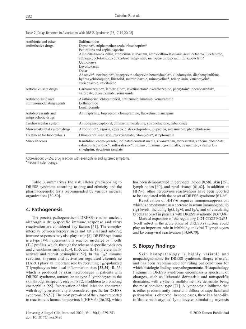
Table 3 summarizes the risk alleles predisposing to DRESS syndrome according to drug and ethnicity and the pharmacogenetic tests recommended by various medical organizations [30-50].
4. Pathogenesis
The precise pathogenesis of DRESS remains unclear, although a drug-specific immune response and virus reactivation are considered key factors [51]. The complex interplay between herpesviruses and antiviral and antidrug immune responses may also play a role [8]. DRESS syndrome is a type IV-b hypersensitivity reaction mediated by T cells (TH2 profile), which, through the release of specific cytokines and chemokines such as IL-4, IL-5, and IL-13, preferentially activate and recruit eosinophils [52]. In this TH2 immune reaction, thymus and activation-regulated chemokine (TARC) plays an important role by recruiting TH2-polarized T lymphocytes into local inflammation sites [53,54]. IL-33, which is produced by skin macrophages in patients with DRESS syndrome, attracts innate type 2 lymphocytes to the skin through its specific receptor ST2, in addition to promoting eosinophilia [55]. Reactivation of viral infection concurrent with drug hypersensitivity is considered specific for DRESS syndrome [56,57]. The most prevalent of the viruses reported to reactivate is human herpesvirus 6 (HHV-6) [56,58], which
has been demonstrated in peripheral blood [8,58], skin [59], lymph nodes [60], and renal tissues [61,62]. In addition to HHV-6, other herpesvirus reactivations have been reported to be associated with the onset of DRESS syndrome [63-66].
Reactivation of HHV-6 requires immunosuppression, which is demonstrated as a decrease in serum immunoglobulin (Ig) levels, including IgG, IgM, and IgA, and of circulating B cells at onset in patients with DRESS syndrome [8,67,68].
Marked expansion of the regulatory CD4+CD25+FOxP3+ T-cell subset in the acute phase of DRESS syndrome could play an important role in inhibiting antiviral T lymphocytes and favoring viral reactivation [14,69,70].
5. Biopsy Findings
Skin h i s topa tho logy i s h igh ly va r iab le and nonpathognomonic for DRESS syndrome. Biopsy is useful and has been recommended for ruling out conditions for which histologic findings are pathognomonic. Histopathology findings in DRESS syndrome encompass a spectrum of changes, such as lichenoid dermatitis and nonspecific dermatitis, with erythema multiforme–like dermatitis being the most dominant type [71]. A lymphocytic infiltrate that is either predominantly dense and diffuse or superficial and perivascular is observed. In some cases, there is a band-like infiltrate with atypical lymphocytes simulating mycosis
Table 2. Drugs Reported in Association With DRESS Syndrome [15,17,19,20,28]
Antibiotic and other Sulfonamides antiinfective drugs Dapsone*, sulphamethoxazole/trimethoprim* Penicillins and cephalosporins Ampicillin/amoxicillin, ampicillin/ sulbactam, amoxicillin-clavulanic acid, cefadroxil, cefepime, cefixime, cefotaxime, ceftazidime, imipenem, meropenem, piperacillin/tazobactam* Quinolones Levofloxacin Other Abacavir*, nevirapine*, boceprevir, telaprevir, benznidazole*, clindamycin, diaphenylsulfone, hydroxychloroquine, linezolid, metronidazole, minocycline*, teicoplanin, vancomycin*, voriconazole, zalcitabineAnticonvulsant drugs Carbamazepine*, lamotrigine*, levetiracetam* oxcarbazepine, phenytoin*, phenobarbital*, valproate, ethosuximide, zonisamideAntineoplastic and Azathioprine, chlorambucil, efalizumab, imatinib, vemurafenib immunomodulating agents Leflunomide LenalidomideAntidepressants and Amitriptyline, bupropion, clomipramine, fluoxetine, olanzapine antipsychotic drugs Cardiovascular system Amlodipine, captopril, diltiazem, mexiletine, spironolactone, tribenosideMusculoskeletal system drugs Allopurinol*, aspirin, celecoxib, dexketoprofen, ibuprofen, metamizole, phenylbutazoneTreatment for tuberculosis Ethambutol, isoniazid, pyrazinamide, rifampicin*, streptomycinMiscellaneous Ranitidine, esomeprazole, iodinated contrast media, rivaroxaban, atorvastatin, codeine phosphate, salazosulfapyridine*, sulfasalazine*, quinine, thiamine, epoetin alfa, cyanamide, vitamin B12 sitagliptin, strontium ranelate
Abbreviation: DRESS, drug reaction with eosinophilia and systemic symptoms.*Frequent culprit drugs.

DRESS Syndrome: Spanish Guidelines
J Investig Allergol Clin Immunol 2020; Vol. 30(4): 229-253© 2020 Esmon Publicidaddoi: 10.18176/jiaci.0480
233
fungoides [1]. Histopathological changes include basket-weave hyperkeratosis, dyskeratosis, lymphocytic exocytosis, and spongiosis. Eosinophils in the dermis or edema may or may not be present.
Histopathology of the skin can highlight various associated inflammatory patterns in a single biopsy [72].
Cutaneous effector lymphocytes comprise a high proportion of polyclonal CD8+ granzyme B+ T lymphocytes [72]. The findings in lymph node biopsies vary from benign reactive hyperplasia induced by viral processes to the presence of atypical lymphocytes that may suggest lymphoma [73]. Liver biopsy reveals an acute hepatitis pattern with lobular inflammation, foci of necrotic hepatocytes, and granulomatous infiltrates with eosinophils. Portal inflammation and cholestasis may also be present [74]. Renal biopsy shows tubulointerstitial nephritis with edema and infiltrates of lymphocytes, histiocytes, eosinophils, and plasma cells [75].
6. Clinical Symptoms
DRESS is characterized by a mixture of symptomatic and asymptomatic features that are variable in both course and time [23,76]. Clinical manifestations often develop 2 to 8 weeks after starting treatment with the causative drug, although rechallenge can result in a reaction within hours to days [23]. Asymptomatic laboratory abnormalities may appear before clinical symptoms. The usual sequence of presentation according to median data in a Spanish DRESS series was fever (11 days), hypogammaglobulinemia (12 days), visceral involvement (20 days), eosinophilia (21 days), and exanthema (23 days) [17].
6.1. Skin Symptoms
Dermatologic manifestations typically begin as a morbilliform eruption that is slightly pruritic and involves the
Table 3. Risk Alleles According to Drug, Ethnicity, and Screening Prevention Strategies
Causative Drug Ethnicity Allele (NPV) Pharmacogenetic Testing Recommendations
Allopurinol Han Chinese, Korean, HLA B* 58:01 (100%) American College of Rheumatology and Clinical Japanese, Thai [41,42] Pharmacogenetics Implementation Consortium (CPIC) [36], Taiwanese Department of Health [43] None from FDA European HLA B* 58:01 (63%) Not recommended for Caucasian population Portuguese population [44,45]Carbamazepine Northern European; HLA-A* 31:01 Warning about possible association [42] Japanese; Korean [43,46] (no FDA recommendation) Spanish Caucasian [47] Canadian Pharmacogenomics Network for drug safety recommends pharmacogenetics testing in patients of all ancestries [48]. Caucasian [43] 8.1 AH (HLA A*01:01: None Cw*07:01: B*08:01: DRB1*03:01: DQA1*05:01: DQB1*02:01) Lamotrigine and Spanish Caucasian [47] HLA-A* 24:02 None phenytoin Salazosulfapyridine Chinese Han (49] HLA B* 13:01 Dapsone Han Chinese patients HLA-B*13:01 None treated for leprosy [43] [50] Piperacillin/tazobactam English population [51] HLA- B62 NoneAbacavira US [52,53] HLA-B* 57:01 (100%) Screening FDA population of risk [41,43] European, African [54] Nevirapine /HIVa Asian: Han Chinese, Thai [55] HLA-B* 35:05 None Italian (Sardinian) [56]; Cw*8 or Cw*08-B*14 None Japanese [57] haplotype Australian HLA-DRB1*01:01; None B35:01 [58] Vancomycin North American HLA-A*32:01 [59] None
Abbreviations: FDA, United States Food and Drug Administration; HLA, human leukocyte antigen; NPV, negative predictive value.aAbacavir and nevirapine hypersensitivity do not completely fit the major criteria for DRESS syndrome.
Cabañas R, et al.
J Investig Allergol Clin Immunol 2020; Vol. 30(4): 229-253 © 2020 Esmon Publicidaddoi: 10.18176/jiaci.0480
234
face, neck, upper extremities, and trunk, progressing towards diffuse, confluent, and infiltrated erythema. The rash can become edematous and includes purpuric lesions, pustules, and even vesicles or bullae in certain cases [19,77]. If the drug is not withheld, the rash may progress to erythroderma or exfoliative dermatitis.
The cutaneous phenotype in DRESS syndrome can be categorized as an urticarial papular exanthem, morbilliform erythema, exfoliative erythroderma, or erythema multiforme–like lesions, which in DRESS syndrome may be prognostic of more severe liver involvement [78]. Skin involvement in DRESS syndrome usually affects more than 50% of the body surface area (BSA). Facial edema usually appears in the periorbital and midfacial region and is symmetric and persistent. Disfiguring facial swelling is recorded in 25% of patients [2]. Mild mucosal involvement (50% of patients) usually involves a single site, most often the mouth or pharynx, and in 15% of cases more than 1 mucous membrane is affected [23].
6.2. Systemic Symptoms
Fever is seen in >90% of patients and generally precedes cutaneous eruptions by several days. It is usually high (>38°C) and spiking [79].
Internal organ involvement occurs in 85%-96% of patients and determines severity; in 50% to 60% of patients, 2 or more organs are involved [18,23,77,80,81]. Any internal organ can be affected. The most common findings are abnormalities of the lymphatic system, blood, and liver followed by the kidneys, heart, and lungs. Severe cases can result in neurologic, gastrointestinal, and endocrine dysfunction [2].
Lymphadenopathy (30%-60% of cases) is often diffuse, with slightly enlarged and tender nodes at various locations [18].
Although any medication can affect any organ, certain drugs have a predilection for specific organs [82].
Liver involvement is frequent in DRESS syndrome (75% of cases) [23]. Hepatosplenomegaly may be present, although involvement is more often asymptomatic and detected in routine liver function tests. While the cholestatic type is the most common, mixed or hepatocellular types can also be detected [80]. DRESS-related liver injury manifests as reversible abnormal liver function results only, although hepatic necrosis may also be found and can lead to liver failure requiring transplantation and even lead to death [80]. Liver involvement is the leading cause of death from DRESS syndrome [18,80,81,83,84].
Renal involvement is found in 10% to 30% of cases, more often in those induced by allopurinol, followed by carbamazepine and dapsone [22]. Older age and preexisting alterations of renal function may be predisposing factors [85-87].
Lung manifestations appear in 5% to 25% of cases, with minocycline being the most common drug affecting the lungs. Respiratory complications include acute interstitial pneumonitis, lymphocytic interstitial pneumonia, pleuritis, and acute respiratory distress syndrome [2,86-88].
Involvement of the heart can take the form of eosinophilic myocarditis or pericarditis, with minocycline and ampicillin being the most frequent culprit drugs [2]. Myocarditis is potentially fatal and can appear months after resolution of the laboratory abnormalities. Patients may present with chest pain, tachycardia, dyspnea, and hypotension [89].
The most frequent gastrointestinal manifestation is gastroenteritis; mucosal erosions can develop and contribute to acute bleeding. Gastrointestinal complications include chronic protein-losing enteropathy, colitis, and pancreatitis [2,87,90].
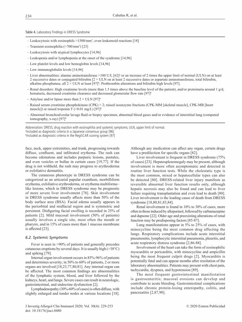
Table 4. Laboratory Findings in DRESS Syndrome
– Leukocytosis with eosinophils >1500/mm3, even leukemoid reactions [18] – Transient eosinophilia (>700/mm3) [23]– Leukocytosis with atypical lymphocytes [14,96] – Leukopenia and/or lymphopenia at the onset of the syndrome [14,96] – Low platelet levels and low hemoglobin levels [14,96]– Low immunoglobulin levels [14,96]– Liver abnormalities: alanine aminotransferase >100 U/L [62]a or an increase of 2 times the upper limit of normal (ULN) on at least
2 successive dates or conjugated bilirubin [2 × ULN on at least 2 successive dates or aspartate aminotransferase, total bilirubin, alkaline phosphatase, all 2 × ULN at least [97]b. Prothrombin alterations and bilirubin high levels [97].
– Renal disorders: High creatinine levels (more than 1.5 times above the baseline level of the patient), and/or proteinuria around 1 g/d, hematuria, decreased creatinine clearance and decreased glomerular flow rate [97]b
– Amylase and/or lipase more than 2 × ULN [97]b – Raised serum creatinine phosphokinase (CPK) > 2; raised isoenzyme fractions (CPK-MM [skeletal muscle], CPK-MB [heart
muscle]) or raised troponin T (>0.01 mg/L) [97]b
– Abnormal bronchoalveolar lavage fluid or biopsy specimen, abnormal blood gases and/or evidence of interstitial lung (computed tomography, x-ray) [97]b
Abbreviation: DRESS, drug reaction with eosinophilia and systemic symptoms; ULN, upper limit of normal.aIncluded as diagnostic criteria in a Japanese consensus group [96]bIncluded as diagnostic criteria in the RegiSCAR scoring system [97]

DRESS Syndrome: Spanish Guidelines
J Investig Allergol Clin Immunol 2020; Vol. 30(4): 229-253© 2020 Esmon Publicidaddoi: 10.18176/jiaci.0480
235
Brain disorders are unusual in DRESS syndrome and include encephalitis, meningitis [87,91], and even cerebral vasculitic-like lesions [92].
Endocrine disorders are rare in the acute phase, being more frequent as long-term sequelae and affecting the thyroid. Pancreatic involvement ranges from pancreatitis to type 1 diabetes mellitus that can develop 3 weeks to 10 months after the onset of DRESS syndrome [2,18,87,93].
Additional manifestations such as myositis, peripheral nerve disorders, uveitis, and salivary gland inflammation may be present [18,87].
Rare cases of shock and multiple organ failure have also been reported [94,95].
6.3. Laboratory Findings
The laboratory findings for DRESS syndrome are shown in Table 4.
7. Prognosis and Outcome
The outcome of DRESS is often unpredictable. Early diagnosis and prompt withdrawal of the culprit drug are often followed by complete recovery [18]. Some culprit drugs, such as allopurinol and anticonvulsants, are associated with a poorer prognosis, and others, such as antibiotics, are associated with a better prognosis [98,99].
A lower BSA affected and milder skin and mucosal involvement correlate with a better prognosis [18].
Severe l iver injury and presence of a typical lymphocytes [99], as well as reactivation of herpesvirus, especially HHV-6 [62], and reactivation of cytomegalovirus (CMV) are associated with a worse prognosis. The Mizukawa scoring system was developed to predict CMV disease and complications and to ensure early intervention with anti-CMV agents [100]. In cases of reactivation of herpesvirus, patients may go on to develop autoimmune disease [58,69], even after resolution of the syndrome.
Serum TARC/CCL17 levels are elevated during acute DRESS syndrome, and TARC/CCL17 has been proposed as a prognostic and diagnostic biomarker [54,101].
The concentration of serum soluble ST2 (an innate type 2 lymphocyte-specific receptor) was proposed as a biomarker of disease, as it correlated with IL-33 and alanine aminotransferase levels at the onset of DRESS syndrome [55].
Elevations in TNF-α and TARC/CCL17 levels during the early stages of the disease enable early recognition of reactivation of HHV-6 [102]. TNF-α and TARC levels also reflect therapeutic responses and may be useful markers of the course of DRESS syndrome [102].
8. Clinical Diagnosis
8.1. When Should We Suspect DRESS Syndrome?
DRESS syndrome should be suspected in any patient under treatment with a new drug initiated in the previous 2-8 weeks who presents any combination of the following: skin eruption
Table 5. Recommended Laboratory Investigations in Patients With Suspected DRESS Syndrome and in Their Follow-up [5,8,19,23,104].
On Admission Follow-up in Acute Phase (at least 2 times/wk) According to Initial Blood Abnormalities and Clinical Course
Complete differential Blood Count including evaluation of atypical lymphocytes -Inflammation markers (CRP, LDH) -Liver function tests (AST, ALT, GGT, alkaline phosphatase, total bilirubin) (Repeat in follow-up if liver involvement) prothrombin time/INRKidney function tests (creatinine, serum urea, urine albumin-to-creatinine ratio (Repeat in follow-up if kidney involvement) or protein-to-creatinine ratio, urine sediment, urinary protein and cells)Other: Blood electrolytes: sodium, potassium. Lipase, amylase Creatine kinase Troponin I Proteinogram and immunoglobulins Herpes virus serology and PCR for HHV-6, HHV-7, CMV, EBVa Exclusion of alternative diagnosis Serology for mycoplasma, chlamydia, HAV, HBV, HCV, parvovirus B19, VHS 1/2. Blood culture Antinuclear antibodies
Abbreviations: ALT, alanine aminotransferase; AST, aspartate aminotransferase; CRP, C-reactive protein; GGT, γ-glutamyl transpeptidase; INR, international normalized ratio; LDH, lactate dehydrogenase; PCR, polymerase chain reaction; HSV, herpes simplex virus. aTesting for herpesvirus infection should be performed at admission and repeated one or more times at 2- to 3-week intervals to detect a change in the antibody titer [5,8].

Cabañas R, et al.
J Investig Allergol Clin Immunol 2020; Vol. 30(4): 229-253 © 2020 Esmon Publicidaddoi: 10.18176/jiaci.0480
236
(mainly if facial edema is associated), fever, lymphadenopathy, eosinophilia, atypical lymphocytes, and signs of liver or kidney involvement. Suspicion will be higher if the patient is on treatment with drugs known to induce DRESS syndrome frequently (Table 2) [1,19,103] (LE3, GRD).
8.2. Which Laboratory Investigations Should Be Performed for the Diagnosis and Follow-up of DRESS Syndrome?
The authors of these guidelines recommend a series of laboratory investigations for diagnosis, assessment of severity, and follow-up (Table 5) and skin biopsy whenever DRESS syndrome is suspected [5,8,19,23,104] (LE4 expert opinion consensus, GRD).
Additional tests that can be performed according to the patient’s symptoms include abdominal ultrasonography, chest x-ray, EKG, echocardiography, computed tomography scan of the brain, neurological evaluation, pulmonary function testing, computed tomography scan of the chest, and evaluation by various specialists (eg, nephrologist, hepatologist, and cardiologist) [79].
8.2.1. Evaluation of kidney injury
Laboratory investigations are recommended for the assessment of renal function and kidney disease (Table 5). Acute kidney injury should be assessed and its severity staged according to the KDIGO clinical practice guideline [105] (LE4 expert opinion consensus, GRD) (Table 6)
8.2.2. Evaluation of liver injury
Liver function tests should be performed (Table 5). Liver injury should be assessed and severity staged according to the DILI Expert Working Group [106] (Table 7) (LE4 expert opinion consensus, GR D).
8.3. Confirmation of a Diagnosis of DRESS Syndrome: Scoring Systems
Different diagnostic scores have been developed to help clinicians to confirm or exclude DRESS syndrome [1,8,19].
RegiSCAR devised a scoring system for DRESS syndrome that is widely accepted and is shown in Table 8 [19]. This group has published a document on the practical application of the diagnostic score, including the specifics for evaluation of the diagnostic features of DRESS syndrome [97]. We strongly recommend the use of the RegiSCAR scoring system for diagnosing DRESS syndrome (LE3, GRD) (Table 8).
8.4. Differentiating DRESS Syndrome From Other Cutaneous and Systemic Diseases and Other SCARs
The differential diagnosis should be made with other diseases that may present with skin rash, systemic symptoms, adenopathy, and fever. These include other SCARs (SJS/TEN and AGEP) (Table 9), bacterial and viral infections (Epstein-Barr virus, CMV, measles, hepatitis virus, influenza virus, parvovirus, and HIV) [1,5,57,67,107]. Other conditions that should also be taken into account include autoimmune diseases (eg, Kikuchi-Fujimoto syndrome, Kawasaki syndrome [108],
Table 6. Staging of Acute Kidney Injury for Severity [105]
Stage Serum Creatinine Urine Output
1 1.5-1.9 times baseline <0.5 mL/kg/h for OR 6-12 h ³0.3 mg/dL (³26.5 µmol/L) increase2 2.0-2.9 times baseline <0.5 mL/kg/h for ³12 h3 3.0 times baseline <0.3 mL/kg/h for OR ³24 h increase in serum creatinine to ³4.0 mg/dL (³353.6 µmol/L) OR Initiation of renal replacement therapy OR In patients <18 y, decrease in eGFR to <35 mL/min per 1.73 m2
Table 7. Clinical Chemistry Criteria for Drug-Induced Liver Injury (DILI) and Staging DILI. Modified from Aithal, Clinical Pharmacology & Therapeutics 2011 [106]
DILI Severity IndexDegree of Severity1. MILD – Elevated alanine aminotransferase (ALT)/alkaline phosphatase (ALP) concentration reaching criteria for DILIa but bilirubin concentration <2× upper limit of normal (ULN).2. MODERATE – Elevated ALT/ALP concentration reaching criteria for DILIa and bilirubin concentration ≥2× ULN, or symptomatic hepatitis.3. SEVERE – Elevated ALT/ALP concentration reaching criteria for DILIa, bilirubin concentration ≥2× ULN, and one of the following: - International normalized ratio ≥1.5 - Ascites and/or encephalopathy, disease duration <26 wk, and absence of underlying cirrhosis - Other organ failure considered to be due to DILI 4. FATAL OR TRANSPLANTATION – Death or transplantation due to DILI – Level of evidence, 2b (exploratory/retrospective cohort studies)
aDILI if any of the following:≥5× ULN for ALT≥2× ULN for ALP (particularly with accompanying elevations in concentrations of 5′-nucleotidase or γ-glutamyl transpeptidase in the absence of known bone diseasedriving the rise in ALP level)>3× ULN for ALT and total bilirubin exceeding 2× ULN

DRESS Syndrome: Spanish Guidelines
J Investig Allergol Clin Immunol 2020; Vol. 30(4): 229-253© 2020 Esmon Publicidaddoi: 10.18176/jiaci.0480
237
Still disease, and acute cutaneous lupus erythematosus), hypereosinophilic syndromes [109], Sézary syndrome, and angioimmunoblastic T-cell lymphoma [27,110].
Taking photographs of the skin lesions and the whole body surface (to evaluate the BSA affected) is of the utmost importance. Sending the images to an expert center may facilitate an earlier diagnosis. This approach also allows a better retrospective evaluation and validation of the case.
It is important not to forget the overlap between SCARs. These cases, while very rare, fulfil the criteria for a definitive or probable diagnosis of at least 2 of AGEP, DRESS syndrome, and SJS-TEN [96,103,113]. An overlap between maculopapular exanthema and DRESS syndrome has also been identified and characterized [114].
Drug-induced eosinophilia may occur with or without other manifestations of adverse drug reactions, such as exanthema or drug fever. Eosinophilia alone requires close observation, because resolution usually occurs within a week or two of drug cessation [17].
9. Identifying the Culprit Drug
9.1. Assessment of Causality Using the Spanish Pharmacovigilance System Algorithm
Many methods have been proposed to assess the causal relationship between an adverse event and a medication taken by a patient [115-117]. The parameters evaluated in the algorithm of the Spanish Pharmacovigilance System
(ASPS) [118] are shown in Table 10. The final case evaluation of each drug is listed as not related (improbable, conditional) or related (possible, probable, or definit).
Whenever possible, we must interview the patient and/or their relatives to obtain more details of all the drugs taken, including over-the-counter drugs and the consumption of herbal or homeopathic products, and dechallenge or rechallenge information (if available). All drugs taken during exposure windows must be recorded (including chronology of drug intake, dose, indication, and clinical course after withdrawal).
The chronology is considered suggestive if the drug was initiated less than 6 months previously and stopped less than 14 days before the index day [23]. The index day is considered to be the day on which prodromal symptoms/signs first occurred, or in their absence, the day of acute rash [17].
We strongly recommend calculating the index day and performing a causality assessment according to the ASPS criteria (Table 10) as soon as DRESS syndrome is suspected (with a score of at least “possible” according to the RegiSCAR criteria shown in Table 8). All drugs in the category of “possible to definite” should be stopped and prohibited provisionally (LE4 expert opinion consensus, GRD).
When a drug is classed as being associated with DRESS syndrome in Spain, a complete adverse reaction report must be submitted to the pharmacovigilance center of the Autonomous Community in order to conduct a second evaluation and to be included in the Spanish Pharmacovigilance System Registry.
Table 8. RegiSCAR Validation Score for DRESS Syndrome 2007 [19]a
Score -1 0 1 2 Min Max
Fever ≥38.5 (core) or >38ºC (axillary) N Y –1 0Enlarged lymph nodes (>1 cm size, at least 2 sites) N/U Y 0 1Eosinophilia N/U 700-1499/µL ≥1500/µL 0 2 10%-19.9% ≥20% (if leukopenia) (if leukopenia) Atypical lymphocytes N/U Y 0 1Skin involvement – Rash extent (%BSA) N/U >50% – Rash suggesting DRESS (≥2 of facial edema, N U Y –2 2 purpura, infiltration, desquamation) – Biopsy suggesting DRESS N Y/UOrgan involvement L/K/Lu/M-H/Pa/Other N/U Y/Y/Y/Y/Y/Y* 0 2Resolution >15 days N Y –1 0Evaluation of other potential causes: Y (None [+] and at least 3 [– ]) – Serology for HAV/HBV/HCV; blood culture; 0 1 – Antinuclear antibody; Chlamydia/MycoplasmaTotal score <2, Excluded; 2-3, Possible; 4-5, Probable; –4 9 >5, Definite
Abbreviations: N, no; Y, yes; U, Unknown; L, liver; K, kidney, Lu, lung; M, muscle; H, heart; Pa, pancreas. aSee reference [97] and text for details about evaluation of organ involvement

Cabañas R, et al.
J Investig Allergol Clin Immunol 2020; Vol. 30(4): 229-253 © 2020 Esmon Publicidaddoi: 10.18176/jiaci.0480
238
Tabl
e 9.
Diff
eren
tial D
iagn
osis
of D
RESS
, SJS
/TEN
, AG
EP, a
nd O
ther
Cut
aneo
us D
iseas
esa
DR
ESS
SJS/
TEN
A
GEP
H
yper
eosi
noph
ilic
Kaw
asak
i Dis
ease
St
ill D
isea
se
Synd
rom
e
Sy
ndro
me
Typi
cal o
nset
2-
6 w
k 1-
3 w
k H
ours
to 2
d (a
ntib
iotic
s)
of e
rupt
ion
4-12
d (o
ther
dru
gs)
Cut
aneo
us
Faci
al e
dem
a D
usky
red,
coa
lesc
ent
Seve
ral d
ozen
small
non
folli
cular
U
rtica
ria,
Con
junc
tival
invo
lvem
ent,
Sa
lmon
col
ored
le
sion
s M
orbi
llifo
rm/
mac
ular
exa
nthe
ma
pust
ules
on
a ba
ckgr
ound
of
angi
oede
ma,
cl
eft p
alat
e, st
raw
berr
y bu
mpy
rash
m
acul
opap
ular
A
typi
cal t
arge
t les
ions
w
ides
prea
d ed
emat
ous
mor
billi
form
to
ngue
, pal
mar
ery
them
a,
exan
them
a w
ith
Bul
lous
and
blis
ter l
esio
ns
eryt
hem
a.
exan
them
a,
hand
ede
ma,
per
iung
ual
scal
ing
M
ucos
al in
volv
emen
t Pr
edom
inan
t in
mai
n sk
info
lds.
papu
lar o
r nod
ular
de
squa
mat
ion,
and
Eryt
hrod
erm
a/
in n
early
all
case
s M
ucos
al in
volv
emen
t rar
e.
infil
trate
po
lym
orph
ous r
ash
exfo
liativ
e de
rmat
itis
Nik
olsk
y (+
) N
o bl
iste
rs
Rar
e bl
iste
rs
Epid
erm
al n
ecro
sis:
N
ikol
sky
(–)
Rar
e pu
stul
es
- <1
0%: S
JS
Muc
osal
lesi
ons
- 10
%-3
0%: o
verla
p
in
freq
uent
-
SJS/
TEN
Pu
rpur
ic e
rupt
ion
- >3
0% T
EN
Infil
trate
d er
ythe
ma
Nik
olsk
y (–
)Sk
in b
iops
y N
onsp
ecifi
c:
Epid
erm
al n
ecro
sis w
ith
Subc
orne
al a
nd/o
r int
raep
ider
mal
Ly
mph
ocyt
ic in
filtra
te.
full
thic
knes
s los
s of
pust
ules
and
per
ivas
cula
r
Eo
sino
phils
, der
mal
ep
ider
mis
in
filtra
te w
ith n
eutro
phils
and
ed
ema
may
be
pres
ent
ed
ema
of th
e pa
pilla
ry d
erm
is
Hem
atol
ogic
al
abno
rmal
ities
-
Eosi
noph
ilia
Yes
No
No
Yes (
>150
0/µL
) N
o Po
ssib
le
- A
typi
cal
Yes
No
No
Poss
ible
N
o N
o
lym
phoc
ytes
Le
ukoc
ytos
is w
ith n
eutro
phili
a
(>70
00/m
m3 )
Syst
emic
in
volv
emen
t -
Lym
phad
e-
Yes
No
No
Yes
Yes
Yes
no
path
y -
Hep
atiti
s Ye
s Ye
s Po
ssib
le b
ut ra
re
Yes
Poss
ible
Po
ssib
le
- O
ther
org
ans
Inte
rstit
ial n
ephr
itis,
Tu
bula
r nep
hriti
s,
Inte
rnal
org
an in
volv
emen
t Ca
rditi
s, pn
eum
oniti
s, Ca
rdio
vasc
ular
abno
rmal
ity,
Pleu
risy,
pn
eum
oniti
s, ca
rditi
s,
trach
eobr
onch
ial n
ecro
sis
Tran
sien
t pre
rena
l fai
lure
en
ceph
alop
athy
, di
arrh
ea, v
omiti
ng, a
nd
peric
ardi
tis
Panc
reat
itis,
diar
rhea
, vom
iting
, ab
dom
inal
pai
n
bo
ne m
arro
w
and
abdo
min
al p
ain
a Mod
ified
from
Fer
nand
ez e
t al.
Trat
ado
de A
lerg
olog
ía 2
016
[1,1
2,23
,111
].Ab
brev
iatio
ns: A
GEP
, acu
te g
ener
alize
d ex
anth
emat
ous
pust
ulos
is; D
RESS
, dru
g re
actio
n w
ith e
osin
ophi
lia a
nd s
yste
mic
sym
ptom
s; SJ
S/TE
N, S
teve
n-Jo
hnso
n sy
ndro
me/
toxi
c ep
ider
mal
nec
roys
is.
DRESS Syndrome: Spanish Guidelines
J Investig Allergol Clin Immunol 2020; Vol. 30(4): 229-253© 2020 Esmon Publicidaddoi: 10.18176/jiaci.0480
239
9.2. Causality Assessment by the Allergist
9.2.1. Clinical History
A detailed clinical history is an essential first step towards an accurate diagnosis of DRESS syndrome [119]. The history must be meticulous, with the full medical background of the patient and the family history of SCARs.
A timeline chart should be constructed to bring together signs and symptoms (eg, fever, eosinophilia, cutaneous symptoms, increase in transaminases), time of onset and resolution, and all of the drugs taken with a latency period that is compatible with DRESS syndrome (ie, initiated less than 6 months previously and stopped less than 14 days
before the index day). The details recorded of the drugs administered should include formulation, dose, route, timing of administration and effect of stopping treatment [23]. This approach and the result of applying the ASPS (Table 10) will enable us to identify the suspect drugs with which to perform our allergy work-up. It is important to take into account that in 18% of cases of DRESS syndrome in the multicenter study by Barbaud et al [120] there were at least 2 different culprit drugs.
9.2.2. Assessment of Causality Using In Vitro Allergy Tests
In vitro diagnostic tests have the advantage over in vivo tests of being absolutely safe. They are based on the property of antigen-specific T cells being activated upon stimulation with the nominal antigen in sensitized patients [121]. They should not be performed before a minimal time interval of 4-8 weeks after the reaction and at least 4 weeks after stopping treatment with systemic corticosteroids. Analysis in the first 6 months to 1 year is recommended, although subsequent test results may be positive [122].
A positive result reflects specific sensitization to the test drug, which is a risk factor but does not prove causality. However, it can support the diagnosis and pinpoint the responsible agent if the patient took several drugs.
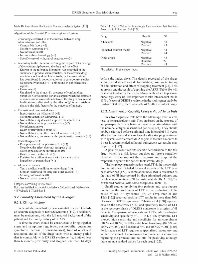
The lymphocyte transformation test (LTT) is the most widely used in vitro test. Detailed technical aspects of the LTT have been described [122]. A stimulation index (SI) is calculated as the ratio of 3H incorporated by drug-stimulated cultures and baseline incorporation of 3H by unstimulated cells. An SI ≥2 is considered positive, with some exceptions (Table 11).
Small studies involving few patients and case reports pointed to the usefulness of LTT in the evaluation of the cause of DRESS syndrome [98,123-129]. Pichler and Tilch [122] reported positive LTT results in more than 50% of cases of DRESS syndrome. Cabañas et al [130] reported data on the sensitivity (73%) and specificity (82%) of LTT in the recovery phase of DRESS syndrome in a series of 41 patients. Comparison of skin tests and LTT confirms a higher sensitivity and specificity of LTT in DRESS syndrome. LTT showed high sensitivity and specificity for anticonvulsants (100% and 100%; P=.008), antituberculosis drugs (87.5% and 100%; P=.004), and ß-lactams (73% and 100%; P=.001) [130]. Performance of LTT requires a specialized laboratory and skilled personnel. Laboratories have reached a consensus regarding the protocol and cut-offs for positivity, although there are no standard values for each drug [122].
Table 10. Algorithm of the Spanish Pharmacovigilance System [118]
Algorithm of the Spanish Pharmacovigilance System
– Chronology, referred to as the interval between drug administration and effect: - Compatible (score +2) - Not fully supported (+1) - No information (0) - Incompatible chronology (–1) - Specific case of withdrawal syndrome (+2). – According to the literature, defining the degree of knowledge of the relationship between the drug and the effect: - Known in the reference literature (+2): recorded in the summary of product characteristics, or the adverse drug reaction was found in clinical trials, or the association has been found in cohort studies or in case-control studies. - Occasionally known (+1): only found in published case reports. - Unknown (0) - Unrelated to the drug (–1): presence of confounding variables. Confounding variables appear when the estimate of a measure of association between the drug exposure and health status is distorted by the effect of ≥1 other variables that are also risk factors for the outcome of interest.– Evaluation of drug withdrawal: - Improvement on withdrawal (+2) - No improvement on withdrawal (–2) - Not withdrawing does not improve the effect (+1) - Not withdrawing improves the effect (–2) - No information (0) - Death or irreversible effect (0) - Not withdrawn, but there is a tolerance effect (+1) - Not withdrawn, improves with symptomatic treatment (+1)– Rechallenge effect: - Reappearance of the positive effect (+3) - Negative, the effect does not reappear (–1) - No re-exposure or no information (0) - Death or irreversible effect (0) - Positive for a different agent with the same active ingredient or parent drug (+1)– Alternative causes: - Yes, a medical condition or other drugs (–3). - Similar likelihood for drug and other causes (–1) - Missing information (0) - No alternative cause (+1)
Table 11. Cut-off Values for Lymphocyte Transformation Test Positivity According to Pichler and Tilch [122]
Drug Result SI
ß-Lactams Negative <3 Positive >3Iodinated contrast media Negative <4 Positive >4Other drugs Negative <2 Doubtful 2-3 Positive >3
Abbreviation: SI, stimulation index.
Categories according to final score:Not classified (lack of data) /Improbable <0/Conditional 1-3/Possible 4-5/Probable 6-7/Definite>8

Cabañas R, et al.
J Investig Allergol Clin Immunol 2020; Vol. 30(4): 229-253 © 2020 Esmon Publicidaddoi: 10.18176/jiaci.0480
240
The fluorescent dye 5,6-carboxyfluorescein diacetate succinimidyl ester may be used as an alternative to the radioactive label for LTT; however, very few reports have been published, and there is no consensus on the analysis of the SI [123,131]. No agreement has been reached on the cut-off for positivity in flow cytometry analysis of CD69 upregulation, as few cases have been published [132].
The analysis of cytokine release by enzyme-linked immunosorbent assay or enzyme-linked immunospot assay (ELISPOT) upon drug-induced stimulation of peripheral blood mononuclear cells is also used by several laboratories. However, no consensus exists on the protocol or criteria for positivity. Positive ELISPOT assays for IFN- production have been reported in DRESS syndrome [50,133-135].
At present, LTT is the best documented assay for in vitro diagnosis of DRESS syndrome [136,137]; the ENDA/EAACI Drug Allergy Interest Group position paper indicates that it might be advisable to perform LTT before in vivo tests in severe reactions with a suspected T-cell mechanism [136].
We strongly recommend that LTT and/or ELISPOT should be available in reference centers managing DRESS syndrome to identify the culprit drug. The tests should be performed before skin tests and incubation with all the drugs indicated by the allergist and with the category of “possible to definite” according to the ASPS [136,137] (LE3, GRD)
9.3.3. Assessment of Causality Using In Vivo Allergy Tests
Skin tests (mainly delayed intradermal reaction) and patch tests are of value in the investigation of T cell–mediated hypersensitivity reactions such as DRESS syndrome [120,138-140].
9.3.3.1. Patch testsPatch tests can prove helpful, with positive results
reported in 32% [141] to 64% of cases in a French multicenter study [120]. Positivity depends to a large extent on the drug. Patch tests are very useful with anticonvulsants [120,141], antibiotics (highest reactivity to ß-lactams and quinolones) [120,139], and proton pump inhibitors [120]. Patch tests always yield negative results with sulfasalazine and allopurinol [120,141].
Results from the largest patch testing series in DRESS syndrome showed this to be a safe procedure with no adverse reactions [120,141-143].
Patch tests should be performed according to European guidelines for skin tests [138,144-146] for a minimum of 4 to 6 weeks after the acute reaction [147] and 4 weeks after stopping treatment with systemic corticosteroids or immunosuppressive therapy [144,148]. Topical corticosteroids should not be applied to the patch test area in the week before the test [144]. Patch testing should be performed 2-6 months after recovery [120] (LE3, GRD).
All culprit drugs suspected according to the clinical history, especially those with the ASPS category of “related”, should be included (LE4 expert opinion consensus, GRD). Testing of chemically or pharmacologically related drugs may provide information on cross-reactivity [133,149,150], and testing available metabolites may improve the results [149] (LE3, GRD).
We recommend using a 10% concentration in petrolatum with the active ingredient. When the commercialized form of the drug is used, it is recommended to test up to a 30% concentration of the final product [120,144] (LE3, GRD).
Concentrations and vehicles previously considered as most adequate for certain drugs should also be chosen. As for ß-lactams, European guidelines and the authors of the present guidelines suggest a concentration of 5% in petrolatum [151,152]. A list with drug concentrations and vehicles used in reported cases of DRESS syndrome is provided in Supplementary File 1 of the online material.
The authors of these guidelines strongly recommend not testing different concentrations of the same suspect drug or using different vehicles simultaneously for safety reasons, because systemic reactions reported after patch tests were the result of such an approach [111,133,153,154] (LE3, GRD).
Special caution is recommended in immunocompromised patients [153,155,156], and testing should start at lower concentrations (LE3, GRD).
Patch tests should be preceded by LTT or performed as the first-line approach if in vitro tests are not available [136] (LE3, GRD).
9.3.3.2. Prick and intradermal tests
If a patch test yields a negative result and a suitable injectable form is available, then prick testing should be performed. If this is negative, subsequent intradermal testing is recommended [98,120,138,157] (LE3, GRD).
Delayed positive reactions to skin prick tests have occasionally been described in patients with DRESS syndrome [120]. Immediate readings should be taken at 20 minutes and delayed readings at 6 and 24 hours according to the European guidelines [145]. The higher sensitivity of intradermal tests compared with patch tests has been reported [120], mainly in reactions to ß-lactams [158].
Although recent studies and case reports support the safety of prick and intradermal tests in DRESS syndrome [42,98,120,157,159], isolated systemic reactions after intradermal tests [120] and prick testing have been reported in HIV-infected patients [155].
Therefore, we recommend that for intradermal testing, the drug should be initially administered at the highest dilution (usually 1/100 of the skin prick test concentration) [145,151], the interval between tests should be extended [145], different concentrations should not be tested on the same day, and special precautions should be adopted with HIV-infected patients [155] (LE3, GRD).
More precise guidelines have been drafted for nonimmediate ß-lactam reactions including SCARs [145,151,158].
9.3.3.3. Controlled re-exposure Test
Since DRESS syndrome is a severe and sometimes life-threatening condition, challenge testing with the suspected culprit drug and cross-reactive drugs is contraindicated [138,147,160].
A search of the literature reveals cases of DRESS syndrome induced by ß-lactams [159,161,162], amikacin [133], and antituberculosis drugs [155,163] in which controlled re-exposure tests were performed under special circumstances.

DRESS Syndrome: Spanish Guidelines
J Investig Allergol Clin Immunol 2020; Vol. 30(4): 229-253© 2020 Esmon Publicidaddoi: 10.18176/jiaci.0480
241
Controlled re-exposure tests with ß-lactamsWe recommend controlled exposure testing with an
alternative ß-lactam (not the culprit) if the benefit outweighs or at least equals the risk. This approach should be guided by the allergy study [98,159,161] (LE3, GRD).
The graded challenge exposure test recommended by Romano et al [158] for nonimmediate ß-lactam allergic reactions is an initial dose of 1/100 of the therapeutic one. In cases with negative results 3 days to 1 week later, a dose of one tenth is given and, if the result is again negative, a full dose can be given after the same interval as used before. We recommend this approach if controlled exposure testing is indicated and with clinical and laboratory monitoring (LE4 expert opinion consensus, GRD).
Controlled re-exposure tests with antituberculosis drugsIn special cases of DRESS syndrome induced by 3 or
4 first-line antituberculosis drugs, challenge testing may be indicated for adequate management of tuberculosis. The availability of in vivo and ex vivo testing to guide rechallenge choices would be extremely helpful in these settings [147]. Two main series of DRESS syndrome induced by antituberculosis drugs have been reported [155,163]. In both series, all antituberculosis drugs were stopped until normalization of skin findings and laboratory values, and then careful re-exposure to each drug was performed independently [155,163]; in the series of Lehloenya et al [155] in particular, this approach was followed after performing allergy tests.
In reference to rechallenge with drugs in patients with SCARs induced by HIV and antituberculosis drugs in low- and middle-income countries, a recent international consensus document [147] stated that if the risk of morbidity and mortality from the disease outweighs or at least equals the risks from the drug reaction, the risk-benefit ratio sways toward sequential rechallenge with potentially implicated drugs. Allergy testing to guide rechallenge choices would be extremely helpful [147] (LE3, GRD).
Given the high specificity and sensitivity of LTT [130,164], we recommend this approach with antituberculosis drugs as a first step in the management of DRESS syndrome induced by these agents followed by patch tests and prick and intradermal tests according to the previous results (LE4 expert opinion consensus, GRD).
Controlled exposure tests should be performed after consulting with an infectious disease specialist if there are no adequate second-line alternatives and guided by the negative results in the allergy tests. The rechallenge should be sequential and cumulative when symptoms resolve and laboratory parameters return to normal [155,163]. Clinical and biological surveillance (temperature and blood tests) should be performed before each administration (LE4 expert opinion consensus, GRD).
As for doses, we recommend the criteria of the French Investigators of Skin Adverse Reactions to Drugs [165], ie, to rechallenge with 1 drug each time, starting with 10-2 on day 1, 10-1 on day 3, a full dose on day 5, and treatment on day 7 (LE4 expert opinion consensus, GRD).
As for other groups of drugs, the authors of the present guidelines recommend controlled re-exposure tests only when different drugs are involved in the reaction and after negative in vitro tests, if available, and in vivo tests, considering that the benefit of treatment with the drug outweighs or at least equals the risk of morbidity and mortality from the drug reaction (LE4 expert opinion consensus, GRD).
Careful risk-benefit assessment in discussion with the patient and informed consent is strongly recommended (LE4 expert opinion consensus, GRD).
We suggest beginning at 10-5 to 10-3 of the full dose and gradually increasing 10-fold with an interval of 3 days to 1 week at 10-2 and 10-1 until the full dose is reached with clinical and laboratory monitoring before each dose. The drug can be reauthorized if the results are negative (LE4 expert opinion consensus, GRD).
Figure 1. Management of DRESS in the acute phase. DRESS indicates drug reaction with eosinophilia and systemic symptoms; ASPS, algorithm of the Spanish Pharmacovigilance System.
Allergy study> 4 wk after withdrawal of corticosteroid
treatment and recovery
– Calculate “index day”– Causality assessment using ASPS (Table 10)
Check RegiSCAR Score criteria (Table 8)
Avoid all possible culprit drugs
Follow-up closely until discharge
Symptomatic and supportive treatment(Tables 13 and 14)
If drugs essential for management of patients, eg, anti-TB drugs,
perform allergy study (Figure 2)
Multidisciplinary approach (dermatologist/pharmacologist/
allergist/organ specialists)
Additional diagnostic tests
(Table 5 )
If score ≥2, possible to definite DRESS
Reaction suspected of being drug-related with acute skin rash/involvement of at least 1 internal organ/
enlarged lymph nodes/abnormalities in blood count/fever >38°C

Cabañas R, et al.
J Investig Allergol Clin Immunol 2020; Vol. 30(4): 229-253 © 2020 Esmon Publicidaddoi: 10.18176/jiaci.0480
242
10. Management and Treatment Recommendations
Management of DRESS syndrome in the acute and recovery phases is summarized in Figures 1-3.
10.1. Withdrawal of the Culprit and Cross-Reacting Drugs
Identification and prompt withdrawal of the offending drug is the mainstay of treatment for patients with DRESS syndrome. It may be enough to obtain remission in some
cases [3]. Prognosis is better with earlier cessation [3,76]. All potentially involved drugs should be stopped. The patient should be educated about the need for a strict avoidance of the offending drug, as well as cross-reacting drugs in the future. Patients who recover from DRESS syndrome may have an increased risk of reaction, even to unrelated drugs; this risk appears to be higher in the first few months following the occurrence of DRESS syndrome [166] and also during the acute phase. Empiric treatment with antibiotics (especially amoxicillin) and NSAIDs should be avoided [167].
Below, we provide specific recommendations for management of DRESS syndrome induced by various groups
Figure 3. Management of DRESS syndrome in the recovery phase by the allergist (part 2). DRESS indicates drug reaction with eosinophilia and systemic symptoms; LTT, lymphocyte transformation test.
Figure 2. Management of DRESS in the recovery phase by the allergist (part 1). DRESS indicates drug reaction with eosinophilia and systemic symptoms; LTT, lymphocyte transformation test.
If negative allergy study, see Figure 3
Possible to definite DRESS
+
+
+
Allergy study ≥4 wk after withdrawal of
corticosteroids
LTT with possible/probable
culprit drugs
Patch testsCulprit drugs identified Inform
first-degree relatives
If negative or doubtful
If negative
If negative/doubtful or not available
Patient referred to allergy service with possible/definite diagnosis
of DRESS
Allergy report informing drugs and cross-reacting drugs
to be avoided in the future “Allergy Passport”
Report the case to pharmacovigilance
authorities
Check RegiSCAR criteria to confirm diagnosis with all available
information
Prick/intradermal tests
Not relevant for the patient
Reauthorize drug intake
Avoid the drug, use alternative therapy
Outweigh risk/benefit Informed consent
Clinical and biological surveillance
Gradual controlled challenge test
Priority for treatment
In the case of different drugs involved in the reaction: If allergy study negative, including LTT,
patch and skin tests
If negative

DRESS Syndrome: Spanish Guidelines
J Investig Allergol Clin Immunol 2020; Vol. 30(4): 229-253© 2020 Esmon Publicidaddoi: 10.18176/jiaci.0480
243
of drugs (eg, which drugs should be prohibited, available alternatives).
10.1.1. Anticonvulsants
Cross-reactivity between aromatic anticonvulsant drugs (eg, phenytoin, phenobarbital, carbamazepine, oxcarbazepine, lamotrigine, felbamate, zonisamide, and primidone) is well documented, varying between 40% and 80% [25,168,169]. These agents should be avoided in the future for antiepileptic drug therapy, as should tricyclic antidepressant agents, which cross-react mainly with amitriptyline [170-171]. Nonaromatic anticonvulsant drugs (gabapentin, topiramate, tiagabine, ethosuximide, pregabalin, and valproic acid) are considered safe [169], as are benzodiazepines and vigabatrin. Given that valproic acid and divalproex are hepatotoxic, caution is advised in patients with liver injury [172,173].
An allergy work-up may prove helpful for identifying the anticonvulsant culprit drug and studying cross-reactivity [173]. It can also guide the introduction of safe alternatives (LE3, GRD).
10.1.2. ß-Lactam antibiotics
Until more evidence becomes available, in cases of ß-lactam–induced DRESS syndrome, we advise against the administration of ß-lactams as a group and performing an allergy study that will guide our decision if the patient needs a drug from this group (see also “Controlled re-exposure tests with ß-lactams” above) (LE4 expert opinion consensus, GRD).
10.1.3. Sulfonamide group
Cross- reac t iv i ty be tween su l fonamide drugs is controversial [174,175]. However, for patients who experience a serious drug reaction with a specific sulfonamide antimicrobial, cross-reactivity would be expected for sulfonamide antimicrobials as a class [176] and should be avoided [5], as should sulfasalazine [176].
Dapsone is a sulfone drug and cross-reactivity could also occur with sulfonamide antimicrobials. However, it is often tolerated in HIV-infected patients with a history of intolerance to sulfonamide antibiotic [177]. The allergy work-up enables us to assess cross-reactivity between sulfa drugs in a specific patient and can guide our decisions on therapy [178].
Another important concern about dapsone is that this drug can persist for up to 35 days in organs; therefore, slow tapering of corticosteroid therapy over at least 1 month with close monitoring of organ function is required in the management of dapsone-induced DRESS syndrome [179].
10.1.4. Antituberculosis drugs
The authors of this guideline provide recommendations on the management of antituberculosis drug–induced DRESS in Table 12.
10.1.5. Iodinated radiocontrast media (IRCM)
DRESS induced by IRCM is rarely reported [180-182], and it is difficult to be aware of it [180]. Cross-reactivity between
IRCM is possible, as is the case with other nonimmediate reactions induced by these agents [182].
We recommend performing allergy tests to identify the specific culprit and to provide an alternative agent that could be safely administered in case of absolute necessity (LE4 expert opinion consensus, GRD).
Table 12. Management of DRESS Syndrome Induced by Antituberculosis Drugs, Expert Consensus
1 Stop all antituberculosis drugs until eosinophilia has almost disappeared and rash and toxic hepatitis have resolved [155,156,163] (LE3, GRD) 2 Symptomatic treatment according to treatment guidelinesa
3 Perform LTT in the acute phase before starting treatment with corticosteroids, if possible, to identify the culprit drug [130,164] Perform LTT [130,164] and allergy work-up in recovery phase (LE4, GRD)4 Start treatment with alternative antituberculosis drugs if possible and/or oral controlled re-exposure (see also in section Controlled re-exposure tests with antituberculosis drugs), independently with first-line agents after negative allergy tests, starting with those less often involved in DRESS syndrome induced by antituberculosis drugs and according to the indications of the infectious disease specialist (LE4 expert consensus, GRD)
aSee Table 14. Stepwise Spanish Guidelines for DRESS Management and Treatment
Table 13. Supportive Measures in the Management of DRESS Syndromea
– Hospital admission or outpatient monitoring in mild cases (with possibility of close monitoring every 48 h) (see also Table 14) (evaluate admission in critical care unit)
– Fluid and electrolyte replacement, nutritional supplementation
– Hemodynamic balance– Life support measures – Gastric protection– Anticoagulation prophylaxis of venous thromboembolism for
adult inpatients if needed– Pain control– Fever management – Avoid empiric NSAIDs (during acute period)– Avoid empiric antibiotic therapy. Avoid amoxicillin (during
acute period) – Skin care and topical treatment– Clinical and laboratory monitoring of organ involvement– Organ specialist consultation to provide timely supportive and
medical measures to prevent organ failure (see also Table 14)
Abbreviations: DRESS, drug reaction with eosinophilia and systemic symptoms; NSAID, nonsteroidal antiinflammatory drug.aAdapted from Clin Mol Allergy, 2016 [3,8,14,23,27,187,188].

Cabañas R, et al.
J Investig Allergol Clin Immunol 2020; Vol. 30(4): 229-253 © 2020 Esmon Publicidaddoi: 10.18176/jiaci.0480
244
Table 14. Stepwise Spanish Guidelines for Management and Treatment of DRESS Syndrome: Expert Consensus
We recommend early management measures (see also Figure 1):
– Prompt withdrawal of suspected and cross-related drugs – Avoid empiric NSAIDs and antibiotics (specially amoxicillin) [8,188] – Evaluation by a multidisciplinary specialist group (dermatologist/pharmacologist/allergist) – Assessment of cutaneous and organ involvement: evaluation for signs of severity – Hospitalization “except mildest nonserious cases” with possibility of close follow-up and laboratory and clinical monitoring every 48 h (LE4 expert opinion consensus, GRD) – Supportive therapy - Antipyretics, H1-antihistamines, emollients, other (see Table 13).
A. If nonserious DRESS syndrome: patients with no organic involvement or only stage 1 DILI [106] or liver involvement below threshold for the definition of DILI (Table 7) (or stage 1 AKI [105] (Table 6):– Symptomatic treatment: - Topical corticosteroids (very high or high potencya) 2-3 times a day for 1 wk [27,104,189,190,191] (LE2+, GRC) - Close CLINICAL and ANALYTICAL follow up (clinical control every 24 h and analytical control at 48-72 h) for reevaluation of severity.B. If serious DRESS syndrome: patients with moderate/severe organ involvement: stage ≥2 DILI [106] (Table 7) or grade ≥2 AKI [105] (Table 6), hemophagocytosis, lung, cardiac or other internal organ involvement or initially nonserious DRESS with unfavorable outcome: - We strongly recommend consultation with an organ specialist - Consider ICU admission in severe cases– We strongly recommend systemic corticosteroid treatment: - If renal injury stage ≥2 AKI (Table 6): Oral prednisone 0.8-1 mg/kg/d for 2-3 wk; tapered down as soon as renal function improves for 4-6 wk (<8 wk) [192] (LE2+, GRC) - If liver injury stages 2 or 3 or 1 DILI (Table 7) [106] but without improvement or worsening after 1 wk of culprit drug withdrawal under close surveillance by hepatologist [193,194] (LE2+, GRC) Oral methylprednisolone 60-120 mg/d or prednisone 40-60 mg/d 3-5 d and then 20 mg/d and taper by 5-10 mg weekly [193] (LE2+, GRC) - If lung or other organ injury oral prednisone or prednisone equivalent 0.5-2 mg/kg/d [3,27,58,104] until clinical improvement and normalization of laboratory parameters are obtained and then tapered 10 mg/wk over the ensuing 6-12 wk [8,14,27,169] (LE3, GRD) If relapse when tapering corticosteroids, return to previous dose and taper more slowly; if this is not effective, then use sparing agents: cyclosporine [195,196] or IVIG [76,197] (LE3, GRD)– In absence of control with corticosteroids or if corticosteroids are contraindicated: - Cyclosporine [27,195,198,199,200,201] 4-5 mg/kg/d for 5-7 d (LE3, GRD) Tapering 50 mg every wk when clinical improvement for approximately 6 wk (LE3, GRD) - Others with lower evidence: IVIG 2 g/kg over 5 d combined with systemic corticosteroids [3,76,104,202-5] (LE3, GRD) Plasmapheresis (especially if DRESS with multiple organ injury) [169, 206-8] (LE3, GRD)– In absence of response to previous treatments: – Cyclophosphamide [209-10] (LE3, GRD) – If confirmation of major viral reactivation and life-threatening signs or viral reactivation suspected of contributing to severe complications (eg, encephalitis, hemophagocytosis, or severe erosive colitis): - Add 1 antiviral to the other treatments [27,104,211-2] (LE3, GRD) Treatment for at least 1 wk. Perform viral load weekly; when 2 consecutive negative results, stop antiviral (LE4 expert consensus, GRD) • Ganciclovir iv: 5 mg/kg • Valganciclovir po: 900 mg/12 h – Organ-specific specialist consultation. Special concern for renal replacement therapy and liver transplantation. (See also Table 15 and Table 16, respectively, for management of renal and liver injury)
Abbreviations: AKI, acute kidney injury; DILI, drug-induced liver injury; DRESS, drug reaction with eosinophilia and systemic syndromes; GR, grade of recommendation; IVIG, intravenous immunoglobulin; LE, level of evidence; NSAID, nonsteroidal anti-inflammatory drugs. aTopical corticosteroids of very high potency (betamethasone dipropionate cream or ointment 0.05%; clobetasol 0.05%; or halobetasol propionate cream or ointment 0.05%) or high potency (triamcinolone acetonide ointment 0.5%; methylprednisolone aceponate cream, lotion, or solution 0.1%; and furoate mometasone ointment 0.1%)

DRESS Syndrome: Spanish Guidelines
J Investig Allergol Clin Immunol 2020; Vol. 30(4): 229-253© 2020 Esmon Publicidaddoi: 10.18176/jiaci.0480
245
10.1.6. Allopurinol
Allopurinol is a frequent culprit of DRESS syndrome. Febuxostat, whose chemical structure is completely different to that of allopurinol, was expected to be a safe option for treatment of affected patients. Nevertheless, cases of febuxostat-allopurinol cross-reactions, probably due
Table 16. Treatment of Liver Injury in DRESS Syndrome: Consensus Expertsa
– Prompt discontinuation of the suspected drugs, supportive, and symptomatic therapy. Avoid hepatotoxic drugs and manage in conjunction with the hepatologist. Monitor liver function every day or every other day.
– If mild DILI or stage 1 [106] or below threshold for the definition of DILI (see Table 7): Follow as indicated in the previous point and if favorable outcome in less than 1 wk, continue monitoring and close follow-up [193,194] (LE3, GRD).
– If moderate or severe DILI (stages 2 or 3) (Table 7) or initially milder stages but without improvement or worsening after 1 week of withdrawal of the culprit drug, treatment with corticosteroids is recommended under surveillance by a hepatologist [193,194] (LE3, GRD).
Methylprednisolone 60-120 mg/d or prednisone 40-60 mg/d 3-5 d and then 20 mg/d and taper to 5-10 mg weekly [82,83].
– In the absence of improvement: Cyclosporine or consider other immunosuppression.
Abbreviations: DILI, drug-induced liver injury; DRESS, drug reaction with eosinophilia; GR, grade of recommendation; LE, level of evidence. aNote: At the earliest signs of liver failure (INR >1.5, development of ascites, or any grade of hepatic encephalopathy), prompt referral to a liver transplant unit is indicated [218].
Table 15. Treatment of Acute Interstitial Nephritis in DRESS Syndrome: Expert Consensus [105,192,213-217]
1. Rapid identification and discontinuation of the offending drug is the cornerstone of the treatment of drug-induced AIN.
2. Early administration of corticosteroids (unless rapid renal function recovery after drug withdrawal [less than 3-5 d], in the case of a stage 1 AKI) (see Table 6 for staging). Renal function must be monitored every day.
– If renal function does not recover after 3-5 d or stage 2-3 AKI, the patient must be sent to nephrology for evaluation:
- Start oral prednisone 0.8-1 mg/kg/d for 2-3 wk, tapered down as soon as renal function improves for 4–6 weeks (< 8 wk).
- Patients who relapse after discontinuation of corticosteroids or do not respond to corticosteroid therapy (after other causes of AIN have been excluded) must be evaluated for change of immunosuppressive drugs.
- The nephrologist may indicate renal replacement therapy.
Abbreviations: AIN, acute interstitial nephritis; AKI, acute kidney injury; DRESS, drug reaction with eosinophilia and systemic symptoms.
to a nonimmunological mechanism, have been reported [183,184]. Patients with renal insufficiency under treatment with febuxostat should be closely monitored, especially if the patient is hypersensitive to allopurinol [184] (LE4 expert opinion consensus, GRD).
10.1.7. Nonsteroidal antiinflammatory drugs (NSAIDs)
DRESS induced by an NSAID is a T cell–mediated reaction in which cross-reactivity with NSAIDs belonging to other groups is not expected [185,186].
Guidelines for the allergy work-up in delayed hypersensitivity reactions induced by NSAIDs have been published elsewhere [186]. If the allergy work-up reveals a specific NSAID to be the culprit drug, the recommendation is to avoid the culprit NSAID and drugs from a chemically related group (LE4 expert opinion consensus, GRD).
10.2. Supportive Treatment
Hospitalization is recommended for all patients except in mild cases, with the possibility of close clinical and analytical follow up every 48 hours [8,14] (LE4 expert opinion consensus, GRD).
Management depends on the severity of the manifestations. Supportive therapy should be provided to stabilize the patient [3]. Those with erythroderma, exfoliative dermatitis, and overlap SJS-TEN require fluids, electrolytes, nutritional support, and even specialized treatment in intensive care or burn units. Cardiac failure may occur [23]. Additional measures include a warm and humid environment and gentle skin care with warm baths/wet dressings and emollient [27].
Recommended supportive measures are shown in Table 13.
10.3. Symptomatic Treatment
A review and update document of the different symptomatic treatments used for DRESS is shown in the online material (File 2), as is the classification based on evidence level regarding treatment with corticosteroids (File 3) and other treatments (File 4).
10.4. Stepwise Spanish Guidelines For Management and Treatment of DRESS Syndrome. Expert Consensus
As an expert committee on adverse drug reactions of the SEAIC and of the consortium PIELenRed and after the review of the most recent scientific publications for the therapeutic management of DRESS syndrome, we make the recommendations that are shown in Table 14.
Specific management of renal and liver injury are also addressed in Tables 15 and 16, respectively.
11. How to Prevent DRESS Syndrome
Primary and secondary prevention recommendations are shown in Table 17 and Table 18, respectively.

Cabañas R, et al.
J Investig Allergol Clin Immunol 2020; Vol. 30(4): 229-253 © 2020 Esmon Publicidaddoi: 10.18176/jiaci.0480
246
Funding
The authors declare that no funding was received for the present study.
Conflicts of Interest
The authors declare that they have no conflicts of interest.
References
1. Bocquet H, Bagot M, Roujeau JC. Drug-induced pseudolymphoma and drug hypersensitivity syndrome (Drug Rash with Eosinophilia and Systemic Symptoms (DRESS) Semin Cutan Med Surg. 1996;15:250-7.
2. Husain Z, Reddy BY, Schwartz RA. DRESS syndrome: Part I. Clinical perspectives. J Am Acad Dermatol. 2013;68(5):693.e1-693.e14.
3. Husain Z, Reddy BY, Schwartz RA. DRESS syndrome: Part II. Management and therapeutics. J Am Acad Dermatol. 2013;68(5):709.e1-709.e9.
4. Mockenhaupt M. Epidemiology and causes of severe cutaneous adverse reactions to drugs. In Pichler WJ (ed): Drug Hypersensitivity. Basel, Karger, 2007; 18-31.
5. Roujeau J-C, Haddad C, Paulmann M, Mockenhaupt M. Management of Nonimmediate Hypersensitivity Reactions to Drugs. Immunol Allergy Clin North Am. 2014;34(3):473-87.
6. Shear NH, Spielberg SP. Anticonvulsant hypersensitivity syndrome. In vitro assessment of risk. J Clin Invest. 1988;82(6):1826-32.
7. Halevy S, Feuerman EJ. Pseudolymphoma syndrome. Dermatologica. 1977;155(6):321-7.
8. Shiohara T, Inaoka M, Kano Y. Drug-induced hypersensitivity syndrome (DIHS): a reaction induced by a complex interplay among herpesviruses and antiviral and antidrug immune responses. Allergol Int. 2006;55(1):1-8.
9. Choudhary S, McLeod M, Torchia D, Romanelli P. Drug Reaction with Eosinophilia and Systemic Symptoms (DRESS) Syndrome. J Clin Aesthet Dermatol. 2013;6(6):31-7.
10. Harbour R, Miller J. A new system for grading recommendations in evidence based guidelines. BMJ. 2001;323(7308):334-6.
11. Orpha News Europe: the newsletter of the rare disease task force. Available from: www.orphanet.net/actor/Europa News/2006/060124.html
12. Bellón T, Roustan G, de Abajo FJ. Reacciones adversas a medicamentos. Boletín Informativo del Centro de Farmacovigilancia de la Comunidad de Madrid. Vol.21, Nº 2, July 2014.
13. Muller P, Dubreil P, Mahé A, Lamaury I, Salzer B, Deloumeaux J, et al. Drug Hypersensitivity Syndrome in a West-Indian population. Eur J Dermatol. 2003;13(5):478-81.
14. Shiohara T, Kano Y, Takahashi R, Ishida T, Mizukawa Y. Drug-induced hypersensitivity syndrome: recent advances in the diagnosis, pathogenesis and management. Chem Immunol Allergy. 2012;97:122-38.
15. Wolfson AR, Zhou L, Li Y, Phadke NA, Chow OA, Blumenthal KG. Drug Reaction with Eosinophilia and Systemic Symptoms (DRESS) Syndrome Identified in the Electronic Health Record Allergy Module. J Allergy Clin Immunol Pract. 2019;7(2):633-40.
Table 18. Secondary Prevention of DRESS. Expert Consensus
– Correct identification of culprit drugs through allergy study.
– A detailed medical report shall be produced indicating clearly which drugs to avoid in the future (as well as those with cross-reactivity) and which alternative drugs will be safely tolerated by the patient
– The patient will carry an allergy passport and a medical allergy report to be shown when attending medical facilities.
– Drug allergy alerts must be recorded in the electronic medical history.
– Avoidance of the causative drug should also be recommended to first-degree family members of patients with DRESS because of genetic factors.
– Any case of drug-induced DRESS must be notified to pharmacovigilance agencies.
– Promoting large collaborative networks researching projects on DRESS and including the cases in national and international registries to promote wider analysis and progression in the knowledge of DRESS and other SCARs is strongly recommended
Abbreviations: DRESS, drug reaction with eosinophilia and systemic symptoms; SCAR, severe cutaneous adverse reaction.
Table 17. Primary Prevention of DRESS Syndrome
– Pre-prescription HLA screening in identified risk populations with specific drugs in which different studies have proved their usefulness and yield: - Allopurinol: HLA B* 58:01 screening in Han Chinese, Thai, and Korean populations and descendants, especially if chronic kidney disease is present. In the case that no HLA-B*5801 genotyping is available, the benefits should be thoroughly assessed and outweigh the possible higher risks before starting therapy [147,219,220] (LE1, GRA) An alternative to allopurinol, such as febuxostat or probenecid should be offered to these patients [221]. - Carbamazepine: HLA*31:01 screening in Caucasian European patients and patients of Japanese origin. If positive for this allele, the use of carbamazepine may be considered if the benefits are thought to exceed the risks [222] (LE1, GRA) - Phenytoin and lamotrigine: HLA-A*24:02 has been associated with DRESS syndrome induced by phenytoin or lamotrigine in the Spanish population. The authors of these guidelines recommend screening in the Spanish population with limited evidence [223] (LE4, GRD) - Dapsone: HLA-B*13:01 screening in patients of Asian descent [41,147,219] (LE1, GRA) - Abacavir: HLA-B*57:01 screening to prevent abacavir hypersensitivity syndrome (a DRESS-like syndrome) already in routine HIV clinical practice in developed countries [147,225] (LE1, GRA)
– Avoid unnecessary treatments.
Abbreviations: DRESS, drug reaction with eosinophilia and systemic symptoms; GR, grade of recommendations; HLA, human leukocyte antigen; LE, level of evidence.

DRESS Syndrome: Spanish Guidelines
J Investig Allergol Clin Immunol 2020; Vol. 30(4): 229-253© 2020 Esmon Publicidaddoi: 10.18176/jiaci.0480
247
16. Hiransuthikul A, Rattananupong T, Klaewsongkram J, Rerknimitr P, Pongprutthipan M, Ruxrungtham K. Drug-induced hypersensitivity syndrome/drug reaction with eosinophilia and systemic symptoms (DIHS/DRESS): 11 years retrospective study in Thailand. Allergol Int. 2016;65(4):432-8.
17. Ramírez E, Medrano-Casique N, Tong HYHY, Bellón T, Cabañas R, Fiandor A, et al. Eosinophilic drug reactions detected by a prospective pharmacovigilance programme in a tertiary hospital. Br J Clin Pharmacol. 2017;83(2):400-15.
18. Cacoub P, Musette P, Descamps V, Meyer O, Speirs C, Finzi L, et al. The DRESS syndrome: A literature review. Am J Med. 2011;124(7):588-97.
19. Kardaun SH, Sidoroff A, Valeyrie-Allanore L, Halevy S, Davidovici BB, Mockenhaupt M, et al. Variability in the clinical pattern of cutaneous side-effects of drugs with systemic symptoms: does a DRESS syndrome really exist? Br J Dermatol. 2007;156(3):609-11.
20. Chen Y-C, Chang C-Y, Cho Y-T, Chiu H-C, Chu C-Y. Long-term sequelae of drug reaction with eosinophilia and systemic symptoms: a retrospective cohort study from Taiwan. J Am Acad Dermatol. 2013;68(3):459-65.
21. Kano Y, Tohyama M, Aihara M, Matsukura S, Watanabe H, Sueki H, et al. Sequelae in 145 patients with drug-induced hypersensitivity syndrome/drug reaction with eosinophilia and systemic symptoms: Survey conducted by the Asian Research Committee on Severe Cutaneous Adverse Reactions (ASCAR). J Dermatol. 2015;42(3):276-82.
22. Chen Y-C, Chiu H-C, Chu C-Y. Drug Reaction With Eosinophilia and Systemic Symptoms. Arch Dermatol. 2010;146(12):1373.
23. Kardaun SH, Sekula P, Valeyrie-Allanore L, Liss Y, Chu CY, Creamer D, et al. Drug reaction with eosinophilia and systemic symptoms (DRESS): An original multisystem adverse drug reaction. Results from the prospective RegiSCAR study. Br J Dermatol. 2013;169(5):1071-80.
24. Pereira de Silva N, Piquioni P, Kochen S, Saidon P. Risk factors associated with DRESS syndrome produced by aromatic and non-aromatic antipiletic drugs. Eur J Clin Pharmacol. 2011;67(5):463-70.
25. Knowles SR, Shapiro LE, Shear NH. Anticonvulsant Hypersensitivity Syndrome. Drug Saf. 1999;21(6):489-501.
26. Chiou CC, Yang LC, Hung SI, Chang YC, Kuo TT, Ho HC, et al. Clinicopathlogical features and prognosis of drug rash with eosinophilia and systemic symptoms: A study of 30 cases in Taiwan. J Eur Acad Dermatology Venereol. 2008;22(9):1044-9.
27. Mockenhaupt M. Drug reaction with eosinophilia and systemic symptoms (DRESS). In: UpToDate. Callen J (Section Ed) Corona R (Deputy Ed).http//www.uptodate.com/contents/drug-reaction-with-eosinophilia-and-systemic-symptoms-dress. Literature review current through: May 2019. Last updated: Jun 03, 2019.
28. Ros J, Muñoz-Couselo E. DRESS syndrome due to vemurafenib treatment: switching BRAF inhibitor to solve a big problem. BMJ Case Rep. 2018;2018:bcr-2018-224379.
29. Hamm RL. Drug-hypersensitivity syndrome: diagnosis and treatment. J Am Coll Clin Wound Spec. 2011;3(4):77-81.
30. Dodiuk-Gad RP, Laws PM, Shear NH. Epidemiology of severe drug hypersensitivity. Semin Cutan Med Surg. 2014;33(1):2-9.
31. Shiohara T, Mizukawa Y. Drug-induced hypersensitivity syndrome (DiHS)/drug reaction with eosinophilia and
systemic symptoms (DRESS): An update in 2019. Allergol Int. 2019;68(3):301-8.
32. Hung S-I, Chung W-H, Liou L-B, Chu C-C, Lin M, Huang H-P, et al. HLA-B*5801 allele as a genetic marker for severe cutaneous adverse reactions caused by allopurinol. Proc Natl Acad Sci U S A. 2005;102(11):4134-9.
33. Kang H-R, Jee YK, Kim Y-S, Lee CH, Jung J-W, Kim SH, et al. Positive and negative associations of HLA class I alleles with allopurinol-induced SCARs in Koreans. Pharmacogenet Genomics. 2011;21(5):303-7.
34. Karlin E, Phillips E. Genotyping for Severe Drug Hypersensitivity. Curr Allergy Asthma Rep. 2014;14(3):418.
35. Gonçalo M, Coutinho I, Teixeira V, Gameiro AR, Brites MM, Nunes R, et al. HLA-B*58:01 is a risk factor for allopurinol-induced DRESS and Stevens-Johnson syndrome/toxic epidermal necrolysis in a Portuguese population. Br J Dermatol. 2013;169(3):660-5.
36. Lonjou C, Borot N, Sekula P, Ledger N, Thomas L, Halevy S, et al. A European study of HLA-B in Stevens-Johnson syndrome and toxic epidermal necrolysis related to five high-risk drugs. Pharmacogenet Genomics. 2008 ;18(2):99-107.
37. Genin E, Chen D-P, Hung S-I, Sekula P, Schumacher M, Chang P-Y, et al. HLA-A*31:01 and different types of carbamazepine-induced severe cutaneous adverse reactions: an international study and meta-analysis. Pharmacogenomics J. 2014;14(3):281-8.
38. Ramírez E, Bellón T, Tong HY, Borobia AM, de Abajo FJ, Lerma V, et al. Significant HLA class I type associations with aromatic antiepileptic drug (AED)-induced SJS/TEN are different from those found for the same AED-induced DRESS in the Spanish population. Pharmacol Res. 2017;115:168-78.
39. Amstutz U, Shear NH, Rieder MJ, Hwang S, Fung V, Nakamura H, et al. Recommendations for HLA-B15:02 and HLA-A31:01 genetic testing to reduce the risk of carbamazepine-induced hypersensitivity reactions. Epilepsia. 2014;55(4):496-506.
40. Yang F, Yang Y, Zhu Q, Chen S-A, Fu X, Yan S, et al. Research on Susceptible Genes and Immunological Pathogenesis of Cutaneous Adverse Drug Reactions in Chinese Hans. J Investig dermatology Symp Proc. 2015 J;17(1):29-31.
41. Zhang F-R, Liu H, Irwanto A, Fu X-A, Li Y, Yu G-Q, et al. HLA-B*13:01 and the dapsone hypersensitivity syndrome. N Engl J Med. 2013;369(17):1620-8.
42. Rutkowski K, Taylor C, Wagner A. HLA B62 as a possible risk factor for drug reaction with eosinophilia and systemic symptoms to piperacillin/tazobactam. J Allergy Clin Immunol Pract. 2017;5(3):829-30.
43. Phillips EJ, Chung WH, Mockenhaupt M, Roujeau JC, Mallal SA. Drug hypersensitivity: Pharmacogenetics and clinical syndromes. J Allergy Clin Immunol. 2011;127(3 SUPPL.):S60-6.
44. Saag M, Balu R, Phillips E, Brachman P, Martorell C, Burman W, et al. High sensitivity of human leukocyte antigen-b*5701 as a marker for immunologically confirmed abacavir hypersensitivity in white and black patients. Clin Infect Dis. 2008;46(7):1111-8.
45. Mallal S, Phillips E, Carosi G, Molina J-M, Workman C, Tomazic J, et al. HLA-B*5701 screening for hypersensitivity to abacavir. N Engl J Med. 2008;358(6):568-79.
46. Chantarangsu S, Mushiroda T, Mahasirimongkol S, Kiertiburanakul S, Sungkanuparph S, Manosuthi W, et al.
Cabañas R, et al.
J Investig Allergol Clin Immunol 2020; Vol. 30(4): 229-253 © 2020 Esmon Publicidaddoi: 10.18176/jiaci.0480
248
HLA-B*3505 allele is a strong predictor for nevirapine-induced skin adverse drug reactions in HIV-infected Thai patients. Pharmacogenet Genomics. 2009;19(2):139-46.
47. Littera R, Carcassi C, Masala A, Piano P, Serra P, Ortu F, et al. HLA-dependent hypersensitivity to nevirapine in Sardinian HIV patients. AIDS. 2006;20(12):1621-6.
48. Gatanaga H, Yazaki H, Tanuma J, Honda M, Genka I, Teruya K, et al. HLA-Cw8 primarily associated with hypersensitivity to nevirapine. AIDS. 2007;21(2):264-5.
49. Yuan J, Guo S, Hall D, Cammett AM, Jayadev S, Distel M, et al. Toxicogenomics of nevirapine-associated cutaneous and hepatic adverse events among populations of African, Asian, and European descent. AIDS. 2011;25(10):1271-80.
50. Konvinse KC, Trubiano JA, Pavlos R, James I, Shaffer CM, Bejan CA, et al. HLA-A*32:01 is strongly associated with vancomycin-induced drug reaction with eosinophilia and systemic symptoms. J Allergy Clin Immunol. 2019;16:1-10.
51. Pavlos R, Mallal S, Ostrov D, Pompeu Y, Phillips E. Fever, rash, and systemic symptoms: understanding the role of virus and HLA in severe cutaneous drug allergy. J Allergy Clin Immunol Pract. 2014;2(1):21-33.
52. Lerch M, Pichler WJ. The immunological and clinical spectrum of delayed drug-induced exanthems. Curr Opin Allergy Clin Immunol. 2004;4(5):411-9.
53. Komatsu-Fujii T, Ohta M, Niihara H, Morita E. Usefulness of rapid measurement of serum thymus and activation-regulated chemokine level in diagnosing drug-induced hypersensitivity syndrome. Allergol Int. 2015;64(4):388-9.
54. Komatsu-Fujii T, Kaneko S, Chinuki Y, Suyama Y, Ohta M, Niihara H, et al. Serum TARC levels are strongly correlated with blood eosinophil count in patients with drug eruptions. Allergol Int. 2017;66(1):116-22.
55. Tsai YG, Liou JH, Hung SI, Chen CB, Chiu TM, Wang CW, et al. Increased Type 2 Innate Lymphoid Cells in Patients with Drug Reaction with Eosinophilia and Systemic Symptoms Syndrome. J Invest Dermatol. 2019;139(8):1722-31.
56. Descamps V, Valance A, Edlinger C, Fillet AM, Grossin M, Lebrun-Vignes B, et al. Association of human herpesvirus 6 infection with drug reaction with eosinophilia and systemic symptoms. Arch Dermatol. 2001;137(3):301-4.
57. Kano Y, Hiraharas K, Sakuma K, Shiohara T. Several herpesviruses can reactivate in a severe drug-induced multiorgan reaction in the same sequential order as in graft-versus-host disease. Br J Dermatol. 2006;155(2):301-6.
58. Ushigome Y, Kano Y, Ishida T, Hirahara K, Shiohara T. Short- and long-term outcomes of 34 patients with drug-induced hypersensitivity syndrome in a single institution. J Am Acad Dermatol. 2013;68(5):721-8.
59. Suzuki Y, Inagi R, Aono T, Yamanishi K, Shiohara T. Human herpesvirus 6 infection as a risk factor for the development of severe drug-induced hypersensitivity syndrome. Arch Dermatol. 1998;134(9):1108-12.
60. Saraya T, Mikoshiba M, Kamiyama H, Yoshizumi M, Tsuchida S, Tsukagoshi H, et al. Evidence for reactivation of human herpesvirus 6 in generalized lymphadenopathy in a patient with drug-induced hypersensitivity syndrome. J Clin Microbiol. 2013;51(6):1979-82.
61. Miyashita K, Shobatake C, Miyagawa F, Kobayashi N, Onmori R, Yonekawa S, et al. Involvement of Human Herpesvirus 6
Infection in Renal Dysfunction Associated with DIHS/DRESS. Acta Derm Venereol. 2016;96(1):114-5.
62. Tohyama M, Hashimoto K, Yasukawa M, Kimura H, Horikawa T, Nakajima K, et al. Association of human herpesvirus 6 reactivation with the flaring and severity of drug-induced hypersensitivity syndrome. Br J Dermatol. 2007;157(5):934-40.
63. Descamps V, Mahe E, Houhou N, Abramowitz L, Rozenberg F, Ranger-Rogez S, et al. Drug-induced hypersensitivity syndrome associated with Epstein-Barr virus infection. Br J Dermatol. 2003;148(5):1032-4.
64. Hashizume H, Takigawa M. Drug-induced hypersensitivity syndrome associated with cytomegalovirus reactivation: immunological characterization of pathogenic T cells. Acta Derm Venereol. 2005;85(1):47-50.
65. Naniwa T, Maeda S, Sawada H, Watanabe Y, Osawa T, Hayami Y, et al. Drug-induced hypersensitivity syndrome associated with a marked increase in anti-paramyxovirus antibody titers in a scleroderma patient. Allergol Int. 2007;56(3):303-8.
66. Picard D, Janela B, Descamps V, D’Incan M, Courville P, Jacquot S, et al. Drug reaction with eosinophilia and systemic symptoms (DRESS): A multiorgan antiviral T cell response. Sci Transl Med. 2010;2(46):46ra62.
67. Kano Y, Inaoka M, Shiohara T. Association between anticonvulsant hypersensitivity syndrome and human herpesvirus 6 reactivation and hypogammaglobulinemia. Arch Dermatol. 2004;140(2):183-8.
68. Aihara Y, Ito S-I, Kobayashi Y, Yamakawa Y, Aihara M, Yokota S. Carbamazepine-induced hypersensitivity syndrome associated with transient hypogammaglobulinaemia and reactivation of human herpesvirus 6 infection demonstrated by real-time quantitative polymerase chain reaction. Br J Dermatol. 2003;149(1):165-9.
69. Takahashi R, Kano Y, Yamazaki Y, Kimishima M, Mizukawa Y, Shiohara T. Defective Regulatory T Cells In Patients with Severe Drug Eruptions: Timing of the Dysfunction Is Associated with the Pathological Phenotype and Outcome. J Immunol. 2009;182(12):8071-9.
70. Ushigome Y, Mizukawa Y, Kimishima M, Yamazaki Y, Takahashi R, Kano Y, et al. Monocytes are involved in the balance between regulatory T cells and Th17 cells in severe drug eruptions. Clin Exp Allergy. 2018;48(11):1453-63.
71. Chi M-H, Hui RC-Y, Yang C-H, Lin J-Y, Lin Y-T, Ho H-C, et al. Histopathological analysis and clinical correlation of drug reaction with eosinophilia and systemic symptoms (DRESS). Br J Dermatol. 2014;170(4):866-73.
72. Ortonne N, Valeyrie-Allanore L, Bastuji-Garin S, Wechsler J, de Feraudy S, Duong T-A, et al. Histopathology of drug rash with eosinophilia and systemic symptoms syndrome: a morphological and phenotypical study. Br J Dermatol. 2015;173(1):50-8.
73. Brown JR, Skarin AT. Clinical mimics of lymphoma. Oncologist. 2004;9(4):406-16.
74. Kleiner DE. The pathology of drug-induced liver injury. Semin Liver Dis. 2009;29(4):364-72.
75. Praga M, González E. Acute interstitial nephritis. Kidney Int. 2010; 77(11):956-61.
76. Bommersbach TJ, Lapid MI, Leung JG, Cunningham JL, Rummans TA, Kung S. Management of Psychotropic Drug-

DRESS Syndrome: Spanish Guidelines
J Investig Allergol Clin Immunol 2020; Vol. 30(4): 229-253© 2020 Esmon Publicidaddoi: 10.18176/jiaci.0480
249
Induced DRESS Syndrome: A Systematic Review. Mayo Clin Proc. 2016;91(6):787-801.
77. Swanson L, Colven RM. Approach to the Patient with a Suspected Cutaneous Adverse Drug Reaction. Med Clin North Am. 2015;99(6):1337-48.
78. Walsh S, Diaz-Cano S, Higgins E, Morris-Jones R, Bashir S, Bernal W, et al. Drug reaction with eosinophilia and systemic symptoms: is cutaneous phenotype a prognostic marker for outcome? A review of clinicopathological features of 27 cases. Br J Dermatol. 2013;168(2):391-401.
79. De A, Rajagopalan M, Sarda A, Das S, Biswas P. Drug Reaction with Eosinophilia and Systemic Symptoms: An Update and Review of Recent Literature. Indian J Dermatol. 2018;63(1):30-40.
80. Lin I-CC, Yang H-CC, Strong C, Yang C-WW, Cho Y-TT, Chen K-LL, et al. Liver injury in patients with DRESS: A clinical study of 72 cases. J Am Acad Dermatol. 2015;72(6):984-91.
81. Mennicke M, Zawodniak A, Keller M, Wilkens L, Yawalkar N, Stickel F, et al. Fulminant liver failure after vancomycin in a sulfasalazine-induced DRESS syndrome: Fatal recurrence after liver transplantation: Case report. Am J Transplant. 2009;9(9):2197-202.
82. Peyrière H, Dereure O, Breton H, Demoly P, Cociglio M, Blayac J-P, et al. Variability in the clinical pattern of cutaneous side-effects of drugs with systemic symptoms: does a DRESS syndrome really exist? Br J Dermatol. 2006;155(2):422-8.
83. Lee T, Lee YS, Yoon SY, Kim S, Bae YJ, Kwon HS, et al. Characteristics of liver injury in drug-induced systemic hypersensitivity reactions. J Am Acad Dermatol. 2013;69(3):407-15.
84. Björnsson E, Olsson R. Outcome and prognostic markers in severe drug-induced liver disease. Hepatology. 2005;42(2):481-9.
85. Augusto J-F, Sayegh J, Simon A, Croue A, Chennebault J-M, Cousin M, et al. A case of sulphasalazine-induced DRESS syndrome with delayed acute interstitial nephritis. Nephrol Dial Transplant. 2009;24(9):2940-2.
86. Matsuno O. Drug-induced interstitial lung disease: mechanisms and best diagnostic approaches. Respir Res. 2012;13(1):39.
87. Kano Y, Ishida T, Hirahara K, Shiohara T. Visceral Involvements and Long-term Sequelae in Drug-induced Hypersensitivity Syndrome. Med Clin North Am. 2010;94(4):743-59.
88. Guillon JM, Joly P, Autran B, Denis M, Akoun G, Debré P, et al. Minocycline-induced cell-mediated hypersensitivity pneumonitis. Ann Intern Med. 1992;117(6):476-81.
89. Kano Y, Shiohara T. The Variable Clinical Picture of Drug-Induced Hypersensitivity Syndrome/Drug Rash with Eosinophilia and Systemic Symptoms in Relation to the Eliciting Drug. Immunol Allergy Clin North Am. 2009;29(3):481-501.
90. Newell BD, Moinfar M, Mancini AJ, Nopper AJ. Retrospective analysis of 32 pediatric patients with anticonvulsant hypersensitivity syndrome (ACHSS). Pediatr Dermatol. 2009;26(5):536-46.
91. Sakuma K, Kano Y, Fukuhara M, Shiohara T. Syndrome of inappropriate secretion of antidiuretic hormone associated with limbic encephalitis in a patient with drug-induced hypersensitivity syndrome. Clin Exp Dermatol. 2008;33(3):287-90.
92. Gaha M, Landry D, Bélair M, Paquet B, Chapdelaine H, Bard C. DRESS syndrome: cerebral vasculitic-like presentation. Neuroradiology. 2015;57(10):1015-21.
93. Descamps V, Bouscarat F, Laglenne S, Aslangul E, Veber B, Descamps D, et al. Human herpesvirus 6 infection associated with anticonvulsant hypersensitivity syndrome and reactive haemophagocytic syndrome. Br J Dermatol. 1997;137(4):605-8.
94. Ben m’rad M, Leclerc-Mercier S, Blanche P, Franck N, Rozenberg F, Fulla Y, et al. Drug-induced hypersensitivity syndrome: clinical and biologic disease patterns in 24 patients. Medicine (Baltimore). 2009;88(3):131-40.
95. Kimmoun A, Dubois E, Perez P, Barbaud A, Levy B. Shock state: an unrecognized and underestimated presentation of drug reaction with eosinophilia and systemic symptoms. Shock. 2013;40(5):387-91.
96. Shiohara T, Takahashi R, Kano Y. Drug-induced hypersensitivity syndrome and viral reactivation. In: Pichler WJ, ed. Drug Hypersensitivity. Basel. Karger; 2007: 251-66.
97. Kardaun SH, Mockenhaupt M, Roujeau J-C. Comments on: DRESS syndrome. J Am Acad Dermatol. 2014;71(5):1000-1000.e2.
98. Cabañas R, Calderón O, Ramírez E, Fiandor A, Prior N, Caballero T, et al. Piperacillin-induced DRESS: Distinguishing features observed in a clinical and allergy study of 8 patients. J Investig Allergol Clin Immunol. 2014;24(6):425-30
99. Tetart F, Picard D, Janela B, Joly P, Musette P. Prolonged Evolution of Drug Reaction With Eosinophilia and Systemic Symptoms. JAMA Dermatology. 2013;150(2):206.
100. Mizukawa Y, Hirahara K, Kano Y, Shiohara T. Drug-induced hypersensitivity syndrome/drug reaction with eosinophilia and systemic symptoms severity score: A useful tool for assessing disease severity and predicting fatal cytomegalovirus disease. J Am Acad Dermatol. 2019;80(3):670-678.e2.
101. Komatsu-Fujii T, Chinuki Y, Niihara H, Hayashida K, Ohta M, Okazaki R, et al. The thymus and activation-regulated chemokine (TARC) level in serum at an early stage of a drug eruption is a prognostic biomarker of severity of systemic inflammation. Allergol Int. 2018;67(1):90-5.
102. Watanabe H. Recent Advances in Drug-Induced Hypersensitivity Syndrome / Drug Reaction with Eosinophilia and Systemic Symptoms. J Immunol Res. 2018;2018:5163129.
103. Shiohara T, Iijima M, Ikezawa Z, Hashimoto K. The diagnosis of a DRESS syndrome has been sufficiently established on the basis of typical clinical features and viral reactivations. Br J Dermatol. 2007;156(5):1083-4.
104. Descamps V, Ben Saïd B, Sassolas B, Truchetet F, Avenel-Audran M, Girardin P, et al. [Management of drug reaction with eosinophilia and systemic symptoms (DRESS)]. Ann Dermatol Venereol. 2010;137(11):703-8.
105. Khwaja A. KDIGO clinical practice guidelines for acute kidney injury. Nephron - Clin Pract. 2012;120(4):179-84.
106. Aithal GP, Watkins PB, Andrade RJ, Larrey D, Molokhia M, Takikawa H, et al. Case definition and phenotype standardization in drug-induced liver injury. Clin Pharmacol Ther. 2011;89(6):806-15.
107. Shiohara T, Kano Y. A complex interaction between drug allergy and viral infection. Clin Rev Allergy Immunol. 2007;33(1-2):124-33.
108. McCrindle BW, Rowley AH, Newburger JW, Burns JC, Bolger AF, Gewitz M et al. Diagnosis, Treatment, and Long-Term Management of Kawasaki Disease: A Scientific Statement for

Cabañas R, et al.
J Investig Allergol Clin Immunol 2020; Vol. 30(4): 229-253 © 2020 Esmon Publicidaddoi: 10.18176/jiaci.0480
250
Health Professionals From the American Heart Association. Circulation. 2017;135(17):e927-99.
109. Valent P, Klion AD, Horny H-P, Roufosse F, Gotlib J, Weller PF, et al. Contemporary consensus proposal on criteria and classification of eosinophilic disorders and related syndromes. J Allergy Clin Immunol. 2012;130(3):607-12.e9.
110. Swerdlow SH, Campo E, Pileri SA, Harris NL, Stein H, Siebert R, et al. The 2016 revision of the World Health Organization classification of lymphoid neoplasms. Blood. 2016;127(20):2375-90.
111. Fernández FJ, López M, Barasona MJ, Cabañas MR, Rodriguez F. Manifestaciones clínicas por órganos y sistemas de las reacciones de hipersensibilidad inducidas por fármacos. Reacciones cutáneas graves. In: Dávila, Jáuregui, Olaguibel y Zubeldia editors. Tratado de Alergologia tomo IV: Ergon; 2016.p.1415-63.
112. Mockenhaupt M. Severe drug-induced skin reactions: clinical pattern, diagnostics and therapy. J Dtsch Dermatol Ges. 2009;7(2):142-60; quiz 161-2.
113. Bouvresse S, Valeyrie-Allanore L, Ortonne N, Konstantinou MP, Kardaun SH, Bagot M, et al. Toxic epidermal necrolysis, DRESS, AGEP: do overlap cases exist? Orphanet J Rare Dis. 2012;7(1):72.
114. Pinto Gouveia M, Gameiro A, Coutinho I, Pereira N, Cardoso JC, Gonçalo M. Overlap between maculopapular exanthema and drug reaction with eosinophilia and systemic symptoms among cutaneous adverse drug reactions in a dermatology ward. Br J Dermatol. 2016;175(6):1274-83.
115. Edwards IR, Lindquist M, Wiholm BE, Napke E. Quality criteria for early signals of possible adverse drug reactions. Lancet. 1990;336(8708):156-8.
116. Clark JA, Klincewicz SL, Stang PE. Spontaneous adverse event signaling methods: classification and use with health care treatment products. Epidemiol Rev. 2001;23(2):191-210.
117. Arimone Y, Bégaud B, Miremont-Salamé G, Fourrier-Réglat A, Molimard M, Moore N, et al. A new method for assessing drug causation provided agreement with experts’ judgment. J Clin Epidemiol. 2006;59(3):308-14.
118. Capellá D, Laporte, JR. La notificación espontánea de reacciones adversas a medicamentos. In: Principios de Epidemiología del Medicamento. 2nd edn. (Laporte JR & Tognoni G editors). Masson-Salvat, Barcelona. 1993. p.147-70.
119. Thong BYH, Mirakian R, Castells M, Pichler W, Romano A, Bonadonna P, et al. A World Allergy Organization international survey on diagnostic procedures and therapies in drug allergy/hypersensitivity. World Allergy Organ J. 2011;4(12):257-70.
120. Barbaud A, Collet E, Milpied B, Assier H, Staumont D, Avenel-Audran M, et al. A multicentre study to determine the value and safety of drug patch tests for the three main classes of severe cutaneous adverse drug reactions. Br J Dermatol. 2013; 168(3):555-62.
121. Lochmatter P, Zawodniak A, Pichler WJ. In vitro tests in drug hypersensitivity diagnosis. Immunol Allergy Clin North Am. 2009;29(3):537-54.
122. Pichler WJ, Tilch J. Review article The lymphocyte transformation test in the diagnosis of drug hypersensitivity. Allergy. 2004; 59(8):809-20.
123. Beeler A, Engler O, Gerber BO, Pichler WJ. Long-lasting reactivity and high frequency of drug-specific T cells after
severe systemic drug hypersensitivity reactions. J Allergy Clin Immunol. 2006;117(2):455-62.
124. Gex-Collet C, Helbling A, Pichler WJ. Multiple drug hypersensitivity - Proof of multiple drug hypersensitivity by patch and lymphocyte transformation tests. J Investig Allergol Clin Immunol. 2005;15(4):293-6.
125. Jurado-Palomo J, Cabañas R, Prior N, Bobolea ID, Fiandor-Román AM, López-Serrano MC, et al. Use of the lymphocyte transformation test in the diagnosis of DRESS syndrome induced by ceftriaxone and piperacillin-tazobactam: two case reports. J Investig Allergol Clin Immunol. 2010;20(5):433-6.
126. Kano Y, Hirahara K, Mitsuyama Y, Takahashi R, Shiohara T. Utility of the lymphocyte transformation test in the diagnosis of drug sensitivity: Dependence on its timing and the type of drug eruption. Allergy Eur J Allergy Clin Immunol. 2007;62(12):1439-44.
127. Karami Z, Mesdaghi M, Karimzadeh P, Mansouri M, Taghdiri MM, Kayhanidoost Z, et al. Evaluation of Lymphocyte Transformation Test Results in Patients with Delayed Hypersensitivity Reactions following the Use of Anticonvulsant Drugs. Int Arch Allergy Immunol. 2016;170(3):158-62.
128. Lochmatter P, Beeler A, Kawabata TT, Gerber BO, Pichler WJ. Drug-specific in vitro release of IL-2, IL-5, IL-13 and IFN-gamma in patients with delayed-type drug hypersensitivity. Allergy. 2009;64(9):1269-78.
129. Yun J, Mattsson J, Schnyder K, Fontana S, Largiadèr CR, Pichler WJ, et al. Allopurinol hypersensitivity is primarily mediated by dose-dependent oxypurinol-specific T cell response. Clin Exp Allergy. 2013;43(11):1246-55.
130. Cabañas R, Calderón O, Ramírez E, Fiandor A, Caballero T, Heredia R, et al. Sensitivity and specificity of the lymphocyte transformation test in drug reaction with eosinophilia and systemic symptoms causality assessment. Clin Exp Allergy. 2018;48(3):325-33.
131. Tsuge I, Okumura A, Kondo Y, Itomi S, Kakami M, Kawamura M, et al. Allergen-specific T-cell response in patients with phenytoin hypersensitivity; simultaneous analysis of proliferation and cytokine production by carboxyfluorescein succinimidyl ester (CFSE) dilution assay. Allergol Int. 2007;56(2):149-55.
132. Beeler A, Zaccaria L, Kawabata T, Gerber BO, Pichler WJ. CD69 upregulation on T cells as an in vitro marker for delayed-type drug hypersensitivity. Allergy. 2008;63(2):181-8.
133. Bensaid B, Rozieres A, Nosbaum A, Nicolas J-F, Berard F. Amikacin-induced drug reaction with eosinophilia and systemic symptoms syndrome: Delayed skin test and ELISPOT assay results allow the identification of the culprit drug. J Allergy Clin Immunol. 2012;130(6):1413-4.
134. Ben-Said B, Arnaud-Butel S, Rozières A, Rodet K, Bérard F, Nicolas J-F, et al. Allergic delayed drug hypersensitivity is more frequently diagnosed in drug reaction, eosinophilia and systemic symptoms (DRESS) syndrome than in exanthema induced by beta-lactam antibiotics. J Dermatol Sci. 2015;80(1):71-4.
135. Klaewsongkram J, Thantiworasit P, Suthumchai N, Rerknimitr P, Sukasem C, Tuchinda P, et al. In vitro test to confirm diagnosis of allopurinol-induced severe cutaneous adverse reactions. Br J Dermatol. 2016;175(5):994-1002.

DRESS Syndrome: Spanish Guidelines
J Investig Allergol Clin Immunol 2020; Vol. 30(4): 229-253© 2020 Esmon Publicidaddoi: 10.18176/jiaci.0480
251
136. Mayorga C, Celik G, Rouzaire P, Whitaker P, Bonadonna P, Rodrigues-Cernadas J, et al. In vitro tests for drug hypersensitivity reactions: an ENDA/EAACI Drug Allergy Interest Group position paper. Allergy Eur J Allergy Clin Immunol. 2016;71(8):1103-34.
137. Porebski G. In vitro assays in severe cutaneous adverse drug reactions: Are they still research tools or diagnostic tests already? Int J Mol Sci. 2017;18(8):1-17.
138. Mirakian R, Ewan PW, Durham SR, Youlten LJF, Dugué P, Friedmann PS, et al. BSACI guidelines for the management of drug allergy. Clin Exp Allergy. 2009;39(1):43-61.
139. Pinho A, Coutinho I, Gameiro A, Gouveia M, Gonçalo M. Patch testing - a valuable tool for investigating non-immediate cutaneous adverse drug reactions to antibiotics. J Eur Acad Dermatol Venereol. 2017;31(2):280-7.
140. Barbaud A. Place of Drug Skin Tests in Investigating Systemic Cutaneous Drug Reactions. In: Pichler WJ, editor. Drug Hypersensitivity. Basel: Karger; 2007.p. 366-79.
141. Santiago F, Gonçalo M, Vieira R, Coelho S, Figueiredo A. Epicutaneous patch testing in drug hypersensitivity syndrome (DRESS). Contact Dermatitis. 2010;62(1):47-53.
142. Giorgini S, Martinelli C, Tognetti L, Carocci A, Giuntini R, Mastronardi V, et al. Use of patch testing for the diagnosis of abacavir-related hypersensitivity reaction in HIV patients. Dermatol Ther. 2011; 24 (6):591-4.
143. Allouchery M, Logerot S, Cottin J, Pralong P, Villier C, Ben Saïd B. Antituberculosis Drug-Associated DRESS: A Case Series. J Allergy Clin Immunol Pract. 2018;6(4):1373-80.
144. Barbaud A, Barbaud A, Gonçalo M, Bruynzeel D, Bircher A. Guidelines for performing skin tests with drugs in the investigation of cutaneous adverse drug reactions. Contact dermatitis 2001; 45:321-8
145. Brockow K, Romano A, Blanca M, Ring J, Pichler W, Demoly P. General considerations for skin test procedures in the diagnosis of drug hypersensitivity. Allergy Eur J Allergy Clin Immunol. 2002;57(1):45-51.
146. Barbaud A. Drug patch testing in systemic cutaneous drug allergy. Toxicology. 2005;209(2):209-16.
147. Phillips EJ, Bigliardi P, Bircher AJ, Broyles A, Chang YS, Chung WH, et al. Controversies in drug allergy: Testing for delayed reactions. J Allergy Clin Immunol. 2019;143(1):66-73.
148. Elzagallaai AA, Knowles SR, Rieder MJ, Bend JR, Shear NH, Koren G. Patch Testing for the Diagnosis of Anticonvulsant Hypersensitivity Syndrome. Drug Saf. 2009;32(5):391-408.
149. Lin Y-T, Chang Y-C, Hui RC-Y, Yang C-H, Ho H-C, Hung S-I, et al. A patch testing and cross-sensitivity study of carbamazepine-induced severe cutaneous adverse drug reactions. J Eur Acad Dermatol Venereol. 2013; 27(3):356-64.
150. Chaabane A, Fadhl N Ben, Chadly Z, Fredj N Ben, Boughattas NA, Aouam K. Captopril-induced DRESS: First reported case confirmed by patch test. Dermatitis. 2013;24(5):255-7.
151. Romano A, Blanca M, Torres MJ, Bircher A, Aberer W, Brockow K, et al. Diagnosis of nonimmediate reactions to beta-lactam antibiotics. Allergy. 2004;59(11):1153-60.
152. Blanca M, Romano A, Torres MJ, Férnandez J, Mayorga C, Rodriguez J, et al. Update on the evaluation of hypersensitivity reactions to betalactams. Allergy Eur J Allergy Clin Immunol. 2009;64(2):183-93.
153. Shebe K, Ngwanya MR, Gantsho N, Lehloenya RJ. Severe recurrence of drug rash with eosinophilia and systemic symptoms syndrome secondary to rifampicin patch testing in a human immunodeficiency virus-infected man. Contact Dermatitis. 2014;70(2):125-7.
154. Caboni S, Gunera-Saad N, Ktiouet-Abassi S, Berard F, Nicolas JF. Esomeprazole-induced DRESS syndrome. Studies of cross-reactivity among proton-pump inhibitor drugs. Allergy. 2007;62(11):1342-3.
155. Lehloenya RJ, Muloiwa R, Dlamini S, Gantsho N, Todd G, Dheda K. Lack of cross-toxicity between isoniazid and ethionamide in severe cutaneous adverse drug reactions: A series of 25 consecutive confirmed cases. J Antimicrob Chemother. 2015;70(9):2648-51.
156. Lehloenya RJ, Todd G, Wallace J, Ngwanya MR, Muloiwa R, Dheda K. Diagnostic patch testing following tuberculosis-associated cutaneous adverse drug reactions induces systemic reactions in HIV-infected persons. Br J Dermatol. 2016;175(1):150-6.
157. Arruti N, Villarreal O, Bernedo N, Audicana MT, Velasco M, Uriel O, et al. Positive Allergy Study (Intradermal, Patch, and Lymphocyte Transformation Tests) in a Case of Isoniazid-Induced DRESS. J Investig Allergol Clin Immunol. 2016;26(2):119-20.
158. Romano A, Valluzzi RL, Caruso C, Maggioletti M, Gaeta F. Non-immediate Cutaneous Reactions to Beta-Lactams: Approach to Diagnosis. Curr Allergy Asthma Rep. 2017;17(4):23.
159. Watts TJ, Li PH, Haque R. DRESS Syndrome due to benzylpenicillin with cross-reactivity to amoxicillin. J Allergy Clin Immunol Pract. 2018;6(5):1766-8.
160. Chiriac AM, Demoly P. Drug allergy diagnosis. Immunol Allergy Clin North Am. 2014;34(3):461-71.
161. Prados-Castaño M, Piñero-Saavedra M, Leguísamo-Milla S, Ortega-Camarero M, Vega-Rioja A. DRESS syndrome induced by meropenem. Allergol Immunopathol. 2015;43(2):233-5.
162. Trubiano JA, Chua KYL, Holmes NE, Douglas AP, Mouhtouris E, Goh M, et al. Safety of cephalosporins in penicillin class severe delayed hypersensitivity reactions. J Allergy Clin Immunol Pract. 2019;31. pii: S2213-2198(19)30894-3.
163. Palmero D, Castagnino J, Musella RM, Mosca C, González Montaner P, de Casado GC. Difficult clinical management of anti-tuberculosis DRESS syndrome. Int J Tuberc Lung Dis. 2013;17(1):76-8.
164. Sun Q, Sha W, Gui X-WW, Xiao Y-JJ, Zeng W-HH, Sun W-WW, et al. Drug-induced lymphocyte stimulation test in the prediction of drug-induced hypersensitivity to antituberculosis drugs. Diagn Microbiol Infect Dis. 2015;82(2):172-6.
165. Barbaud A. Adverse reactions to drugs after SCAR. DHM 2018, Amsterdam 19-21 April 2018 (oral presentation).
166. Picard D, Vellar M, Janela B, Roussel A, Joly P, Musette P. Recurrence of drug-induced reactions in DRESS patients. J Eur Acad Dermatol Venereol. 2015;29(4):801-4.
167. Shiohara T, Kano Y. Drug reaction with eosinophilia and systemic symptoms (DRESS): incidence, pathogenesis and management. Expert Opin Drug Saf. 2017;16(2):139-47.
168. Hirsch LJ, Arif H, Nahm EA, Buchsbaum R, Resor SR, Bazil CW. Cross-sensitivity of skin rashes with antiepileptic drug use. Neurology. 2008;71(19):1527-34.

Cabañas R, et al.
J Investig Allergol Clin Immunol 2020; Vol. 30(4): 229-253 © 2020 Esmon Publicidaddoi: 10.18176/jiaci.0480
252
169. Fernando SL. Drug-reaction eosinophilia and systemic symptoms and drug-induced hypersensitivity syndrome. Australas J Dermatol. 2014;55(1):15-23.
170. Seitz CS, Pfeuffer P, Raith P, Bröcker E-B, Trautmann A. Anticonvulsant hypersensitivity syndrome: cross-reactivity with tricyclic antidepressant agents. Ann Allergy, Asthma Immunol. 2006;97(5):698-702.
171. Rosado Ingelmo A, García Robaina JC, García-Avilés C, Vila Albeida C. Reacciones de hipersensibilidad a fármacos poco habituales. In: Dávila, Jáuregui, Olaguibel y Zubeldia editors. Tratado de Alergologia tomo IV: Ergon; 2016. p. 1611-44.
I172. Meik S, Arias M, Fernández Mego L, López MC, Abeldaño A, Perellano G. Anticonvulsant hypersensitivity syndrome (DRESS syndrome): report of 4 cases. Dermatol Argent. 2010:16(4):272-7.
173. Galindo PA, Borja J, Gómez E, Mur P, Gudín M, García R, et al. Anticonvulsant drug hypersensitivity. J Investig Allergol Clin Immunol. 2002; 12(4):299-304.
174. Strom BL, Schinnar R, Apter AJ, Margolis DJ, Lautenbach E, Hennessy S, et al. Absence of cross-reactivity between sulfonamide antibiotics and sulfonamide nonantibiotics. N Engl J Med. 2003;349(17):1628-35.
175. Wulf NR, Matuszewski KA. Sulfonamide cross-reactivity: is there evidence to support broad cross-allergenicity? Am J Health Syst Pharm. 2013;70(17):1483-94.
176. Zawodniak A, Lochmatter P, Beeler A, Pichler WJ. Cross-reactivity in drug hypersensitivity reactions to sulfasalazine and sulfamethoxazole. Int Arch Allergy Immunol. 2010;153(2):152-6.
177. May SM, Motosue MS, Park MA. Dapsone is often tolerated in HIV-infected patients with history of sulfonamide antibiotic intolerance. J Allergy Clin Immunol Pract. 2017;5(3):831-3.
178. Monge-Ortega OPP, Cabañas R, Fiandor A, Domínguez-Ortega J, González-Muñoz M, Quirce S, et al. Overlap between DRESS syndrome and exanthema induced by sulfadiazine in a patient treated with sulfamethoxazole: Utility of the lymphocyte transformation test for identification of the culprit drug. J Investig Allergol Clin Immunol. 2018;28(2):132-4.
179. Sener O, Doganci L, Safali M, Besirbellioglu B, Bulucu F, Pahsa A. Severe dapsone hypersensitivity syndrome. J Investig Allergol Clin Immunol. 2006;16(4):268-70.
180 . Kanny G, Pichler W, Morisset M, Franck P, Marie B, Kohler C, et al. T cell-mediated reactions to iodinated contrast media: Evaluation by skin and lymphocyte activation tests. J Allergy Clin Immunol. 2005;115(1):179-85.
181. Belhadjali H, Bouzgarrou L, Youssef M, Njim L, Zili J. DRESS syndrome induced by sodium meglumine ioxitalamate. Allergy. 2008;63(6):786-7.
182. Macías EM, Muñoz-Bellido FJ, Velasco A, Moreno E, Dávila I. Dress syndrome involving 2 unrelated substances: Imipenem and iodinated contrast media. J Investig Allergol Clin Immunol. 2013;23(1):56-7.
183. Lien YHH, Logan JL. Cross-Reactions Between Allopurinol and Febuxostat. Am J Med. 2017;130(2):e67-8.
184. Paschou E, Gavriilaki E, Papaioannou G, Tsompanakou A, Kalaitzoglou A, Sabanis N. Febuxostat hypersensitivity: Another cause of DRESS syndrome in chronic kidney disease? Eur Ann Allergy Clin Immunol. 2016;48(6):251-5.
185. Kowalski ML, Asero R, Bavbek S, Blanca M, Blanca-Lopez N, Bochenek G, et al. Classification and practical approach to the
diagnosis and management of hypersensitivity to nonsteroidal anti-inflammatory drugs. Allergy. 2013;68(10):1219-32.
186. Ortega N, Doña I, Moreno E, Audicana MT, Barasona MJ, Berges-Gimeno MP, et al. Practical guidelines for diagnosing hypersensitivity reactions to nonsteroidal anti-inflammatory drugs. J Investig Allergol Clin Immunol. 2014; 24(5):308-23.
187. Yacoub MR, Berti A, Campochiaro C, Tombetti E, Ramirez GA, Nico A, et al. Drug induced exfoliative dermatitis: State of the art. Clin Mol Allergy. 2016;14(1):1-12.
188. Mardivirin L, Valeyrie-Allanore L, Branlant-Redon E, Beneton N, Jidar K, Barbaud A, et al. Amoxicillin-induced flare in patients with DRESS (drug reaction with eosinophilia and systemic symptoms): Report of seven cases and demonstration of a direct effect of amoxicillin on human herpesvirus 6 replication in vitro. Eur J Dermatol. 2010;20(1):68-73.
189. Um SJ, Lee SK, Kim YH, Kim KH, Son CH, Roh MS, et al. Clinical features of drug-induced hypersensitivity syndrome in 38 patients. J Investig Allergol Clin Immunol. 2010;20(7):556-62.
190. Funck-Brentano E, Duong T-A, Bouvresse S, Bagot M, Wolkenstein P, Roujeau J-C, et al. Therapeutic management of DRESS: a retrospective study of 38 cases. J Am Acad Dermatol. 2015;72(2):246-52.
191. Descamps V, Ranger-Rogez S. DRESS syndrome. Jt Bone Spine. 2014;81(1):15-21.
192. Fernandez-Juarez G, Perez JV, Caravaca-Fontán F, Quintana L, Shabaka A, Rodriguez E, et al. Duration of Treatment with Corticosteroids and Recovery of Kidney Function in Acute Interstitial Nephritis. Clin J Am Soc Nephrol. 2018;13(12):1851-8.
193. Hu PF, Wang PQ, Chen H, Hu XF, Xie QP, Shi J, et al. Beneficial effect of corticosteroids for patients with severe drug-induced liver injury. J Dig Dis. 2016;17(9):618-27.
194. Hou FQ, Zeng Z, Wang GQ. Hospital Admissions for Drug-Induced Liver Injury: Clinical Features, Therapy, and Outcomes. Cell Biochem Biophys. 2012;64(2):77-83.
195. Harman KE, Morris SD, Higgins EM. Persistent anticonvulsant hypersensitivity syndrome responding to ciclosporin. Clin Exp Dermatol. 2003;28(4):364-5.
196. Wang L, Mei XL. Drug reaction with eosinophilia and systemic symptoms: Retrospective analysis of 104 cases over one decade. Chin Med J (Engl). 2017;130(8):943-9.
197. Singer EM, Wanat KA, Rosenbach MA. A case of recalcitrant DRESS syndrome with multiple autoimmune sequelae treated with intravenous immunoglobulins. JAMA dermatology. 2013;149(4):494-5.
198. Kirchhof MG, Wong A, Dutz JP. Cyclosporine treatment of drug-induced hypersensitivity syndrome. JAMA Dermatology. 2016;152(11):1254-7.
199. González-Ramos J, Noguera-Morel L, Tong HY, Ramírez E, Ruiz-Bravo E, Bellón T, et al. Two cases of overlap severe cutaneous adverse reactions to benznidazole treatment for asymptomatic Chagas disease in a nonendemic country. Br J Dermatol. 2016;175(3):604-7.
200. Zuliani E, Zwahlen H, Gilliet F, Marone C. Vancomycin-induced hypersensitivity reaction with acute renal failure: resolution following cyclosporine treatment. Clin Nephrol. 2005;64(2):155-8.
201. Lee JH, Park H-KK, Heo J, Kim TO, Kim GH, Kang DH, et al. Drug Rash with Eosinophilia and Systemic Symptoms (DRESS)
DRESS Syndrome: Spanish Guidelines
J Investig Allergol Clin Immunol 2020; Vol. 30(4): 229-253© 2020 Esmon Publicidaddoi: 10.18176/jiaci.0480
253
syndrome induced by celecoxib and anti-tuberculosis drugs. J Korean Med Sci. 2008;23(3):521-5.
202. Scheuerman O, Nofech-Moses Y, Rachmel A, Ashkenazi S. Successful Treatment of Antiepileptic Drug Hypersensitivity Syndrome With Intravenous Immune Globulin. Pediatrics. 2004;107(1):e14.
203. Cumbo-Nacheli G, Weinberger J, Alkhalil M, Thati N, Baptist AP. Anticonvulsant hypersensitivity syndrome: Is there a role for immunomodulation? Epilepsia. 2008;49(12):2108-12.
204. Kito Y, Ito T, Tokura Y, Hashizume H. High-dose intravenous immunoglobulin monotherapy for drug-induced hypersensitivity syndrome. Acta Derm Venereol. 2012;92(1):100-1.
205. Fields KS, Petersen MJ, Chiao E, Tristani-Firouzi P. Case reports: treatment of nevirapine-associated dress syndrome with intravenous immune globulin (IVIG). J Drugs Dermatol. 2005;4(4):510-3.
206. Shaughnessy KK, Bouchard SM, Mohr MR, Herre JM, Salkey KS. Minocycline-induced drug reaction with eosinophilia and systemic symptoms (DRESS) syndrome with persistent myocarditis. J Am Acad Dermatol. 2010;62(2):315-8.
207. Higuchi M, Agatsuma T, Iizima M, Yamazaki Y, Saita T, Ichikawa T, et al. A Case of Drug-Induced Hypersensitivity Syndrome With Multiple Organ Involvement Treated With Plasma Exchange. Ther Apher Dial. 2005;9(5):412-6.
208. Lo MH, Huang CF, Chang LS, Kuo HC, Chien SJ, Lin IC, et al. Drug reaction with eosinophilia and systemic symptoms syndrome associated myocarditis: A survival experience after extracorporeal membrane oxygenation support. J Clin Pharm Ther. 2013;38(2):172-4.
209. Laban E, Hainaut-Wierzbicka E, Pourreau F, Yacoub M, Sztermer E, Guillet G, et al. Cyclophosphamide Therapy for Corticoresistant Drug Reaction With Eosinophilia and Systemic Symptoms (DRESS) Syndrome in a Patient With Severe Kidney and Eye Involvement and Epstein-Barr Virus Reactivation. Am J Kidney Dis. 2010;55(3):e11-4.
210. Esposito AJ, Murphy RC, Toukatly MN, Amro OW, Kestenbaum BR, Najafian B. Acute kidney injury in allopurinol-induced DRESS syndrome: a case report of concurrent tubulointerstitial nephritis and kidney-limited necrotizing vasculitis. Clin Nephrol. 2017 Jun;87(6):316-9.
211. Moling O, Tappeiner L, Piccin A, Pagani E, Rossi P, Rimenti G, et al. Treatment of DIHS/DRESS syndrome with combined N-acetylcysteine, prednisone and valganciclovir - a hypothesis. Med Sci Monit. 2013;18(7):CS57-62.
212. Asano Y, Kagawa H, Kano Y, Shiohara T. Cytomegalovirus Disease During Severe Drug Eruptions. Arch Dermatol. 2009;145(9):1030-6.
213. Ang C-C, Wang Y-S, Yoosuff E-LM, Tay Y-K. Retrospective analysis of drug-induced hypersensitivity syndrome: a study of 27 patients. J Am Acad Dermatol. 2010;63(2):219-27.
214. González E, Gutiérrez E, Galeano C, Chevia C, de Sequera P, Bernis C, et al. Early steroid treatment improves the recovery of renal function in patients with drug-induced acute interstitial nephritis. Kidney Int. 2008; 73(8):940-6.
215. Raza MN, Hadid M, Keen CE, Bingham C, Salmon AH. Acute tubulointerstitial nephritis, treatment with steroid and impact on renal outcomes. Nephrology. 2012;17(8):748-53.
216. Su T, Gu Y, Sun P, Tang J, Wang S, Liu G, et al. Etiology and renal outcomes of acute tubulointerstitial nephritis: a single-center prospective cohort study in China. Nephrol Dial Transplant. 2018;33(7):1180-8.
217. Ramachandran R, Kumar K, Nada R, Jha V, Gupta KL, Kohli HS. Drug-induced acute interstitial nephritis: A clinicopathological study and comparative trial of steroid regimens. Indian J Nephrol. 2015;25(5):281-6.
218. Verma S, Kaplowitz N. Diagnosis, management and prevention of drug-induced liver injury. Gut. 2009;58(11):1555-64.
219. White KD, Abe R, Ardern-Jones M, Beachkofsky T, Bouchard C, Carleton B, et al. SJS/TEN 2017: Building Multidisciplinary Networks to Drive Science and Translation. J allergy Clin Immunol Pract. 2018;6(1):38-69.
220. Saito Y, Stamp LK, Caudle KE, Hershfield MS, McDonagh EM, Callaghan JT, et al. Clinical Pharmacogenetics Implementation Consortium (CPIC) guidelines for human leukocyte antigen B (HLA-B) genotype and allopurinol dosing: 2015 update. Clin Pharmacol Ther. 2016;99(1):36-7.
221. Quach C, Galen BT. HLA-B*5801 Testing to Prevent Allopurinol Hypersensitivity Syndrome: A Teachable Moment. JAMA Intern Med. 2018;178(9):1260-1.
222. Phillips EJ, Sukasem C, Whirl-Carrillo M, Müller DJ, Dunnenberger HM, Chantratita W, et al. Clinical Pharmacogenetics Implementation Consortium Guideline for HLA Genotype and Use of Carbamazepine and Oxcarbazepine: 2017 Update. Clin Pharmacol Ther. 2018;103(4):574-81.
223. Ramírez E, Bellón T, Tong HY, Borobia AM, de Abajo FJ, Lerma V, et al. Significant HLA class I type associations with aromatic antiepileptic drug (AED)-induced SJS/TEN are different from those found for the same AED-induced DRESS in the Spanish population. Pharmacol Res. 2017;115:168-78.
224. White KD, Chung W-H, Hung S-I, Mallal S, Phillips EJ. Evolving models of the immunopathogenesis of T cell-mediated drug allergy: The role of host, pathogens, and drug response. J Allergy Clin Immunol. 2015;136(2):219-34
225. Martin MA, Hoffman JM, Freimuth RR, Klein TE, Dong BJ, Pirmohamed M, et al. Clinical Pharmacogenetics Implementation Consortium Guidelines for HLA-B Genotype and Abacavir Dosing: 2014 update. Clin Pharmacol Ther. 2014 May 21;95(5):499-500.
Manuscript received July 1, 2019; accepted for publication January 7, 2020.
Rosario Cabañas
Allergy UnitHospital Universitario La PazInstitute for Health Research (IdiPAZ)Madrid, Spain E-mail: [email protected]