Embed Size (px)
Citation preview

Is intra-articular gas within the SI Joints a confounding factor in the
false negative diagnosis of sacroiliitis?
Dr. Omar Azmat*, Dr. Zaid Jibri, Dr. Vimarsha Swami, Dr. Babak Maghdoori, Dr. Kent Greep, Dr. Jacob Jaremko, Dr. Robert Lambert
University of Alberta Hospital, Edmonton. Canada
SP_014 Scientific Paper

The authors have nothing to disclose

Clinical Background

Clinical Background/ Relevance
The relevance of gas within the sacroiliac remains indeterminate
There have been case-control studies that assessed the presence of intra-articular gas in patients with chonodrocalcinosis (1) and following pelvic trauma (2), demonstrating no statistically significant difference.
The hypothesis has always been that intra-articular gas within the sacroiliac joint relates to degenerative joint disease
The term ‘intra-articular emptiness’ has also been used in the context of osteoarthritis and inactive arthritis (3).

Pathophysiology of Intra-Articular Gas
Presence of gas within a joint may be related to the vacuum phenomenon.
The gas contains nitrogen and elements of oxygen and carbon dioxide in the same proportion as found in the blood stream.
Vacuum phenomenon has been described in synovial joints under traction, which creates distraction and a negative pressure allowing blood gases to accumulate (4)
Vacuum arthrogram of a normal hip taken during tractio (T). Cartilage (1), limbus articularis (2), ligamentum tere(3)
Image source from reference number 4

Pathophysiology of intra-articular gas continued.
Intra-articular gas has been described to have a higher prevalence rate in the elderly population (5, 6), the cause of which other than osteoarthropathy is unknown.
Found to be higher in females (5,6), that may relate to wider, more mobile sacroiliac joints due to gestational and hormonal etiologies.
Other causes of intra-articular gas includes open fractures and infections with gas forming organisms

Aims & Objectives

Aims & Objectives
Utilizing a case-control population to analyze the prevalence of intra-articular gas within the sacroiliac joints
and analyze whether it predicts exclusion of sacroiliitis.

Material & Methods

Materials & Methods
CT scans with a minimal slice thickness of 3mm and including part or whole of the sacroiliac joints were included.
Number of cases = 126 patients with symptoms of low back pain
83 males and 43 females with mean age 49 years (range 23-77)
63 cases had a clinical diagnosis of spondyloarthritis (SpA) and the other 63 cases clinically proven not to have spondyloarthritis (age & gender matched between the two groups)

Readers Three Musculoskeletal Radiologists
(One fellowship trained Musculoskeletal Radiologist with more than 10 years of experience in MSK Radiology and two MSK Radiology Fellows)
All three readers underwent a preliminary Clinical Training Exercise, which involved a review of text and visual definitions, followed by a reliability exercise of 61 cases (not included in the final reading exercise)
During the training and reading exercise, the readers remained blinded to patient demographics, clinical history and clinical diagnosis. They were also blinded to other reader’s scoring.

Scoring methodology Scoring of Case ‘Diagnosis’:
0 = Normal
1 = Spondyloarthropathy (even if unilateral)
2 = Osteoarthritis
3 = Other (trauma, infection, tumor etc)
4 = No diagnosis (where no final diagnosis can be made)
Diagnosis was marked twice. Initially following a brief overview of the case (pre) and finally after analyzing all individual pathology (post). For this presentation, only the ‘post’ diagnosis has been considered.
For the final analysis, binary numerals were created as followed:
0 = Not spondyloarthropathy (including above mentioned 0,2,3,4 categories)
1 = Spondyloarthopathy (category 1)
Scoring KL (Kellgren and Lawrence) Grading
Modified for CT as follows
G0: 100% not SpA (spondyloarthropathy)
G1: Subtle doubtful disease, could be early SpA
G2: Mild SpA, differential diagnosis of mild osteoarthritis
G3: Confident to be SpA
G4: Complete/ near complete ankylosis of SpA
Radiological categorization of the modified New York (NY) criteria for
Ankylosing Spondylitis
Each/both joint(s) KL grade 0= NY 0
Each/both joint(s) KL grade 1 = NY 0
One joint KL grade 2 = NY 0
Both joints KL grade 2 = NY 1
Each (or both) joint(s) KL grade 3 = NY 1
Each (or both) joint(s) KL grade 4 = NY 1
Where NY1, satisfies the radiological criteria for Ankylosing Spondylitis.

Scoring of Intra-articular Gas
Binary scoring of Intra-articular Gas
No intra-articular gas = 0
Presence of intra-articular gas = 1
Each sacroiliac joint was scored separately and the final score of gas was:
Gas in one joint = 1
Gas in both joints = 1
No gas in either joint = 0
For discrepancies, the majority (at least two of the three readers) score was arbitrated and considered as the final score.

Analysis Statistical Analysis The patients were analyzed and compared in 8 groups:
1. Males with SpA versus females with SpA
2. Males <45 years with SpA versus females < 45 years with SpA
3. Males ≥ 45 years with SpA versus females ≥ 45 years with SpA
4. Control males versus control females
5. Control males < 45 years versus control females < 45 years
6. Control males ≥ 45 years versus control females ≥ 45 years
7. All males < 45 years versus all males ≥ 45 years
8. All females < 45 years versus all females ≥ 45 years

Analysis The prevalence of intra-articular gas was calculated for
each group
The statistical significance of difference in prevalence of intra-articular gas between the groups was calculated (Chi-Square analysis and two tailed p-values).
Reader correlation was measured using pooled kappa values

Results

Prevalence
Cohort comprised 83 males and 43 females (total n = 126), with half having a clinical diagnosis of SpA (63 patients) and the other clinical proven not to have SpA (63 patients).
Intra-articular gas was present in 14 subjects with a diagnosis of SpA (14/63 = 22%) and in 36 subjects from the control group (36/63 = 57% )

Prevalence of intra-articular gas
With Spa
With Spa <
45 years
With Spa ≥
45 years
Control Control
< 45 years
Control ≥ 45
All < 45 years
All ≥ 45
years
Males 23.81 33.33 7.14 60.98 73.33 53.85 55.56 37.5 Females 60 28.57 87.5 52.38 33.33 66.67 31.25 75
0 10 20 30 40 50 60 70 80 90
100
Axi
s Ti
tle
All figures are quoted in percentiles
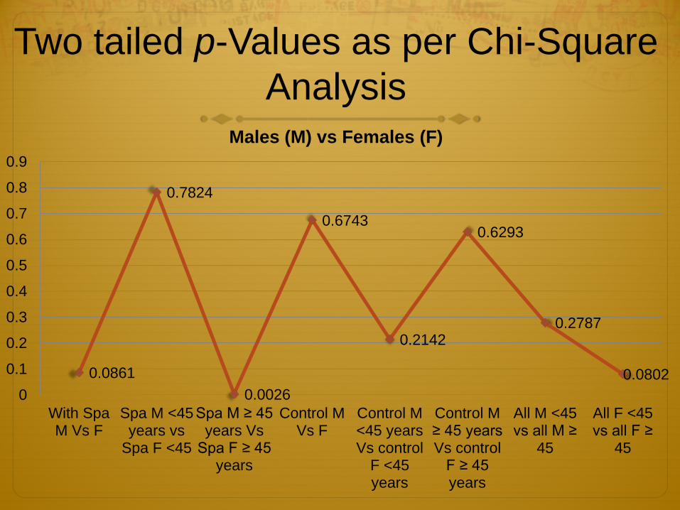
Two tailed p-Values as per Chi-Square Analysis
0.0861
0.7824
0.0026
0.6743
0.2142
0.6293
0.2787
0.0802 0
0.1
0.2
0.3
0.4
0.5
0.6
0.7
0.8
0.9
With Spa M Vs F
Spa M <45 years vs
Spa F <45
Spa M ≥ 45 years Vs
Spa F ≥ 45 years
Control M Vs F
Control M <45 years Vs control
F <45 years
Control M ≥ 45 years Vs control
F ≥ 45 years
All M <45 vs all M ≥
45
All F <45 vs all F ≥
45
Males (M) vs Females (F)

False Negative Diagnosis of SpA made by the readers and presence of intra-articular gas
M SpA F SpA M Spa <45
M SpA >45
F SpA <45
F SpA >45
False negative (number of cases)
4 7 2 2 2 5
False negative cases, with intra-articular gas
0 (0%) 6 (85.71%)
0 (0%) 0 (0%) 1 (50%) 5 (100%)

Reader Correlation Inter-observer reader correlation
Pooled kappa values
For radiological diagnosis: 0.672
Presence of intra-articular gas (for individual joint) : 0.72 – 0.74
Examples of Intra-articular Gas

Axial and coronal reformatted CT images of the sacroiliac joints demonstrating bilateral joint space gas (arrows)
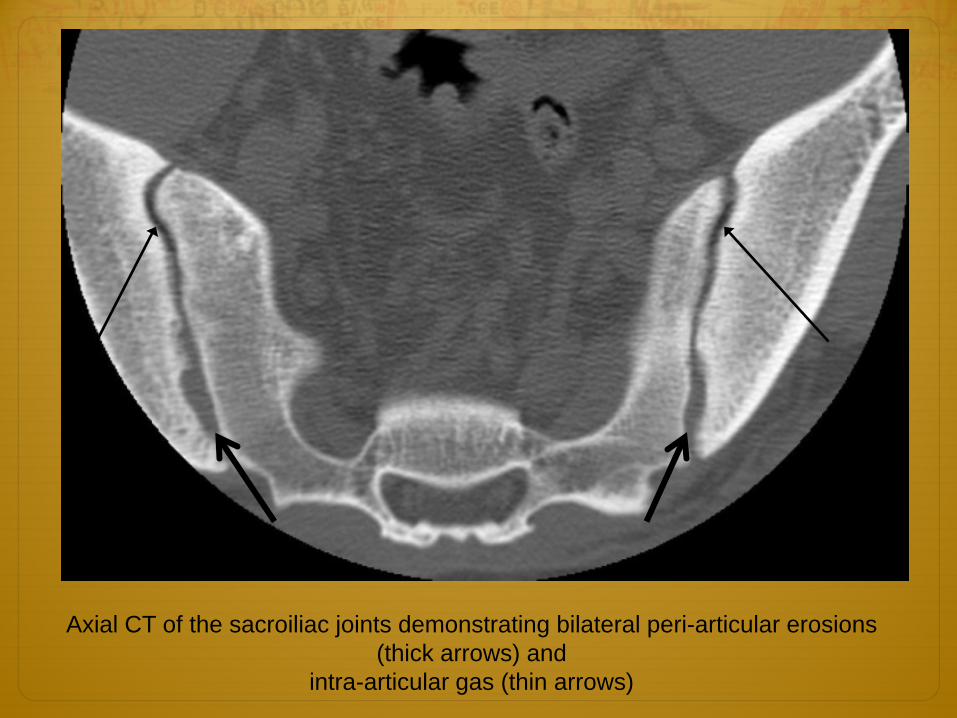
Axial CT of the sacroiliac joints demonstrating bilateral peri-articular erosions (thick arrows) and
intra-articular gas (thin arrows)
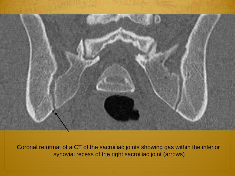
Coronal reformat of a CT of the sacroiliac joints showing gas within the inferior synovial recess of the right sacroiliac joint (arrows)

Conclusion
1. Intra-articular gas in the sacroiliac joints is a frequent finding regardless of age, sex or diagnosis and should not be considered only in cases of osteoarthritis.
2. Intra-articular gas may be contributory to the false negative diagnosis of spondyloarthritis in females aged over 45 years.

References 1. Shorter AM, Burrows DA, Cockshott WP. Is the vacuum sign in the sacro-iliac joint a
useful radiological sign of chondrocalcinosis? Diagn Imaging Clin Med. 1984;53(3):141-3.
2. Sze MJ, Mulligan MJ. Reliability of vacuum phenomenon in the sacroiliac joint as a sign of traumatic injury. Emerg Radiol. 2002 Jul;9(2):100-2. Epub 2002 Mar 21.
3. Ana Isabel García Díez, Xavier Tomás Batllé, Jaume Pomés Talló et al. Sacroiliac Joints: Osteoarthritis or Arthritis. Reumatol Clin. 2009;5(1):40-43.
4. Vegter J, Van Den Broek JA. The diagnostic value of traction during radiography in diseases of the hip. J Bone Joint Surg Br 1983;65: 428–32
5. Faflia CP, Prassopoulos PK, Daskalogiannaki ME, et al. Variation in the appearance of the normal sacroiliac joint on pelvic CT. Clin Radiol. 1998;53:742–6
6. Lo SS, Atceken Z, Carone M et al. Sacroiliac joint vacuum phenomenon--underreported finding. Clin Imaging. 2011 Nov-Dec;35(6):465-9. doi: 10.1016/j.clinimag.2010.10.011.


![A single intra-articular injection of 2.0% non-chemically ... · i.e., much longer than intra-articular corticosteroid injections [14–27]. Intra-articular HA even seems to offer](https://img.dokumen.tips/doc/110x75/5e6e7a63d7b9dc553774f316/a-single-intra-articular-injection-of-20-non-chemically-ie-much-longer.jpg)