Embed Size (px)
Citation preview

Pulp Regeneration—Translational Opportunities
Sources of Dentin-Pulp Regeneration Signals andTheir Modulation by the Local MicroenvironmentFanny Chmilewsky, PhD,* Charlotte Jeanneau, PhD,* Jacques Dejou, PhD,*†
and Imad About, PhD*
Abstract
Many aspects of dentin pulp tissue regeneration havebeen investigated, and it has been shown that dentinpulp has a high regeneration capacity. This seems tobe because of the presence of progenitor cells andinductive regeneration signals from different origins.These signals can be liberated after the acidic dissolu-tion of carious dentin as well as from pulp fibroblastsand endothelial cells in cases of traumatic injury. Thus,both carious lesions and pulp cells provide the requiredmediators for complete dentin-pulp regenerationincluding reparative dentin secretion, angiogenesis,and innervation. Additionally, all dentin pulp insultsincluding carious ‘‘infection,’’ traumatic injuries, applica-tion of restorative materials on the injured dentin pulp,and subsequent apoptosis are known activators of thecomplement system. This activation leads to the produc-tion of several biologically active fragments responsiblefor the vascular modifications and the attraction of im-mune cells to the inflammatory/injury site. Among these,C5a is involved in the recruitment of pulp progenitorcells, which express the C5a receptor. Thus, in additionto dentin and pulp cells, plasma should be considered asan additional source of regeneration signals. (J Endod2014;40:S19–S25)Key WordsComplement, odontoblast, progenitor cells, pulp-dentinregeneration
From the *Aix Marseille Universit�e, Centre National de laRecherche Scientifique, Institut des Sciences du MovementUnit�e Mixte de Recherche 7287; †Service d’Odontologie, Assis-tance Publique-Hopitaux de Marseille, Hopital Timone, Mar-seille, France.
This paper is based on a presentation from the InternationalAssociation for Dental Research (IADR) Pulp Biology andRegeneration Group Satellite Meeting, which was held March24–26, 2013 in San Francisco, California.
Address requests for reprints to Dr Imad About, Aix-Marseille Universit�e, CNRS, ISM UMR 7287, 13288, Marseillecedex 09, France. E-mail address: [email protected]/$ - see front matter
Copyright ª 2014 American Association of Endodontists.http://dx.doi.org/10.1016/j.joen.2014.01.012
JOE — Volume 40, Number 4S, April 2014
Tissue regeneration strategies require 3 major elements: stem cells, scaffolds, andactivation signals. Although many studies were performed to develop a ‘‘suitable’’
scaffold for each particular tissue, numerous investigations were devoted to betterunderstand the stem cells’ properties and potential in tissue regeneration bothin vitro and in vivo (1, 2). However, the signals that orchestrate the regenerationprocesses were not given much attention. Indeed, stem cells are resting cellsresiding beside differentiated cells within an intact and functional tissue. With fewexceptions, the regenerative function of these cells is induced only after tissueinjury in cases of trauma or under pathological conditions. Stem cell inductionappears as the result of activation signals originating from the modifiedmicroenvironment. This pinpoints the fact that understanding what happens incase of pathology/injury is essential in understanding the role of activation signalsin the regeneration processes (3).
This is of prime importance in cases of dentin-pulp regeneration, which occursunder carious or traumatic injuries. These events lead to an inflammatory reaction,which is believed to be the initial step of tissue regeneration. Within the tooth,dentin-pulp regeneration can take multiple forms depending on the causative agentand the severity of the tissue alteration. This varies from a ‘‘simple’’ up-regulation ofthe underlying odontoblast synthetic activity, which leads to regenerating a protectivereactionary dentin, to more severe dentin-pulp injuries. These usually imply completepulp dentin regeneration including not only reparative dentin synthesis but also angio-genesis and innervation. This regeneration seems to be dependent not only on the inter-action of the dentin with the pulp but also on the interaction between different pulp celltypes (4) and between the progenitor cells and other components of the immune/in-flammatory response such as the complement biologically active C5a fragment (5).Thus, dentin-pulp regeneration in case of pulp injury requires the activation and pro-liferation of progenitor cells as well as their migration and differentiation at the injurysite. All these processes are orchestrated by signaling molecules. In this review, wediscuss the origin of the regeneration signals under normal and pathological conditionsand their modulation by inflammation and restorative procedures.
Pulp Progenitor Cell Differentiation In VitroIs Dependent on the Culture Conditions
It has long been established that dental pulp contains progenitor cells capable ofdentin regeneration (6). Although other localizations and niches cannot be excluded, 3independent investigations using 3 different approaches have clearly identified a spe-cific origin of these cells in the perivascular area (1, 7, 8). When isolated withspecific surface markers such as CD34/vascular endothelial growth factor receptor 2(VEGFR2), these cells can undergo adipogenic, chondrogenic, neuronal, andodontoblastic differentiation depending on the culture conditions (1, 6).Additionally, these progenitor cells are heterogenous and comprise a subpopulationof CD31�/CD146�, which can undergo endothelial differentiation when culturedwith a medium containing basic fibroblast growth factor 2 (FGF-2) and vascularendothelial growth factor (VEGF). This has been shown by the formation of tubularstructures on Matrigel (San Jose, CA) and the expression of many endothelial cellmarkers (1). Taken together, these data clearly indicate that modifying the culture con-ditions/local environment affects the cell fate/differentiation potential.
Origin of Dentin-Pulp Regeneration Signals S19

Pulp Regeneration—Translational Opportunities
The Transplantation Site Affectsthe Pulp Progenitor's Fate
Similar results indicate that the microenvironment also controls thefate of progenitor cells in vivo. This effect is nicely shown through trans-plantation experiments. When STRO-1–positive pulp progenitor cellswere mixed with hydroxyapatite/tricalcium phosphate ceramic powderfollowed by transplantation into immunocompromised mice, dentin-pulp–like tissue was generated (9). Similarly, when they were trans-planted on dentin slices, pulp cells generated tubular reparative dentinon the slice surface (10). Surprisingly, when progenitor cells from exfo-liated teeth were transplanted with hydroxyapatite in vivo, bone regener-ation was obtained (11). This was also best shown in autotransplantationexperiments in which progenitor cell transplantation with b-tricalciumphosphate into swinemandibular critical bone defects led to bone regen-eration (2). Interestingly, when pulp progenitor cells that express CD34and VEGFR2but not CD31 or CD146were injected into an ischemic site ofa mouse hind limb, they led to re-establishment of blood flow and vascu-larization (1). On the other hand, when the same cells were labeled withfluorescent Dil-acetylated-LDL (Biomedical Technologies Inc, Schiltigh-eim, France) dye and autotransplanted into amputated pulp of dog teethin a scaffold of collagen type I and type III, the DiI-labeled cells wereobserved in the amputated area closely related to newly formed capil-laries and expressed several proangiogenic factors implying trophicaction on endothelial cells (12). Similarly, when stem cells from exfoli-ated deciduous teeth were seeded onto tooth slice/scaffolds and im-planted subcutaneously into immunodeficient mice, they differentiatedinto functional odontoblasts and endothelial cells only after the additionof VEGF (13).
These data show clearly that pulp progenitors differentiate intoodontoblastic cells when transplanted into the pulp and into othercell types in vitro or when transplanted into other sites in vivo. Thisindicates that the regeneration signals affecting progenitor cell differen-tiation originate from the local environment.
Injuries and Restorative Materials Modifythe Dentin-Pulp Microenvironment
Dentin pulp is subject to injuries with various degrees of severity.These can be caused by carious or traumatic injuries and the subsequentapplication of a restorative material. All these injuries are responsible fordentin-pulp modifications. Indeed, dentin bioactive molecules such asgrowth factors can be solubilized and liberated from the dentin by acidsthat can be produced during the carious injury progression because ofbacterial metabolism. These factors can also be released from the dentinduring the total-etching restorative procedures in which dentin etchingdoes not only remove the smear layer and smear plugs but also resultsin a superficial demineralization of the underlying dentin (14). Thus,during dentin demineralization that occurs after carious irritation, thesesignaling molecules can be released from the dentin matrix and diffuse toreach the pulp cells. The secretory activity of the odontoblasts is thenstimulated, leading to the production of a reactionary dentin (15–17).This effect has also been shown by the fact that both calciumhydroxide and mineral trioxide aggregate can solubilize transforminggrowth factor beta 1 (TGF-b1) from the dentin (18, 19). Similarly,both materials together with Biodentine (Septodont, Saint-Maur-des-Foss�es, France) have been shown to induce the secretion of significantquantities of TGF-b1 from cultured human pulp fibroblasts (20). Giventhe well-established role of this growth factor in progenitor cell recruit-ment and odontoblast differentiation, this explains, at least in part, thetherapeutic success of these materials in direct pulp capping or indeep cavities. Opposed to these bioactive materials, a decrease in TGF-b1 secretion was observed after applying the Xeno III self-etching adhe-
S20 Chmilewsky et al.
sive (Dentsply DeTrey GmbH, Konstanz, Germany). Additionally, it hasbeen reported that resinous monomers reduce the secretion of othergrowth factors from pulp cells such as FGF-2 and appear to be respon-sible for the intracellular accumulation of dentin sialoprotein and osteo-nectin within the endoplasmic reticulum (21). These proteins areinvolved in extracellular matrix mineralization and need to be secretedto accomplish their function. This accumulation explains why afterpulp cells were incubated even with nontoxic concentrations of resinousmonomers the mineralization was inhibited (22).
Dentin Acts as a Reservoir of RegenerationSignals
Many growth factors are assumed to play a role in pulp-dentinregeneration (23, 24). Among these factors, VEGF, platelet-derivedgrowth factors, FGF-2, and TGF-b1 are secreted and sequestered inthe dentin (25, 26). These factors can be liberated by the acidicenvironment at the carious site and diffuse via the dentin tubules tostimulate the underlying odontoblasts to secrete a reactionary dentinlocally. Among these factors, TGF-b1, TGF-b3, and bonemorphogenetic protein 7 have been shown to up-regulate odontoblastsecretory activity (25, 27).
On the other hand, the odontoblasts that border the dental pulp actas sensors of external stimuli. These cells have cytoplasmic processesthroughout the dentin and lie in close proximity to sensory unmyelin-ated nerve fibers, which extend from the dental pulp to the inner halfof the dentin (28). Emerging evidence supports a role for odontoblastsas sensory cells that may sense external stimuli and transduce the signalto nearby neural cells, which is supported by the close apposition of theodontoblasts to the dentinal nerve terminals (29). Recent data haveshown that these cells express transient receptor potential (TRP) ionchannels (30). These channels are known for their capacity to directlymediate nociceptive functions because of thermal and chemical stimuli.Odontoblasts express transient receptor potential vanilloide 1 that re-sponds to noxious heat and transient receptor potential cation channelmelastin 8 and ankyrin A1 that mediate cool and noxious cold sensa-tions, respectively. In addition, these receptors are sensitive to chemicalstimulation (30). Thus, although the dentin pulp is the main target ofbacteria/toxins and restorative materials, odontoblasts deposit a protec-tive reactionary dentin because they act as sensor cells that are respon-sive to dentin-solubilized regeneration signals.
Pulp Cells Secrete Regeneration Signalsafter Pulp Injury
Severe caries lesions or deep cavity preparations may lead toodontoblast destruction. However, a new generation of odontoblast-like cells is responsible for reparative dentin apposition within thepulp chamber (31). This results in tubular dentin secretion discontin-uous from the physiological dentin in terms of tubular structure. Thereparative dentin secretion is a complex process requiring the presenceof responsive progenitor cells as well as the appropriate signals for theiractivation and differentiation. Again, growth factors liberated undercaries lesions play a role in reparative dentin secretion; TGF-b1, FGF-2, and BMP-2 and -4 seem to be involved in the proliferation and dif-ferentiation of pulp cells (32, 33).
However, after surgical pulp amputation, healing can occur withhard tissue formation in germ-free animals independent of growth fac-tor release from the dentin (34, 35). This is suggestive of theautoreparative pulp potential after traumatic injuries.
Indeed, investigating pulp fibroblasts’ and endothelial cells’ secre-tory activity revealed some interesting aspects related to their functionsboth under normal and pathological conditions. It has been shown that
JOE — Volume 40, Number 4S, April 2014
Pulp Regeneration—Translational Opportunities
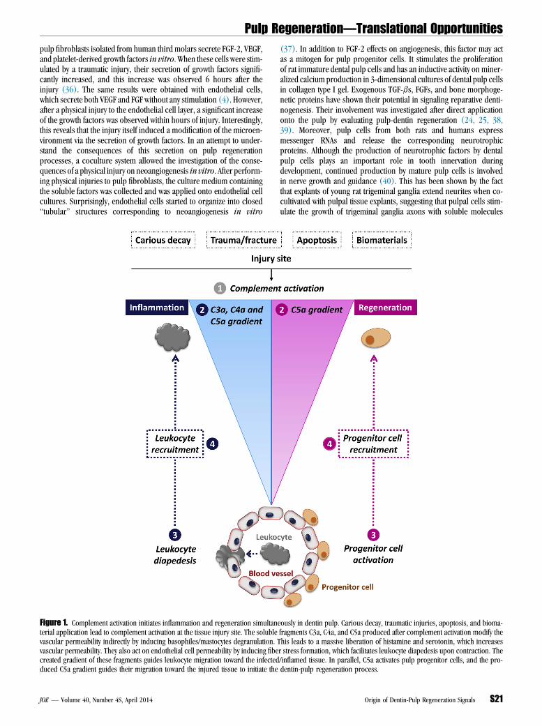
pulp fibroblasts isolated from human third molars secrete FGF-2, VEGF,and platelet-derived growth factors in vitro. When these cells were stim-ulated by a traumatic injury, their secretion of growth factors signifi-cantly increased, and this increase was observed 6 hours after theinjury (36). The same results were obtained with endothelial cells,which secrete both VEGF and FGF without any stimulation (4). However,after a physical injury to the endothelial cell layer, a significant increaseof the growth factors was observed within hours of injury. Interestingly,this reveals that the injury itself induced a modification of the microen-vironment via the secretion of growth factors. In an attempt to under-stand the consequences of this secretion on pulp regenerationprocesses, a coculture system allowed the investigation of the conse-quences of a physical injury on neoangiogenesis in vitro. After perform-ing physical injuries to pulp fibroblasts, the culture medium containingthe soluble factors was collected and was applied onto endothelial cellcultures. Surprisingly, endothelial cells started to organize into closed‘‘tubular’’ structures corresponding to neoangiogenesis in vitroFigure 1. Complement activation initiates inflammation and regeneration simultanterial application lead to complement activation at the tissue injury site. The solublevascular permeability indirectly by inducing basophiles/mastocytes degranulation. Tvascular permeability. They also act on endothelial cell permeability by inducing fibecreated gradient of these fragments guides leukocyte migration toward the infectedduced C5a gradient guides their migration toward the injured tissue to initiate the
JOE — Volume 40, Number 4S, April 2014
(37). In addition to FGF-2 effects on angiogenesis, this factor may actas a mitogen for pulp progenitor cells. It stimulates the proliferationof rat immature dental pulp cells and has an inductive activity on miner-alized calcium production in 3-dimensional cultures of dental pulp cellsin collagen type I gel. Exogenous TGF-bs, FGFs, and bone morphoge-netic proteins have shown their potential in signaling reparative denti-nogenesis. Their involvement was investigated after direct applicationonto the pulp by evaluating pulp-dentin regeneration (24, 25, 38,39). Moreover, pulp cells from both rats and humans expressmessenger RNAs and release the corresponding neurotrophicproteins. Although the production of neurotrophic factors by dentalpulp cells plays an important role in tooth innervation duringdevelopment, continued production by mature pulp cells is involvedin nerve growth and guidance (40). This has been shown by the factthat explants of young rat trigeminal ganglia extend neurites when co-cultivated with pulpal tissue explants, suggesting that pulpal cells stim-ulate the growth of trigeminal ganglia axons with soluble molecules
eously in dentin pulp. Carious decay, traumatic injuries, apoptosis, and bioma-fragments C3a, C4a, and C5a produced after complement activation modify thehis leads to a massive liberation of histamine and serotonin, which increasesr stress formation, which facilitates leukocyte diapedesis upon contraction. The/inflamed tissue. In parallel, C5a activates pulp progenitor cells, and the pro-dentin-pulp regeneration process.
Origin of Dentin-Pulp Regeneration Signals S21

Figure 2. Complement activation in vivo. Immunohistochemistry was used to investigate complement activation through the formation of the cytolytic membraneattack complex (C5b–9) in (A and C) intact and (B and D–F) carious teeth. (C) Although C5b–9 complex was not detected in noncarious dentin of the intact tooth,(D) its labeling was intense in the carious tooth, and (E) the labeling correlated to the bacterial location within the dentin tubules. (F) No labeling was observed inthe controls. (A and B) Hematoxylin-eosin stain. Arrowheads indicate caries lesions, arrows indicate C5b–9 binding of bacteria in dentin tubules, and the asteriskindicates reparative dentin. Scale bars: A–D and F = 500 mm, E = 20 mm.
Pulp Regeneration—Translational Opportunities
(41). This clearly shows that any local tissue modification has directconsequences on dentin-pulp regeneration.
Complement Activation Produces EssentialMolecules Initiating Inflammation
The coagulation and complement systems are efficient plasma cas-cades that play essential roles in inflammation and immune defensemechanisms. The complement system is a major component of the im-mune system, consisting of a proteolytic cascade of more than 30 plasmaand cellular proteins (42). It is activated via 3 principle pathways:
1. The classic pathway is mainly activated by the formation of antigen-antibody complexes but can also be activated by biomaterials (43),apoptotic/necrotic cells, and pathogen-associated molecularpatterns.
2. The lectin pathway is known to be activated directly by pathogen sur-face and especially by carbohydrates and other pathogen-associatedmolecular patterns.
3. The alternative pathway is constitutively activated by spontaneous hy-drolysis but may be triggered directly by foreign surfaces like micro-organisms or man-made biomaterials (44).
S22 Chmilewsky et al.
The complement system activation by either pathway leads to theproduction of 3 kinds of effectors that allow the complement systemto fulfill 3 essential roles regarding immune defense mechanisms. First,C3b and C5b opsonins coat the pathogen surface to enhance its clear-ance by the phagocytic system. Second, the membrane attack complex(C5b–9) fixation into the lipid membrane of cells and pathogens leadsto transmembrane pore formation and cell lysis. Finally, the anaphyla-toxins (C3a, C4a, and especially C5a) are important mediators ofinflammation inducing the vascular permeability by stimulating hista-mine and serotonin release from mastocytes and basophils and fiberstress formation in endothelial cells. Moreover, these anaphylatoxinsare powerful chemotactic factors of leukocyte recruitment to the com-plement activation site (Fig. 1).
Complement Activation Is Also Involvedin the Regeneration Process
Tissue regeneration and inflammation are tightly linked processes,and tissue regeneration is completely dependent on the inflammatoryreaction. This has been clearly shown in the context of myocardialinfarction in which the corticoids used to decrease inflammation signif-icantly reduced the tissue regeneration process (45). The complement
JOE — Volume 40, Number 4S, April 2014

Figure 3. (A) The expression of C5aR by pulp progenitor STRO-1–sorted cells. (a) Although STRO-1 was not expressed by unsorted cells, (b) this surface markerwas expressed by all STRO-1–sorted cells. Similarly, (c) although only a few unsorted cells showed the expression of C5aR, (d) all STRO-1–sorted cells appeared toexpress C5aR. (e) Merged images of STRO-1 and C5aR revealed that some unsorted cells expressed C5aR, (f) whereas STRO-1–sorted cells coexpressed bothSTRO-1 and C5aR. No immunostaining was observed with the (g) negative unsorted or (h) on STRO-1–sorted cells. Scale bars = 50 mm. (B) C5a induces selectivepulp progenitor STRO-1–sorted cell migration. (a) A schematic representation of the migration system used (m-slide chemotaxis 3-dimensional chamber cross-section). Pulp cells were seeded into a C5a gradient and followed for 48 hours. (b and c) Representative plots of cell migration over a 48-hour period: (b) unsortedcells’ trajectory plots and (c) STRO-1–sorted cells’ trajectory plots. (b) No directed migration can be measured for unsorted cells, (c) whereas STRO-1–sorted cellsmigrate toward the C5a gradient.
Pulp Regeneration—Translational Opportunities
is known to play a major role in initiating and amplifying the inflamma-tory reaction. This role is mainly mediated by the C3a and C5a anaphy-latoxins. Although the importance of C3a still raises some debates (46),that of C5a is well established. Indeed, C5a has a powerful chemotacticeffect on C5aR-expressing cells. Basically, this receptor is mainly ex-pressed by immune cells such as neutrophils, eosinophils, basophils/mastocytes, monocytes/macrophages, dendritic cells, and B and T lym-phocytes (47, 48). However, in addition to various immune cells,several nonimmune cells express C5aR including endothelial cells;astrocytes, cells from skin, intestine, and heart; and humanmesenchymal stem cells (49). This receptor is also expressed by cellsinvolved in the mineralization process, such as osteoblasts (50). Inaddition, it has been shown that some stem/progenitor cells expressthis receptor (51).
C5a and Tissue RegenerationSeveral investigations showed the involvement of C5a in tissue
regeneration. This has been studied in the liver, which has a veryhigh regeneration capacity (51). Indeed, when C5-deficient micewere subjected to a toxic damage after CCL4 chemokine treatment, theirliver could not regenerate. However, liver regeneration was observedafter injecting C5 or C5a to the treated animals (52). This role hasalso been shown in cardiac tissue regeneration in which C5a not onlyinduced the migration of progenitor cells but also their differentiation(53). After human bone fracture, a significant increase of C5aR expres-
JOE — Volume 40, Number 4S, April 2014
sion was observed in mesenchymal progenitors during osteogenic dif-ferentiation. This study also showed that C5a induces the migration ofmesenchymal progenitor cells as well as osteoblasts showing C5ainvolvement in fractured bone regeneration (54).
Dental Pulp Is Subject to Various InsultsLeading to Complement Activation
Dental pulp is subject to various types of injuries, which lead to aninflammatory reaction. These include carious decay, traumatic injuries/fractures, and the therapeutic application of a restorative material. It hasbeen shown that biomaterials that contain free OH, NH3, or carboxylicacid groups are involved in the complement activation (55, 56). It hasalso been reported that Ni(2+) and Co(2+) induce complementactivation in plasma samples as shown by the cleavage ofcomplement factor C3 to C3b (55). Additionally, during the manage-ment of pulp lesions, odontoblast and pulp cells may undergo apoptosis(56). All these events are well-established complement activationmech-anisms (Fig. 1).
Complement Activation andDentin-Pulp Regeneration
It has been shown that carious injuries, like any bacterial infec-tion, activate complement as revealed by the cytolytic membraneattack complex (C5b–9) formation in human third molars (5).This complex binds onto the bacterial membrane as shown in the
Origin of Dentin-Pulp Regeneration Signals S23

TABLE 1. Sources of Dentin-Pulp Regeneration Signals
Sources Insults Mediators Effects
Dentin/odontoblast(14–18, 23, 24, 37–39)
Carious FGF-2, VEGF, PDGF, TGF-b1.
FGF-2, VEGF, PDGF, TGF-b1
Progenitor and pulp cell proliferation,angiogenesis, directing progenitorcell migration, odontoblasticdifferentiation
Pulp fibroblasts (27, 31, 32) Carious, trauma/biomaterial
Endothelial cells (4) Carious, trauma FGF-2, VEGF, Pulp cell proliferation, angiogenesisPlasma (5) Carious, trauma C5a Directing progenitor cell migration
FGF-2, basic fibroblast growth factor 2; PDGF, platelet-derived growth factor; TGF-b1, transforming growth factor beta 1; VEGF, vascular endothelial growth factor.
Pulp Regeneration—Translational Opportunities
tubules of decayed dentin and pulp (Fig. 2A–F). This activation im-plies the generation of biologically active fragments including a C5agradient production. In an attempt to understand the potential roleof this fragment in dentin-pulp regeneration, a cell migration designwas used to study the interactions between pulp progenitor cells andthe C5a fragment (5).
Using the magnetic cell sorting approach of human pulp progen-itor cells with the mesenchymal STRO-1 stem cell marker, it has beenshown that the isolated pulp progenitors also express C5aR as shownby immunofluorescence double staining (Fig. 3A). This has also beenshown in human teeth in which both STRO-1 and C5aR were coex-pressed by the same cells in the perivascular area, which correspondsto the perivascular niche of pulp progenitor cells (5). When STRO-1–sorted progenitor cells expressing C5aR were subjected to a C5agradient in a migration chamber, they migrated toward a C5a gradient,whereas the nonsorted cells exhibited random movements (Fig. 3B).The interaction specificity between C5a and STRO-1–sorted progenitorcells has been shown by adding the C5aR antagonist W54011, whichsignificantly inhibited the binding (5).
Thus, after complement activation, a C5a gradient is produced,and this gradient guides the pulp progenitor cells to the complementactivation site/inflammatory site (Fig. 1). This clearly shows the involve-ment of C5a in 1 of the early and essential steps of dentin-pulp regen-eration. Indeed, after odontoblast disintegration in deep/severecarious/traumatic injuries, a migration of progenitor cells is requiredto replace the missing odontoblasts and to secrete a protective repara-tive dentin. Although the C5a fragment is produced within minutes ofcomplement activation and provides a very quick signal for initiatingthe inflammatory reaction, it is also involved in the regeneration processvia the recruitment of pulp progenitor cells.
These findings do not exclude the involvement of other mecha-nisms during this critical step. Indeed, dentin and pulp matrix compo-nents (57), liposaccharides (58), and stromal cell–derived factor 1(SDF-1) (59) have been shown to contribute to pulp cell migration.Interestingly, among the cells expressing SDF-1 receptor, a high per-centage was STRO-1 positive and had progenitor cell properties suchas colony formation, high proliferation rate, a multilineage differentia-tion potential, and expressed chemokine receptor 4 (CXCR4) (60, 61).Also, it is well established that CXCR4-expressing cells are responsive toSDF-1, which can attract CXCR4-positive cells from their niches inresponse to stimulations related to tissue damage or infection (60).In line with this, it has been shown that SDF-1 and CXCR4 messengerRNA expression increases in inflamed dental pulps (59).
These data contribute to a better understanding of the mecha-nisms involved in progenitor cell migration to the infectious/injurysite in order to regenerate the damaged dentin. Thus, in addition tothe signals produced from the dentin, pulp fibroblasts, and endothe-lial cells, complement activation and its consequences to pulp regen-eration clearly show that the plasma should be considered as animportant additional source providing C5a as a new signal involvedin dentin-pulp regeneration.
S24 Chmilewsky et al.
ConclusionsRegeneration signals of the dentin pulp have different sources
(Table 1). The soluble extracellular matrix molecules of dentin, pulpfibroblasts, and endothelial cells are involved in complete dentin-pulp regeneration. This review adds to our understanding thatcomplement activation is involved in the regeneration process byinducing progenitor cell recruitment. This regeneration process israther complex implying many signals and starts very quickly duringthe inflammatory reaction after any dentin-pulp damage.
AcknowledgmentsSupported by funding from Aix-Marseille University and CNRS.The authors deny any conflicts of interest related to this study.
References1. Iohara K, Zheng L, Wake H, et al. A novel stem cell source for vasculogenesis in
ischemia: subfraction of side population cells from dental pulp. Stem Cells 2008;26:2408–18.
2. Zheng Y, Liu Y, Zhang CM, et al. Stem cells from deciduous tooth repair mandibulardefect in swine. J Dent Res 2009;88:249–54.
3. About I. Dentin–pulp regeneration: the primordial role of the microenvironmentand its modification by traumatic injuries and bioactive materials. Endod Top2013;28:61–89.
4. About I. Dentin regeneration in vitro: the pivotal role of supportive cells. Adv DentRes 2011;23:320–4.
5. Chmilewsky F, Jeanneau C, Laurent P, et al. Pulp progenitor cell recruitment is selec-tively guided by a C5a gradient. J Dent Res 2013;92:532–9.
6. About I, Bottero MJ, De Denato P, et al. Human dentin production in vitro. Exp CellRes 2000;258:33–41.
7. Shi S, Gronthos S. Perivascular niche of postnatal mesenchymal stem cells in humanbone marrow and dental pulp. J Bone Miner Res 2003;18:696–704.
8. T�ecl�es O, Laurent P, Zygouritsas S, et al. Activation of human dental pulp progenitor/stem cells in response to odontoblast injury. Arch Oral Biol 2005;50:103–8.
9. Gronthos S, Brahim J, Li W, et al. Stem cell properties of human dental pulp stemcells. J Dent Res 2002;81:531–5.
10. Batouli S, Miura M, Brahim J, et al. Comparison of stem-cell-mediated osteogenesisand dentinogenesis. J Dent Res 2003;82:976–81.
11. Miura M, Gronthos S, Zhao M, et al. SHED: stem cells from human exfoliated decid-uous teeth. Proc Natl Acad Sci U S A 2003;100:5807–12.
12. Iohara K, Zheng L, Ito M, et al. Regeneration of dental pulp after pulpotomy by trans-plantation of CD31(-)/CD146(-) side population cells from a canine tooth. RegenMed 2009;4:377–85.
13. Sakai VT, Zhang Z, Dong Z, et al. SHED differentiate into functional odontoblasts andendothelium. J Dent Res 2010;89:791–6.
14. Van Meerbeek B, Inokoshi S, Braem M, et al. Morphological aspect of the resin-dentin interdiffusion zone with different dentin adhesive systems. J Dent Res1992;71:1530–40.
15. About I, Laurent-Maquin D, Lendahl U, et al. Nestin expression in embryonic andadult human teeth under normal and pathological conditions. Am J Pathol 2000;157:287–95.
16. Goldberg M, Smith AJ. Cells and extracellular matrices of dentin and pulp: a biolog-ical basis for repair and tissue engineering. Crit Rev Oral Biol Med 2004;15:13–27.
17. Tziafas D, Smith AJ, Lesot H. Designing new treatment strategies in vital pulp therapy.J Dent 2000;28:77–92.
18. Graham L, Cooper PR, Cassidy N, et al. The effect of calcium hydroxide on solubi-lisation of bio-active dentine matrix components. Biomaterials 2006;27:2865–73.
JOE — Volume 40, Number 4S, April 2014

Pulp Regeneration—Translational Opportunities
19. Tomson PL, Grover LM, Lumley PJ, et al. Dissolution of bio-active dentine matrixcomponents by mineral trioxide aggregate. J Dent 2007;35:636–42.20. Laurent P, Camps J, About I. Biodentine induces TGF-b1 release from human pulp
cells and early dental pulp mineralization. Int Endod J 2012;45:439–48.21. Diamanti E, Mathieu S, Jeanneau C, et al. Endoplasmic reticulum stress and miner-
alization inhibition mechanism by the resinous monomer HEMA. Int Endod J 2013;46:160–8.
22. About I, Camps J, Mitsiadis TA, et al. Influence of resinous monomers on the differ-entiation in vitro of human pulp cells into odontoblasts. J Biomed Mater Res 2002;63:418–23.
23. Iohara K, Nakashima M, Ito M, et al. Dentin regeneration by dental pulp stem celltherapy with recombinant human bone morphogenetic protein 2. J Dent Res 2004;83:590–5.
24. Unda FJ, Martin A, Hernandez C, et al. FGFs-1 and -2, and TGF beta 1 as inductivesignals modulating in vitro odontoblast differentiation. Adv Dent Res 2001;15:34–7.
25. Sloan AJ, Smith AJ. Stimulation of the dentine-pulp complex of rat incisor teeth bytransforming growth factor-beta isoforms 1-3 in vitro. Arch Oral Biol 1999;44:149–56.
26. Goldberg M, Lacerda-Pinheiro S, Jegat N, et al. The impact of bioactive molecules tostimulate tooth repair and regeneration as part of restorative dentistry. Dent ClinNorth Am 2006;50:277–98.
27. Sloan AJ, Rutherford RB, Smith AJ. Stimulation of the rat dentine-pulp complex bybone morphogenetic protein-7 in vitro. Arch Oral Biol 2000;45:173–7.
28. Byers MR, N€arhi MV. Dental injury models: experimental tools for understandingneuroinflammatory interactions and polymodal nociceptor functions. Crit RevOral Biol Med 1999;10:4–39.
29. Maurin JC, Couble ML, Didier-Bazes M, et al. Expression and localization of reelin inhuman odontoblasts. Matrix Biol 2004;23:277–85.
30. El Karim IA, Linden GJ, Curtis TM, et al. Human odontoblasts express functionalthermo-sensitive TRP channels: implications for dentin sensitivity. Pain 2011;152:2211–23.
31. Fitzgerald M, Chiego DJ, Heys DR. Autoradiographic analysis of odontoblast replace-ment following pulp exposure in primate teeth. Arch Oral Biol 1990;35:707–15.
32. Martin P. Wound healing. Aiming for perfect skin regeneration. Science 1997;276:75–81.
33. Shiba H, Nakamura S, Shirakawa M, et al. Effects of basic fibroblast growth factor onproliferation, the expression of osteonectin (SPARC) and alkaline phosphatase, andcalcification in cultures of human pulp cells. Dev Biol 1995;170:457–66.
34. Inoue T, Shimono M. Repair dentinogenesis following transplantation into normaland germ-free animals. Proc Finn Dent Soc 1992;88:183–94.
35. Tsuji T, Takel K, Inoue T, et al. An experimental study on wound healing of surgicalexposed dental pulps in germ-free rats. Bull Tokyo Dent Coll 1987;28:35–8.
36. Tran-Hung L, Laurent P, Camps J, et al. Quantification of angiogenic growth factorsreleased by human dental cells after injury. Arch Oral Biol 2008;52:9–13.
37. Tran-Hung L, Mathieu S, About I. Role of human pulp fibroblasts in angiogenesis.J Dent Res 2006;85:819–23.
38. Mathieu S, Jeanneau C, Sheibat-Othman N, et al. Usefulness of controlled release ofgrowth factors in investigating the early events of dentin-pulp regeneration. J Endod2013;39:228–35.
39. Nakashima M. Induction of dentin in amputated pulp of dogs by recombinant hu-man bone morphogenetic proteins-2 and -4 with collagen matrix. Arch Oral Biol1994;39:1085–9.
40. Nosrat IV, Smith CA, Mullally P, et al. Dental pulp cells provide neurotrophic supportfor dopaminergic neurons and differentiate into neurons in vitro; implications for
JOE — Volume 40, Number 4S, April 2014
tissue engineering and repair in the nervous system. Eur J Neurosci 2004;19:2388–98.
41. Lillesaar C, Eriksson C, Johansson CS, et al. Tooth pulp tissue promotes neuriteoutgrowth from rat trigeminal ganglia in vitro. J Neurocytol 1999;28:663–70.
42. Ricklin D, Hajishengallis G, Yang K, et al. Complement-a key system for immune sur-veillance and homeostasis. Nat Immunol 2010;11:785–97.
43. Tengvall P, Askendal A, Lundstr€om II. Ellipsometric in vitro studies on the activa-tion of complement by human immunoglobulins M and G after adsorption to meth-ylated silicon. Colloids Surf B Biointerfaces 2001;20:51–62.
44. Andersson J, Ekdahl KN, Larsson R, et al. C3 adsorbed to a polymer surface can forman initiating alternative pathway convertase. J Immunol 2002;168:5786–91.
45. Kloner RA, Fishbein MC, Lew H, et al. Mummification of the infarcted myocardiumby high dose corticosteroids. Circulation 1978;57:56–63.
46. Busche MN, Stahl GL. Role of the complement components C5 and C3a in a mousemodel of myocardial ischemia and reperfusion injury. GMS 2010;8:1612–3174.
47. Morelli A, Larregina A, Chuluyan E, et al. Expression and modulation of C5a receptor(CD88) on skin dendritic cells. Chemotactic effect of C5a on skin migratory den-dritic cells. Immunology 1996;89:126–34.
48. Werfel T, Oppermann M, Bergemann G, et al. C5a receptors are detectable on mastcells in normal human skin and in psoriatic plaques but not in weal and flare re-actions or in uticaria pigmentosa by immunohistochemistry. Arch Dermatol Res1997;289:83–6.
49. Schraufstatter I, DiScipio RG, Zhao M, et al. C3a and C5a are chemotactic factors forhuman mesenchymal stem cells which cause prolonged ERK1/2 phosphorylation.J Immunol 2009;182:3827–36.
50. Ignatius A, Schoengraf P, Kreja L, et al. Complement C3a and C5a modulate osteo-clast formation and inflammatory response of osteoblasts in synergism with IL-1b.J Cell Biochem 2011;112:2594–605.
51. Michalopoulos GK, DeFrances MC. Liver regeneration. Science 1997;276:60–6.52. Mastellos D, Papadimitriou JC, Franchini S, et al. A novel role of complement: mice
deficient in the fifth component of complement (C5) exhibit impaired liver regen-eration. J Immunol 2001;166:2479–86.
53. Lara-Astiaso D, Izarra A, Estrada JC, et al. Complement anaphylatoxins C3a and C5ainduce a failing regenerative program in cardiac resident cells. Evidence of a role forcardiac resident stem cells other than cardiomyocyte renewal. Springerplus 2012;1:63–78.
54. Ignatius A, Ehrnthaller C, Brenner RE, et al. The anaphylatoxin receptor C5aR is pre-sent during fracture healing in rats and mediates osteoblast migration in vitro.Trauma 2011;71:952–60.
55. Acevedo F, Vesterberg O. Nickel and cobalt activate complement factor C3 fasterthan magnesium. Toxicology 2003;185:9–16.
56. Nilsson B, Korsgren O, Lambris J, et al. Can cells and biomaterials in therapeuticmedicine be shielded off from innate immune recognition? Trends Immunol2010;31:32.
57. Smith JG, Smith AJ, Shelton RM, et al. Recruitment of dental pulp cells by dentine andpulp extracellular matrix components. Exp Cell Res 2012;318:2397–406.
58. Wang MC, Hung PS, Tu HF, et al. Lipopolysaccharide induces the migration of hu-man dental pulp cells by up-regulating miR-146a. J Endod 2012;38:1598–603.
59. Suzuki T, Lee CH, Chen M, et al. Induced migration of dental pulp stem cells forin vivo pulp regeneration. J Dent Res 2011;90:1013–8.
60. Jiang L, Peng WW, Li LF, et al. Isolation and identification of CXCR4-positive cellsfrom human dental pulp cells. J Endod 2012;38:791–5.
61. Jiang L, Peng WW, Li LF, et al. Proliferation and multilineage potential of CXCR4-positive human dental pulp cells in vitro. J Endod 2012;38:642–7.
Origin of Dentin-Pulp Regeneration Signals S25


![Pulp Dentin Complex[1] / orthodontic courses by Indian dental academy](https://img.dokumen.tips/doc/110x75/577cce141a28ab9e788d4145/pulp-dentin-complex1-orthodontic-courses-by-indian-dental-academy.jpg)
