Embed Size (px)
Citation preview

Università degli Studi di Milano
GRADUATE SCHOOL OF VETERINARY SCIENCES
FOR ANIMAL HEALTH AND FOOD SAFETY Director: Prof. Vittorio Dell’Orto
Doctoral Program in Veterinary Clinical Sciences
Some perinatal endocrine and morphological
aspects of canine species
Tea Meloni
Tutor: Coordinator: Prof. ssa Maria Cristina Veronesi Prof. Valentino Bontempo
Academic Year: 2014-2015

2

3
To myself,
for how passion can become a work

4

5
Index
1. Foreword……………………………………………………… 11
2. Canine pregnancy……………………………………………. 17
2.1 Fertilization…………………………………………... 19
2.2 Embryogenesis……………………………………… 19
2.2.1 From the zygote to the blastocyst…………………… 19
2.2.2 From the implantation to the organogenesis…………... 21
2.2.3 Maternal recognition of pregnancy…………………... 22
2.3 Placentation and fetal fluids………………………….. 23
2.3.1 Placenta………………………………………… 23
2.3.2 Amniotic fluid…………………………………… 25
2.3.3 Allantoic fluid…………………………………… 29
2.3.4 Fetal fluids in Veterinary Medicine…………………. 30
2.3.5 Endocrine functions of placenta……………………... 31
2.4 Fetal growth………………………………………….. 33
2.4.1 The period of fetus………………………………… 33
2.4.2 Endocrine regulation………………………………. 33
2.4.3 Fetal maturation………………………………….. 35
2.5 Normal pregnancy……………………………………. 36
2.5.1 Physiological changes in the dam…………………….. 36
2.5.2 Duration of pregnancy and prediction……………......... 37
of the parturition date
3. Parturition in canine species………………………………….. 45
3.1 Endocrine and mechanical events……………………... 47
3.2 Stages of whelping…………………………………….. 49

6
4. Preparation for birth and neonatal adaptation……………. 53
4.1 Hypothalamic-pituitary-adrenal axis………………... 55
4.1.1 Modulation……………………………………. 55
4.1.2 Glucocorticoid secretion………………………….. 56
4.1.3 Cortisol functions………………………………. 58
4.2 Effects of parturition on the newborn viability…….. 61
4.3 Fetal distress……………………………………….. 63
4.4 Neonatal adaptation………………………………... 67
4.4.1 Cardiocirculatory system………………………… 67
4.4.2 Respiratory system……………………………… 68
4.4.3 Digestive system………………………………… 69
4.4.4 Urinary system…………………………………. 70
4.4.5 Muscular and skeletal system…………………….. 71
4.4.6 Immune system…………………………………. 71
4.4.7 Neonatal mortality…………………………….... 73
5. Non-invasive methods for perinatal investigations………... 77
6. References……………………………………………………. 85
7. Objectives…………………………………………………….. 117
8. Insulin-like growth factor-I and non-esterified fatty acids… 123
8.1 Introduction…………………………………………. 125
8.2 Insulin-like growth factor-I…………………………... 125
8.3 Non-esterified fatty acids…………………………….. 128
8.4 References…………………………………………… 129

7
9. IGF-I and NEFA concentrations in fetal fluids of term…….. 137
pregnancy dog
9.1 Abstract………………………………………………. 139
9.2 Introduction…………………………………………... 140
9.3 Materials and methods………………………………... 142
9.4 Results……………………………………………….... 144
9.5 Discussion……………………………………………. 146
9.6 Conclusions…………………………………………... 149
9.7 References……………………………………………. 149
10. Immunoglobulin G and lysozyme……………………………. 155
10.1 Introduction…………………………………………... 157
10.2 Transplacental antibodies transfer…………………….. 157
10.3 Lysozyme……………………………………………... 160
10.4 References…………………………………………….. 161
11. Immunoglobulin G and lysozyme concentrations…………… 167
in canine fetal fluids at term of pregnancy
11.1 Abstract………………………………………………... 169
11.2 Introduction…………………………………………… 170
11.3 Materials and methods………………………………… 172
11.4 Results…………………………………………………. 175
11.5 Discussion……………………………………………... 177
11.6 References……………………………………………… 181
12. Cortisol levels in hair and nails of newborn puppies………….. 185
12.1 Introduction……………………………………………. 187
12.2 Cortisol assessment in biological matrices………………. 187
12.3 Hair and nails for hormonal measurements…………….. 188
12.4 References……………………………………………… 192

8
13. Hair and nails as new, non-invasive matrices for…………… 199
long time-frame cortisol analysis in newborn dogs
13.1 Abstract………………………………………………. 201
13.2 Introduction…………………………………………... 202
13.3 Materials and methods………………………………... 204
13.4 Results……………………………………………….... 205
13.5 Discussion……………………………………………. 207
13.6 Conclusions…………………………………………... 211
13.7 References…………………………………………….. 212
14. Bacterial infection in the newborn……………………………. 217
14.1 Neonatal bacterial infections in humans and…………... 219
large animals
14.2 Bacterial infections in canine newborn………………… 220
14.2.1 Septicaemia predisposing factors……………………… 220
14.2.2 Bacterial aetiology and clinical course…………………. 221
14.2.3 Diagnosis…………………………………………. 224
14.3 References……………………………………………... 224
15. A survey on bacterial involvement in neonatal mortality……. 233
in dogs
15.1 Abstract………………………………………………... 235
15.2 Introduction…………………………………………… 236
15.3 Materials and methods………………………………… 237
15.4 Results………………………………………………… 238
15.5 Discussion…………………………………………….. 243
15.6 References……………………………………………... 246
16. Age estimation in large and giant newborn puppies…………. 251
through the hindlimb ossification centers evaluation and
morphometry of hindlimb long bones, skull, and body
16.1 Introduction…………………………………………… 253

9
16.2 Materials and methods…………………………………. 256
16.3 Results…………………………………………………. 260
16.4 Discussion……………………………………………... 272
16.5 Conclusions……………………………………………. 278
16.6 References……………………………………………… 280
17. General discussion……………………………………………… 289
Acknowledgements………………………………………………… 297

10

11
CHAPTER 1
Foreword

12

13
1. Foreword
Recently the interest for the canine neonatology, mainly aimed to the
improvement of survival after birth and to the correct management of the
newborn puppies, is increasing.
In mammals, the newborn represents the result of the prenatal intrauterine
development, along which several factors play a crucial role to ensure the fetal
growth, well-being, and protection. In this respect, the placenta partially
maintains the best environment for the conceptus and it was recognized that
fetal fluids, especially the amniotic fluid, contain many important substances
involved in fetal growth, development, and well-being.
Both the late pregnancy and neonatal period are considered the most stressful
stages for the fetus and newborn, respectively. In fact, an hypothalamic-pituitary-
adrenal (HPA) axis activation of the subject occurs during these phases and the
consequent cortisol release leads to the final multi-organ maturation in the fetus
and even allows the trigger of parturition, whereas in the newborn it is associated
to the gradual adaptation to the extrauterine life.
The process of birth represents the transition from the harmless intrauterine life
to the outside world, where numerous factors could become causes of neonatal
morbidity and mortality. Newborn puppies (as well as kittens) are much less
mature than newborns of many other domestic species; therefore, their
management is quite challenging for the veterinary practitioner, because of the
small size and immature multi-organ functions. In particular, the immune system
is not fully developed, so that, at the beginning of their extrauterine life, all
mammals newborns depend on the passive immunity acquired by the mother,
through the placenta and/or colostrum.
Along the neonatal period, several physiologic changes normally take place,
ensuring the maturation and adaptation of the newborn. Concurrently, also the
whole skeleton conformation undergoes a gradual evolution. That said, canine
species is characterized by a wide variety of breeds encompassing many body
shapes and sizes; additionally, the growth speed appears extremely rapid and
influenced by the breed, sex, genetics, environment, endocrine as well as
metabolic factors, diet, and pathologic conditions.

14
Aims of the thesis
Because of the recognized importance of the perinatal challenging period for the
survival and health of the offspring, the present thesis was aimed to investigate
some aspects of the perinatal phase, i.e. the time ranging between the last third
of gestation until the first 30 days after birth, in the dog.
To the author knowledge, the amniotic and allantoic composition throughout
pregnancy, as well as the role of the amniotic and allantoic compounds involved
in the fetal growth and development, and in preparation for birth, were not yet
fully investigated in canine species. Furthermore, the hormonal mechanisms
which dominate both the final phase of gestation and the beginning of the
extrauterine life were not fully clarified in dogs. The present thesis was aimed,
first of all, to provide a deeper knowledge about fetal fluids composition, by
evidencing the presence and the concentrations of some factors (IGF-I, NEFA,
IgG, and lysozyme) necessary for fetal and neonatal growth, development, and
well-being, and to evaluate the HPA axis activation in the newborn, by
measuring cortisol levels in many biological matrices. Both these studies were
carried on in the respect of the animal welfare, through not-invasive techniques.
In fact, IGF-I, NEFA, IgG, and lysozyme levels were assessed in fetal fluids
collected during elective cesarean sections, performed because of the high risk of
dystocia or previous history of troubles at parturition, whereas cortisol
concentrations were analyzed in both hair and nails collected by born dead
newborn puppies or puppies dead spontaneously within 7 days of age. The
present thesis represents only a starting point; further researches are needed to
improve the clinical application, for instance by evaluating the possible
correlations between both IGF-I and NEFA and fetal growth diseases, or by
assaying cortisol levels in healthy and sick newborn puppies not only at birth, but
during the neonatal phase and, if possible, even later, to monitor the last period
of intrauterine development as well as the neonatal adaptation after birth.
The neonatal period represents another really interesting phase, along which
indispensable physiologic changes have to take place to ensure the survival after
birth. In particular, the present thesis was aimed to investigate the real bacterial
involvement in canine neonatal mortality, since the bacterial infections are
considered as the second main cause of neonatal loss in dogs. Despite the impact

15
of bacterial infections on neonatal death, only few studies were published about
the incidence of septicaemia in newborn puppies.
Additionally, in canine species, also the information availability about the skeletal
development are limited, above all within the first month of age. The previous
studies about the evaluation of the ossification centers appearance and fusion are
difficult to compare each other because, in most cases, the breeds enrolled, age
and method of examination, anatomical compartments investigated, as well as
the aim of the research, were not homogeneus. Thus, it could be useful the
creation of a radiographic data-base concerning the ossification centers
appearance during the first month of age in newborn puppies, in order to
investigate the normal growth, as well as to detect a new easy tool to estimate
their real age, above all in case of illegal import. At this regard, the third aim of
the present thesis was to investigate the timing appearance of hindlimb
ossification centers, as well as the radiographic and anatomical morphometry of
the hindlimb long bones, skull, and body, in large and giant sized puppies dead
spontaneously within the first 30 days of age.

16

17
CHAPTER 2
Canine pregnancy

18

19
2. Canine pregnancy
2.1 Fertilization
The term “fertilization” indicates the union of male and female pronuclei, which
marks the beginning of the embryonic development. In most bitches, this
process occurs in metaphase II oocytes, from 90 hours after ovulation (Reynaud
et al., 2005).
In the oviduct, spermatozoa change their motility pattern from a progressive,
linear motility into a hyperactive frenzied motion, probably in order to facilitate
the interaction with the oocyte. To date, it is well known that spermatozoa show
particular proteins on their plasma membrane surfaces, which bind specifically to
the glycoproteins of zona pellucida. Then, the acrosomal reaction, during which
the fusion between the spermatozoa plasma membrane and the outer acrosomal
membrane occurs, leads to a vesiculation that allows for both the digestion and
penetration of the zona pellucida by the acrosomal enzymes. When the
spermatozoon reaches the perivitelline space, the oocyte plasma membrane fuses
with the equatorial segment of spermatozoon. Thus, the oocyte undergoes
several changes aimed to the early embryogenesis, such as the cortical reaction.
During the first and second meiotic divisions, cortical granules fuse with the
oocyte plasma membrane, releasing their contents into the perivitelline space to
prevent the polyspermy. After the inclusion of the spermatozoon nucleus within
the oocyte cytoplasm, the fusion between the male and female pronuclei, or
syngamy, can occur (Senger, 2003).
2.2 Embryogenesis
A successful preattachment phase during pregnancy requires the progression
from zygote to blastocyst, as well as the development of a functional
trophoblast. Canine embryonic and fetal development occurs within 61 days
from the fertilization, a relatively short interval compared to many other
mammals (Pretzer, 2008).
2.2.1 From the zygote to the blastocyst
The “period of the ovum” defines the interval between days 2 and 17 after LH
peak (Pretzer, 2008). Shortly thereafter the syngamy, the embryo, now defined

20
“zygote”, undergoes several cleavage divisions. The reaching of the uterotubal
junction takes place in 7-10 days for canine zygotes compared to 3-4 days typical
of the other species (Reynaud et al., 2005; Pretzer, 2008; Reynaud et al., 2012).
The first mitotic division generates a two-celled embryo, formed by blastomeres,
each of them representing more or less one-half of the zygote; subsequently,
each blastomere is subjected to additional divisions, giving rise to the morula. To
the author knowledge, the data available concerning the oocytes maturation
timing in the bitch are still controversial (Tsutsui, 1975; Renton et al., 1991;
Bysted et al., 2001; Concannon et al., 2001; Reynaud et al., 2005; Reynaud et al.,
2012). The continuous divisions of the morula cells lead to a separation between
inner and outer cells, with the consequent blastocyst development. The
blastocyst is formed by an inner cell mass, a cavity called blastocoele, and a single
outer layer of cells, named trophoblast. The inner cell mass of the blastocyst will
form the embryonic body, whereas the trophoblastic cells will give rise to the
chorion, the fetal component of the placenta. During the morula stage, some
fluid can accumulate inside, thanks to an active sodium pump and particular
junctions, which allow for intercellular communication and alter the cellular
permeability. The combination among the pression increase caused by the fluid
accumulation in the blastocoele, the degradation of the zona pellucida by some
proteolytic enzymes, released by the trophoblastic cells, and the blastocyst
contractions, causes the rupture of the zona pellucida, with following hatching of
the blastocyst (Senger, 2003). The timing of the preattachment embryogenesis
depends on species; in the bitch, the blastocyst hatching from the zona pellucida
usually occurs 13-15 days after ovulation (Senger, 2003), even if this process can
be delayed until day 19 after LH surge (Concannon et al., 2001).
The passage of the developing embryos into the uterus can occur as early as 16-
cells stage, but more commonly as morulae or early blastocysts (Pretzer, 2008;
Reynaud et al., 2012), 8-10 days after ovulation (Reynaud et al., 2012). An
intrauterine and transcornual migration of the blastocysts is possible from day 12
to 17 after LH peak (Pretzer, 2008). The beginning of the fixation and
implantation was suggested by day 17-19 post LH surge (Pretzer, 2008) or 16-17
after ovulation (Reynaud et al., 2012).

21
2.2.2 From the implantation to the organogenesis
The “period of the embryo” includes the interval ranging from 19 to 35 days
after LH surge. The most important event within this phase is defined
“gastrulation”, along which the blastula becomes a trilaminar structure consisting
of an outer ectodermal, a middle mesodermal, and an inner endodermal layer
(Pretzer, 2008). During the early embryonic development, the endoderm forms
under the inner cell mass and grows downward on the inner surface of the
trophoblast, giving rise to the yolk sac, a transient extraembryonic membrane
that regresses as the conceptus growth progresses. Simultaneously, the
mesoderm develops between the endoderm and the embryo, surrounding the
yolk sac and pushing against the trophectoderm (previous trophoblastic cells).
Additionally, the mesoderm begins to fold upward, forming the amniotic folds
around the embryo. While the amniotic folds fuse above the embryo, the
mesoderm fuses with the trophectoderm cells forming the chorion, giving rise to
a double sac around the conceptus. The inner sac is defined amnion and it
consists of trophectoderm and mesoderm; it creates the amniotic cavity, filled
with fluid responsible for the mechanical protection of fetus. The chorion
completely surrounds the conceptus. At the same time, the allantois, a
diverticulum from the primitive gut which collects embryonic wastes, develops,
surrounded by the mesoderm. As the embryo grows, the allantois continues to
expand and fuses with the chorion, forming the allantochorion. The
allantochorionic membrane represents the fetal contribution to the placenta and
provides the surface for the attachment to the endometrium (Senger, 2003).
The ectodermal, mesodermal, and endodermal layers are indispensable for both
the extraembryonic membranes formation and the following embryonic
organogenesis. Especially, ectoderm forms the skin epidermis and the neural
tissue; endoderm differentiates into the inner layer of the gastrointestinal and
respiratory tracts, whereas the urogenital, circulatory, as well as muscular skeletal
systems, derive from the middle mesoderm (Pretzer, 2008). Regarding the
reproductive organs, the ectodermal layer gives rise to the mammary glands,
hypothalamus, pituitary lobes, caudal vagina, vestibule, and penis or clitoris,
whereas the mesodermal layer differentiates into the gonads, uterus, cervix,
cranial vagina, epididymis, and accessory sex glands (Pretzer, 2008).

22
The vesicles can be observed ultrasonographically as 1-2 mm round anechoic
structures as early as day 19 after LH peak, whereas the canine embryo by days
22-23 and the heartbeat on days 23-24. The development of the dog embryo
starts with the head folds and neural tube closure, and continues with the somite
formation, appearance of branchial clefts, lens and otic placodes, cardiac bulge,
and finally growth of limb buds. An early embryo is characterized by a minimal
anatomic differentiation: only the heartbeat flickering motion and the anechoic
area in the head can be detected by ultrasounds. The first detectable abdominal
viscera are the stomach and urinary bladder, on days 29-33 and 31-35
respectively. The skeleton appears as hyperechoic structure on days 29-33
(Pretzer, 2008).
At 23 days of gestation, canine embryo is 10 mm in length and a prominent
thoracic limb bud, otic and lens placodes, and both mandibular and maxillary
processes are evident. By 25 days, the embryo is 14 mm long; at this stage, the
mammary ridge is present, whereas the vertebral structures and the dental lamina
are forming. In 28 days embryo (17 mm), the process of ossification is occurring
in the mandible, maxilla, frontal bone, and clavicle. By 30 days, the embryo
appears 19 mm in length; the eyelids and external ear are developing, as well as
sensory hair on the muzzle, chin, and eyebrows. Simultaneously, the intestine
fills the available abdominal cavity and appears herniated through the umbilical
stalk. Also nipples, forelimb digits, and a prominent genital tubercle are
detectable. The 33 days embryo (27 mm) is characterized by the ossification of
nasal, incisive, palatine, zygomatic, parietal bones, ribs, and the midshaft of
several long bones limbs. Additionally, canine teeth are developing, the fusion of
palatal shelves is visible, and the hindpaws digits appear distinct (Pretzer, 2008).
2.2.3 Maternal recognition of pregnancy
Luteolysis must be prevented and progesterone maintained sufficiently high to
carry on the established pregnancy. Normally, the maternal recognition of
gestation, through which the fetus signals to the mother its presence, represents
an important step. In the bitch, this is not necessary since, contrary to the
females of the other species, the lifespan of corpora lutea is the same during and
without pregnancy (Senger, 2003).
However, pre-implantation canine embryos are able to express enzymes and
cytokines that regulate the trophoblast growth and promote some endometrial

23
changes (Schäfer-Somi, 2012). In particular, Schäfer-Somi et al. (2008) suggested
an active role of the embryo before and during the invasion, reporting some
genes expression in canine pre-implantation uterus and embryo. In the bitch, it
was demonstrated that pre-implantation uterus and dioestrus uterus express
similar factors; however, the factors exclusively detected in pregnant uterus are
interleukin-4, CD8, and interferon γ, which modulate the intrauterine response
towards a predominance of Th2 cells, aimed to the maintenance of pregnancy in
other species. At placentation sites, also the expression of insulin-like growth
factor-II (IGF-II) and granulocyte-macrophage colony-stimulating factor (GM-
CSF) was reported (Beceriklisoy et al., 2009).
Both the intrauterine growth and angiogenesis were already investigated
(Beceriklisoy et al., 2009; Schäfer-Somi et al., 2009; Schäfer-Somi et al., 2012),
evidencing an important increase of some growth and angiogenesis factors
during pre-implantation and implantation (Schäfer-Somi et al., 2012).
Additionally, the progesterone receptors were observed to be down-regulated
inside the pregnant uterus except at placentation sites to carry on the gestation;
conversely, a significant up-regulation of the intrauterine leukemia inhibitory
factor (LIF) was documented (Schäfer-Somi et al., 2009).
The influence of maternal hormones on the embryo growth and secretion of
cytokines, growth factors, and enzymes still has to be investigated (Schäfer-Somi,
2012).
2.3 Placentation and fetal fluids
2.3.1 Placenta
One of the most important events during embryonic development is the
formation of the extraembryonic membranes, which are essential for the
metabolic, gaseous, and hormonal exchange, and hence, for the embryo survival.
The term “implantation” means the attachment of the placental membranes to
the endometrium. The placenta is a transient organ, necessary for both metabolic
interchange between the conceptus and dam, and endocrine control (King, 1982;
Chucri et al., 2010). Furthermore, it holds a great immunological role in the
antibodies transfer, tolerance and regulation of fetal development, cytokines
release, and in the helper and cytotoxic lymphocytes (Michelon et al., 2006;
Chucri et al., 2010).

24
The fetal contribution to the placenta is the chorion, characterized by small
projections, the chorionic villi, on its surface. According to the chorionic villi
distribution, placentae are classified in diffuse, zonary, discoid, and cotiledonar.
The zonary placenta is typical of dogs and cats, and it is formed by a transfer
zone, a pigmented zone, and a relatively nonvascular zone. In this type of
placenta, a band of tissue surrounds the middle of the conceptus, where the
transfer of nutrients occurs (Miglino et al., 2006). The pigmented zone consists in
an highly pigmented ring at either end of the central zone, which represents local
regions of marginal hematomes (Miglino et al., 2006), probably involved in the
iron transfer to the fetus in both dog and cat (Leiser and Enders, 1980; Leiser
and Kaufmann, 1994). The third region is represented by the transparent zone
on the distal ends of the chorion, likely responsible for the direct absorption of
substances from the uterine lumen (Miglino et al., 2006).
Miglino et al. (2006) deeply investigated the features of the fetal membranes and
the degree of elaboration of both amnion and allantois in canine placentae from
20, 24, 35, 45, and 55 days of pregnancy. At day 20 of gestation, the yolk sac was
prominent compared to the embryo and completely vascularized. An avascular
amnion entirely surrounded the embryo. The internal surface of the early
placental girdle did not show blood vessels. At day 24, the embryo became more
distinct and the fetal membranes easy to distinguish. The amnion contained
translucent amniotic fluid, whereas the allantoic sac was full of a clear yellow
fluid. The central part of the allantoic sac represented the placental contacts of
the endometrium; it surrounded the embryo almost totally in canine species.
Two hematomas started to appear from 22 to 25 days of pregnancy, as green
borders of the placental girdle, originated by blood extravasation from maternal
capillaries. Between 25 and 30 days of gestation, the blood vessels from the
umbilical cord began to supply both the yolk sac and placental girdle. The
amnion was supplied by few very fine vessels of allantoic origin, which allow for
the nutrient diffusion. Fetal and maternal tissues of the placental girdle began to
indent each other, especially from day 35 onwards, and involved particularly the
fetal and maternal vascular systems. Up to day 45 of pregnancy, the allantoic sac
appeared prominent because of its increased size and developing vasculature. At
day 53, the fetus was almost entirely developed inside the amnion, which was
completely enveloped by the allantois (Miglino et al., 2006).

25
According to the number of tissue layers between maternal and fetal blood,
placentae can be classified as epitheliochorial, syndesmochorial,
endotheliochorial, and hemochorial. The endotheliochorial placentation is typical
of dogs and cats, and it is characterized by a complete erosion of both the
endometrial epithelium and underlying interstitium, with following exposure of
maternal capillaries to the epithelial cells of the chorion (Senger, 2003; Aralla et
al., 2013).
2.3.2 Amniotic fluid
Production and removal
Amniotic fluid (AF) is a wonderfully complex and unique fluid that nourishes
and protects the fetus. During embryogenesis, its volume increases faster than
embryo and later it provides mechanical protection and essential nutrients and
molecules for the growing fetus (Underwood et al., 2005). In early fetal phase,
the biochemical composition of the AF in humans is really similar to the fetal
extracellular fluid, probably due to transudation across the fetal skin before
keratinization (Liu et al., 2008). In human beings, a fast bi-directional diffusion
occurs between the fetus and AF across the not keratinized skin and the surfaces
of the amnion, placenta, and umbilical cord, absolutely permeable to water and
solutes (Underwood et al., 2005). The same phenomenon was reported in sheep,
that showed an important transfer of water and electrolytes across the skin until
late development. On the contrary, little is known in other domestic species
(Fresno et al., 2012).
After the fetal skin keratinization, the human AF volume is determined by
several factors, which include the AF circulation. Amniotic fluid originates from
secretions by the respiratory tract, oral cavity, gastrointestinal tract, as well as by
excretion of fetal urine (Underwood et al., 2005). Its removal predominately
occurs by fetal swallowing, even if a significant intramembranous pathway
promotes the passage of fluid and solutes from the amniotic cavity to the fetal
circulation. Nevertheless, the AF volume is only minimally affected, probably
due to other control mechanisms, such as the compensation, hormonal changes,
and uterine perfusion (Underwood et al., 2005). This was documented by the fact
that fetal esophageal or intestinal atresia causes polyhydramnios in only 50% and
60% of cases, respectively. Compensation is evident in sheep where esophagus
ligation leads to an increased absorption of AF into the fetal circulation without

26
any variation in the total fluid volume. Several researches suggested that passive
diffusion is only partially responsible for the intramembranous fluid absorption
and that many solutes diffuse in the opposite direction, from fetus to AF. Brace
et al. (2004) reported that probably much larger shifts of fluid and solutes
occurred by the AF transfer into the fetal circulation perhaps via a trans-cellular
vesicular transport mechanism. Vascular endothelial growth factor (VEGF)
seemed to mediate this process in the ovine fetal membranes (Cheung, 2004).
Recent studies performed on fetal sheep during late pregnancy elucidated the
regulation mechanisms of the AF volume; interestingly, it is mainly regulated by
modulating the rate of intramembranous absorption of the amniotic water and
solutes into the fetal vessels, although also fetal urine production, lung fluid
secretion, and fetal swallowing are considered essential amniotic inflows and
outflows (Brace et al., 2014; Brace and Cheung, 2014).
It is likely that even the hormonal changes play a crucial role in AF volume
regulation. In early gestation, there is not a significant number of receptors for
estrogen or progesterone in fetal membranes. However, as pregnancy progresses,
receptors for decidual prolactin are widely expressed by both fetal and maternal
tissues. It was ascertained that, in humans, decidual prolactin has an effect on
amniotic permeability, despite this is probably not the only hormonal or growth
factor-dependent mechanism (Underwood et al., 2005).
Finally, uterine perfusion affects AF volume. Maternal dehydration induces an
increased fetal plasma osmolality, with following increased fetal production of
arginine vasopressin. The result is represented by an increase in the osmolality of
both fetal urine and AF. The direct injection of arginine vasopressin into ovine
AF causes an increase in fetal urine and AF osmolality, besides a decrease in fetal
urine output; nevertheless, AF volume does not change, suggesting a reverse
intramembranous flow from the isotonic fetal circulation to the hypertonic AF
(Mann et al., 1996).
Nutritive properties
Amniotic fluid contains several nutrients essential for fetal development and
well-being, such as carbohydrates, proteins, lipids, lactate, pyruvate, electrolytes,
enzymes, and hormones. Before the fetal skin keratinization, amino acids diffuse
from the placenta and fetal circulation into AF. Later in gestation, diffusion
through the placental membranes continues and is increased by fetal urinary

27
excretion of amino acids. Some amino acids are more present in AF than in
maternal serum, such as taurine in humans, whereas most show lower levels in
AF compared to maternal and fetal blood (Underwood et al., 2005). Glutamine
appears particularly important in rapidly dividing cells, as demonstrated by its
uptake from the AF by the intestine in fetal sheep (Bloomfield et al., 2002).
Arginine also keeps a crucial role in fetal and placental development; indeed its
metabolites work as key regulators of placental angiogenesis, trophoblast growth,
and embryogenesis. In sheep, the levels of arginine and its metabolites increase
rapidly in both amniotic and allantoic fluids in early pregnancy and remain
elevated in AF throughout gestation. As gestation progresses, the swallowed
metabolites in AF promote the proliferation and differentiation of intestinal
epithelial cells (Kwon et al., 2003).
The role of swallowed carbohydrates and lipids in AF remains still controversial
(Underwood et al., 2005). In fact, infusions of dextrose or dextrose with amino
acids directly into AF in case of growth-restricted rabbit fetuses did not improve
fetal growth, whereas an infusion of bovine AF supported organ and somatic
growth (Buchmiller et al., 1994). Furthermore, in fetal rabbits with esophagus
ligation, the amniotic infusion of glucose or glucose with amino acids enhanced
organ weights and fetal growth (Mulvihill et al., 1985). In the attempt to
demonstrate the nutritive value of fetal swallowing, the esophageal ligation in
fetal rabbits was performed to prevent swallowing, followed by some infusions
into the gut distal to the ligature. Those animals infused with lactated Ringer
solution showed poor gut development, whereas those infused with bovine AF
had more normal gut maturation (Mulvihill et al., 1986). Also in fetal sheep,
improved fetal organ growth was obtained by esophageal infusion of AF
(Trahair et al., 2000). Additionally, trophic effects of AF were proved on cultured
human fetal small intestinal cells. These studies suggested that growth factors
found in AF keep a primary role in fetal growth and development: among these,
the epidermal growth factor (EGF), transforming growth factor alpha (TGF-α),
transforming growth factor beta-1 (TGF-β1), insulin-like growth factor I (IGF-
I), free fatty acids (FFA), erythropoietin (EPO), and granulocyte colony-
stimulating factor (G-CSF) (Hagenfeldt and Hagenfeldt, 1976; Urban and
Iwaszkiewicz-Pawlowska, 1986; Underwood et al., 2005).

28
Protective function
Amniotic fluid plays a double protective role, by providing a supportive cushion
and keeping a significant immune defensive role. Many of the substances
belonging to the innate immune system were detected in AF and they are
thought to have significant antimicrobial properties; among these, the α-
defensins, lactoferrin, lysozyme, bactericidal/permeability-increasing protein,
calprotectin, secretory leukocyte protease inhibitor, psoriasin, and cathelicidin
are included. These potent antimicrobials exerted broad-spectrum activity against
several infectious agents. The α-defensins seem to be the most important; in fact,
they are present in significant concentrations in AF of women without evidence
of infection. Their AF levels increase in case of preterm labor, preterm
premature rupture of membranes, and chorioamnionitis. Lactoferrin was
detected in human milk, as well as in human AF at 20 weeks gestation. Its high
concentrations were found in case of preterm labor and amnionitis. Lactoferrin
has both bacteriostatic and bacteriocidal activity, since its enzymatic digestion at
acid pH releases a potent microbicidal peptide called lactoferricin, that showed
antimicrobial effects against viruses, protozoa, and fungi (Underwood et al.,
2005).
In mammals, the passive immunity from maternal antibodies represents a vital
component of the immune protection to prevent neonatal diseases, as the
neonatal immune system is not fully efficient at birth. Compared to humans, in
which a significant amount of immunoglobulins are transferred transplacentally,
dogs have an endotheliochorial placenta with four layers separating fetal and
maternal blood. This type of placentation results in very little maternal
immunoglobulin transfer to the fetus, with reported transplacental
immunoglobulin passage ranging from 5% to 10% (Tizard, 2009; Chucri et al.,
2010; Evermann and Wills, 2011).
The protective activity of the ‘‘cellular’’ innate immune system within AF was
less well clarified. The presence of mononuclear phagocytes in AF are limited in
normal gestations, whereas their number increases in cases of neural tube
defects. Normally, the neutrophils are not identified in the AF of healthy fetuses,
but represent a useful marker of fluid infection. Granulocyte colony-stimulating
factor (G-CSF) and macrophage colony-stimulating factor (M-CSF) were
assessed in AF of healthy term and preterm fetuses. Increased concentrations of

29
G-CSF were noted in the serum of women with subclinical chorioamnionitis, in
the cord blood of neonates with infection, fetal distress, premature rupture of
membranes, and meconium staining of AF, as well as in the AF, neonatal urine,
and bronchoalveolar fluid of neonates with intra-amniotic infection (IAI). It is
unknown if G-CSF and M-CSF play a preventive defense role in the AF or are
just excreted during infection (Underwood et al., 2005).
Diagnostic role
Amniocentesis was regarded a valuable tool in assessing fetal well-being since the
1970s. It is commonly offered to women who will be at least 35 years old at the
time of full-term delivery or who have other risk factors for a chromosomal
abnormality. Assessment of AF keeps an important role also in the prenatal
diagnosis of neural tube defects and inborn errors of metabolism, as well as
hematologic and genetic diseases. Furthermore, AF investigations were
performed on patients with preterm labor and/or premature rupture of
membranes to investigate possible IAI. At this regard, AF indicators strongly
suggestive of infection include high concentrations of matrix metalloproteinase
(e.g., MMP-9), interleukins (e.g., IL-6 and IL-1b), tumor necrosis factor (TNF-α),
G-CSF, elevated white blood cell count, low glucose, and the presence of
bacteria. In human gestation, amniocentesis represents an helpful tool also in
prenatal diagnosis of cytomegalovirus, toxoplasma, and parvovirus B-19
infection. Assessment of fetal lung maturity, through the determination of the
lecithin/sphingomyelin ratio and/or the presence of phosphatidyl glycerol in AF
became a well-accepted procedure. Nevertheless, other superior techniques were
more recently proposed for evaluation of fetal lung maturity, such as the
detection of lamellar body counts in AF, the surfactant to albumin ratio in AF,
and electrical conductivity of AF (Underwood et al., 2005).
2.3.3 Allantoic fluid
The principle mechanisms for the accumulation of allantoic fluid (AL) during the
early human pregnancy include probably the transmembrane transport and the
secretory activity of the extra-embryonic membranes. However, in late gestation
AL originates mainly from the mesonephros, metanephros, and kidney
secretions, becoming more similar to fetal urine. Towards the end of pregnancy,

30
fetal urine is diverted into the amniotic fluid from the allantoic sac through the
urethra, since the urachus occludes progressively (Li et al., 2005).
2.3.4 Fetal fluids in Veterinary Medicine
The biochemical composition of both AM and AL was widely investigated in the
past in Veterinary Medicine, focusing in particular on bovine (Baetz et al., 1976;
Wintour et al., 1986) and ovine (Wales and Murdoch, 1973; Georgiev, 1975;
Wintour et al., 1986). Several researches evidenced that the biochemical and
metabolic processes taking place in the fetus lead to systematic changes in fetal
fluids volume and composition, above all in amniotic fluid, as gestation
progresses, reflecting putative variations in metabolic and transport activity
(Baetz et al., 1976; Wintour et al., 1986; Li et al., 2005; Peter, 2013).
Interestingly, the biochemical constituents of bovine fetal fluids were reported to
change between day 115 and 265 of gestation (Baetz et al., 1976). On the
contrary, in sheep it was documented that the fetal fluids composition varies also
in early pregnancy, between day 22 and 44 (Wales and Murdoch, 1973).
Nevertheless, fetal fluids homeostasis was still not fully clarified in cattle. The
fetus maintains its plasma volume by balancing the volume and composition of
both AM and AL, through continuous exchange between maternal and fetal
circulation. It is well known that several hormones are likely involved in the
regulation of fetal fluids homeostasis, but these hormonal regulators work only
when the fetus becomes able to synthesize them. Before this period, the
electrolytic composition of fetal fluids must be regulated by maternal hormones
or autocrine/paracrine factors (Li et al., 2005). In cattle, during the first trimester
of pregnancy, AL accumulation appears faster than AM formation, but after this
phase the amniotic volume exceeds the allantoic one until approximately 150
days of gestation. Later, the AL volume increases rapidly, whereas AM
accumulates more slowly (Arthur, 1957; Wintour et al., 1986; Peter, 2013).
Transmembrane transport and secretory activity of the extraembryonic
membranes probably represent the major mechanisms responsible for fluids
accumulation before the placentomes formation. With further development, the
secretions from the mesonephros, metanephros, and kidneys contribute to the
AL composition, whereas secretions from the buccal cavity, respiratory tract,
gut, and not yet keratinized fetal skin mainly give rise to the AM (Baetz et al.,
1976; Wintour et al., 1986). The most recent researches in bovine species were

31
aimed to investigate the biochemical composition and amino acids profile of
fetal fluids in case of somatic cell nuclear transfer pregnancies (Li et al., 2005;
Zhou et al., 2014).
In the last years, several studies were performed about fetal fluids characteristics
in mares. Among these, Zanella et al. (2014) determined the biochemical profile
of both AM and AL in mares during initial, mid, and final phases of gestation,
whereas Pirrone et al. (2012) investigated the amniotic fluid and blood lactate
concentrations in mares and foals along the early post-partum period, to verify
the usefulness of this parameter in the evaluation of the foal health. In fact
nowadays, the researches on fetal fluids are directing toward the detection of
some substances that could have a diagnostic role in some gestational or
neonatal pathologies (Pirrone et al., 2012; Canisso et al., 2015).
Concerning the small carnivores, to the author knowledge, only one recent study
provided some essential data about the feline fetal fluids (Fresno et al., 2012),
since the biochemical and electrolyte composition, as well as their role in fetal
metabolism, were not previously documented neither in dog nor in cat. Based on
this research, feline fetal fluids composition does not represent the result of
simple filtration from maternal blood, since the fetus seems to be actively
involved in the final biochemical characteristics of both AM and AL throughout
pregnancy. Amniotic and allantoic fluids tend to have a similar biochemical
composition in cat, probably due to the poor vascularization of amnion and to
the diffusion from allantois vessels to amniotic cells. Also in feline species, some
variations in fetal fluids composition occur along gestation, as the reflex of
changes in metabolic and transfer activity and differences in the contribution of
both fetal and placental tissues to the amniotic and allantoic compartments.
Thus, the fetal fluids remain an interesting topic to better document in canine
species.
2.3.5 Endocrine functions of placenta
In mammals, placenta is the major endocrine organ during gestation, as it
produces several hormones aimed to stimulate ovarian function, maintain
pregnancy, influence fetal growth, stimulate mammary function, and assist in
parturition. First of all, progesterone secretion is mandatory to stimulate the
endometrial glands secretion, as well as to inhibit the myometrial contractions.

32
Blood progesterone levels increase gradually in pregnant female, but the timing
of progesterone peak and its absolute concentrations vary significantly among
species. During early pregnancy, progesterone is always produced by the corpus
luteum, even if the following maintenance of gestation depends on species. In
pregnant bitch, the corpus luteum is the only source of progesterone (Verstegen-
Onclin and Verstegen, 2008; Papa and Hoffmann, 2011; Kowalewski, 2012) and
its function is regulated by several species-specific mechanisms, among which
the independence of gonadotropic support in the first third of dioestrus
(Kowalewski, 2012). Recently, it was documented that PGE2 represents one of
the most important luteotropic factors, but afterwards prolactin becomes the
main one (Kowalewski, 2012). Concerning the prolactin, Kowalewski et al. (2011)
strongly suggested that this hormone could play a crucial role not only in
maintaining the canine corpus luteum, but also in regulating the placenta
function.
In humans, the fetal membranes represent one of the major sites of both
prostaglandins synthesis and metabolism (Myatt and Sun, 2010). Prostaglandins
have an important role in the initiation and maintenance of labor (Gibb, 1998;
Myatt and Sun, 2010), since they are powerful stimulants for the pregnant
myometrium. The amount reaching the myometrium depends on the expression
of both the prostaglandin synthases (PGHS) in amnion and chorion and 15
hydroxy prostaglandin dehydrogenase (PGDH) in chorion trophoblast, which
balance the synthesis and metabolism, respectively (Myatt and Sun, 2010). At this
regard, several authors showed that very little amount of prostaglandin can pass
the fetal membranes without being converted to an inactive metabolite (Nakla et
al., 1986; Bennett et al., 1990; Mitchell et al., 1993). Conversely, at term labor, the
increased prostaglandin synthesis, as well as the low PGDH activity and
expression in chorionic trophoblast, were demonstrated (Pomini et al., 2000).
The influence of corticotropin-releasing hormone (CRH) (McKeown and
Challis, 2003), cortisol, and progesterone (Patel et al., 2003) on the regulation of
PGHS and PGDH activity was widely studied in both norüal and pathologic
pregnancies (Van Meir et al., 1996; Casciani et al., 2008).
Among the several placental products, also estrogens and relaxin are included,
above all towards the end of gestation. Relaxin can be secreted by placenta
and/or ovary depending on species. In the bitch, it reaches detectable plasma

33
concentrations at approximately 25-30 days of pregnancy (Concannon et al.,
2001) and peaks between days 40 and 50 (Linde Forsberg, 2010).
The placenta serves also as a metabolic exchange organ between the fetus and
dam. Gases and water pass by simple diffusion, whereas active transport pumps
were detected in placenta for sodium, potassium, and calcium. Glucose and
amino acids are transported by facilitated diffusion. Maternal proteins do not
cross the placental barrier, except some immunoglobulins, as well as the lipids.
Anyway, the fetus is able to synthesizes the most proteins from the amino acids
transferred by the dam. Additionally, the placenta hydrolyzes triglycerides and
maternal phospholipids to synthesize new lipids for the conceptus. Large peptide
hormones do not cross the placenta, contrary to other ones with a smaller
molecular weight, such as steroids, thyroid hormone, and catecholamines, that
can do it easily. Vitamins and minerals can be transferred to the fetus at variable
rates (Senger, 2003).
2.4 Fetal growth
2.4.1 The period of fetus
The “period of fetus” refers to the interval from day 35 of gestation until birth.
A 35 days fetus can be finally recognized as canine because of some
characteristic external features, such as the pigmentation development, growth of
hair and claws, eyelids closure and fusion, growth of external ear, trunk
elongation, and sexual differentiation (Pretzer, 2008). By day 40, the eyes closure
and lids fusion were observed; also the elimination of the physiological umbilical
hernia was detected, as well as the claws formation on all digits. At 45 days of
gestation, the color markings appear and the body hair begins to grow. By 55
days, all deciduous teeth are calcified. The last bones to ossify at 57 days include
the basihyoid, sacral wings of S1, and talus (Pretzer, 2008).
2.4.2 Endocrine regulation
Several hormones are able to affect the fetal growth, acting on both tissue
accretion and differentiation, above all along late gestation. Their actions may
partially be mediated by other growth factors (insulin-like growth factors, IGFs)
(Fowden, 1995). IGFs exert profound effects on somatic growth and cellular
proliferation of many tissues, including the placenta (Fowden, 2003). Both IGF-I

34
and IGF-II are expressed in fetal tissues and present in fetal circulation, with
higher levels of IGF-II during late pregnancy. The expression of IGFs genes is
specifically regulated in each tissue and can be affected by nutritional and
endocrine conditions in utero. Deletion of these genes retards the fetal growth
(Anthony et al., 1995); conversely, an overexpression leads to the fetal
overgrowth (Fowden, 2003). Furthermore, IGFs affect the growth of individual
fetal tissues and influence the utilization of nutrients by fetal and placental units.
IGFs circulating concentrations and tissue expression are reduced by
undernutrition and deficiency of nutritionally sensitive hormones, such as
insulin, thyroxine, and glucocorticoids (Fowden, 2003). Finally, IGFs play an
essential role in bone metabolism. Interestingly, Akcakus et al. (2006) reported
higher IGF-I levels in umbilical cord of LGA (large for gestational age) human
neonates at delivery compared to SGA (small for gestational age) infants; in
addition, the whole body bone mineral density was demonstrated to be higher in
LGA neonates than in normal ones.
Insulin stimulates the fetal growth by increasing the mitotic rate and nutrient
availability for the accretion of tissues. It minimally affects the tissue
differentiation and maturation in utero, contrary to cortisol that keeps a critical
role in differentiation and maturation of tissues and promotes the transition
from fetal to adult modes of growth regulation, by inducing switch from IGF-II
to IGF-I gene expression in fetal liver (Fowden, 1995). In canine species, as well
as in humans, cortisol holds a critical role in fetal multi-organ maturation, above
all in lung development; nevertheless, to the author knowledge, specific
information concerning the adrenal glands development in canine fetus and the
cortisol role in triggering for parturition are totally lacking. In the bitch, it was
only reported that the plasmatic cortisol increases near to whelping, but there are
no data about cortisol levels changes in fetal plasma during gestation (Veronesi,
2013).
Thyroxine affects both fetal tissue accretion and differentiation through a
combination of metabolic and non-metabolic mechanisms, whereas the pituitary
growth hormone is minimally involved in the control of fetal growth (Fowden,
1995).
Fetal hormones, therefore, promote growth and development in utero, by
altering both metabolism and gene expression of fetal tissues (Fowden, 1995).

35
The interactions of the genome with the availability of oxygen and glucose, as
well as the endocrine responses to changes in their supply, largely affect the fetal
growth. Insulin and thyroid hormones are controlled by glucose and oxygen
levels respectively, and they influence the fetal growth partially via IGF-I.
Circulating IGFs are regulated by the glucose availability to the fetus. The
materno-fetal transfer of substrates depends on the placental transfer capacity
and placental utilization of those substances. The fetus checks the latter through
its blood concentrations of oxygen and glucose, and possibly IGF-I. In the
mother, placental hormones and proteins (progesterone, placental lactogen,
placental growth factors) increase the circulating IGFs and alter both the stability
and IGF-binding proteins levels. These changes may direct metabolic and
growth adaptation of the mother to gestation, which promote an adequate
transport of substrates to the developing fetus (Owens, 1991). Hormonal
functions ensure that fetal growth rate is commensurate with the nutrient supply
and that pre-partum maturation occurs in preparation for the extrauterine life
(Fowden, 1995).
2.4.3 Fetal maturation
To the author knowledge, contrary to the other animal species, the availability of
information about canine fetus development remains scarce. The fetal growth
curves, only based on the ultrasonographic measurement of abdominal, cardiac,
and biparietal diameter, were principally aimed to the prediction of the
parturition date. Unfortunately, there are only few studies about the organic
functional development; in most cases, the fetal heart rate was evaluated by
repeated ultrasonographic observations. In this respect, it was reported that,
during the second half of pregnancy, the fetal heart rate ranges from 170 to 260
beats for minute, with inter- and intra-fetus variability (Veronesi, 2013). Some
studies performed on dog fetuses during the last 3 weeks of gestation in order to
investigate the effect of hypoxia, induced by the compression of maternal
abdominal aorta, on fetal heart rate, tissues, and plasmatic levels of pH, pO2,
and pCO2, demonstrated that the notable decrease of fetal heart rate, few
seconds after the beginning of the compression, represents an early signal of
fetal hypoxia (Monhelt et al., 1988). However, in both humans and other
domestic animals, it was suggested that the correct interpretation of fetal heart
rate changes would require continuous and prolonged recordings. Recently, the
blood flow of uterine and umbilical arteries was evaluated from 44 days of

36
pregnancy to the parturition; in the future, this parameters could be useful for
the diagnosis and monitoring of pathologic pregnancies (Veronesi, 2013).
2.5 Normal pregnancy
2.5.1 Physiological changes in the dam
Along gestation, the increased metabolic requirement implies some maternal
physiological changes. Blood volume increases by 40% to compensate for the
large amounts of blood and fluids lost at whelping. The volume increase is
primarily formed by plasma, with a following haemodiluition (the hematocrit is
30-35% at term) (Smith, 2007; Linde Forsberg, 2010). An increase in cardiac
output was documented, due to an enhanced heart rate and stroke volume
(Linde Forsberg, 2010). Lùcio et al. (2009) investigated the peri-partum
hemodynamic status of bitches with normal birth or dystocia, by evaluating the
heart rate, systolic and diastolic blood pressure, and glucose level pre-partum,
intra-partum, immediately after whelping, and after 1 hour. Heart rate was high
in all cases, and blood pressure was generally normal; although systolic and
diastolic blood pressures were highest during intra-partum stage and sometimes
during the immediate post-partum phase, significant differences were not
observed between groups. Blood glucose levels were always within the normal
range, despite lower values in pre-partum period.
Since the cranial displacement of the diaphragm due to the pregnant uterus, the
functional residual capacity of the lungs is decreased and oxygen consumption
along gestation increases by 20%. Furthermore, pregnant bitches show delayed
gastric emptying, as a consequence of a decreased gastric motility and stomach
displacement (Linde Forsberg, 2010). During pregnancy, the physiological
increase in progesterone levels induces the growth hormone (GH) secretion, that
may cause the insulin resistance, above all in middle-aged and older bitches that
are pregnant or were recently in oestrus. Additionally, a reduced production of
glucose via gluconeogenesis, glycogenolysis, and lipolysis was proved. The
condition of type 2 diabetes is usually limited to the gestational phase,
nevertheless some bitches seldom develop a pre-partum hypoglycaemia, with
following muscle weakness, convulsions or collapse (Linde Forsberg, 2010).
Towards the term of pregnancy, the mineralization of the fetal skeleton,
lactation, and myometrial activity increase the need of calcium. The inappetence

37
and respiratory alkalosis from panting may reduce the availability of free calcium,
leading to deficient secretion of parathyroid hormone (PTH), with a resulting
decrease in blood calcium levels (Hollinshead et al., 2010; Linde Forsberg, 2010).
Concerning the hormonal situation, increased concentrations of 15-keto-
dihydroprostaglandin F2α (PGF2α) were documented 24-36 hours before
whelping and again at the onset of parturition (Concannon et al., 1989; Veronesi
et al., 2002; Verstegen-Onclin and Verstegen, 2008; Linde Forsberg, 2010), with
consequent decrease of progesterone levels (Veronesi et al., 2002). Furthermore,
serum cortisol concentrations increase at delivery, remaining high for 12 hours
and declining to basal values after 36 hours (Veronesi et al., 2002; Linde
Forsberg, 2010). Olsson et al. (2003) evaluated plasma levels of vasopressin,
oxytocin, cortisol, and PGF2α metabolite during whelping, demonstrating that
probably these hormones play different roles. Generally, all the hormonal
concentrations appeared higher at birth of the first puppy. Vasopressin and
cortisol levels remained high also at birth of the second puppy, then declined;
oxytocin was high throughout parturition, whereas PGF2α metabolite until 1h
after whelping. Plasma vasopressin concentrations were strongly correlated with
cortisol, but less with PGF2α metabolite and not significantly with oxytocin.
Baan et al. (2008) examined the hormonal changes in spontaneous and induced
parturition in dogs. Based on these findings, PGF2α metabolite concentrations
increased before whelping in both groups, with lower values in induced bitches;
the metabolite levels reached a maximum in both groups during whelping and
quickly decreased later, remaining elevated in induced group. In both groups
cortisol reached similar maximum concentrations during the last 30 hours before
the expulsion onset. During 3 days post-partum, cortisol was higher in induced
group compared to spontaneous one. In both groups, estradiol-17-beta
decreased, whereas prolactin increased between late gestational period and 30
hours before parturition.
Another study investigated the plasma oxytocin levels during late gestation and
parturition in canine species, highlighting higher and more variable
concentrations during the expulsive stage of whelping than during late pregnancy
(Klarenbeek et al., 2007).

38
2.5.2 Duration of canine pregnancy and prediction of the parturition date
The assessment of the gestational age and fetal maturation, as well as the
prediction of parturition day, is of considerable clinical importance in the bitch
to provide obstetrical assistance during spontaneous whelping, but especially in
case of threatened abortion, prolonged gestation, preterm labor, previous history
of dystocia, or elective cesarean section (Kim et al., 2007; Lopate, 2008; Linde
Forsberg, 2010; Veronesi, 2013). Since the gestation length in canine species is
relatively short (only 63 days from ovulation) compared to other domestic
species, the accuracy in prediction of the parturition date is absolutely
indispensable to ensure the complete fetal maturity (Lopate, 2008; Veronesi,
2013). Therefore, fetuses are immature at birth; the most development of major
organ systems occurs during the last days of gestation to guarantee the
extrauterine survival, even if it continues for several weeks-months after birth.
Additionally, since the type of canine placenta, the overcoming of the estimated
parturition date by more than 2 days implies the need of more nutritional
support, with following intrauterine fetal death. Thus, it is critical to ensure the
achievement of the maximal gestational age, without overcome it, prior to
delivery (Lopate, 2008).
The keys for timing the duration of canine pregnancy are both the preovulatory
LH and the concomitant increase in serum progesterone concentrations, rather
than the insemination date or estrus onset (Kutzler et al., 2003; Kim et al., 2007;
Smith, 2007). In fact, many studies evidenced a minimal correlation between the
onset of estrus and the ovulation timing; thus, it does not represent an accurate
predictor of ovulation or parturition date. Since the extreme variability of the
estrous cycle length and receptive behavior in the bitch, as well as the sperm
lifespan in female reproductive tract, the breeding date is not an accurate method
either to estimate the gestational age (Rendano et al., 1984; Shille and Gontarek,
1985; Johnston et al., 2001; Lopate, 2008). Furthermore, the pregnancy length
could be more variable in case of multiple mating (Veronesi, 2013). However,
the canine full-term pregnancy was reported to last 57-72 days from the
insemination (Concannon et al., 1983; Rendano, 1983; Rendano et al., 1984;
Johnston et al., 2001; Kim et al., 2007; Lopate, 2008; Linde Forsberg, 2010;
Veronesi, 2013).
Fortunately, two breeding management methods, commonly used in clinical
practice, can be adjusted to accurately predict the date of whelping: the

39
assessment of the ovulation timing, by serial measurements of serum
progesterone concentrations, and transabdominal ultrasonography (Kim et al.,
2007).
The duration of canine gestation is 65±1 days from the preovulatory serum LH
peak (Concannon et al., 1983; Kim et al., 2007; Linde Forsberg, 2010; Veronesi,
2013), which coincides with the initial sharp rise in serum progesterone
concentrations to ≥1.5 ng/ml (Kutzler et al., 2003; Linde Forsberg, 2010). It is
recommended to perform serial preovulatory serum progesterone measurements
to estimate the day of the LH peak (day 0), followed by transabdominal
ultrasonography for the confirmation (Kutzler et al., 2003; Kutzler et al., 2003;
Kim et al., 2007). The preovulatory progesterone measurement was based on the
fact that the ovulation occurs approximately 2 days after the serum LH peak (day
0) (Concannon et al., 1983; Rendano et al., 1984; Shille and Gontarek, 1985;
Johnston et al., 2001; Kim et al., 2007; Lopate, 2008) and the coincident increase
of progesterone levels represents the cheaper and easier tool to estimate day 0
and to plan the mating (Goodman, 2001; Kutzler et al., 2003; Kim et al., 2007).
By analyzing the serum progesterone concentrations before mating, the accuracy
of prediction date of parturition within an interval of ±1, ±2, and ±3 days was
67%, 90%, and 100%, respectively (Linde Forsberg, 2010).
Pregnancy may be diagnosed as early as 19-21 days, when the conceptuses are
approximately 1 cm in diameter (Shille and Gontarek, 1985; England et al., 1990;
Nyland and Mattoon, 2002; Lopate, 2008). Fetal heartbeats and movement may
be detected as early as day 23 (England et al., 1990; Nyland and Mattoon, 2002;
Lopate, 2008). Davidson and Baker (2009) reported that ultrasonography allows
for the evaluation of early fetal cardiac motion at 21-22 days post LH peak, as
well as fetal movements at 31-32 days, and also the fetal heart rate, enabling the
assessment of viability. Several ultrasonographic fetal measurements seem to be
useful to accurately predict the parturition date: among these, the embryonic
vesicle diameter, crown-rump length, body diameter, and biparietal diameter are
included (Kutzler et al., 2003). At least two measurements were recommended on
≥2 fetuses (Kim et al., 2007). Both the diameter of the inner chorionic cavity on
day 18-37 following ovulation (Cartee and Rowles, 1984; Shille and Gontarek,
1985; England et al., 1990; Yeager et al., 1992; Luvoni and Grioni, 2000; Son et al.,
2001; Nyland and Mattoon, 2002; Beccaglia and Luvoni, 2006; Luvoni and
Beccaglia, 2006; Lopate 2008) and the fetal head diameter on days 37-38 to

40
parturition showed the best correlation with gestational age and parturition date
(Beccaglia and Luvoni, 2006; Lopate, 2008; Davidson and Baker, 2009; Linde
Forsberg, 2010; Veronesi, 2013). It was demonstrated that these parameters are
well correlated to the prediction of parturition date (±1 day), above all in small-
and medium-sized bitches (Veronesi, 2013). Between 70 and 77% of whelping
dates were predicted within 1 day based on the biparietal diameter and the inner
chorionic cavity, respectively, whereas between 85 and 86% within 2 days
(Beccaglia and Luvoni, 2006). In early to mid-pregnancy (<37-40 days), the use
of the inner chorionic cavity was between 64 and 91% accurate (±1d) in both
small and medium breeds, and between 85 and 88% accurate in large breeds
(±2d) to estimate the day of parturition (Luvoni and Grioni, 2000; Son et al.,
2001; Beccaglia and Luvoni, 2006; Luvoni and Beccaglia, 2006; Levstein-
Volanski, 2008; Lopate, 2008). In late pregnancy (>40days), the biparietal
diameter is the most accurate measurement tool (Son et al., 2001; Kutzler et al.,
2003; Luvoni and Beccaglia, 2006; Levstein-Volanski, 2008; Lopate, 2008). The
accuracy of the biparietal diameter measurements within 1 day of actual
parturition was 64-75% in small breeds and 65% in medium, and within 2 days
this increased to 85-88% and 81-86%, respectively (Son et al., 2001; Beccaglia
and Luvoni, 2006; Luvoni and Beccaglia, 2006; Levstein-Volanski, 2008). The
measurement formulas for medium-sized bitches could be used also for giant
and toy breeds, if corrected for the extremes in size; specifically, it is
recommended to subtract 2d for giant breed bitches (>40kg) and add 1d for
small-breed bitches (<9kg), after gestational age was calculated (Kutzler et al.,
2003; Lopate, 2008). Finally, also the deep portion of fetal diencephalo-
telencephalic vesicle, that can be visualized from days 35 to 58 as a symmetric
anechoic area found on sagittal midline in fetal skull, was reported as another
tool to determine the gestational age. It represents fetal thalamus and primordial
basal nuclei (Beccaglia and Luvoni, 2004; Beccaglia et al., 2008).
Ultrasonography and radiography are considered the best methods to assess the
fetal maturation (Smith, 2007; Lopate, 2008). B-mode and four-dimensional
colour Doppler ultrasonography were employed to assess the diameter of
pregnancy structures, as well as fetal size, during gestation (Kutzler et al., 2003;
Linde Forsberg, 2010). Thus, this allows to confirm the pregnancy and to predict
the parturition date, through the fetal measurements and the evaluation of the
organs development progression, above all when the information about the

41
mating date and progesterone concentrations measurements are not available
(England and Allen, 1990; Yeager et al., 1992; Nyland and Mattoon, 2002;
Levstein-Volanski, 2008; Lopate, 2008; Veronesi, 2013). The embryo, oblong
and adjacent to the wall of the uterus, appears for the first time within the
gestational sac by days 25 or 26. The heartbeat is first visible at 25-26d, whereas
at days 27-28 the embryo is suspended by fetal membranes. The placenta can be
detected as early as day 26-27 as a distinct structure lining the uterus; it becomes
zonary by days 29-31, and the edges curl inward by days 32-34. The embryo is
located dependently in the chorionic cavity by days 29-33. The bladder is first
visible between 35 and 39 days; the stomach 36-39d; kidney and eyes 39-47d;
intestine 57-63d. The peristalsis is evident between 62 and 64 d (England et al.,
1990; Yeager et al., 1992; Nyland and Mattoon, 2002; Levstein-Volanski, 2008;
Lopate, 2008). Other fetal structures used to time pregnancies by
ultrasonography are fetal limb buds, first detectable on day 33-35; eyes, kidney,
and liver on day 39-47; and intestine on day 57-63 (Linde Forsberg, 2010).
As the progesterone concentrations, the accuracy of parturition date estimation
was not significantly affected by the litter size (Lopate, 2008). Some studies
reported no litter size effect on the gestation length (Kutzler et al., 2003; Luvoni
and Beccaglia, 2006), whereas other suggested longer gestation in smaller litter
size and shorter in larger litter size (Okkens et al., 1993; Okkens et al., 2001; Eilts
et al., 2005; Beccaglia and Luvoni, 2006; Bobic Gavrilovic et al., 2007; Linde
Forsberg, 2010). A recent study, performed on Drever bitches, proposed that
each additional puppy above the average for the breed results in a shortening of
pregnancy length by 0.25 days, and for each puppy less than the breed average a
corresponding lengthening of pregnancy occurs (Bobic Gavrilovic et al., 2007).
Some important differences in fetal growth rates in late gestation were correlated
to the maternal body weight (Kutzler et al., 2003; Kutzler et al., 2003; Kim et al.,
2007). In previous studies, fetal growth was linear from days 17 to 30,
subsequently became exponential (England, 1998; Kim et al., 2007). After day 30,
fetuses of small bitches (<9kg) grew slower, whereas fetuses of giant bitches
(>40kg) grew faster, compared to medium and large breeds (Kutzler et al., 2003;
Kutzler et al., 2003; Kim et al., 2007). When corrected for the dam bodyweight,
the overall accuracy for parturition date prediction by ultrasound method was
75% for the day 65±1d prediction, 87% for the day 65±2d prediction, and 100%

42
for the day 65±3d prediction (Kutzler et al., 2003; Kutzler et al., 2003; Kim et al.,
2007).
Furthermore, the gestation length likely depends on the breed (Linde Forsberg,
2010). In that regard, German Shepherds (Okkens et al., 1993; Okkens et al.,
2001) and Hound dogs (Eilts et al., 2005) seem to have a shorter gestation length,
whereas West Highland White Terrier dogs a longer one (Okkens et al., 1993;
Okkens et al., 2001).
Radiographic approach can help to estimate the gestational age and the number
of fetuses, but not to determine fetal readiness for birth, because there is some
overlap of radiographic detail. The fetus may be completely mineralized as early
as 58 days after LH surge, but at this stage it would not survive ex-utero (Lopate,
2008). By radiography, the fetal skeleton is rarely visible before day 42 (Linde
Forsberg, 2010). The structures more commonly employed to determine the
stage of pregnancy are the following: the skull on day 45-49 after LH peak;
scapula, humerus, and femur 46-51 days; radius, ulna, and tibia 50-53 days; pelvic
bones and ribs on day 53-59; coccygeal vertebrae, fibula, calcaneus, distal
extremities 55-64 day after LH peak; teeth on day 58-63 (Rendano, 1983;
Rendano et al., 1984; Toal et al., 1986; Johnston et al., 2001; Lopate, 2008).

43

44

45
CHAPTER 3
Parturition in canine species

46

47
3. Parturition in canine species
3.1 Endocrine and mechanical events
In mammals, at the end of gestation, the fetus promotes a cascade of complex
endocrine/biochemical events, working as the trigger for the onset of
parturition. The fetal hypothalamic-pituitary-adrenal (HPA) axis is mandatory for
the beginning of delivery. Towards the term of gestation, the available
intrauterine space becomes limited for fetus, causing fetal stress. Such stress
induces the release of adrenal corticotrophin (ACTH) by the fetal anterior
pituitary, that stimulates the fetal adrenal cortex to release corticoids. The
increase of fetal corticoids induces a cascade of events which change dramatically
the endocrine condition of the dam (Senger, 2003). Although the fetus was often
considered as the trigger for parturition, to the author knowledge this remains
still unproven in canine species (Veronesi, 2013). In dogs, it is generally believed
that stress, caused by the reduction in the placental nutritional supply, stimulates
the fetal HPA axis, resulting in the release of adrenocorticosteroid hormone,
considered the trigger for parturition (Linde Forsberg, 2010). In mammals, these
endocrine variations lead to the removal of the myometrial “progesterone
block”, allowing for the onset of myometrial contractions, and to the increase of
reproductive tract secretions, especially by the cervix. The first event occurs due
to the conversion of progesterone into estradiol by some enzymes induced by
fetal cortisol; this marks the beginning of the first stage of whelping. In addition,
fetal corticoids promote the placental synthesis of PGF2α, which is involved in
the removal of the “progesterone block” (Senger, 2003). Even in the bitch, it
was documented that the progesterone decreases in maternal plasma, while the
PGF2α metabolites increase, before the parturition, but the activation
mechanisms of these hormonal changes have to be clarified (Veronesi, 2013). In
canine species, the increase in both fetal and maternal cortisol is thought to
stimulate the release of PGF2α from the fetoplacental tissue, with consequent
plasma progesterone concentrations decline. Kowalewski et al. (2010) showed the
pre-partum increase of PGF2α as a consequence of a strong up-regulation of
PTGS2 (cyclooxygenase 2, COX2) in the fetal trophoblast with the withdrawal
of progesterone having a signaling function (Linde Forsberg, 2010). In
mammals, as both estradiol and prostaglandin become elevated, the myometrium
begins to display noticeable contractions. The increase of estradiol and PGF2α

48
levels, associated with the simultaneous regression of corpus luteum and
progesterone decline, creates the ideal condition for the onset of the uterine
contractions. The dilation of the cervix and fetal entry into the cervical canal
promote the first stage of parturition (Senger, 2003). In the bitch, concurrently
with the gradual decrease in plasma progesterone levels before whelping, a
progressive qualitative change was documented in uterine electrical activity;
specifically, a significant increase in uterine activity occurs during the last 24
hours before parturition, with the final decrease in plasma progesterone
concentration to below 2 ng/ml. Probably, the change in the
oestrogen:progesterone concentrations ratio causes the placental separation and
cervical dilation in dogs, despite the oestrogen increase was not detected before
whelping, contrary to many other species (Linde Forsberg, 2010). In mammals,
the high pressure on the cervix, guaranteed by myometrial contractions as well as
fetal movements, stimulates cervical pressure-sensitive neurons which synapse
with hypothalamic oxytocin-producing neurons. Oxytocin, released into the
systemic circulation from the posterior pituitary lobe, facilitates the myometrial
contractility induced previously by estradiol and PGF2α. While the pressure
against the cervix is increasing, the oxytocin secretion and thus the strength of
myometrial contractions reach the peak, so that the fetus enters the cervical
canal and the first stage of parturition is complete (Senger, 2003). In canine
species, only few studies investigated oxytocin plasmatic levels in the bitch
during gestation and whelping, evidencing higher and more variable plasmatic
concentrations during the expulsive phase than during pregnancy. However to
date, the relationship between the oxytocin secretion and the myometrial
contractility was not still elucidated. It was only reported that the bitches
affected by dystocia showed lower plasmatic levels of oxytocin, vasopressin, and
PGF2α metabolite, compared to normal subjects (Bergström et al., 2010;
Veronesi, 2013).
Relaxin is another important hormone involved in successful whelping. It is a
glycoprotein produced by the corpus luteum or placenta, depending on species
(Senger, 2003). In the pregnant bitch, this hormone is produced primarily by the
placenta, even if it was also detected in the ovaries and uterus (Linde Forsberg,
2010). In mammals, the relaxin synthesis is stimulated by PGF2α and promotes
the softening of the cervical connective tissue and the elasticity of the pelvic
ligaments, preparing the birth canal to the fetal passage (Senger, 2003; Linde

49
Forsberg, 2010). In canine species, the role of the relaxin in the functional
regulation of the cervix and myometrium remains to clarify, despite high
plasmatic levels were detected in the bitch during late gestation and parturition
(Veronesi, 2013). Relaxin concentrations increase gradually along the last two-
thirds of pregnancy and it usually decline abruptly after whelping, even if they
can be detected for up to 9 weeks post-partum (Linde Forsberg, 2010).
Among the dramatic effects of estradiol increase before parturition, the onset of
the secretory activity of the female reproductive tract is included. Both the cervix
and vagina start to produce mucus, which washes out the cervical seal of
pregnancy and lubricates the birth canal. While myometrial contractions increase,
the fetus begins to press on the fetal membranes, leading to their rupture, with
subsequent loss of amniotic and allantoic fluids. During the passage through the
birth canal, the fetus becomes hypoxic and starts to move, promoting in turn
further myometrial contractions. The uterine contractions, associated to
abdominal muscle contractions of the dam, lead to the fetal expulsion, which
marks the second stage of whelping.
In most species, the expulsion of fetal membranes quickly follows the fetal
expulsion and it characterizes the third stage of parturition. Expulsion of the
fetal membranes requires the dislodgement of the chorionic villi from the crypts
of the maternal side of placenta, which is believed to be induced by powerful
vasoconstriction of arteries in the villi (Senger, 2003).
3.2 Stages of whelping
Several days before parturition, the bitch may become restless, secluded or
excessively attentive, and anorexic. She may exhibit nesting behavior 12-24 hours
before whelping, likely due to the uterine contractions increase. In primiparous
bitches, lactation may be noted less than 24 hours before parturition, whereas
after several pregnancies colostrum can be detected as early as 1 week before
whelping (Linde Forsberg, 2010; Veronesi, 2013). The decline in rectal
temperature, due to the final abrupt decrease in progesterone concentrations,
represents the most consistent change (Veronesi et al., 2002; Smith, 2007; Linde
Forsberg, 2010; Rickard, 2011; Veronesi, 2013). During the last week of canine
gestation, the rectal temperature fluctuates and finally declines sharply 8-24
hours before whelping. Approximately 10-14 hours later, the plasma
progesterone level is lower than 2 ng/ml (Smith, 2007; Linde Forsberg, 2010;

50
Veronesi, 2013). This hormone is thought to be thermogenic, thus a decrease in
rectal temperature follows the progesterone decline. After that, the rectal
temperature rises again and may become higher than normal (Linde Forsberg,
2010; Veronesi, 2013). To assess the pre-partum decrease in body temperature
properly, measurements should start at least two weeks before the predicted
parturition date and made twice a day (Veronesi et al., 2002; Veronesi, 2013);
subsequently, every 1-2 hours while the temperature is decreasing, and less
frequently when the temperature is increasing again (Veronesi et al., 2002; Linde
Forsberg, 2010). The rectal temperature in miniature-sized breeds can fall to
35°C, in medium-sized bitches to around 36°C, whereas it seldom falls below
37°C in bitches of giant breeds. This difference is probably due to the different
ratio between surface area and body volume; the hair coat may also have an
influence (Linde Forsberg, 2010).
The relaxation of the pelvic and abdominal musculature, as well as of the
perineal region, that occurs before whelping due to relaxin, is a consistent but
subtle indicator of impending parturition (Linde Forsberg, 2010).
The first stage of parturition usually lasts 6-12 hours, but it may be 24 or 36
hours long, especially in a nervous primiparous females (Senger, 2003; Linde
Forsberg, 2010; Rickard, 2011). This phase is characterized by vaginal relaxation,
cervical dilation, and intermittent uterine contractions, without abdominal
straining. Restless behavior, panting, tearing up, rearranging of bedding,
shivering, anorexia, and occasionally vomiting may be noted (Linde Forsberg,
2010; Rickard, 2011). The propulsive efforts of the uterus push the fetal
membranes ahead of the fetus, promoting the cervix dilation. During pregnancy,
the fetuses are oriented 50% caudally and 50% cranially within the uterus, but
during the first stage of whelping they rotate on their long axis, resulting in 60-
70% of puppies in anterior and 30-40% in posterior presentation (Linde
Forsberg, 2010).
The duration of the second stage of whelping is usually 3-12 hours, seldom 24
hours (Senger, 2003; Linde Forsberg, 2010; Rickard, 2011). During this phase,
the rectal temperature rises to normal or slightly above normal. Internally, the
first fetus moves toward the pelvis, and the subsequent uterine contractions are
coupled to abdominal straining. As the fetus enters the birth canal, the
allantochorionic membrane may rupture and a clear discharge may be observed

51
(Linde Forsberg, 2010; Rickard, 2011). The first fetus is usually delivered,
covered by the amniotic membrane, within 4 hours since the onset of the second
stage. The interval between births ranges between 5 minutes and 2 hours in
normal uncomplicated whelping. In almost 80% of cases, fetuses are delivered
alternatively from the two uterine horns (Linde Forsberg, 2010).
The third stage of canine parturition usually occurs immediately after the fetal
expulsion, but it can occurs 15 minutes after the delivery of each fetus (Senger,
2003; Linde Forsberg, 2010; Rickard, 2011). However, more fetuses may be born
before the passage of their placenta. The bitch normally eats the placenta. Lochia
(the greenish post-partum discharge of fetal fluids and placental remains) may be
detected for up to 3 weeks or even more. Normally uterine involution is
complete after 12-15 weeks (Linde Forsberg, 2010).

52

53
CHAPTER 4
Preparation for birth
and
neonatal adaptation

54

55
4. Preparation for birth and neonatal adaptation
4.1 Hypothalamic-pituitary-adrenal axis
4.1.1 Modulation
In most animal species, birth is triggered by the activation of the fetal
hypothalamic-pituitary-adrenal (HPA) axis; consequently, during late gestation,
an enhance in fetal plasma glucocorticoids was reported (Challis et al., 2000;
Challis et al., 2001; Schwartz and McMillen, 2001). The timing of HPA axis
activation depends on species and it is still unknown in carnivores (Vannucchi et
al., 2012). Cortisol is a corticosteroid hormone, that is synthesized by the adrenal
glands under the regulation of the anterior pituitary adrenocorticotropic
hormone (ACTH) which, in turn, is stimulated by the corticotrophin-releasing
hormone (CRH), secreted by the hypothalamic paraventricular nucleus.
Corticotrophin-releasing hormone (CRH) and arginine-vasopressin (AVP) work
as the principal modulators of HPA axis (Mastorakos and Illas, 2003).
Corticotrophin-releasing hormone is mainly produced by hypothalamus, even if
it was detected in theca and stroma cells, as well as in corpus luteum cells, of
human and rat ovaries. Furthermore, even the cytoplasm of the endometrial
glandular cells contains CRH, and myometrium shows receptors for this
hormone. Both fetal and maternal CRH may regulate the implantation process,
by acting on the cytotrophoblast cells. Along human gestation, maternal plasma
CRH levels rise exponentially from the first trimester of pregnancy, due to the
placenta and fetal membranes production. The presence of CRH-binding
protein, that restricts the bioactivity of circulating CRH, was documented only in
plasma and amniotic fluid of human beings. Maternal secretion of pituitary
ACTH, as well as plasma ACTH concentrations, increase during gestation,
paralleling to the rise of plasma cortisol levels, since pregnancy is a physiologic
period of hypercortisolism. CRH was assessed in fetal hypothalamus by 12th
week of gestation and its fetal plasma concentrations appeared 50% less than
those in maternal plasma, due to the slower placental secretion in fetal
compartment. Also ACTH can be detected in fetal plasma at 12 weeks
pregnancy, with higher levels before 34 weeks. Although the increased adrenal
steroid synthesis in fetus, the cortisol of maternal origin predominates in fetal
circulation, at least in non human primates. Fetal adrenal glands synthesize
cortisol from the progesterone supplied by placenta, even if another source is

56
represented by the amniotic fluid, where cortisol is converted from cortisone by
the choriodecidua. In humans, maternal plasma CRH, ACTH, and cortisol levels
rise during normal delivery and decline after 4 days, without any correlation
between maternal ACTH and cortisol concentrations at this stage. In sheep,
placental CRH promotes the fetal production of ACTH, which in turn leads to a
peak of fetal cortisol secretion that triggers parturition. During post-partum
period, HPA axis generally recovers from its activation during gestation
(Mastorakos and Illas, 2003).
Arginine-vasopressin (AVP) was assessed in fetal hypothalamus; it is usually
detectable in human fetal neurohypophysis at 11 to 12 weeks of pregnancy and
strongly increases over the next 12 to 16 weeks. The role of AVP is unclear,
despite parturition seems to promote its fetal release (Mastorakos and Illas,
2003).
4.1.2 Glucocorticoid secretion
Glucocorticoids are involved in fetal maturation, regulation of immune response,
and several physiological changes correlated with gestation (Gonzales et al., 1986;
Whittle et al., 2001; Myatt and Sun, 2010). Studies performed on animals
suggested a critical role of fetal cortisol even in the initiation of parturition
(Challis et al., 2000; Whittle et al., 2001; Jenkin and Young, 2004; Myatt and Sun,
2010). In sheep, it was demonstrated that the maturing HPA axis leads to the
release of increasing amount of cortisol towards the end of pregnancy. At
placental level, this hormone induces the expression of some enzymes aimed to
the synthesis of both estrogen and prostaglandin, which may stimulate uterine
contractions (Anderson et al., 1975; Flint et al., 1978; Ma et al., 1999; Myatt and
Sun, 2010). Specifically, cortisol is able to stimulate the P450 C17 hydroxylase, that
produces estrogen from progesterone and prostaglandin synthases 2 (PGHS-2),
essential for prostaglandin synthesis (Wu et al., 2001; Myatt and Sun, 2010). In
humans, the estrogen synthesis depends on the precursor
dehydroepiandrosterone sulfate (DHEAS) because of the lack of P450 C17 in
placenta, despite the ability of cortisol to up-regulate the expression of
prostaglandin synthesizing enzymes and the CRH synthesis in placenta and fetal
membranes (McLean et al., 1995; Karalis et al., 1996; Blumenstein et al., 2000;
Challis et al., 2000; Cheng et al., 2000). Thus, both progesterone and estrogen
levels rise with gestational age in women, although the progressive increase of

57
glucocorticoid concentrations in maternal and fetal circulation, as well as in the
amniotic fluid (Casey et al., 1985; Challis et al., 2000; Myatt and Sun, 2010).
Dehydroepiandrosterone and DHEAS are mainly produced by human fetal
adrenal glands along pregnancy, because the enzyme 3beta-hydroxysteroid
dehydrogenase (3β-HSD), responsible for the cortisol synthesis from cholesterol,
is expressed in fetal adrenal glands since the last trimester of gestation (Mesiano
and Jaffe, 1997; Myatt and Sun, 2010). Successively, the 3β-HSD expression
leads to the cortisol synthesis by the human fetal adrenal glands (Ohrlander et al.,
1976; Mesiano and Jaffe, 1997). In comparison to other animals, the increase of
the fetal cortisol concentration appears slower and less extent at the end of
pregnancy, contrary to the hormonal level in maternal circulation and amniotic
fluid, that rise dramatically (Blankstein et al., 1980; Myatt and Sun, 2010). Since it
is well known that high concentrations of circulating glucocorticoids are
teratogenic for the growing fetus, the 11β-hydroxysteroid dehydrogenase type 2
(11β-HSD2) ensures low cortisol levels in fetal circulation, by converting cortisol
into biologically inactive cortisone in placenta and fetal tissues (Shams et al.,
1998; Seckl et al., 2000; Myatt and Sun, 2010). In humans, the expression of 11β-
HSD2 starts early in gestation as a self-protective mechanism, so that the
biologically inactive cortisone represents the major glucocorticoid hormone in
fetal circulation (Murphy, 1981; Stewart et al., 1994; Myatt and Sun, 2010).
However, the glucocorticoids amount increase occurs at term of pregnancy in
fetal circulation, since their critical role in fetal organ maturation and parturition.
In this respect, several factors appear involved, such as weakened placental
glucocorticoid barrier, scarce 11β-HSD2 or increased 11β-HSD1 expression, and
increased cortisol synthesis (Ohrlander et al., 1976; Tanswell et al., 1977; Seron-
Ferre et al., 1978; Murphy, 1981; Giannopoulos et al., 1982; Diaz et al., 1998;
Alfaidy et al., 2003; Myatt and Sun, 2010). It was evidenced that both the
expression and activity of 11β-HSD1 in fetal membranes rise with gestational
age, paralleling to the cortisol levels in the amniotic fluid and fetal circulation, as
this enzyme regenerates cortisol from cortisone (Tanswell et al., 1977; Blankstein
et al., 1980; Alfaidy et al., 2003; Myatt and Sun, 2010). Cortisol/cortisone ratio
increases in amniotic fluid as gestation progresses, with higher values than in
cord serum, suggesting fetal membranes as another source for cortisol secretion
during pregnancy (Blankstein et al., 1980; Myatt and Sun, 2010). Interestingly,
this ratio appears significantly lower in the amniotic fluid of infants who develop

58
respiratory distress syndrome, proving the importance of cortisol source in fetal
lung maturation (Smith et al., 1977).
4.1.3 Cortisol functions
Multiple endocrine factors play an essential role in the transition to extrauterine
life, and particularly in the regulation of the lung development, in several species,
including rodents, primates, and humans (Bolt et al., 2001). Specifically, the
physiological increase of endogenous cortisol concentrations, some days before
parturition, is of outmost importance for fetal final maturation, since in humans
high cortisol levels in fetal circulation are strongly correlated to the structural and
functional lung maturation (Bonanno and Wapner, 2009; Vannucchi et al., 2012).
At this regard, several events occur, such as the surfactant protein and
phospholipid production, cellular maturation and differentiation, changes in
interstitial tissue components, and regulation of the pulmonary fluids
metabolism (Bolt et al., 2001; Vannucchi et al., 2012). In human medicine, the
production of pulmonary surfactant by type II pneumocytes may be reliably
correlated with the degree of fetal lung maturity and readiness for extrauterine
life (Vestweber, 1997; Vannucchi et al., 2012). Scarce surfactant synthesis, due to
pulmonary immaturity, represents the main cause of respiratory distress
syndrome, one of the most significant pathological conditions of fetal
immaturity (Miyoshi et al., 1998; Vannucchi et al., 2012). To date, antenatal
corticosteroids administration to pregnant women is considered a routine
treatment to artificially induce pulmonary fetal maturation and significantly
decrease the incidence of respiratory distress syndrome in premature newborns
(Peltoniemi et al., 2007; Vannucchi et al., 2012). Even in ovine fetus it was
documented that the antenatal steroids exposure improves the adaptation after
birth by accelerating both parenchymal and vascular lung maturation (Houfflin-
Debarge et al., 2005).
Since the effects of glucocorticoids on both fetal canine lung and endogenous
serum cortisol concentrations were not clearly delineated, Vannucchi et al. (2012)
investigated this topic, suggesting that, despite the down-regulation on the HPA
axis and the induction of premature whelping, maternal betamethasone
treatment was able to provide similar vitality to the newborns puppies born by
cesarean section at 58 days after ovulation when compared to the untreated
neonates born at term. Additionally, Regazzi (2011) documented a structural

59
pulmonary maturation simultaneously to an improvement on neonatal clinical
conditions in response to canine prenatal betamethasone administration.
The crucial role of cortisol in regulating surfactant synthesis in fetal lung was
widely described in both humans and animals (Snyder et al., 1981; Gonzales et al.,
1986; Mendelson et al., 1986; Myatt and Sun, 2010). During the intrauterine
development, the type II alveolar cells synthesize surfactant, intermittently
discharged into the amniotic fluid (Snyder et al., 1988; Van Golde et al., 1988;
Pryhuber et al., 1991; Myatt and Sun, 2010). Surfactant is a particular mixture
formed by phospholipids, nonpolar lipids, and proteins. Surfactant protein A
(SP-A) is one of the most plentiful apoproteins specifically associated with
pulmonary surfactant (Floros and Phelps, 1997) and its concentration in
amniotic fluid increases abruptly along the third trimester of human pregnancy
(Snyder et al., 1988; Pryhuber et al., 1991; Myatt and Sun, 2010). The surfactant
phospholipids provide a source of arachidonic acid, useful for prostaglandin
synthesis by amnion (Lopez Bernal et al., 1989; Newman et al., 1993) and it was
documented that SP-A is actively involved in the regulation of immune function
in fetal lung (Crouch and Wright, 2001). Recently, Condon et al. (2004)
evidenced that SP-A injection into the amniotic fluid caused preterm labor in
mouse, suggesting that prostaglandin-synthesizing enzymes in fetal membranes
could be possible targets for this apoprotein. In fetal membranes, the SP-A
expression seems to be stimulated by cortisol within the physiological range
reached in the amniotic fluid during late gestation (Sun et al., 2006a). Based on
previous findings, it was speculated that both SP-A originated from fetal lung via
amniotic fluid and SP-A synthesized locally in fetal membranes may participate
in the onset of labor, by inducing the prostaglandin synthesis at term of
pregnancy, which may parallel increase the expression of 11β-HSD1 in fetal
membranes and cortisol in the amniotic fluid from the third trimester of
gestation ongoing (Gibb, 1998; Sun et al., 2006a; Myatt and Sun, 2010).
Accumulating evidences indicated that this positive feedback loop would
promote the fetal organ maturation and the trigger of parturition in humans, by
ensuring abundant biologically active glucocorticoids and prostaglandin in fetal
membranes or in amniotic fluid (Myatt and Sun, 2010).
Preterm birth could be due to a precocious activation of fetal HPA axis,
reflecting a normal fetal response to an adverse intrauterine environment.
Hypothalamic-pituitary-adrenal axis development is associated with a gradual

60
increase of ACTH and adrenal corticosteroids (cortisol in sheep and humans) in
the fetal circulation during the last days of pregnancy, beyond an enhanced
expression of mRNA encoding CRH in hypothalamus, proopiomelanocortin
(POMC) in pituitary gland, and ACTH receptors and steroidogenic enzymes in
fetal adrenal (Challis et al., 2000; Challis et al., 2001). At term, high levels of
cortisol act on placenta/trophoblast derived cells to promote the expression of
prostaglandin synthase type II (PGHS-II). In human gestation, cortisol also
decreases the expression of 15-hydroxyprostaglandin dehydrogenase (PGDH) in
chorionic trophoblast cells. Towards term of pregnancy, the increased synthesis
and decreased metabolism lead to an enhanced output of primary prostaglandin,
which in turn increases the activity of 11beta-hydroxysteroid dehydrogenase in
human fetal membranes. Increased chorionic 11betaHSD-1 results in the rise of
local generation of cortisol from cortisone, with further paracrine/autocrine
stimulation of prostaglandin output. Increased fetal cortisol contributes to the
maturation of the organ systems required for postnatal extrauterine survival
(Challis et al., 2001).
Although the specific mechanisms initiating parturition can be different among
animal species, glucocorticoids were proposed as the factors synchronizing the
fetal maturation with the trigger of labor in nearly all species investigated (Jenkin
and Young, 2004; Myatt and Sun, 2010). In humans, chorioamnionitis is the
most common type of infection and it was recognized as the leading cause for
preterm labor. The membranes infection causes the activation of macrophages,
which release proinflammatory cytokines, such as IL-1b and TNFα, and activate
local stromal cells that in turn release proinflammatory cytokines (Smaill, 1996).
Both IL-1b and TNFα stimulate prostaglandin synthesis in fetal membranes, as
well as increase the estrogen and CRH production in placenta (Petraglia et al.,
1990; Nestler, 1993). Furthermore, it was proved that IL-1b and TNFα induce
11betaHSD-1 expression in ovary, kidney, adipose tissue, and osteoblast cells
(Escher et al., 1997; Cooper et al., 2001; Tomlinson et al., 2001; Yong et al., 2002),
but also in fibroblast cells and trophoblast cells derived from human fetal
membranes (Sun and Myatt, 2003; Li et al., 2006). The increased regeneration of
cortisol might control the inflammation, but also promote fetal organ maturation
and initiate parturition, to protect the fetus from deteriorating detrimental effects
of infection.

61
Similar changes occur in case of fetal hypoxemia, that decreases the fetal
pituitary ACTH response but increases fetal adrenal responsiveness. Excessive
levels of feto-placental glucocorticoids, derived from maternal administration of
synthetic corticosteroids or strong endogenous fetal cortisol production, result in
the intrauterine growth retardation (IUGR). Therefore, fetuses exposed to high
maternal glucocorticoids concentrations in late pregnancy are growth restricted
with altered postnatal HPA responsiveness and insulin resistance of type 2
diabetes. In conclusion, the fetal HPA activity is crucial not only for determining
the gestational length, but even to predict pathophysiologic adjustments in later
life (Challis et al., 2000; Challis et al., 2001).
4.2 Effects of parturition on the newborn viability
More than 65% of mortality in puppies occurs at parturition or during the first
week of age (Linde Forsberg, 2010). Despite the elevated perinatal loss rates and
the clear effect of whelping on the neonatal mortality (Davidson, 2003; Indrebø
et al., 2007; Veronesi, 2013), to date only scarce information are available about
the normal and pathologic clinical course of whelping (Veronesi, 2013). During
the first two weeks of age, essential adaptations to countless factors have to take
place simultaneously with the development of vital functions that were not
performed during intrauterine life. For instance, pulmonary respiration develops
to guarantee the efficiency of gas exchange, known previously as a placental
activity. Therefore, the neonatal period is critical, so that it is crucial to study the
typical progression of the newborn adaptation to improve the puppies
management (Vannucchi et al., 2012).
The successful immediate adaptation to the extrauterine life depends on
morphological, physiological, and biochemical maturation of the pulmonary
parenchyma (Vannucchi et al., 2012). Normally at birth, humans and most
domestic animals undergo a short period of fetal asphyxia due to the uterine
contractions, which might result in neonatal transient hypercapnia and acidaemia
(Ruth and Raivio, 1988). At the onset of labor, the hypoxia stimulates fetal
maturation, by inducing the production of pulmonary surfactant (Martin and
Crump, 2003). To date, only limited data concerning blood gas parameters in
canine newborns are available (Lúcio et al., 2009), nevertheless the necropsy
performed on several newborn puppies, dead intra-partum or early after birth,
supported the role of neonatal asphyxia as the main cause of death (Linde
62
Forsberg, 2010; Veronesi, 2013). As in other species, the oxygenation of the
canine fetus during delivery is threatened by the reduction in the uterine blood
flow, as well as in the maternal part of placenta, the detachment of the zonary
placenta, and the traction on the umbilical cord. Based on some observations
during cesarean sections, it was presumed that the placenta remains attached to
the uterine wall until shortly before or shortly after the fetal entry into the pelvic
cavity; however, the exact moment of its total detachment was not still
understood (van der Weiden et al., 1981; Veronesi, 2013). The umbilical cord of
the newborn puppies is short, so probably it undergoes traction during the fetal
advancement in the birth canal, with following vasoconstriction of the umbilical
vessels.
The adjustment of all the new vital functions, such as spontaneous respiration,
adaptation of cardiovascular system, development of specific metabolic
functions, redirecting of bloodstream, acquisition of muscular tonus, and
sensitization to external stimuli, require some times (Crissiuma et al., 2005). Thus,
a depression of vital functions should be considered normal immediately after
birth, in response to the transition between fetal and extrauterine life (Lúcio et
al., 2009). Based on the respiratory system examination, 78% of eutocic canine
neonates exhibit irregular respiration associated with mild/moderate breath
sounds immediately at birth, that decreases to 28% within 5 minutes (Silva et al.,
2009). Uterine contractions during whelping cause a physiological reduction in
blood flow from mother to fetus, decreasing partially the oxygen pressure in fetal
blood. When hypoxia is severe or long lasting, it produces a significant pO2
reduction and reflex cerebral vasodilatation, combined with peripheral
vasoconstriction (Siristatidis et al., 2004). Indeed, it is not unexpected that, also in
normal spontaneous delivery, a moderate or severe combined respiratory-
metabolic acidosis occurs in almost every newborn puppy, as the pulmonary
expansion starts gradually and does not allow gas exchange to reach immediate
balance (van der Weiden et al., 1989; Lúcio et al., 2009; Veronesi, 2013). Lúcio et
al. (2009) reported that, one hour after birth, puppies still exhibited acidosis
because, despite a significant increase, pH values remained below the reference
values. This period is not sufficient to achieve full recovery, although a
considerable improvement thanks to tachypnoic response. Moreover,
physiological hypoxia and increase in neonatal metabolism favour anaerobiosis,
which maintains the state of metabolic acidosis. The fast pCO2 decrease during

63
the first 15 minutes after birth proved that the respiration balances the
respiratory acidosis very quickly. The most newborn puppies, included those
affected by severe acidosis, reaches a normal acid-base balance within 3 hours
after birth (van der Weiden et al., 1989). The high glycogen levels, stored in fetal
tissues during late pregnancy, and the transient body temperature reduction after
birth represent the main factors responsible for the neonatal resistance against
intra-partum asphyxia (Veronesi, 2013).
In humans, main disorders during neonatal period are secondary to hypoxia
(Siristatidis et al., 2004). In pigs, prolonged or intermittent asphyxia in utero or
during labor decreases the newborns viability and reduces the adaptation ability
to the extrauterine life (Herpin et al., 1996). Canine newborns born by dystocic
labor exhibit higher degrees of depression at birth than those born by normal
whelping (Lúcio et al., 2009). Particularly, Silva et al. (2009) observed irregular
respiratory pattern in 87% of cases at birth and in 62% of subjects after 5
minutes. Regarding the newborn puppies born by cesarean section, respiratory
rate at 60 minutes of life was lower compared to the other ones; additionally,
respiratory alterations were found in 70% of cases at birth and in 45% of
subjects after 5 minutes. Probably when the cesarean section is performed
before the physiological onset of whelping, the mechanisms responsible for final
pulmonary maturation are not appropriately activated. Consequently, the lack of
stimuli from compression along the vaginal canal in neonates born by cesarean
section reduces reflex respiration (Vannucchi et al., 2012).
The Apgar scoring system is an easy and reliable method for evaluating both
human and animal neonates. Nevertheless, its use is not yet widespread in
veterinary medicine. A recent study performed by Veronesi et al. (2009) assessed
a modified Apgar scoring system for routine evaluation of newborn puppies,
based on the detection of heart rate, respiratory effort, reflex irritability, motility,
and mucus color. Specifically, Apgar score at 5 minutes after birth was proved to
be a good indicator of the newborn viability and short-term survival prognosis.
4.3 Fetal distress
The term “fetal distress” indicates a fetal well-being compromise, likely due to an
unsuitable uterine environment. Cardiotocography remains the cornerstone for
the diagnosis of fetal distress, despite its high sensitivity and low specificity
(Vintzileos et al., 1995; Tharmaratnam, 2000; Wiberg-Itzel et al. 2008; Heinis et

64
al., 2011). This technique reduced the intra-partum fetal mortality but not long-
term neonatal morbidity or incidence of cerebral palsy. Obviously, the diagnosis
is simple when there are clear signs of fetal compromise, such as late
decelerations in presence of IUGR and oligohydramnios. Unfortunately, the
signs of fetal compromise are often subtle and not so easily observable; among
these, a meconium change in the amniotic fluid, rising base-line fetal heart rate,
absent accelerations, atypical variable decelerations, or a combination of more
findings are included (Tharmaratnam, 2000). Monhelt et al. (1988) recorded in
dog the fetal heart rate, transcutaneous pO2 and pCO2, and tissue pH during
hypoxic episodes obtained by blocking the maternal abdominal aorta. Thus, the
late deceleration of fetal heart rate represents an early sign of fetal hypoxia,
whereas the fetal blood pH decrease beyond that level normally seen during
labor occurs later.
Since the heart tracings often were not reassuring but only few fetuses were
really hypoxic, a diagnostic test seemed to be necessary. In 1962 the sampling of
blood from the fetus scalp was introduced during labor to analyze pH values
(Bretscher and Saling, 1967). This technique was regarded the ideal tool for the
identification of intra-partum fetal hypoxia and, to date, it represents a diagnostic
test in cases with suspicious cardiotocographic findings. Arbitrarily, a pH <7.20
was chosen as cut-off value to recommend intervention. However, the pH
analysis is complicated, needs a relatively large amount of blood (30-50 μl), and
sampling failure rates of 11-20% were reported (Westgren et al., 1998; Tuffnell et
al., 2006). Moreover, it does not discriminate between respiratory and metabolic
acidaemia, the latter associated with poor neonatal outcome (Goldaber et al.,
1991; Low et al., 1994; MacLennan, 1999; Heinis et al., 2011). For this reason, full
blood gas analysis is considered better than pH alone (Low et al., 1994; Low et al.,
1997). It was documented that lactate increases gradually as the metabolic
acidosis progresses and represents a better marker than pH for immediate
intervention, since it rises in subcutaneous tissue before pH decreases (Heinis et
al., 2011; Holzmann et al., 2011). Lactate is a typical metabolite in anaerobic
metabolism and reflects tissue hypoxia (Wiberg-Itzel et al., 2008; Rubak and
Henriksen, 2010; Holzmann et al., 2011). Studies performed on the umbilical
cord blood demonstrated a correlation between high lactate levels and fetal
metabolism for anaerobic glycolysis, which takes place in fetal oxygen-deprived
tissues. This finding may be of clinical importance when fetal distress or fetal

65
hypoxemia is caused by perinatal events (Borruto et al., 2006). Interestingly,
animal and human studies established that fetal scalp blood lactate levels
adequately represent lactate concentrations in the fetal circulation and that fetal
lactate production is endogenous (Kastendieck et al., 1988; Nordström et al.,
1996). Recently, lactate was judged a good predictor of severe neonatal
morbidity and intra-partum surveillance (Kruger et al., 1999; Wiberg-Itzel et al.,
2008; East et al., 2010; Wiberg et al., 2010; Holzmann et al., 2011); thus, it could
represent a useful option in clinical practice since only a blood microvolume is
required for the assessment (Schimojo et al., 1993). The comparison between the
analysis of pH and lactate in fetal scalp blood showed significantly fewer failures
in sampling with lactate analysis and no differences in short-term neonatal
outcome (Westgren et al., 1998). Borruto et al. (2006) documented increased
lactate levels in asphyctic infants and a clear correlation between lactic acidosis
and fetal distress; furthermore, low Apgar scores were observed in neonates with
moderate or severe asphyxia at birth. Concerning the influence of the type of
delivery, lactate concentrations seemed higher in case of instrumental labor
compared to spontaneous one. In distressed group, severe variable decelerations
were generally recorded, by intra-partum cardiotocography, during the second
stage of labor. The incidence of neonatal Apgar scores < or =7 in neonates with
abnormal baseline fetal heart rate was higher than in those with severe variable
decelerations, mild variable decelerations, and transient tachycardia. The duration
of the active second stage of labor was significantly correlated with the presence
of fetal lactate.
Also in canine species, the fetal viability evaluation is necessary above all towards
the parturition date or when the bitch overcomes the predicted parturition day
without signs of impending whelping (Veronesi, 2013). Newborn viability
evaluation and early detection of fetal distress could contribute to reduce the
mortality at birth, since in dogs a high neonatal mortality rate is reported
subsequent to complicated and uncomplicated whelping (Groppetti et al., 2010).
Fetal heart rate is an excellent indicator of fetal stress. Normally, fetal heart rate
is 2-3 times that of the bitch (220-240bpm) (Verstegen et al., 1993; Zone and
Wanke, 2001; Nyland and Mattoon, 2002). According to some authors, heart
rate between 180 and 200 bpm is indicative of slight fetal stress (Zone and
Wanke, 2001; Veronesi, 2013) and requires an obstetrical intervention within 2-3
hours, whereas rate consistently <180 bpm is typical of severe fetal distress

66
secondary to hypoxia (Zone and Wanke, 2001; Rickard, 2011; Veronesi, 2013)
and the prognosis is usually inauspicious if there is no an immediate intervention
(Veronesi, 2013). Nevertheless, other studies reported heart rates lower than the
previous ones (<140-160 bpm) as markers of sustained fetal stress due to
hypoxia (Verstegen et al., 1993; Linde Forsberg, 2010); this cases require an
immediate cesarean section (Linde Forsberg, 2010; Rickard, 2011). Intermittent
uterine contractions can cause temporary, substantial reduction in fetal heart
rate, but it should return normal within 1-2 minutes and remain within normal
range if there is no fetal stress (Lopate, 2008). Zone and Wanke (2001) suggested
the correlation between the fetal bowel movements and fetal compromise. In
fact, bowel movements were observed in all the puppies with severe fetal
distress, whereas in only 40% of puppies with slight fetal distress. According to
some authors, the lack of fetal movements would be, independently by the fetal
heart rate, a negative prognostic index for the canine species, as well as for
humans (Veronesi, 2013). Fetal stress can also be detected by examining both
the fetal fluids and feto-placental units (Zone and Wanke, 2001). At this regard,
an increase in the echodensity of fetal fluids may indicate the premature placental
separation with the consequent passage of meconium or hemorrhage into fetal
fluids. Any variations in the fluids volume may suggest the rupture of fetal
membranes, abnormalities of placental function or of fetal swallowing. Placenta
edema or thickening may signal abnormalities or alterations in blood flow,
decreased placental drainage, or placentitis (Lopate, 2008). IUGR may be
suspected if abdominal:biparietal diameter ratio is less than 2 from day 48 to
birth (Zone and Wanke, 2001).
According to Marchini et al. (2005), even the insulin-like growth factor binding
protein-1 and interleukin-6 can be considered markers of fetal stress in human
neonates during delivery at term gestation.
Finally, as in humans, umbilical vein lactate and tocodynamometry could provide
valuable clinical information to improve the management of both mother and
newborn, even in canine species. In both humans and dogs, fetal lactate level is
considered an objective indicator of fetal distress and a valid predictor of
neonatal survival. In fact, fetal acidosis recognition by the umbilical lactate
measurement, combined to Apgar score classification and uterine activity
monitoring during delivery, represents an advanced system in the evaluation of
human and canine newborn patients. Umbilical lactate concentration was proved

67
to be useful to predict canine neonatal mortality within 48 hours from birth,
using 5mmol/L of umbilical vein lactate as cut-off value to distinguish between
healthy and distressed puppies. Higher values were related with distressed pups,
whereas lower values characterized vigorous pups. Lactate concentrations lower
than 5mmol/L and Apgar score higher than 9, related to mean delivery time of
105 minutes with effective uterine contractions (10mmHg of strength or more,
frequency from 4 to 12 contractions per hour, and 2-5 min in duration) should
be considered good prognostic factors in canine labor and neonatology
(Groppetti et al., 2010).
4.4 Neonatal adaptation
Despite several aspects of the neonatal physiology have to be still elucidated, the
principle characteristics of canine neonatal physiology are already known. In
mammals, the birth marks the fetal passage from the protective intrauterine life
to the outside environment, characterized by multiple risk factors. The
intrauterine environment guarantees, not only the exchanges by the placenta, but
also the maintenance of a constant temperature and mechanical protection.
During delivery, the fetus has to undergo several maturational and adaptation
processes to ensure its independence from the mother. The feto-neonatal
transition represents a critical phase, from which depends the following
adaptation of the newborn to the extrauterine life. Any possible abnormality at
delivery and, consequently during the neonatal phase, could lead to the survival
failure in the first hours after birth or predispose the neonate to morbidity and
mortality (Veronesi, 2013). Puppies are born much less mature than newborns of
many other domestic species, and thus are more dependent on care during the
first days of age. The neonatal physiology is characterized by an immature organ
function, which undergoes dramatic changes, especially during the first 4-6
weeks after birth, to guarantee the correct adaptation to the extrauterine
environment. A brief descriptions of the major changes occurring during the
neonatal multi-systemic adaptation in the dog are therefore reported in the
following paragraphs.
4.4.1 Cardiocirculatory system
The adaptation process of the cardiocirculatory system is probably the most
clamorous event during the feto-neonatal transition. It is characterized by the
breakdown of the placental circle, opening of the pulmonary circulation, and
68
closure of some typical fetal ways (Rickard, 2011). Several changes occur
normally within few minutes or during the first hours/days after birth, leading to
consequent blood flow variations, which are dramatically important to promote
the pulmonary gas exchange, as well as to allow the neonatal survival.
4.4.2 Respiratory system
The key event for the transition to the extrauterine life is the beginning of the
autonomous respiration. Fetal breathing movements, combined with the
contractions of the intercostal muscles and diaphragm, occur already during the
last phase of pregnancy to train the diaphragm in preparation for birth and
promote the lungs growth. Furthermore, simultaneously to the adrenal gland
functional development, the fetal cortisol secretion stimulates the final multi-
organ maturation in preparation for postnatal life (Nathanielsz, 1998; Challis et
al., 2000; Veronesi, 2013). In the late 1960s, Liggins (1968-1969) was the first to
suggest a positive effect of glucocorticoids on fetal maturation in lambs,
reporting good results with the use of prenatal corticosteroids in mothers at risk
of preterm delivery. Since then, many studies confirmed the clinical benefits of
prenatal corticosteroids for prevention of respiratory distress syndrome. The
exact mechanism of the lung maturation induced by glucocorticoid remains still
unknown, but recent advances in molecular genetics increased the knowledge in
human and lambs about the regulation of pulmonary development and
surfactant production (McCormick and Mendelson, 1994; Ballard et al., 1997;
Reichardt et al., 1998). In this respect, glucocorticoids probably act on structural
lung growth and development (Schittny et al., 1998; Whitsett and Stahlman,
1998), antioxidant enzymes (Saugstad, 1998), lung tissue growth factors
(Saugstad, 1998; Jaskoll et al., 1996), inflammatory mediators (Vyas and Kotheca,
1997), as well as on regulation of pulmonary absorption (Zhou et al., 1996),
beyond on stimulation of surfactant synthesis (Gross, 1990). Since the activation
of the HPA axis is fundamental to maintain homeostasis in response to stress, a
low secretory capacity of the adrenal cortex may cause a decreased stress
response during acute illness in preterm newborns, with following increased
morbidity (Bolt et al., 2002; Watterberg, 2004). However, Fujitaka et al. (1997)
suggested that premature infants are able to secrete glucocorticoids like at term
newborns, even if in case of prematurity the fetal zone of the cortex, associated
with a predominance of cortisone, remains functional for a longer time than in
control. Scott and Watterberg (1995) found an inverse relationship between

69
gestational age and cortisol concentrations in preterm newborn babies, with the
highest cortisol values in youngest infants and the lower values in subjects with
the highest ventilatory requirements or that received surfactant, contrary to other
authors which detected significantly increased basal cortisol concentrations in
full-term infants with respiratory distress compared to normal newborns (Das et
al., 2002; Soliman et al., 2004).
Since cortisol also works as the fetal trigger for the onset of calving, there is an
efficient synchronization between the control of parturition and fetal lung
maturation in cattle (Comline et al., 1974; Hunter et al., 1977). Because synthetic
corticosteroids can pass the placental barrier, they can reach the fetal circulation
when given to the mother, accelerating lung maturation in ovine (Houfflin-
Debarge et al., 2005) and surfactant production in calves (Zaremba et al., 1997).
Calves that are born before some 90% of the normal gestation length usually
suffer from respiratory problems after birth, most likely associated with
inadequate synthesis of surfactant (Bleul, 2009). Anyway, little is known about
the physiological effects of prenatal glucocorticoid treatment on premature
newborn calves. Normally cortisol concentrations, high at birth, tend to decrease
during the first week of age and decline markedly after feed intake (Lee et al.,
1995; Hadorn et al., 1997; Hammon and Blum, 1998). Neonatal calves born by
cesarean section seem to be more predisposed to develop a respiratory distress
syndrome, and therefore a respiratory acidosis, during the first hours of life
(Cambier et al., 2000). Some authors found higher cortisol levels in fetal lambs
affected by hypoxemia compared to controls (Gardner et al., 2001; Gardner et al.,
2002). In foal and lambs, since fetal cortisol increases late in gestation,
cardiovascular and endocrine function maturation could be delayed, and the
cortisol increases during the early postnatal life to continue these maturational
events (O’Connor et al., 2005). Panzani et al. (2009) reported high cortisol levels
in foals at birth and a decline to basal values within 3 hours after foaling, in
agreement with other studies (Rossdale et al., 1982; Silver et al., 1991), in which
premature foals exhibited significantly low cortisol concentrations. Moreover, no
postnatal rise in cortisol levels was observed in these subjects.
4.4.3 Digestive system
At birth, the digestive system of the newborn is adequate only for the digestion
and absorption of the maternal milk, with an increased enzymatic activity just

70
before parturition to ensure a full efficiency in the first hours of life, and
undergoes changes in parallel to the variations of maternal secretions. However,
the neonatal digestive system is immature both structurally and functionally. In
newborn puppy the muscular layer of the small intestine is about 50% thinner
compared to that found in adult dog. Some pancreatic enzymes are lacking, as
well as the α-glycosidases are not well developed, with subsequent problems for
the digestion of sucrose and maltose. The digestive capacity of the stomach and
pancreas increases gradually paralleling to the puppies age (Casal, 2010; Peterson,
2011; Rickard, 2011; Veronesi, 2013).
After birth, the gastrointestinal system is steril; in the first 24 hours of age, it
undergoes a bacterial colonization, which gives rise to the microbial flora with
progressive increase of anaerobic bacteria (Casal, 2010; Fitzgerald and Newquist,
2011; Rickard, 2011; Veronesi, 2013). The intestinal permeability to
macromolecules declines about 24-48 hours after birth, likely due to the rapid
maturation of the intestinal wall, which acquires hydrolytic properties against
macromolecules and becomes a protective barrier against the “bacterial
translocation”. Since the third week of age, some important changes of the
digestive system occur in association with the replacement of milk by solid food,
such as the doubling of the intestinal wall thickness. The introduction of solid
food provides both a source and substrate for new bacterial growth, which
replace those induced by milk feeding and constitute the final intestinal microbial
flora (Peterson, 2011; Veronesi, 2013).
At birth, also the liver is not able to perform neither gluconeogenesis nor
glycogenolisis, and the mechanisms of biotransformation, detoxification, and
elimination are reduced (Veronesi, 2013). Many of the microsomal enzymes,
generally involved in drug metabolism, are not functional during the neonatal
period. In fact, the liver acquires the almost complete functional capacity from
the 8 weeks of age onward (Casal, 2010; Fitzgerald and Newquist, 2011; Root
Kustritz, 2011).
4.4.4 Urinary system
At birth, the nephrogenesis is not complete but continues until 3 weeks of age,
with structural and functional changes in all kidney components (Casal, 2010;
Fitzgerald and Newquist, 2011; Root Kustritz, 2011).

71
4.4.5 Muscular and skeletal system
Most of the maturation and growth processes going on in the newborn already
start during intrauterine life, closely regulated by maternal and fetal endocrine
mechanisms. Along human pregnancy, the placenta appears strictly involved in
the regulation of fetal growth (Murphy et al., 2006). There are little data to
suggest a direct role for estrogen and progesterone in fetal growth regulation, but
some studies demonstrated some correlations between the concentrations of
these hormones and birth weight or placental weight (Mucci et al., 2003; Mucci et
al., 2004).
After birth, the newborns show a creeping movement because of the neurologic,
skeletal, and muscular immaturity. Regarding the skeleton, “fontanellae” are
detectable on the skull. Along the neonatal and pediatric periods, there is a
progressive growth of cartilagineous elements and bone mineralization, with
remodeling influenced by simultaneous neurologic development (Veronesi,
2013). Normal puppies can lift their head already at birth, try to push themselves
up on their forelimbs or scoot along using their hindlimbs by about 10 days of
age, and attempt to walking by 14 days (Root Kustritz, 2011). However, a real
locomotory capacity is reached by the third week of age.
4.4.6 Immune system
After birth, the exposure to the new environment and foreign antigens requires
the establishment of appropriate defense responses. The neonatal immunity
competence is not so much efficient until 6 weeks of age, and it becomes fully
efficient at about sixteen weeks. In the bitch, the endotheliochorial placenta
allows a limited transfer of immunoglobulins to fetus; in fact, in the dog only 5-
10% of the neonatal antibodies derives from the transplacental passage during
the last third of pregnancy. Consequently, the newborn puppies are dependent
on the passive immunity derived from the colostrum, produced in the first 36-48
hours after whelping (Chappuis, 1998; Day, 2007; Casal, 2010; Evermann and
Wills, 2011; Fitzgerald and Newquist, 2011; Rickard, 2011; Veronesi, 2013). The
colostrum provides several nutritional substances, included vitamins and
electrolytes, and allows the transfer of passive immunity, such as
immunoglobulins, mainly IgG and IgA, and less IgM, and bioactive substances,
such as the lysozyme, lattoferrin, lattoperoxidase, interferon, and trypsin
inhibitor, oligosaccharides complexes, mucines, cytokines, chemokines, and

72
leucocytes (Fitzgerald and Newquist, 2011; Veronesi, 2013) . Furthermore, the
colostrum contains the lipase, which promotes the lipids digestion. The
particular structural and functional characteristics of the digestive system ensure,
immediately after birth, the intestinal permeability to these macromolecules,
allowing the transfer of the passive immunity. Although not specifically
documented in the dog, it is assumed that this transfer takes place thanks to
different mechanisms, such as low concentration of intestinal proteolytic
enzymes and the transient expression of the intestinal FcγR, immunoglobulin
receptor, allowing absorption of IgG into the neonatal vascular and lymphatic
circulations (Day, 2007). IgM and IgA are also absorbed, but it is not specifically
known whether colostrum IgA are significantly absorbed and re-secreted or
largely remain within the intestinal lumen in this species (Day, 2007). In canine
species, gut “closure” seems to begin as early as 4-8 hours after birth and to be
complete within 16-36 hours (Casal, 2010; Evermann and Wills, 2011; Fitzgerald
and Newquist, 2011; Rickard, 2011; Chastant-Maillard et al., 2012). This could be
the result of changes in intestinal pH and proteolytic enzymes, as well as the loss
of specific receptors (Casal, 2010). The immunoglobulins concentration is
maximal at delivery, but declines drastically in the following 2 days, paralleling to
the transition from colostrum to milk (Evermann and Wills, 2011; Veronesi,
2013). The maternally derived antibodies may last until 6-16 weeks after birth,
then they decrease to undetectable limits (Evermann and Wills, 2011). As
maternal antibody decreases, endogenous immunoglobulins secretion steadily
increases. It is well known that, unfortunately, the presence of maternal
antibodies represents a clear obstacle to the effectiveness of vaccines in the
neonatal period; in fact, the “blanketing” phenomenon refers to the
neutralization of the vaccine antigen by the maternal antibodies. Furthermore, in
vaccinated puppies the maternal immunity inhibits the immune response against
the vaccine antigens for which the passive antibodies are specific. As maternal
antibodies are decreasing, there is a “susceptibility window” in which maternal
immunoglobulins are not enough to ensure an adequate immunity protection,
but are enough to interfere with the active immune response in the newborn at
vaccination (Chappuis, 1998; Day, 2007; Morein et al., 2007; Evermann and
Wills, 2011). Unfortunately, the beginning and duration of this critical period are
very changeable among different litters and even among the puppies within the
same litter (Veronesi, 2013). The point at which a newborn puppy becomes
immune competent (generally between 6 and 12 weeks of age) is thus

73
determined by the concentration of colostrum immunoglobulin ingested.
Puppies not receiving colostrum are able to mounting a protective immune
response much sooner compared to those with maternal passive immunity, as
early as 2 weeks of age (Day, 2007). In general, puppies begin life with a
competent but immature immune system that matures over the first 6 to 12
months of life (Chappuis, 1998; Evermann and Wills, 2011), but the
development of the immune system remains still to clarify in canine species,
since the substantial changes are poorly documented (Day, 2007).
4.4.7 Neonatal mortality
The neonatal adaptation represents one of the most critical phase during the
entire life of a living being, so that any failure along this process or some peculiar
features of the same newborn can potentially cause the death of the subject. The
reported neonatal mortality rate in dogs ranges from 9 to 34% subsequent to
complicated and uncomplicated whelping, with a greatest risk during the first
week of age (Davidson, 2003; Peterson, 2011). Neonatal death was documented
to be firstly due to dystocia (Münnich et al., 1996; Moon et al., 2000; Moon-
Massat and Erb, 2002), whereas the bacterial infection was recognized to be the
second main cause of puppies mortality, since it frequently evolves to
septicaemia within the first 14-21 days of age (Sager and Remmers, 1990;
Poffenbargerm et al., 1991; Münnich et al., 1995; Van der Beek et al., 1999;
Davidson, 2003; Daniels and Spencer, 2011). Several factors should be
considered in the predisposition to neonatal septicaemia, among which the bitch,
whelping, environment, the same newborn, as well as the other subjects living in
the breeding (Sager and Remmers, 1990; Poffenbargerm et al., 1991; Go et al.,
1994; Münnich et al., 1995; Van der Beek et al., 1999; Johnston et al., 2001;
Davidson, 2003; Peterson et al., 2011). The infection often spreads from mother
to fetus during gestation, delivery or, after whelping, through infected maternal
secretions, such as vaginal and oronasal discharges, faeces, and milk (Sager and
Remmers, 1990; Münnich, 2004). Bacterial translocation, that is the passage of
live bacteria through intact intestinal barrier, is recognized as another cause of
systemic disease, above all in newborn (Go et al., 1994; Dahlinger et al., 1997).
In canine species, the microbial organisms most frequently associated with
neonatal mortality are Escherichia coli, Staphylococcus aureus and Staphylococcus
pseudintermedius, Streptococcus canis, Streptococcus dysgalactiae subsp equisimilis, and

74
Streptococcus equi subsp zooepidemicus, and Klebsiella pneumoniae (Askaa et al., 1978;
Mosier, 1981; Sager and Remmers, 1990; Johnston et al., 2001; Davidson, 2003;
Daniels and Spencer, 2011).
The neonatal septicaemia can have a hyperacute or subacute evolution. The
hyperacute form is often characterized by the asymptomatic death of the
newborn puppy (Askaa et al., 1978; Davidson, 2003; Daniels and Spencer, 2011),
whereas in case of subacute course the newborn, apparently healthy and with a
normal weight at birth, dies within the first days of age, showing really unspecific
symptoms (Johnston et al., 2001). Typical clinical signs include decrease in weight
gain, loss of the sucking reflex, weakness, cyanosis, persistent watery diarrhea,
hematuria, unusual vocalizations, abdominal distension and/or pain,
hypothermia, sloughing of the extremities, and coma (Johnston et al., 2001;
Davidson, 2003). The clinical management of infected newborn puppy is very
difficult because of the fast onset of unspecific symptoms and disease course;
thus, prognosis is often poor (Johnston et al., 2001; Davidson, 2003).

75

76

77
CHAPTER 5
Non-invasive methods
for
perinatal investigations

78

79
5.Non-invasive methods for perinatal investigations
Because the recent increasing interest in small animals neonatology, the
development of specific methods for assessing both fetal and neonatal well-
being became necessary to ensure an early diagnosis of abnormal pregnancy or
fetal distress or to improve the management of pathologic newborns,
respectively. At this regard, as in the other domestic species, the non-invasive
techniques would be more advisable in the respect of the animal welfare.
Since the last decades, the early stages of gestation were monitored in bovine
species by ultrasonography (Curran et al., 1986; Muller and Wittkowski, 1986),
contrary to the final phases of pregnancy, during which the uterus and fetus were
not so widely assessed. Along the last trimester of gestation, the indirect
evaluation of the bovine conceptus is possible through the fetal fluids and
placental membranes observation (Buczinski et al., 2007; Baska-Vincze et al.,
2014). Despite placental investigation and placentomes size detection by
ultrasounds could be useful in assessing any abnormality in fetal growth during
late pregnancy in cattle (Wallace et al., 2000), to the author knowledge these
methods were not commonly employed (Heyman et al., 2002), in contrast to
mares (Reef et al., 1996) and ewes (Anthony et al., 2003) which worked as animal
models for human research. In both humans (Manning, 1999) and horses (Reef
et al., 1995; Reef et al., 1996) ultrasonographic biophysical profiles were reported
and are currently used in practice. Particularly, anomalies of the utero-placental
unit were demonstrated to be important factors in the detection of fetal well-
being in diseased mares (Reef et al., 1996; Pantaleon et al., 2003).
Beyond the placenta, also abnormalities in fetal fluids can reflect pathologic
conditions, and transabdominal ultrasonography could be useful as a diagnostic
tool in several species (Buczinski et al., 2007; Baska-Vincze et al., 2014).
Hydrallantois is a common hydropic condition in cattle and it is due to placental
anomalies, whereas hydramnios is usually secondary to congenital fetal
abnormalities (Troy, 1993). Both these conditions are detectable by ultrasounds,
even if a precise description was not yet documented (Buczinski et al., 2007).
Even a decreased fetal fluids amount is an abnormal finding in humans and
horses; in fact, human oligohydroamnios is frequently associated with
intrauterine growth retardation, intra-partum asphyxia, and fetal death (Moore,
1997), whereas in equine species a decrease in fetal fluid depth often indicates a

80
poor foal outcome (Reef et al., 1996). Additionally, in mares, abnormal
echogenicity of amniotic fluid was documented in case of placentitis,
septicaemia, and peri-partum asphyxia syndrome (Reef et al., 1996), as well as in
ruminants, in which the repeated observation of debris in fetal fluids may mean a
compromised fetus (Jonker, 2004). Finally, more recently the equine amniotic
fluid and blood were collected to evaluate the lactate concentrations in both
mares and foals in early post-partum period, since the lactate was judged as an
important marker of hypoxia and a useful indicator for prognosis in horse foals
(Pirrone et al., 2012).
In canine species, fetal fluids ultrasonographic characteristics along normal or
pathologic pregnancies were scarcely investigated. To the author knowledge,
only Zone and Wanke (2001) found a significant correlation between the fetal
compromise and both the fetal fluids and feto-placental units. Interestingly, an
increase in the fetal fluids echodensity may indicate the premature placental
separation with following passage of meconium or hemorrhage into fetal fluids,
whereas any variations in the fluids amount may suggest the rupture of fetal
membranes, anomalies of placental function or of fetal swallowing. Placenta
edema or thickening may signal abnormalities or alterations in blood flow,
decreased placental drainage, or placentitis. Furthermore, the intrauterine growth
retardation may be suspected if the abdominal:biparietal diameter ratio is less
than 2 from day 48 to birth (Zone and Wanke, 2001). Also the fetal fluids
composition in dogs received scarce scientific interest. Likely to the horse, more
recently the umbilical vein lactate level was proved to be an objective indicator
of fetal distress and a valid predictor of neonatal survival even in dogs
(Groppetti et al., 2010).
Among the several important functions, another key role of the amniotic fluid
was described in cattle and humans regarding the estimation of fetal lung
maturity, based on the lecithin to sphingomyelin ratio (Eigemann et al., 1984;
Moore, 1997) or the lamellar body count, that still remains valuable in human
neonatology (Roiz-Hernandez et al., 2002). The amniotic fluid collection is
performed by amniocentesis, which is considered safe in women (Norwitz and
Levy, 2013) contrary to cows and mares (Schmidt et al., 1991; Callan et al., 2002).
Despite in women both amniocentesis and chorionic villus sampling are
commonly performed, non-invasive testing are developing to avoid direct
contact with the growing fetus/placenta and all the related risks. Among these,

81
the first-trimester risk assessment, maternal serum analyte screening, and
sonographic fetal structure survey are included to verify the risk of a fetal
aneuploidy. The first-trimester risk assessment is generally performed at 11-14
weeks pregnancy through the sonographic nuchal translucency and the
measurement in maternal serum of β-human chorionic gonadotropin (β-HCG)
and pregnancy-associated plasma protein-A, whereas the maternal serum analyte
screening is usually offered at 15-20 weeks gestation and it measures the
circulating levels of alpha-fetoprotein, β-HCG, unconjugated estriol, and inhibin-
A in maternal serum. Finally, the sonographic fetal structure survey, commonly
offered at 18-20 weeks pregnancy, allows to identify possible major structural
defects, often associated to chromosomal abnormality, or typical soft markers of
fetal aneuploidy (Norwitz and Levy, 2013).
Beyond the fetal annexes, also the assessment of the fetus at term represents a
reliable method to detect some anomalies. In humans, the fetal breathing and
body movements, as well as the fetal heart rate and heart rate variability, seem to
be suitable as good markers of fetal well-being (Bocking, 2003), whereas in
equine species, several parameters, such as the fetal heart rate, fetal breathing
and body movements, fetal aortic diameter, and fetal eye size were investigated
by non-invasive techniques (fetal electrocardiography, transabdominal Doppler
and ultrasonography) (Reef et al., 1996; Pantaleon, 2003). The fetal breathing and
body movements were documented even in ewes (Bocking and Harding, 1986),
whereas only limited data are available about these parameters in cattle (Bocking,
2003). In canine species both the correlation between the fetal heart rate and
fetal compromise (Verstegen et al., 1993; Zone and Wanke, 2001; Nyland and
Mattoon, 2002), and between the fetal bowel movements and fetal distress
(Zone and Wanke, 2001), were studied. However to date, the fetal heart rate
appears the safest parameter to assess the fetal well-being towards term of
gestation in several species, even if its interpretation is often complicated, as in
humans (Manning, 1999), horse (Reef et al., 1996; Nagel et al., 2010; Baska-
Vincze et al., 2015), sheep (George et al., 2004), cattle (Jonker et al., 1993; Jonker
et al., 1996; Baska-Vincze et al., 2014), and dog (Verstegen et al., 1993; Zone and
Wanke, 2001; Nyland and Mattoon, 2002). Nevertheless, in human beings,
recently fetal scalp blood lactate was recognized as the best predictor of severe
neonatal morbidity and intra-partum surveillance, since it represents the perfect

82
marker of hypoxia (Kruger et al., 1999; Wiberg-Itzel et al., 2008; East et al., 2010;
Wiberg et al., 2010; Holzmann et al., 2011).
Although the majority of the above mentioned tools are related to fetal normal
growth, it should be stressed that the term “growth” strongly differs from the
concept of “maturity”. Determining if the newborn is sufficiently mature for the
extrauterine life is of critical importance for its outcome. In this respect,
investigations performed on non-invasive biological matrices seem a promising
technique to assess fetal and neonatal adaptation and well-being (Kindahl et al.,
2002; Buczinski et al., 2007).

83

84

85
References
CHAPTER 6

86

87
References
Askaa, J., Jacobsen, K.B., Soerensen, M. (1978) Neonatal infections in
puppies caused by Escherichia Coli serogroups 04 and 025. Nord Vet Med 30,
486-8.
Akcakus, M., Koklu, E., Kurtoglu, S., Kula, M., Koklu, S. (2006) The
relationship among intrauterine growth, insulin-like growth factor I (IGF-I),
IGF-binding protein-3, and bone mineral status in newborn infants. Amer J
Perinatol 23(8), 473-480.
Alfaidy, N., Li, W., Macintosh, T., Yang, K., Challis, J. (2003) Late gestation increase in 11beta-hydroxysteroid dehydrogenase 1 expression in human fetal membranes: a novel intrauterine source of cortisol. J Clin Endocrinol Metab 88, 5033-5038. Anderson, A.B., Flint, A.P., Turnbull, A.C. (1975) Mechanism of action of glucocorticoids in induction of ovine parturition: effect on placental steroid metabolism. J Endocrinol 66, 61-70. Anthony, R.V., Pratt, S.L., Liang, R., Holland, M.D. (1995) Placental-fetal
hormonal interactions: impact on fetal growth. J Anim Sci 73(6), 1861-71.
Anthony, R.V., Scheaffer, A.N., Wright, C.D., Regnault, T.R.H. (2003)
Ruminant models of prenatal growth restriction. Reprod Suppl 61, 183-194.
Aralla, M., Groppetti, D., Caldarini, L., Cremonesi, F., Arrighi, S. (2013)
Morphological evaluation of the placenta and fetal membranes during canine
pregnancy from early implantation to term. Res Vet Sci 95(1), 15-22.
Arthur, G.H. (1957) Some notes on the quantities of fetal fluids in ruminants,
with special reference to “hydrops amnii”. Br Vet J 113, 17-28.
Baan, M., Taverne, M.A., de Gier, J., Kooistra, H.S., Kindahl, H.,
Dieleman, S.J., Okkens, A.C. (2008) Hormonal changes in spontaneous and
aglepristone-induced parturition in dogs. Theriogenology 69(4), 399-407.
Baetz, A.L., Hubbert, W.T., Gram, C.K. (1976) Changes of biochemical
constituents in bovine foetal fluids with gestational age. Am J Vet Res 37, 1047-
52.

88
Ballard, P.L., Ning, Y., Polk, D.H., Ikegami, M., Jobe, A.H. (1997)
Glucocorticoid regulation of surfactant components in immature lambs. Am J
Physiol 273(5 Pt 1), 1048-57.
Baska-Vincze, B., Baska, F., Szenci, O. (2014) Transabdominal
ultrasonographic evaluation of fetal well-being in the late-term mare and cow.
Acta Vet Hung 62(4), 439-51.
Baska-Vincze, B., Baska, F., Szenci, O. (2015) Fetal heart rate and fetal heart
rate variability in Lipizzaner broodmares. Acta Vet Hung 63(1), 89-99.
Beccaglia, M., Luvoni, G.C. (2006) Comparison of the accuracy of two ultrasonographic measurements in predicting the parturition date in the bitch. J Small Anim Pract 47, 670-3. Beccaglia, M., Faustini, M., Luvoni, G.C. (2008) Ultrasonographic study of deep portion of diencephalo-telencephalic vesicle for the determination of gestational age of the canine foetus. Reprod Dom Anim 43(3), 367-70. Beccaglia, M., Luvoni, G.C. (2004) Ultrasonographic study during pregnancy of the growth of an encephalic portion in the canine foetus. Vet Res Commun 28 161-4. Beceriklisoy, H.B., Schäfer-Somi, S., Kücükaslan, I., Agaoglu, R., Gültiken, N., Ay, S.S., Kaya, D., Aslan, S. (2009) Cytokines, growth factors and prostaglandin synthesis in the uterus of pregnant and nonpregnant bitches. Reprod Domest Anim 44 (Suppl 2), 115-119. Bennett, P.R., Chamberlain, G.V., Patel, L., Elder, M.G., Myatt, L. (1990) Mechanisms of parturition: the transfer of prostaglandin E2 and 5-hydroxyeicosatetraenoic acid across fetal membranes. Am J Obstet Gynecol 162, 683-687. Bergström, A., Fransson, B., Lagerstedt, A.S., Kindahl, H., Olsson, U., Olsson, K. (2010) Hormonal concentrations in bitches with primary uterine inertia. Theriogenology 73(8), 1068-75. Blankstein, J., Fujieda, K., Reyes, F.I., Faiman, C., Winter, J.S. (1980) Cortisol, 11-desoxycortisol, and 21-desoxycortisol concentrations in amniotic fluid during normal pregnancy. Am J Obstet Gynecol 137, 781-784.

89
Bleul, U. (2009) Respiratory distress syndrome in calves. Vet Clin Food Anim 25(1), 179-193. Bloomfield, F.H., Breier, B.H., Harding, J.E. (2002) Fate of (125)I-IGF-I
administered into the amniotic fluid of late-gestation fetal sheep. Pediatr Res 51,
361-9.
Bloomfield, F.H., van Zijl, P.L., Bauer, M.K., Harding, J.E. (2002) Effects
of intrauterine growth restriction and intraamniotic insulin-like growth factor I
treatment on blood and amniotic fluid concentrations and on fetal gut uptake of
amino acid in late gestation ovine fetuses. J Pediatr Gastroenterol Nutr 35, 287-
97.
Blumenstein, M., Hansen, W.R., Deval, D., Mitchell, M.D. (2000) Differential regulation in human amnion epithelial and fibroblast cells of prostaglandin E(2) production and prostaglandin H synthase-2 mRNA expression by dexamethasone but not tumour necrosis factor-alpha. Placenta 21, 210-217. Bobic Gavrilovic, B., Andersson, K., Linde Forsberg, C. (2007) Reproductive patterns in the domestic dog-a retrospective study of the Drever breed. Theriogenology 70(5), 783-794. Bocking, A.D. (2003) Assessment of fetal heart rate and fetal movements in detecting oxygen deprivation in-utero. Eur J Obstet Gynecol Reprod Biol 110, S108-S112. Bocking, A.D., Harding, R. (1986) Effects of reduced uterine blood flow on electrocortical activity, breathing, and skeletal muscle activity in fetal sheep. Am J Obstet Gynecol 154, 655-662. Bolt, R.J., van Weissenbruch, M.M., Lafeber, H.N., Delemarre-van de Waal, H.A. (2001) Glucocorticoids and lung development in the fetus and preterm infant. Pediatr Pulmonol 32(1), 76-91. Bolt, R.J., van Weissenbruch, M.M., Popp-Snijders, C., Sweep, C.G.J., Lafeber, H.N., Delemarre-van de Wall, H.A. (2002) Fetal growth and the function of the adrenal cortex in preterm infants. J Endocrinol Metab 87(3), 1194-1199.

90
Bonanno, C., Wapner, R. (2009) Antenatal corticosteroid treatment: what’s happened since Drs Liggins and Howie? Am J Obstet Gynecol 200, 448-457. Borruto, F., Comparetto, C., Wegher, E., Treisser, A. (2006) Screening of foetal distress by assessment of umbilical cord lactate. Clin Exp Obstet Gynecol 33(4), 219-22. Brace, R.A., Cheung, C.Y. (2014) Regulation of amniotic fluid volume: evolving concepts. Adv Exp Med Biol 814, 49-68. Brace, R.A., Anderson, D.F., Cheung, C.Y. (2014) Regulation of amniotic fluid volume: mathematical model based on intramembranous transport mechanisms. Am J Physiol Regul Integr Comp Physiol 307(10), R1260-73. Brace, R.A., Vermin, M.L., Huijssoon, E. (2004) Regulation of amniotic fluid volume: intramembranous solute and volume fluxes in late gestation fetal sheep. Am J Obstet Gynecol 191, 837-46. Bretscher, J., Saling, E. (1967) pH values in the human fetus during labor. Am J Obstet Gynecol 97, 906-11. Buchmiller, T.L., Kim, C.S., Chopourian, H.L., Fonkalsrud, E.W. (1994) Transamniotic fetal feeding: enhancement of growth in a rabbit model of intrauterine growth retardation. Surgery 116, 36-41. Buczinski, S.M.C., Fecteau, G., Lefebvre, R.C., Smith, L.C. (2007) Fetal well-being assessment in bovine near-term gestations: Current knowledge and future perspectives arising from comparative medicine. Can Vet J 48, 178-183. Callan, R.J., Schanckel, J.A., Van Campen, H., Mortimer, R.G., Cavender, J.A., Williams, E.S. (2002) Percutaneous collection of fetal fluids for detection of bovine viral diarrhea virus in cattle. J Am Vet Med Assoc 220, 1348-1352. Cambier, C., Clerbaux, T., Detry, B., Beerens, D., Frans, A., Gustin, P. (2000) Blood oxygen binding in double-muscled calves and dairy calves with conventional muscle conformation. Am J Vet Res 61(3), 299-304. Canisso, I.F., Ball, B.A., Scoggin, K.E., Squires, E.L., Williams, N.M., Troedsson, M.H. (2015) Alpha-fetoprotein is present in the fetal fluids and is increased in plasma of mares with experimentally induced ascending placentitis. Anim Reprod Sci, doi: 10.1016/j.anireprosci.2014.12.019
91
Cartee, R.E., Rowles, T. (1984) Preliminary study of the ultrasonographic diagnosis of pregnancy and fetal development in the dog. Am J Vet Res 45, 1259-65. Casal, M. (2010) Management and critical care of the neonate. In: England, G., von Heimendahl, A. (eds) BSAVA Manual of Canine and Feline Reproduction and Neonatology, second ed. BSAVA, Gloucester, pp. 135-146. Casciani, V., Marinoni, E., Bocking, A.D., Moscarini, M., Di Iorio, R., Challis, J.R. (2008) Opposite effect of phorbol ester PMA on PTGS2 and PGDH mRNA expression in human chorion trophoblast cells. Reprod Sci 15, 40-50. Casey, M.L., MacDonald, P.C., Mitchell, M.D. (1985) Despite a massive increase in cortisol secretion in women during parturition, there is an equally massive increase in prostaglandin synthesis. A paradox? J Clin Invest 75, 1852-1857. Challis, J.R., Matthews, S.G., Gibb, W., Lye, S.J. (2000) Endocrine and paracrine regulation of birth at term and preterm. Endocr Rev 21, 514-550. Challis, J.R., Sloboda, D., Matthews, S., Holloway, A., Alfady, N., Howe, D., Fraser, M., Newnham, J. (2000) Fetal hypothalamic-pituitary adrenal (HPA) development and activation as a determinant of the timing of birth, and of postnatal disease. Endocr Res 26(4), 489-504. Challis, J.R., Sloboda, D., Matthews, S.G., Holloway, A., Alfady, N., Patel, F.A., Whittle, W., Fraser, M., Moss, T.J., Newnham, J. (2001) The fetal placental hypothalamic-pituitary-adrenal (HPA) axis, parturition and post natal health. Mol Cell Endocrinol 185, 135-44. Chappuis, G. (1998) Neonatal immunity and immunization in early age: lessons from veterinary medicine. Vaccine 16(14/15), 1468-1472. Chastant-Maillard, S., Freyburger, L., Marcheteau, E., Thoumire, S., Ravier, J.F., Reynaud, K. (2012) Timing of the Intestinal Barrier Closure in Puppies. Reprod Dom Anim 47(Suppl 6), 190-193. Cheng, Y.H., Nicholson, R.C., King, B., Chan, E.C., Fitter, J.T., Smith, R. (2000) Glucocorticoid stimulation of corticotrophin-releasing hormone gene

92
expression requires a cyclic adenosine 3',5'-monophosphate regulatory element in human primary placental cytotrophoblast cells. J Clin Endocrinol Metab 85, 1937-1945. Cheung, C.Y. (2004) Vascular endothelial growth factor activation for intramembranous absorption: a critical pathway for amniotic fluid volume regulation. J Soc Gynecol Investig 11, 63-74. Chucri, T.M., Monteiro, J.M., Lima, A.R., Salvadori, M.L.B., Kfoury Junior, J.R., Miglino, M.A. (2010) A review of immune transfer by the placenta. J Reprod Immunol 87, 14-20. Comline, R.S., Hall, L.W., Lavelle, R.B., Nathanielsz, P.W., Silver, M. (1974) Parturition in the cow: endocrine changes in animals with chronically implanted catheters in the foetal and maternal circulations. J Endocr 63(3), 451-472. Concannon, P., Tsutsui, T., Shille, V. (2001) Embryo development,
hormonal requirements and maternal responses during canine pregnancy. J
Reprod Fertil Suppl 57, 169-79.
Concannon, P.W., McCann, J.P., Temple, M. (1989) Biology and
endocrinology of ovulation, pregnancy and parturition in the dog. J Reprod
Fertil Suppl 39, 3-25
Concannon, P.W., Whaley, S., Lein, D., Wissler, R.. (1983) Canine gestation length: variation related to time of mating and fertile life of sperm. Am J Vet Res 44, 1819-1821. Condon, J.C., Jeyasuria, P., Faust, J.M., Mendelson, C.R. (2004) Surfactant protein secreted by the maturing mouse fetal lung acts as a hormone that signals the initiation of parturition. Proc Natl Acad Sci USA 101, 4978-4983. Cooper, M.S., Bujalska, I., Rabbitt, E., Walker, E.A., Bland, R., Sheppard, M.C., Hewison, M., Stewart, P.M. (2001) Modulation of 11beta-hydroxysteroid dehydrogenase isozymes by proinflammatory cytokines in osteoblasts: an autocrine switch from glucocorticoid inactivation to activation. J Bone Miner Res 16, 1037-1044. Crissiuma, A.L., Labarthe, N.V., Soares, A.M.B., Juppa, C.J. Jr, Mannarino, R., Gershony, L.C. (2005) Aspectos cardiorrespiratórios e ácidos-

93
básicos do período de transição fetal-neonatal em cães. Clínica Veterinária 57, 36-46.
Crouch, E., Wright, J.R. (2001) Surfactant proteins a and d and pulmonary host defense. Annu Rev Physiol 63, 521-554. Curran, S., Pierson, R.S., Ginther, O.J. (1986) Ultrasonographic appearance of the bovine conceptus from days 20 to 60. J Am Vet Med Assoc 189, 1295-1302. Dahlinger, J., Marks, S.L., Hirsh, D.C. (1997) Prevalence and identity of translocating bacteria in healthy dogs. J Vet Intern Med 11(6), 319-22. Daniels, J., Spencer, E. (2011) Bacterial infections. In: Peterson, M.E., Kutzler, M.A. (eds) Small Animal Pediatrics. The first 12 month of life. Elsevier Saunders, Missouri, pp. 113-118. Das, B.K., Agarwal, P., Agarwal, J.K., Mishra, O.P. (2002) Serum cortisol and thyroid hormone levels in neonates with sepsis. Indian J Pediatr 69(8), 663-665. Davidson, A.P. (2003) Approaches to reducing neonatal mortality in dogs. In:
Concannon, P.W., England, G., Verstegen, J., Linde-Forsberg, C. (eds) Recent
advances in small animals reproduction. New York, USA.
Davidson, A.P., Baker, T.W. (2009) Reproductive ultrasound of the bitch and queen. Topics Comp Anim Med 24(2), 55-63. Day, M.J. (2007) Immune System Development in the Dog and Cat. J Comp Path 137, S10-S15. Diaz, R., Brown, R.W., Seckl, J.R. (1998) Distinct ontogeny of glucocorticoid and mineralocorticoid receptor and 11beta-hydroxysteroid dehydrogenase types I and II mRNAs in the fetal rat brain suggest a complex control of glucocorticoid actions. J Neurosci 18, 2570-2580. East, C.E., Leader, L.R., Sheehan, P., Henshall, N.E., Colditz, P.B. (2010) Intrapartum fetal scalp lactate sampling for fetal assessment in the presence of a non-reassuring fetal heart rate trace. Cochrane Database Syst Rev 17(3), CD006174.

94
Eigemann, U.J.E., Schoon, H.A., Jahn, D., Grunert, E. (1984) Neonatal respiratory distress syndrome in the calf. Vet Rec 114, 141-144. Eilts, B.E., Davidson, A.P., Thompson, R.A., Paccamonti, D.L., Kappel DG (2005) Factors influencing gestation length in the bitch. Theriogenology 64, 242-51. England, G.C.W. (1998) Ultrasonographic assessment of abnormal pregnancy. Vet Clin North Am Small Anim Pract 28, 849-868. England, G.C.W., Allen, W.E. (1990) Studies on canine pregnancy using B-mode ultrasound: diagnosis of early pregnancy and the number of conceptuses. J Small Anim Pract 31, 321-3. England, G.C.W., Allen W.E., Porter, D.J. (1990) Studies on canine pregnancy using B-mode ultrasound: development of the conceptus and determination of gestational age. J Small Anim Pract 31, 324-9. Escher, G., Galli, I., Vishwanath, BS, Frey, B.M., Frey, F.J. (1997) Tumor necrosis factor alpha and interleukin 1beta enhance the cortisone/cortisol shuttle. J Exp Med 186, 189-198. Evermann, J.F., Wills, T.B. (2011) Immunologic development and immunization. In: Peterson, M.E., Kutzler, M.A. (eds) Small Animal Pediatrics-The first 12 months of life. Elsevier Saunders, St. Louis, Missouri, pp. 104-112. Fitzgerald, K.T., Newquist, K.L. (2011) Husbandry of the neonate. In: Peterson, M.E., Kutzler, M.A. (eds) Small Animal Pediatrics-The first 12 months of life. Elsevier Saunders, St. Louis, Missouri, pp. 44-52. Flint, A.P., Kingston, E.J., Robinson, J.S., Thorburn, G.D. (1978) Initiation of parturition in the goat: evidence for control by foetal glucocorticoid through activation of placental C21-steroid 17alpha-hydroxylase. J Endocrinol 78, 367-378. Floros, J., Phelps, D. (1997) Pulmonary Surfactant. In: Yaksh, T., Lynch, III C, Zapol, W., Maze, M., Biebuck, J., Saidman, L. (eds) Anesthesia: Biologic Foundations. Lippincott Raven, Philadelphia, pp. 1259-1279. . Fowden, A.L. (2003) The insulin-like growth factors and feto-placental growth. Placenta 24(8-9), 803-12.

95
Fowden, A.L. (1995) Endocrine regulation of fetal growth. Reprod Fertil Dev
7(3), 351-63.
Fresno, L., Rodriguez-Gil, J.E., Rigau, T., Pastor, J., Rivera del Alamo,
M.M. (2012) Modulation of the biochemical composition of amniotic and
allantoic fluids as a control mechanism of feline foetal development. Placenta
33(6), 522-7.
Fujitaka, M., Jinno, K., Sakura, N., Takata, K., Yamasaki, T., Inada, J.,
Sakano, T., Horino, N., Kidani, K., Ueda, K. (1997) Serum concentrations of
cortisone and cortisol in premature infants. Metabolism 46(5), 518-21.
Gardner, D.S., Fletcher, A.J.W., Fowden, A.L., Giussani, D.A. (2001)
Plasma adrenocorticotropin and cortisol concentrations during acute hypoxemia
after a reversible period of adverse intrauterine conditions in the ovine fetus
during late gestation. Endocrinology 142(2), 589-598.
Gardner, D.S., Fletcher, A.J.W., Bloomfield, M.R., Fowden, A.L.,
Giussani, D.A. (2002) Effects of prevailing hypoxaemia, acidaemia or
hypoglycaemia upon the cardiovascular, endocrine and metabolic responses to
acute hypoxaemia in the ovine fetus. J Physiol 540, 351-66.
George, S., Gunn, A.J., Westgate, J.A., Brabyn, C., Guan, J., Bennett, L.
(2004) Fetal heart rate variability and brain stem injury after asphyxia in preterm
fetal sheep. Am J Physiol Regul Integr Comp Physiol 287, 925-933.
Georgiev, S. (1975) Study of the amniotic fluid of sheep in the normal course of
pregnancy and in abortion. Vet Med Nauki 12(5), 37-44.
Giannopoulos, G., Jackson, K., Tulchinsky, D. (1982) Glucocorticoid metabolism in human placenta, decidua, myometrium and fetal membranes. J Steroid Biochem 17, 371-374. Gibb, W. (1998) The role of prostaglandins in human parturition. Ann Med 30, 235-241. Go, L.L., Ford, H.R., Watkins, S.C., Healey, P.J., Albanese, C.T., Donhalek, A., et al. (1994) Quantitative and morphologic analysis of bacterial translocation in neonates. Arch Surg 129, 1184-90.

96
Goldaber, K.G., Gilstrap, L.C., Leveno, K.J., Dags, J.S., McIntire, D.D.
(1991) Pathologic fetal academia. Obstet Gynecol 78, 1103-7.
Gonzales, L.W., Ballard, P.L., Ertsey, R., Williams, M.C. (1986) Glucocorticoids and thyroid hormones stimulate biochemical and morphological differentiation of human fetal lung in organ culture. J Clin Endocrinol Metab 62, 678-691. Goodman, M. (2001) Ovulation timing. Concepts and controversies. Vet Clin
North Am Small Anim Pract 31(2), 219-35, v.
Groppetti, D., Pecile, A., Del Carro, A.P., Copley, K., Minero, M.,
Cremonesi, F. (2010) Evaluation of newborn canine viability by means of
umbilical vein lactate measurement, apgar score and uterine tocodynamometry.
Theriogenology 74(7), 1187-96.
Gross, I. (1990) Regulation of fetal lung maturation. Am J Physiol 259(6 Pt 1),
337-344.
Hadorn, U., Hammon, H., Bruckmaier, M., Blum, J.W. (1997) Delaying
colostrum intake by one day has important effects on metabolic traits and on
gastrointestinal and metabolic hormones in neonatal calves. J Nutr 127(10),
2011-2023.
Hagenfeldt, L., Hagenfeldt, K. (1976) Individual free fatty acids in amniotic
fluid and in plasma of pregnant women. Br J Obstet Gynaecol 83(5), 383-6.
Hammon, H., Blum, J.W. (1998) Metabolic and endocrine changes in neonatal
calves. In: Proceedings of Symposium on Growth in Ruminants: Basic Aspects,
Theory and Practice for the Future, Berne, Switzerland, pp. 39-48.
Heinis, A.M., Spaanderman, M.E., Gunnewiek, J.M., Lotgering, F.K.
(2011) Scalp blood lactate for intra-partum assessment of fetal metabolic
acidosis. Acta Obstet Gynecol Scand 90, 1107-14.
Herpin, P., Le Dividich, J., Hulin, J.C., Fillaut, M., De Marco, F., Bertin,
R. (1996) Effects of the level of asphyxia during delivery on viability at birth and
early postnatal vitality of newborn pigs. J Anim Sci 74, 2067-2075.
97
Heyman, Y., Chavatte-Palmer, P., LeBourhis, D., Camous, S., Vignon, X.,
Renard, J.P. (2002) Frequency and occurrence of late-gestation losses from
cattle cloned embryos. Biol Reprod 66, 6-13.
Hollinshead, F.K., Hanlon, D.W., Gilbert, R.O., et al. (2010) Calcium,
parathyroid hormone, oxytocin and pH profiles in the whelping bitch.
Theriogenology 73, 1276-1283.
Holzmann, M., Cnattingius, S., Nordström, L. (2011) Outcome of severe intrapartum academia diagnosed with fetal scalp blood sampling. J Perinat Med 39(5), 545-8. Houffin-Debarge, V., Deruell, P., Jaillard, S., Magnenant, E., Riou, Y., Devisme, I., Puech, F., Storme, L. (2005) Effects of antenatal glucocorticoids on circulatory adaptation at birth in the ovine fetus. Biol Neonate 88(2), 73-78. Hunter, J.T., Fairclough, R.J., Peterson, A.J., Welch, R.A.S. (1977) Foetal and maternal hormonal changes preceding normal bovine parturition. Acta Endocr 84(3), 653-662. Indrebø, A., Trangerud, C., Moe, L. (2007) Canine neonatal mortality in four
large breeds. Acta Vet Scand 49(Suppl I), S2.
Jaskoll, T., Choy, H.A., Melnick, M. (1996) The glucocorticoid-glucocorticoid
receptor signal transduction pathway, transforming growth factor-beta, and
embryonic mouse lung development in vivo. Pediatr Res 39(5), 749-759.
Jenkin, G., Young, I.R. (2004) Mechanisms responsible for parturition; the use of experimental models. Anim Reprod Sci 82-83, 567-581. Johnston, S.D., Root Kustritz, M., Olson, P.N.S. (2001) Pregnancy. In: Johnston, S.D., Root Kustritz, M., Olson, P.N.S. (eds) Canine and feline theriogenology. WB Saunders Co., Philadelphia, pp. 66-104. Johnston, S.D., Root Kustritz, M., Olson, P.N.S. (2001) The neonate-From Birth to weaning. In: Johnston, S.D., Root Kustritz, M., Olson, P.N.S. (eds) Canine and feline theriogenology. WB Saunders Co., Philadelphia, pp. 146-67. Jonker, F.H. (2004) Fetal death: Comparative aspects in large domestic animals. Anim Reprod Sci 82, 415-430.

98
Jonker, F.H., Van Geijn, H.P., Chan, W.W., Rausch, W.D., Van der Weijden, G.C., Taverne, M.A.M. (1996) Characteristics of fetal heart rate changes during the expulsive stage of bovine parturition in relation to fetal outcome. Am J Vet Res 57, 1373-1381. Jonker, F.H., Van Oord, H.A., Van der Weijden, G.C., Taverne, M.A.M. (1993) Fetal heart rate patterns and the influence of myometrial activity during the last month of gestation in cows. Am J Vet Res 54, 158-163. Karalis, K., Goodwin, G., Majzoub, J.A. (1996) Cortisol blockade of progesterone: a possible molecular mechanism involved in the initiation of human labor. Nat Med 2, 556-560. Kastendieck, E., Paulick, R., Martius, J. (1988) Lactate in fetal tissue during hypoxia; correlation to lactate, pH and base deficit in the fetal blood. Eur J Obstet Gynecol Reprod Biol 29, 61-71. Kim, Y.H., Travis, A.J., Meyers-Wallen, V.N. (2007) Parturition prediction
and timing of canine pregnancy. Theriogenology 68(8), 1177-1182.
Kindahl, H., Kornmatitsuk, B., Königsson, K., Gustafsson, H. (2002)
Endocrine changes in late bovine pregnancy with special emphasis on fetal well-
being. Domestic Anim Endocrinol 23(1-2), 321-8.
King, G.J. (1982) Comparative placentation in ungulates. J Exp Zool 31, 588-602. Klarenbeek, M., Okkens, A.C., Kooistra, H.S., Mol, J.A., Bevers, M.M.,
Taverne, M.A. (2007) Plasma oxytocin concentration during late pregnancy and
parturition in the dog. Theriogenology 68, 1169-1176.
Kowalewski, M.P. (2012) Endocrine and molecular control of luteal and
placental function in dogs: a review. Reprod Domest Anim 47 (Suppl6), 19-24.
Kowalewski, M.P., Beceriklisoy, H.B., Pfarrer, C., Aslan, S., Kindahl, H.,
Kücükaslan, I., Hoffmann, B. (2010) Canine placenta: a source of prepartal
prostaglandins during normal and antiprogestin-induced parturition.
Reproduction 139(3), 655-64.
Kowalewski, M.P., Michel, E., Gram, A., Boos, A., Guscetti, F.,
Hoffmann, B., Aslan, S., Reichler, I. (2011) Luteal and placental function in

99
the bitch: spatio-temporal changes in prolactin receptor (PRLr) expression at
dioestrus, pregnancy and normal and induced parturition. Reprod Biol
Endocrinol 9, 109.
Kruger, K., Hallberg, B., Blennow, M., Kublickas, M., Westgren, M. (1999)
Predictive value of fetal scalp blood lactate concentration and pH as marker for
neurologic disability. Am J Obste Gynecol 181, 1072-8.
Kutzler, M.A., Mohammed, H.O., Lamb, S.V., Meyers-Wallen, V.N. (2003)
Accuracy of canine parturition date prediction from the initial rise in
preovulatory progesterone concentration. Theriogenology 60(6), 1187-1196.
Kutzler, M.A., Yeager, A.E., Mohammed, H.O., Meyers-Wallen, V.N. (2003) Accuracy of canine parturition date prediction using fetal measurements obtained by ultrasonography. Theriogenology 60(7), 1309-1317. Kwon, H., Wu, G., Bazer, F.W., Spencer, T.E. (2003) Developmental changes in polyamine levels and synthesis in the ovine conceptus. Biol Reprod 69, 1626-34.
Lee, C.Y., Head, H.H., Feinstein, C.R., Hayen, J., Simmen, F.A. (1995) Endocrine changes and circulating insulin-like growth factors in newborn calves fed colostrum, milk or milk replacer. Asian J Anim Sci 8, 51-58. Leiser, R., Enders, A.C. (1980) Light and electron microscopic study of the near term paraplacenta of the domestic cat. II. Paraplacental hematoma. Acta Anat 106(3), 312-26. Leiser, R., Kaufmann, P. (1994) Placental structure: in a comparative aspect. Exp Clin Endocrinol 102, 122-34. Levstein-Volanski, R. (2008) Evaluation of tests commonly used to predict parturition date in the bitch. DVSc. Thesis. University of Guelph, Canada, pp. 76-101, 111-12. Li, N., Wells, D.N., Peterson, A.J., Lee, R.S.F. (2005) Perturbations in the
biochemical composition of foetal fluids are apparent in surviving bovine
somatic cell nuclear transfer pregnancies in the first half of pregnancy. Biol
Reprod 73, 139-48.

100
Li, W., Gao, L., Wang, Y., Duan, T., Myatt, L., Sun, K. (2006) Enhancement of cortisolinduced 11beta-hydroxysteroid dehydrogenase type 1 expression by interleukin 1beta in cultured human chorionic trophoblast cells. Endocrinology 147, 2490-2495. Liu, H., Zheng, Z., Wintour, E.M. (2008) Aquaporins and fetal fluid balance.
Placenta 29, 804-47.
Liggins, G.C. (1968) Premature parturition after infusion of corticotrophin or cortisol into foetal lambs. J Endocrinol 42(2), 323-329. Liggins, G.C. (1969) Premature delivery of foetal lambs, infused with glucocorticoids. J Endocrinol 45(4), 515-523. Linde Forsberg, C. (2010) Pregnancy diagnosis, normal pregnancy and
parturition in the bitch. In: England, G., von Heimendahl, A. (eds) BSAVA
Manual of Canine and Feline Reproduction and Neonatology, second ed.
BSAVA, Gloucester, pp. 89-97.
Lopate, C. (2008) Estimation of gestational age and assessment of canine fetal
maturation using radiology and ultrasonography: a review. Theriogenology 70,
397-402.
Lopez Bernal, A., Newman, G.E., Phizackerley, P.J., Turnbull, A.C. (1989) Effect of lipid and protein fractions from fetal pulmonary surfactant on prostaglandin E production by a human amnion cell line. Eicosan 2, 29-32. Low, J.A., Lindsay, B.G., Derrick, E.J. (1997) Threshold of metabolic
acidosis associated with newborn complications. Am J Obstet Gynecol 177,
1391-4.
Low, J.A., Panagiotopolous, C., Derrick, E.J. (1994) Newborn complications
after intrapartum asphyxia with metabolic acidosis in the term fetus. Am J
Obstet Gynecol 170, 1081-7.
Lùcio, C.F., Silva, L.C., Rodrigues, J.A., Veiga, G.A., Vannucchi, C.I.
(2009) Peripartum haemodynamic status of bitches with normal birth or
dystocia. Reprod Domest Anim 44 Suppl 2, 133-6.

101
Lùcio, C.F., Silva, L.C., Rodrigues, J.A., Veiga, G.A., Vannucchi, C.I.
(2009) Acid-base changes in canine neonates following normal birth or dystocia.
Reprod Domest Anim 44 Suppl 2, 208-10.
Luvoni, G.C., Beccaglia, M. (2006) The prediction of parturition date in canine pregnancy. Reprod Dom Anim 41, 27-32. Luvoni, G.C., Grioni, A. (2000) Determination of gestational age in medium and small size bitches using ultrasonographic fetal measurements. J Small Anim Pract 41, 292-4. Ma, X.H., Wu, W.X., Nathanielsz, P.W. (1999) Differential effects of natural and synthetic glucocorticoids on cytochrome 17alpha-hydroxylase (P-45017alpha) and cytochrome P-450 side-chain cleavage (P-450scc) messenger ribonucleic acid in the sheep placenta. Am J Obstet Gynecol 180, 1215-1221. MacLennan, A. (1999) A template for defining a causal relation between acute intrapartum events and cerebral palsy: international consensus statement. BMJ 319, 1054-9. Mann, S.E., Nijland, M.J., Ross, M.G. (1996) Ovine fetal adaptations to chronically reduced urine flow: preservation of amniotic fluid volume. J Appl Physiol 81, 2588-94. Manning, F.A. (1999) The fetal biophysical profile score. Obstet Gynecol Clin North Am 26, 557-577. Marchini, G., Hagenäs, L., Kocoska-Maras, L., Berggren, V., Hansson, L.O. (2005) Insulin-like growth factor binding protein-1 and interleukin-6 are markers of fetal stress during parturition at term gestation. J Pediatr Endocrinol Metab 18(8), 777-83. Martin, P.A., Crump, M.H. (2003) The Adrenal Gland. In: Pineda, M.H., Dooley, M.P. (eds) McDonald’s Veterinary Endocrinology and Reproduction. Iowa State Press, Ames, IA, pp. 165-200. Mastorakos, G., Illas, I. (2003) Maternal and fetal hypothalamic-pituitary-adrenal axis during pregnancy and parturition. Ann N Y Acad Sci 997, 136-49.

102
McCormick, S.M., Mendelson, C.R. (1994) Human SP-A1 and SP-A2 genes are differentially regulated during development and by cAMP and glucocorticoids. Am J Physiol 266(4 Pt 1), 367-374. McKeown, K.J., Challis, J.R. (2003) Regulation of 15-hydroxy prostaglandin dehydrogenase by corticotrophin-releasing hormone through a calcium-dependent pathway in human chorion trophoblast cells. J Clin Endocrinol Metab 88, 1737-1741. McLean, M., Bisits, A., Davies, J., Woods, R., Lowry, P., Smith, R. (1995) A placental clock controlling the length of human pregnancy. Nat Med 1, 460-463. Mendelson, C.R., Chen, C., Boggaram, V., Zacharias, C., Snyder, J.M. (1986) Regulation of the synthesis of the major surfactant apoprotein in fetal rabbit lung tissue. J Biol Chem 261, 9938-9943. Mesiano, S., Jaffe, R.B. (1997) Developmental and functional biology of the primate fetal adrenal cortex. Endocr Rev 18, 378-403. Michelon, T., Silveira, J.G., Graudens, M., Neumann, J. (2006) Imunologia da Gestação. Rev Ass Méd Rio Gr Sul 50, 145-151. Miglino, M.A., Ambrósio, C.E., dos Santos Martins, D., Wenceslau, C.V.,
Pfarrer, C., Leiser, R. (2006) The carnivore pregnancy: the development of the
embryo and fetal membranes. Theriogenology 66(6-7), 1699-702.
Mitchell, B.F., Rogers, K., Wong, S. (1993) The dynamics of prostaglandin metabolism in human fetal membranes and decidua around the time of parturition. J Clin Endocrinol Metab 77, 759-764. Miyoshi, M.H., Guinsburg, R., Kopelman, B.I. (1998) Síndrome do Desconforto Respiratório do Recém-Nascido. In: Kopelman, B.I., Guinsburg, R. (eds) Distúrbios Respiratórios No Período Neonatal. Atheneu, São Paulo, pp. 63-74. Monhelt, A.G., Stone, M.L., Abitbol, M.M. (1988) Fetal heart rate and
transcutaneous monitoring during experimentally induced hypoxia in the fetal
dog. Pediatr Res 23(6), 548-52.

103
Moon, P.F., Erb, H.N., Ludders, J.W., Gleed, R.D., Pascoe, P.J. (2000)
Perioperative risk factors for puppies delivered by cesarean section in the United
States and Canada. J Am Anim Hosp Assoc 36, 359-68.
Moon-Massat, P.F., Erb, H.N. (2002) Perioperative factors associated with
puppy vigor after delivery by cesarean section. J Am Anim Hosp Assoc 38, 90-6.
Moore, T.R. (1997) Clinical assessment of amniotic fluid. Clin Obstet Gynecol
40, 303-316.
Morein, B., Blomqvist, G., Hu, K. (2007) Immune Responsiveness in the
Neonatal Period. J Comp Path 137, S27-S31.
Mosier, J.E. (1981) Canine Pediatrics-The neonate. In: Proceedings of the 48th
American Animal Hospital Association annual meeting, pp. 339-47.
Mucci, L.A., Lagiou, P., Hsieh, C.C., Tamimi, R., Hellerstein, S., Vatten,
L., Adami, H.O., Cnattingius, S., Trichopoulos, D. (2004) A prospective
study of pregravid oral contraceptive use in relation to fetal growth. BJOG
111(9), 989-995.
Mucci, L.A., Lagiou, P., Tamimi, R.M., Hsieh, C.C., Adami, H.O.,
Trichopoulos, D. (2003) Pregnancy estriol, estradiol, progesterone and
prolactin in relation to birth weight and other birth size variables (United States).
Canc Caus Control 14(4), 311-318.
Muller, E., Wittkowski, G. (1986) Visualization of male and female
characteristics of bovine fetuses by real time ultrasonography. Theriogenology
25, 571-574.
Mulvihill, S.J., Albert, A., Synn, A., Fonkalsrud, E.W. (1985) In utero
supplemental fetal feeding in an animal model: effects of fetal growth and
development. Surgery 98, 500-5.
Mulvihill, S.J., Stone, M.M., Fonkalsrud, E.W., Debas, H.T. (1986) Trophic
effect of amniotic fluid on fetal gastrointestinal development. J Surg Res 40, 291-
6.
Münnich, A., Gruβel, T., Oelzner, J. (1996) Disease in newborn puppies-the
influence of parturition and maternal health. In: Proceedings of the 3rd

104
International Symposium on Reproduction of Dogs, Cats and Exotic carnivores,
p. 69.
Münnich, A., Grüssel, T., Leopold, T. (1995) [Experiences in diagnosis and
therapy of puppy diseases in the first days of life]. Tierarztl Prax 23(5), 497-501.
Murphy, B.E. (1981) Ontogeny of cortisol-cortisone interconversion in human tissues: a role for cortisone in human fetal development. J Steroid Biochem 14, 811-817. Murphy, V.E., Smith, R., Giles, W.B., Clifton, V.L. (2006) Endocrine
regulation of human fetal growth: the role of the mother, placenta, and fetus.
End Rev 27(2), 141-169.
Myatt, L., Sun, K. (2010) Role of fetal membranes in signaling of fetal
maturation and parturition. Int J Dev Biol 54, 545-553.
Nagel, C., Aurich, J., Aurich, C. (2010) Determination of heart rate and heart
rate variability in the equine fetus by fetomaternal electrocardiography.
Theriogenology 73(7), 973-83.
Nakla, S., Skinner, K., Mitchell, B.F., Challis, J.R. (1986) Changes in prostaglandin transfer across human fetal membranes obtained after spontaneous labor. Am J Obstet Gynecol 155, 1337-1341. Nathanielsz, P.W. (1998) Comparative studies on the initiation of parturition. Eur J Obstet Gynecol Reprod Biol 78(2), 127-32. Nestler, J.E. (1993) Interleukin-1 stimulates the aromatase activity of human placental cytotrophoblasts. Endocrinology 132, 566-570. Newman, G.E., Phizackerley, P.J., Lopez Bernal, A. (1993) Utilization by human amniocytes for prostaglandin synthesis of [1-14C]arachidonate derived from 2-[1-14C]arachidonylphosphatidylcholine associated with human fetal pulmonary surfactant. Biochim Biophys Acta 1176, 106-112. Nordström, L., Ingemarsson, I., Westgren, M. (1996) Fetal monitoring with
lactate. Baillieres Clin Obstet Gynaecol 10, 225-42.
Norwitz, E.R., Levy, B. (2013) Noninvasive Prenatal Testing: The Future Is
Now. Rev Obstet Gynecol 6(2), 48-62.

105
Nyland, T.G., Mattoon, J.S. (2002) Ovaries and uterus. In: Small animal diagnostic ultrasound. WB Saunders Co., Philadelphia, pp. 231-49. O’Connor, S.J., Gardner, D.S., Ousey, J.C., Holdstock, N., Rossdale, P., Edwards, C.M.B., Fowden, A.L., Giussani, D.A. (2005) Development of baroreflex and endocrine responses to hypotensive stress in newborn foals and lambs. Plugers Arch- Eur J Physiol 450(5), 298-306. Ohrlander, S., Gennser, G., Eneroth, P. (1976) Plasma cortisol levels in human fetus during parturition. Obstet Gynecol 48, 381-387. Okkens, A.C., Teunissen, J.M., Van Osch, W., Van Den Brom, W.E., Dieleman, S.J., Kooistra, H.S. (2001) Influence of litter size and breed on the duration of gestation in dogs. J Reprod Fertil Suppl 57, 193-7. Okkens, A.C., Hekerman, T.W.M., De Vogel, J.W.A., Van Haaften, B. (1993) Influence of litter size and breed on variation in length of gestation in the dog. Vet Q 15, 160-1. Olsson, K., Bergström, A., Kindahl, H., Lagerstedt, A.S. (2003) Increased plasma concentrations of vasopressin, oxytocin, cortisol and the prostaglandin F2alpha metabolite during labour in the dog. Acta Physiol Scand 179(3), 281-7. Owens, J.A. (1991) Endocrine and substrate control of fetal growth: placental
and maternal influences and insulin-like growth factors. Reprod Fertil Dev 3(5),
501-17.
Pantaleon, L.G., Bain, F.T., Zent, W., Powell, D.G. (2003) Equine fetal
growth and development. Compend Contin Educ Pract Vet 25, 470-477.
Panzani, S., Villani, M., Govoni, N., Kindahl, H., Faustini, M., Romano,
G., Veronesi, M.C. (2009) 15-Ketodihydro-PGF2alpha and cortisol plasma
concentrations in newborn foals after spontaneous or oxytocin-induced
parturition. Theriogenology 71(5), 768-774.
Papa, P.C., Hoffmann, B. (2011) The corpus luteum of the dog: source and
target of steroid hormones? Reprod Domest Anim 46(4), 750-6.
Patel, F.A., Funder, J.W., Challis, J.R. (2003) Mechanism of cortisol/progesterone antagonism in the regulation of 15-hydroxyprostaglandin

106
dehydrogenase activity and messenger ribonucleic acid levels in human chorion and placental trophoblast cells at term. J Clin Endocrinol Metab 88, 2922-2933. Peltoniemi, O.M., Kari, M.A., Tammela, O., Lehtonen, L., Marttila, R., Halmesmaki, E., Jouppila, P., Hallman, M. (2007) Randomized trial of a single repeat dose of prenatal betamethasone treatment in imminent preterm birth. Pediatrics 119, 290-298. Peter, A.T. (2013) Bovine placenta: A review on morphology, components, and defects from terminology and clinical perspectives. Theriogenology 80(7), 693-705. Peterson, M.E. (2011) The digestive system. In: Peterson, M.E., Kutzler, M.A. (eds) Small Animal Pediatrics-The first 12 months of life. Elsevier Saunders, St. Louis, Missouri, pp. 351-367. Petraglia, F., Garuti, G.C., De Ramundo, B., Angioni, S., Genazzani, A.R., Bilezikjian, L.M. (1990) Mechanism of action of interleukin-1 beta in increasing corticotropin-releasing factor and adrenocorticotropin hormone release from cultured human placental cells. Am J Obstet Gynecol 163, 1307-1312. Pirrone A., Mariella, J., Gentilini, F., Castagnetti, C. (2012) Amniotic fluid
and blood lactate concentrations in mares and foals in the early postpartum
period. Theriogenology 78(6), 1182-1189.
Poffenbargerm, E.M., Olson, N.P., Ralston, S.L., Chandler, M.L. (1991)
Canine neonatology. Part II. Disorders of the neonate. Comp Small Anim 13,
25-37.
Pomini, F., Patel, F.A., Mancuso, S., Challis, J.R. (2000) Activity and expression of 15-hydroxyprostaglandin dehydrogenase in cultured chorionic trophoblast and villous trophoblast cells and in chorionic explants at term with and without spontaneous labor. Am J Obstet Gynecol 182, 221-226. Pretzer, S.D. (2008) Canine embryonic and fetal development: a review.
Theriogenology 70(3), 300-303.
Pryhuber, G.S., Hull, W.M., Fink, I., McMahan, M.J., Whitsett, J.A. (1991) Ontogeny of surfactant proteins A and B in human amniotic fluid as indices of fetal lung maturity. Pediatr Res 30, 597-605.

107
Reef, V.B., Vaala, W.E., Worth, L.T., Spencer, P.A., Hammett, B. (1995) Ultrasonographic evaluation of the fetus and intrauterine environment in healthy mares during late gestation. Vet Radiol Ultrasound 36, 533-541. Reef, V.B., Vaala, W.E., Worth, L.T., Sertich, P.L., Spencer, P.A. (1996) Ultrasonographic assessment of fetal well-being during late gestation: Development of an equine biophysical profile. Equine Vet J 28, 200-208. Regazzi, F.M. (2011) Lung Morphometric and Function Changes in Canine Neonates After Prenatal Corticoterapy. Thesis, University of São Paulo, Brazil, p. 101. Reichardt, H.M., Kaestner, K.H., Tuckermann, J., Kretz, O., Wessely, O., Bock, R., Gass, P., Schmid, W., Herrlich, P., Angel, P., Schutz, G. (1998) DNA binding of the glucocorticoid receptor is not essential for survival. Cell 93(4), 531-541. Rendano, V.T., Lein, D.H., Concannon, P.W. (1984) Radiographic evaluation of prenatal development in the Beagle: correlation with time of breeding, LH release, and parturition. Vet Radiol 25(3), 132-41. Rendano, V.T. (1983) Radiographic evaluation of fetal development in the bitch and fetal death in the bitch and queen. In: Current veterinary therapy, vol. VIII. WB Saunders Co., Philadelphia, pp. 947-52. Renton, J.P., Boyd, J.S., Eckersall, P.D., Ferguson, J.M., Harvey, M.J., Mullaney, J., Perry, B. (1991) Ovulation, fertilization and early embryonic development in the bitch (Canis familiaris). J Reprod Fertil 93, 221-231. Reynaud, K., Fontbonne, A., Saint-Dizier, M., Thoumire, S., Marnier, C.,
Tahir, M.Z., Meylheuc, T., Chastant-Maillard, S. (2012) Folliculogenesis,
ovulation and endocrine control of oocytes and embryos in the dog. Reprod
Dom Anim 47 (Suppl 6), 66-6.
Reynaud, K., Fontbonne, A., Marseloo, N., Thoumire, S., Chebrout, M.,
Viaris de Lesegno, C., Chastant-Maillard, S. (2005) In vivo meiotic
resumption, fertilization and early embryonic development in the bitch.
Reproduction 130, 193-201.

108
Rickard, V. (2011) Birth and the first 24 hours. In: Peterson, M.E., Kutzler,
M.A. (eds) Small Animal Pediatrics-The first 12 months of life. Elsevier
Saunders, St. Louis, Missouri, pp. 11-19.
Roiz-Hernandez, J., Navarro-Solis, E., Carreon-Valdez, E. (2002) Lamellar
bodies as a diagnostic test of fetal lung maturity. Int J Gynecol Obstet 77, 217-
221.
Root Kustritz, M.V. (2011) History and physical examination of the neonate.
In: Peterson, M.E., Kutzler, M.A. (eds) Small Animal Pediatrics-The first 12
months of life. Elsevier Saunders, St. Louis, Missouri, pp. 20-27.
Rossdale, P.D., Silver, M., Ellis, L., Frauenfelder, H. (1982) Response of the
adrenal cortex to tetracosactrin (ACTH1-24) in the premature and full-term foal.
J Reprod Fertil Suppl 32, 545-553.
Rubak, S.L., Henriksen, T.B. (2010) Lactate measurement in umbilical cord
blood in neonates. Ugeskr Laeger 172(5), 364-8.
Ruth, V.J., Raivio, K.O. (1988) Perinatal brain damage: predictive value of
metabolic acidosis and the Apgar score. BMJ 297, 24-27.
Sager, M., Remmers, C. (1990) Ein Beitrag zur perinatalen Welpensterblichkeit
beim Hund. Tierarztl Prax 18(4), 415-9.
Saugstad, O.D. (1998) Chronic lung disease: the role of oxidative stress. Biol
Neonate 74 Suppl 1, 21-28.
Schäfer-Somi, S. (2012) Early canine pregnancy-A battle for successful growth
and angiogenesis. Reprod Dom Anim 47(Suppl 6), 165-168.
Schäfer-Somi, S., Beceriklisoy, H.B., Budik, S., Kanca, H., Aksoy, O.A., Polat, B., Cetin, Y., Ay, S.S., Aslan, S. (2008) Expression of genes in the canine preimplantation uterus and embryo - implications for an active role of the embryo before and during invasion. Reprod Domest Anim 43, 656-663. Schäfer-Somi, S., Klein, D., Beceriklisoy, H.B., Sabitzer, S., Ay, S.S., Agaoglu, A.R., Kücükaslan, A.I., Kaya, D., Aksoy, O.A., Aslan, S. (2009) The establishment of canine pregnancy-progesterone receptor and leukemia inhibitory factor in the uterus of pregnant and non-pregnant bitches. Reprod Domest Anim 44(Suppl 2), 109-114.

109
Schäfer-Somi, S., Reinbacher, E., Sabitzer, S., Klein, D., Kanca, H., Beceriklisoy, H.B., Aksoy, O.A., Kucukaslan, I., Macun, H.C., Aslan, S. (2012) Vascular Endothelial (VEGF) and Epithelial Growth Factor (EGF) as well as Platelet Activating Factor (PAF) and receptors are expressed in the early pregnant canine uterus. Reprod Domest Anim, doi:10.1111/j.1439-053. Schimojo, N., Naka, K., Uenoyama, H., Hamamoto, K., Yoshioka, K., Okuda, K. (1993) Electrochemical assay system with single-use electrode strip for measuring lactate in whole blood. Clin Chem 39, 2312-4. Schittny, J.C., Djonov, V., Fine, A., Burri, P.H. (1998) Programmed cell death contributes to postnatal lung development. Am J Respir Cell Mol Biol 18(6), 786-793. Schmidt, A.R., Williams, M.A., Carleton, C.L., Darien, B.J., Derksen, F.J. (1991) Evaluation of transabdominal ultrasound-guided amniocentesis in the late gestational mare. Equine Vet J 23, 261-265. Schwartz, J., McMillen, I.C. (2001) Fetal hypothalamic-pituitary-adrenal axis on the road of parturition. Clin Exp Pharmacol Physiol 28(1-2), 108-12. Scott, S.M., Watterberg, K.L. (1995) Effect of gestational age, postnatal age,
and illness on plasma cortisol concentrations in premature infants. Pediatr Res
37(1), 112-6.
Seckl, J.R., Cleasby, M., Nyirenda, M.J. (2000) Glucocorticoids, 11betahydroxysteroid dehydrogenase, and fetal programming. Kidney Int 57, 1412-1417. Senger, P.L. (2003): Spermatozoa in the female tract. In: Senger, P.L. (ed) Pathways to pregnancy and parturition, second ed. Current Conceptions Inc., Pullman, WA, pp. 266-283. Senger, P.L. (2003): Early embryogenesis and maternal recognition of pregnancy. In: Senger, P.L. (ed) Pathways to pregnancy and parturition, second ed. Current Conceptions Inc., Pullman, WA, pp. 284-303. Senger, P.L. (2003): Placentation, the endocrinology of gestation and parturition. In: Senger, P.L. (ed) Pathways to pregnancy and parturition, second ed. Current Conceptions Inc., Pullman, WA, pp. 304-325.

110
Seron-Ferre, M., Lawrence, C.C., Siiteri, P.K., Jaffe, R.B. (1978) Steroid production by definitive and fetal zones of the human fetal adrenal gland. J Clin Endocrinol Metab 47, 603-609. Shams, M., Kilby, M.D., Somerset, D.A., Howie, A.J., Gupta, A., Wood, P.J., Afnan, M., Stewart, P.M. (1998) 11Beta-hydroxysteroid dehydrogenase type 2 in human pregnancy and reduced expression in intrauterine growth restriction. Hum Reprod 13, 799-804. Shille, V.M., Gontarek, J. (1985) The use of ultrasonography for pregnancy diagnosis in the bitch. J Am Vet Med Assoc 187, 1021-5. Silva, L.C., Lúcio, C.F., Veiga, G.A., Rodrigues, J.A., Vannucchi, C.I. (2009) Neonatal clinical evaluation, blood gas and radiographic assessment after normal birth, vaginal dystocia or caesarean section in dogs. Reprod Domest Anim 44 Suppl 2, 160-3. Silver, M., Fowden, A.L., Knox, J., Ousey, J., Cash, R., Rossdale, P.D. (1991) Relationship between circulating tri-iodothyronine and cortisol in the perinatal period in the foal. J Reprod Fertil Suppl 44, 619-626. Siristatidis, C., Salamalekis, E., Kassanos, D., Loghis, C., Creatsas, G. (2004) Evaluation of fetal intrapartum hypoxia by middle cerebral and umbilical artery Doppler velocimetry with simultaneous cardiotocography and pulse oximetry. Arch Gynecol Obstet 270, 265-270. Smaill, F. (1996) Infection during pregnancy. In: Hillier, S., Kitchener, H., Neilson, J. (eds) Scientific Essentials of Reproductive Medicine. Saunders, London, p. 369. Smith, B.T., Worthington, D., Maloney, A.H. (1977) Fetal lung maturation. III. The amniotic fluid cortisol/cortisone ratio in preterm human delivery and the risk of respiratory distress syndrome. Obstet Gynecol 49, 527-531. Smith, F.O. (2007) Challenges in small animal parturition-timing elective and emergency cesarean sections. Theriogenology 68(3), 348-53. Snyder, J.M., Kwun, J.E., O’Brien, J.A., Rosenfeld, C.R., Odom, M.J. (1988) The concentration of the 35-kDa surfactant apoprotein in amniotic fluid from normal and diabetic pregnancies. Pediatr Res 24, 728-734.

111
Snyder, J.M., Mendelson, C.R., Johnston, J.M. (1981) The effect of cortisol on rabbit fetal lung maturation in vitro. Dev Biol 85, 129-140. Soliman, A.T., Taman, K.H., Rizk, M.M., Nasr, I.S., Alrimawy, H., Hamido, M.S.M. (2004) Circulating adrenocorticotropic hormone (ACTH) and cortisol concentrations in normal, appropriate-for-gestational-age newborns versus those with sepsis and respiratory distress: Cortisol response to low-dose and standard-dose ACTH tests. Metabolism 53(2), 209-214. Son, C., Jeong, K., Kim, J., Park, I., Kim, S., Lee, C. (2001) Establishment of the prediction table of parturition day with ultrasonography in small pet dogs. J Vet Med Sci 63, 715-21. Stewart, P.M., Murry, B.A., Mason, J.I. (1994) Type 2 11 beta-hydroxysteroid dehydrogenase in human fetal tissues. J Clin Endocrinol Metab 78, 1529-1532. Sun, K., Brockman, D., Campos, B., Pitzer, B., Myatt, L. (2006a) Induction of surfactant protein A expression by cortisol facilitates prostaglandin synthesis in human chorionic trophoblasts. J Clin Endocrinol Metab 91, 4988-4994. Sun, K., Myatt, L. (2003) Enhancement of glucocorticoid-induced 11betahydroxysteroid dehydrogenase type 1 expression by proinflammatory cytokines in cultured human amnion fibroblasts. Endocrinology 144, 5568-5577. Tanswell, A.K., Worthington, D., Smith, B.T. (1977) Human amniotic membrane corticosteroid 11-oxidoreductase activity. J Clin Endocrinol Metab 45, 721-725. Tharmaratnam, S. (2000) Fetal distress. Baillieres Best Pract Res Clin Obstet Gynaecol 14(1), 155-72. Tizard, I.R. (2009) Immunity in the fetus and newborn. In: Veterinary Immunology: An Introduction, seventh ed. Elsevier, USA, pp. 221-233. Toal, R.L., Walker, M.A., Henry, G.A. (1986) A comparison of real-time ultrasound, palpation and radiography in pregnancy detection and litter size determination in the bitch. Vet Radiol 27, 102-8. Tomlinson, J.W., Moore, J., Cooper, M.S., Bujalska, I., Shahmanesh, M., Burt, C., Strain, A., Hewison, M., Stewart, P.M. (2001) Regulation of

112
expression of 11beta-hydroxysteroid dehydrogenase type 1 in adipose tissue: tissue-specific induction by cytokines. Endocrinology 142, 1982-1989. Trahair, J.F., Sangild, P.T. (2000) Fetal organ growth in response to oesophageal infusion of amniotic fluid, colostrum, milk or gastrin-releasing peptide: a study in fetal sheep. Reprod Fertil Dev 12, 87-95. Troy, E. (1993) Hydropic conditions of the bovine uterus. Bov Pract 27, 183-184. Tsutsui, T. (1975) Studies on the reproduction in the dog. V. On cleavage and transport of fertilized ova in the oviduct. Japan J Anim Reprod 21, 70-75. Tuffnell, D., Haw, W.L., Wilkinson, K. (2006) How long does a fetal scalp blood sample take? BJOG 113, 332-4. Underwood, M.A., Gilbert, W.M., Sherman, M.P. (2005) Amniotic fluid: not
just fetal urine anymore. J Perinatol 25, 341-348.
Urban, J., Iwaszkiewicz-Pawlowska, A. (1986) Concentration of free fatty
acids (FFA) in amniotic fluid and maternal and cord serum in cases of
intrauterine growth retardation. J Perinat Med 14(4), 259-62.
Van der Beek, S., Nielen, A.L., Schukken, Y.H., Braskamp, E.W. (1999)
Evaluation of genetic, common-litter, and within-litter effects on preweaning
mortality in a birth cohort of puppies. Am J Vet Res 60(9), 1106-10.
van der Weiden, G.C., Taverne, M.A., Dieleman, S.J., Wurth, Y., Bevers, M.M., van Oord, A.H. (1989) Physiological aspects of pregnancy and parturition in dogs. J Reprod Fert, Suppl, 39, 211-224. van der Weiden, G.C., Taverne, M.A., Okkens, A.C., Fontijne, P. (1981) The intra-uterine position of canine fetuses and their sequence of expulsion at birth. J Small Anim Pract, 22, 503-510. Van Golde, L.M., Batenburg, J.J., Robertson, B. (1988) The pulmonary surfactant system: biochemical aspects and functional significance. Physiol Rev 68, 374-455. Van Meir, C.A., Sangha, R.K., Walton, J.C., Matthews, S.G., Keirse, M.J., Challis, J.R. (1996) Immunoreactive 15-hydroxyprostaglandin dehydrogenase
113
(PGDH) is reduced in fetal membranes from patients at preterm delivery in the presence of infection. Placenta 17, 291-297. Vannucchi, C.I., Silva, L.C., Lúcio, C.F., Regazzi, F.M., Veiga, G.A., Angrimani, D.S. (2012) Prenatal and neonatal adaptations with a focus on the respiratory system. Reprod Domest Anim 47 Suppl 6, 177-81. Veronesi, M.C. (2013): Preparazione alla nascita ed effetti del parto sul neonato. In: Veronesi, M.C., Castagnetti, C., Taverne, M.A.M. (eds) Neonatologia veterinaria. EdiSES, Napoli, pp. 3-9. Veronesi, M.C. (2013): Cenni di fisiologia neonatale. In: Veronesi, M.C., Castagnetti, C., Taverne, M.A.M. (eds) Neonatologia veterinaria. EdiSES, Napoli, pp. 11-30. Veronesi, M.C., Battocchio, M., Marinelli, L., Faustini, M., Kindahl, H., Cairoli, F. (2002) Correlations among body temperature, plasma progesterone, cortisol and prostaglandin F2alpha of the periparturient bitch. J Vet Med A Physiol Pathol Clin Med 49(5), 264-8. Veronesi, M.C., Panzani, S., Faustini, M., Rota, A. (2009) An Apgar scoring system for routine assessment of newborn puppies viability and short term survival prognosis. Theriogenology 72, 401-407. Verstegen, J.P., Silvia, L.D.M., Onclin, K., Donnay, I. (1993) Echocardiographic study of heart rate in dog and cat fetuses in utero. J Reprod Fertil Suppl 47, 174-80. Verstegen-Onclin, K., Verstegen, J. (2008) Endocrinology of pregnancy in the dog: a review. Theriogenology 70(3), 291-9. Vestweber, J.G. (1997) Respiratory problems of newborn calves. Vet Clin North Am Food Anim Pract 13, 411-424. Vintzileos, A.M., Nochimson, D.J., Antsaklis, A., Varvarigos, I., Guzman, I., Knuppel, R.A. (1995) Comparison of intrapartum electronic fetal heart monitoring versus intermittent auscultation in detecting fetal academia at birth. Am J Obstet Gynecol 173, 1021-4. Vyas, J., Kotecha, S. (1997) Effects of antenatal and postnatal corticosteroids on the preterm lung. Arch Dis Child 77(2), 147-150.

114
Wales, R.G., Murdoch, R.N. (1973) Changes in the composition of sheep
foetal fluids during early pregnancy. J Reprod Fertil 33, 197-205.
Wallace, J.M., Bourke, D.A., Aitken, R.P., Palmer, R.M., Da Silva, P.,
Cruickshank, M.A. (2000) Relationship between nutritionally mediated
placental growth restriction and fetal growth, body composition and endocrine
status in adolescent sheep. Placenta 21, 100-108.
Watterberg, K.L. (2004) Adrenocortical function and dysfunction in the fetus and neonate. Semin Neonatol 9(1), 13-21. Westgren, M., Kuger, K., Ek, S., Grunevald, C., Kublickas, M., Naka, K., et al. (1998) Lactate compared with pH analysis at fetal scalp blood sampling:a propspective randomized study. Br J Obstet Gynaecol 105, 29-33. Whitsett, J.A., Stahlman, M.T. (1998) Impact of advances in physiology, biochemistry, and molecular biology on pulmonary disease in neonates. Am J Respir Crit Care Med 157(4 Pt 2), 67-71. Whittle, W.L., Patel, F.A., Alfaidy, N., Holloway, A.C., Fraser, M., Gyomorey, S., Lye, S.J., Gibb, W., Challis, J.R. (2001) Glucocorticoid regulation of human and ovine parturition: the relationship between fetal hypothalamic-pituitaryadrenal axis activation and intrauterine prostaglandin production. Biol Reprod 64, 1019-1032. Wiberg, N., Kallen, K., Herbst, A., Olofsson, P. (2010) Relation between umbilical cord blood pH, base deficit, lactate, 5-minute Apgar score and development of hypoxic ischemic encephalopathy. Acta Obstet Gynecol Scand 89, 1263-9. Wiberg-Itzel, E., Lipponer, C., Norman, M., Herbst, A., Prebensen, D., Hansson, A., Bryngelsson, A-L., Christoffersson, M., Sennström, M., Wennerholm, U-B., Nordström, L. (2008) Determination of pH or lactate in fetal scalp blood in management of intrapartum fetal distress: randomized controlled multicenter trial. BMJ 336(7656), 1284-7. Wintour, E.M., Laurence, B.M., Lingwood, B.E. (1986) Anatomy, physiology and pathology of the amniotic and allantoic compartments in the sheep and cow. Aust Vet J 63, 216-221.

115
Wu, W.X., Ma, X.H., Yoshizato, T., Shinozuka, N., Nathanielsz, P.W. (2001) Increase in prostaglandin H synthase 2, but not prostaglandin F2alpha synthase mRNA in intrauterine tissues during betamethasone-induced premature labor and spontaneous term labor in sheep. J Soc Gynecol Investig 8, 69-76. Yeager, A.E., Mohammed, H.O., Meyers-Wallen, V., Vannerson, L., Concannon, P.W. (1992) Ultrasonographic appearance of the uterus, placenta, fetus, and fetal membranes throughout accurately timed pregnancy in Beagles. Am J Vet Res 53, 342-51. Yong, P.Y., Harlow, C., Thong, K.J., Hillier, S.G. (2002) Regulation of 11betahydroxysteroid dehydrogenase type 1 gene expression in human ovarian surface epithelial cells by interleukin-1. Hum Reprod 17, 2300-2306. Zanella, L.F., Takahira, R.K., Melo e Oña, C.M., Oña Magalhães, L.C.,
Prestes, N.C. (2014) Biochemical profile of amniotic and allantoic fluid during
different gestational phases in mares. J Equine Vet Sci 34(3), 203-206.
Zaremba, W., Grunert, E., Aurich, J.E. (1997) Prophylaxis of respiratory distress syndrome in premature calves by administration of dexamethasone or a prostaglandin F2 alpha analogue to their dams before parturition. Am J Vet Res 58(4), 404-407. Zhou, H., Gao, Y., Raj, J.U. (1996) Antenatal betamethasone therapy augments nitric oxide-mediated relaxation of preterm ovine pulmonary veins. J Appl Physiol 80(2), 390-396. Zhou, W., Gosch, G., Guerra, T., Broek, D., Wu, G., Walker, S., Polejaeva, I. (2014) Amino acids profiles in first trimester amniotic fluids of healthy bovine cloned pregnancies are similar to those of IVF pregnancies, but not nonviable cloned pregnancies. Theriogenology 81(2), 225-9. Zone, M.A., Wanke, M.M. (2001) Diagnosis of canine fetal health by ultrasonography. J Reprod Fertil 57, 215-9.

116

117
CHAPTER 7
Objectives

118

119
7. Objectives
In human medicine, beyond the ultrasounds examination, the monitoring of the
fetal fluids characteristics represents the best tool to investigate the fetal and
neonatal well-being. To the author knowledge, this interesting topic was more or
less deeply documented in all domestic animals, except in canine species.
Particularly, the characteristics of the fetal fluids during the different stages of
the normal pregnancy, as well as the possible fetal fluids changes correlated to
fetal diseases, are totally lacking in dog. The present thesis represents only a
starting point, aimed to investigate the features of the fetal fluids belonging to
normal, viable, and well developed newborn puppies born by normal term
gestations. Specifically, the insulin-like growth factors-I and non-esterified fatty
acids concentrations were assessed in amniotic and allantoic fluids because of
their critical role in fetal and neonatal well-being and development. Furthermore,
as their protective fetal role, also the immunoglobulin G and lysozyme levels
were measured in both fetal fluids. Further researches are needed to verify any
possible correlation between the fetal fluids composition and fetal development
anomalies or maternal and fetal/neonatal diseases in general, and to find any
possible relationship between the specific maternal or fetal pathological
conditions and the presence, as well as the amount, of some substances.
Additionally, it would be interesting to investigate the possible relationship
between the fetal fluids anomalies and neonatal impaired viability.
Since the not invasive techniques are considered more advisable to investigate
the fetal and neonatal well-being, beyond the fetal fluids, the hair and nails
appear as the newest biological matrices in which hormonal concentrations
measurement is possible. Hair and nails cortisol levels represent the perfect
biomarker for monitoring the HPA axis activity over long periods. In fact, along
their growth, both hair and nails accumulate this hormone from blood, thus
providing a retrospective picture of previous long-term cortisol accumulation.
To date, little is known about hair cortisol analysis in canine species, whereas the
nails cortisol concentrations were never assessed. Also in this case, the present
thesis represents only a starting point, aimed firstly to verify the hormone levels
detectability in both hair and nails belonging to normal developed dead newborn
puppies. Further researches are obviously needed to better investigate the
cortisol concentrations in case of prematurity or fetal diseases, as well as to study

120
hormonal variations in both healthy and sick puppies during the neonatal and
pediatric periods. Moreover, it could be interesting to perform the same analysis
even on the dam hair to clarify the real origin of cortisol and the effects of
maternal conditions on newborns hair hormone levels.
It is well known that the neonatal period in dog represents a phase of high
susceptibility to bacterial infection, since the immune competence of puppies is
not fully achieved until several weeks after birth. Indeed, the septicaemia was
identified as the main cause of neonatal death during the first 2-3 weeks of age
and it can show a hyperacute or subacute course. In both cases, the medical
management of the infected newborn puppy is really difficult because of the
sudden onset of unspecific clinical signs and the fast fatal disease course. Despite
the high percentage of neonatal losses in canine species, in the last years only few
studies investigated the role of bacterial infections in newborn puppies mortality.
For this reason, the present thesis aimed to clarify the real involvement of
bacterial infections in neonatal mortality in dog, beyond to evaluate the
antibiotics susceptibility of the isolated bacteria to improve the clinical
management of both litter and dam in case of neonatal septicaemia.
In canine species, also the information available about the skeletal development
are limited, above all along the first month of age, and the previous studies about
the evaluation of the ossification centers appearance and fusion are difficult to
compare each other because, in most cases, the breeds enrolled, age and method
of examination, anatomical compartments investigated, as well as the aim of the
researches, do not appear homogeneus. Thus, the present research was aimed to
study the normal neonatal growth and to find new, easily performable methods
to estimate the biological age of the puppies, often illegally imported younger
than 2 months of age. At this regard, it could be useful the creation of a
radiographic data-base concerning the ossification centers appearance during the
first month of age in puppies. Indeed, the last aim of the present thesis was to
verify the timing appearance of hindlimb ossification centers, as well as the
radiographic and anatomical morphometry of the hindlimb long bones, skull,
and body, in large-giant sized puppies dead spontaneously within the first 30
days of age.

121
General aims
1. IGF-I and NEFA concentrations in fetal fluids of term pregnancy dogs.
2. Immunoglobulin G and lysozyme concentrations in fetal fluids of term
pregnancy dogs.
3. Hair and nails as new, non-invasive matrices for long time-frame cortisol
analysis in newborn dogs.
4. A survey on bacterial involvement in neonatal mortality in dogs.
5. Age estimation in large and giant newborn puppies through the hindlimb
ossification centers evaluation and morphometry of hindlimb long bones,
skull, and body.

122

123
CHAPTER 8
Insulin-like growth factor-I
and
non-esterified fatty acids

124

125
8.1 Introduction
Towards the end of pregnancy and during the early postnatal period, the
newborn puppies begin the adaptation to the extrauterine life, going to meet
several physiological changes. This process occurs despite incomplete maturation
of their endocrine system, which continues its development along the postnatal
phase. Under the regulation of HPA axis, several hormones and growth factors
work together to ensure the normal somatic growth.
In mammals, fetal growth and well-being are regulated by many factors and the
placenta contributes to maintain the best environment for the fetus. The fetal
fluids, especially the amniotic fluid, should be considered as a marvelously
complex and dynamic milieu that changes along pregnancy; in fact, it is
recognized to contain important factors involved in fetal growth and
development, and even in fetal protection (Underwood et al., 2005).
8.2 Insulin-like growth factor-I
Among these substances, growth factors, such as IGF family, were widely
studied in the last decades. In mammals, the insulin-like growth factors (IGF-I
and IGF-II) hold a crucial role in promoting fetal and postnatal growth
(Monzavi and Cohen, 2002; Randhawa and Cohen, 2005), brain and inner ear
development (Netchine et al., 2011), and metabolism (Randhawa and Cohen,
2005). In this respect, previous studies revealed that mice with a homozygous
defect of IGF-I gene showed a severe embryonic and postnatal growth
retardation (Baker et al., 1993; Liu et al., 1993; Powell-Braxton et al., 1993),
beyond neurological defects (Beck et al., 1995). Furthermore, cases of humans
with complete IGF-I deficiency, due to a gene deletion, were characterized by
low weight at birth, growth failure, mental retardation, and sensorineural
deafness (Woods et al., 1996; Camacho-Hubner et al., 2002). When fetal growth is
concerned, IGF-I seems to play a more important role than GH (Collett-Solberg
and Cohen, 2000; Cohen and Rosenfeld, 2002), such as documented by the
mating between GH-deficient mice and IGF-I transgenic mice that gave rise to
offspring with normal body weight and linear growth (Behringer et al., 1990).
Moreover, IGF-I has an essential role in post-natal growth, as supported by a
severe post-natal growth arrest in mice with IGF-I gene deletion (Baker et al.,
1993; Liu et al., 1993).

126
In human beings, the intrauterine secretion of IGFs occurs primarily in response
to fetal nutrition and insulin levels (Chard, 1994), and the IGF-I concentrations
are strongly regulated within each tissue, based on its developmental stage (Allan
et al., 2001; Monzavi and Cohen, 2002). In childhood and adult life IGF-I is
secreted by the liver, under GH control and on the basis of nutritional status
(Netchine et al., 2011), whereas IGF-II release is GH-independent (Monzavi and
Cohen, 2002). IGF-I production increases during the last trimester of pregnancy
in humans (Gluckman et al., 1983; Lassarre et al., 1991; Monzavi and Cohen,
2002) and sheep (Gluckman and Butler, 1983), and continues after birth
(Gluckman et al., 1983; Gluckman and Butler, 1983; Randhawa and Cohen, 2005;
Netchine et al., 2011), with a peak at puberty. Differently, IGF-II concentrations
are low at birth, increase in the first week of age, and then remain stable
(Monzavi and Cohen, 2002). In humans, IGFs activity is regulated by IGF
binding proteins (IGFBPs) and IGFBP proteasis (Monzavi and Cohen, 2002),
but unfortunately how these hormones regulate the fetal growth remains poorly
understood (Randhawa and Cohen, 2005).
IGFs seem to be involved in common pathological processes, such as fetal
growth restriction (FGR) in humans and mice, due to mutations or targeted
deletions of the IGF ligands, as well as the IGF-I receptor and its main signaling
molecule (Randhawa and Cohen, 2005). Specifically, it was suggested that
homozygous defect of IGF-I gene in mice (Baker et al., 1993; Liu et al., 1993;
Powell-Braxton et al., 1993) and IGF-I gene deletion in humans (Woods et al.,
1996; Camacho-Hubner et al., 2002) are responsible for this fetal pathology. In
the early neonatal period, umbilical cord serum (Lassarre et al., 1991; Verhaeghe
et al., 1993; Giudice et al., 1995; Ostlund et al., 1997), serum (Leger et al., 1996), or
plasma (Ohkawa et al., 2009) IGF-I concentrations were closely correlated with
gestational age at birth (Verhaeghe et al., 1999), birth weight and size (Verhaeghe
et al., 1993; Ostlund et al., 1997; Ohkawa et al., 2009), and pathologic conditions,
such as intrauterine growth retardation (IUGR) (Lassarre et al., 1991; Giudice et
al., 1995; Leger et al., 1996; Ostlund et al., 1997). Interestingly, after birth, low
IGF-I levels were found in IUGR and in small-for-gestational-age (SGA) babies
(Lassarre et al., 1991; Verhaeghe et al., 1993; Giudice et al., 1995; Leger et al.,
1996; Ostlund et al., 1997; Ogilvy-Stuart et al., 1998; Monzavi and Cohen, 2002;
Ohkawa et al., 2009), whereas high IGF-I concentrations were detected in large-
for-gestational-age (LGA) infants (Lassarre et al., 1991; Verhaeghe et al., 1993;

127
Giudice et al., 1995; Monzavi and Cohen, 2002). IGF-I null mice showed
retarded growth and defects in development of brain, muscle, bone, and lung,
and were sterile, whereas in humans the genetic IGF-I defects caused prenatal
and postnatal growth retardation, sensorineural deafness, and intellectual deficit
(Netchine et al., 2011). In humans and sheep, a negative correlation was reported
between IUGR and IGF-I in amniotic fluid (Gomez Roig et al., 2005; Eremia et
al., 2006). Additionally, in women a difference in amniotic fluid IGF-I
concentrations was documented between normally developed and IUGR
pregnancies, and those levels were thus related to birth weight (Delmis et al.,
1992). IGF-I plasma concentrations were measured also in term foals and no
differences were found on the basis of birth weight or gender (Panzani et al.,
2013), as opposed to the human babies in which females showed higher
circulating IGF-I levels than males (Ibáñez et al., 2008). Moreover, premature
foals did not have lower IGF-I concentrations when compared to healthy term
ones (Panzani et al., 2013), whereas several studies performed on human
neonates documented that prematurity was characterized by lower IGF-I plasma
levels (Giudice et al., 1995). Even in sheep fetus, IUGR was associated with
decreased fetal IGF-I concentrations (Oliver et al., 1996).
The literature reports that IGFBP-3 represents the primary binding protein,
extending the IGF half life in circulation (Randhawa and Cohen, 2005), whereas
elevated IGFBP-1 levels inhibite fetal growth, probably by sequestering fetus-
derived IGF-I (Chard, 1994). At this purpose, several studies revealed that
decreased concentrations of IGF-I (Giudice et al., 1995; Leger et al., 1996),
IGFBP-3 (Giudice et al., 1995; Leger et al., 1996), with increased levels of
IGFBP-1 (Verhaege et al., 1993; Chard, 1994; Ostlund et al., 1997), were
associated with IUGR. Especially, IGFBP-1 concentrations in fetal cord serum
collected in the third trimester appeared higher in term SGA babies and in
preterm AGA newborns than in term AGA ones (Verhaeghe et al., 1993;
Ostlund et al., 1997; Verhaeghe et al., 1999), whereas IGFBP-3 levels seemed to
be lower in IUGR neonates (Giudice et al., 1995). On the contrary, LGA infants
showed elevated IGF-I and IGFBP-3, and decreased IGFBP-1 (Giudice et al.,
1995). Even in placenta, decreased expression levels of hPGH, IGF-I, and
IGFBP-1 were found in case of pregnancies complicated by FGR (Koutsaki et
al., 2011).
128
8.3 Non-esterified fatty acids
Fetal growth and development require an adequate supply also of fatty acids,
that represent important constituents of the membrane lipids and essential
intracellular mediators of gene expression (Jump, 2004; Sampath and Ntambi,
2004; Hulbert et al., 2005; Schmitz and Ecker, 2008).
The fetus depends on the maternal supply of long-chain polyunsaturated fatty
acids (LCPUFA) for its growth and development. During late pregnancy, the
enhanced lipid catabolism causes a maternal hyperlipidemia, giving LCPUFA
available for the fetus (Duttaroy, 2009). Little is known about the transfer of
these molecules from placenta to fetal circulation (Gil-Sánchez et al., 2011), but
the placental lipoprotein lipase (LPL) activity seems to be essential for the
provision of free fatty acids to the fetus; in fact, placental LPL function appears
reduced in preterm IUGR (Magnusson et al., 2004). The fatty acids deficiency
during embryological organogenesis may be devastating, above all for
neurological development (Duttaroy, 2009). Previous studies investigated the
relationship between maternal docosahexaenoic acid intake along gestation and
lactation and cognitive functions in later childhood (Helland et al., 2003; Jensen et
al., 2005), demonstrating that children of women with low fish intake during
pregnancy had an increased risk of poor cognitive and behavioural outcome
(Oken et al., 2005; Hibbeln et al., 2007). In humans it was documented that, at
birth, the non-esterified fatty acids (NEFA) concentrations appeared higher in
umbilical vein compared to umbilical artery, and positively correlated to maternal
levels (Lewis et al., 2011). Concentrations of NEFA were assessed also in healthy
and pathologic newborn foals, born by spontaneous or induced parturition, to
evaluate how the type of delivery and health status could influence the metabolic
profile (Panzani et al., 2012). Regarding the fatty acids level in human amniotic
fluid, a previous study reported that no correlation existed between amniotic
fluid free fatty acids (FFA) and maternal plasma ones, suggesting that amniotic
fluid FFA may partially derive from fetus renal excretion (Hagenfeldt and
Hagenfeldt, 1976). Furthermore, it was demonstrated that, in human pregnancy
complicated by IUGR, FFA amount in amniotic fluid is almost three times
higher than in normal gestation (Urban and Iwaszkiewicz-Pawlowska, 1986).
Fetal fluids amount and composition were largely studied in both humans and
some domestic animals, but few data are available about their composition in

129
canine species. In particular, to the author knowledge, IGF-I and NEFA were
never investigated in dog fetal fluids. For these reasons, the present study was
aimed to verify the presence of IGF-I and NEFA in both amniotic and allantoic
fluids, collected from normal developed and viable newborn puppies born at
term of normal pregnancies, evaluating possible differences in their
concentrations between the two fluids and finding any possible correlation
between their levels and breed size or fetal gender.
8.4 References
Allan, G.J., Flint, D.J., Patel, K. (2001)Insulin-like growth factor axis during
embryonic development. Reproduction 122(1), 31-39.
Baker, J., Liu, J.P., Robertson, E.J., et al. (1993) Role of insulin-like growth
factors in embryonic and postnatal growth. Cell 75, 73-82.
Beck, K.D., Powell-Braxton, L., Widmer, H-R., Valverde, J., Hefti, J.
(1995) Igf1 gene disruption results in reduced brain size, CNS
hypomyelinization, and loss of hippocampal granule and striatal parvalbumin-
containing neurons. Neuron 14, 717-30.
Behringer, R., Lewin, T., Quaife, C.J., et al. (1990) Expression of insulin-like
growth factor I stimulates normal somatic growth in growth hormone-deficient
transgenic mice. Endocrinol 127(3), 1033-40.
Camacho-Hubner, C., Woods, K.A., Clark, A.J., Savage, M.O. (2002)
Insulin-like growth factor (IGF)-I gene deletion. Rev Endocr Metab Disord 3(4),
357-361.
Chard, T. (1994) Insulin-like growth factors and their binding proteins in
normal and abnormal human fetal growth. Growth Regul 4(3), 91-100.
Cohen, P., Rosenfeld, R.G. (2002) Growth disorders. In: Sperling, M. (ed)
Textbook of Pediatric Endocrinology, second ed. Elsevier Saunders,
Philadelphia.
Collett-Solberg, P.F., Cohen, P. (2000) Genetics, chemistry, and function of
the IGF/IGFBP System. Endocrine 12, 1-16.

130
Delmis, J., Drazancic, A., Ivanisevic, M., Suchanek, E. (1992) Glucose,
insulin, HGH and IGF-I levels in maternal serum, amniotic fluid and umbilical
venous serum: a comparison between late normal pregnancy and pregnancy
complicated with diabetes and fetal growth retardation. J Perinat Med 20(1), 47-
56.
Duttaroy, A.K. (2009) Transport of fatty acids across the human placenta: A
review. Prog Lipid Res 48, 52-61.
Eremia, S.C., de Boo, H.A., Bloomfield, F.H., Oliver, M.H., Harding, J.E.
(2006) Fetal and amniotic insulin-like growth factor-I supplements improve
growth rate in intrauterine growth restriction fetal sheep. Endocrinology 148(6),
2963-72.
Gil-Sánchez, A., Demmelmair, H., Parrilla, J.J., Koletzko, B., Larqué, E.
(2011) Mechanisms involved in the selective transfer of long chain
polyunsaturated fatty acids to the fetus. Front Genet 57(2), 1-8.
Giudice, L.C., de Zegher, F., Gargosky, S.E., Dsupin, B.A., de las
Fuentes, L., Crystal, R.A., Hintz, R.L., Rosenfeld, R.G. (1995) Insulin-like
growth factors and their binding proteins in the term and preterm human fetus
and neonate with normal and extremes of intrauterine growth. J Clin Endocrinol
Metab 80(5), 1548-55.
Gluckman, P.D., Butler, J.H. (1983) Parturition-related changes in insulin-like
growth factors-I and –II in the perinatal lamb. J Endocrinol 99(2), 223-232.
Gluckman, P.D., Johnson-Barrett, J.J., Butler, J.H., Edgar, B.W., Gunn, T.R.
(1983) Studies of insulin-like growth factor-I and -II by specific radioligand
assays in umbilical cord blood. Clin Endocrinol (Oxf) 19(3), 405-13.
Gómez Roig, M.D., Sabrià, J., Valls, C., Borràs, M., Miró, E., Ponce, J.,
Laïlla Vicens, J.M. (2005) The use of biochemical markers in prenatal diagnosis
of intrauterine growth retardation: insulin-like growth factor I, leptin, and alpha-
fetoprotein. Eur J Obstet Gynecol Reprod Biol 120, 27-32.
Hagenfeldt, L., Hagenfeldt, K. (1976) Individual free fatty acids in amniotic
fluid and in plasma of pregnant women. Br J Obstet Gynaecol 83(5), 383-6.
131
Helland, I.B., Smith, L., Saarem, K., Saugstad, O.D., Drevon, C.A. (2003)
Maternal supplementation with very-long-chain n-3 fatty acids during pregnancy
and lactation augments children’s IQ at 4 years of age. Pediatrics 111(1), e39-44.
Hibbeln, J.R., Davis, J.M., Steer, C., Emmett, P., Rogers, I., Williams, C.,
Golding, J. (2007) Maternal seafood consumption in pregnancy and
neurodevelopmental outcomes in childhood (ALSPAC study): an observational
cohort study. Lancet 369(9561), 578-85.
Hulbert, A.J., Turner, N., Storlien, L.H., Else, P.L. (2005) Dietary fats and
membrane function: implications for metabolism and disease. Biol Rev Camb
Philos Soc 80(1), 155-69.
Ibáñez, L., Sebastiani, G., Lopez-Bermejo, A., Díaz, M., Gómez-Roig,
M.D., de Zegher, F. (2008) Gender specificity of body adiposity and circulating
adiponectin, visfatin, insulin, and insulin growth factor-I at term birth: relation to
prenatal growth. J Clin Endocrinol Metab 93(7), 2774-2778.
Jensen, C.L., Voigt, R.G., Prager, T.C., Zou, Y.L., Fraley, J.K., Rozelle,
J.C., et al. (2005) Effects of maternal docosahexaenoic acid intake on visual
function and neurodevelopment in breastfed term infants. Am J Clin Nutr 82(1),
125-32.
Jump, D.B. (2004) Fatty acid regulation of gene transcription. Crit Rev Clin Lab
Sci 41(1), 41-78.
Koutsaki, M., Sifakis, S., Zaravinos, A., Koutroulakis, D., Koukoura, O.,
Spandidos, D.A. (2011) Decreased placental expression of hPGH, IGF-I and
IGFBP-1 in pregnancies complicated by fetal growth restriction. Growth Horm
IGF Res 21(1), 31-36.
Lassarre, C., Hardouin, S., Daffos, F., Forestier, F., Frankenne, F.,
Binoux, M. (1991) Serum insulin-like growth factors and insulin-like growth
factor binding proteins in the human fetus. Relationships with growth in normal
subjects and in subjects with intrauterine growth retardation. Pediatr Res 29(3),
219-25.
Leger, J., Noel, M., Limal, J.M., Czernichow, P. (1996) Growth factors and
intrauterine growth retardation. II. Serum growth hormone, insulin-like growth
factor (IGF) I, and IGF-binding protein 3 levels in children with intrauterine

132
growth retardation compared with normal control subjects: prospective study
from birth to two years of age. Study Group of IUGR. Pediatr Res 40(1), 101-7.
Lewis, R.M., Hanson, M.A., Burdge, G.C. (2011) Umbilical venous-arterial
plasma composition differences suggest differential incorporation of fatty acids
in NEFA and cholesteryl ester pools. Brit J Nutr 106, 463-467.
Liu, J.P., Baker, J., Perkins, A.S., et al. (1993) Mice carrying null mutations of
the genes encoding insulin-like growth factor I (Igf-I) and type I IGF receptor
(IgfIr). Cell 75, 59-72.
Magnusson, A.L., Waterman, I.J., Wennergren, M., Jansson, T., Powell,
T.L. (2004) Triglyceride hydrolase activities and expression of fatty acid binding
proteins in the human placenta in pregnancies complicated by intrauterine
growth restriction and diabetes. J Clin Endocrinol Metab 89(9), 4607-14.
Monzavi, R., Cohen, P. (2002) IGFs and IGFBPs: role in health and disease.
Best Pract Res Clin Endocrinol Metab 16, 433-447.
Netchine, I., Azzi, S., Le Bouc, Y., Savage, M.O. (2011) IGF-I molecular
anomalies demonstrate its critical role in fetal, postnatal growth and brain
development. Best Pract Res Clin Endocrinol Metab 25, 181-190.
Ogilvy-Stuart, A.L., Hands, S.J., Adcock, C.J., Holly, J.M., Matthews,
D.R., Mohamed-Ali, V., Yudkin, J.S., Wilkinson, A.R., Dunger, D.B.
(1998) Insulin, insulin-like growth factor I (IGF-I), IGF-binding protein-1,
growth hormone, and feeding in the newborn. J Clin Endocrinol Metab 83(10),
3550-7.
Ohkawa, N., Shoji, H., Kitamura, T., Suganuma, H., Yoshikawa, N.,
Suzuki, M., Lee, T., Hisata, K., Shimizu, T. (2009) IGF-I, leptin and active
ghrelin levels in very low birth weight infants during the first 8 weeks of life.
Acta Pediatr 99, 37-41.
Oken, E., Wright, R.O., Kleinman, K.P., Bellinger, D., Amarasiriwardena,
C.J., Hu, H., et al. (2005) Maternal fish consumption, hair mercury, and infant
cognition in a US Cohort. Environ Health Perspect 113(10), 1376-80.

133
Oliver, M.H., Harding, J.E., Breier, B.H., Gluckman, P.D. (1996) Fetal
insulin-like growth factor (IGF)-I and IGF-II are regulated differently by glucose
or insulin in the sheep fetus. Reprod Fertil Dev 8, 167-172.
Ostlund, E., Bang, P., Hagenäs, L., Fried, G. (1997) Insulin-like growth
factor I in fetal serum obtained by cordocentesis is correlated with intrauterine
growth retardation. Hum Reprod 12(4), 840-4.
Panzani, S., Castagnetti, C., Prandi, A., Faustini, M., Zamboni, A.,
Veronesi, M.C. (2013) Insulin-like growth factor I: could it be a marker of
prematurity in the foal? Theriogenology 79, 495-501.
Panzani, S., Comin, A., Galeati, G., Romano, G., Villani, M., Faustini, M.,
Veronesi, M.C. (2012) How type of parturition and health status influence
hormonal and metabolic profiles in newborn foals. Theriogenology 77(6), 1167-
77.
Powell-Braxton, L., Hollinshead, P., Warburton, C., et al. (1993) IGF-I is
required for normal embryonic growth in mice. Genes Dev 7, 2609-17.
Randhawa, R., Cohen, P. (2005) The role of the insulin-like growth factor
system in prenatal growth. Mol Genet Metab 86, 84-90.
Sampath, H., Ntambi, J.M. (2004) Polyunsaturated fatty acid regulation of
gene expression. Nutr Rev 62(9), 333-9.
Schmitz, G., Ecker, J. (2008) The opposing effects of n-3 and n-6 fatty acids.
Prog Lipid Res 47(2), 147-55.
Underwood, M.A., Gilbert, W.M., Sherman, M.P. (2005) Amniotic fluid: not
just fetal urine anymore. J Perinatol 25, 341-348.
Urban, J., Iwaszkiewicz-Pawlowska, A. (1986) Concentration of free fatty
acids (FFA) in amniotic fluid and maternal and cord serum in cases of
intrauterine growth retardation. J Perinat Med 14(4), 259-62.
Verhaeghe, J., Coopmans, W., van Herck, E., van Schoubroeck, D.,
Deprest, J.A., Witters, I. (1999) IGF-I, IGF-II, IGF binding protein 1, and C-
peptide in second trimester amniotic fluid are dependent on gestational age but
do not predict weight at birth. Pediatr Res 46(1), 101-8.

134
Verhaeghe, J., Van Bree, R., Van Herck, E., et al. (1993) C- peptide, insulin-
like growth factors I and II, and insulin-like growth factor binding protein-1 in
umbilical cord serum: correlation with birth weight. Am J Obstet Gynecol
169(1), 89-97.
Woods, K.A., Camacho-Hubner, C., Savage, M.O., et al. (1996) Intrauterine
growth retardation and postnatal growth failure associated with deletion of the
insulin-like growth factor I gene. New Engl J Med 335(18), 1363-67.

135

136

137
CHAPTER 9
IGF-I and NEFA concentrations
in fetal fluids
of term pregnancy dogs
Published in:
Theriogenology, 81(9),1307-1311, 2014

138

139
9. IGF-I and NEFA concentrations in fetal fluids of
term pregnancy dogs
Meloni Ta*, Comin Ab, Rota Ac, Peric Tb, Contri Ad, Veronesi MCa
a Department of Health, Animal Science and Food Safety, Faculty of Veterinary
Medicine, Università degli Studi di Milano, via G. Celoria 10, 20133 Milan, Italy
b Department of Food Science, University of Udine, via Sondrio 2, 33100 Udine,
Italy
c Ambulatorio Veterinario Associato Dr. Pellegrini-Rota, via Ungaretti 69, 24030
Almenno San Bartolomeo, Bergamo, Italy
d Department of Veterinary Clinical Sciences, University of Teramo, viale Crispi
212, 64100 Teramo, Italy
9.1 Abstract
Insulin-like growth factor-I (IGF-I) and non-esterified fatty acids (NEFA) play
an essential role in fetal growth and development. To date, fetal fluids IGF-I and
NEFA levels at term canine pregnancy are unknown and could be related to the
neonatal development and breed size. For these reasons, the aims of the present
study were as follows: 1) to evaluate IGF-I and NEFA concentrations in fetal
fluids collected from normally developed and viable newborn puppies born at
term of normal pregnancies; 2) to assess possible differences between IGF-I and
NEFA levels in amniotic compared to allantoic fluid; 3) to detect possible
relationship between breed body size and IGF-I and NEFA amniotic and
allantoic concentrations; 4) to evaluate possible differences in IGF-I fetal fluids
levels between male and female puppies; and 5) to assess possible correlations
between the two hormones in each type of fluid. The study enrolled 25 pure
breed bitches submitted to elective Cesarean section at term because of the high
140
risk of dystocia or previous troubles at parturition. At surgery, amniotic and
allantoic fluids were collected and assayed for IGF-I and NEFA. IGF-I and
NEFA amounts in both amniotic and allantoic fluids of different breed size
bitches (small: ≤10 kg; medium: 11-25 kg; large: 26-40 kg) were detected, as well
as the effect of gender on IGF-I levels. On a total of 73 amniotic and 76
allantoic samples collected by normal, viable, and mature newborns, the mean
IGF-I concentration was significantly higher in amniotic than in allantoic fluid in
all three groups, but the amniotic IGF-I levels were significantly lower in small
and medium size bitches when compared with large ones. No significant
differences were found in allantoic IGF-I concentrations among size groups. A
significant effect of the puppy gender on IGF-I content in both fetal fluids was
not reported. Regarding NEFA, in all the three groups, the mean NEFA
concentration did not significantly differ between amnion and allantois, but in
both fetal fluids, higher NEFA levels were detected in samples belonging to
small breeds when compared with medium and large. These data strongly
indicated that, also in the dog, a relation between fetal fluids IGF-I and NEFA
concentrations and breed size exists. Further research is needed to elucidate the
possible role of IGF-I and NEFA in the pathologic conditions related to canine
fetal growth.
9.2 Introduction
In the last years, an increasing interest for the canine neonatology, mainly aimed
to the improvement of after birth survival and to the correct management of the
newborn puppies, was observed. In mammals, the newborn is the result of a
prenatal intrauterine development, which in the last phase of gestation involves a
lot of complex mechanisms leading the fetus to be ready for the challenging
neonatal adaptation [1-3].
In contrast to humans [4-6], literature about canine fetal fluids composition is
scarce and the role of amniotic compounds involved in the process of fetal
growth and development, as well as in the fetal well-being and in preparation for
birth, was not yet investigated. Therefore, amniocentesis is still an unused
procedure for the routine assessment of fetal well-being or for prenatal diagnosis
of diseases in dogs.
In the last decades, among the multitude of substances detectable in fetal fluids,
growth factors, such as IGF family, were examined in animal models, focusing

141
on human neonatal studies. In mammals, in fact, the insulin-like growth factors
(IGF-I and IGF-II) have a crucial role in promoting fetal and postnatal growth
[7, 8], brain [9-12] and inner ear development [9], and metabolism [7]. Insulin-
like growth factors-I is also supposed to be involved in fetal diseases, such as the
intrauterine growth retardation (IUGR), in humans and mice [7]. In the early
human neonatal period, umbilical cord serum [13-16], serum [17], or plasma [18]
IGF-I levels were closely related with birth weight and size [13, 15, 18], and
eventual IUGR [13, 14, 16, 17]. Additionally, higher amniotic fluid IGF-I
concentrations were demonstrated in normally developed infants when
compared with IUGR neonates and it was, therefore, supposed that IGF-I could
be related to birth weight [19]. Plasma IGF-I levels were measured also in term
foals and no difference were noted on the basis of birth weight or gender [20], in
contrast to the human babies, in which females showed higher circulating IGF-I
concentrations than males [21]. In the sheep, IUGR fetuses were found to be
associated with decreased circulating IGF-I levels [22].
In humans, the fetus totally depends on the maternal supply of long-chain
polyunsaturated fatty acids, which are indispensable for growth and development
[23]. Fatty acids are also recognized to be involved, as gluconeogenic precursors,
in the maintenance of normoglycemia in the newborn, an essential mechanism
of neonatal adaptation in humans. At birth, the limited carbohydrates stores of
the neonate, rapidly depleted, imply the mobilization of fat depots [24, 25]. It
was reported that, in human amniotic fluid, free fatty acids (FFA) concentrations
are not correlated to maternal plasma FFA, suggesting that amniotic fluid FFA
may partially derive from fetus renal excretion [26]. Furthermore, it was
demonstrated that, in human pregnancy complicated by IUGR, FFA amount in
amniotic fluid is almost three times higher than in normal gestation [27].
The newborn puppy, as the human neonate, is strongly dependent on the limited
stores of glucose for normoglycemia maintenance and the risk of hypoglycemia
after birth is considered higher in small size breeds when compared with
medium and large [28]; thus, fat stores mobilization could be hypothesized also
in newborn puppies.
Because of the lack of knowledge about IGF-I and NEFA in canine fetal fluids,
and of the possible relationship with neonatal development and breed size, the
aims of the present study were as follows:: 1) to evaluate whether or not IGF-I

142
and NEFA are detectable in amniotic and allantoic fluids collected from
normally developed and viable newborn puppies born at term of normal
pregnancies; 2) to assess possible differences between IGF-I and NEFA
concentrations in amniotic compared to allantoic fluid; 3) because of the wide
heterogeneity of body size in dog breeds, the study was also aimed to detect
possible relationship between breed body size and IGF-I and NEFA amniotic
and allantoic levels; 4) to evaluate possible differences in IGF-I fetal fluids
concentrations between male and female puppies; and 5) to assess possible
correlations between the two hormones in each type of fluid.
9.3 Materials and methods
9.3.1 Animals and samples collection
The study enrolled 25 owned bitches, 2 to 9 years old, 1 to 7 parity, belonging to
several pure breeds and, for this reason, distributed into three different groups
according to the breed size: small (bodyweight ≤10 kg, n=16), medium
(bodyweight 11-25 kg, n=4), and large (bodyweight 26-40 kg, n=5). Elective
Cesarean section was performed in all the cases for health of both mothers and
puppies; in fact, these bitches belonged to particular breeds at high risk of
dystocia or with a previous history of troubles at parturition. The female dogs
were fully monitored from before the mating or artificial insemination (AI),
throughout the gestation, until parturition. They showed a body condition score
ranging between 2.5/5 and 3/5 and were healthy at a general examination,
regularly vaccinated, and submitted to parasites prophylaxis. The bitches were
monitored at estrus by vaginal cytology and plasma progesterone (P4) analysis
[29], suggesting the single natural mating or AI with fresh semen timed with
regard to predicted ovulation. Pregnancy was detected by ultrasounds at 20 to 28
days after the sole mating or AI, when the fetal measurements of the inner
chorionic cavity allowed the estimation of parturition date [30]. At the same
time, the viability of fetuses was evaluated on the basis of early fetal cardiac
motion [31]. During the second half of gestation, all the bitches were fed with
commercial foods formulated specifically for pregnant female dogs. At 40 to 45
days of pregnancy, the parturition date was additionally predicted by measuring
the biparieal diameter by ultrasonography [30] and the fetal viability was re-
evaluated. At the predicted parturition day, the bitches were submitted to
elective Cesarean section only if plasma P4 concentrations were ≤2 ng/ml; on

143
the contrary, when plamatic P4 was still above 2 ng/ml, a daily fetal viability and
plasma P4 concentrations check was planned.
Before the surgery, all the owners signed an informed consent, not only for the
Cesaeran section, but also specifically to allow the collection of fetal fluids for
research purposes.
The anesthesia was performed following the same protocol in all the bitches in
order to minimize the negative impact on puppy newborns: premedication with
atropine (0.02 mg/kg IM) and metoclopramide (0.2 mg/kg SC), cefazolin (25
mg/kg IV), mask oxygenation, induction with propofol (4-6 mg/kg slowly IV),
lidocaine infiltration on the site of surgical incision (2 mg/kg), and maintenance
using isofluorane in oxygen. The elective Cesarean section was conventionally
performed by a ventral midline laparotomy. As soon as all fetuses were removed,
tramadol (3 mg/kg IV) and oxytocin (0.15 IU/kg IM) were injected to the dams.
Fetal fluids were collected from every fetus of each female dog, contemporary at
the fetal extraction. A veterinarian, particularly skilled in newborns management,
took cares of the puppies, while another one promptly recovered the fluids from
each sac at the time of fetal membranes opening, with sterile syringe without
needle and only in few instances using it, but avoiding any touch to the
newborns. The collected fluids were immediately centrifuged at 1000 x g for 20
minutes and stored at -20° C until analysis for IGF-I and NEFA.
At birth, the neonates were evaluated for normal viability by appearance, pulse,
grimace, attitude, respiration score (≥7) [32], maturity, sex, absence of gross
physical malformations, and weighted before nursing. Only newborn puppies
with a birth weight within the breed reference range reported by the Italian
Kennel Club (ENCI) were considered.
9.3.2 IGF-I analysis
IGF-I concentrations in fetal fluids samples were analyzed by RIA after an
acid/ethanol extraction to release IGFs from binding proteins. The RIA was
performed according to manufacturer’s instructions. Insulin-like growth factor-I
was determined using an antibody distributed by Novozymes Biopharma
(Thebarton, Australia). Recombinant human IGF-I (Novozymes Biopharma)
was used as the radioligand and unlabelled ligand. The tracer was prepared with
Na 125I by the iodogen method [33]. The minimum detectable dose of IGF-I

144
was 2.7 pg⁄tube. Intra- and inter-assay coefficients of variation were 4.4% and
9.1%, respectively.
9.3.3 NEFA analysis
Enzymatic-colorimetric methods were used to determine NEFA concentrations
in fetal fluids (Wako Chemicals, Richmond, VA, USA). Intra- and inter-assay
coefficients of variation were 2.7% and 5.5%, respectively. Assay sensitivity was
0.140 μEq⁄ l.
9.3.4 Statistical analysis
In this study, the data were presented as mean ± standard deviation (SD). The
normal distribution of IGF-I and NEFA concentrations in the different breed
size groups (small, medium, and large), genders (male and female puppies), and
fluids (amniotic and allantoic) was tested using the Kolmogorov-Smirnov
normality test. The significance was assumed for P<0.05. The homoscedasticity
in different groups was tested using the Levene’s test. Because the data were
normally distributed in all groups, an ANOVA was performed, considering the
bitch size, the puppy gender, and the fluid as a fixed factor, whereas the bitch
and the puppy as a random factor. When appropriate, the post-hoc analysis was
performed using the Scheffè test. The interactions between the bitch size*fluid
type and between gender*fluid type were also tested. A possible correlation
between amniotic and allantoic IGF-I and NEFA concentrations was assessed
by Pearson correlation test. Statistical analyses were performed using SPSS 15.0
(SPSS Inc., Chicago, IL, USA) and the significance level was set at P<0.05.
9.4 Results
9.4.1 Clinical results
The 25 gestations, with normal clinical course, provided a total of 96 newborn
puppies at Cesarean section. The neonatal examination evidenced 85 normal
neonates, 38 females and 47 males, whereas the remaining 11 newborn puppies
were excluded because they were born dead or affected by neonatal
malformations (anasarca, cleft palate). Out of 85 normal puppies, a total of 73
amniotic and 76 allantoic samples were analyzed for IGF-I and NEFA
concentrations.

145
9.4.2 IGF-I and NEFA concentrations
The data concerning IGF-I and NEFA concentrations in both amniotic and
allantoic fluid were normally distributed. Furthermore, the Levene’s test
confirmed that all groups were homoscedastic. The mean IGF-I concentration
was significantly (P<0.01) higher in amniotic (34.2±13.72 ng/ml) than in
allantoic (22.8±14.69 ng/ml) fluid, independently by the breed size. Regarding
the puppy gender, no significant differences were observed in IGF-I levels
between males and females, neither in amniotic (34.4±14.6 ng/ml vs. 34.7±12.9
ng/ml) nor in allantoic fluids (24.3±16.2 ng/ml vs. 20.4±11.5 ng/ml). Taking
into account the breed size, it was found that amniotic IGF-I concentrations
were significantly lower (P<0.05) in small and medium size groups when
compared with large size bitches (Table 1), whereas significant differences were
not evidenced in allantoic IGF-I levels among size groups (Table 1). A
significant interaction was detected between breed size and IGF-I fluid
concentration (P<0.01). Moreover, a significant effect of the bitch (P<0.01), but
not of the puppy, on IGF-I levels in both fetal fluids existed. No significant
correlations were evidenced between IGF-I concentrations in amniotic and
allantoic fluids.
In all the breeds, the mean NEFA concentration did not significantly differ
between amnion and allantois. In both amniotic and allantoic fluids, NEFA
levels were significantly (P<0.05) higher in small breeds when compared with
medium and large ones (Table 1). A significant effect of the bitch, or of the
puppy, on NEFA concentrations in both fetal fluids was not detected.
Significant correlations were not highlighted neither between NEFA levels in
amniotic and allantoic fluids nor between IGF-I and NEFA concentrations in
amniotic and allantoic fluids.

146
Table 1: IGF-I and NEFA concentrations (mean ± SD) in amniotic and allantoic fluids of bitches
with different body weight.
IGF-I (ng/ml)
NEFA (μEq⁄ l)
Bitches
(25)
Amniotic fluid
(73)
Allantoic fluid
(76)
Amniotic fluid
(73)
Allantoic fluid
(76)
Small: <10 kg
(16)
32±13.21a
(38)
23± 15.01
(41)
44±34.80a
(38)
38 ± 27.04a
(41)
Medium: 11-25 kg
(4)
29.8 ± 13.19a
(18)
26.5 ± 18.54
(16)
22.7 ± 10.99b
(18)
36 ±18b
(16)
Large: 26-40 kg
(5)
43.6 ± 11.46b
(17)
19.6 ± 9.49
(19)
14.8 ± 10.74b
(17)
11.9 ± 11.22b
(19)
a,bValues with different superscript letters in the same column differ significantly (P<0.05).
9.5 Discussion
In mammals, fetal growth and well-being are regulated by several factors and the
placenta contributes to maintain the best environment for the fetus. Fetal fluids,
especially the amniotic fluid, should not be simply considered as fetal urines, but
instead a marvelously complex and dynamic milieu changing along pregnancy; in
fact, it is recognized to contain important factors involved in fetal growth and
development, and also in fetal protection [5].
To date, little is known about the composition of amniotic and allantoic fluids in
domestic species. Previous studies examined fetal fluids in bovine [34], ovine [35,
36], and, only recently, in cat [37]. On the contrary, in canine species, according
to the authors knowledge, the composition of both fetal fluids was not yet fully
investigated. For this reason, the present study was designed to provide
information about IGF-I and NEFA fetal fluids concentrations in dog. In
particular, amniotic and allantoic fluids belonging to mature, viable, and normally
developed pure breed newborn puppies were collected, assessing any possible
relation to the breed body size.
Despite it should be interesting to study the perinatal amniotic and allantoic
composition associated to the changes occurring in both maternal and neonatal

147
circulations, in the total respect of animal welfare, blood sampling from puppies,
and also by the dam, were not performed. The collection of fetal fluids during
Cesaeran section represented, therefore, an alternative, not invasive technique
for the investigation of prenatal amniotic and allantoic composition.
The present study strongly demonstrated that IGF-I and NEFA concentrations
are detectable in both amniotic and allantoic fluids of canine species and that a
correlation between these substances and breed size exists, although the number
of bitches grouped on the basis of breed size was unbalanced. In fact, the small
breeds group was the largest because of the higher susceptibility to dystocia of
small breed bitches when compared to medium and large.
In dogs at physiological term pregnancies, IGF-I concentrations appeared higher
in amniotic than in allantoic fluid, independently by the breed size. Considering
the intimate relationship between the fetus and the amniotic fluid, this finding
probably suggests a role of the fetus itself contributing to the amniotic IGF-I
concentrations. Additionally, IGF-I levels were significantly higher in amniotic
fluids collected from fetuses belonging to large breeds when compared with
small and medium. This result seems to demonstrate that amniotic IGF-I could
be used as an indicator of growth potential in dogs, as previously suggested in
the same species for IGF-I in serum [38], and it is in agreement with the low
IGF-I amniotic concentrations reported in human IUGR [19]. Regarding the
possible influence of fetal gender, the lack of significant differences in IGF-I
levels between male and female puppies in both fluids is in contrast to the higher
IGF-I circulating levels detected in human females when compared with male
newborns [21]. A significant effect of the bitch on IGF-I concentrations in both
fetal fluids was highlighted; in this respect, IGF-I levels vary among puppies
belonging to different litters, whereas IGF-I concentrations appear similar
among puppies within the same litter. This finding could therefore suggest that
the major contribution to IGF-I fetal fluids composition derives from the
maternal compartment or from the interaction of maternal and paternal effects,
at least when normal maternal and fetal conditions are concerned.
After birth, normoglycemia is very important for the newborn adaptation to the
extrauterine life. The newborn puppy, such as the human neonate, must have
sufficient stores of glycogen and gluconeogenic precursors, like fatty acids,
adequate concentrations of hepatic enzymes for gluconeogenesis and

148
glycogenolysis, and a functional endocrine system to ensure a normal glucose
production. The newborns are strongly dependent on glucose intake but, at
birth, their limited carbohydrates stores are rapidly depleted, leading to the
mobilization of fat depots [24, 25, 28]. In the present study, no significant
differences were found between amniotic and allantoic fluids NEFA
concentrations. However, significant differences were noted about NEFA levels
in both fluids when breed size was considered; interestingly, NEFA
concentrations were higher in amniotic and allantoic fluids belonging to small
size dogs than in medium and large breeds, and this result seems to be in
agreement with higher NEFA levels reported in human neonates affected by
IUGR [27]. Despite the fact that in the present study only normal small size
puppies, and not IUGR neonates, were investigated, this finding probably
suggests that, also in dogs, NEFA amniotic concentrations could be indicative of
fetal, or maternal, or maternal and fetal fat mobilization to cope with the high
energetic request at time of parturition. On the contrary to IGF-I, a significant
effect of the bitch on NEFA amount in amniotic and allantoic fluids was not
detected.
9.6 Conclusions
The present study demonstrates that IGF-I and NEFA are detectable in canine
amniotic and allantoic fluids collected at term of pregnancy from normal,
mature, and viable pure breed puppies, and that these substances are strongly
related to the breed size. Insulin-like growth factor-I could be considered as a
possible indicator of growth potential, whereas NEFA as a marker of fat
mobilization in response to energy request. Further research is needed to verify
the potential relationship between IGF-I and NEFA fetal fluids concentrations
and fetal growth pathologic conditions.

149
9.7 References
[1] Senger PL. Placentation, the endocrinology of gestation and parturition. In:
Senger PL, editor. Pathways to pregnancy and parturition. Second edition.
Pullman, WA: Current Conceptions Inc; 2003. p. 304-25.
[2] Fowden AL. Endocrine regulation of fetal growth. Reprod Fertil Dev 1995;
7: 351-63.
[3] Thorburn GD, Nicol DH, Bassett JM, Shutt DA, Cox RI. Parturition in the
goat and sheep-changes in corticosteroids, progesterone, oestrogens and
prostaglandin. J Reprod Fertil Suppl 1972; 16: 61-84.
[4] Guoyang L, Norwitz ER. Revisiting amniocentesis for fetal lung maturity
after 36 weeks’ gestation. Rev Obstet Gynecol 2008; 1: 61-8.
[5] Underwood MA, Gilbert WM, Sherman MP. Amniotic fluid: not just fetal
urine anymore. J Perinatol 2005; 25: 341-8.
[6] Wilson RD. Amniocentesis and chorionic villus sampling. Curr Opin Obstet
Gynecol 2000; 12: 81-6.
[7] Randhawa R, Cohen P. The role of the insulin-like growth factor system in
prenatal growth. Mol Genet Metab 2005; 86: 84-90.
[8] Monzavi R, Cohen P. IGFs and IGFBPs: role in health and disease. Best
Pract Res Clin Endocrinol Metab 2002; 16(3): 433-447.
[9] Netchine I, Azzi S, Le Bouc Y, Savage MO. IGF1 molecular anomalies
demonstrate its critical role in fetal, postnatal growth and brain development.
Best Pract Res Clin Endocrinol Metab 2011; 25: 181-190.
[10] Camacho-Hubner C, Woods KA, Clark AJ, Savage MO. Insulin-like growth
factor (IGF)-I gene deletion. Rev Endocr Metab Disord 2002; 3(4): 357-361.
[11] Woods KA, Camacho-Hubner C, Savage MO, Clark AJL. Intrauterine
growth retardation and postnatal growth failure associated with deletion of the
insulin-like growth factor I gene. N Engl J Med 1996; 335(18): 1363-67.

150
[12] Beck KD, Powell-Braxton L, Widmer H-R, Valverde J, Hefti J. Igf1 gene
disruption results in reduced brain size, CNS hypomyelinization, and loss of
hippocampal granule and striatal parvalbumin-containing neurons. Neuron 1995;
14:717-30.
[13] Ostlund E, Bang P, Hagenäs L, Fried G. Insulin-like growth factor I in fetal
serum obtained by cordocentesis is correlated with intrauterine growth
retardation. Hum Reprod, 1997; 12(4): 840-4.
[14] Giudice LC, de Zegher F, Gargosky SE, Dsupin BA, de las Fuentes L,
Crystal RA, Hintz RL, Rosenfeld RG. Insulin-like growth factors and their
binding proteins in the term and preterm human fetus and neonate with normal
and extremes of intrauterine growth. J Clin Endocrinol Metab 1995; 80(5): 1548-
55.
[15] Verhaege J, Van Bree R, Van Herck E, Laureys J, Bouillon R, Van Assche
FA. C- peptide, insulin-like growth factors I and II, and insulin-like growth
factor binding protein-1 in umbilical cord serum: correlation with birth weight.
Am J Obstet Gynecol 1993; 169(1): 89-97.
[16] Lassarre C, Hardouin S, Daffos F, Forestier F, Frankenne F, Binoux M.
Serum insulin-like growth factors and insulin-like growth factor binding proteins
in the human fetus. Relationships with growth in normal subjects and in subjects
with intrauterine growth retardation. Pediatr Res 1991; 29(3): 219-25.
[17] Leger J, Noel M, Limal JM, Czernichow P. Growth factors and intrauterine
growth retardation. II. Serum growth hormone, insulin-like growth factor (IGF)
I, and IGF-binding protein 3 levels in children with intrauterine growth
retardation compared with normal control subjects: prospective study from birth
to two years of age. Study Group of IUGR. Pediatr Res 1996; 40(1): 101-7.
[18] Ohkawa N, Shoji H, Kitamura T, Suganuma H, Yoshikawa N, Suzuki M,
LeeT, Hisata K, Shimizu T. IGF-I, leptin and active ghrelin levels in very low
birth weight infants during the first 8 weeks of life. Acta Paediatr 2010; 99: 37-
41.
[19] Delmis J, Drazancic A, Ivanisevic M, Suchanek E. Glucose, insulin, HGH
and IGF-I levels in maternal serum, amniotic fluid and umbilical venous serum:

151
a comparison between late normal pregnancy and pregnancy complicated with
diabetes and fetal growth retardation. J Perinat Med 1992; 20(1): 47-56.
[20] Panzani S, Castagnetti C, Prandi A, Faustini M, Zamboni A, Veronesi MC.
Insulin-like growth factor I: could it be a marker of prematurity in the foal?
Theriogenology 2013; 79: 495-501.
[21] Ibáñez L, Sebastiani G, Lopez-Bermejo A, Díaz M, Gómez-Roig MD, de
Zegher F. Gender specificity of body adiposity and circulating adiponectin,
visfatin, insulin, and insulin growth factor-I at term birth: relation to prenatal
growth. J Clin Endocrinol Metab 2008; 93(7): 2774-2778.
[22] Eremia SC, de Boo HA, Bloomfield FH, Oliver MH, Harding JE. Fetal and
amniotic insulin-like growth factor-I supplements improve growth rate in
intrauterine growth restriction fetal sheep. Endocrinology 2007; 148(6):2963-
2972.
[23] Duttaroy AK. Transport of fatty acids across the human placenta: a review.
Prog Lip Res 2009; 48: 52-61.
[24] McGowan JE. Neonatal hypoglycemia. Pediatr Review 1999; 20: e6-e15.
[25] Sperling MA. Carbohydrate metabolism: insulin and glucagon. In:
Tulchinsky D, Little AB, editors. Maternal-fetal endocrinology, Philadelphia: WB
Saunders; 1994, p. 380-400.
[26] Hagenfeldt L, Hagenfeldt K. Individual free fatty acids in amniotic fluid and
in plasma of pregnant women. Br J Obstet Gynaecol 1976; 83(5): 383-6.
[27] Urban J, Iwaszkiewicz-Pawlowska A. Concentration of free fatty acids
(FFA) in amniotic fluid and maternal and cord serum in cases of intrauterine
growth retardation. J Perinat Med 1986; 14(4): 259-62.
[28] Fitzgerald KT, Newquist KL. Husbandry of the neonate. In: Peterson ME,
Kutzler MA, editors. Small Animal Pediatrics. The first 12 month of life,
Elsevier Saunders, St. Louis, Missouri; 2011, p. 44-52.
[29] von Heimendahl A, England GCW. Determining breeding status. In:
England G, von Heimendahl A, editors. BSAVA Manual of Canine and Feline
Reproduction and Neonatology, 2nd ed, BVSAVA, Gloucester; 2010, p. 44-50.

152
[30] Beccaglia M, Luvoni GC. Comparison of the accuracy of two
ultrasonographic measurements in predicting the parturition date in the bitch. J
Small Anim Pract 2006; 47: 670–673.
[31] Davidson AP, Baker TW: Reproductive ultrasound of the bitch and queen.
Top Companion Anim Med, 2009; 24(2): 55-63.
[32] Veronesi MC, Panzani S, Faustini M, Rota A. An Apgar scoring system for
routine assessment of newborn puppy viability and short-term survival
prognosis. Theriogenology 2009; 72: 401-07.
[33] Salacinski PR, McLean C, Sykes JEC, Clement-Jones VV, Lowry PJ.
Iodination of proteins, glycoproteins, and peptides using a solid-phase oxidizing
agent, 1,3,4,6-tetrachloro-3,6-diphenyl glycoluril (Iodogen). Anal Biochem,
1981; 117, 136-146.
[34] Baetz AL, Hubbert WT, Gram CK. Changes of biochemical constituents in
bovine foetal fluids with gestational age. Am J Vet Res 1976; 37: 1047-52.
[35] Wales RG, Murdoch RN. Changes in the composition of sheep foetal fluids
during early pregnancy. J Reprod Fertil 1973; 33: 197-205.
[36] Bor NM, Karpuzoglu T, Hamzadi T, Edguer E, Kis M. Role of fetal skin in
circulation of amniotic fluid. Arch Int Physiol Biochim 1970; 78: 69-78.
[37] Fresno L, Rodriguez-Gil JE, Rigau T, Pastor J, Rivera del Alamo MM.
Modulation of the biochemical composition of amniotic and allantoic fluid as a
control mechanism of feline foetal development. Placenta 2012; 33: 522-527.
[38] White ME, Hathaway MR, Dayton WR, Henderson T, Lepine AJ.
Comparison of insulin-like growth factor-I concentration in mammary secretions
and serum of small- and giant-breed dogs. Am J Vet Res 1999; 60(9): 1088-91.

153

154

155
CHAPTER 10
Immunoglobulin G
and
lysozyme

156

157
10.1 Introduction
The placenta is a transient organ, typically present during gestation, and
responsible for protection, nutrition, respiration, and endocrine control (King,
1982; Chucri et al., 2010). In fact, it is able to transfer several substances from the
maternal circulation to fetal one. However, at the same time, carbon dioxide,
water, hormones, and residual metabolism products can pass from the
fetoplacental circulation to the maternal one. Furthermore, the placenta holds a
great immunological role in the antibodies transfer, tolerance and regulation of
fetal development, cytokines release, and in the helper and cytotoxic
lymphocytes (Michelon et al., 2006; Chucri et al., 2010).
10.2 Transplacental antibodies transfer
In mammals, the passive immunity from maternal antibodies represents a vital
component of the immune protection to survive in the extrauterine environment
during the first months of age (Tizard, 2009). Indeed at birth, the neonatal
immune system is not fully efficient and, however, the immune response to an
antigenic stimulation would be a primary, not protective, response.
The transfer of maternal antibodies during pregnancy varies according to
species, based on the type of placenta (Enders and Blankenship, 1999; Marques
et al., 2007b; Chucri et al., 2010). Both the immune transfer and placental
permeability were reported to be inversely proportional to the “placental
barrier”, that is the number of tissue layers interposed between the maternal and
fetoplacental circulation systems (Porter, 1976; Leiser et al., 1994; Chucri et al.,
2010). In equines, swines, and ruminants, the placenta, epitheliochorial in the
first two species and syndesmochorial in the last one, prevents the antibodies
transfer to the fetus during pregnancy, so that the passive immune transfer
depends completely on the colostrum intake (Dall’Ara, 2005; Giguère and
Polkes, 2005; Morein et al., 2007; Crisman and Scarratt, 2008; Chucri et al., 2010).
In endotheliochorial and hemochorial placentation, typical of small carnivores
and humans respectively, the infiltration of the trophoblast cells through the
uterine epithelium is characteristic. In endotheliochorial placenta of the dog and
cat, the uterine endothelium intimately connects with the fetal chorionic villi
(Moffett and Loke, 2006; Chucri et al., 2010) and fetomaternal circulation is of
simple crosscurrent type (Chucri et al., 2010). Specifically, four cellular layers

158
separate maternal and fetal circulation systems: the maternal uterine
endothelium, chorion, fetal mesenchyme, and fetal endothelium (Miglino et al.,
2006; Chucri et al., 2010). Placental hematomas represent another important
feature of the endotheliochorial placenta, since in these areas the phagocytosis of
fetal cells occurs from elements of maternal blood (Amoroso, 1952).
Additionally, Enders and Carter (2004) localized the iron transfer in carnivores
hematophagous areas. Stoffel et al. (1998) reported that the canine placenta
showed also the endotheliochorial labyrinth of placental belt and the placental
free zone, considered as two other areas of transplacental exchanges.
In comparison to humans, in which a significant amount of immunoglobulin is
transferred transplacentally, the type of placentation in canine species results in
very little maternal immunoglobulin transfer to the fetus, with reported
transplacental immunoglobulin passage ranging from 5% to 10% (Tizard, 2009;
Chucri et al., 2010; Evermann et al., 2011).
The placental transport of immunoglobulin was deeply investigated in humans.
In this species, only the class of immunoglobulin G (IgG) is transferred from
mother to fetus (Simister, 2003; Kane and Acquah, 2009). Immunoglobulin G
concentrations in fetal blood rise from early in the second trimester to the term
of gestation, since the most antibodies passage occurs during the third trimester
(Simister, 2003). The timing of the beginning in immunoglobulin transport
across the placenta is still controversial. Maternofetal transfer of radiolabeled
IgG was minimally detected at around 12 weeks gestation, in agreement with low
IgG levels present in the villous stroma of placenta at 8-10 weeks pregnancy
(Bright and Ockleford, 1995; Simister, 2003). Several studies documented an
increase in fetal IgG concentrations in the second trimester; particularly, more
recent researches revealed a smooth rise from 13-18 weeks pregnancy (Garty et
al., 1994; Malek et al., 1996; Simister, 2003). According to Simister (2003), the
immune transfer starts at 16 weeks gestation, whereas Saji et al. (1999) asserted
that the passage through placenta begins slowly at approximately 20 weeks
pregnancy. Anyway, fetal IgG levels continue to increase along the third
trimester (Garty et al., 1994; Malek et al., 1994; Malek et al., 1996; Simister, 2003;
Chucri et al., 2010). The greatest rate of antibodies transfer to fetus occurs after
34 weeks gestation, so that premature newborns do not receive fully protective
levels of antibodies (Landor, 1995). In women, during the last trimester of
pregnancy, transported IgG crosses the syncytiotrophoblast covering the
chorionic villi and the fetal capillaries endothelium. It was evidenced that the

159
neonatal Fc receptor, FcRn, mediates the IgG transmission across the
syncytiotrophoblast (Moraes-Pinto et al., 2001; Radulescu et al., 2005; Simister,
2003). FcRn is expressed in the syncytiotrophoblast by late in the first trimester,
but at this developmental stage the syncytiotrophoblast covers a continuous
cytotrophoblast layer. Cytotrophoblast cells may prevent further villi penetration
by IgG, as they neither express FcRn, nor contain IgG. As pregnancy progresses,
the syncytiotrophoblast expands, whereas the cytotrophoblast becomes
discontinuous; thus, the surface area for IgG uptake from maternal blood is
increased and this also allows the access to the fetal vessel endothelium. The
mechanism of IgG transport across the endothelium of fetal capillaries remains
to understand. Normally at term, human fetal IgG exceeds maternal levels
(Longsworth et al., 1945; Kohler and Farr, 1966; Simister, 2003). IgG1 represents
the most efficiently transported subclass, whereas IgG2 the least one (Garty et
al., 1994; Malek et al., 1996; Malek et al., 1996; Simister, 2003). It was
demonstrated that low birth weight neonates have reduced circulating IgG
concentrations compared to those of similar gestational age with normal birth
weight (Catty et al., 1979; Simister, 2003), since probably placental insufficiency
limits IgG transport (Sierig et al., 2002). Additionally, according to Bianco et al.
(1983), mean IgG levels in the human amniotic fluid resulted higher in case of
infection compared to those in normal amniotic fluid.
The mechanism of transplacental passive immunity in dogs was not yet clearly
illustrated. However, it is already known that IgG is the primary antibody
transferred, as this antibody was detected in fetal capillaries (Stoffel et al., 2000).
Early researches reported both prenatal and postnatal antibodies transfer in
canine species, nevertheless the morphological evidence of the transplacental
transport route was investigated more recently by Stoffel et al. (2000). In the
bitch, some antigenic-specific passive immunity is conferred to fetuses during
the last third of pregnancy. The presence of IgG was clearly demonstrated in all
the layers of the materno-fetal barrier in the labyrinth zone. Immunoreactivity
appeared particularly prominent in maternal basement membrane material, as
well as in the syncytiotrophoblast. In the haemophagous zone, IgG was very
abundant in cytotrophoblast cells, but not detectable in the underlying
mesenchyme and fetal capillaries. Since maternal antibodies seem to be degraded
in the cytotrophoblast cells of the haemophagous zone, materno-fetal transfer of
IgG is likely restricted to a subpopulation of maternal vessels in the labyrinth
160
zone. However, further studies are needed to clarify the mechanism of transport
and its restriction to individual vessels (Stoffel et al., 2000).
10.3 Lysozyme
Lysozyme is an hydrolytic enzymes, able to catalyse the hydrolysis of the β-(1,4)-
glycosidic bond between N-acetylmuramic acid and N-acetylglucosamine in
peptidoglycan, the major bacterial cell wall polymer (Ogundele, 1998; Callewaert
and Michiels, 2010). Thus, one of its recognized function is the antibacterial
defence, as it lyses mostly Gram-positive and a few Gram-negative bacteria, or
induces their aggregation (Witholt et al., 1976). Among the anti-inflammatory
functions, lysozyme was shown to inhibit the chemotaxis of activated leucocytes,
mitogen-induced lymphoblastogenesis, and autologous mixed lymphocyte
reaction (Gordon et al., 1979). Furthermore, Ogundele (1998) reported its ability
to inhibit the haemolytic activity of serum complement, highlighting a new anti-
inflammatory action of this substance. Finally, in some animals, lysozyme keeps
even a digestive role.
In humans, c-type lysozyme was detected in several body fluids (tears, saliva,
airway fluid, breast milk, urine, serum, cerebrospinal fluid, cervical mucus, and
amniotic fluid), in tissues, including the respiratory and intestinal tracts, and in
the lysosomal granules of neutrophils and macrophages (Callewaert and
Michiels, 2010).
As above mentioned, lysozyme was found also in human amniotic fluid
(Schlievert et al., 1977; Barling et al., 1985), where it works mainly as one of the
antimicrobial system factors (Schlievert et al., 1977; Mega et al., 1981). Amniotic
lysozyme was supposed to originate from several source, such as the placenta
itself, saliva, urine, and fetal cord serum (Umezu et al., 1985). In human amniotic
fluid, its content was suggested to resemble index of fetal maturity; indeed,
normal amniotic fluid lysozyme level increases gradually from the early stage of
gestation to the late part of the second trimester, and rapidly after 32 weeks
(Hisanaga et al., 1982). In addition, Porto et al. (1990) reported that lysozyme in
the amniotic fluid could be an indicator of fetal distress.
Seeing the placental type in dogs, colostrum and milk represent the major
sources of immunity, thanks to both specific and non-specific factors. At this
regard, Rota et al. (2008) documented the presence of IgG and lysozyme in peri-
partum mammary secretions of the bitch. Interestingly, whereas IgG class was

161
high at the beginning and declined significantly 1-2 days after whelping,
lysozyme tended to remain high until 5 days after parturition. Another study
(Schäfer-Somi et al., 2005), assessing the immunoglobulin presence in nasal
secretions of canine newborns, demonstrated the predominance of IgG during
the first 3 days of age. These findings highlighted the importance of the early
colostrum adsumption after birth for IgG absorption, whereas the prolonged
presence of lysozyme ensured an extended functional non-specific immunity for
the puppy, as reported in other mammals (Sarwar et al., 2001). The lysozyme
levels were investigated also in mammary secretions of cows (Król et al., 2010),
donkey jennies (Veronesi et al., 2014), and mares (Sarwar et al., 2001).
Because of the lack of knowledge about IgG and lysozyme content in canine
fetal fluids, the aims of the present study were to assess the detectability of these
substances in both amniotic and allantoic fluids, collected by full developed and
viable puppies born at term of normal pregnancies, evaluating IgG and lysozyme
concentrations in both fluids and any possible correlation between their levels
and some maternal and neonatal factors.
10.4 References
Amoroso, E.C. (1952) Alanto-chorionic differentiations in the carnívora. J Anat 86, 481-482. Barling, P.M., John, M.J., Walsh, J.R., Niall, H.D. (1985) The isolation and characterization of lysozyme from human foetal membranes: a comparison with the enzyme from other sources. Comp Biochem Physiol B 81(2), 509-13. Bianco, J.D., Gibbs, R.S., Krebs, L.F. (1983) A controlled study of amniotic fluid immunoglobulin levels in intraamniotic infection. Obstet Gynecol 61(4), 450-3. Bright, N.A., Ockleford, C.D. (1995) Cytotrophoblast cells: a barrier to maternofetal transmission of passive immunity. J Histochem Cytochem 43, 933-44.

162
Callewaert, L., Michiels, C.W. (2010) Lysozymes in the animal kingdom. J Biosci 35(1), 127-160. Catty, D., Drew, R., Seger, R. (1979) Transmission of IgG subclasses to the human fetus. In: Hemmings, W.A. (ed) Protein Transmission Through Living Membranes. Elsevier, London, pp. 37-43. Chucri, T.M., Monteiro, J.M., Lima, A.R., Salvadori, M.L.B., Kfoury Junior, J.R., Miglino, M.A. (2010) A review of immune transfer by the placenta. J Reprod Immunol 87, 14-20. Crisman, M.V., Scarratt, W.K. (2008) Immunodeficiency disorders in horses. Vet Clin North Am Equine Pract 24(2), 299-310. Enders, A.C., Blankenship, T.N. (1999) Comparative placental structure. Adv Drug Deliv Rev 38, 3-15. Enders, A.C., Carter, A.M. (2004) What can comparative studies of placental structures tell us?-a review. Placenta 25, 1-7. Evermann, J.F., Wills, T.B. (2011) Immunologic development and immunization. In: Peterson, M.E., Kutzler, M.A. (eds) Small Animal Pediatrics-The first 12 months of life. Elsevier Saunders, St. Louis, Missouri, pp. 104-112. Garty, B.Z., Ludomirsky, A., Danon, Y.L., Peter, J.B., Douglas, S.D. (1994) Placental transfer of immunoglobulin G subclasses. Clin Diagn Lab Immunol 1, 667-9. Giguère, S., Polkes, A.C. (2005) Immunologic disorders in neonatal foals. Vet Clin Equine 21, 241-272. Gordon, L.I., Douglas, S.D., Kay, N.E., Yamada, O., Osserman, E.F., Jacob, H.S. (1979) Modulation of neutrophil function by lysozyme. Potential negative feedback system of inflammation. J Clin Invest 64, 226-232. Hisanaga, S., Umezu, T., Shimokawa, H., Maesato, S. (1982) Amniotic fluid lysozyme content in normal and abnormal pregnancy. Nihon Sanka Fujinka Gakkai Zasshi 34(4), 541-4. Kane, S.V., Acquah, M.D. (2009) Placental transport of immunoglobulins: a clinical review for gastroenterologists who prescrible therapeutic monoclonal

163
antibodies to women during conception and pregnancy. Am J Gastroenterol 104, 228-233. King, G.J. (1982) Comparative placentation in ungulates. J Exp Zool 31, 588-602. Kohler, P.F., Farr, R.S. (1966) Elevation of cord over maternal IgG immunoglobulin: evidence for an active placental IgG transport. Nature 210(40), 1070-1. Król, J., Litwińczuk, Z., Brodziak, A., Barlowska, J. (2010) Lactoferrin, lysozyme and immunoglobulin G content in milk of four breeds of cows managed under intensive production system. Pol J Vet Sci 13(2), 357-61. Landor, M. (1995) Maternal-fetal transfer of immunoglobulins. Ann All Ast Immunol 74, 279-283. Leiser, R., Kaufmann, P. (1994) Placental structure: in a comparative aspect. Exp Clin Endocrinol 102, 122-134. Longsworth, L.G., Curtis, R.M., Pembroke, J.R. (1945) The electrophoretic analysis of maternal and fetal plasmas and sera. J Clin Invest 24, 46-53. Malek, A., Sager, R., Kuhn, P., Nicolaides, K.H., Schneider, H. (1996) Evolution of maternofetal transport of immunoglobulins during human pregnancy. Am J Reprod Immunol 36, 248-55. Malek, A., Sager, R., Schneider, H. (1994) Maternal-fetal transport of immunoglobulin G and its subclasses during the third trimester of human pregnancy. Am J Reprod Immunol 32, 8-14. Marques, R.S., Vulcano, M., Cazerta, S.M.M., Miglino, M.A., Neto, A.C.A., Pereira, F.T.V. (2007b). Caracterização morfológica da região intercaruncular de vacas e búfalas gestantes. Biotemas 20, 103-114. Mega, M., Giorgino, F.L., Dal Muttuo, U., Stevanin, A., De Negri, M. (1981) Antimicrobic activity of the amniotic fluid. Clin Exp Obstet Gynecol 8(4), 173-4. Michelon, T., Silveira, J.G., Graudens, M., Neumann, J. (2006) Imunologia da Gestação. Ass Méd Rio Gr Sul 50, 145-151.

164
Miglino, M.A., Ambrósio, C.E., dos Santos Martins, D., Wenceslau, C.V.,
Pfarrer, C., Leiser, R. (2006) The carnivore pregnancy: the development of the
embryo amd fetal membranes. Theriogenology 66(6-7), 1699-702.
Moffett, A., Loke, C. (2006) Immunology of placentation in eutherian mammals. Nature 6, 584-594. Moraes-Pinto, M.I., Lazzeti, A.V., Farhat, C.K. (2001) Transporte transplacentário de anticorpos: implicações na proteção de recém-nascido e em estratégias de imunização. Rev Paul Pediatr 19, 87-92. Morein, B., Blomqvist, G., Hu, K. (2007) Immune Responsiveness in the Neonatal Period. J Comp Path 137, S27-S31. Ogundele, M.O. (1998) A novel anti-inflammatory activity of lysozyme: modulation of serum complement activation. Mediat Inflamm 7, 363-365. Dall’Ara, P. (2005) Caratteristiche del sistema immunitario nelle diverse specie animali. In: Poli, G., Cocilovo, A., Dall’Ara, P., Martino, P.A., Ponti, W. (eds) Microbiologia e immunologia veterinaria, second ed. UTET, Torino, pp. 749-782. Porter, P. (1976) Immunoglobulin mechanisms in health and nutrition from birth to weaning. Proc Nutr Soc 35(3), 273-282. Porto, M.H., Ribeiro, M.A., Cavanha-Neto, M., Franco Junior, J.G., Zugaib, M., Carneiro-Sampaio, M.M. (1990) Amniotic fluid lysozyme activity in fetal distress. Braz J Med Biol Res 23(5), 403-8. Radulescu, L., Antohe, F., Jinga, V., Ghetie, V.R., Simionescu, M. (2005) Neonatal Fc receptors discriminates and monitors the pathway of native and modified immunoglobulin G in placental endothelial cells. Hum Immunol 65, 578-585. Rota, A., Puricelli, M.L., Servida, F., Panzani, S., Dall’Ara, P., Veronesi, M.C. (2008) IgG e lisozima nelle secrezioni mammary periparto nella cagna. Atti Società Italiana Scienze Veterinaria (SISVet) 62, 53-54. Saji, F., Samejima, Y., Kamiura, S., Koyama, M. (1999) Dynamics of immunoglobulins at the feto-maternal interface. Rev Reprod 4, 81-89.

165
Sarwar, A., Enbergs, H., Klug, E. (2001) Influences of parity, age and mineral and trace element mixture on lysozyme activity in mare’s milk during early lactation period. Veterinarski Arhiv 71(3), 139-147. Schäfer-Somi, S., Bär-Schadler, S., Aurich, J.E. (2005) Immunoglobulins in nasal secretions of dog puppies from birth to six weeks of age. Res Vet Sci 78(2), 143-50. Schlievert, P., Johnson, W., Galask, R.P. (1977) Amniotic fluid antibacterial mechanisms: newer concepts. Seminar Perinatol 1(1), 59-70. Sierig, G., Labitzke, B., Diez, U., Kiess, W., Borte, M. (2002) Natural history of serum immunoglobulin concentrations in low birth weight infants and association with respiratory tract infections. Biol Neonate 82, 159-165. Simister, N.E. (2003) Placental transport of immunoglobulin G. Vaccine 21, 3365-3369. Stoffel, M.H., Friess, A.E., Hartmann, S.H. (2000) Ultrastructural evidence of transplacental transport of immunoglobulin G in bitches. J Reprod Fertil 118, 315-326. Stoffel, M.H., Gilli, U., Friess, A.E. (1998) Scanning electron microscopy of the canine placenta. In: Motta, P.M. (ed) Progress in Clinical and Biological Research: News Trends in Microanatomy Of Reproduction. Alan Liss, New York, pp. 291-300. Tizard, I.R. (2009) Immunity in the fetus and newborn. In: Veterinary Immunology: An Introduction, seventh ed. Elsevier, USA, pp. 221-233. Umezu, T., Hisanaga, S., Shimokawa, H., Kashiwabara, Y., Maesato, S. (1985) Origin of lysozyme in amniotic fluid. Nihon Sanka Fujinka Gakkai Zasshi 37(2), 301-3. Veronesi, M.C., Dall’Ara, P., Gloria, A., Servida, F., Sala, E., Robbe, D. (2014) IgG, IgA, and lysozyme in Martina Franca donkey jennies and their foals. Theriogenology 81(6), 825-31. Witholt, B., Heerikhuizen, H.V., De Leij, L. (1976) How does lysozyme penetrate through the bacterial outer membrane? Biochim Biophys Acta 443, 534-544.

166

167
CHAPTER 11
Immunoglobulins G and lysozyme
concentrations
in canine fetal fluids
at term of pregnancy
Published in:
Theriogenology, 83(4), 766-71, 2015

168

169
11. Immunoglobulins G and lysozyme
concentrations in canine fetal fluids at term of
pregnancy
Dall’Ara Pa, Meloni Tb*, Rota Ac, Servida Fa, Filipe Ja, Veronesi MCb
a Department of Veterinary Science and Public Health, Università degli Studi di
Milano, via G. Celoria 10, 20133 Milan, Italy
b Department of Health, Animal Science and Food Safety, Università degli Studi
di Milano, via G. Celoria 10, 20133 Milan, Italy
c ECAR Resident, Ambulatorio Veterinario Pellegrini-Rota, via Ungaretti 69,
24030 Almenno San Bartolomeo, Bergamo, Italy
11.1 Abstract
In the dog, the endotheliochorial placenta allows only the 5 to 10% transfer of
maternal antibodies to the fetus, but the timing and the factors influencing the
immunoglobulin G (IgG) transplacental transport were not fully investigated.
The aims of the present study were the following: 1) to assess the presence of
both IgG and lysozyme in amniotic and allantoic fluids collected from fully
developed and viable newborn puppies born by elective Cesarean section at
term, and possible correlations between amniotic and allantoic IgG and
lysozyme levels; 2) to verify possible differences in IgG and lysozyme
concentrations between the two fluids; and 3) to detect possible differences in
IgG and lysozyme fetal fluids levels in relation to the maternal breed body size
and parity, as well as to the neonatal gender. The study, performed on 41
purebred bitches submitted to elective Caesarean section at term, enrolled 142
puppies, 74 males and 68 females, born mature, viable, without gross
malformations, and with a normal weight. At surgery, a total of 129 amniotic and

170
84 allantoic samples were collected for IgG and lysozyme analysis. Class G
immunoglobulins and lysozyme were detected in both fluids, but IgG
concentrations were higher (P<0.01) in amniotic fluid. Moreover, a significant
positive correlation (P<0.01) between IgG amniotic and allantoic levels, but not
for lysozyme, was observed. A significant effect of the maternal parity (P<0.05),
but not of the breed body size, on the amniotic IgG concentrations was found,
whereas the newborn gender was not associated to different IgG or lysozyme
amniotic and allantoic levels. Given the significant contributions of fetal fluids to
fetal and neonatal health, the results reported that the amniotic and allantoic
fluids play a role in the immune protection of the fetus/newborn also in canine
species. However, additional research is needed to better elucidate both the
origin of IgG and lysozyme and the factors influencing the wide interindividual
variations.
11.2 Introduction
Immediately after birth, the newborn is highly susceptible to infectious diseases
because of the immune system immaturity and sensitivity to tolerogenic signals.
Although the adaptive immune system is not completely developed and the full
immunologic competence is normally reached several weeks after birth, the
newborn should cope with the new harmful extrauterine environment, where
several factors are frequently responsible for neonatal morbidity and mortality
[1]. Therefore, the neonatal immunologic protection depends on the immunity
passively acquired by the mother through the placenta or colostrum, or both [2].
In eutherian mammals, the placenta is a transient organ, responsible not only for
the mechanical protection, nutrition, respiration, and endocrine control of the
fetus during pregnancy [3], but also for the immunologic defense guaranteed by
the antibodies transfer. The canine placenta is classified as endotheliochorial [4],
so that the uterine endothelium connects intimately with the fetal chorionic villi.
Furthermore, the maternal and fetal circulation systems are separated by four
tissue layers: the maternal uterine vessels endothelium, chorion, fetal
mesenchyme, and fetal endothelium [5]. In the bitch, only 5 to 10% of maternal
antibodies is transferred to the fetus, whereas the transit of the remainder
passive immunity is guaranteed after birth by colostrum [6]. The mechanism of
transplacental passive immunity in canine species is still not completely clarified.
However, the finding of the immunoglobulins of class G (IgG) within the fetal

171
capillaries [7] demonstrated that IgG represent the primary antibodies
transferred via the placenta in the dog. In addition, in the newborn puppy, the
serum IgG concentration is documented to be around 5 to 10% of the adult
level [8].
Contrary to humans, in which both the timing and factors influencing the
transplacental transport of IgG were widely studied [9], in canine species the
knowledge about these topics are very scarce. The literature reports that IgG
placental transfer in humans depends on several factors, such as the maternal
levels of immunoglobulins and parity, gestational age, maternal vaccination
during pregnancy, and neonatal birth weight [9, 10]. It was suggested that the
IgG levels in the newborn usually are well correlated with the maternal ones;
nevertheless, the amount of IgG transferred was reported to depend on the
number of cell surface specific receptors (FcRn) [9]. Concerning the possible
influence of the neonatal gender, at the authors knowledge, it was not associated
to different IgG placental transport in human beings, even if amniotic
angiogenin levels were found significantly lower in amniotic fluid samples
collected from pregnancies with a male than those with a female fetus [11].
The immune system includes also the action of several nonspecific factors, such
as lysozyme. This important hydrolytic enzyme is particularly able in the lysis of
peptidoglycan within the bacterial cell wall, especially against Gram-positive
bacteria. Lysozyme was detected in many human biological samples, among
which the amniotic fluid [12-14] where it shows mainly antimicrobial and
antiinflammatory properties. Amniotic lysozyme was supposed to originate from
several source, such as the placenta itself, saliva, urines, and fetal cord serum
[15], and its amount was suggested to be an index of fetal maturity [16].
Unfortunately, to the authors knowledge, the presence as well as the
concentrations of lysozyme in dog fetal fluids were never investigated.
Because the information about IgG and lysozyme content in canine fetal fluids
are lacking, the aims of the present study were to 1) assess the detectability of
IgG and lysozyme in amniotic and allantoic fluids collected by fully developed
and viable puppies born at term of normal pregnancies, and possible correlations
between amniotic and allantoic IgG and lysozyme levels; 2) verify possible
differences in IgG and lysozyme concentrations between the two fluids; and 3)

172
detect possible differences in IgG and lysozyme fetal fluids levels in relation to
the maternal breed body size and parity, as well as to the neonatal gender
11.3 Materials and methods
11.3.1 Animals and samples collection
The study was performed in the year 2012, when the national regulations did not
require an istitutional approval for the use of waste biological material obtained
during routine clinical management of patients. Fourty-one owned bitches, 2 to
10 years old, 11 primiparous and 30 multiparous, were enrolled. Because the
female dogs belonged to five different breeds, they were grouped on the basis of
breed size: small (bodyweight ≤10 kg, n=30) and medium-large (bodyweight
>10 kg, n=11). Particularly, the first group included Chihuahua and Maltese,
whereas the second group enrolled Shar-Pei, Maremma, and Leonberger. All the
bitches underwent the elective Cesarean section because of the high risk of
dystocia of the breeds enrolled or previous history of troubles at parturition.
These female dogs were monitored from before the mating, throughout the
gestation, until whelping. They were healthy at a general examination, regularly
vaccinated and submitted to parasites prophylaxis. The last vaccination occurred
between one and two months before the expected date of mating. Only four
dogs were vaccinated during pregnancy against Canine Herpes Virus, according
to the manufacturer instructions. The breeding time determination, as well as the
pregnancy detection and monitoring, were performed as previously reported
[17]. At the predicted parturition day, the bitches underwent the elective
Cesarean section only if plasma P4 concentrations were 2 ng/ml or lesser. On
the contrary, when P4 in plasma was still above 2 ng/ml, a daily fetal viability
and plasma P4 levels check was planned.
Before the surgery, all the owners signed an informed consent, both for the
Cesarean section and the fetal fluids collection for research purposes.
The same anesthetic protocol was used in all the bitches in order to minimize the
negative impact on puppy newborns viability. Cesarean section was performed
by celiotomy, as reported by Meloni et al. [17], and fetal fluids were collected
from every fetus of each female contemporary at fetal extraction. Specifically, the
fluids were recovered from each sac at the time of fetal membranes opening.
First, the allantoic sac was minimally sliced and the allantoic fluid was collected

173
with a sterile syringe without needle; second, the amniotic sac was opened and
the incision edges were hold to avoid the possible contamination and allow the
collection of the amniotic fluid, always by using a sterile syringe without needle.
In case of fluids mixture, or even when whatever contamination was suspected,
the samples were not included in the study. The collected fluids were
immediately centrifuged at 1000 x g for 20 minutes and stored at -20° C until
analysis for IgG and lysozyme, for a time not exceeding 3 months. The
collection of the fetal fluids absolutely did not modify neither the course of
surgery nor the newborn care, since a single operator was totally devoted to the
fluids collection.
At birth, the neonates were evaluated for maturity, viability by APGAR score
[18], absence of gross physical malformations, and gender, and weighted before
nursing. Body weight at birth was compared to the breed reference range
reported by the Italian Kennel Club (ENCI).
11.3.2 IgG analysis
Immunoglobulins G concentrations in amniotic and allantoic fluid were analyzed
by an ELISA catching (Dog IgG Quantitation Set; Bethyl Laboratories, Inc.,
Montgomery, TX, USA). Ninety-six-well microtitre plates were coated for 1
hour at room temperature with 100 µl/well of capture affinity purified antibody
(sheep anti-dog IgG) diluted 1:100 in coating buffer (carbonate-bicarbonate
buffer, 0.05 M; pH 9.6); at the end of the incubation, the plates were washed five
times with Tris buffered saline (TBS) of 50 mM, pH 8.0 with 0.05% Tween 20
(TBST). Successively, the plates were postcoated with 200 µl/well of TBST for
30 minutes, and then washed again five times. Seven twofold dilutions of
reference dog antibodies were used (from 500-7.8 ng/ml). On the basis of many
set up assays (performed to evaluate the optimal dilutions of both fluids
depending on the expected results), the amniotic and allantoic fluids were diluted
differently; the amniotic fluids were diluted 1:5000, whereas the allantoic fluids
were diluted 1:500. All dilutions were prepared in TBST. In each well, 100 µl of
each reference serum or sample dilution were added in duplicate and incubated
for 1 hour at room temperature, and then washed five times before adding 100
µl/well of conjugate (sheep anti-dog IgG horseradish peroxidase [HRP]
conjugate, diluted 1: 100000), the latter incubated for 1 hour at room
temperature. The wells were washed five times before adding 100 µl/well of

174
substrate-chromogen (H2O2 and 3,3’,5,5’-tetramethylbenzidine [TMB]), left for
15 minutes in the dark; the reaction was stopped by adding 100 µl/well of stop
solution (sulfuric acid, 0.18 M), and the plates were read at 450 nm with an
ELISA microplate reader (Titertek Multiskan, Labsystem), expressing the result
as optical density.
11.3.3 Lysozyme analysis
Lysozyme concentration was assayed using a micromethod assay [19], starting
from the classic lysoplate method [20]. A suspension of Micrococcus lysodeikticus
cell walls (Sigma) at a final concentration of 0.6 mg/ml in 1% low
electroendosmosis and sulfate content agarose prepared in 0.066-M phosphate
buffer, pH 6.6 was poured in aliquots of 50 ml into 15-cm diameter Petri dishes.
Just before use, 44 wells, 3-mm diameter, were punched into the gel with a cork
borer and evacuated with a Pasteur pipet connected to a water aspirator. Samples
and standard solutions (10-5-2.5-1.25-0.625 μg/ml) from chicken egg white
lysozyme (Sigma), prepared in the same buffer used for the gel medium, were
placed in the wells in duplicate (10 μl/well). After plates incubation at 24°C for
18 hours, the diameter of the cleared zone of lysis was measured with a special
ruler. Exponential standard curves from the size of the cleared zones of the
lysozyme standard solutions, from which the lysozyme concentration in the
unknown samples was estimated, were used.
11.3.4 Statistical analysis
Data are shown as mean ± standard deviation (SD). The normal distribution of
IgG and lysozyme concentrations in the two types of fluid (amniotic and
allantoic), according to the maternal breed body size (small and medium/large)
and parity groups (primiparous and multiparous), as well as in the newborn
gender groups (male and female), was tested using both the Kolmogorov-
Smirnov and the Shapiro-Wilk normality tests. A possible correlation between
amniotic and allantoic IgG and lysozyme levels was assessed by the Spearman
correlation test. Because the data were not normally distributed in all tested
groups, the homoscedasticity in different groups was tested using the
nonparametric Levene test, followed by an ANOVA test, considering the type of
fluid, the bitch size, and the gender of puppy as a fixed factor, whereas both the
bitch and puppy as a random factor. The interactions between the bitch size and
fluid type as well as between the puppy gender and fluid type were also tested.

175
Statistical analyses were performed using SPSS 21.0 (SPSS Inc., Chicago, IL,
USA). In all cases, the significance level was set at P<0.05.
11.3 Results
11.4.1 Clinical findings
At the end of the 41 normal pregnancies, 142 puppies were born mature, viable,
without gross malformations, and with a normal weight, by Cesarean section;
three additional puppies were excluded because they were born dead. Of 142
normal puppies, 74 males and 68 females, a total of 129 amniotic and 84
allantoic samples were collected for IgG and lysozyme analysis.
11.4.2 IgG and lysozyme concentrations
Both IgG and lysozyme were present and detectable in amniotic and allantoic
fluids. However, IgG concentration was significantly higher (P<0.01) in the
amniotic as compared with the allantoic fluid (0.14 ± 0.17 mg/ml vs. 0.06 ± 0.06
mg/ml, respectively), whereas a significant difference was not evidenced in the
mean level of lysozyme between amniotic and allantoic fluids (Table 1).
Regarding the possible correlation between the two fluids, the Spearman rho test
showed a statistically significant positive correlation (P<0.01) between the IgG
concentrations in amniotic and allantoic fluids, but not between the lysozyme
levels in the two fluids.
Table 1: IgG and lysozyme concentrations (mean ± SD) in amniotic and allantoic fluids belonging to
the 41 bitches.
IgG (mg/ml) Lysozyme (μg/ml)
Amniotic fluid (129)
Allantoic fluid (84)
Amniotic fluid (129)
Allantoic fluid (84)
0.14 ± 0.17a 0.06 ± 0.06b 1.9 ± 1.76 3.1 ± 1.58
a,bValues with different superscript letters in the same line differ significantly (P<0.01).
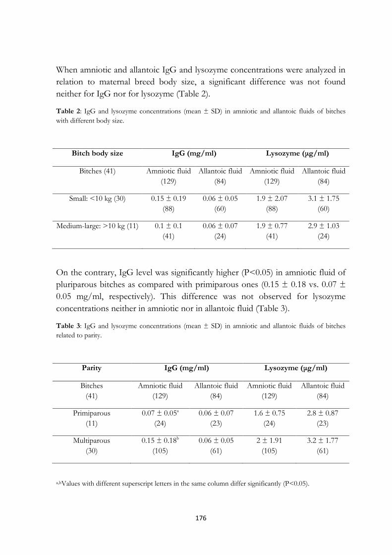
176
When amniotic and allantoic IgG and lysozyme concentrations were analyzed in
relation to maternal breed body size, a significant difference was not found
neither for IgG nor for lysozyme (Table 2).
Table 2: IgG and lysozyme concentrations (mean ± SD) in amniotic and allantoic fluids of bitches
with different body size.
Bitch body size IgG (mg/ml) Lysozyme (μg/ml)
Bitches (41) Amniotic fluid
(129)
Allantoic fluid
(84)
Amniotic fluid
(129)
Allantoic fluid
(84)
Small: <10 kg (30) 0.15 ± 0.19
(88)
0.06 ± 0.05
(60)
1.9 ± 2.07
(88)
3.1 ± 1.75
(60)
Medium-large: >10 kg (11) 0.1 ± 0.1
(41)
0.06 ± 0.07
(24)
1.9 ± 0.77
(41)
2.9 ± 1.03
(24)
On the contrary, IgG level was significantly higher (P<0.05) in amniotic fluid of
pluriparous bitches as compared with primiparous ones (0.15 ± 0.18 vs. 0.07 ±
0.05 mg/ml, respectively). This difference was not observed for lysozyme
concentrations neither in amniotic nor in allantoic fluid (Table 3).
Table 3: IgG and lysozyme concentrations (mean ± SD) in amniotic and allantoic fluids of bitches
related to parity.
Parity IgG (mg/ml) Lysozyme (μg/ml)
Bitches
(41)
Amniotic fluid
(129)
Allantoic fluid
(84)
Amniotic fluid
(129)
Allantoic fluid
(84)
Primiparous
(11)
0.07 ± 0.05a
(24)
0.06 ± 0.07
(23)
1.6 ± 0.75
(24)
2.8 ± 0.87
(23)
Multiparous
(30)
0.15 ± 0.18b
(105)
0.06 ± 0.05
(61)
2 ± 1.91
(105)
3.2 ± 1.77
(61)
a,bValues with different superscript letters in the same column differ significantly (P<0.05).

177
In addition, IgG and lysozyme levels in both fluids did not differ significantly
when newborn gender was considered (Table 4).
Table 4: IgG and lysozyme concentrations (mean ± SD) in amniotic and allantoic fluids of bitches
related to fetal gender.
Fetal gender IgG (mg/ml) Lysozyme (μg/ml)
Fetuses
(142)
Amniotic fluid
(129)
Allantoic fluid
(84)
Amniotic fluid
(129)
Allantoic fluid
(84)
Males
(74)
0.14 ± 0.16
(66)
0.06 ± 0.06
(47)
1.8 ± 0.73
(6)
3 ± 1.34
(47)
Females
(68)
0.13 ± 0.17
(63)
0.05 ± 0.05
(37)
2 ± 2.41
(63)
3.1 ± 1.85
(37)
11.4 Discussion
To the authors knowledge, this is the first study in which IgG and lysozyme
were investigated in dog fetal fluids collected from mature, viable, and normal
newborn puppies, born by elective Cesarean section at term. The present
research reported that both IgG and lysozyme are detectable in canine amniotic
and allantoic fluids at term pregnancy, suggesting that, also in the dog, both
fluids provide fetal protection, not only by mechanical cushioning but also as a
part of the innate immune system, which includes antimicrobial effectors, such
as lysozyme, lactoferrin, α-dephensin, and so forth [21]. The amniotic fluid is
known to play a wide variety of roles to allow the correct development and
growth of a healthy fetus. Normally, both maternal and fetal compartments
concur in amniotic fluid composition, whereas allantoic fluid could be
considered as fetal urine. Thus, all the compounds found within the allantoic
fluid presumably result from the fetal excretion; on the contrary, the substances
detected in the amniotic fluid could originate from the maternal, fetal, or both
compartments.
A high interindividual variation was found for amniotic and allantoic fluids, as
suggested by the wide standard deviations, even within each litter, especially for

178
the amniotic IgG. This high interindividual variation could be due to a
multiplicity of causes, including the maternal concentrations of immunoglobulins
and the rate of transplacental transfer, as reported for the final levels of
antibodies in the human neonatal blood [22].
Despite the different type of placenta between dogs and humans, the amniotic
IgG mean concentration (0.14 mg/ml) was similar to that documented for the
women amniotic fluid (0.11-0.17 mg/ml) [23]. Additionally, like in humans, this
IgG level represents about 1/100 of the serum concentration of adult individuals
(10-20 mg/ml in dogs and 8-18 mg/ml in humans). In the human amniotic fluid,
beyond the predominant maternal IgG, different molecular forms of fetal
immunoglobulins were detected [24]. In the present study, it was not possible to
report the exact origin of the amniotic IgG. Thus, it may be speculated that, even
in dogs, the final amount of amniotic IgG could derive from a cumulative
contribution of both maternal and fetal counterparts. Because in women the IgG
placental transfer depends, among several factors, also by the maternal levels of
immunoglobulins, the evaluation of circulating IgG in the bitches would have
been interesting. Unfortunately, despite all bitches were vein-cannulated during
surgery, the analysis of maternal blood IgG and lysozyme was not performed
because most of the owners did not agree for the use of a maternal blood sample
for research purposes. Thus, it was not possible to better elucidate the real
maternal contribution on amniotic IgG and lysozyme. In this study, IgG were
detected also in the allantoic fluid, although at significantly lower concentrations
(about one half) in comparison with the amniotic fluid. This finding could, on
one hand, be explained by a direct fetal production of IgG, but, on the other
hand, the presence of IgG in the fetal urine could be merely due to the
functional immaturity of the kidney in the canine fetus. In fact, the renal
glomeruli are still developing and permeable at large molecules like antibodies,
that can therefore pass through the kidney in the urine and then in the allantoic
fluid [25].
Beyond other essential substances, the fetal fluids contain lysozyme (mean 1.9
and 3.1 μg/ml in the amniotic and allantoic fluid, respectively), in concentrations
similar to the lower levels reported in the serum of adult dogs (2-13 μg/ml;
unpublished data). Also for lysozyme, it could be supposed a maternal, fetal, or
materno-fetal origin. Even if more homogeneous levels (as evidenced by the
narrow standard deviations) were found in the allantoic as compared with

179
amniotic fluid, no significant differences were detected in lysozyme
concentrations between the two fluids. Several studies highlighted the presence
of a multitude of innate antimicrobial peptides in the vernix caseosa and
amniotic fluid in humans [26]. In women, these antimicrobial peptides act as
important immunomodulators, working synergistically to provide a first defense
line and promote an interaction between the adaptive and innate immune system
in the newborn [27]. It is reasonable to suppose that, even in the dog, lysozyme
could be active in fetal fluids similarly to what suggested for humans, providing a
first line of defense against infectious agents.
No significant differences were found between IgG or lysozyme amniotic or
allantoic concentrations and maternal breed body size. This is in agreement with
what observed in humans, because a relation between amniotic IgG levels and
race/ethnicity was not reported. In a study performed by Kennedy et al. [28],
considering the immune response to antirabies vaccination in dogs, small size
breeds were considered “high responders”, in comparison with large size breeds.
It is interesting to note the finding of significantly (P<0.05) higher IgG amniotic
concentrations in samples collected from multiparous compared with
primiparous bitches, which further reinforce the hypothesis of a major maternal
contribution to the final amniotic IgG amount. This result appears in
disagreement with the lack of correlation between maternal parity, and age, and
amniotic fluid IgG levels reported for humans [9, 10].
A possible effect of the fetal gender on amniotic and allantoic IgG and lysozyme
concentrations was not highlighted, confirming that, also in canine species, both
fluids belonging to female or male newborns have similar immunoglobulins and
antimicrobial lysozyme levels.
A final consideration concerns the four dogs undergone the vaccination against
Canine Herpes Virus during pregnancy. Although the statistical analysis on the
possible differences in IgG (and lysozyme) concentrations between the fetal
fluids collected by vaccinated and nonvaccinated bitches was not performed
because of the paucity of samples belonging to vaccinated subjects (11 amniotic
and allantoic samples), just at a descriptive analysis, the IgG levels resulted very
similar in the two groups of samples (mean 0.04 vs. 0.06 mg/ml in samples from
vaccinated and nonvaccinated bitches, respectively). In humans, it was
documented that, even if several vaccines are given to pregnant women to

180
guarantee the fetal protection during gestation or the neonatal protection after
birth, many factors may limit the efficacy of antibodies placental transfer after
maternal vaccination [9].
In conclusion, the results of the present study reported that the amniotic and
allantoic fluids play a role in the immune protection of the fetus/newborn also in
canine species. Class G immunoglobulins and lysozyme were detected in both
amniotic and allantoic fluids, but IgG levels were higher in amniotic fluid. A
significant effect of the maternal parity, but not of the breed body size, on the
amniotic IgG concentrations was observed, whereas the newborn gender was
not associated to different IgG or lysozyme amniotic or allantoic levels. Given
the significant contributions of fetal fluids to fetal and neonatal health, additional
research is needed to better elucidate both the origin of IgG and lysozyme and
the factors influencing the wide interindividual variations.

181
11.6 References
[1] Barrios C, Brawand P, Berney M, Brandt C, Lambert PH, Siegrist CA.
Neonatal and early life immune responses to various forms of vaccine antigens
qualitatively differ from adult responses: predominance of a Th2-biased pattern
which persists after adult boosting. Eur J Immunol 1996; 26: 1489-1496.
[2] Schultz RD, Thiel B, Mukhtar E, Sharp P, Larson LJ. Age and long-term
protective immunity in dogs and cats. J Comp Path 2010; 142:S102-108.
[3] King BF, Pinheiro PBN, Hunter RL. The fine structure of the placental
labyrinth in the sloths, Bradypus tridactylus. Anat Rec 1982; 202:15-22.
[4] Miglino MA, Ambrosio CE, dos Santos Martins D, Valverde Wenceslau C,
Pfarrer C, Leiser R. The carnivore pregnancy: The development of the embryo
and fetal membranes. Theriogenology 2006; 66:1699-1702.
[5] Leiser R, Kaufmann P. Placental structure: in a comparative aspect. Exp Clin
Endocrinol 1994; 102:122-134.
[6] Tizard IR. Immunity in the fetus and newborn. In: Tizard IR, editor.
Veterinary immunology An introduction, 8th ed, St. Louis, Missouri: Saunders
Elsevier; 2009, p. 223-238.
[7] Stoffel MH, Friess AE, Hartmann SH. Ultrastructural evidence of
transplacental transport of immunoglobulin G in bitches. J Repr Fert 2000;
118:315-326.
[8] Day MJ. Immune system development in the dog and cat. J Comp Pathol
2007; 137 Suppl 1:S10-15.
[9] Palmeira P, Quinello C, Silveira-Lessa AL, Zago CA, Carneiro-Sampaio M.
IgG Placental Transfer in Healthy and Pathological Pregnancies. Clin Dev
Immunol 2012; ID 985646.
[10] Gelber SE, Bongiovanni AM, Jean-Pierre C, Linhares IM, Skupski DW,
Witkin SS. Antibodies to the 70 kDa heat shock protein in midtrimester amniotic
fluid and intraamniotic immunity. Am J Obstet Gynecol 2007; 197:278.e1-4.
[11] Poggi SH, Spong CY, Ghidini A, Ossandon M. Gender differences in
amniotic fluid cytokine levels. J Matern Fetal Neonatal Med 2004; 15:367-371.

182
[12] Schlievert P,Johnson W, Galask RP Amniotic fluid antibacterial
mechanisms: newer concepts. Seminars in Perinatology 1977; 1:59-70.
[13] Barling PM, John MJ, Walsh JR, Niall HD. The isolation and
characterization of lysozyme from human foetal membranes: a comparison with
the enzyme from other sources. Comp Biochem Physiol B 1985; 81:509-513.
[14] Callewaert L, Michiels CW. Lysozymes in the animal kingdom. J Biosci
2010; 35(1):127-160.
[15] Umezu T, Hisanaga S, Shimokawa H, Kashiwabara Y, Maesato S. Origin of
lysozyme in amniotic fluid. Nihon Sanka Fujinka Gakkai Zasshi 1985; 37:301-
303.
[16] Hisanaga S, Umezu T, Shimokawa H, Maesato S. Amniotic fluid lysozyme
content in normal and abnormal pregnancy. Nihon Sanka Fujinka Gakkai Zasshi
1982; 34:541-544.
[17] Meloni T, Comin A, Rota A, Peric T, Contri A, Veronesi MC. IGF-I and
NEFA concentrations in fetal fluids of term pregnancy dogs. Theriogenology
2014; 81:1307-1311.
[18] Veronesi MC, Panzani S, Faustini M, Rota A. An Apgar scoring system for
routine assessment of newborn puppy viability and short-term survival
prognosis. Theriogenology 2009; 72:401-407.
[19] Piantedosi D, Servida F, Cortese L, Puricelli M, Benedetti V, Di Loria A, et
al. Colostrum and serum lysozyme levels in Mediterranean buffaloes (Bubalus
bubalis) and in their newborn calves. Vet Rec 2010; 166:83-85.
[20] Osserman EF, Lawlor DP. Serum and urinary lysozyme (muramidase) in
monocytic and monomyelocytic leukemia. J Exp Med 1966; 124:951-952.
[21] Underwood MA, Gilbert WM, Sherman MP. Amniotic fluid: not just fetal
urine anymore. J Perinatol 2005; 25:341-348.
[22] Chucri TM, Monteiro JM, Lima AR, Salvadori MLB, Kfoury Junior
JR, Miglino MA. A review of immune transfer by the placenta. J Reprod
Immunol 2010; 87:14-20.

183
[23] Cederqvist LL, Ewool LC, Bonsnes RW, Litwin SD. Detectability and
pattern of immunoglobulins in normal amniotic fluid throughout gestation. Am J
Obstet Gynecol 1978; 130:220-224.
[24] Quan CP, Forestier F, Bouvet JP. Immunoglobulins of the human amniotic
fluid. Am J Reprod Immunol 1999; 42:219-225.
[25] Schäfer-Somi S, Bar Schadler S, Aurich J E. Proteinuria and
immunoglobulinuria in neonatal dogs. Vet Rec 2005; 157:378-382.
[26] Akinbi HT, Narendran V, Pass AK, Markart P, Hoath SP. Host defense
proteins in vernix caseosa and amniotic fluid. Am J Obstet Gynecol 2004;
191(6):2090-6.
[27] Yoshio H, Lagercrantz H, Gudmundsson GH, Agerberth B. First line of
defense in early human life. Semin Perinatol 2004; 28:304-311.
[28] Kennedy LJ, Lunt M, Barnes A, McElhinney L, Fooks AR, Baxter DN,
Ollier WE. Factors influencing the antibody response of dogs vaccinated against
rabies. Vaccine 2007; 25:8500-8507.

184

185
CHAPTER 12
Cortisol levels
in hair and nails
of newborn puppies

186

187
12.1 Introduction
The late pregnancy and the neonatal period represent two most stressful stages,
respectively for mammals fetus and newborn. In canine species, natimortality
and neonatal losses account to up 30%, so that a full knowledge about the fetal
development physiology during the last intrauterine phase, as well as about the
process of birth and neonatal adaptation, seems to be necessary to reduce the
impact of canine neonatal death. However, unlike other domestic animals
species, these topics remain still not completely clarified in dogs.
12.2 Cortisol assessment in biological matrices
Toward the end of gestation, the fetal HPA axis activation of the subject occurs
and, in response to adrenocorticotrophic hormone, the adrenals release cortisol
(C), beyond other hormones which were recognized the major steroids produced
by the fetus itself (Gitau et al., 2001). During the last days of pregnancy, C is
involved in multi-organs final maturation (Bolt et al., 2001) and trigger of
parturition process, whereas during the first month of age it is released in
response to some important neonatal physiologic changes and gradual
adaptation to the extrauterine life (Thorburn et al., 1972; Fowden, 1995).
Cortisol is the most common glucocorticoid in humans, non-human primates,
and many larger mammals, in which it is considered the primary end point stress
hormone. In fact, it was judged a perfect biomarker of acute or chronic stress
and a useful tool for long-term monitoring of HPA axis activity in both humans
and animals (Russell et al., 2011). This ability to reflect stress levels over long
periods of time was limited probably because of the nature of the traditional
matrices in which the hormone levels were measured. Indeed, in the most
studies, C concentrations were regularly assessed through serum, saliva, and
urine. Both serum and saliva samples provide an instantaneous view of the C
levels; therefore, they can be used to test only acute changes, since are subject to
major physiological daily fluctuations. Total serum C includes both protein-
bound and free cortisol; consequently, it could result affected in case of changes
in the levels of cortisol-binding globulin. Instead, in saliva only unbound and
bioactive hormone can be measured and the collection is less invasive.
Moreover, both serum and saliva samples require specific procedures for the
storage. On the contrary, urine provide a short-term view of C levels, which

188
follow a particular diurnal rhytm. Urinary C reflects only the free portion of the
hormone and requires the refrigeration or freezing of the sample (Russell et al.,
2011; Meyer and Novak, 2012; Stalder and Kirschbaum, 2012; Maidana et al.,
2013).
12.3 Hair and nails for hormonal measurement
In humans and animals, hair represents the newest biological matrix in which
hormone levels measurement is possible. For decades, hair analysis was used for
monitoring exposure to exogenous compounds, overall drugs of abuse. More
recently, there was a growing interest in quantifying endogenously produced
compounds (Gow et al., 2010). Cortisol concentrations analysis in hair samples is
a non invasive technique and it allows to have a long-term retrospective picture
of the hormone previous accumulation; in fact, hair C results by the
accumulation and incorporation from plasma over a period of months before
sample collection. Additionally, hair C levels are not affected by circadian
hormonal variations or by factors inducing short-term changes (Russell et al.,
2011; Meyer and Novak, 2012; Stalder and Kirschbaum, 2012; Maidana et al.,
2013).
Cortisol is supposed to enter the hair at the shaft medulla through passive
diffusion from blood. This diffusion is amplified by high lipid solubility and low
protein binding, suggesting the preferential deposition of unbound cortisol.
Additional C may coat the outer cuticle from sebaceous and eccrine secretions
(Raul et al., 2004; Pragst and Balikova, 2006). In a recent study, Russell et al.
(2014) documented that human sweat contains C concentrations comparable
with saliva, thus perfuse sweating after intense exercise may increase hormone
levels detected in hair. Whether the hair C is representative of systemic
concentrations remains under discussion. Most authors suggested that hair C
content is representative of systemic levels, even if local hormone production
may participate; indeed, it was demonstrated that hair follicles contain a
functional equivalent of the HPA axis and can synthesize C after stimulation by
CRH (Ito et al., 2005).
Hair C measurement was reported as validated method in humans (Raul et al.,
2004; Sauvé et al., 2007; Kirschbaum et al., 2009; Gow et al., 2010, D’Anna-
Hernandez et al., 2011), grizzly bears (Macbeth et al., 2010), rhesus monkeys
(Davenport et al., 2006), cows (Comin et al., 2011; Comin et al., 2013; Del Rosario

189
Gonzàlez-de-la-Vara et al., 2011), horses (Comin et al., 2012), dogs, and cats
(Accorsi et al., 2008), even if the number of studies in small animals is absolutely
lower than those performed in humans.
During gestation it was demonstrated that a strong maternal stress can negatively
affect fetal HPA axis regulation (Van den Bergh et al., 2007) and that abnormal
patterns of C are associated with consequent miscarriage, increased fetal activity,
premature birth, and decreased birth weight in women (Nepomnaschy et al.,
2006; Field and Diego, 2008). Interestingly, D’Anna-Hernandez et al. (2011)
reported a gradual increase of C amounts during human pregnancy, highlighting
the correlation between hair and salivary C levels in pregnant and post-partum
women. Yamada et al. (2007) evaluated hair C concentrations in newborns,
founding higher values in hospitalized neonates than in healthy ones, probably
due to their more stressful condition.
In a recent study on bovine species, hair C levels were analysed in clinically or
physiologically compromised cows, as well as in clinically normal lactating cows
at least two month after calving. The findings demonstrated a significant positive
correlation between hair C and severity of clinical condition, and significant
differences between hair C levels recorded in each disease subgroup compared
with clinical healthy cows (Comin et al., 2013).
Cortisol concentrations were assessed even in equine hair. Samples collected
from normal, healthy foals at birth, 30 and 60 days of age, revealed a significant
decline of C levels from birth to two months of age, with high inter-individual
variations, underlining a progressive adaptation of the newborn to the
extrauterine environment (Comin et al., 2012). Silver et al. (1984) reported low
plasma C concentrations in premature foals, thus analysis of hair samples could
be interesting in case of prematurity.
To date, little is known about the hair C detection in canine species, even less in
cats. In dogs Bryan et al. (2013) evidenced that a single hair sample is better than
multiple samples of saliva or faeces to evaluate long-term C levels; in this study
no correlations were found neither between hair and faeces nor between hair
and saliva, in contrast to previous papers (Accorsi et al., 2008; Bennett and
Hayssen, 2010). In canine species, a difference in C concentrations was detected
on the basis of different coat colour: particularly, black dogs had less C in hair
than non black dogs and, within the same subject, black hair was lower in C than

190
not black one. In contrast to humans (Kirschbaum et al., 2009), the average
amount of C did not differ between proximal and distal hair sections (Bennett
and Hayssen, 2010), probably due to the different hair length and hygiene. To
date, the possible influence of gender on the hormonal concentrations was
investigated in plasmatic samples, suggesting higher plasma C levels in adult male
dogs than in female ones (Mongillo et al., 2014), whereas the previous literature
reported no sex-related differences in basal concentrations in canine species
(Reimers et al., 1984; Garnier et al., 1990; Reimers et al., 1990; Hennessy et al.,
1997). Previous researches documented that basal C levels appeared to be
influenced also by breed body size and age (Reimers et al., 1990). Indeed, smaller
breeds tended to have higher basal C concentrations than larger breeds, as well
as nursing pups had higher levels than older dogs (Reimers et al., 1984; Reimers
et al., 1990; Hennessy et al., 1997). Furthermore, in stress situations females
showed higher C concentrations than males, as well as juvenile puppies seemed
to have lower C levels than older subjects (Garnier et al., 1990; Hennessy et al.,
1998). To date, to the author knowledge, only one study analysed C profile in
canine neonates plasma, to investigate whether maternal corticosteroid treatment
can improve neonatal vitality and alter maternal and neonatal endogenous C
concentrations (Vannucchi et al., 2012).
Recently, in adult dogs, hair C measurement was used for the diagnosis of
hypercortisolism (Corradini et al., 2013; Ouschan et al., 2013), whereas very few
studies were performed on hair C levels (Accorsi et al., 2008; Finkler and Terkel,
2010; Galuppi et al., 2013).
Although hair represents a promising matrix for hormones measurement in
newborns, in fetus it starts to grow in late pregnancy (at about 45 days of
gestation in dog), so that its analysis provides information limited to the last
period of intrauterine development.
Another valuable matrix, providing cumulative cortisol exposure, is nails
(Warnock et al., 2010; Maidana et al., 2013), collected without invasiveness and
stored at room temperature. The nails start growing around the eighth week of
pregnancy in humans, whereas by 40 days of gestation they are already formed in
dogs (Pretzer, 2008), so that the cumulative information cover a longer window
of time in comparison to the hair (Palmeri et al., 2000; de Berker et al., 2007; Ben
Khelil et al., 2011). Similarly to the hair, the hormones passively diffuse from

191
blood to the matrix of nails and are incorporated into the keratin along nail
growth (Ben Khelil et al., 2011). Recently, C was assessed in fingernails of adults
humans beings (Warnock et al., 2010; Ben Khelil et al., 2011). In humans, fetal
HPA axis starts to work as early as 8 weeks gestation (Mesiano and Jaffe, 1997),
thus this hormone is available for incorporation into the nail throughout large
part of pregnancy. The nails growth rate was investigated and it was reported
variable between 1.9 and 4.4 mm/month, with a reasonable guide of 3
mm/month, or 0.1 mm/day (de Berker et al., 2007). Under physiologic
conditions, several maternal and fetal factors could influence C nails
accumulation in fetus and newborn. Among maternal factors, the breed, age, and
parity should be considered, even though Tegethoff et al. (2011) did not report
an effect of these parameters on the concentrations of other steroids in infants
nails. The neonatal main potential influencing factors are the age at the time of
collection and gender. Since the hormones chronic incorporation, the analysis of
nails collected at birth provides a retrospective picture of the hormones
accumulation between the time of nails appearance to the time of birth.
Subsequent timed nails sampling would provide information about the
hormones incorporation in the interval between the two collections, as
previously reported for hair C concentrations in newborn foals (Comin et al.,
2012). Concerning the fetal gender, several researches documented sex
differences in some components of the HPA stress system, demonstrating the
sex-specificity of many central and peripheric signals (Goel et al., 2014). In
human infants nails, Tegethoff et al. (2011) did not evidence differences between
male and female newborns in some steroids levels.
Johnson et al. (2004), ten years ago, assessed cortisol concentrations in equine
hoof to clarify the role of glucocorticoids in the pathogenesis of laminitis.
Indeed, this hormone resulted detectable in the hoof, that could be maybe
employed also in the newborn foals to perform retrospective hormonal
evaluations. Recently, the C assessment in nails was validated even in bovine
species, in which the claws, beyond the hair, can thus be used to provide
retrospective hormonal information (Comin et al., 2014). However, further
studies are required to validate this technique in other animal species.
In the purpose to improve knowledge about fetal final stage of development and
neonatal biology in canine species, the present study was aimed to assess the
detectability of C in newborn puppies, by using new, non invasive matrices, such

192
as hair and nails, evaluating possible differences in C levels according to the
newborn gender, breed size, and age, and, relatively to the hair, detecting the
possible effect of the coat colour on the hormonal concentrations. Furthermore,
the final aim was to evaluate possible correlations in C levels between the two
biological matrices.
12.4 References
Accorsi, P.A., Carloni, E., Valsecchi, P., Viggiani, R., Gamberoni, M.,
Tamanini, C., Seren, E. (2008) Cortisol determination in hair and faeces from
domestic cats and dogs. Gen Comp Endocrinol 155, 398-402.
Ben Khelil, M., Tegethoff, M., Meinlschmidt, G., Jamey, C., Ludes, B.,
Raul, J.S. (2011) Simultaneous measurement of endogenous cortisol, cortisone,
dehydroepiandrosterone, and dehydroepiandrosterone sulfate in nails by use of
UPLC-MS-MS. Anal Bioanal Chem 401(4), 1153-1162.
Bennett, A., Hayssen, V. (2010) Measuring cortisol in hair and saliva from
dogs: coat color and pigment differences. Dom Anim Endocrinol 39, 171-180.
Bolt, R.J., Van Weissenbruch, M.M., Lafeber, H.N., Wall, H.A.V. (2001)
Glucocorticoids and lung development in the fetus and preterm infant. Pediatr
Pulmonol 32, 76-91.
Bryan, H.M., Adams, A.G., Invik, R.M., Wynne-Edwards, K.E., Smits,
J.E.G. (2013) Hair as a meaningful measure of baseline cortisol levels over time
in dogs. J Am Assoc Lab Anim Sci 52(2), 189-196.
Comin, A., Peric, T., Corazzin, M., Veronesi, M.C., Meloni, T., Zufferli,
V., Cornacchia, G., Prandi, A. (2013) Hair cortisol as a marker of
hypothalamic-pituitary-adrenal axis activation in Friesian dairy cows clinically or
physiologically compromised. Livestock Science 152(1), 36-41.
Comin, A., Peric, T., Magrin, L., Corazzin, M., Cornacchia, G., Prandi, A.
(2014) Study of progesterone and cortisol concentrations in the Italian Friesian
claw. J Dairy Sci 97(9), 5491-6.

193
Comin, A., Prandi, A., Peric, T., Corazzin, M., Dovier, S., Bovolenta, S.
(2011) Hair cortisol levels in dairy cows from winter housing to summer
highland grazing. Livestock Science 138(1), 69-73.
Comin, A., Veronesi, M.C., Montillo, M., Faustini, M., Valentini, S.,
Cairoli, F., Prandi, A. (2012) Hair cortisol levels as a retrospective marker of
hypothalamic-pituitary-adrenal axis activity in horse foals. Vet J 194(1), 131-132.
Corradini, S., Accorsi, P.A., Boari, A., Beghelli, V., Mattioli, M., Famigli-
Bergamini, P., Fracassi, F. (2013) Evaluation of hair cortisol in the diagnosis
of hypercortisolism in dogs. J Vet Intern Med 27, 1268-1272.
D’Anna-Hernandez, K.L., Ross, R.G., Natvig, C.L., Laudenslager, M.L.
(2011) Hair cortisol levels as a retrospective marker of hypothalamic-pituitary
axis activity throughout pregnancy: Comparison to salivary cortisol. Physiol
Behav 104, 348-353.
Davenport, M.D., Tiefenbacher, S., Lutz, C.K., Novak, M.A., Meyer, J.S.
(2006) Analysis of endogenous cortisol concentrations in the hair of rhesus
macaques. Gen Comp Endocrinol 147, 255-261.
De Berker, D.A.R., Andre, J., Baran, R. (2007) Nail biology and nail science.
Inter J Cosm Sci 29, 241-275.
Del Rosario Gonzalez-de-la-Vara, M., Valdez, R.A., Lemus-Ramirez, V.,
Vázquez-Chagoyán, J.C., Villa-Godoy, A., Romano, M.C. (2011) Effects of
adrenocorticotropich ormone challenge and age on hair cortisol concentrations
in dairy cattle. Can J Vet Res 75, 216-221.
Field, T., Diego, M. (2008) Cortisol: the culprit prenatal stress variable. Int J
Neurosci 118, 1181-205.
Finkler, H., Terkel, J. (2010) Cortisol levels and aggression in neutered and
intact free-roaming female cats living in urban social groups. Physiol Behav 99,
343-347.
Fowden, A.L. (1995) Endocrine regulation of fetal growth. Reprod Fertil Dev
7(3), 351-363.

194
Galuppi, R., Leveque, J.F., Beghelli, V., Bonoli, C., Mattioli, M.,
Ostanello, F., Tampieri, M.P., Accorsi, P.A. (2013) Cortisol levels in cats’
hair in presence or absence of Microsporum canis infection. Res Vet Sci 95(3),
1076-80.
Garnier, F., Benoit, E., Virat, M., Ochoa, R., Delatour, P. (1990) Adrenal
cortical response in clinically normal dogs before and after adaptation to a
housing environment. Lab Anim 24, 40-3.
Gitau, R., Fisk, N.M., Teixeira, J.M., Cameron, A., Glover, V. (2001) Fetal
hypothalamic-pituitary-adrenal stress responses to invasive procedures are
independent of maternal responses. J Clin Endocrinol Metab 86, 104-109.
Goel, N., Workman, J.L., Lee, T.T., Innala, L., Viau, V. (2014) Sex
differences in the HPA axis. Comprehensive Physiology 4(3), 1121-1155.
Gow, R., Thomson, S., Rieder, M., Van Uum, S., Koren, G. (2010) An
assessment of cortisol analysis in hair and its clinical applications. Forensic Sci
Int 196, 32-37.
Hennessy, M.B., Davis, H.N., Williams, M.T., Mellott, C., Douglas, C.W.,
Voith, V.L. (1997) Plasma cortisol levels of dogs at a county animal shelter.
Physiol Behav 62, 485-90.
Hennessy, M.B., Williams, M.T., Miller, D.D., Douglas, C.W., Voith, V.L.
(1998) Influence of male and female petters on plasma cortisol and behavior: can
human interaction reduce the stress of dogs in a public animal shelter? Appl
Anim Behav Sci 61, 63-77.
Ito, N., Ito, T., Kromminga, A., Bettermann, A., Takigawa, M., Kees, F.,
Straub, R.H., Paus, R. (2005) Human hair follicles display a functional
equivalent of the hypothalamic-pituitary-adrenal axis and synthesize cortisol.
FASEB J 19, 1332-1334.
Johnson, P.J., Ganjam, V.K., Slight, S.H., Kreeger, J.M., Messer, N.T.
(2004) Tissue-specific dysregulation of cortisol metabolism in equine laminitis.
Equine Vet J 36(1), 41-5.

195
Kirschbaum, C., Tietze, A., Skoluda, N., Dettenborn, L. (2009) Hair as a
retrospective calendar of cortisol production-increased cortisol incorporation
into hair in the third trimester of pregnancy. Psychoneuroendocr 34, 32-37.
Macbeth, B.J., Cattet, M.R.L., Stenhouse, G.B., Gibeau, M.L., Janz, D.M.
(2010) Hair cortisol concentration as a noninvasive measure of long-term stress
in free-ranging grizzly bears (Ursus arctos): considerations with implications for
other wildlife. Can J Zool 88, 935-949.
Maidana, P., Bruno, O.D., Mesch, V. (2013) A critical analysis of cortisol
measurements: an update. Medicina (B Aires) 73(6), 579-84.
Mesiano, S., Jaffe, R.B. (1997) Developmental and functional biology of the
primate fetal adrenal cortex. Endocr Rev 18, 378-403.
Meyer, J.S., Novak, M.A. (2012) Minireview: Hair cortisol: a novel biomarker
of hypothalamic-pituitary-adrenocortical activity. Endocrinology 153(9), 4120-7.
Mongillo, P., Prana, E., Gabai, G., Bertotto, D., Marinelli, L. (2014) Effect
of age and sex on plasma cortisol and dehydroepiandrosterone concentrations in
the dog (Canis familiaris). Res Vet Sci 96, 33-38.
Nepomnaschy, P.A., Welch, K.B., McConnell, D.S., Low, B.S.,
Strassmann, B.I., England, B.G. (2006) Cortisol levels and very early
pregnancy loss in humans. Proc Natl Acad Sci USA 103, 3938-42.
Ouschan, C., Kuchar, A., Mostl, E. (2013) Measurement of cortisol in dog
hair: a noninvasive tool for the diagnosis of hypercortisolism. Vet Dermatol 24,
428-e94.
Palmeri, A., Pichini, S., Pacifici, R., Zuccaro, P., Lopez, A. (2000) Drugs in
nails: physiology, pharmacokinetics and forensic toxicology. Clin Pharmacokinet
38(2), 95-110.
Pragst, F., Balikova, M.A. (2006) State of the art in hair analysis for detection
of drug and alcohol abuse. Clin Chim Acta 370, 17-49.
Pretzer, S.D. (2008) Canine embryonic and fetal development: A review.
Theriogenology 70(3), 300-303.

196
Raul, J., Cirimele, V., Ludes, B., Kintz, P. (2004) Detection of physiological
concentrations of cortisol and cortisone in human hair. Clin Biochem 37(12),
1105-11.
Reimers, T.J., Lawler, D.F., Sutaria, P.M., Correa, M.T., Erb, H.N. (1990)
Effects of age, sex, and body size on serum concentrations of thyroid and
adrenocortical hormones in dogs. Am J Vet Res 51, 454-7.
Reimers, T.J., Mummery, L.K., McCann, J.P., Cowan, R.G., Concannon,
P.W. (1984) Effects of reproductive state on concentrations of thyroxine, 3,5,3’-
triiodothyronine and cortisol in serum of dogs. Biol Reprod 31(1), 148-54.
Russell, E., Koren, G., Rieder, M., Van Uum, S.H. (2011) Hair cortisol as a
biological marker of chronic stress: Current status, future directions and
unanswered questions. Psyconeuroendocrinol 37(5), 589-601.
Russell, E., Koren, G., Rieder, M., Van Uum, S.H. (2014) The detection of
cortisol in human sweat: implications for measurement of cortisol in hair. Ther
Drug Monit 99(1), 90-e.
Sauvé, B., Koren, G., Walsh, G., Tokmakejian, S., VanUum, S. (2007)
Measurement of cortisol in human hair as a biomarker of systemic exposure.
Clin Invest Med 30, E183-E191.
Silver, M., Ousey, J.C., Dudan, F.E., Fowden, A.L., Knox, J., Cash, R.S.,
Rossdale, P.D. (1984) Studies on equine prematurity 2: Post natal
adrenocortical activity in relation to plasma adrenocorticotrophic hormone and
catecholamine levels in term and premature foals. Equine Vet J 16(4), 278-286.
Stalder, T., Kirschbaum, C. (2012) Analysis of cortisol in hair-state of the art
and future directions. Brain Behav Immun 26(7), 1019-1029.
Tegethoff, M., Raul, J., Jamey, C., Ben Khelil, M., Ludes, B.,
Meinlschmidt, G. (2011) Dehydroepiandrosterone in nails of infants: A
potential biomarker of intrauterine response to maternal stress. Biol Psychol 87,
414-420.
Thorburn, G.D., Nicol, D.H., Bassett, J.M., Shutt, D.A., Cox, R.I. (1972)
Parturition in the goat and sheep-changes in corticosteroids, progesterone,
oestrogens and prostaglandin. J Reprod Fertil Suppl 16, 61-84.

197
Van den Bergh, B.R.H., Van Calster, B., Smits, T., Van Huffel, S., Lagae,
L. (2007) Antenatal maternal anxiety is related to HPA-axis dysregulation and
self-reported depressive symptoms in adolescence: A prospective study on the
fetal origins of depressed mood. Neuropsychopharmacol 33, 536-45.
Vannucchi, C.I., Regazzi, F.M., Barbosa, M.M.M., Silva, L.G.C., Veiga,
G.A.L., Lúcio, C.F., Angrimani, D.S., Nichi, M., Furtado, P.V., Oliveira,
C.A. (2012) Cortisol profile and clinical evaluation of canine neonates exposed
antenatally to maternal corticosteroid treatment. Reprod Dom Anim 47(Suppl.
6), 173-176.
Warnock, F., McElwee, K., Seo, R.J., McIsaac, S., Seim, D., Ramirez-
Aponte, T., Macritchie, K.A., Young, A.H. (2010) Measuring cortisol and
DHEA in fingernails: a pilot study. Neuropsychiatr Dis Treat 6, 1-7.
Yamada, J., Stevens, S., de Silva, N., Gibbins, S., Beyene, J., Taddio, A., et
al. (2007) Hair cortisol as a potential biologic marker of chronic stress in
hospitalized neonates. Neonatology 92, 42-49.

198

199
CHAPTER 13
Hair and nails
as new, non-invasive matrices
for long time-frame cortisol analysis
in newborn dogs

200

201
13. Hair and nails as new, non-invasive matrices for
long time-frame cortisol analysis in newborn dogs
Veronesi MCa, Comin Ab, Meloni T a*, Faustini M, Rota Ac, Prandi Ab
a Department of Health, Animal Science and Food Safety, Faculty of Veterinary
Medicine, Università degli Studi di Milano, via G. Celoria 10, 20133 Milan, Italy b Department of Food Science, University of Udine, via Sondrio 2, 33100 Udine,
Italy c Ambulatorio Veterinario Associato Dr. Pellegrini-Dr. Rota, via Ungaretti 69,
24030 Almenno San Bartolomeo, Bergamo, Italy
13.1 Abstract
The last intrauterine fetal stage of development and the neonatal period
represent the most challenging phases for the mammals offspring. In the dog,
the knowledge about the final intrauterine fetal development and biology, as well
as about the neonatal physiology, remains still scarce. Hormonal changes
occurring in the last intrauterine fetal phase and during the early neonatal age are
still not completely clear, probably because of the invasiveness related to the
collection of the more common biological matrix, represented by circulating
blood.
Toward the end of gestation, during parturition and after birth, the HPA axis is a
key system regulating several physiologic processes, and its activity was
investigated for a long time by blood analysis, considered an invasive and single
point measurement. In respect to animal welfare and for a more correct long
time-frame retrospective investigation, non-invasive hormonal studies were
performed firstly in the hair of both humans and animals and, more recently, in
the nails of human beings. This study was aimed to assess C in hair and nails of
newborn puppies, and to evaluate the possible influence of the newborn gender,
breed body size, and age on hair and nails C concentrations. The results obtained
from 165 newborn puppies evidenced that hair and nails C levels were highly
correlated each other (P<0.0001), although the C accumulation in the two
matrices was different in relation to the class of age. Moreover, the puppies age

202
influenced both hair and nails C concentrations (P<0.05), with premature
newborns showing higher values when compared to term born-dead puppies or
puppies dead between 1 and 30 days of age. The present study demonstrated
that C is quantifiable in hair and nails of newborn dogs, so that both matrices
appear as interesting tools for new, non-invasive, long time-frame perinatal and
neonatal researches also in canine species.
13.2 Introduction
The last intrauterine fetal stage of development and the neonatal period
represent the most challenging phases for the mammals offspring. Contrary to
other domestic species, in the dog the knowledge about the final intrauterine
fetal development and biology, as well as about the neonatal physiology, remains
still scarce. Hormonal changes occurring in the last intrauterine fetal phase and
during the early neonatal period are still not completely clarified, probably
because of the invasiveness related to the collection of the more common
biological matrix, represented by circulating blood.
Toward term of pregnancy, the fetal hypothalamic-pituitary-adrenal axis (HPA)
becomes a key system regulating several physiologic processes, such as the fetal
multi-organs final maturation [1], the response to stress [2], and the triggering of
parturition [3]. After birth and along the neonatal period, when the newborn
undergoes a series of physiologic and metabolic changes necessary for survival
and health, the HPA system still keeps a primary role in the neonatal adaptation
to the extrauterine life [4, 5].
Activation of the HPA axis results in a cascade of endocrine responses. Cortisol
(C), the main hormone of this axis, represents an appropriate biological endpoint
in the investigation of HPA axis function. In response to the
adrenocorticotrophic hormone (ACTH), the adrenals produce C, considered the
best biomarker of chronic stress both for humans and animals [6]. In several
animal species, HPA activity in the fetus, as well as in the newborn, was in the
past investigated mainly by fetal blood [7] or cord blood analysis [8], but these
methods were limited due to the invasiveness of sampling and to the single point
measurement [2]. These limitations arose the need to detect alternative, non-
invasive techniques for biological matrices collection, useful for the study of long
time-frame hormonal changes in respect to both humans and animals welfare.

203
Alternatively to blood analysis, C levels were therefore assessed in the saliva,
urine, and faeces, collected without invasiveness. Nevertheless, the saliva still
remains representative of a single point hormonal measurement, whereas the
urine and faeces assess the HPA activity over a period of only 24 hours or less.
Recently, some studies reported C measurements in the hair of humans [9-13]
and animals [14-19], providing the evidence that the hair could be considered a
useful biological matrix for the non-invasive and long time-frame retrospective
investigation of previous hormone accumulation. Interesting findings were
reported about the effect of the pregnant condition on hair C accumulation.
Particularly, D’Anna-Hernandez et al. found higher level of C in the hair of
women in late pregnancy compared with those in early gestation or in the post-
partum period, stressing the role of C in human late pregnancy [13].
Thanks to the lack of invasiveness at collection and the long time-window
hormonal accumulation, the hair was proved to be an interesting tool for the
study of C accumulation even in newborn babies. Indeed, hair C measurement
was used to assess the effect of hospitalization in preterm neonates, in
comparison with healthy ones [20]. The study highlighted that hospitalized
newborns showed higher hair C levels compared with healthy babies. More
recently, Comin et al. collected the hair of newborn horse foals to investigate the
changes in C concentrations from birth to 60 days of age [18]. The authors
reported the decline of hair C levels from birth to 30 and 60 days of age,
probably as a response of the HPA axis during the neonatal adaptation to the
extrauterine environment. Although hair C concentration was previously
investigated in adult dogs [15, 21], the authors are not aware of similar studies in
the newborn puppy.
In humans, another valuable matrix providing a long-term view of C
accumulation is fingernail [22-24], collected without invasiveness and easily
stored at room temperature. Similarly to the hair, also fingernails analysis allows
the assessment of hormones accumulated over a long-time period, passively
diffused from capillaries to the matrix of nails, and incorporated into the keratin
along nail growth [24]. To the authors knowledge, to date similar studies were
not performed neither in newborn babies nor in animals. Both the hair and nails,
thanks to lack of invasiveness at collection and because of the long time-frame
retrospective accumulation, could therefore represent interesting biological

204
matrices for the study of C accumulation during the fetal and neonatal periods
also in canine species.
The possible effect of some parameters, such as the ethnicity or race, gender,
age, and coat colour on hair C accumulation was widely investigated in humans
and animals, providing conflicting results [6].
In the purpose to improve knowledge about the final stage of fetal development
and neonatal biology in dogs, by using non-invasive methods, the aims of the
present study were: 1) to assess the detectability of C in newborn puppies,
through new, non-invasive matrices, such as hair and nails; 2) to evaluate
possible influence of the newborn gender, breed size, and age on C
concentrations, in both hair and nails; 3) relatively to the hair, to detect also the
possible effect of coat colour on C levels; 4) to evaluate possible correlations and
differences between the C concentrations in the two matrices.
13.3 Materials and methods
13.3.1 Animals
The study was performed on 165 spontaneously dead purebred puppies, 80
females and 85 males, belonging to several canine breeds, without gross physical
malformations. All the owners and breeders gave a written informed consent to
the collection of hair and nails from dead puppies, for research purposes.
According to maternal body weight, the 165 subjects were classified as belonging
to small size breed (bodyweight ≤10 kg) (N = 60), or to a merged class of
medium-large size breed (bodyweight >10 kg) (N = 105). Based on the age at
death, all newborns were grouped in three classes of age, as follows: 1)
premature: when puppies were delivered between 2 and 1 weeks before the
expected date of whelping, estimated by ultrasonographic parturition date
prediction [25] (N=25); 2) fresh, term-born-dead puppies (puppies delivered
dead at the predicted term of pregnancy and puppies born alive at term, but dead
within 24 hours since birth (N=97); 3) puppies dead between 1 and 30 days of
age (N=43).
205
13.3.2 Hair and nails samples collection
A proper amount of hair (at least 20 mg) was carefully shaved from each puppy.
Hair was collected closest to the skin always from the same area [26], including
only the back and the dorsal portion of the neck. Given the conflicting results
reported in bibliography concerning the influence of coat colour on C
concentrations, when in the same puppy both light and dark hair areas were
detected in a sufficiently large surface to allow the collection, both dark and light
hair were separately collected. The nails of all the fingers were trimmed and
pooled (at least 4 mg). Hair and nails samples were separately stored in dry tubes,
at room temperature, until analysis.
13.3.3 Hair and nails cortisol assays
Hair strands and nails were washed in 5 ml isopropanol to minimize the risk of
extracting C from outside these biological samples and to ensure the removal of
any steroids on their surface. Hair and nails C was extracted with methanol and
measured by RIA [27]. All the samples were freeze-dried as described by Comin
et al. [27] and the dry weights were calculated.
13.3.4 Statistical analysis
Data regarding C levels in hair and nails were statistically analysed by ANOVA,
in order to assess: a) possible differences in hair or nails C concentrations
according to the newborn gender and to breed size; b) possible differences in
hair or nails C levels according to the newborn class of age; c) possible
differences in hair C concentrations according to the coat colour. Furthermore,
the Tukey test was used to define the possible differences in hair and nails C
levels among the three classes of age. Differences between C concentrations in
the two matrices within each class of age were assessed by t-test corrected for
unequal variances. Finally, possible correlations between hair and nails C levels
were analysed by Spearman correlation test. Significance was set at P<0.05.
13.4 Results
Cortisol was detectable in all the hair and nails samples belonging to the 165
newborn puppies. Specifically, in the hair of these subjects, the overall C
concentration (mean ± SD) was 65.2 ± 52.23 pg/mg, whereas in the 165 pooled
samples of nails, the overall C concentration was 62.6 ± 58.8 pg/mg. The

206
Spearman correlation test revealed a highly significant positive correlation
between hair and nails C levels (R=0.68, P<0.0001).
The statistical analysis did not show any difference in C concentrations neither in
the hair nor in the nails in relation to the newborn gender or breed body size
(Table 1), whereas a significant effect of the class of age was found on both hair
and nails C levels, with significant higher values in premature compared with
born dead puppies or puppies dead between 1 and 30 days of age (Figure 1).
From the 165 newborns, based on the coat colour, 68 light and 108 dark hair
samples were collected and analysed for C concentrations. The results showed a
mean (± SD) of 67.6 ± 55.81 pg/mg in light hair compared to 63.0 ± 50.06
pg/mg in dark hair, without significant differences.
When the possible differences in C levels between the two matrices within each
class of age were assessed, the statistical analysis documented the absence of
differences between the C concentrations in the two matrices within the class of
premature puppies (mean ± SD: 123.22 ± 103.41 vs. 79.81 ± 87.49 pg/mg, in
the hair and nails, respectively), whereas in term born-dead subjects C levels
were higher (P<0.05) in the hair compared to the nails (mean ± SD: 66.97 ±
36.56 vs. 53.92 ± 49.91 pg/mg, in the hair and nails, respectively). On the
contrary, in the class of age ranging between 1 and 30 days, C concentrations
were significantly higher (P<0.0001) in the nails compared to the hair (mean ±
SD: 42.97 ± 18.84 vs 77.37 ± 55.29 pg/mg, in the hair and nails, respectively).
Table 1: Hair and nails C concentrations in the 165 puppies, according to newborn gender and breed
size (mean ± SD).
Newborn gender Breed size
Females
(N=80)
Males
(N=85)
Small
(N=60)
Medium-large
(N=105)
Hair C (pg/mg) 57.4 ± 43.22 71.9 ± 59.03 57.9 ± 45.85 84.6 ± 61.77
Nails C (pg/mg) 60.1 ± 60.76 65.1 ± 57.12 59.0 ± 57.74 64.6 ± 59.52
Figure 1: Concentrations of C in hair and nails of the 165 puppies, grouped according to the class of
age (mean ± SD).

207
a, b and A, B P<0.05
13.5 Discussion
The present study showed for the first time that C is detectable in hair and nails
of newborn dogs, demonstrating the usefulness of both hair and nails as non-
invasive, alternative matrices for perinatal hormonal investigations, also in canine
species.
Because the body hair and color marking in dogs appear 44-46 days post coitum
[28], it is reasonable to assume that the collection of hair at birth should reflect
the hormones accumulation from the time of hair appearance to the time of
puppies delivery. Thus, in canine species, the C concentrations measured in hair
collected at birth would depict the hormone accumulated during the last two-
three weeks of pregnancy. To the authors knowledge, specific studies about the
proportion of maternal C crossing the placenta and activating the fetal HPA axis
are lacking in the dog. Nevertheless, some researches in humans and other
animal species demonstrated that maternal C activates the fetal pituitary-adrenal
axis [29-31], so that C accumulated in the fetal hair during gestation could be
produced by the mother, by the fetus itself, or by both mother and fetus.
0
50
100
150
200
250
prematures Born dead 1-30 d
Co
rtis
ol co
ncen
trati
on
(p
g/
mg
)
Newborns class of age
hair C
nails C
A
B
B
a
b
b

208
The authors are not aware of references about the exact timing of nails first
appearance in canine fetus but, at 30 days of gestation, full visible nails are
usually detectable (authors observation). This means that C measured in nails
collected at birth would reflect a longer time of accumulation when compared to
the hair, whereas the question about the exact maternal, fetal, or cumulative
origin of the hormone still remains unsolved.
The overall mean concentrations of C were similar in hair and nails (65 vs. 63
pg/mg), and C levels in these two biological samples were highly correlated,
suggesting that both matrices are representative of the HPA activity in the
newborn puppy. Thus, even the nails could work as an interesting tool for non-
invasive, long time-frame fetal and neonatal studies in canine species. In
addition, nails seem to be more useful compared to the hair for two main
reasons. Firstly because the amount of hair necessary for the analysis is relevant
(about 20 mg) as regard to the small size of newborn puppies, so that a large
surface of the body should be shaved. This was not a problem for the current
study because all the subjects enrolled were dead, but the future application of
this procedure to viable newborn puppies could be a problem. The nails, instead,
require a smaller amount of sample to enable the hormone measurement.
Secondly, the earlier appearance of the nails during the fetal development allows
the analysis of C accumulated during a longer time in comparison to the hair.
When the possible influence of the newborn gender or breed body size on hair
and nails C concentrations was assessed, no significant effect was found, most
likely because of the wide standard deviations, indicating high inter-individual
variations. Slightly higher mean C levels were detected in the hair of males
compared to females puppies, nevertheless the difference between the two
genders appeared not significant, in agreement with the absence of sex influence
on hair C concentrations reported by Comin et al. in newborn horse foals [18].
Literature from humans provided conflicting results about this topic. In fact,
while some authors reported the absence of a confounding effect of sex on hair
C concentrations [9, 32-34], Dettenborn et al. suggested higher values in males
when compared to females, in adult humans [35].
The lack of breed body size effect on hair C concentrations seems to be in
contrast with previous findings reported by Fraser et al. [36], who showed a

209
positive correlation between body mass index and urinary C levels in humans.
The different biological matrix used could be the reason for different results.
The statistical analysis failed to find any significant difference in C
concentrations between light and dark hair. Both in humans and animals, the
conflicting results about the effect of hair colour on C levels were fully debated
in the review by Russell et al. [6], so that the lack of a significant effect in
newborn puppies is not surprising.
Concerning the nails, the possible effect of the gender, body mass index, or age
on C concentrations was still not investigated in the few studies performed in
humans, although the effect of gender on adult human nails growth [37] and
possible C accumulation [22] was reported.
The most relevant result was the high significant effect of the class of age on C
levels in the hair, as well as in the nails, with significantly higher values in
puppies delivered prematurely when compared to puppies dead at birth or within
the first 30 days of age. A previous investigation in human infants [20]
highlighted higher hair C concentrations in hospitalized preterm born infants
compared to healthy, born at term, neonates. Since the hospitalized infants were
born at younger gestational age, in comparison to the healthy newborns, the
effect of age could create a potential confusion [6]. On the other hand, it is well
known that in women, in the third trimester of pregnancy, hair C levels appear
higher than those during the first trimester or the post-partum period [13].
Because in the present study it was not possible to ascertain the maternal or fetal
(or both) origin of C accumulated in the hair and nails of premature puppies, it is
reasonable to suppose that the higher C levels observed in both matrices
belonging to premature puppies could be the result of maternal increased C
circulating concentrations, as reported for late pregnant women. Unfortunately,
in the present study hair was not collected from pregnant bitches, so that this
hypothesis still remains to be proved. In humans, it was suggested that even early
detection of stress stimuli by the placenta induces the release of CRH with
consequent increased risk for preterm birth [38]. It is however known that the
effects of glucocorticoids in primates parturition is rather different from other
animals species [39].
Interestingly, also higher nails C levels were detected in premature compared to
born dead subjects and puppies dead within the first 30 days of age. The only

210
one study investigating this hormone in human nails was performed on students
aged between 18 and 24 years, and the effect of age was not assessed [22].
Decreasing hair C concentrations from birth to 60 days of age were reported in
newborn horse foals by Comin et al. [18]. At birth, hair C median levels in horse
foals were about 55 pg/mg, at 30 days about 30 pg/mg, and at 60 days of age
about 16 pg/mg. In the present study, hair C mean concentrations in term-born
dead puppies were about 72 pg/mg, and between 1 and 30 days of age about 48
pg/mg, a bit higher than in newborn horse foals. Unfortunately, unlike the horse
foals, hair C concentrations measured in puppies aged 1 to 30 days enrolled a
more heterogeneous age population, explaining, at least partially, the wide inter-
individual variation. In addition, also the cause of death could have influenced
the C accumulation within this class of age. On the other hand, a wide inter-
individual variation in hair C concentrations was observed in all the three classes
of age, even when the puppies gender or the breed body size were concerned.
This finding highlights that many are the parameters affecting the C
accumulation in the hair of canine fetuses and newborns. Comparing the mean
hair C level measured in puppies of all the three classes of age with data reported
by Accorsi et al. [15] in adult dogs, the hair C mean concentration in newborn
puppies was definitely higher than in adults (about 123 pg/mg in premature
puppies, about 72 pg/mg in term born-dead puppies, and 48 pg/mg in 1-30 days
old puppies vs. 2.1 pg/mg in adult dogs, respectively).
It is interesting to note that, also in the nails, the class of age affected
significantly the C levels. Unfortunately, the authors are not aware of studies
investigating the effect of age in nails C concentrations neither in humans nor in
other animals. However, in an elegant review about nail biology [40], it was
suggested that nails growth is affected by several factors, among which the age
of the subject, with children under 14 having faster growth than adults.
It was finally surprising the finding of the reverse C accumulation in the hair and
nails between term born-dead puppies and puppies 1 to 30 days old, suggesting a
different pattern of C incorporation in the two matrices in the perinatal period
of dogs that deserves further investigations.

211
13.6 Conclusions
This study, for the first time, demonstrated that C is quantifiable in hair and nails
of newborn dogs, so that both matrices are interesting for non invasive, long
time-frame fetal and neonatal studies also in canine species. The most relevant
finding was the significant effect of the class of age on both hair and nails C
concentrations, with higher values in puppies delivered prematurely compared to
term-born dead puppies or puppies dead within the first 30 days of age.
Newborn gender, breed body size and, relatively to the hair, the coat colour do
not seem to affect hair or nails C accumulation during the foetal development or
the first 30 days after birth. The maternal or fetal origin (or both) of this
hormone, as well as the influence of gender, breed size, or coat colour deserve
further investigations in normal puppies, but also open new perspectives for the
study of all the maternal and fetal factors that could affect fetal and/or neonatal
development and well-being. Finally, also the different pattern of C
accumulation between the hair and nails within different classes of age appears
an interesting topic to investigate much deeper.

212
References
[1] Bolt RJ, Van Weissenbruch MM, Lafeber HN, Wall HAV. Glucocorticoids
and lung development in the fetus and preterm infant. Pediatr Pulm 2001; 32:
76-91.
[2] Tegethoff M, Raul JS, Jam C, Ben Khelil M, Meinlschmidt G.
Dehydroepiandrosterone in nails of infants: a potential biomarker of intrauterine
responses to maternal stress. Biol Psychol 2011; 87(3): 414-420.
[3] Thorburn GD, Hollingworth SA, Hooper SB. The trigger for parturition in
sheep: fetal hypothalamus or placenta? J Dev Physiol 1991; 15(2): 71-79.
[4] Thorburn GD, Nicol DH, Bassett JM, Shutt DA, Cox RI. Parturition in the
goat and sheep-changes in corticosteroids, progesterone, oestrogens and
prostaglandin. J Reprod Fertil Suppl 1972; 16: 61-84.
[5] Fowden AL. Endocrine regulation of fetal growth. Reprod Fert Develop
1995; 7(3): 351-363.
[6] Russell E, Koren G, Rieder M, Van Uum S. Hair cortisol as a biological
marker of chronic stress: Current status, future directions and unanswered
questions. Psyconeuroendocrino 2012; 37(5): 589-601.
[7] Kajantie E, Raivio T, Jänne OA, Hovi P, Dunkel L, Andersson S. Circulating
glucocorticoid bioactivity in the preterm newborn after antenatal betamethasone
treatment. J Clin Endocr Metab 2004; 89(8): 3999-4003.
[8] Gitau R, Menson E, Pickles V, Fisk NM, Glover V, MacLachlan N.
Umbilical cortisol levels as an indicator of the fetal stress response to assisted
vaginal delivery. Eur J Obstet Gyn R B 2001; 98(1): 14-17.
[9] Raul J, Cirimele V, Ludes B, Kintz P. Detection of physiological
concentrations of cortisol and cortisone in human hair. Clin Biochem 2014;
37(12): 1105-1111.
[10] Sauvé B, Koren G, Walsh G, Tokmakejian S, VanUum S. Measurement of
cortisol in human hair as a biomarker of systemic exposure. Clin Invest Med
2007; 30: E183-E191.

213
[11] Kirschbaum C, Tietze A, Skoluda N, Dettenborn L. Hair as a retrospective
calendar of cortisol production-increased cortisol incorporation into hair in the
third trimester of pregnancy. Psychoneuroendocrino 2009; 34: 32-37.
[12] Gow R, Thomson S, Rieder M, Van Uum S, Koren G. An assessment of
cortisol analysis in hair and its clinical applications. Forensic Sci Int 2010; 196:
32-37.
[13] D’Anna-Hernandez KL, Ross RG, Natvig CL, Laudenslager ML. Hair
cortisol levels as a retrospective marker of hypothalamic-pituitary axis activity
throughout pregnancy: Comparison to salivary cortisol. Physiol Behav 2011; 104:
348-353.
[14] Davenport MD, Tiefenbacher S, Lutz CK, Novak MA, Meyer JS. Analysis
of endogenous cortisol concentrations in the hair of rhesus macaques. Gen
Comp Endocr 2006; 147: 255-261.
[15] Accorsi PA, Carloni E, Valsecchi P, Viggiani R, Gamberoni M, Tamanini C,
Seren E. Cortisol determination in hair and faeces from domestic cats and dogs.
General and Comparative Endocrinology 2008; 155: 398-402.
[16] Macbeth BJ, Cattet MRL, Stenhouse GB, Gibeau ML, Janz DM. Hair
cortisol concentration as a noninvasive measure of long-term stress in free-
ranging grizzly bears (Ursus arctos): considerations with implications for other
wildlife. Endocrinology 2010; 88: 935-949.
[17] Del Rosario Gonzalez-de-la-Vara M, Valdez RA, Lemus-Ramirez V,
Vázquez-Chagoyán JC, Villa-Godoy A, Romano MC. Effects of
adrenocorticotropich ormone challenge and age on hair cortisol concentrations
in dairy cattle. Can J Vet Res 2011; 75: 216-221.
[18] Comin A, Veronesi MC, Montillo M, Faustini M, Valentini S, Cairoli F,
Prandi A. Hair cortisol level as a retrospective marker of hypothalamic-pituitary-
adrenal axis activity in horse foals. Vet J 2012; 194: 131-132.
[19] Comin A, Peric T, Corazzin M, Veronesi MC, Meloni T, Zufferli V,
Cornacchia G, Prandi A. Hair cortisol as a marker of hypothalamic-pituitary-
adrenal axis activation in Friesian dairy cows clinically or physiologically
compromised. Livest Sci 2013; 152: 36-41.

214
[20] Yamada J, Stevens S, de Silva N, Gibbins S, Beyene J, Taddio A, Newman
C, Koren G. Hair cortisol as a potential biologic marker of chronic stress in
hospitalized neonates. Neonatology 2007; 92: 42-49.
[21] Bryan HM, Adams AG, Invik RM, Wynne-Edwards KE, Smits JEG. Hair as
a meaningful measure of baseline cortisol levels over time in dogs. J Am Assoc
Lab Anim Sci 2013; 52(2): 189-196.
[22] Warnock F, McElwee K, Seo RJ, McIsaac S, Seim D, Ramirez-Aponte T,
Macritchie KA, Young AH. Measuring cortisol and DHEA in fingernails: a pilot
study. Neuropsychiatr Dis Treat 2010, 6: 1-7.
[23] Maidana P, Bruno OD, Mesch V. A critical analysis of cortisol
measurements: an update. Medicina-Buenos Aire 2013 ; 73(6): 579-584.
[24] Ben Khelil M, Tegethoff M, Meinlschmidt G, Jamey C, Ludes B, Raul J.
Simultaneous measurement of endogenous cortisol, cortisone,
dehydroepiandrosterone, and dehydroepiandrosterone sulfate in nails by use of
UPLC-MS-MS. Anal Bioanal Chem 2011; 401: 1153-1162.
[25] Beccaglia M, Luvoni GC. Comparison of the accuracy of two
ultrasonographic measurements in predicting the parturition date in the bitch. J
Small Anim Pract 2006; 47: 670-673.
[26] Yamanashi Y, Morimura N, Mori Y, Hayashi M, Suzuki J. Cortisol analysis
of hair of captive chimpanzees (Pan troglodytes). Gen Comp Endocr 2013; 194:
55-63.
[27] Comin A, Peric T, Magrin L, Corazzin M, Cornacchia G, Prandi A. Study of
progesterone and cortisol concentrations in the Italian Friesian claw. J Dairy Sci
2014; 97(9): 5491-5496.
[28] Sinowatz F. The integumentary system. In: Hyttel P, editor. Essential of
Domestic Animal Embryology, Philadelphia: Saunders; 2010, p. 316-329.
[29] Mesiano S, Jaffe RB. Developmental and functional biology of the primate
fetal adrenal cortex. Endocr Rev 1997; 18(3): 378-403.
[30] Smith R, Mesiano S, Eng-Cheng Chan, Brown S, Jaffe RB. Corticotropin-
Releasing Hormone Directly and Preferentially Stimulates

215
Dehydroepiandrosterone Sulfate Secretion by Human Fetal Adrenal Cortical
Cells. J Clin Endocrinol Metab 1998; 83(8): 2916-2920.
[31] Wadhwa PD. Psychoneuroendocrine processes in human pregnancy
influence fetal development and health. Psychoneuroendocrino 2005; 30: 724-
743.
[32] Gao W, Xie Q, Jin J, Qiao T, Wang H, Chen L, Deng H, Lu Z. HPLC-FLU
detection of cortisol distribution in human hair. Clin. Biochem 2010; 43: 677-
682.
[33] Thomson S, Koren G, Fraser LA, Rieder M, Friedman TC, Van Uum SH.
Hair analysis provides a historical record of cortisol levels in Cushing’s
syndrome. Exp Clin Endocr Diab 2010; 118: 133-138.
[34] Manenschijn L, Koper JW, Lamberts SW, van Rossum EF. Evaluation of a
method to measure long term cortisol levels. Steroids 2011; 76: 1032-1036.
[35] Dettenborn L, Tietze A, Bruckner F, Kirschbaum C. Higher cortisol
content in hair among long-term unemployed individuals compared to controls.
Psychoneuroendocrino 2010; 35: 1404-1409.
[36] Fraser R, Ingram MC, Anderson NH, Morrison C, Davies E, Connell JM.
Cortisol effects on body mass, blood pressure, and cholesterol in the general
population. Hypertension 1999; 33(6): 1364-1368.
[37] Gupta GR, Dhruw VK, Athawal BK. Human nail growth pattern and
medicolegal aspect. J Ind Acad Forensic Med 2005; 27(2): 971-973.
[38] Sandman CA, Glynn L, Schetter CD, Wadhwa P, Garite T, Chicz-DeMet A,
Hobel C. Elevated maternal cortisol early in pregnancy predicts third trimester
levels of placental corticotropin releasing hormone (CRH): priming the placental
clock. Peptides 2006; 27(6): 1457-1463.
[39] Li XQ, Zhu P, Myatt L, Sun K. Roles of glucocorticoids in human
parturition: a controversial fact? Placenta 2014; 35(5): 291-296.
[40] de Berker DA, Andre J, Baran R. Nail biology and nail science. Int J Cosmet
Sci 2007; 29(4): 241-275.

216

217
CHAPTER 14
Bacterial infections
in the newborn

218

219
14.1 Neonatal bacterial infections in humans and large animals
Neonatal bacterial infections continue to be a significant health care burden in
both humans and domestic animal species.
In humans, neonatal sepsis represents a common problem especially in infants
with a very low birth weight. Group B streptococcal infection is still considered
the major cause of neonatal bacteremia, although the intra-partum antibiotic
prophylaxis strongly decreased its incidence. However, some researches
performed on preterm neonates documented also Escherichia coli (E. coli) as a
common responsible for the early-onset sepsis. In fact, this bacterium was
recognized as the major pathogen of neonatal bacteremia in premature infants
and the second cause in term babies. It was documented that Gram-positive
organisms are mainly involved in late-onset sepsis, with coagulase-negative
staphylococci responsible for 48% of infections (Shah and Padbury, 2014).
In calves, the bacterial enteritis remains to date the most frequent neonatal
disease and E. coli appears the first bacterial organism involved, above all the
enterohaemorragic strains within the first week of age. This enteritis can leads to
the colisepticaemia, more commonly within the first 3 days of age and with a
high mortality rate (Butler and Clarke, 1994; Taverne, 2013). Respiratory diseases
represent the second most important cause of morbidity and mortality in calves,
nevertheless the information about their incidence is scarce, probably due to the
breeder lack of interest. In bovine species, the respiratory diseases have a
multifactorial etiology, being usually caused by viral infections, that predispose
successively to bacterial complications. At this regard, Mannheimia haemolytica,
Pasteurella multocida, and Histophilus somni were reported to be the microbial
organisms likely involved (Taverne, 2013; Jamali et al., 2014; Taylor et al., 2014;
Francoz et al., 2015). Interestingly, Histophilus somni was judged able to cause fatal
septicaemia in 10% of cases, with neurologic and poliarthritic clinical signs
(Taverne, 2013).
Several studies performed on equine species documented the bacterial infection
as a leading cause of death in foals during the first few weeks of age (Henson
and Barton, 2001; Marsh and Palmer, 2001; Corley et al., 2007; Theelen et al.,
2014). Both Gram-negative and Gram-positive bacteria were reported to be
involved: specifically, among the first, E. coli, Pasteurella spp., Salmonella spp.,
Klebsiella spp., Actinobacillus spp., Enterobacter, and Pseudomonas spp. are included,
whereas, among the second, β-haemolytic streptococci, stafilococci spp., and

220
Corynebacterium are recognized (Castagnetti, 2013). However, E. coli and other
Gram-negative organisms remain the most common isolates from septicaemic
neonatal foals, although the Gram-positive prevalence is increasing (Marsh and
Palmer, 2001; Theelen et al., 2014). The gastrointestinal tract is considered the
principle portal of entry for bacteria (Henson and Barton, 2001; Marsh and
Palmer, 2001; Corley et al., 2007). In fact, the strong association between
bacteremia and enteritis, as well as with pneumonia and omphalitis, was
evidenced in equine newborn (Magdesian, 2005; Hollis et al., 2008; Castagnetti,
2013).
The incidence of septicaemia was investigated also in piglets (Bressack et al.,
1987; Graage et al., 2014). In this species, group B streptococci and Staphylococcus
aureus were documented as the major responsible for this pathology.
14.2 Bacterial infections in canine newborn
Neonatal mortality rate in puppies was reported to range from 9-10% to 26-
34%, subsequent to complicated and uncomplicated whelpings, with a greatest
risk during the first week of age (Mosier, 1981; Davidson, 2003; Peterson, 2011).
The most neonatal deaths occur due to any kind of dystocia (Münnich et al.,
1996; Moon et al., 2000; Moon-Massat and Erb, 2002); in this regard, differences
in the number of stillbirths were not observed according to the puppies gender
and their frequency seemed to be not dependent on the number of newborns for
litter, contrary to post-partum mortality, that is more common in large litters. A
recent study evidenced, however, that perinatal mortality is significantly
influenced by breed, litter number and size, and bitch age (Tønnessen et al.,
2012). The second cause of neonatal morbidity and mortality in canine species is
represented by bacterial infections, which can be remain localized or become
generalized leading to septicaemia, the most frequent cause of mortality within
the first 14 days of age (Sager and Remmers, 1990; Poffenbargerm et al., 1991;
Münnich et al., 1995; Van der Beek et al., 1999; Davidson, 2003; Daniels and
Spencer, 2011).
14.2.1 Septicaemia predisposing factors
In the predisposition to septicaemia several factors should be considered, such as
the bitch, the process of whelping, the environment, the newborn itself, and the
221
overcrowding in the kennel. Maternal predisposing factors include uterine and
transplacental infections, high bacterial contamination in the parturition canal,
puerperal endometritis and mastitis and/or other diseases (gastroenteritis,
periodontitis, dermatitis), unhealthy diet along pregnancy or nursing, lactation
diseases (agalactia) or poor quality of colostrum, whereas dystocia, prolonged
expulsive phase or mishandling midwives represent possible risk factors during
delivery. At birth, unsuitable temperature, overcrowding, and/or poor cleaning
of breeding may adversely affect on healthy status of the newborn puppies. The
infection could spread from mother to fetus during gestation, delivery or, after
whelping, throught infected maternal secretions, as vaginal and oronasal
discharges, faeces, and milk (Sager and Remmers, 1990; Münnich et al., 1996;
Münnich and Lübke-Becker, 2004). Also some specific conditions of the
newborn itself can predispose to septicaemia: low birth weight (Indrebø, 2007),
prematurity, congenital abnormalities, hypoxia, hypothermia, hypoglycemia,
localized infections (omphalitis, gastroenteritis, pneumonia, dermatitis), and
inadequate intake of colostrum, as well as the spread use of some antibiotics
(ampicillin), and inbreeding (Sager and Remmers, 1990; Poffenbargerm et al.,
1991; Go et al., 1994; Münnich et al., 1995; Van der Beek et al., 1999; Johnston et
al., 2001; Davidson, 2003; Peterson et al., 2011; Philbey et al., 2013). Therefore,
bacterial colonization is allowed and defensive capabilities of newborn, still not
completely efficient, are ineffective. Even bacterial traslocation, that is the
passage of live bacteria through intact intestinal barrier, is recognized as a cause
of systemic disease, above all in newborns (Go et al., 1994; Dahlinger, 1997).
14.2.2 Bacterial aetiology and clinical course
Among bacteria, the organisms most frequently associated with neonatal
mortality in dogs are E. coli, staphylococci (above all Staphylococcus aureus),
streptococci (in particular Streptococcus canis), and klebsielle (especially K.
pneumoniae) (Askaa et al., 1978; Mosier, 1981; Sager and Remmers, 1990; Johnston
et al., 2001; Davidson, 2003; Daniels and Spencer, 2011).
E. coli is a normal saprophytic bacterium of enteric flora but some strains can
induce, with different mechanisms of action, enteritis and always fatal
septicaemia. The newborn puppy enteric epithelium is more permeable to E. Coli
than adult dogs (Young et al., 1983). The main infection sources are represented
by mother (Bjurström and Linde-Forsberg, 1992; Münnich and Lübke-Becker,

222
2004), or other subjects living in the kennel, and environment (Allen and
Dagnall, 1982; Münnich, 2008). E. Coli was the most frequently isolated bacterial
species from the vagina of bitches with neonatal mortality, as reported by
Bjurström (1993) and subsequently confirmed by Münnich and Lübke-Becker
(2004), which showed that 60% of E. Coli strains isolated from dead puppies
were also found in vaginal samples of the dams, demonstrating their maternal
origin. However, E. Coli can be normally isolated in bitch vagina (Bjurström and
Linde-Forsberg, 1992). The role of canine milk is still discussed because, in the
most cases of neonatal septicaemia, bacteria isolated from canine milk do not
seem to be the primary cause of neonatal mortality (Schäfer-Somi et al., 2003;
Münnich and Lübke-Becker, 2004).
Among staphylococci, the principal pathogenic species are S. aureus e S.
intermedius, the first responsable for pyodermitis and the second for localized
infections (otitis, mastitis, pyometra). S. aureus was described as a frequent cause
of neonatal septicaemia (Sager and Remmers, 1990; Münnich et al., 1995),
whereas S. intermedius is unlikely to be a major cause of septicaemia in newborns.
The process of neonatal colonization by S. intermedius in puppies is still discussed:
it seems to happen soon after birth (Saijonmaa-Koulumies and Lloyd, 2002) or
gradually during the first weeks of age (Allaker et al., 1992), with a speed
proportional to the extent of environmental and/or maternal contamination.
Genetic relationship between bacterial strains in puppies and their mothers was
documented for staphylococci as well as for E. Coli (Münnich, 2008).
Streptococci are opportunistic pathogens that normally reside in upper
respiratory, intestinal, lower urinary and genital tracts (Bjurström and Linde-
Forsberg, 1992) and could be responsible for localized infections (overall
dermatitis and pneumonia) or septicaemia in dogs of all ages (Lamm et al., 2010).
β-hemolytic streptococci are the most pathogen streptococci for the animals and the
more frequently isolated strains in canine species are S. canis, S. dysgalactiae ssp
equisimilis, and S. equi ssp zooepidemicus (Vela et al., 2006; Lamm et al., 2010).
Streptococcal septicaemia is one of the most common cause of miscarriage or
neonatal death in the dog (Kornblatt et al., 1982; Kornblatt et al., 1983;
Bjurström, 1993): puppies can become infected during the intrauterine life or at
the time of birth with the passage through vagina (Vela et al., 2006). Indeed,
streptococci often were isolated from bitches vaginal discharges (Greene and
Prescott , 1998). Consumption of contamined milk represents another possible
source of infection, but at a less extent (Schafer-Somi et al., 2003).

223
Klebsielle normally reside in the intestinal tract and the most important species is
K. pneumoniae. As concerns at K. pneumonia, the use of antibiotics that reduce
resistance to colonization, nutrition with commercial formulas, and nosocomial
transmission seem to predispose the newborn puppy to the infection.
Also Proteus mirabilis and Pseudomonas aeruginosa were isolated in cases of neonatal
death. P. mirabilis, largely widespread in nature and often in faeces, overall in
dogs, can induce diarrhea in young subjects. P. aeruginosa, instead, is a bacterium
frequently involved in diseases primarily caused by other bacteria;
immunosuppressive or too long antibiotic therapies are predisposing factors for
the infection. P. aeruginosa pathogenic activity often causes suppurative
inflammation, localized or generalized, with a possible fatal exitus.
Hyperacute form of septicaemia is characterized by fast fatal exitus, with
asymptomatic death of the newborn puppy (Askaa et al., 1978; Davidson, 2003;
Daniels and Spencer, 2011), whereas the subacute form is defined as “fading
puppy syndrome”. In this latter the newborns, apparently healthy and with a
normal weight at birth, die within the earliest days of age (Johnston et al., 2001;
Indrebø, 2007).
The clinical management of infected newborn puppy is very difficult because of
the fast onset of aspecific symptoms and disease course; thus, the prognosis is
often poor. Clinical signs compatible with a possible septicaemia may not be
noted because death often occurs suddenly or include a decrease in weight gain,
loss of the sucking reflex, weakness, cyanosis, persistent wathery diarrhea,
hematuria, unusual vocalizations, abdominal distension and/or pain,
hypothermia, necrosis and sloughing of the extremities and coma (Johnston et
al., 2001; Davidson, 2003). In newborn puppy the knowledge about
pharmacodynamics and pharmacokynetics is scarce. Third generation
cephalosporins and fluorquinolones were reported previously as the most
effective drugs in case of neonatal septicaemia (Johnston et al., 2001; Davidson
2003; Münnich, 2008; Daniels and Spencer, 2011; Veronesi, 2013). Nevertheless,
unfortunately the bacterial antimicrobial resistance and multiresistance are
emerging, probably due to the spread use of the antibiotics (Guardabassi et al.,
2004; Milani et al., 2012).

224
14.2.3 Diagnosis
Post-mortem examination of the newborn puppy, including necropsy,
bacteriology, and histological analysis (Farstad, 2003; Jonhson, 2006; Morris et
al., 2007; Veronesi, 2013), could be helpful to confirm the suspected diagnosis of
neonatal septicaemia and to identify the possible cause of death. The isolation of
the involved bacterial strains would allow to perform the antimicrobial
susceptibility test, for choosing the most rationale therapy in surviving
littermates as well as for better managing of the further pregnancies in bitches
with a previous history of neonatal losses (Veronesi, 2013).
Despite the high rate of neonatal bacterial infections evidenced in canine species,
to date the literature provides only scarce information about this interesting
topic. Thus, the present study was born to investigate the real bacterial aetiology
of the neonatal mortality in dogs and to evaluate the in vitro antibiotic sensitivity
of the isolated bacterial strains.
14.3 References
Allaker, R.P., Jensen, L., Lloyd, D.H., Lamport, A.I. (1992) Colonization of
neonatal puppies by staphylococci. Brit Vet J 148(6), 523-528.
Allen, W.E., Dagnall, G.J.R. (1982) Some observations on the aerobic
bacterial flora of the genital tract of the dog and bitch. J Small Anim Pract 23(6),
325-335.
Askaa, J., Jacobsen, K.B., Soerensen, M. (1978) Neonatal infections in
puppies caused by Escherichia Coli serogroups 04 and 025. Nord Vet Med 30,
486-488.
Bjurström, L. (1993) Aerobic bacteria occurring in the vagina of bitches with
reproductive disorders. Acta Vet Scand 34(1), 29-34.

225
Bjurström, L., Linde-Forsberg, C. (1992) Long-term study of aerobic bacteria
of the genital tract in breeding bitches. Am J Vet Res 53(5), 665-669.
Bressack, M.A., Morton, N.S., Hortop, J. (1987) Group B Streptococcal
Sepsis in the Piglet: Effects of Fluid Therapy on Venous Return, Organ Edema
and Organ Blood Flow. Circ Res 61(5), 659-69.
Butler, D.G., Clarke, R.C. (1994) Diarrhoea and dysentery in calves. In: Gyles,
C.L. (ed) Escherichia coli in domestic animals and humans. CAB International,
Wallingford (UK), pp. 91-109.
Castagnetti, C. (2013) Patologie infettive del puledro. In: Veronesi, M.C.,
Castagnetti, C., Taverne, M.A.M. (eds) Neonatologia Veterinaria. EdiSES,
Napoli, pp. 237-360.
Corley, K., Pearce, G., Magdesian, K., et al. (2007) Bacteraemia in neonatl
foals: Clinicopathological differences between Gram-positive and Gram-negative
infections, and single organism and mixed infections. Equine Vet J 39, 84-89.
Dahlinger, J., Marks, S.L., Hirsh, D.C. (1997) Prevalence and identity of
translocating bacteria in healthy dogs. J Vet Intern Med 11(6), 319-322.
Daniels, J., Spencer, E. (2011) Bacterial infections. In: Peterson, M.E., Kutzler,
M.A. (eds) Small Animal Pediatrics. The first 12 month of life. Elsevier
Saunders, Missouri, pp. 113-118.
Davidson, A.P. (2003) Approaches to reducing neonatal mortality in dogs. In:
Concannon, P.W., England, G., Verstegen, J., Linde-Forsberg, C. (eds) Recent
Advances in Small Animal Reproduction. Ithaca, NY, USA, International
Veterinary Information Service.
Farstad, W. (2003) Infectious diseases of the neonate-a review. In: Proceedings
of the 2nd Course Reproduction in Companion, Exotic and Laboratory Animals,
22-27 September 2003, Hannover, Germany, 20.1-20.7.

226
Francoz, D., Buczinski, S., Bèlanger, A.M., Forté, G., Labrecque, O.,
Tremblay, D., Wellemans, V., Dubuc, J. (2015) Respiratory pathogens in
Québec dairy calves and their relationship with clinical status, lung consolidation,
and average daily gain. J Vet Intern Med 29(1), 381-7.
Go, L.L., Ford, H.R., Watkins, S.C., et al. (1994) Quantitative and
morphologic analysis of bacterial translocation in neonates. Arch Surg 129, 1184-
1190.
Graage, R., Ganter, M., Verspohl, J., Strommenger, B., Waldmann, K.H.,
Baumgärtner, W., Hennig-Pauka, I. (2014) Septicaemia in piglets associated
with a positive finding of a methicillin-resistant S. aureus strain. Tierärzt Prax 3,
163-168.
Greene, C.E., Prescott, J.F. (1998) Streptococcal and other Gram-positive
bacterial infections. In: Greene, C.E. (ed) Infectious diseases of the dog and cat,
second ed. W.B. Saunders, Philadelphia, pp. 205-214.
Guardabassi, L., Schwarz, S., Lloyd, D.H. (2004) Pet animals as reservoirs of
antimicrobial-resistant bacteria. J Antimicrob Chemother 54, 321-332.
Henson, S., Barton, M. (2001) Bacterial isolates and antibiotic sensitivity
patterns from septicemic neonatal foals: A 15 years retrospective study (1986-
2000). In: Proceedings of the Dorothy Havemeyer Foundation Neonatal
Septicemia Workshop 3, Talliores, France, 350-352.
Hollis, A.R., Wilkins, P.A., Palmer, J.E., Boston, R.C. (2008) Bacteremia in
Equine Neonatal Diarrhea: A Retrospective Study. J Vet Intern Med 22, 1203-
1209.
Indrebø, A., Trangerud, C., Moe, L. (2007) Canine neonatal mortality in four
large breeds. Acta Vet Scand 49(Suppl I), S2.
Jamali, H., Rezagholipour, M., Fallah, S., Dasdrania, A., Chelliah, S.,
Velappan, R.D., Wey, K.S.C., Ismail, S. (2014) Prevalence, characterization

227
and antibiotic resistance of Pasteurella multocida isolated from bovine
respiratory infection. Vet J 202(2), 381-383.
Johnson, C.A. (2006) Falsa gravidanza, patologie della gravidanza, parto e
periodo postpartum. In: Nelson, R.W., Couto, C.G. (eds) Medicina interna del
cane e del gatto, third ed. Elsevier, Milano, pp. 905-924.
Johnston, S.D., Root Kustritz, M., Olson, P.N.S. (2001) The neonate - From
Birth to weaning. In: Johnston, S.D., Root Kustritz, M., Olson, P.N.S. (eds)
Canine and feline theriogenology. Saunders, Philadelphia, pp. 146-167.
Kornblatt, A.N., Adams, R.L., Barthold, S.W. (1982) Canine neonatal deaths
associated with group B streptococcal septicaemia. Lab Anim Sci 32, 428.
Kornblatt, A.N., Adams, R.L., Barthold, S.W., Cameron, G.A. (1983)
Canine neonatal deaths associated with group B streptococcal septicaemia. J Am
Vet Med Assoc 183, 700-701.
Lamm, C.G., Ferguson, A.C., Lehenbauer, T.W., Love, B.C. (2010)
Streptococcal infection in dogs: A retrospective study of 393 cases. Vet Pathol
47(3), 387-395.
Magdesian, G.K. (2005) Neonatal foal diarrhea. Vet Clin Equine 21, 295-312.
Marsh, P., Palmer, J. (2001) Bacterial isolates from blood and their
susceptibility patterns in critically ill foals: 543 cases (1991-1998). J Am Vet Med
Assoc 218(10), 1608-1610.
Milani, C., Corrò, M., Drigo, M., Rota, A. (2012) Antimicrobial resistance in
bacteria from breeding dogs housed in kennels with differing neonatal mortality
and use of antibiotics. Theriogenology 78, 1321-28.
Moon, P.F., Erb, H.N., Ludders, J.W., Gleed, R.D., Pascoe, P.J. (2000)
Perioperative risk factors for puppies delivered by cesarean section in the United
States and Canada. J Am Anim Hosp Assoc 36, 359-368.

228
Moon-Massat, P.F., Erb, H.N. (2002) Perioperative factors associated with
puppy vigor after delivery by cesarean section. J Am Anim Hosp Assoc 38, 90-
96.
Morris, A., Harrison, L.M., Partridge, S.M. (2007) Practical and theoretical
aspects of postmortem bacteriology. Curr Diagnost Pathol 13, 65-74.
Mosier, J.E. (1981) Canine Pediatrics. The neonate. In: Proceedings of the 48th
American Animal Hospital Association annual meeting, 4-10 April 1981, Atlanta,
Georgia.
Münnich, A. (2008) The pathological newborn in small animals: the neonate is
not a small adult. Vet Res Commun 32(Suppl I), S81-S85.
Münnich, A., Grüssel, T., Leopold, T. (1995) Experiences in diagnosis and
therapy of puppy diseases in the first days of life. Tierarztl Prax 23(5), 497-501.
Münnich, A., Grüssel, T., Oelzner, J. (1996) Disease in newborn puppies-the
influence of parturition and maternal health. In: Proceedings of the 3rd
International Symposium on Reproduction of Dogs, Cats and Exotic carnivores,
12-14 September 1996, Veldhoven, The Netherlands, 69.
Münnich, A., Lübke-Becker, A. (2004) Escherichia coli infections in newborn
puppies-clinical and epidemiological investigation. Theriogenology 62, 562-575.
Peterson, M.E. (2011) Neonatal mortality. In: Peterson, M.E., Kutzler, M.A.
(eds) Small Animal Pediatrics. The first 12 month of life. Elsevier Saunders,
Missouri, pp. 82-87.
Philbey, A.W., Taylor, D.J., Robb, A., Gibbons, J.F., Irvine, R.M.,
Thompson, H. (2013) Staphylococcal dermatitis/pododermatitis and
septicaemia in neonatal puppies. Vet Res 173(17), 424.
Poffenbargerm, E.M., Olson, N.P., Ralston, S.L., Chandler, M.L. (1991)
Canine neonatology. Part II. Disorders of the neonate. Comp Small Anim 13,
25-37.

229
Sager, M., Remmers, C. (1990) Ein Beitrag zur perinatalen Welpensterblichkeit
beim Hund. Tierarztl Prax 18(4), 415-419.
Saijonmaa-Koulumies, L.E., Lloyd, D.H. (2002) Colonization of neonatal
puppies by Staphylococcus intermedius. Vet Dermatol 13, 123-130.
Schäfer-Somi, S., Spergser, J., Breitenfellner, J., Aurich, J.E. (2003)
Bacteriological status of canine milk and septicaemia in neonatal puppies-a
retrospective study. J Vet Med B Infect Dis Vet Public Health 50(7), 343-346.
Shah, B.A., Padbury, J.F. (2014) Neonatal sepsis An old problem with new
insights. Virulence 5(1), 170-178.
Taverne, M.A.M. (2013) Bovino-Patologie respiratorie. In: Veronesi, M.C.,
Castagnetti, C., Taverne, M.A.M. (eds) Neonatologia Veterinaria. EdiSES,
Napoli, pp. 359-366.
Taverne, M.A.M. (2013) Bovino-Enterite neonatale. In: Veronesi, M.C.,
Castagnetti, C., Taverne, M.A.M. (eds) Neonatologia Veterinaria. EdiSES,
Napoli, pp. 367-380.
Taylor, J.D., Holland, B.P., Step, D.L., Payton, M.E., Confer, A.W. (2014)
Nasal isolation of Mannheimia haemolytica and Pasteurella multocida as
predictors of respiratory disease in shipped calves. Res Vet Sci doi:
10.1016/j.rvsc.2014.12.015
Tønnessen, R., Sverdrup Borge, K., Nødtvedt, A., Indrebø, A. (2012)
Canine perinatal mortality: A cohort study of 224 breeds. Theriogenology 77,
1788-1801.
Van der Beek, S., Nielen, A.L., Schukken, Y.H., Brascamp, E.W. (1999)
Evaluation of genetic, common-litter, and within-litter effects on preweaning
mortality in a birth cohort of puppies. Am J Vet Res 60(9), 1106-1110.

230
Vela, A.I., Falsen, E., Simarro, I., Rollan, E., Collins, M.D., Domínguez,
L., Fernandez-Garayzabal, J.F. (2006) Neonatal mortality in puppies due to
bacteremia by Streptococcus dysgalactiae subsp. dysgalactiae. J Clin Microbiol
44(2), 666-668.
Veronesi, M.C. (2013) Patologie neonatali. In: Veronesi, M.C., Castagnetti, C.,
Taverne, M.A.M. (eds) Neonatologia Veterinaria. EdiSES, Napoli, pp. 93-144.
Young, R.S.K., Yagel, S.K., Towfighi, J. (1983) Systemic and neuropathologic
effects of E. coli endotoxin in neonatal dogs. Pediatr Res 17, 349-353.

231

232

233
CHAPTER 15
A survey
on bacterial involvement
in neonatal mortality
in dogs
Published in:
Veterinaria Italiana, 50(4), 293-299, 2014

234

235
15. A survey on bacterial involvement in neonatal
mortality in dogs
Tea Meloni(1), Piera A Martino(2), Valeria Grieco(2*), Maria C Pisu(3), Barbara
Banco(2), Alessandro Rota(4), Maria C Veronesi(1)
(1) Department of Health, Animal Science and Food Safety, Faculty of
Veterinary Medicine, Università degli Studi di Milano, via G. Celoria 10, 20133
Milano, Italy.
(2) Department of Veterinary Science and Public Health, Faculty of Veterinary
Medicine, Università degli Studi di Milano, via G. Celoria 10, 20133 Milano,
Italy.
(3) VRC Centro di Referenza Veterinario, corso Francia 19, 10138 Torino, Italy.
(4) Ambulatorio Veterinario Associato Dr. Pellegrini-Rota, via Ungaretti 69,
24030 Almenno San Bartolomeo, Bergamo, Italy.
15.1 Abstract
Bacterial infections represent the second cause of neonatal morbidity and
mortality in dogs, so the present study aimed to investigate the bacterial
involvement in canine neonatal mortality and to evaluate the antibiotic
susceptibility of the isolated bacteria. Fifty-one newborn purebred puppies, born
dead or dead within 28 days of age, belonging to 36 different litters, were
enrolled and the following procedures were performed on their fresh dead
bodies: necropsy, collection of swabs by liver, kidney, lung, small bowel, and
possible thoracic and/or abdominal effusion, for both bacteriological
examination and antimicrobial susceptibility testing, and collection of samples by
the same organs for histology. About 47% of total swabs were positive at
bacteriology (pure bacterial culture or bacterial association). In 65% of the
newborns puppies the mortality could be attributed to a bacterial infection.
Although the high multidrug resistance, the most effective antimicrobials were
third generation cephalosporins and fluoroquinolones. In case of neonatal
236
mortality, bacterial culture and antimicrobial susceptibility testing become
essential for a targeted therapy in surviving littermates and for the management
of following pregnancies in bitches with recurrent neonatal loss.
15.2 Introduction
Neonatal mortality rate in canine species ranges from 9 to 34% (8, 15, 25, 37),
with a greatest risk during the first week of age (8, 25, 26, 30). Beyond dystocia
(22, 23, 28), bacterial infection was identified as the second main cause of
neonatal death in dogs. Indeed, the consequent septicaemia is thought to be the
most common responsible for puppies mortality within the first 21 days of age
(7, 27, 35, 37). Bacterial infection often spreads from mother to foetus during
pregnancy, delivery or, after whelping, through infected maternal secretions, in
particular vaginal and oronasal discharges, faeces, and milk (29, 33). Bacterial
translocation is also recognized as a cause of neonatal systemic disease (6, 10).
The microbial organisms most frequently associated with neonatal death are
Escherichia coli (1, 13), Staphylococcus aureus and Staphylococcus pseudintermedius (27,
32), Streptococcus canis, Streptococcus dysgalactiae subsp equisimilis, and Streptococcus equi
subsp zooepidemicus (11, 19), and Klebsiella pneumoniae (8, 26). Proteus mirabilis and
Pseudomonas aeruginosa were also isolated in cases of neonatal loss (26).
Septicaemia may have a hyperacute evolution, with sudden death of the neonates
(1, 7, 8, 37), or a subacute course (14, 16, 37). The clinical management of
septicaemic newborn puppy is very difficult because of the sudden onset of
unspecific symptoms and the fast disease course. Thus, treatment is usually
delayed and unsuccessful, and the prognosis is poor.
Post-mortem examination, including necropsy and additional investigations (15,
37), could be helpful to identify the possible cause of neonatal mortality. The
detection of involved bacteria allows for performing the subsequent
antimicrobial susceptibility testing (AST), essential to choose the most
appropriate therapy of surviving littermates and for a better management of
following pregnancies in the same bitch.
For all these reasons, the aims of the present study were the investigation of the
bacterial involvement in puppies neonatal mortality and the evaluation of the
antibiotic susceptibility of the isolated bacteria.

237
15.3 Materials and methods
15.3.1 Animals
The study was performed in Northern Italy, between January 2012 and May
2013, on 51 full term newborn puppies, belonging to 36 litters of 17 breeds. All
these puppies were stillborn or dead during the neonatal period, considered as
the first 28 days of age (8, 37). The 36 bitches, 2-8 years old, 20 primiparous and
16 pluriparous, were healthy before mating, regularly submitted to a vaccination
program, and correctly dewormed. Among these, 7 female dogs revealed
previous isolated or recurrent, not investigated, neonatal losses. In all the bitches
the last gestation showed a normal clinical course.
15.3.2 Necropsy and sampling
Only fresh dead puppies (stored at 4°C for an elapsing time from death to
necropsy of maximum 4 hours) underwent the necropsy, that was mainly
focused to a correct bacteriological investigation. For each newborn, body size,
maturity, sex, and weight were recorded; gross malformations, when present,
were detected. After the careful opening of the abdominal cavity, swabs were
collected from liver, kidney, and small bowel, avoiding any possible
contamination, and from abdominal effusion, if present. Afterwards, the same
organs were also sampled for histology and the specimens were immediately
fixed in 10% buffered formalin solution. Finally, the thoracic cavity was opened;
lung and possible thoracic effusion were sampled as reported for the abdomen.
Only one specimen was collected for each organ.
15.3.3 Bacteriological examination and antimicrobial susceptibility testing
The swabs were immediately plated on Petri plates with first isolation medium
(TSA with 5% sheep blood, Oxoid, Milan, Italy), by streaking technique, to
obtain the growth of bacterial colonies. Plates were incubated at 37°C for 24
hours under aerobic conditions; swabs collected by lung and effusions were also
incubated in modified atmosphere in a candle jar (5% CO2). After the first
incubation, all the plates that were bacteriological negative underwent a second

238
incubation at 37°C for 24 hours; the plates were considered sterile when
bacterial colonies were not observed after the second incubation.
Isolated bacteria were identified by using different techniques: the macroscopic
observation of colonies morphology on blood-agar plates, Gram-stain reaction,
cellular morphology, and biochemical tests, particularly catalase and oxidase tests
(Oxichrome Reagent, Remel-Oxoid, Milan, Italy). For the identification of
Gram-negative bacteria (Enterobacteriaceae), the growth on selective and
differential medium Mc Conkey (Oxoid, Milan, Italy) was evaluated. Moreover,
commercially available specific miniaturized methods (“API-20E”®, “API-
20NE”®, “API-20STAPH”®, Bio-Mériéux, Craponne, France), as well as
selective and differential media, such as Mannitol Salt Agar (Oxoid, Milan, Italy)
and Brilliant Green Agar (Oxoid, Milan, Italy), were carried out to achieve the
biochemical characterization of bacteria (4).
For each cultured bacterial species, susceptibility to the most common
antimicrobial drugs was investigated according to CLSI guidelines (5).
Ampicillin, amoxicillin, amoxicillin and clavulanic acid, cephalexin, ceftriaxone,
enrofloxacin, metronidazole, spiramycin, polymyxins, and trimethoprim-
sulfamethoxazole were tested. All the bacteria were classified as being
susceptible, intermediate, or resistant to antibiotics.
15.3.4 Histology
Samples were evaluated for possible inflammatory lesions, bacterial emboli
within the blood vessels or internal organs, and bacterial growth within the deep
tissues.
Four-micrometer-thick serial sections were obtained from each paraffin block
and stained with hematoxylin-eosin (H&E).
15.4 Results
15.4.1 Clinical findings
Out of the 51 newborn purebred puppies (32 born by euthocic delivery and 19
by dystocic delivery), 9 were born dead and 42 died within 28 days after birth.
Among the latter ones, 4 newborns died suddenly, 20 displayed noticeable
symptoms within 48 hours from birth, whereas the other 18 showed clinical

239
signs after 2 days of age. In 26 of 36 litters, more than one newborn puppy was
affected by the same symptoms. Clinical signs were always unspecific, sometimes
associated, and distributed as follows: lethargy (54.9%), loss of suckling reflex
(41.2%), abnormal vocalizations (21.6%), diarrhea (17.6%), failed weight gain
(13.7%), hypothermia (5.9%), dermatitis (5.9%), conjunctivitis (5.9%), rigidity
(5.9%), dyspnoea (2%), convulsions (2%), jaundice/regurgitation (2%), and heart
murmur (2%). Clinical course was fast in 35 puppies, with death within 48 hours
since the onset of clinical signs.
15.4.2 Necropsy
Necropsy evidenced that all the newborns were well developed, mature, and of
the correct size for the belonging breed. Gross malformations were never
detected. Thoracic effusion was found in 6 puppies, whereas abdominal effusion
in 4 subjects. In most cases, gross organic lesions were not present, although
areas of lung atelectasis were observed in 16 puppies.
A total of 214 swabs were collected. In all the 51 subjects the following organs
were sampled both for bacteriology and histology: liver, kidney, lung, and small
bowel. Swabs were taken also from thoracic effusion and abdominal effusion in
6 and 4 cases, respectively.
15.4.3 Bacteriology and antimicrobial susceptibility test
From a total of 214 swabs, 101 (47.2%) were bacteriological positive, whereas
113 (52.8%) were bacteriological negative. For 39 of 51 (76.5%) newborn
puppies, at least one organ resulted positive at bacteriological examination. In 87
of 101 positive swabs (86.1%), the following bacteria were isolated in pure
culture: E. coli, Enterococcus faecalis, Staphylococcus aureus, haemolytic Escherichia coli,
Proteus mirabilis, β-haemolytic streptococci, Klebsiella pneumoniae, Staphylococcus
pseudintermedius, Bacillus, and Streptococcus faecalis. Their distribution in the organic
swabs is reported in Table I. In the other 14 postive samples (13.9%), different
bacterial associations were found, as showed in Table II.

240
Table I - Organic distribution of bacterial strain isolated in pure culture in 87 samples collected from
full term newborn puppies in Northern Italy, between January 2012 and May 2013.
Bacteria Liver Kidney Lung Small
bowel
Abdominal
effusion
Thoracic
effusion
Total
samples
E. coli 12 10 14 13 1 - 50
Haemolytic E. coli - 2 1 2 - - 5
S. aureus 1 2 2 1 - 1 7
β-haemolytic S. - - 3 - - - 3
K. pneumoniae - 1 1 - - - 2
P. mirabilis 2 1 1 1 - - 5
S. pseudintermedius - - 1 - - - 1
E. faecalis 1 2 1 6 - - 10
Bacillus - - - 1 - - 1
S. faecalis - - - 1 - - 1
A. viridans - - 1 - - 1 2a
aA. viridans was isolated in lung and thoracic effusion samples belonging to the same subject.

241
Table II - Organic distribution of bacterial association isolated in 14 samples, collected from full term
newborn puppies in Northern Italy, between January 2012 and May 2013.
Bacterial
association Liver Kidney Lung
Small
bowel
Abdominal
effusion
Thoracic
effusion
Total
samples
E. coli +
K. pneumoniae 2 2 - 2 1 - 7
E. coli + E. faecalis 1 - - 1 - - 2
Haemolytic E. coli +
E. faecalis - - 1 1 - - 2
P. mirabilis + E. coli +
P. aeruginosa - - - 1 - - 1
P. mirabilis + E. coli - - - 1 - - 1
E. coli +
haemolytic E. coli - - - 1 - - 1
On the basis of these results, it is reasonable to suppose that 27 of 51 subjects
(52.9%) died because of a localized or systemic single bacterial infection.
Interestingly, E. coli was involved in 15 cases and haemolytic E. coli in 4 newborn
puppies, β-haemolytic streptococci were isolated in 3 subjects, E. faecalis was found in
2 cases, whereas in 3 newborn puppies were detected P. mirabilis, K. pneumoniae
and S. aureus, respectively.
In 6 of 51 subjects (11.8%) the death was probably due to a bacterial co-
infection. In the first newborn puppy the association between E. coli and K.
pneumoniae was isolated in the kidney, liver, and small bowel, in addition to
Aerococcus viridans in the lung and thoracic effusion, whereas in the second subject
E. coli and K. pneumoniae were identified in the kidney, liver, and abdominal

242
effusion. In the third neonate, the lethal association was represented by K.
pneumoniae in the kidney and E. coli in the lung, whereas in the fourth subject
death might be attributed to P. mirabilis in the kidney, liver, and small bowel, P.
aeruginosa in the small bowel, and S. aureus in the lung. In the fifth case the
newborn puppy probably died because of E. coli in all organic samples and
abdominal effusion, in addition to E. faecalis in the liver, whereas in the sixth
newborn the mortality could be due to haemolytic E. coli in the kidney, lung, and
small bowel, and E. faecalis in the lung.
Of the 51 newborn puppies, 6 (11.8%) revealed only physiologic bacteria in the
small bowel, and 12 (23.5%) were characterized by having all the organic samples
negative at bacteriological examination.
The AST was performed in 33 cases: the most effective drugs were third
generation cephalosporins (25 cases, 75.7%) and fluoroquinolones (20 cases,
60.6%). In 29 cases (87.9%), a multidrug resistance (resistance to at least 4
antibiotics) of the bacterial strains was noted, whereas in 2 cases (6.1%) cases the
isolated bacteria were resistant to all the tested antibiotics.
15.4.4 Histology
Histological examination evidenced a multi-organ morphology characterized by
typical findings of an on-going maturation process. Despite gross lesions were
detected during necropsy in only 8 of 51 puppies, noteworthy histological
findings were found in 16 cases (31.4%). The most affected organ was the lung
(12 cases, 75%) and the lesions included fibrino-purulent, purulent and
necrotizing bronchopneumonia or pneumonia (5), necrosis (3), and oedema (6).
In particular, 3 newborn puppies showed more than one of these pulmonary
alterations simultaneously. In 4 subjects (25%) only, or also, the kidney was
affected by some lesions, such as cortical or tubular necrosis (3) and acute
infarctions (1). Furthermore, in 2 newborn puppies (12.5%) the liver was
characterized by multifocal necrosis. However, bacterial aggregates were never
detected. Hyperemia was found in all the samples belonging to 9 of the 51
puppies (17.6%).

243
15.5 Discussion
Despite the high percentage of neonatal death rate in dog, in the last decades few
studies investigated the role of bacterial infections in newborn puppies mortality
(7, 27, 31, 32, 37).
Neonatal mortality rate is underestimated by both owners and breeders, as
proved by the about 20% of bitches which have experienced neonatal loss,
whose causes have not been investigated. In over 70% of cases, multiple puppies
in the same litter were affected by neonatal diseases, showing that the breeders
should consider with great attention also the first sign of sickness even in a single
newborn. In agreement with extant literature, also in the present study, clinical
signs, when present, were not specific, abrupt, and were followed by fast and
fatal clinical course, not allowing a possible patient treatment (8, 16, 37).
Post-mortem examination confirmed the limited value of necropsy alone;
indeed, in most cases gross organic lesions were not or minimally detectable, as
previously reported (7, 19, 33, 37). The usefulness of necropsy is to allow the
collection of samples for bacteriology, AST, and, at a less extent, histology.
On a total of 214 collected swabs, more than 47% was bacteriological positive. It
is reasonable to believe that in 65% of puppies bacterial infection might have
been involved in neonatal death, confirming that bacteria could play an
important role in canine neonatal mortality (19, 33). In about 23% of newborns
all the swabs were bacteriological negative, whereas in about 12% of cases only
physiological bacteria were isolated in the small bowel.
Literature reports that the bacterial organisms most frequently responsible for
neonatal mortality in dogs are E. coli, staphylococci, streptococci, and Klebsiella
spp.; however, P. mirabilis and P. aeruginosa can be also isolated (2, 7, 8, 11, 16, 25,
26, 32, 33, 37). The results of this study confirmed that the just mentioned
bacteria, alone or in association, are often involved in neonatal death in dogs,
above all E. coli (19 of 51 cases). The newborn puppy enteric epithelium is more
permeable to E.coli than the adult one (38) and the most important infection
sources are represented by mother (2, 3, 29), or other subjects living in the
kennel, as well as the environment (26). Among staphylococci, the principal
pathogenic species are thought to be S. aureus and S. pseudintermedius. Despite the
former usually being described as frequent cause of neonatal septicaemia (27,
32), in the present study S. aureus was probably responsible for neonatal death in
only 1 case. Streptococcal septicaemia is another common cause of miscarriage

244
or neonatal loss in the dog (11, 17, 18, 26), nevertheless in this research only 3
newborn puppies died because of β-haemolytic streptococci infection. Also
streptococci often have a maternal origin (2, 3, 36); the consumption of
contamined milk represents another possible, but unusual, source of infection
(33). Regarding K. pneumoniae, it was likely responsible for neonatal loss in only 1
case, as well as P. mirabilis. The use of antibiotics that reduce resistance to
colonization, nutrition with commercial formulas, and iatrogenic transmission
seem to predispose the newborn puppy to K. pneumoniae infection.
In 4 puppies it was supposed that less frequent, but potentially pathogenic
bacteria, such as E. faecalis (2), A. viridans (1), and P. aeruginosa (1), contributed to
the neonatal death. Enterococcus faecalis is a bacterium belonging to the normal
endogenous flora of humans and animals but its intestinal excess or systemic
diffusion represent a real problem, above all in the newborn. Aerococcus viridans is
a Gram-positive coccus rarely found as human pathogen (20), but in literature it
has been reported that, in vulnerable patients, this organism could have a
clinically significant role in systemic infections (34); this fact might happen also
in canine species. Pseudomonas aeruginosa is frequently involved in diseases caused
primarily by other bacteria; immunosuppressive or too long antibiotics therapies
are predisposing factors for the infection.
In 14 of 101 positive samples, bacterial associations were isolated, as already
reported by other authors (33), and in 6 out of 51 newborn puppies death was
probably due to the bacterial co-infection.
Obviously, other possible causes of mortality should be considered in those
cases in which all the swabs were bacteriological negative (12) or only
physiological bacteria were isolated in the small bowel (6).
Beyond bacterial detection, the present study was also aimed to assess antibiotic
sensitivity of isolated bacteria. In 33 cases the antimicrobial susceptibility was
tested: the most effective drugs were third generation cephalosporins, in
agreement with data reported by other authors (7, 8, 16, 26, 37), and
fluoroquinolones. In 88% of cases the bacterial strains showed a multidrug
resistance, and in 6% of cases the bacteria were resistant to all the tested
antibiotics. It has been already demonstrated that bacterial antimicrobial
resistance and multiresistance represent an emerging problem (12). The spread
use of antibiotics by many breeders to reduce the neonatal mortality might be
responsible for the dam vagina colonization by opportunistic pathogens and the
selection of resistant bacteria, which may cause septicaemia in newborn puppies

245
(21). Therefore, the AST should be strongly recommended after bacterial
detection, to optimize the efficacy of therapy and to avoid dangerous bacterial
resistance (7, 8, 37).
Histology, reported as a useful tool for septicaemia diagnosis, was of limited
value in the present study. Indeed, bacterial emboli within the blood vessels or
internal organs, or bacterial growth within the deep tissues (7, 9) were never
seen, in contrast to what reported by others authors (19, 36). A possible
explanation for this finding could be the fast course of bacterial infection in
newborn puppies, so that the neonates died before the establishment of the
typical histological changes (24).
The present study demonstrates the involvement of bacterial infections in
neonatal mortality in canine species and the alarming antibiotic resistance of the
isolated bacterial strains.
Neonatal loss should not be underestimated by owners and breeders, and
necropsy, coupled to bacteriological examination and AST, should be always
suggested in even isolated neonatal mortality occurrence.

246
15.6 References
1. Askaa J., Jacobsen K.B. & Soerensen M. 1978. Neonatal infections in puppies
caused by Escherichia Coli serogroups 04 and 025. Nord Vet Med, 30, 486-8.
2. Bjurström L. 1993. Aerobic bacteria occurring in the vagina of bitches with
reproductive disorders. Acta Vet Scand, 34(1), 29-34.
3. Bjurström L. & Linde-Forsberg C. 1992. Long-term study of aerobic bacteria
of the genital tract in breeding bitches. Am J Vet Res, 53(5), 665-9.
4. Carter G.R. & Wise D.J. 2004. Diagnostic veterinary bacteriology and
micology: an overview. In Essentials of veterinary bacteriology and micology
(G.R. Carter, D.J. Wise, eds). 6th Ed, Iowa State Press, Blackwell, USA, 93-6.
5. Clinical and Laboratory Standards Institute. 2012. M02-A11, Performance
Standards for Antimicrobial Disk Susceptibility Tests; Approved Standard-
Eleventh Edition.
6. Dahlinger J., Marks S.L. & Hirsh D.C. 1997. Prevalence and identity of
translocating bacteria in healthy dogs. J Vet Intern Med, 11(6), 319-22.
7. Daniels J. & Spencer E. 2011. Bacterial infections. In Small Animal Pediatrics.
The first 12 month of life (M.E. Peterson, M.A. Kutzler, eds). Elsevier
Saunders, Missouri, 113-18.
8. Davidson A.P. 2003. Approaches to reducing neonatal mortality in dogs. In
Recent Advances in Small Animal Reproduction (P.W. Concannon, G.
England, J. Verstegen, C. Linde-Forsberg, eds). Ithaca, NY, USA:
International Veterinary Information Service.
9. Farstad W. 2003. Infectious diseases of the neonate-a review. In Proc. 2nd
Course Reproduction in Companion, Exotic and Laboratory Animals, 22-27
September 2003, Hannover, Germany, 20.1-20.7.
10. Go L.L., Ford H.R., Watkins S.C. et al. 1994. Quantitative and morphologic
analysis of bacterial translocation in neonates. Arch Surg, 129, 1184-90.

247
11. Greene C.E. & Prescott J.F. 1998. Streptococcal and other Gram-positive
bacterial infections. In: Infectious diseases of the dog and cat (C.E. Greene,
ed). 2nd Ed, W.B. Saunders, Philadelphia, 205-14.
12. Guardabassi L., Schwarz S. & Lloyd D.H. 2004. Pet animals as reservoirs of
antimicrobial-resistant bacteria. J Antimicrob Chemother, 54, 321-32.
13. Hoskins J.D. 2001. Puppy and kitten losses. In Veterinary Pediatrics Dogs and
Cats from Birth to Six Months (J.D. Hoskins, ed). 3rd Ed, W.B. Saunders,
Philadelphia, 57-61.
14. Indrebø A., Trangerud C. & Moe L. 2007. Canine neonatal mortality in four
large breeds. Acta Vet Scand, 49(Suppl I), S2. doi:10.1186/1751-0147-49-S1-
S2.
15. Johnson C.A. 2006. Falsa gravidanza, patologie della gravidanza, parto e
periodo postpartum. In Medicina interna del cane e del gatto (R.W. Nelson,
C.G. Couto, eds). 3rd Ed, Elsevier, Milano, 905-24.
16. Johnston S.D., Root Kustritz M. & Olson PNS. 2001. The neonate - From
Birth to weaning. In Canine and feline theriogenology (S.D. Johnston, M.
Root Kustritz, PNS Olson, eds). Saunders, Philadelphia, 146-67.
17. Kornblatt A.N., Adams R.L. & Barthold S.W. 1982. Canine neonatal deaths
associated with group B streptococcal septicaemia. Lab Anim Sci, 32, 428.
18. Kornblatt A.N., Adams R.L., Barthold S.W. & Cameron G.A. 1983. Canine
neonatal deaths associated with group B streptococcal septicaemia. J Am Vet
Med Assoc, 183, 700-701.
19. Lamm C.G., Ferguson A.C., Lehenbauer T.W. & Love B.C. 2010.
Streptococcal infection in dogs: A retrospective study of 393 cases. Vet Pathol,
47(3), 387-395. doi: 10.1177/0300985809359601.
20. Leite A., Vinhas-Da-Silva A., Felício L., Vilarinho A.C. & Ferreira G. 2010.
Aerococcus viridans urinary tract infection in a pediatric patient with
secondary pseudohypoaldosteronism. Rev Argent Microbiol, 42(4), 269-70.

248
21. Milani C., Corrò M., Drigo M. & Rota A. 2012. Antimicrobial resistance in
bacteria from breeding dogs housed in kennels with differing neonatal
mortality and use of antibiotics. Theriogenology, 78, 1321-28.
22. Moon P.F., Erb H.N., Ludders J.W., Gleed R.D. & Pascoe P.J. 2000.
Perioperative risk factors for puppies delivered by cesarean section in the
United States and Canada. J Am Anim Hosp Assoc, 36, 359-68.
23. Moon-Massat P.F. & Erb H.N. 2002. Perioperative factors associated with
puppy vigor after delivery by cesarean section. J Am Anim Hosp Assoc, 38, 90-
6.
24. Morris A., Harrison L.M & Partridge S.M. 2007. Practical and theoretical
aspects of postmortem bacteriology. Current Diagnostic Pathology, 13, 65-74.
25. Mosier J.E. 1981. Canine Pediatrics - The neonate. In Proc. 48th American
Animal Hospital Association annual meeting, 4-10 April 1981, Atlanta,
Georgia.
26. Münnich A. 2008. The pathological newborn in small animals: the neonate is
not a small adult. Vet Res Commun, 32(Suppl I), S81-S85.
27. Münnich A., Grüssel T. & Leopold T. 1995. [Experiences in diagnosis and
therapy of puppy diseases in the first days of life]. Tierarztl Prax, 23(5), 497-
501.
28. Münnich A., Grüssel T. & Oelzner J. 1996. Disease in newborn puppies-the
influence of parturition and maternal health. In Proc. 3rd International
Symposium on Reproduction of Dogs, Cats and Exotic carnivores, 12-14
September 1996, Veldhoven, The Netherlands, 69.
29. Münnich A. & Lübke-Becker A. 2004. Escherichia coli infections in newborn
puppies-clinical and epidemiological investigation. Theriogenology, 62, 562-575.
30. Peterson M.E. 2011. Neonatal mortality. In Small Animal Pediatrics. The first
12 month of life (M.E. Peterson, M.A. Kutzler, eds). Elsevier Saunders,
Missouri, 82-7.

249
31. Poffenbarger E.M., Olson N.P., Ralston S.L. & Chandler M.L. 1991. Canine
neonatology. Part II. Disorders of the neonate. Comp Small Anim, 13, 25-37.
32. Sager M. & Remmers C. 1990. Ein Beitrag zur perinatalen Welpensterblichkeit
beim Hund. Tierarztl Prax, 18(4), 415-9.
33. Schäfer-Somi S., Spergser J., Breitenfellner J. & Aurich J.E. 2003.
Bacteriological status of canine milk and septicaemia in neonatal puppies-a
retrospective study. J Vet Med B Infect Dis Vet Public Health, 50(7), 343-6.
34. Uh Y., Son J.S., Jang I.H., Yoon K.J. & Hong S.K. 2002. Penicillin-resistant
Aerococcus viridans bacteremia associated with granulocytopenia. J Korean
Med Sci, 17(1), 113-5.
35. Van der Beek S., Nielen A.L., Schukken Y.H. & Brascamp E.W. 1999.
Evaluation of genetic, common-litter, and within-litter effects on preweaning
mortality in a birth cohort of puppies. Am J Vet Res, 60(9), 1106-10.
36. Vela A.I., Falsen E., Simarro I., Rollan E., Collins M.D., Domínguez L. &
Fernandez-Garayzabal J.F. 2006. Neonatal mortality in puppies due to
bacteremia by Streptococcus dysgalactiae subsp. dysgalactiae. J Clin Microbiol, 44(2),
666-668. doi: 10.1128/JCM.44.2.666-668.2006.
37. Veronesi M.C. 2013. Patologie neonatali. In Neonatologia veterinaria (M.C.
Veronesi, C. Castagnetti, M.A.M. Taverne, eds). EdiSES, Napoli, 93-144.
38. Young R.S.K., Yagel S.K. & Towfighi J. 1983. Systemic and neuropathologic
effects of E. coli endotoxin in neonatal dogs. Pediatr Res, 17, 349-53.

250

251
Age estimation
in large and giant newborn puppies
through
the hindlimb ossification centers
evaluation
and
morphometry
of the hindlimb long bones, skull, and
body
CHAPTER 16

252

253
16.1 Introduction
In humans, several skeletal segments were studied as markers for age estimation.
In particular, in dead human beings, the cranial suture closure, dentition, and
epiphyseal closure represent the more commonly examined areas, whereas in
children and adolescents, the teeth mineralization status, long bones diaphyseal
lenght, and developmental status of the epiphysis are usually studied (Schmeling
et al., 2007; Cunha et al., 2009; Boyne et al., 2010; Franklin, 2010). However, to
date the radiographic exam of the left hand holds a primary role in the
estimation of skeletal age in living people, until the end of skeletal maturation
(Cunha et al., 2009; Schmidt et al., 2013b).
Generally, also both the appearance and fusion of the ossification centers (OCs)
can provide some important information to estimate the age in dead as well as in
living people (Cunha et al., 2009; Schmidt et al., 2013a). At this regard, some
researches reported the Dual-energy X-ray Absorptiometry (DEXA) as a useful
tool to investigate the OCs appearance (Panattoni et al., 1999; Panattoni et al.,
2000). Unfortunately, it was demonstrated that the growth pattern depends both
on the ethnicity and age, and it could be affected by nutritional and individual
factors; thus, often there is not a perfect correspondence between the biological
and the chronological age (Cunha et al., 2009).
As in humans, generalization about normal growth are difficult to make in
canine species, mainly due to the wide variety of breeds encompassing many
body shapes and sizes. The dog is one of the few species characterized by a large
variety of breed sizes, which makes metabolic and growth rates significantly
different. Breed-specific differences in growth patterns might be expected
because of the huge variations in size, temperament, and coat type, which are
able to affect the energy requirements (Hawthorne et al., 2004), as reported by
Rainbird and Kienzle (1990). Little information were published on breed-specific
changes in puppies growth patterns; furthermore, most studies documented only
limited data on single breeds, such as 8-34 month old and 6-20 weeks old
Labrador Retrievers (Alexander and Wood, 1987; Booles et al., 1994), as well as
0-12 weeks old and 0-7 weeks old German Shepherds (Schroeder and Smith,
1994; Elmaz et al., 2008).

254
The growth and weight gain represent an important indicator of health in
puppies, above all within the neonatal period, but they imply a gradual
development of the whole skeletal system.
In mammals, two distinct ossification processes occur simultaneously: the
intramembranous ossification, which produces many craniofacial bones directly
from mesenchymal condensations (Percival and Richtsmeier, 2013), and the
endochondral ossification, that gives rise to the most skeletal bones (Mackie et
al., 2011). Particularly, endochondral bone growth starts with the proliferation,
maturation, and hypertrophy of chondrocytes, organized in OCs, to allow the
mineralization of cartilaginous matrix to form an osseous tissue (Panattoni et al.,
1999; Wongdee et al., 2012). Endochondral ossification begins during the
intrauterine life and continues until the early adulthood, whereas the
development of the secondary OCs generally occurs after birth (Evans and de
Lahunta, 2013).
In dogs, it is well-known that limbs bones develop from one or more OCs, and
both their appearance and fusion occur within different timing. Skeleton of
growing dogs was investigated by morphometric, radiographic,
photodensitometric, and bone mineral density (BMD) studies aimed to establish
breed standard (Onar, 1999; Onar and Günes, 2003; Driver et al., 2010; Schmidt
et al., 2011), to investigate abnormal skeletal development (Breit et al., 2004;
Vanden Berg-Foels et al., 2006) and to exclude some pathologies (Todhunter et
al., 1997; Emmerson et al., 2000; Meomartino et al., 2002; Mostafa et al., 2009;
Doskarova et al., 2010; Vanden Berg-Foels et al., 2011), to quantify the long bone
healing (Zotti et al., 2004), to assess BMD variations in different breeds (Markel
et al., 1994), and to evaluate the resistance of canine spine to traumatic lesions
(Zotti et al., 2011). Many papers were aimed to estimate the age in growing dogs
by evaluating the OCs. Most of them enrolled medium and large breed dogs, as
Beagles (Hare, 1961; Chapman, 1965; Yonamine et al., 1980; Mahler and Havet,
1999), German Shepherds (Hare, 1961; Gustaffson et al., 1975; Charjan et al.,
2002; Elmaz et al., 2008), and Greyhounds (Smith, 1960a-b, 1964; Gustaffson et
al., 1975; Riser, 1975). Nevertheless, it is really difficult to compare the respective
findings because in most cases the methods, breeds, age of examination,
anatomical compartments investigated, as well as the research purposes,
appeared not homogeneous.

255
Regarding the morphometry, only few indications were documented about the
morphometric changes of skeleton and BMD in dogs (Delaquerriere-Richardson
et al., 1982; Helmsmuller et al., 2013). Furthermore, studies about the
employment of DEXA to investigate the OCs appearance are really scarce in
veterinary medicine.
Aim of the study
At present, at least in Italy, the illegal import of puppies represent an emerging
problem. Based on D. Lgs. 576/2013, in Italy the import of puppies younger
than 3 months and 21 days is allowed without the dam and without a valid
vaccination for rabies only with a certificate demonstrating that, from the birth
to the movement, there was no contact between the subject and other animals
affected by rabies. In our country, the obligation to move puppies at least 3
months and 21 days old is mainly due to the fact that the vaccine against rabies
can be performed from 3 months of age on forward and it becomes protective 3
weeks after the administration. However, some countries allow to perform this
vaccination early, by using expressely registered vaccins, since the European
Community did not impose any specific age to start the vaccination against
rabies. Indeed, in these cases, the competent authorities of the puppies home
town should declare and certify on the animal passport the possible employment
of a specific vaccine which can be used in subjects younger than 3 months of
age.
Although the strict supervision, the illegal import of puppies often too young
continues, especially from the East Europe countries. Thus, new, easily
performable, non invasive methods would be necessary to estimate the biological
age of illegal imported puppies. In canine species, to date the dentition
examination remains the most widespread tool for age estimation in growing
subjects. Nevertheless, especially in case of illegally imported puppies younger
than 1 month of age, this method is not so useful since the teeth growth in dogs
usually starts around 3-4 weeks of age (Veronesi, 2013). At this regard, the
evaluation of limbs OCs appearance and the morphometric measurements of the
limbs long bones, skull, and body length could provide better guarantees.
Additionally, to the author knowledge, the researches about the age estimation
by evaluating the OCs and morphometry are scarce in dog, above all within the
first month of age.
256
Thus, the present study was aimed to: 1) evaluate the chronological appearance
of the OCs in hindlimb of large-giant sized purebred puppies, dead
spontaneously within the first month of age and with a normal weight for the
respective breed and age at the time of death; 2) study the morphometry of
hindlimb long bones, skull, and whole body of normal newborn puppies, by
both radiographic and anatomical approaches; 3) detect possible correlations
among age, body weight, and radiographic measurements, as well as among age,
body weight, and anatomical measures; 4) assess possible correlations among
radiographic measurements themselves, as well as among anatomical ones; 5)
found possible correlations between radiographic and anatomical measurements;
6) evidence possible correlations among femoral BMD, age, and body weight, as
well as between femoral BMD and radiographic measurements, and between
femoral BMD and anatomical ones; 7) evaluate and quantify the trends of the
ossification process and the architectural changes of OCs, through histological
sections of hindlimb long bones.
16.2 Materials and methods
The present study enrolled 79 newborn puppies, 43 males and 36 females,
belonging to several large and giant size breeds, dead spontaneously within 30
days of age. The respective owners signed an informed consent for the use of
each cadaver for research purposes. After death, the subjects were weighted,
immediately chilled at 4°C, and stored for at maximum 12 hours. Radiographic
projections, densitometric analysis, radiographic and anatomical measurements,
as well as long bones histological sampling were performed as soon as possible,
always within 24 hours after death for each puppy.
Based on the age-at-death, all the newborns were grouped as follows: Group 1
(premature), Group 2 (born dead to 6 days of age), Group 3 (7 to 14 days of
age), Group 3 (15 to 25 days of age), and Group 4 (26 to 30 days of age). The
subjects were classified as premature when they were delivered between 2 and 1
weeks before the expected date of whelping, estimated by ultrasonographic
parturition date prediction (Veronesi, 2013).
16.2.1 Radiographic examination
Radiographic studies were performed by using two CR systems (Agfa ADC
COMPACT® e FCR Fuji Capsula X) assembled with a radiological unit

257
(ARCOM-Simply) with double focal spot (0.6 and 1.3 mm), 32 Kw of nominal
anode input power and inherent filtration of 0.7 mm Al eq. The focal spot-film
distance was 100 cm and the central ray was perpendicular to the film in all the
radiographs. For every newborn puppy, several radiographic projections were
performed: latero-lateral (LL) and dorso-ventral (DV) of the head, medio-lateral
(ML) and caudo-cranial (Cd-Cr) of the hindlimb, as well as latero-lateral (LL) and
dorso-ventral (DV) of the whole body.
Latero-lateral views of both the head and whole body were obtained placing the
dogs on left lateral recumbency with the forelimbs and the hindlimbs
superimposed, by fixing them at the radiographic cassettes with radiolucent
adhesive tape. The head was placed in lateral position. Dorso-ventral views were
performed placing the subjects on ventral recumbency, with the flexed arms
externally in contact with the radiographic cassettes. Medio-lateral views of the
hindlimb were obtained by placing the dogs on the side of the radiographed
limb; the controlateral one was carefully moved cranially, superimposed with the
body, and fixed with radiolucent adhesive tape. Caudo-cranial views were
performed placing the subjects on ventral recumbency, extending the
radiographed hindlimb caudally and fixing it at the radiographic cassettes with
radiolucent adhesive tape.
All images were stored in an Apple data base and both post-processing
evaluation and radiographic measurements were performed by OsiriXPRO
software (Apple®).
16.2.2 Ossification centers evaluation
The appearance of a radiopaque area on radiographs at the level of the
corresponding bone was considered as the standard for OC evaluation (Hare,
1959b). The following hindlimb OCs were considered in the 79 subjects
examined: ilium (os ilium), ischium (os ischii), pubis (os pubis), proximal and distal
epiphyses of femur (os femoris) and tibia (tibia), fibula, patella, and tarsal bones
(ossa tarsi).
16.2.3 Radiographic measurements of the hindlimb long bones, skull, and body length
The length of the hindlimb long bones, skull, and body was measured on
radiographs in all the 79 newborns enrolled. Measurements of femoral and tibial
lengths were taken on the ML projections, between the most proximal and distal

258
points of the long bones (Alpak et al., 2004). At birth, only the long bones
diaphysis appear radiopaque, thus in newborn puppies the long bones length
corresponds to the length of diaphysis (Riser, 1973).
On the LL projection of the head, the skull length (SL) was measured, whereas
the measurements of neurocranium width (NW) and zygomatic width (ZW)
were taken on the DV projection of the head. Particularly, the SL was measured
both from the anterior end of the interincisive suture to the external occipital
protuberance (SL1), and from the anterior end of the interincisive suture to the
aboral border of the occipital condyles (SL2). The NW was measured from the
most lateral point of the brain case to the one of the other side, whereas the ZW
from the most lateral point of the zygomatic arch to the most lateral point of the
other (Onar, 1999; Onar and Günes, 2003; Alpak et al., 2004).
The vertebral column length (VCL) was measured on the LL projection of the
whole body, from the first cervical vertebra to the ischiatic tuberosity (Evans and
de Lahunta, 2013).
In order to reduce the inaccuracies, each measurement was performed three
times by the same single operator.
16.2.4 Densitometric analysis
Densitometric analysis was performed only on 10 subjects out of 79; among
these, 2 newborn puppies belonged to Group 1, 3 subjects to Group 2, 2
puppies to Group 3, 2 to Group 4, and only 1 to Group 5. All the subjects were
scanned by mean of a DEXA device (Hologic QDR-1000, Hologic, Waltham,
MA, USA). The cadaver was scanned standing horizontally, with a disto-
proximal direction, and in a medio-lateral projection. Before scanning, the unit
was always calibrated by mean of own calibration phantom (Hologic Calibration
Phantom, Hologic). The general BMD of the femur was calculated and it was
expressed as grams of bone mineral on the scanned site area (g/cm2) (Panattoni
et al., 1999; Zotti et al., 2011). Each scan was performed by the same single
operator.
16.2.5 Anatomical measurements of hindlimb long bones, skull, and body length
Anatomical measures of the hindlimb long bones, skull, and body length were
taken only on the 10 puppies selected for the densitometric examination. Before

259
the skeletonization of the hindlimb, femoral and tibial lengths were measured
using a caliber, with and without the skin, and finally after the skeletonization.
Measures were carried out on the lateral sides of each limb examined. The
femoral length was measured between the most proximal and distal points of the
bone, as well as the tibial length (Alpak et al., 2004).
The skull (Alpak et al., 2004) and VCL (Evans and de Lahunta, 2013)
measurements were performed using a caliber, without skeletonization of the
cadavers.
In order to reduce the inaccuracies, each measurement was taken three times by
the same single operator.
16.2.6 Histological analysis
Histological analysis was performed only on 9 out of those 10 subjects selected
for anatomical measurements. After the hindlimb skeletonization, proximal and
distal epiphysis of femur, calcaneus, talus, and IV tarsal bone were sampled for
the histological examination. All the samples were immediately fixed in buffered
10% formalin (Bio-Optica, Milan, Italy) for 2-3 days; then, they were decalcified
firstly with 45% formic acid (Sigma Chemical Company, St. Louis, USA) for 2-3
days at 4°C, and later with 15% 0.5M EDTA solution (Sigma Chemical
Company, St. Louis, USA) (pH 8.0) for other 7 days at 4°C, according to Ozaki
et al. (2010) with a slight modification. Finally, the samples were dehydrated in
graded alcohol and xylene series, and embedded in paraffin. Serial sections (4
μm) were mounted on the glass slides, previously treated with Vectabond
(Vector Laboratories, Burlingame, CA, USA) to enhance the tissue adherence,
and they were stained with hematoxilin-eosin and Thichrome Staining (Masson-
Bio-Optica).
The OCs detectable by histological examination were classified in type 0
(OCT0), 1 (OCT1), 2 (OCT2), and 3 (OCT3).
16.2.7 Statistical analysis
Statistical analysis was performed with the SPSS 20.0 (SPSS Inc., Chicago, IL,
USA). In the present study, the data were presented as mean±standard deviation
(SD). Firstly, the Pearson correlation coefficient was employed to assess the
following possible correlations: 1) among age, body weight, and radiographic

260
measurements of hindlimb long bones, skull, and body length, of the 79 subjects,
as well as 2) among radiographic measurements themselves belonging to the 79
newborn puppies. The significance level was set at P<0.01. Secondly, the
Covariance analysis (ANCOVA) was performed to show any possible influence
of gender, age, and body weight, on each radiographic measurement, considering
both the gender and age as categoric variables, whereas the body weight as
covariate. Furthermore, the Tukey test was used to investigate the possible
differences among the mean values of the different classes of age. The
significance level was set at P<0.05. Then, a multiple regression of all the
variables considered towards the age was performed by using Foreward Stepwise
program in order to find an equation, based on those measurements most
significantly correlated with age, to estimate the real puppies age.
Since the densitometric analysis, as well as the anatomical measurements, were
performed only on 10 and 9 subjects out of the 79, respectively, the data
distribution resulted not normal; thus, a non parametric Rho Spearman test was
carried out to assess the following possible correlations: 1) between femoral
BMD and both age and body weigth, 2) between femoral BMD and radiographic
measurements, and 3) between femoral BMD and anatomical measurements.
The significance level was set at P<0.05.
Furthermore, the non parametric Rho Spearman test was also employed to
assess the following possible correlations: 1) among anatomical measurements
themselves, 2) between anatomical measurements and both age and body weight,
and 3) between anatomical and radiographic measurements. The significance
level was set at P<0.05.
16.3 Results
In the present study 79 purebred puppies, belonging to large and giant size
breeds (25kg<large<40kg; giant>40kg), 36 females and 43 males, dead
spontaneously within 30 days of age, were enrolled. Particularly, 14 different
breeds were involved: Bull Mastiff (n=28), Labrador Retrievers (n=10), Golden
Retrievers (n=1), Boxer (n=4), Afghan Hound (n=5), German Shepherd (n=3),
Belgian Shepherd (n=1), Giant Schnauzer (n=5), Hovawart (n=1), Maremma
(n=6), Rottweiler (n=6), Saint Bernard (n=1), Leonberger (n=7), and Alaskan
Malamute (n=1).

261
The Group 1 (premature) included 18 puppies, the Group 2 (born dead to 6
days of age) 51 subjects, the Group 3 (7 to 14 days of age) was composed by 6
puppies, whereas both the Group 4 (from 15 to 25 days of age) and Group 5
(from 26 to 30 days of age) included only 2 subjects. The newborns were not
grouped based on the week of age in the attempt to increase the extent of some
classes of age and, however, on the basis of some skeletal resemblances.
16.3.1 Radiographic evaluation of the ossification centers
The diaphysis of all the hindlimb long bones were identified by radiographic
examination already after birth, also in premature puppies (Fig.1), and no
differences were macroscopically detected between right and left hindlimbs. The
distribution of the hindlimb OCs percentage of radiographic detection,
according to the group of age, in the 79 examined puppies is shown in Table 1
(Fig.2-7).
Figure 1- Dorso-ventral projection of the whole body in a premature newborn puppy.
Long bones diaphysis are already present.

262
OCs/Age Group 1
(n=18)
Group 2
(n=51)
Group 3
(n=6)
Group 4
(n=2)
Group 5
(n=2)
Ischium 100% 100% 100% 100% 100%
Ilium 100% 100% 100% 100% 100%
Pubis 44.44% 96.08% 100% 100% 100%
Femoral diaphysis 100% 100% 100% 100% 100%
Femoral head 0% 1.96% 16.67% 100% 100%
Greater trochanter 0% 0% 0% 0% 0%
Lesser trochanter 0% 0% 0% 0% 0%
Femoral trochlea 0% 1.96% 0% 100% 100%
Medial condyle 0% 0% 0% 100% 100%
Lateral condyle 0% 0% 0% 100% 100%
Patella 0% 0% 0% 0% 0%
Tibial diaphysis 100% 100% 100% 100% 100%
Proximal epiphysis 0% 0% 0% 50% 100%
Tibial tuberosity 0% 0% 0% 0% 0%
Cochlea 0% 0% 0% 50% 100%
Medial malleolus 0% 0% 0% 0% 0%
Calcaneus 100% 100% 100% 100% 100%
Talus 0% 90.20% 100% 100% 100%
Central tarsal bone 0% 0% 0% 50% 100%
I tarsal bone 0% 0% 0% 0% 100%
II tarsal bone 0% 0% 0% 0% 100%
III tarsal bone 0% 0% 0% 50% 100%
IV tarsal bone 0% 3.92% 16.67% 100% 100%
Table 1-Distribution of the radiographic detection percentage of the hindlimb OCs in the 79 examined
large and giant sized puppies, according to the group of age

263
Figure 2- Latero-lateral projection of the whole body in a born dead newborn puppy.
The calcaneus and talus are already detectable.
Figure 3- Caudo-cranial projection of the hindlimb in a 7-days old puppy.
Calcaneus and talus are visible, beyond the pubis in the pelvis.
.
Figure 4- Caudo-cranial projection of the hindlimb in a 14-days old puppy.
Calcaneus and talus appear more developed.

264
Figure 5- Latero-lateral projection of the hindlimb in a 21-days old puppy.
The femoral head and IV tarsal bone are detectable.
Figure 6- Caudo-cranial projection of the hindlimb in a 25-days old puppy.
Femoral distal epiphysis, tibial proximal epiphysis, central and IV tarsal bones become visible.
Figure 7- Hindlimb in a 30-days old puppy. All the major hindlimb OCs are detectable.

265
16.3.2 Radiographic measurements
All the mean values (±SD) of the skull, body, and hindlimb long bones lengths
(expressed in cm) in the 79 subjects are summarized in Table 2.
Measurement
(cm)/Age
Group 1
(n=18)
Group 2
(n=51)
Group 3
(n=6)
Group 4
(n=2)
Group 5
(n=2)
Skull length 1 4.29±0.26 5.33±0.55 5.48±0.51 7.13±0.92 9.65±0.52
Skull length 2 4±0.28 5.09±0.56 5.21±0.38 6.61±0.70 9.12±0.46
Neurocranium width 2.55±0.23 2.95±0.27 3.14±0.23 4.28±0.19 5.27±0.18
Zygomatic width 3.41±0.17 3.76±0.37 4.11±0.48 4.97±0.52 6.39±0.19
Vertebral column length 13.74±1.10 16.73±2.29 17.23±1.38 21.92±1.84 30.60±1.17
Femoral length 1.90±0.15 2.49±0.32 2.70±0.36 3.35±0.42 5.67±0.19
Tibial length 1.66±0.20 2.29±0.31 2.54±0.32 3.23±0.62 5.25±0.06
Table 2 - Mean values (±SD) of the skull, body, and hindlimb long bones lengths (expressed in cm) in
the 79 large and giant size puppies, according to the group of age
All the radiographic measurements of the hindlimb long bones, skull, and body
lengths resulted highly correlated (P<0.01) among themselves. Furthermore, it
was found a significant positive correlation (P<0.01) among these radiographic
measures, age, and bodyweight. Interestingly, the neurocranium width and tibial
length were detected as the most signicantly correlated parameters with age.
The ANCOVA, followed by Tukey test, allowed to evidence a significant
positive correlation (P<0.05) between all the radiographic measurements and
both age and bodyweight. In particular, all the mean values of the radiographic
measures of hindlimb long bones, skull, and body lengths appeared statistically
different (P<0.05) in the 5 groups of age, except the mean values of the
neurocranium width, skull length, vertebral column length, as well as both
femoral and tibial lengths, which did not seem statistically different between the
Groups 2 (born dead-6 days) and 3 (7-14 days). Regarding the correlation
between the bodyweight and every radiographic measurement, it was possible to

266
calculate a specific equation to express this correlation (Table 3). Statistic analysis
failed to show any correlation between the gender and radiographic measures,
except for the zygomatic width (3.87±0.74 in females vs. 3.77±0.46 in males,
P<0.05).
Radiographic measurement Correlation with bodyweight (BW)
Skull length 1 4.01+(0.0028*BW)
Skull length 2 3.81+(0.0026*BW)
Neurocranium width 2.27+(0.0015*BW)
Zygomatic width 3.01+(0.0018*BW)
Vertebral column length 12.28+(0.0096*BW)
Femoral length 1.62+(0.0019*BW)
Tibial length 1.46+(0.0018*BW)
Table 3 - Correlation between radiographic measures and bodyweight
The multiple regression of all the variables considered towards the age,
performed by using Foreward Stepwise program, selected the neurocranium
width (NW) as well as femoral (FL) and tibial lengths (TL) as the most predictive
variables to estimate the newborn age. Thus, it was possible to create an
equation, based on these variables, aimed to the estimation of age in large and
giant sized puppies within the first month of post-natal life: age=-
33,773+(NW*9,677)+(TL*21,462)-(FL*17,563) (r=0.895; r²=0.802). In the
attempt to avoid the possible influence of the bodyweight, also the latter was
added in the multiple regression, changing the equation in the following way:
age= -24,1717+(NW*11,479)+(TL*24,447)-(FL*17,680) (r=0.903; r²=0.816).
16.3.3 Densitometric findings
The densitometric study was performed only on 10 subjects; among these, 2
newborn puppies belonged to Group 1, 3 subjects to Group 2, 2 puppies to
Group 3, 2 to Group 4, and only 1 to Group 5. The mean values of femoral
BMD were reported in Table 4. The minimum value was 0.1516 g/cm2 , whereas

267
the maximum was 0.3107 g/cm2; the first value belonged to a premature
newborn puppy, whereas the second one to a 30-days old puppy.
Group 1
(n=2)
Group 2
(n=3)
Group 3
(n=2)
Group 4
(n=2)
Group 5
(n=1)
Femoral BMD
(g/cm2)
0.15 0.20±0.02 0.24±0.02 0.23±0.01 0.31
Table 4 - Mean values±SD of femoral BMD in the 10 puppies evaluated, according to the group of age
The Rho Spearman test revealed a significant positive correlation between the
femoral BMD and both age and bodyweight (P<0.01). Additionally, it was
evidenced also a significant correlation between femoral BMD and both
radiographic and anatomical measurements of hindlimb long bones, skull, and
body lengths (P<0.05).
16.3.4 Anatomical measurements
Anatomical analysis was performed on 9 out of the 10 puppies selected for
densitometric examination, except the oldest one. The mean (±SD) values (cm)
of the anatomical measurements of hindlimb long bones, skull, and body
lengths, performed in 9 puppies are summarized in Table 5.

268
Measurement
(cm)/Age
Group 1
(n=2)
Group 2
(n=3)
Group 3
(n=2)
Group 4
(n=2)
Skull length 1 4.47±0.75 5.45±0.48 6±0.09 7.58±0.68
Skull length 2 4.55±0.87 5.38±0.50 5.45±0.45 7.35±0.68
Neurocranium width 2.52±0.54 3.11±0.42 2.98±0.02 4.48±0.16
Zygomatic width 2.82±0.26 3.53±0.46 3.98±0.12 4.5±0.52
Vertebral column
length
13.86±2.21 16.98±1.24 18.25±0.40 21.32±0.87
Femur with skin 2.73±0.09 3.61±0.60 3.73±0.19 4.22±0.07
Tibia with skin 2.4±0.38 3.02±0.30 3.53±0.05 4.32±0.49
Femur without skin 2.47±0.19 3.19±0.37 3.62±0.02 4.43±0.19
Tibia without skin 2.38±0.35 2.88±0.50 3.32±0.31 4.08±0.68
Scheletonized femur 2.63±0.42 3.28±0.35 3.4±0.09 4.35±0.40
Scheletonized tibia 2.30±0.23 3.08±0.26 3.13±0.14 4±0.56
Tarsus thickness 0.35±0.07 0.5±0.1 0.55±0.07 0.75±0.07
Table 5 - Mean (±SD) values (cm) of the anatomical measurements in the 9 puppies, according to the
first 4 groups of age
Firstly, the Rho Spearman test evidenced a significant correlation (P<0.01)
among all the anatomical measurements themselves. Secondly, it was
demonstrated a significant positive correlation (P<0.01) between these
anatomical measures and both age and bodyweight. In particular, the femoral
and tibial lengths seemed to be the most correlated anatomical measures with
age. Finally, the statistical analysis showed also a significant correlation between
anatomical and radiographic measurements (P<0.01).

269
16.3.5 Histological findings
The histological and histochemical analysis confirmed the OCs presence of the
proximal and distal epiphysis of femur, calcaneus, talus, and IV tarsal bone,
previously identified by radiographs. Based on the present findings, the OCs
were classified in type 1 (OCT1), 2 (OCT2), and 3 (OCT3). The histological
section was able to detect also the OC of type 0 (OCT0), not identified by X-ray
images. Furthermore, it was possible to describe even an earlier stage of
development, defined “resting” condition. The term “resting” refers to the
earliest phase of development, characterized by small lacunae, very few
chondrocytes, and mytosis. In OCT0 hypertrophic lacunae, containing
hypertrophic, flattened, or degenerated chondrocytes were evident, and the
extracellular matrix around the lacunae had occasionally a granular aspect (Fig.
8). The OCT1 showed chondrocytes and lacunae as the OCT0, but some of
them appeared empty and confluent, and their invasion by the vessel of the
cartilage canals started. In the long bone, OCT1 had a spherical organization. In
OCT2, most lacunae became fused togheter, delimited by septa of cartilage
extracellular matrix and invased by vessels. Occasionally, it was possible to
observe neo-formed bone tissue, closed to the septa (Fig. 9). In long bones,
OCT2 showed a spherical organization. OCT3 was characterized by an
interweaving of septa covered by neo-formed bone tissue and ossification cells
(Fig. 10). Bone marrow cells began to be evident between the septa. In femoral
head, the shape of center became hemispherical at this stage. In Table 6, the
timing of histological OCs appearance observed in calcaneus, talus, and femur, is
reported.

270
Age Calcaneus Talus IV tarsal
bone
Femoral
head
Femoral
trochlea
Femoral
condyles
Premature OCT3 Resting - - - -
Premature OCT3 Resting - - - -
Born dead OCT3 OCT0 - - - -
Born dead OCT3 OCT1-2 - - - -
Born dead OCT3 OCT1 - - - -
7 days OCT3 OCT2 - - - -
14 days OCT3 OCT3 OCT0 Resting Resting Resting
20 days OCT3 OCT3 OCT1 OCT2-3 OCT2-3 OCT2-3
25 days OCT3 OCT3 OCT3 OCT3 OCT3 OCT3
Table 6 - Timing of OCs histological appearance in calcaneus, talus, IV tarsal bone, and femur.

271
Figure 8- OCT0 in femoral head of a 7-days old puppy.
Figure 9- OCT2 in femoral head of a 20-days old puppy.
Figure 10- OCT3 in femoral head of a 25-days old puppy.

272
16.4 Discussion
The age estimation in human beings, as well as in dogs, represents a really
common necessity. In dead people, the cranial suture closure, dentition, and
epiphyseal closure are considered the best markers for this purpose, whereas in
living people the radiographic examination of the left hand plays a critical role
until the end of maturation process. In children, the best methods allowing the
estimation of the real age seem to be the tooth mineralization, longitudinal bones
length measurement, and developmental status of epiphysis (Schmeling et al.,
2007; Cunha et al., 2009). In canine species, the dentition examination remains
the most widespread tool for age estimation in growing subjects. Nevertheless,
in case of puppies younger than 1 month of age, this method is not so useful
since the teeth growth usually starts around 3-4 weeks of age (Veronesi, 2013).
Then, the evaluation of limbs OCs appearance and the morphometric
measurements of the limbs long bones, skull, and body length could provide
better guarantees. Consequently, the present study was born to find objective
parameters to estimate as accurately as possible the age of puppies less than 30
days old.
Firstly, all the long bones diaphysis were observed in the hindlimb since the birth
(Riser, 1973), even in premature newborn puppies.
Concerning the evaluation of the OCs appearance in the pelvis, both the ischium
and ilium were present at birth, as reported unanimously by the literature,
whereas the pubis was not constant neither in the Group 1 (premature) nor in
Group 2 (born dead-6 days) (44.44% and 96.08%, respectively), but it appeared
definitely in Group 3 (7-14 days), partially in agreement with Evans and de
Lahunta (2013) but in contrast to other older researches (Hare, 1960b-1961;
Smith, 1964; Chapman, 1965; Ticer, 1984). The acetabular bone, iliac crest,
ischiatic tuberosity, and ischial arch were not evident within the first month of
age, as previously reported by Ticer (1984).
Regarding the OCs in the hindlimb, the femoral head was observed in only 1 out
of 51 (1.96%) newborn puppies belonging to the Group 2 (born dead-6 days),
successively in 1 out of 6 (16.67%) cases in Group 3 (7-14 days), and in 100% of
the subjects in Group 4 (15-25 days), in agreement with previous studies (Hare,
1960a-1961; Chapman, 1965; Ticer, 1984; Zoetis et al., 2003). Both the greater
and lesser trochanters did not appear within the first month of age, as showed by

273
previous papers (Bressou et al., 1957; Hare, 1960a-1961; Chapman, 1965; Fukuda
and Matsuoka, 1980; Ticer, 1984; Zoetis et al., 2003), whereas both the femoral
trochlea and medial as well as lateral condyles became constantly evident in the
Group 4 (15-25 days), in agreement with other authors (Ticer, 1984; Zoetis et al.,
2003). The femoral trochlea was evident in only 1 out of 51 (1.96%) cases of the
Group 2 (born dead-6 days), but it was probably due to an early bone
development.
The patella did not appear within the first month of age, as previously
demonstrated by Hare (1960a) but contrary to Hare (1961), which reported its
appearance in German Shepherds at 30 days of age.
Concerning the tibia OCs, both the proximal epiphysis and cochlea were
observed in 50% (1 out of 2) of cases in the Group 4 (15-25 days) and constantly
in the Group 5 (26-30 days). The appearance of the proximal epiphysis seemed
to be in agreement with what reported by Fukuda and Matsuoka (1980), as well
as the appearance of cochlea confirmed some previous findings (Bressou et al.,
1957; Hare, 1960a-1961; Chapman, 1965; Fukuda and Matsuoka, 1980; Zoetis et
al., 2003). Both the tibial tuberosity and medial malleolus did not appear within
the first month of age, as demonstrated by several athors (Bressou et al., 1957;
Hare, 1960a-1961; Chapman, 1965; Fukuda and Matsuoka, 1980; Ticer, 1984;
Zoetis et al., 2003).
Regarding the tarsal bones, it was possible to observe the presence of calcaneus
from the time of birth, even in premature subjects, confirming the data
previously reported in all the breeds described (Bressou et al., 1957; Hare, 1960a-
1961; Chapman, 1965; Fukuda and Matsuoka, 1980; Ticer, 1984; Zoetis et al.,
2003). Talus became constant in the Group 3 (7-14 days), whereas its appearance
was less homogeneous (90.20%) in the Group 2 (born dead-6 days), partially in
contrast with all the previous researches, reporting that talus was always
detectable at birth in medium-large sized dogs (Bressou et al., 1957; Hare, 1960a-
1961; Chapman, 1965; Fukuda and Matsuoka, 1980; Ticer, 1984; Zoetis et al.,
2003). Central tarsal bone appeared in 50% (1 out of 2) of the subjects in the
Group 4 (15-25 days) and in 100% (2 out of 2) of cases in Group 5 (26-30 days),
in agreement with Hare (1960a-1961) but in contrast to some studies
documenting an earlier appearance of this bone in German Shepherd and Collie
(Bressou et al., 1957; Hare, 1961; Chapman, 1965). Both the I and II tarsal bones

274
became evident only in the Group 5 (26-30 days), confirming what suggested by
Bressou et al. (1957). The III tarsal bone appeared in 50% (1 out of 2) of the
subjects in the Group 4 (15-25 days) and constantly in the Group 5 (26-30 days),
in agreement with other studies (Bressou et al., 1957; Hare, 1961; Chapman,
1965; Ticer, 1984) but contrary to Hare (1960a), which reported a later
appearance of it in English Bulldog. The IV tarsal bone was evident only in
3.92% (2 out of 51) of the puppies belonging to the Group 2 (born dead-6 days),
in 16.67% (1 out of 6) of cases in the Group 3 (7-14 days), whereas it became
constant in the Group 4 (15-25 days). All these findings seem to be in agreement
with some previous studies, since different timing of appearance were reported
depending on the breed examined (Bressou et al., 1957; Hare, 1960a-1961;
Chapman, 1965; Ticer, 1984). Thus, the present study appears in agreement with
the previous literature about the OCs appearance, probably because the most
previous researches enrolled medium and large breed dogs.
The real innovative contribution of the present study is represented by the
morphometric measurements of hindlimb long bones, skull, and body lengths,
since this topic, as well as its possible correlation with age, was scarcely
investigated up to now in canine newborns. During dog gestation, some fetal
morphometric measurements, performed by ultrasounds, are often employed to
estimate the gestational age and the date of parturition (Beccaglia and Luvoni,
2006). However, most of the morphometric researches were performed on adult
dogs, above all on canine skull, since it is the most important criterion in
determining the standard breeds. In this respect, Schmidt et al. (2011) examined
Cavalier King Charles Spaniel skull by computed tomography images and Driver
et al. (2010) by magnetic resonance to study syringomyelia in the same breed,
whereas Yildiz et al. (1993) and Onar et al. (1997) investigated German Shepherd
dogs skull by anatomical approach. To the author knowledge, only few studies
exist about the skull in puppies and they enrolled mainly German Shepherd dogs
from 40 to 105 days of age (Onar, 1999; Onar and Günes, 2003). To date,
morphometric measurements of the skull in newborn puppies are almost totally
lacking, in fact only in one study (Elmaz et al., 2008) German Shepherd puppies
were measured at several different body sites every fourteen days from birth
until weaning.
Radiographic morphometry was employed to evaluate canine hip joint through
the acetabular angle of retrotorsion (Doskarova et al., 2010), as well as the dorsal

275
acetabular rim view and the centre-edge angle (Meomartino et al., 2002). In
addition, Osmond et al. (2006) and Mostafa et al. (2009) examined the
morphometric characteristics of the pelvic limbs in dogs with and without
cranial cruciate ligament rupture. To the author knowledge, the canine skeletal
development was investigated by morphometric approach only by Delaquerriere-
Richardson et al. (1982) and Helmsmuller et al. (2013). Both these studies were
performed on Beagle dogs but the first investigated the correlation among age,
body weight, radiographic morphometrical measurements, and x-ray
photodensitometry in subjects 13 and 21 months old, whereas the second
monitored the ontogenetic development of dogs between 9 and 51 weeks of age.
In the present study, the radiographic measurements of hindlimb long bones,
skull, and body lengths showed a highly significant positive correlation, which
revealed an increase in body size parallel to the skull size, as previously
demonstrated by Alpak et al. (2004) in small, medium, and large sized dogs.
Regarding the morphometric measurements of the skull, it was possible to note
that the skull length seems to increase more than the width, in agreement with
Onar and Günes (2003), probably due to the fact that the study involved mainly
dolichocephalic breeds. Furthermore, this research evidenced a significant
positive correlation between radiographic measurements of hindlimb long bones,
skull, and body lengths, and both the age and body weight. Previously, Onar
(1999) suggested that all the skull morphometric measures increased with age,
whereas Elmaz et al. (2008) documented a positive correlation among some
morphological characteristics, age, and body weight. Interestingly, in the present
study the age showed the highest correlation with both the neurocranium width
and tibial length, measured by radiographic approach. Delaquerriere-Richardson
et al. (1982) and Connolly et al. (2004) found a strong positive correlation
between morphometric measurements of femur and age in dogs and in pigs,
respectively. The findings of the present study are in agreement with those
researches, since radiographic femoral and tibial lengths were highly correlated
with age. The correlation between the neurocranium width and group of age
confirmed the approach used to estimate the fetal gestational age in the second
half of pregnancy in the bitch, by measuring the biparietal diameter (Beccaglia
and Luvoni, 2006), suggesting that also in the first post-natal month this measure
can be used for the assessment of newborn age. The mean values of the
morphometric measurements of hindlimb long bones, skull, and body lengths

276
appeared statistically different in the 5 groups of age, in agreement with the
gradual growth reported by Onar (1999), except the Group 2 and 3 which
differed only for zygomatic width. This could be explained by the different
number of the subjects enrolled in these two groups or it might suggest a slower
development during the first 14 days of age compared to the following period.
Regarding the body weight, it showed the highest correlation with the zygomatic
width, even if the difference of the latter with the neurocranium width was
minimal. However, it was found a correlation between the body weight and
every radiographic measurement, expressed by an equation. Based on these
findings, different gender did not affect the radiographic measurements, except
the zygomatic width, in agreement with the literature, despite previous studies
suggested significant ontogenetic differences between genders in large breeds
(Helmsmuller, 2013).
Beyond the just mentioned points, the most important result of the present
study was the specific correlation among the radiographic measurements of the
neurocranium width and both tibial and femoral lengths, obtained by multiple
regression and expressed as an equation, by which it could be possible to
estimate the age of large and giant sized newborn puppies. This equation could
represent an easy, quick, and non invasive tool to assess the actual age of illegally
imported newborn puppies. Obviously, further researches on a larger number of
animals are needed to verify the effective usefulness and the real sensitivity of
this method, in large as well as in medium and small breeds.
To the author knowledge, no densitometric researches were performed to
evaluate the ossification in newborn puppies, as well as the possible correlation
between the long bones BMD and long bones length was not yet investigated.
Several papers exist about the employment of DEXA to measure the BMD in
human fetuses, neonates, and infants for an evaluation of both the skeletal
development and conceptual/biological age (Braillon et al., 1992; Salle et al.,
1992; Brunton et al., 1993; Tsukahara et al., 1993; Panattoni et al., 1999-2000;
Partyka, 2013). In canine species, DEXA was used to measure the BMD of
healed femura after fracture fixation (Muir et al., 1995), to check the vertebral
BMD in Boxer (Zotti et al., 2004), to test the canine spine resistence to traumatic
lesions (Zotti et al., 2011), and to study the densitometric properties of limbs
(Markel et al., 1994; Emmerson et al., 2000). In the present study, because of the
small size of the examined animals, it was not possible to choice accurately a

277
specific area in the metaphysis, as well as in the diaphysis, to examine the
progression of the ossification process along the bone and to describe possible
differences in BMD, as previously made in humans (Panattoni et al., 1999-2000).
Indeed, the general femoral BMD was calculated, demonstrating a significant
positive correlation between femoral BMD and both age and body weight. This
correlation was already documented by Panattoni et al. (1999) and Partyka (2013)
in humans, as well as by Delaquerriere-Richardson (1982) in dogs. Furhtermore,
a significant correlation was detected between the femoral BMD and both all
radiographic and anatomical measurements.
From the anatomical point of view, all the anatomical measurements of the
hindlimb long bones, skull, and body lengths, performed only on 9 subjects,
resulted significantly correlated among themselves. Additionally, they appeared
significantly and positively correlated with the age and body weight; in particular,
the tibial and femoral lengths showed the highest correlation with the age, as
reported by Connolly et al. (2004) for fetal pigs femur length and by Partyka
(2013) for fetal human femur diameter. Thus, it would be very interesting to
increase the number of the subjects in every class of age to verify if the tibial
and/or femoral lengths, measured by an anatomical approach, could represent a
more practical method to estimate the age in newborn puppies within the first
month of age. Finally, it was found a significant correlation between anatomical
measurements and radiographic ones.
This is the first study which described the appearance and morphological
changes of the OCs in femur and some tarsal bones during the first post-natal
month. All the bones samples were well preserved. The histological analysis
confirmed the progression of the ossification grade inside the centers with
growing age. The ossification process occurs as in other species (Burkus et al.,
1993; Lefebvre and Bhattaram, 2010). While in the rabbit (Rivas and Shapiro,
2002) it was demonstrated that long bone and epiphyseal development progress
through sixteen structural stages, starting from 12 days-old embryos up to 18
month of age, the present study examined only the first month of age; thus, it
was possible to identify only four structural stages. Based on the present study, a
correlation between the histological pattern of organization and the radiographic
aspect of the OCs seems to exist, according with Pazzaglia et al. (2011).
Hypertrophic chondrocytes and enlarged lacunae (OCT1) indicated a calcium
salt deposition on the cartilage matrix between cells and corresponded to the

278
first opacity in the middle of the cartilagineous epiphysis observed in the early
phase of the OCs development. When the same OC assumed a rounded
contour, it corresponded to the formation of a more structured center with
calcified trabeculae and neo-formed bone tissue (OCT2-OCT3). Moreover, a
further indication of the developmental change of the OCs was the variation
from a spherical form (OCT2) to an hemispheric one (OCT3), more similar to
the final shape of the bone epiphysis. This was evident also in radiographs,
above all in femur, as reported by Yonamine et al. (1980). The lack of the
correspondence between the morphological appearance of OCT0 and
radiographic aspect was probably due to the low level of mineralization,
indicating a very precocious stage of ossification.
16.5 Conclusions
The present study describes homogeneously the skeletal growth and
development of large and giant breed sized puppies within the first month of
age. The neonatal growth was recognized to be a gradual process, that occurs
simultaneously in the body and limbs as well as in the skull. This was confirmed
by the OCs radiographic and histological evaluation, the morphometric
measurements of the hindlimb long bones, skull, and body by both radiographic
and anatomical approaches, as well as by the densitometric analysis.
Furthermore, it was verified also by the significant correlations among the
morphometric findings, age, and body weight. The evaluation of OCs
appearance on radiographs could be a useful tool to estimate the age in newborn
puppies, even if during the first 14 days of age only minimal variations in the
OCs development were detected. In this respect, the radiographic measures of
the hindlimb long bones, skull, and body lengths were demonstrated to provide
better guarantees; specifically, the neurocranium width, tibial and femoral
lengths, measured by radiographic approach, resulted the most correlated
measurements with the age. Then, they were included in a special equation that,
in the future, could become the best and more practical method to estimate the
age in living newborn puppies, as well as it was already done in canine fetuses
with biparietal diameter to estimate the gestational age. Obviously, further
studies are required to verify the reliability of this equation, by increasing the
number of the subjects enrolled. Furthermore, it would be interesting to
investigate if a similar equation for the age estimation can be create on the basis
of some anatomical measurements; so, even the radiographic examination could

279
be avoided, making as practical as possible the procedures performed for the age
estimation in newborn puppy.

280
16.6 References
Alexander, J.E., Wood, L.L.H. (1987) Growth studies in Labrador Retrievers
fed a caloric-dense diet: time restricted versus free choice feeding. Canine Pract
14, 41-47.
Alpak, H., Mutus, R., Onar, V. (2004) Correlation analysis of the skull and
long bone measurements of the dog. Ann Anat 186, 323-330.
Beccaglia, M., Luvoni, G.C. (2006) Comparison of the accuracy of two
ultrasonographic measurements in predicting the parturition date in the bitch. J
Small Anim Pract 47, 670-3.
Booles, D., Poore, D.W., Legrand-Defretin, V., Burger, I.H. (1994) Body
composition of male and female Labrador Retriever puppies at 20 wk of age. J
Nutr 124, 2624s-2625s.
Boyne, M.S., Thame, M., Osmond, C., Fraser, R.A., Gabay, L., Reid, M.,
Forrester, T.E. (2010) Growth, body composition, and the onset of puberty:
longitudinal observations in Afro-Caribbean children. J Clin Endocrinol Metab
95, 3194-200.
Braillon, P.M., Salle, B.L., Brunet, J., Glorieux, F.H., Delmas, P.D.,
Meunier, P.J. (1992) Dual energy x-ray absorptiometry measurement of bone
mineral content in newborns: validation of the technique. Pediatr Res 32, 77-80.
Breit, S., Kunzel, W., Seiler, S. (2004) Variation in the ossification process of
the anconeal and medial coronoid processes of the canine ulna. Res Vet Sci 77,
9-16.
Bressou, C., Pomriaskinsky-Kobozieff, N., Kobozieff, N. (1957) Étude
radiologique de l’ossification du squelette du pied du chien aux divers stade de
son évolution, de la naissance à l’âge adulte. Ree Méd Vet Alfort 133, 449-464.
Brunton, J.A., Bayley, H.S., Atkinson, S.A. (1993) Validation and application
of dual-energy x-ray absorptiometry to measure bone mass and body
composition in small infants. Am J Clin Nutr 58, 839-45.

281
Burkus, J.K., Ganey, T.M., Ogden, J.A. (1993) Development of the cartilage
canals and the secondary center of ossification in the distal chondroepiphysis of
the prenatal human femur. Yale J Biol Med 66, 193-202.
Chapman, W. (1965) Appearance of ossification centers and epiphyseal closure
as determined by radiographic techniques. J Am Vet Med Assoc 147, 138-141.
Charjan, R.Y., Bhamburkar, V.R., Banubakode, S.B., Dalvi, R.S.,
Nandeswar, R.C., Kadukar, V.H. (2002) Radiographic study on status of
developing canine pectoral limb bones. Indian J Vet Anat 14, 43-50.
Connolly, S.A., Jaramillo, D., Hong, J.K., Shapiro, F. (2004) Skeletal
development in fetal pig specimens: MR imaging of femur with histologic
comparison. Radiology 233, 505-14
Cunha, E., Baccino, E., Martrille, L., Ramsthaler, F., Prieto, J., Schuliar,
Y., Lynnerup, N., Cattaneo, C. (2009) The problem of aging human remains
and living individuals: a review. Forensic Sci Int 193, 1-13.
Delaquerriere-Richardson, L., Anderson, C., Jorch, U.M., Cook, M. (1982)
Radiographic morphometry and radiographic photodensitometry of the femur in
the Beagle at 13 and 21 months. Am J Vet Res 43, 2255-2258.
Doskarova, B., Myllar, M., Paral, V. (2010) Morphometric assessment of the
canine hip joint using the acetabular angle of retrotorsion. Vet Comp Orthop
Traumatol 23, 326-31.
Driver, C.J., Rusbridge, C., McGonnell, I.M., Volk, H.A. (2010)
Morphometric assessment of cranial volumes in age-matched Cavaier King
Charles spaniels with and without syringomyelia. Vet Rec 167, 978-9.
Elmaz, O., Aksoy, O.A., Zonturlu, A., Dikmen, S. (2008) The determination
of growth performance and some morphological characteristics effective on
development curves of German Shepherd puppies during the suckling period.
Pol J Vet Sci 11(4), 367-70.
Emmerson, T.D., Lawes, T.J., Goodship, A.E., Rueux-Mason, C., Muir,
P. (2000) Dual-energy x-ray absorptiometry measurement of bone-mineral
density in the distal aspect of the limbs in racing Greyhounds. Am J Vet Res
61(10), 1214-9.

282
Evans, H.E., de Lahunta, A. (2013) Miller’s anatomy of the dog, fourth ed.
Elsevier Health Sciences, Philadelphia.
Franklin, D. (2010) Forensic age estimation in human skeletal remains: current
concepts and future directions. Leg Med 12, 1-7.
Fukuda, S., Matsuoka, O. (1980) Comparative studies on maturation process
of secondary ossification centers of long bones in the mouse, rat, dog and
monkey. Jikken Dobutsu 29(3), 317-26.
Gustaffson, P.O., Olsson, S.E., Kasstrom, H., Wennman, B. (1975)
Skeleton development of greyhounds, German Shepherd dogs and their
crossbreds offspring. Acta Radiol Suppl 344, 81-107.
Hare, W.C. (1959b) Radiographic Anatomy of the Canine Pectoral Limb. Part
II. Developing Limb. J Am Vet Med Assoc 135, 305-310.
Hare, W.C. (1960a) Radiographic Anatomy of the Canine Pelvic Limb. Part I.
Developing Limb. J Am Vet Med Assoc 136, 603-11.
Hare, W.C. (1960b) Radiographic Anatomy of the Canine Pelvic Limb. Part II.
Fully developed limb. J Am Vet Med Assoc 136, 542-549.
Hare, W.C. (1961) The ages at which the centers of ossification appear
roentgenographically in the limb bones of the dog. Am J Vet Res 22, 825-35.
Hawthorne, A.J., Booles, D., Nugent, P.A., Gettinby, G., Wilkinson, J.
(2004) Body-Weight Changes during Growth in Puppies of Different Breeds. J
Nutr 134, 2027s-2030s.
Helmsmuller, D., Wefstaedt, P., Nolte, I., Schilling, N. (2013) Ontogenic
allometry of the Beagle. BMC Vet Res 9, 203.
Lefebvre, V., Bhattaram, P. (2010) Vertebral skeletogenesis. Curr Top Dev
Biol 90, 291-317.
Mackie, E.J., Tatarczuch, L., Mirams, M. (2011) The skeleton: a multi-
functional complex organ: the growth plate chondrocyte and endochondral
ossification. J Endocrinol 211, 109-21.

283
Mahler, S., Havet, T. (1999b) Secondary ossification centre at the acetabular
dorsal rim in a dog: radiographic and MRI observation. Rev Med Vet 150, 433-
440.
Markel, M.D., Sielman, E., Bodganske, J.J. (1994) Densitometric properties
of long bones in dogs, as determined by use of dual-energy x-ray absorptiometry.
Am J Vet Res 55(12), 1750-6.
Meomartino, L., Fatone, G., Potena, A., Brunetti, A. (2002) Morphometric
assessment of the canine hip joint using the dorsal acetabular rim view and the
centre-edge angle. J Small Anim Pract 43(1), 2-6.
Mostafa, A.A., Griffon, D.J., Thomas, M.W., Constable, P.D. (2009)
Morphometric characteristics of the pelvic limbs of Labrador Retrievers with
and without cranial cruciate ligament deficiency. Am JVet Res 70(4), 498-507.
Muir, P., Markel, M.D., Bogdanske, J.J., Johnson, K.A. (1995) Dual-energy
x-ray absorptiometry and force-plate analysis of gait in dogs with healed femora
after leg-lengthening plate fixation. Vet Surg 24, 15-24.
Onar, V. (1999) A Morphometric Study on the Skull of the German Shepherd
dog (Alsatian). Anat Histol Embryol 28, 253-256.
Onar, V., Günes, H. (2003) On the Variability of Skull Shape in German
Shepherd (Alsatian) Puppies. Anat Rec Part A 272A, 460-466.
Onar, V., Mutu, R., Kahvecioglu, O. (1997) Morphometric analysis of the
foramen magnum in German Shepherd dogs (Alsatians). Ann Anat 179, 563-
568.
Osmond, C.S., Marcellin-Little. D.J., Harrysson, O.L., Kidd, L.B. (2006)
Morphometric assessment of the proximal portion of the tibia in dogs with and
without cranial cruciate ligament rupture. Vet Radiol Ultrasound 47(2), 136-41.
Panattoni, G.L., D’Amelio, P., Di Stefano, M., Isaia, G.C. (2000)
Ossification centers of human femur. Calcif Tissue Int 66(4), 255-8.
Panattoni, G.L., D’Amelio, P., Di Stefano, M., Sciolla, A., Isaia, G.C.
(1999) Densitometric study of developing femur. Calcif Tissue Int 64(2), 133-6.

284
Partyka, C. (2013) Anthropometric, densitometric and histometric
investigations into the development of the femoral bone in human foetuses. Ann
Acad Med Stetin 59(1), 91-9.
Pazzaglia, U.E., Beluffi, G., Benetti, A., Bondioni, M.P., Zarattini, G.
(2011) A Review of the Actual Knowledge of the Processes Governing Growth
and Development of Long Bones. Fetal Pediatr Pathol 30, 199-208.
Percival, C.J., Richtsmeier, J.C. (2013) Angiogenesis and intramembranous
osteogenesis: Dev Dyn 242(8), 909-22.
Rainbird, A., Kienzle, E. (1990) Untersuchungen zum Energiebedarf dei
Hundes in Abhaengigkeit von Rassezugehaerigkeit und Alter-Investigations on
the energy requirements of dogs in relation to breed and age. Kleintierpraxis 4,
145-158.
Riser, W.H. (1973) Growth and development of the normal canine pelvis, hip
joints and femurs from birth to maturity: a radiographic study. J Am Vet Radiol
Soc 14, 24-34.
Riser, W.H. (1975) The dog as a model for the study of hip dysplasia. Growth,
form, and development of the normal and dysplastic hip joint. Vet Pathol 12,
229-334.
Rivas, R., Shapiro, F. (2002) Structural stages in the development of the long
bones and epiphyses: a study in the New Zealand white rabbit. J Bone Joint Surg
Am 84-A, 85-100.
Salle, B.L., Braillon, P., Glorieux, F.H., Brunet, J., Cavero, E., Meunier,
P.J. (1992) Lumbar bone mineral content measured by dual-energy X-ray
absorptiometry in newborns and infants. Acta Paediatr 81, 953-8.
Schmeling, A., Geserick, G., Reisinger, W., Olze, A. (2007) Age estimation.
Forensic Sci Int 165, 178-81.
Schmidt, M.J., Neumann, A.C., Amort, K.H., Failing, K., Kramer, M.
(2011) Cephalometric measurements and determination of general skull type of
Cavalier King Charles Spaniels. Vet Radiol Ultrasound 52, 436-40.

285
Schmidt, S., Nitz, I., Ribbecke, S., Schulz, R., Pfeiffer, H., Schmeling, A.
(2013a) Skeletal age determination of the hand: a comparison of methods. Int J
Leg Med 127, 691-8.
Schmidt, S., Schiborr, M., Pfeiffer, H., Schmeling, A., Schulz, R. (2013b)
Age dependence of epiphyseal ossification of the distal radius in ultrasound
diagnostics. Int J Leg Med 127, 831-8.
Schroeder, G.E., Smith, G.A. (1994) Food intake and growth of German
Shepherd puppies. J Small Anim Pract 35, 587-591.
Smith, R.N. (1960a) Radiological observations on the limb of young
greyhounds. J Small Anim Pract 1, 84-90.
Smith, R.N. (1960b) Epiphyseal fusion in the grey-hound. Vet Rec 72, 75-79.
Smith, R.N. (1964) The pelvis of the young dog. Vet Rec 76, 975-979.
Ticer, J.W. (1984) Radiographic Technique in Veterinary Practice, second ed.
Elsevier Saunders, Philadelphia.
Todhunter, R.J., Zachos, T.A., Gilbert, R.O., Erb, H.N., Williams, A.J.,
Burton-Wurster, N., Lust, G. (1997) Onset of epiphyseal mineralization and
growth plate closure in radiographically normal and dysplastic Labrador
retriever. J Am Vet Med Assoc 210(10), 1458-62.
Tsukahara, H., Sudo, M., Umezaki, M., Fujii, Y., Kuriyama, M.,
Yamamoto, K., Ishii, Y. (1993) Measurement of lumbar spinal bone mineral
density in preterm infants by dual-energy X-ray absorptiometry. Biol Neonate
64, 96-103.
Vanden Berg-Foels, W.S., Schwager, S.J., Todhunter, R.J., Reeves, A.P.
(2011) Femoral Head Shape Differences During Development May Identify
Hips at Risk of Degeneration. Ann Biomed Eng 39(12) 2955-2963.
Vanden Berg-Foels, W.S., Todhunter, R.J., Schwager, S.J., Reeves, A.P.
(2006) Effect of Early Postnatal Body Weight on Femoral Head Ossification
Onset and Hip Osteoarthritis in a Canine Model of Developmental Dysplasia of
the Hip. Pediatr Res 60(5), 549-554.

286
Veronesi, M.C. (2013) Preparazione alla nascita ed effetti del parto sul neonato.
In: Veronesi, M.C., Castagnetti, C., Taverne, M.A.M. (eds) Neonatologia
veterinaria. EdiSES, Napoli, pp. 3-9.
Veronesi, M.C. (2013) Cenni di fisiologia neonatale. In: Veronesi, M.C.,
Castagnetti, C., Taverne, M.A.M. (eds) Neonatologia veterinaria. EdiSES,
Napoli, pp. 11-30.
Wongdee, K., Krishnamra, N., Charoenphandhu, N. (2012) Endochondral
bone growth, bone calcium accretion, and bone mineral density: how are they
related? J Pediatr Surg 62, 299-307.
Yildiz, B., Serbest, A., Yilmaz, O., Kirbiyik, H. (1993) The comparison of
the head measures of Turkish and German shepherd dog breeds. J Fac Vet Med
Univ Uludağ 1, 35-39.
Yonamine, H., Ogi, N., Ishikawa, T., Ichiki, H. (1980) Radiographic studies
on skeletal growth of the pectoral limb of the beagle. Nihon Juigaku Zasshi 42,
417-25.
Zoetis, T., Tassinari, M.S., Bagi, C., Walthall, K., Hurtt, M.E. (2003)
Species comparison of postnatal bone growth and development. Birth Defects
Res B Dev Reprod Toxicol 68(2), 86-110.
Zotti, A., Gianesella, M., Gasparinetti, N., Zanetti, E., Cozzi, B. (2011) A
preliminary investigation of the relationship between the “moment of resistance”
of the canine spine, and the frequency of traumatic vertebral lesions at different
spinal levels. Res Vet Sci 90 179-84.
Zotti, A., Isola, M., Sturaro, E., Menegazzo, L., Piccinini, P., Bernardini,
D. (2004) Vertebral Mineral Density Measured by Dual-energy X-ray
Absorptiometry (DEXA) in a Group of Healthy Italian Boxer Dogs. J Vet Med
A 51(5), 254-258.

287

288

289
CHAPTER 17
General discussion
and
conclusions

290

291
17. General discussion and conclusions
Because of the increasing interest in small animals neonatology and the scarce
knowledge about the fetal fluids features of the bitch, contrary to humans and
other domestic species, the first aim of the present thesis was to investigate the
characteristics of the fetal fluids belonging to normal, viable, and well developed
newborn puppies born by normal term gestations. This was performed in the
attempt to find another non invasive method, beyond the ultrasounds
examination, to monitor the fetal and neonatal well-being.
The first study documented that IGF-I and NEFA concentrations are
detectable in both amniotic and allantoic fluids of canine species. At
physiological term pregnancy, IGF-I levels were higher in amniotic than in
allantoic fluid, independently by the breed size, suggesting a fetal contribution to
the amniotic IGF-I concentrations. Furthermore, IGF-I levels appeared
significantly higher in amniotic fluid collected from fetuses belonging to large
breeds compared to small and medium ones, demonstrating that amniotic IGF-I
could be used as an indicator of growth potential also in dogs. Finally, a
significant effect of the bitch on IGF-I concentrations was reported in both fetal
fluids; thus, the major contribution to IGF-I fetal fluids composition could
derive from the maternal compartment, or from the interaction of maternal and
paternal effects. Concerning on NEFA, significant differences were noted in
both fluids when breed size was considered, with higher values in amniotic and
allantoic fluids belonging to small size dogs than in medium and large breeds,
suggesting that probably, also in dogs, amniotic NEFA could be used as a
marker of fat mobilization in response to an energy request. Further researches
are needed to verify the potential relationship between IGF-I and NEFA fetal
fluids levels and fetal or maternal pathologic conditions.
The second study demonstrated the detectability of IgG and lysozyme in canine
amniotic and allantoic fluids at term pregnancy, documenting also in the dog the
essential role of both fluids in the fetal protection, not only from a mechanical
perspective. A high inter-individual variation was detected for amniotic and
allantoic fluids, likely depending on the maternal concentrations of
immunoglobulins and the rate of transplacental transfer. Despite the different
type of placenta between dogs and humans, the amniotic IgG mean level was
similar to that documented for the women, with IgG concentration representing

292
about 1/100 of the serum level of adult individuals. Lower IgG concentrations
were detected in the allantoic compared to the amniotic fluid, probably due to a
direct fetal IgG production. A significant effect of the maternal parity, but not of
the breed body size, was observed, whereas the newborn gender was not
associated to different IgG or lysozyme amniotic or allantoic concentrations.
Concerning the lysozyme, its levels in fetal fluids appeared similar to the lower
concentrations reported in the serum of adult dogs. Although no significant
differences were found in lysozyme levels between the two fluids, it is reasonable
to suppose that, even in the dog, lysozyme could work in fetal fluids similarly to
what suggested for humans, providing a first line of defense against infectious
agents. Since the significant contributions of fetal fluids to fetal and neonatal
health, additional research is needed to better elucidate both the origin of IgG
and lysozyme and the factors influencing the wide inter-individual variations.
Given that the non invasive techniques are considered more advisable to study
the fetal and neonatal well-being and development, beyond the fetal fluids, the
hair and nails become the newest biological matrices in which hormonal
concentrations measurement is possible. Hair and nails cortisol levels represent
the perfect biomarker to monitor the fetal and neonatal HPA axis activity over
long periods, as hair and nails accumulate gradually this hormone from blood,
providing a retrospective picture of previous long-term hormonal accumulation.
Since little is known about hair cortisol analysis in canine species, whereas the
nails cortisol concentrations assessment was never performed, the third aim of
this thesis was to verify the cortisol levels detectability in both hair and nails
belonging to normal dead newborn puppies. The third study documented that
C is quantifiable in hair and nails of newborn dogs, thus both matrices could be
interesting for non invasive, long time-frame fetal and neonatal studies also in
canine species. The most relevant result was the significant effect of the class of
age on both hair and nails C concentrations, with higher values in premature
puppies compared to term-born dead puppies or puppies dead within the first 30
days of age. Newborn gender, breed body size and, relatively to the hair, the coat
colour do not affect hair or nails C accumulation during the fetal development or
the first month after birth. Further researches become essential to investigate the
maternal or fetal origin (or both) of this hormone, as well as the influence of
gender, breed size, or coat colour in normal puppies. Additionally, also the
different pattern of C accumulation between the hair and nails within different

293
classes of age could be an interesting topic to investigate much deeper. Finally,
further study would be obviously required to better investigate the hormonal
levels in case of prematurity or fetal diseases, as well as to study its variations in
both healthy and sick puppies during the neonatal and pediatric periods. The
employment of these new biological matrices opens new perspectives for the
study of all the maternal and fetal factors that could affect fetal and/or neonatal
development and well-being
After the protective intrauterine life, the newborn has to face the dangerous
world outside. In dog, the neonatal period represents a phase of high
susceptibility to bacterial infection, since the immune competence is not fully
reached until several weeks after birth. Indeed, the septicaemia was identified as
the main cause of neonatal death during the first 2-3 weeks of age and it can
show a hyperacute or subacute course. In both cases, the medical management
of the infected newborn puppy is really difficult because of the sudden onset of
unspecific clinical signs and the fast disease course. Despite the high percentage
of neonatal losses in canine species, in the last years only few studies
documented the role of bacterial infections in newborn puppies mortality. For
this reason, the fourth aim of the present thesis was to clarify the real
involvement of bacterial infections in canine neonatal mortality, beyond to
evaluate the antibiotics susceptibility of the isolated bacteria to improve the
clinical management of both litter and dam in case of neonatal septicaemia. The
fourth study confirmed that bacteria play an important role in canine neonatal
mortality, since it is reasonable to believe that in 65% of puppies bacterial
infection might have been involved in neonatal death. E. coli, staphylococci,
streptococci, and Klebsiella spp., but also P. mirabilis and P. aeruginosa were judged
the organisms most frequently involved, alone or in association, in neonatal
losses, above all E. coli. In some cases, less common but potentially pathogenic
bacteria were isolated, such as E. faecalis and A. viridans. Based on the
antimicrobial susceptibility test, the most effective drugs were third generation
cephalosporins and fluoroquinolones, even if the bacterial antibiotic resistance
and multiresistance represent an emerging problem.
In canine species, also the data available about the skeletal development in the
neonatal period are limited. Thus, the present research was aimed to find new,
easily performable methods to estimate the biological age of the puppies, often

294
illegally imported younger than 2 months of age, and to investigate the normal
growth. Particularly, the last aim of the present thesis was to verify the timing
appearance of the hindlimb ossification centers, as well as the radiographic and
anatomical morphometry of the hindlimb long bones, skull, and body, in large
and giant sized puppies dead spontaneously within the first 30 days of age.
The fifth study showed that the neonatal growth occurs gradually as the age
progresses and simultaneously in the body, limbs, and skull. This was confirmed
by the radiographic and histological ossification centers evaluation, the
morphometric measurements of the hindlimb long bones, skull, and body, as
well as by densitometric analysis. Furthermore, it was testified also by the
significant correlations among the morphometry, age, and body weight. The
evaluation of OCs appearance on radiographs could be a useful tool to estimate
the neonatal age in puppies, even if in the first 14 days of age only few changes
can be observed. On the contrary, the radiographic measures of the hindlimb
long bones, skull, and body lengths seem to provide better guarantees;
specifically, the neurocranium width, tibial and femoral lengths resulted the most
correlated radiographic measurements with the age. Based on them, a special
equation was obtained and, in the future, it could become the best and more
practical method to estimate the age in living newborn puppies. Obviously,
further studies are required to verify the reliability of this equation, by increasing
the number of the subjects enrolled. Additionally, it would be more advisable to
find a similar equation based on the anatomical measurements, to estimate the
neonatal age through an even more practical and fast technique compared to the
radiographic examination.

295

296

297
Acknowledgments
At the end of this my “journey” at university, I would like to thank firstly my
tutor, Cristina. Thank you for these tiring three years, for your helpfulness and
your essential teaching, but above all for your criticisms that improved my
projects and for the moral support in all bad moments.
Thank you also to professor Cairoli, for his advices and for the laughs that often
comforted me. Thank you for have trying to teach me the diplomacy, essential in
some occasions, anyway to date I am not so sure that it belongs to me.
Many thanks to all the professors and colleagues which collaborated on my PhD
projects: to prof. Prandi and dr. Comin for their “worrying” suggestions and
hormonal mesurements; to prof. Dall’Ara and dr. Servida for the
immunoglobulin and lysozyme assessment; to prof. Martino and prof. Grieco for
the neonatal bacteriology and histopathology, respectively; to prof. Di
Giancamillo and prof. Modina for their indispensable contribution in the
radiographic and anatomical project; to Massimo Faustini for the statistical
elaboration. Special thanks to Gilberto for all the hours spent together in x-rays
room, really thank you for the time that you dedicated me.
Thank you to my colleagues Sara, for having supporting me in despondency and
madness moments, and Alessandro, for providing me infinite raw material for
my projects and for the few carefree moments.
Thanks to all friends from Germany. To Andrea and Uwe for those wonderful
three months a long way from home, for confirming me how interesting is to
work in animal reproduction, for the endless travels also during the night, and
for their hospitality. You made me feel like at home. Many thanks to all the
german collagues, Myriam, Julia, Cathy, and Jacqueline, for the cheerful time
spent together in the clinic.
To my mother and father for your essential support, your interesting in my job,
but above all for your immense love. I love you so much, and I hope you are
proud of me.
And last but not last… Thanks to Alberto, for sharing all my victories and
defeats in the last ten years and, then, also during this PhD experience. I
perfectly know that in some moments I am really unbearable! My life would not

298
have been the same without you… Certainly, your life would have been more
quiet without me!!!