Embed Size (px)
Citation preview

Some ABCA3 mutations elevate ER stress and initiate
apoptosis of lung epithelial cells
Nina Weichert

Aus der Kinderklinik und Kinderpoliklinik
im Dr. von Haunerschen Kinderspital
der Ludwig-Maximilians-Universität München
Direktor: Prof. Dr. med. Dr. sci. nat. Christoph Klein
Some ABCA3 mutations elevate ER stress and initiate
apoptosis of lung epithelial cells
Dissertation
zum Erwerb des Doktorgrades der Humanmedizin
an der Medizinischen Fakultät der
Ludwig-Maximilians-Universität zu München
Vorgelegt von
Nina Weichert
aus Heidelberg
2011

Mit Genehmigung der Medizinischen Fakultät der Universität München
1. Berichterstatter: Prof. Dr. Matthias Griese
2. Berichterstatter: Prof. Dr. Dennis Nowak
Mitberichterstatter: Priv. Doz. Dr. Angela Abicht
Prof. Dr. Michael Schleicher
Mitbetreuung durch den
promovierten Mitarbeiter: Dr. Suncana Kern
Dekan: Herr Prof. Dr. med. Dr. h. c. Maximilian Reiser,
FACR, FRCR
Tag der mündlichen Prüfung: 24.11.2011

I
Table of Contents
1.Abstract ................................................................................................................... 1
2.Zusammenfassung................................................................................................. 2
3.Intoduction .............................................................................................................. 3
3.1 Pediatric interstitial lung disease ............................................................................... 3
3.1.1 Epidemiology of pILD............................................................................................... 3
3.1.2 Classification of pILD ............................................................................................... 4
3.1.3 Genetic surfactant dysfunction disorders................................................................. 5
3.1.4 Clinical diagnostics and therapy of pILD.................................................................. 6
3.1.5 Prognosis of pILD .................................................................................................... 6
3.2 ATP- binding cassette protein A3 (ABCA3) ............................................................... 7
3.2.1 ABC transporters ..................................................................................................... 7
3.2.2 General on the ABCA3 protein ................................................................................ 8
3.2.3 Function of ABCA3 .................................................................................................. 8
3.2.3.1 Localization and lamellar body biogenesis ........................................... 8
3.2.3.2 ABCA3 is a lipid transporter .................................................................. 9
3.2.3.3 Categorization of ABCA3 mutations ..................................................... 9
3.2.4 ABCA3 in lung disease .......................................................................................... 10
3.2.4.1 Early and late onset of lung disease ................................................... 10
3.2.4.2 Genotype-phenotype interplay............................................................ 11
3.2.4.3 Outer stressor ..................................................................................... 11
3.2.4.4 Histopathological pattern .................................................................... 11
3.2.4.5 Therapy............................................................................................... 12
3.2.5 ABCA3 mutations in this study............................................................................... 12
3.3 How misfolded proteins disturb cell homeostasis.................................................. 14
3.3.1 Function of the endoplasmic reticulum .................................................................. 14
3.3.2 Induction of the quality control system by unfolded proteins ................................. 15
3.3.3 Induction of apoptosis ............................................................................................ 17
3.4 ER stress and apoptosis contribute to disease pathogenesis .............................. 18
3.5. An objective ............................................................................................................... 20
4. Materials ............................................................................................................... 21
4.1 Chemicals.................................................................................................................... 21
4.2 Equipment ................................................................................................................... 21
4.3 Enzymes and kits ....................................................................................................... 22
4.4 Primers ........................................................................................................................ 23

II
4.5 Vectors ........................................................................................................................ 24
4.6 Antibodies ................................................................................................................... 25
4.7 Bacterial strains and cell lines .................................................................................. 27
5. Methods................................................................................................................ 27
5.1 Molecular biological methods ................................................................................... 27
5.1.1 Cloning strategy: Generation of pUB6-ABCA3-HA vector ..................................... 27
5.1.2 Site-directed point mutagenesis............................................................................. 28
5.1.3 DNA- sequencing................................................................................................... 29
5.1.4 E.coli DH5 culture .................................................................................................. 29
5.1.5 Generation of competent E.coli DH5 .................................................................... 29
5.1.6 Transformation of E.coli DH5 ................................................................................ 30
5.1.7 Plasmid-DNA isolation ........................................................................................... 30
5.1.8 Restriction .............................................................................................................. 30
5.2 Mamalian cell culture ................................................................................................. 31
5.2.1 Media and growth conditions ................................................................................. 31
5.2.2 Mycoplasma testing ............................................................................................... 31
5.2.3 Transfection of A549 cells...................................................................................... 31
5.3 Biochemical methods ................................................................................................ 31
5.3.1 Whole cell lysates preparation ............................................................................... 31
5.3.2 Crude membrane preparation................................................................................ 32
5.3.3 Determination of protein concentration .................................................................. 32
5.3.4 Deglycosylation-assay ........................................................................................... 32
5.3.5 SDS-Polyacrylamide Gel Electrophoreses (SDS-PAGE) ...................................... 33
5.3.6 Western Blotting..................................................................................................... 33
5.3.7 Liposome preparation and NBD-lipid uptake ......................................................... 34
5.3.8 RNA isolation from A549 cells ............................................................................... 34
5.3.9 RT-PCR ................................................................................................................. 34
5.3.10 Densitrometric analysis of the intensity of protein and DNA bands ..................... 35
5.3.11 Immunofluorescence............................................................................................ 35
5.3.12 FACS analyses .................................................................................................... 36
5.3.13 Statistical analyses .............................................................................................. 36
6. Results.................................................................................................................. 37
6.1 Creating a model system to analyse the effect of ABCA3 mutations on alveolar
type II cells´homeostasis ................................................................................................. 37
6.1.1 Generation of pUB6-hABCA3 vector and site-directed point mutagenesis............ 37
6.1.2 Optimization of transfection procedure in A549 cell line ........................................ 39
6.2 General characterization of R43L, R280C and L101P ABCA3 mutations.............. 40

III
6.2.1 Localization and trafficking..................................................................................... 41
6.2.2 ABCA3 WT and mutant protein processing and maturation in A549 cells ............. 44
6.2.3 Glycosylation of ABCA3 protein............................................................................. 45
6.2.4 Functional assay .................................................................................................... 46
6.2.5 Biogenesis of LAMP3+ vesicles induced by ABCA3-WT protein expression ......... 47
6.3 Induction of ER stress by mutant ABCA3 expression............................................ 48
6.3.1 L101P and R280C mutations upregulate ER stress marker BiP ........................... 49
6.3.2 ABCA3 mutants influence expression of Hsp90 .................................................... 50
6.3.3 No influence on calnexin expression by ABCA3 mutations ................................... 50
6.3.4 L101P and R280C mutations increase susceptibility of A549 cells to ER stress... 51
6.4 L101P and to a lesser extent R280C mutation induce apoptosis of A549 cells ... 54
6.4.1 Annexin V/PI staining............................................................................................. 54
6.4.2 GSH decrease ....................................................................................................... 55
6.4.3 Caspase 3 activation.............................................................................................. 55
6.5 Prolonged ER stress leads to apoptosis through caspase 4 activation in A549
cells expressing R280C and L101P mutations .............................................................. 58
6.6 Epithelial-mesenchymal transition in A549 cells expressing ABCA3 defect
proteins ............................................................................................................................. 59
6.6.1 Induction of epithelial-mesenchymal transition in A549 cell line ............................ 60
6.6.2 Induction of epithelial-mesenchymal transition in A549 cells expressing ABCA3
defect protein .................................................................................................................. 61
7. Discussion ........................................................................................................... 64 7.1 General characterization of the ABCA3 mutations R43L, R280C and L101P ....... 64
7.2 Induction of ER stress and UPR system by ABCA3 mutants ................................ 67
7.3 Induction of apoptotic cell death by ABCA3 mutations ......................................... 68
7.4 EMT in alveolar type II cells overexpressing ABCA3 mutations............................ 69
7.5 Conclusion .................................................................................................................. 71
7.6. Future directions ....................................................................................................... 72
8. References ........................................................................................................... 73
9. List of tables ........................................................................................................ 79
10. List of figures..................................................................................................... 80
11. Danksagung ....................................................................................................... 82
12. Publication list ................................................................................................... 83
IV
List of Abbreviations
α1-AT α 1 antitrypsin
A549 human type II lung adenocarcinoma epithelial
cell line
ABCA1 ATP binding cassette A1
ABCA3 transporter ATP-binding cassette A3 transporter
ABCA4 ATP binding cassette A4
AIP acute interstitial pneumonia
ASK1 apoptosis signaling-regulating kinase 1
ATF6 activating transcription factor 6
ATF4 activating transcription factor 4
ATP adenosine tripshospate
BAL broncho-alveolar lavage
BiP binding protein= GRP78
BIP bronchioloitis obliterans with interstitial
pneumonia
bp base pair
BSA bovine serum albumin
Ca 2+ calcium
cAMP cyclic adenosine monophosphate
Cer ceramid
CFTR Cystic Fibrosis transmembrane conductance
regulator
CHOP CEBP homology protein= growth arrest and DNA
damage inducible gene 153
Cl- chloride
COP cryptogenic organizing pneumonia
DAPI 4´,6-Diamidin-2´-phenylindol-dihydrochloride
DIP desquamative interstitial pneumonia
DLCO diffusing capacity of the lung for carbon
monoxide
DNA deoxyribunucleotid acid
DPLD diffuse parenchymal lung disease
DPPC dipalmitoyl phsophatidylcholine
DTT dithiothreitol
ECAD e-cadherin
ECL enhanced chemiluminescence

V
ECMO extracorporeal membrane oxygenation
E.coli escherichia coli
EEA-1 early endosome antigen-1
EDTA ethylenediaminetetraacetic acid
eIF2 eukaryotic translation initiation factor 2
EMT epithelial-mesenchymal transition
ER endoplasmatic reticulum
ERAD ER-associated degradation
ERS European Respiratory Society
FACS fluorescence activated cell sorting
FBS fetal bovine serum
FEV1 1 second forced expiratory volume
FRET fluorescence resonance energy transfer
FVC forced vital capacity
GFP green fluorescence protein
GIP giant-cell interstitial pneumonia
GM130 golgi matrix protein of 130 kDa
GM-CSF granulocyte-macrophage colony-stimulating
factor
HA hemagglutinin
hABCA3 human ABCA3
HDL high-density lipids
HEPES 4-(2-hydroxyethyl)-1-piperazineethanesulfonic
acid
Hsp90 heat shock protein 90
HRCT high-resolution computer tomography
HRP horseradish peroxidase
hXBP1 hybrid XBP1
IL-1 interleucin 1
ILD interstitial lung disease
IPF idiopathic pulmonary fibrosis
IPP idiopathic pulmonary pneumonitis
IRE1 ionsitol-requiring enzyme 1
JNK c-Jun Nh2-terminal kinase
LAMP3 anti-human CD 63
LB lamellar body
LIP lymphoid interstitial pneumonia

VI
LPC lysophosphatidylcholin
mRNA messenger ribonucleic acid
NaCl natrium chloride
NSIP nonspecific interstitial pneumonia
OD optical density
OLB open lung biopsy
PAGE polyacrylamide gel electrophoresis
PAP pulmonary alveolar proteinosis
PBS phosphate buffered saline
PC phosphatidylcholine
PE phosphatidylethanolamine
PERK ER resident transmembrane protein kinase
PFT pulmonary function test
PG phosphatidylglycerol
PI phosphatidylinositol
pILD pediatric interstitial lung disease
PS phosphatidylserine
PVDF polyvindylidene fluoride
RB-IP respiratory-bronchiolitis interstitial pneumonia
RDS respiratory distress syndrome
RNA ribonucleic acid
ROS Reactive oxygen species
RPMI Rosewell Park Memorial Insitute: cell culture
medium
rRNA ribosomal ribonucleic acid
RSV respiratory synsytial virus
RT-PCR reverse transcriptase polymerase chain reaction
SD standard deviation
SDS sodium dodecyl sulphate
SFTPB mutation in the SP-B gene
SFTPC mutation in SP-C gene
siRNA small interfering ribonucleic acid
SM sphingomyeline
SP-B surfactant protein B
SP-C surfactant protein C
sXBP1 spliced XBP1
TBB transbronchial biopsy

VII
TBS tris-buffered saline
Tfb transformation buffer
TGF- tissue growth factor-
TLC total lung capacity
TM tunicamycin
TNF- tumor necrose factor-
UIP usual interstitial pneumonia
UPR unfolded protein response
uXBP1 unspliced XBP1
VATB video-assistant thoracic biopsy
wt wild type
XBP1 X-box-binding protein 1
YFP yellow fluorescence protein

1
1. Abstract
Mutations in the gene coding for the ATP binding cassette protein A3 (ABCA3) are
known as the most frequent genetic cause of fatal neonatal respiratory distress syndrome
and chronic interstitial lung disease (ILD) of children. ABCA3 transporter is localized to the
limiting membrane of lamellar bodies, organelles for assembly and storage of pulmonary
surfactant in alveolar epithelial type II cells. It transports surfactant phospholipids into
lamellar bodies and is essential for their biogenesis. ABCA3 mutations can result in either
functional defects of the correctly localized ABCA3 or trafficking/folding defects where
mutated ABCA3 remains in the endoplasmic reticulum (ER).
This study showed previously not examined cellular dysfunction in cultured lung
epithelial A549 cells overexpressing the three ABCA3 mutations R43L, R280C and L101P.
All three mutations were found in children with ABCA3-associated lung disease either with
fatal neonatal respiratory distress syndrome (L101P and R43L) or chronic pediatric ILD
(R280C). Cell biology of R43L and R280C mutations was studied here for the first time.
L101P mutation was used as a known example of the trafficking/folding defect leading to the
ER retention of ABCA3 protein.
Human lung epithelial A549 cells were transfected with vectors containing wild type
ABCA3 or one of the three ABCA3 mutant forms, R43L, R280C and L101P, C-terminally
tagged with YFP or hemagglutinin-tag. Localization/trafficking properties were analyzed by
immunofluorescence and ABCA3 deglycosylation. Uptake of fluorescent NBD-labeled lipids
into lamellar bodies was used as a functional assay. ER stress and apoptotic signaling were
examined through RT-PCR based analyses of XBP1 splicing, immunoblotting or FACS
analyses of stress- and apoptosis-proteins, Annexin V surface staining and determination of
the intracellular glutathion level. Induction of epithelial-mesenchymal transition (EMT) was
assessed by immunoblotting.
It was demonstrated that two ABCA3 mutations, which affect ABCA3 protein
trafficking/folding and lead to partial (R280C) or complete (L101P) retention of ABCA3 in the
ER compartment, can elevate ER stress and susceptibility to it and induce apoptosis in A549
cells. A549 cells expressing L101P additionally gain a mesenchymal phenotype. R43L
mutation, resulting in a functional defect of the properly localized ABCA3, had no effect on
intracellular stress and apoptotic signaling.
These data suggest that expression of partially or completely ER localized ABCA3
mutant proteins induce raised intracellular stress and apoptotic cell death of the affected
cells, which are factors that might contribute to the pathogenesis of genetic ILD via a fatal
ER-stress/apoptosis/fibrogenesis-axis.

2
2. Zusammenfassung
Mutationen im ATP-Bindekassetten-Protein A3 (ABCA3) sind die häufigsten bekannten
genetischen Ursachen für schwere Atemnotsyndrome bei Neugeborenen und für chronische
interstitielle Lungenerkrankungen (ILD) im Kindesalter. Der ABCA3-Transporter ist in der äußeren
Membran von Lamellarkörperchen lokalisiert, dem Speicherort von Surfactant in Alveolar Typ II
Zellen. Er transportiert Phospholipide in die Lamellarkörperchen und ist essentiell für deren
Bildung. ABCA3 Mutationen zeigen entweder einen funktionellen Defekt des korrekt lokalisierten
ABCA3 Proteins oder einen Transport-/Proteinfaltungs-Defekt des mutierten ABCA3 Proteins,
welches im Endoplasmatischen Retikulum (ER) verbleibt.
Diese Studie zeigt die zuvor noch nicht untersuchten zellulären Fehlfunktionen der
Lungenepithelzellen A549, welche die drei ABCA3 Mutationen R43L, R280C und L101P
überexprimieren. Alle drei Mutationen wurden bei Kindern mit einer ABCA3-assoziierten
Lungenerkrankung gefunden. Diese Kinder waren entweder an einem schweren neonatalen
Atemnotsyndrom (L101P und R43L) oder einer chronischen kindlichen ILD (R280C) erkrankt.
Zellbiologische Untersuchungen der R43L und R280C Mutationen wurden in dieser Studie zum
ersten Mal durchgeführt. Die L101P Mutation wurde als bekanntes Beispiel eines Transport-/
Proteinfaltungs-Defektes verwendet, der zu einer Akkumulation des ABCA3 Proteins im ER führt.
Humane Lungenepithelzellen A549 wurden mit Vektoren transfiziert, die das ABCA3-WT
Protein oder eine der drei ABCA3 Mutationen R43L, R280C und L101P, C-terminal fusioniert mit
einem YFP oder Hämagglutinin-Tag, enthalten. Lokalisations-/Transport-Eigenschaften wurden
mittels Immunfluoreszenz und eines Deglykosylierungs-Assays analysiert. Die Aufnahme von
fluoreszierenden NBD-markierten Lipiden in Lamellarkörperchen wurde als funktioneller Assay
angewandt. ER Stress und Apoptosesignale wurden untersucht anhand von RT-PCR
basierenden Messungen des XBP1-Splicing, Immunoblotting und FACS-Analysen von Stress-/
Apoptose-Proteinen, Annexin V-Färbung und Bestimmung des intrazellulären Glutathion-
Gehaltes. Die Induktion der epithelialen-mesenchymalen Transformation wurde anhand von
Immunoblotting bemessen.
Es zeigte sich, dass in A549 Zellen die zwei ABCA3 Mutationen, die Transport/Faltung
des ABCA3 Proteins beeinflussen, zu partiellem (R280C) oder vollständigem (L101P) Verbleiben
von ABCA3 im ER führen, ER Stress sowie die Anfälligkeit dafür erhöhen und Apoptose
induzieren. A549 Zellen, die L101P überexprimieren, erlangten zusätzlich einen mesenchymalen
Phänotyp. Die R43L Mutation zeigte einen funktionellen Defekt des richtig lokalisierten ABCA3
Proteins und hatte keinen Effekt auf intrazellulären Stress und Apoptosesignale.
Diese Daten lassen vermuten, dass die Expression von teilweise oder vollständig im ER
verbleibendem mutiertem ABCA3 Protein in den betroffenen Zellen erhöhten intrazellulären
Stress und Apoptose verursacht und damit ein Faktor ist, der zu der Pathogenese genetischer
ILD über eine ER-Stress/Apoptose/Fibrogenese-Achse beitragen kann.

3
3. Introduction 3.1 Pediatric interstitial lung disease (pILD)
Interstitial lung disease (ILD) in infants and children, also called diffuse parenchymal
lung disease (DPLD), comprises a heterogeneous group of rare respiratory disorders
affecting the pulmonary interstitium (1). Some entities are unique to children (1). These
disorders are associated with high morbidity and mortality (2). Limited by the rarity of
pediatric interstitial lung disease (pILD), these disorders are still poorly investigated and
understood. Common characteristics of pILD are respiratory symptoms such as dyspnea,
tachypnea, cyanosis, cough, wheezing and haemoptysis, that persist longer than three
months, accompanied by abnormal pulmonary function tests (PFTs), impaired gas exchange,
frequent respiratory infections and infiltrates on the chest x-ray (1,3).
3.1.1 Epidemiology of pILD
To date, epidemiological analysis of pILD is difficult to obtain and the development of
prospective trials requires a systematic multicenter cooperation. Current knowledge is based
on case reports presenting patients´ clinical features and course of disease. The estimative
prevalence of ILDs in children is much lower than reported in adults (4-7): A national survey
in the United Kingdom and Ireland revealed an estimated prevalence of 3,6 cases/million (4).
A study conducted in Germany assessed an incidence of 1,32 new cases of pILD per 1
million of children per year (2). Parental consanguinity is confirmed in pILD in 7 % (8).
Furthermore, in reviewing the family history it was shown that siblings suffered from similar
disease in 10 %, suggesting a genetic influence (8). One main group of pILD is related to
genetic surfactant dysfunction (2,9, Fig.1). Among these genetic surfactant abnormalities
mutations in the ABCA3 gene are most frequent (2).
Fig.1: Distribution of pediatric ILD in Germany. pILD are classified into two groups: entities occurring in infants (1-4) and at all ages (5-8). In infants, surfactant dysfunction disorders are the most prevalent entities, whereas ABCA3 mutations were the most frequent mutations in genes leading to surfactant abnormalities. According to (2)

4
3.1.2. Classification of pILD
Many attempts were made on an accurate classification scheme of pILD. For a long
time, pILD was tried to fit into the adult´ s classification. Limited by different disease
frequency, histopathological features and clinical outcome a direct adaption of the adult
scheme failed.
A novel classification scheme of pILD was proposed by Deutsch and coworkers (10).
For that multicenter study lung biopsies of 187 affected children less than 2 years old were
collected from 1999 to 2004 in North America. Deutsch and coworkers provide a
categorization scheme of almost all known entities in pILD (Fig.2). It mainly comprises two
groups: entities prevalent in infants and entities in all ages. As shown in Figure 2 both groups
consists of four subgroups. In entities seen in neonates and infants, the subgroups contain
diffuse developmental disorders, alveolarization abnormalities, specific conditions of
undefined etiology and surfactant disorders. Essential for surfactant production are the genes
for the surfactant protein B and C gene (SFTPB and SFTPC) and the ATP- binding cassette
protein A3 (ABCA3) gene. Therefore, abnormalities in these genes are involved in surfactant
dysfunction and cause respiratory disease. Entities at all ages include disorders related to
systemic diseases, in immune-intact hosts, in immunocompromised hosts and disorders
masquerading as ILD.
Diffuse Lung Disease in Childhood
Infants All Ages
• Diffuse developmental disorders • Disorders related to systemic
disease processes
• Growth abnormalities • Disorders to the normal host
• Specific conditions of undefined
etiology, e.g. neuroendocrine
cell hyperplasia or pulmonary
interstitial glycogenesis
• Disorders of the
immunocompromised host
• Surfactant disorders as
surfactant protein B or C
mutations, ABCA3 mutations
• Disorders masquerading as
ILD
Fig.2: Classification of diffuse lung disease in children. This scheme provides a categorization of almost all known diffuse pediatric lung disease entities two groups: entities prevalent in infants and occurring at all ages. According to (10).
5
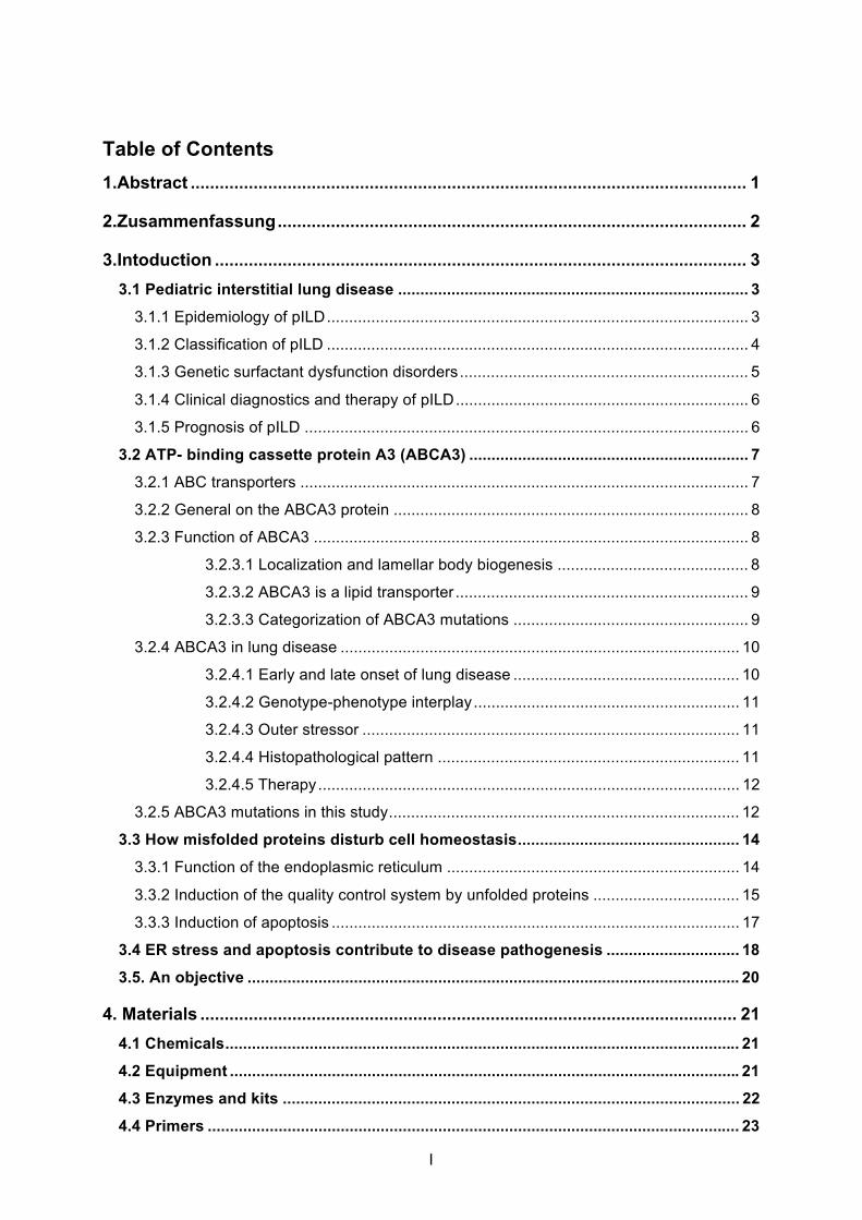
3.1.3 Genetic surfactant dysfunction disorders
Pulmonary surfactant is a complex mixture of 90 % lipids (mostly phospholipids) and
10 % surfactant-specific proteins A-D (11). It forms a film at the alveoli-air interface to reduce
surface tension in the alveoli and to prevent alveolar collapse at the end of expiration.
Alveolar type II cells (AT II) produce the surfactant mixture, which is stored within 120-180
lamellar bodies until it is secreted (12-14). Lamellar bodies (LB) are characterized by multiple
lamellar structures and one surrounding outer membrane (15, Fig.3).
Mutations in the genes encoding for surfactant protein B (SP-B) and C (SP-C) as well
as for the ATP-binding cassette protein A3 (ABCA3) were found in genetic surfactant
deficiency syndromes (16-22). In 1993, for the first time a genetic mechanism for fatal
respiratory distress syndrome (RDS) was discovered in a full-term infant. An autosomal
recessively inherited mutation in the SP-B gene (SFTPB) was identified to be the underlying
cause for surfactant protein B deficiency and genetic respiratory disease (23). Since then, an
increasing number of mutations in SFTPB have been identified in full-term infants presenting
with severe RDS and a rapid progressive and fatal course (24-26). Also mutations in the SP-
C gene (SFTPC) have been identified in newborns with RDS, but usually in SFTPC-
associated lung disease, disease onset occurs later than in neonatal period and patients
present with a more chronic clinical course (16,18).
Recently in 2004, when mutations in SFTPB or SFTPC have been excluded in
newborns with neonatal lung disease unresponsive to exogenous surfactant, mutations in the
ABCA3 gene were recognized as a cause of lethal RDS and imbalance in surfactant
composition (22, 27). Today, mutations in the ABCA3 gene are known to cause not only fatal
neonatal RDS but also chronic pILD in older children with a milder course (21). With more
than 500 identified mutations, ABCA3 deficiency is the most frequent known cause of genetic
ILD today (own unpublished data, 22, 27, 28).
Fig.3: A representative electron micrograph of a lamellar body in A549 cells (provided by our research group). LBs show many regular concentric lamellae and appear like “onion-skin” (diameter: 0.1-2.4 μm).
6
3.1.4 Clinical diagnostics and therapy of pILD
Clinical signs and symptoms like hypoxia, tachypnea, dyspnea, retractions, failure to
thrive, crackles, cough, wheeze and frequent respiratory infections are observed in infants
and children with suspected diffuse parenchymal lung disease, but clinical presentation
varies from asymptomatic to severe respiratory distress (29). As a first diagnostic study when
plain chest x-ray provides insufficient insight, a high-resolution CT (HRCT) can show the
most common feature of widespread ground-glass opacities, secondary honeycomb,
interlobular septal and air wall thickening and fibrotic lesions (29). Lung biopsy and the
histological analysis of lung tissue are often indispensable and crucial for the final diagnosis
of pILD (8). According to the latest data and the increasing number of genetic pILD (2, 30,
31), the diagnostic workup of newborns and infants with probable genetic-associated lung
disease should also include a DNA blood sample testing for mutations in the genes encoding
for SP-C, SP-B and ABCA3.
The current treatment of pILD is based on the clinical experience from small case series
in children and from clinical trials in adults. Affected newborns with severe respiratory
distress onset directly after birth require mechanical ventilation and application of exogenous
surfactant. Generally, therapy aims for the suppression of inflammation and prevention of
fibrogenesis (29). Over half of the patients receive systemic steroids and a minor group
profits additionally from further immunosuppressive, anti-inflammatory or anti-fibrotic drugs
as hydroxychloroquine, azathioprine and methrotrexate for weeks, months or years (2, 10).
As children are sometimes unresponsive to drug-therapy, the ultimate option is lung
transplantation. Today controlled clinical trials in children are conducted for evidence based
treatment recommendations.
3.1.5 Prognosis of pILD
Today morbidity and mortality in pILD is pretty high, but also varying between different
entities. The overall mortality in pILD is around 15% and 21% (9, 29). Whereas better
prognosis and lower mortality is reported for children with neuroendocrine cell hyperplasia of
infancy or pulmonary interstitial glycogenesis, other forms of pILD, especially genetic
surfactant deficiency syndromes, are associated with worse outcome. Particularly for
ABCA3-related lung disease mortality is pretty high (10, 22).
7
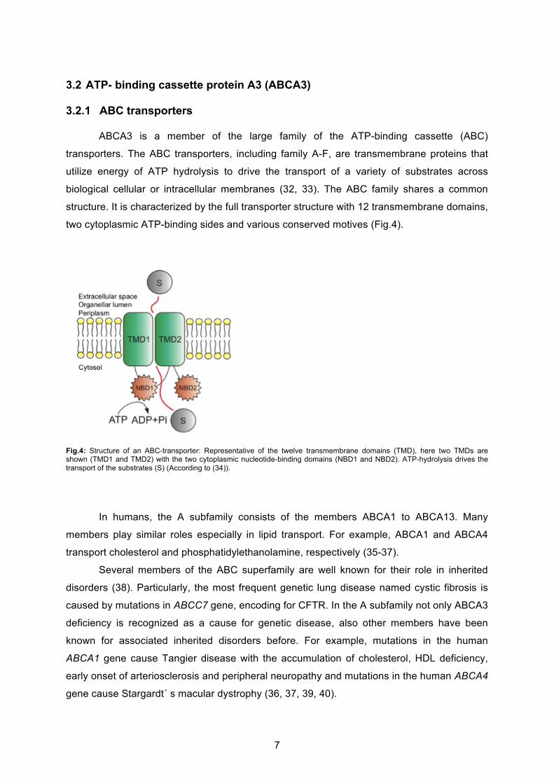
3.2 ATP- binding cassette protein A3 (ABCA3)
3.2.1 ABC transporters ABCA3 is a member of the large family of the ATP-binding cassette (ABC)
transporters. The ABC transporters, including family A-F, are transmembrane proteins that
utilize energy of ATP hydrolysis to drive the transport of a variety of substrates across
biological cellular or intracellular membranes (32, 33). The ABC family shares a common
structure. It is characterized by the full transporter structure with 12 transmembrane domains,
two cytoplasmic ATP-binding sides and various conserved motives (Fig.4).
Fig.4: Structure of an ABC-transporter: Representative of the twelve transmembrane domains (TMD), here two TMDs are shown (TMD1 and TMD2) with the two cytoplasmic nucleotide-binding domains (NBD1 and NBD2). ATP-hydrolysis drives the transport of the substrates (S) (According to (34)).
In humans, the A subfamily consists of the members ABCA1 to ABCA13. Many
members play similar roles especially in lipid transport. For example, ABCA1 and ABCA4
transport cholesterol and phosphatidylethanolamine, respectively (35-37).
Several members of the ABC superfamily are well known for their role in inherited
disorders (38). Particularly, the most frequent genetic lung disease named cystic fibrosis is
caused by mutations in ABCC7 gene, encoding for CFTR. In the A subfamily not only ABCA3
deficiency is recognized as a cause for genetic disease, also other members have been
known for associated inherited disorders before. For example, mutations in the human
ABCA1 gene cause Tangier disease with the accumulation of cholesterol, HDL deficiency,
early onset of arteriosclerosis and peripheral neuropathy and mutations in the human ABCA4
gene cause Stargardt´ s macular dystrophy (36, 37, 39, 40).

8
3.2.2 General on the ABCA3 protein First, ABCA3 protein was found in thyroid tissue (33), followed by a wide variety of
other tissue as brain, heard, kidney, stomach, pancreas and platelets. However, its highest
protein expression levels are found in lung tissue, particularly in alveolar type II cells (41-43).
The expression level of ABCA3 is developmentally regulated, increases prior to birth and is
upregulated in a glucocorticoid-dependent manner in vitro (41, 43, 44).
The ABCA3 protein is encoded by a single gene mapped to human chromosome
16p13.3 with a size of 80 kB, transcribed into 6500bp mRNA and translated into a 1704
amino acid protein (45). Mutations in ABCA3 gene are inherited in an autosomal-recessive
manner. According to the general structure of an ABC transporter, ABCA3 consists of 12
transmembrane domains, two extracellular loops (EL) and two nucleotide-binding domains
(NBD) (Fig.5).
Fig.5: Structure of the ABCA3 transporter in a lipid bilayer membrane (yellow): According to the general structure of an ABC transporter, ABCA3 is a transporter with 12 transmembrane domains (green), two extracellular loops (EL) and two nucleotide-binding domains (NBD). More than 500 mutations are identified, some of them are colored blue (frameshift mutation), red (missense mutation) or black (nonsense mutation) in this scheme. The author of this model is Dr. Suncana Kern, she provided it to our research group and I use it with her permission.
3.2.3 Function of ABCA3
3.2.3.1 Localization and lamellar body biogenesis
ABCA3 is highly expressed in alveolar type II cells (ATII) and localizes to the limiting
membrane of lamellar bodies (LBs) (41, 42), intracellular organelles for assembly and
storage of pulmonary surfactant. ABCA3 protein plays a key role in the biogenesis of LBs
9
and is crucial for their morphology. In electron microscope studies of healthy lungs, LBs
appear as “onion-skin” like, regular concentric arranged lamellae (Fig.3). In children with
ABCA3 mutations, LB morphology is abnormal (22). Immature LBs with “tightly packed
concentric membranes” (46) show small electro-dense bodies within, appearing as “fried-
egg” inclusions (46-48). This LB-morphology is characteristic for ABCA3 deficiency and LB
morphology can be an indicator to distinguish between the entities of genetic surfactant
dysfunction disorders. Whereas in SP-B deficiency large and disorganized composite bodies
and multiple vesicular inclusions appear (49, 50), in SP-C deficiency vesicular bodies with
aggregates of small vesicles with electro-dense cores can be observed (51-53).
Furthermore, ABCA3 protein is not only essential for LB morphology but also for their
formation. In Abca3 knock-out mice presenting with acute respiratory failure normal and
mature LBs are absent and mice die within hours after birth (54-56). Moreover, in in vitro
studies, the ABCA3 overexpression in kidney cells (HEK293 cells) induces de novo synthesis
of lamellar body-like vesicles (57).
3.2.4.2 ABCA3 is a lipid transporter
ABCA3 expression is not only essential for biogenesis of mature LBs as the
intracellular organelle for assembly and storage of surfactant, but is also responsible for the
composition and homeostasis of pulmonary surfactant. ABCA3 is a lipid transporter, which
drives the transmembrane translocation of surfactant phospholipids from the cytoplasm into
LBs. In vitro studies show that ABCA3 overexpression promotes the uptake of natural
choline-phospholipids into LBs (58, 59). When ABCA3 expression is down-regulated by
siRNA, phosphatidylcholine, sphingomyelin and cholesterol uptake are significantly
decreased (57). Furthermore, in human and mice with ABCA3 deficiency,
phosphatidylcholine species and phosphatidylglycerol are dramatically reduced in
bronchoalveolar lavage and lung tissue, respectively (27, 54, 60). Thereby surfactant mixture
looses its function and surface tension is increased (27). Since the last steps in SP-B and
SP-C processing occur inside of functional LBs, ABCA3 deficiency in human and mouse
leads to accumulation of SP-B and SP-C precursors and lack of mature forms (28, 55, 60).
3.2.3.1.1 Categorization of ABCA3 mutations
Mutations in the ABCA3 gene are classified into two categories (61): Type I mutations
are characterized by abnormal intracellular ABCA3 protein localization and impaired
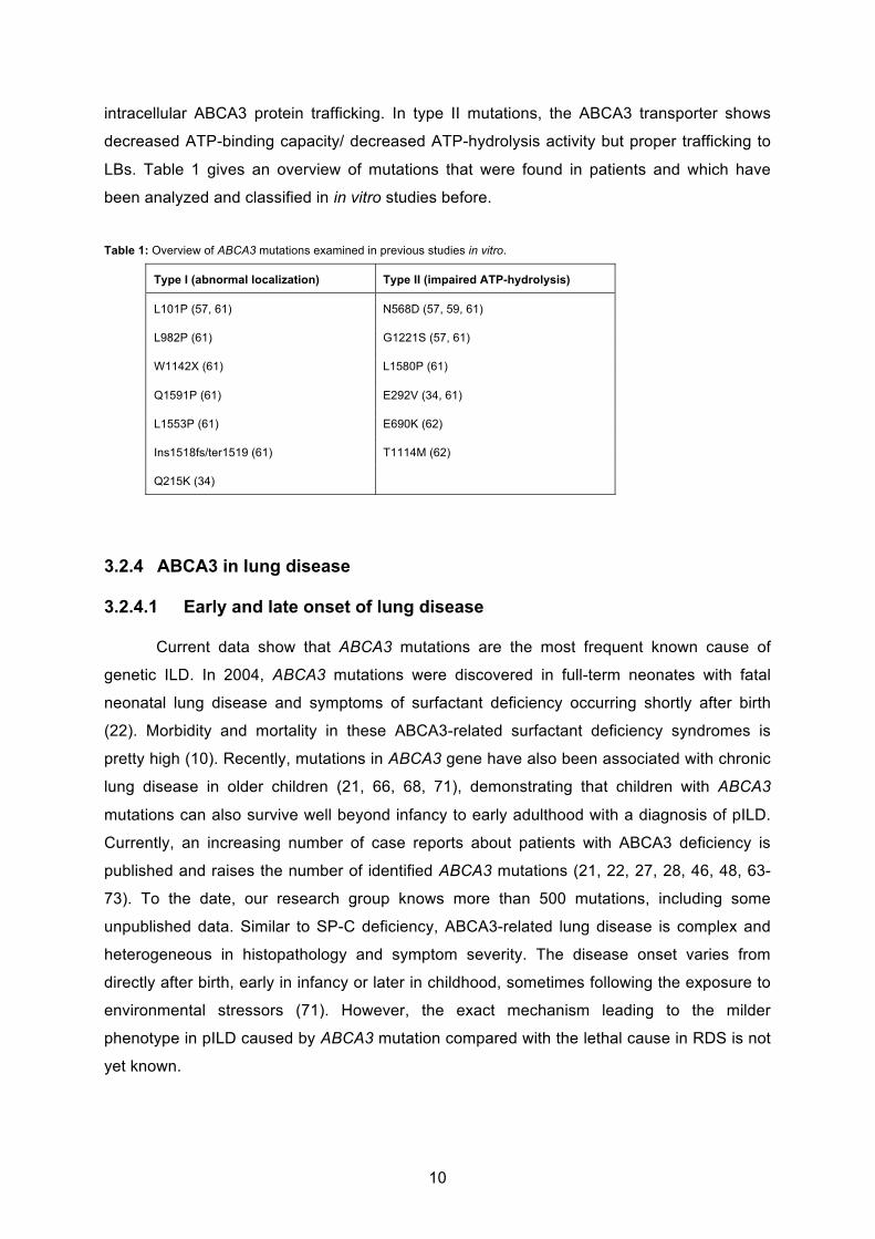
10
intracellular ABCA3 protein trafficking. In type II mutations, the ABCA3 transporter shows
decreased ATP-binding capacity/ decreased ATP-hydrolysis activity but proper trafficking to
LBs. Table 1 gives an overview of mutations that were found in patients and which have
been analyzed and classified in in vitro studies before.
Table 1: Overview of ABCA3 mutations examined in previous studies in vitro.
Type I (abnormal localization) Type II (impaired ATP-hydrolysis)
L101P (57, 61) N568D (57, 59, 61)
L982P (61) G1221S (57, 61)
W1142X (61) L1580P (61)
Q1591P (61) E292V (34, 61)
L1553P (61) E690K (62)
Ins1518fs/ter1519 (61) T1114M (62)
Q215K (34)
3.2.4 ABCA3 in lung disease
3.2.4.1 Early and late onset of lung disease
Current data show that ABCA3 mutations are the most frequent known cause of
genetic ILD. In 2004, ABCA3 mutations were discovered in full-term neonates with fatal
neonatal lung disease and symptoms of surfactant deficiency occurring shortly after birth
(22). Morbidity and mortality in these ABCA3-related surfactant deficiency syndromes is
pretty high (10). Recently, mutations in ABCA3 gene have also been associated with chronic
lung disease in older children (21, 66, 68, 71), demonstrating that children with ABCA3
mutations can also survive well beyond infancy to early adulthood with a diagnosis of pILD.
Currently, an increasing number of case reports about patients with ABCA3 deficiency is
published and raises the number of identified ABCA3 mutations (21, 22, 27, 28, 46, 48, 63-
73). To the date, our research group knows more than 500 mutations, including some
unpublished data. Similar to SP-C deficiency, ABCA3-related lung disease is complex and
heterogeneous in histopathology and symptom severity. The disease onset varies from
directly after birth, early in infancy or later in childhood, sometimes following the exposure to
environmental stressors (71). However, the exact mechanism leading to the milder
phenotype in pILD caused by ABCA3 mutation compared with the lethal cause in RDS is not
yet known.

11
3.2.4.2 Genotype-phenotype interplay
Some data show a genotype-phenotype correlation in lung disease caused by ABCA3
mutations, suggesting that patients with a type I homozygous ABCA3 mutation or with type I /
type II compound heterozygous mutations die within the neonatal period, whereas patients
with a type II homozygous ABCA3 mutation survive beyond neonatal period und occur with
milder phenotype (21, 22). In contrast, some clinical experience on ABCA3-related lung
disease is inconsistent with the idea of a genotype-phenotype correlation (47, 48, 62, 66,
unpublished data from our research group), demonstrating a significant phenotypic
heterogeneity of lung disease associated with ABCA3 mutations from fatal RDS to milder
course in pILD that is independent from genotype. However, the mechanism leading to
phenotypic heterogeneity of lung disease associated with ABCA3 deficiency is still poorly
understood.
3.2.4.3 Outer stressor
Since the variety of clinical appearance in ABCA3 deficient patients can not completely
be explained by the genotype-phenotype correlation, clinical experience suggests a link
between the late onset of genetic ILD and exposure to outside stress factors, such as
respiratory infections and cigarette smoke (71), speculating that outer stressors accelerated
the patients´ underlying disease course.
3.2.4.4 Histopathological pattern
Hyperplastic alveolar type II cells, accumulation of alveolar macrophages and
proteinaceous material in distal air spaces characterize the histological pattern in patients
with ABCA3 deficiency (22, 47). In further course of the disease, interstitial thickening and
fibroblast proliferation in interstitial spaces are found in the histological pattern (46, 47). Lung
biopsy findings show the histology of CPI (chronic pneumonitis of infancy), DIP
(desquamative interstitial pneumonia), NSIP (nonspecific interstitial pneumonia), PAP
(pulmonary alveolar proteinosis) and pulmonary fibrosis, whereas DIP is seen in the majority
of the patients (47).
12
3.2.4.5 Therapy
To the date, there are no causative therapeutic options available for ABCA3-related
surfactant deficiency. The therapeutic strategy concentrates on the reduction of symptoms
and the limitation of inflammation and fibrogenesis. Oxygen application, mechanical
ventilation and exogenous surfactant application can help to stabilize vital function
temporarily in case of acute respiratory distress. Symptomatic treatment of ABCA3-related
lung disease includes drugs, which are administrated in the management of ILD as well like
steroids and hydroxychloroquine (29). The ultimate therapeutic option in end-stage
respiratory failure is lung transplantation.
3.2.4.6 ABCA3 mutations in this study
In this study, the clinically relevant ABCA3 mutations R43L, R280C and L101P, which
either were found in newborns with respiratory distress syndrome (RDS) or in children with
chronic pILD, were examined. Table 2 provides an overview on the patients, their clinical
feature and outcome.
Brasch and colleagues reported from a full-term female newborn, delivered
uneventfully after a normal pregnancy (28). Directly after birth she developed severe
respiratory distress, required intense supportive care, mechanical ventilation and exogenous
surfactant application and died at the age of 47 days. The patient had a sibling who has died
briefly after birth from respiratory failure before. Analyzing the ABCA3 gene, which is
inherited in an autosomal-recessive fashion, in the healthy parents and one healthy sibling
showed that they are heterozygous for the G T mutation at nucleotide 128 (first
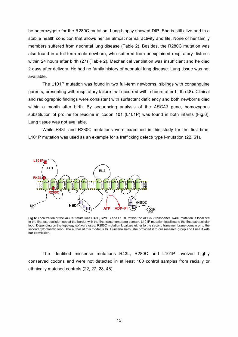
extracellular loop), which causes an R43L (Arg > Leu) amino acid substitution (Fig.6). Lung
biopsy of the patient revealed chronic pneumonitis of infancy, absent ABCA3 protein
expression, reduced mature SP-B expression (SP-C analysis not done) and electro-dense
bodies in type II pneumocytes. Appropriate material for mutation analysis was not available
from the patient. Furthermore, Somaschini (48) and Garmany (27) and coworkers, both
reported from two full-term newborns that were compound heterozygote for the R43L
mutation (R43L/R1482W and R43L/P264R, respectively) and had a similar clinical picture as
the reported female patient above, including severe respiratory distress syndrome. Both
newborns required intense supportive care. One patient died at the age of 30 days, the other
patient received lung transplantation. The histological examination showed DIP and CIP,
respectively (Table 2).
Unpublished data from our research group include a female patient, 15 years old,
presenting with milder respiratory symptoms consistent with chronic pILD. She was found to
13
be heterozygote for the R280C mutation. Lung biopsy showed DIP. She is still alive and in a
stabile health condition that allows her an almost normal activity and life. None of her family
members suffered from neonatal lung disease (Table 2). Besides, the R280C mutation was
also found in a full-term male newborn, who suffered from unexplained respiratory distress
within 24 hours after birth (27) (Table 2). Mechanical ventilation was insufficient and he died
2 days after delivery. He had no family history of neonatal lung disease. Lung tissue was not
available.
The L101P mutation was found in two full-term newborns, siblings with consanguine
parents, presenting with respiratory failure that occurred within hours after birth (48). Clinical
and radiographic findings were consistent with surfactant deficiency and both newborns died
within a month after birth. By sequencing analysis of the ABCA3 gene, homozygous
substitution of proline for leucine in codon 101 (L101P) was found in both infants (Fig.6).
Lung tissue was not available.
While R43L and R280C mutations were examined in this study for the first time,
L101P mutation was used as an example for a trafficking defect/ type I-mutation (22, 61).
Fig.6: Localization of the ABCA3 mutations R43L, R280C and L101P within the ABCA3 transporter. R43L mutation is localized to the first extracellular loop at the border with the first transmembrane domain. L101P mutation localizes to the first extracellular loop. Depending on the topology software used, R280C mutation localizes either to the second transmembrane domain or to the second cytoplasmic loop. The author of this model is Dr. Suncana Kern, she provided it to our research group and I use it with her permission.
The identified missense mutations R43L, R280C and L101P involved highly
conserved codons and were not detected in at least 100 control samples from racially or
ethnically matched controls (22, 27, 28, 48).
14
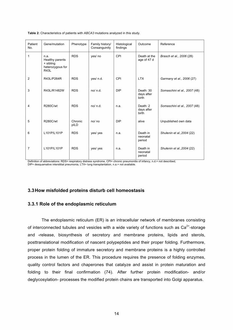
Table 2: Characteristics of patients with ABCA3 mutations analyzed in this study.
Definition of abbreviations: RDS= respiratory distress syndrome, CPI= chronic pneumonitis of infancy, n.d.= not described, DIP= desquamative interstitial pneumonia, LTX= lung transplantation, n.a.= not available.
3.3 How misfolded proteins disturb cell homeostasis
3.3.1 Role of the endoplasmic reticulum
The endoplasmic reticulum (ER) is an intracellular network of membranes consisting
of interconnected tubules and vesicles with a wide variety of functions such as Ca2+-storage
and -release, biosynthesis of secretory and membrane proteins, lipids and sterols,
posttranslational modification of nascent polypeptides and their proper folding. Furthermore,
proper protein folding of immature secretory and membrane proteins is a highly controlled
process in the lumen of the ER. This procedure requires the presence of folding enzymes,
quality control factors and chaperones that catalyze and assist in protein maturation and
folding to their final confirmation (74). After further protein modification- and/or
deglycosylation- processes the modified protein chains are transported into Golgi apparatus.
Patient No.
Gene/mutation Phenotype Family history/ Consanguinity
Histological findings
Outcome Reference
1 n.a. Healthy parents + sibling heterozygous for R43L
RDS yes/ no CPI Death at the age of 47 d
Brasch et al., 2006 (28)
2 R43L/P264R RDS yes/ n.d. CPI LTX Garmany et al., 2006 (27)
3 R43L/R1482W RDS no/ n.d. DIP Death: 30 days after birth
Somaschini et al., 2007 (48)
4 R280C/wt RDS no/ n.d. n.a. Death: 2 days after birth
Somaschini et al., 2007 (48)
5 R280C/wt Chronic pILD
no/ no DIP alive Unpublished own data
6 L101P/L101P RDS yes/ yes n.a. Death in neonatal period
Shulenin et al.,2004 (22)
7 L101P/L101P RDS yes/ yes n.a. Death in neonatal period
Shulenin et al.,2004 (22)
15
3.3.2 Induction of the quality control system by unfolded proteins
Failure of posttranslational protein modification and an increasing number of unfolded
proteins retained within the ER lumen cause ER stress. Therefore, the ER offers a
cytoprotective quality control mechanism named the unfolded protein response (UPR). The
UPR system prevents the cell from unfolded defect proteins exit the ER and induces cellular
adaption to disturbed cell homeostasis (74, 75). Three mechanisms regulate cellular
adaption: first, general inhibition of protein translation to decrease the protein load in the ER,
second, transcriptional induction of genes encoding chaperones to maintain the protein-
folding capacity of the ER and third, the induction of proteins belonging to the ER-associated
degradation (ERAD), that is responsible for the transport of misfolded proteins to the cytosol
and subsequent degradation (76-79). If UPR fails to resolve ER stress and to restore cell
homeostasis, apoptotic cell-death pathway is initiated (80).
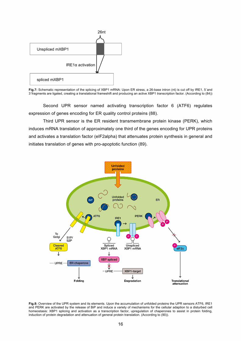
In non stressed cells, the binding protein (BiP)- also named glucose regulated protein
(GRP78)- binds to the three UPR signal transducers that are localized to ER membrane to
maintain them in an inactive state (81, Fig.8). Upon ER stress and accumulation of unfolded
proteins in the ER lumen, BiP is released from the three UPR sensors to assist in protein
folding. Thus, UPR sensors are activated to induce the cellular adaption to disturbed cell
homeostasis.
First UPR signal transducer, ionsitol-requiring enzyme 1 (IRE1), a transmembrane
serine/ threonine receptor protein kinase, is activated upon accumulation of unfolded proteins
in the ER lumen. IRE1 cleaves mRNA encoding for the basic leucine zipper transcription
factor X-box binding protein 1 (XBP1), a key transcription factor of UPR elements, in the
cytoplasm (82). The XBP1-mRNA is cleaved at two sites, the 5´ and 3´ fragments are ligated
and a 26- base intron is cut off (82, Fig.7). The result is a translational frameshift and the
active XBP1 transcription factor (83, 84). Active XBP1 is responsible for the transcription of
genes encoding a wide variety of UPR elements such as chaperones and folding enzymes
as well as ERAD elements (85, 86). Furthermore, IRE1 induces degradation of several
mRNAs, thereby preventing the cells from a high protein load during ER stress (87).
16
Fig.7: Schematic representation of the splicing of XBP1 mRNA: Upon ER stress, a 26-base intron (nt) is cut off by IRE1, 5´and 3´fragments are ligated, creating a translational frameshift and producing an active XBP1 transcription factor. (According to (84))
Second UPR sensor named activating transcription factor 6 (ATF6) regulates
expression of genes encoding for ER quality control proteins (88).
Third UPR sensor is the ER resident transmembrane protein kinase (PERK), which
induces mRNA translation of approximately one third of the genes encoding for UPR proteins
and activates a translation factor (eIF2alpha) that attenuates protein synthesis in general and
initiates translation of genes with pro-apoptotic function (89).
Fig.8: Overview of the UPR system and its elements. Upon the accumulation of unfolded proteins the UPR sensors ATF6, IRE1 and PERK are activated by the release of BiP and induce a variety of mechanisms for the cellular adaption to a disturbed cell homeostasis: XBP1 splicing and activation as a transcription factor, upregulation of chaperones to assist in protein folding, induction of protein degradation and attenuation of general protein translation. (According to (90)).
17
3.3.3 Induction of apoptosis
Prolonged ER stress caused by accumulation of unfolded proteins in the ER lumen and
the inability to restore cell homeostasis induces programmed cell death (=apoptosis).
Apoptosis is a highly conserved cellular mechanism. It is essential for cell selection, cell
homeostasis and tissue morphogenesis. A wide variety of stimuli can trigger cell surface
death receptors (extrinsic pathway) or initiate the intrinsic pro-apoptotic pathway via
mitochondria stimulation. Upon ER accumulation of misfolded proteins the three UPR
sensors not only initiate cytoprotective mechanisms, but also contribute to the induction of
programmed cell death via upregulation of pro-apoptotic proteins such as CHOP, Ca2+-
release, induction of mitochondrial stress (mitochondrial apoptosis) and ER stress (80).
Pro-apoptotic signals are transduced by a family of regulated proteolytic enzymes
named caspases. Caspases are a family of highly conserved aspartate-specific cysteine
proteases, which induce the pro-apoptotic pathway and contribute to the destruction of cell
structures and mechanisms as well as to the deregulation of protein synthesis (91). The
caspase-cascade is a complex signaling pathway. Most caspases are present in cells as
longer inactive precursors and become sequentially activated by proteolytic cleavage through
an active upstream caspase or through autocatalysis (91). In mammalian species, 15 known
members are divided into two groups: initiator caspases, which are activated by death
receptors, and effector caspases that cleave proteins involved in programmed cell death
events. The activation of the down-stream executioner caspase 3 can be either conducted
by the initiator caspase 9 that is activated in a mitochondria-dependent signaling way
(mitochondrial apoptosis) by increased intracellular stress (92) or by caspase 8 that is
activated through stimulation of the surface death receptor Fas. The executioner caspases 3,
6, 7 cleave proteins involved in DNA processing and several structural proteins.
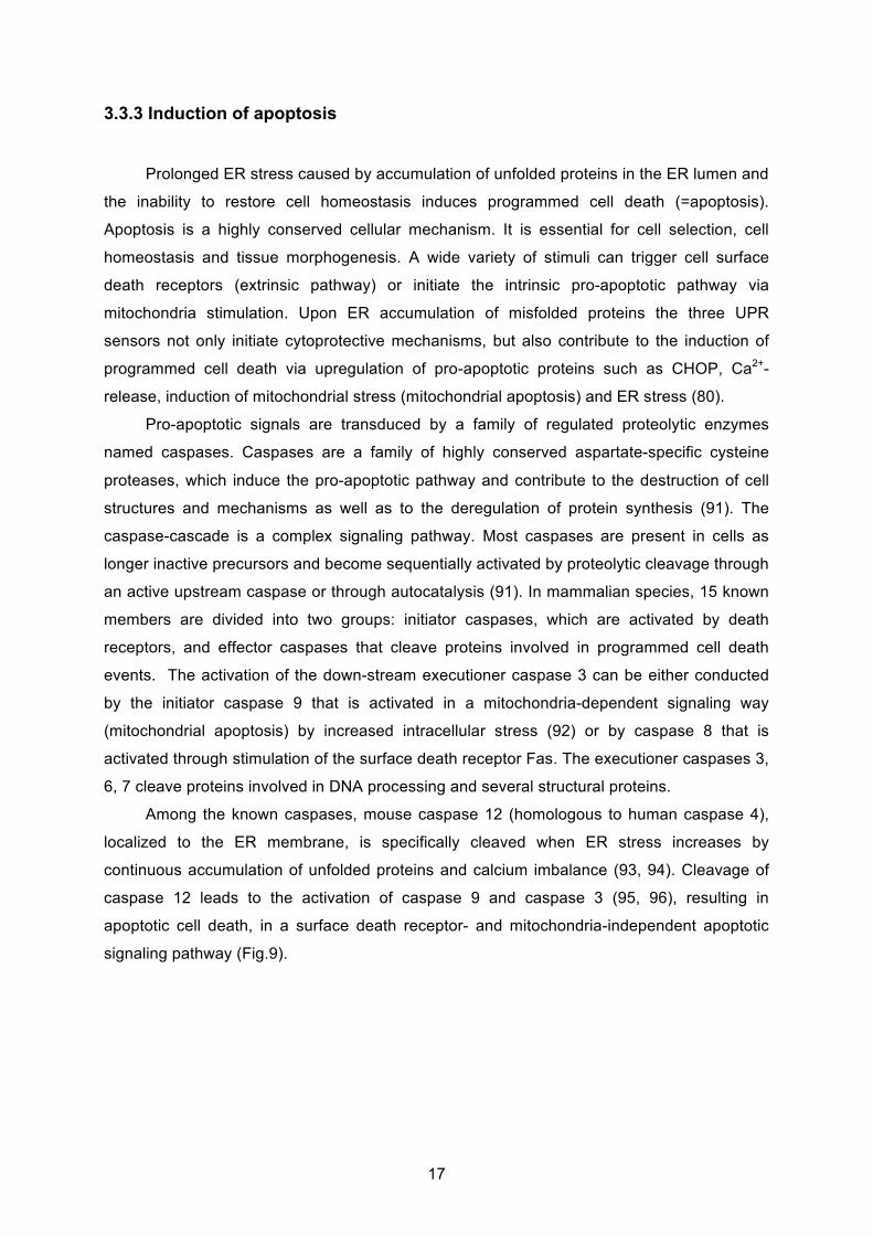
Among the known caspases, mouse caspase 12 (homologous to human caspase 4),
localized to the ER membrane, is specifically cleaved when ER stress increases by
continuous accumulation of unfolded proteins and calcium imbalance (93, 94). Cleavage of
caspase 12 leads to the activation of caspase 9 and caspase 3 (95, 96), resulting in
apoptotic cell death, in a surface death receptor- and mitochondria-independent apoptotic
signaling pathway (Fig.9).
18
Fig.9: Scheme of the mechanism how ER stress causes apoptotic cell death: Increasing ER stress leads to the cleavage of the ER membrane-bound caspase 12 in mouse (homologous human caspase 4), which catalysis the cleavage of caspase 9. Active caspase-9 catalysis the cleavage of procaspase-3, thereby promoting the further conduction of apoptosis. (According to (96))
Vital cells generate and maintain an asymmetric double layer cell membrane of
uncharged and charged phospholipids species. The negatively charged phosphatidylserines
(PS) are mainly located in the inner leaflet facing the cytosol. During apoptosis, plasma
membranes loose their feature of asymmetry by externalization of PS to the outer leaflet of
the plasma membrane in a caspase-dependent way (97). Expression of PS on the cell
surface is a signal for macrophages for phagocytosis. Furthermore, Annexin V binds to
negatively charged PS on plasma membranes of apoptotic cells and can be used as a
Molecular Imaging agent in visualizing apoptotic cells in patients and in in vitro studies (98,
99).
3.5 ER stress and apoptosis contribute to disease pathogenesis
Recent data show that prolonged accumulation of unfolded proteins within the ER
accompanied by simultaneous failure of the UPR system contribute to the pathogenesis of
several disorders, such as metabolic disease, atherosclerosis, neurodegenerative disease
and conformational diseases (100-102). Furthermore, ER stress and apoptosis in alveolar
type II cells play a role in lung disease, especially in pathogenesis of idiopathic pulmonary
fibrosis (IPF) and genetic SP-C-associated pulmonary fibrosis (103-105). Moreover, in in
vitro studies in lung epithelial A549 cells expressing SP-C mutations, which cause misfolding
and aggregation of the SP-C pre-protein, ER stress increases, UPR is activated and
apoptotic cell death is initiated (106).
Fibrosis is one of the hallmarks documented in ABCA3-associated lung disease (21,
19
68, 71). Today, it is known that idiopathic pulmonary fibrosis is not primarily connected to
chronic and unresolved inflammation and fibrosis can occur without inflammation (107).
Fibroblastic foci in the lung may underlie areas of unresolved chronic alveolar epithelial
injury, ongoing lung repair and abnormal wound healing (29, 107, 108).
Under such conditions of epithelial micro-injury alveolar type II cells contribute
significantly to the alteration of the normal lung architecture, aberrant tissue remodeling and
fibrogenesis and induce activation and migration of fibroblasts (107). Recently, it was shown
that ATII cells undergo epithelial-mesenchymal transition (EMT) under specific conditions
and change their characteristics and morphology to a fibroblast-like feature (109). Thereby
ATII may contribute to lung fibrogenesis as an additional source of myofibroblasts in the
pathogenesis of ILD (110). Moreover, alveolar epithelial cells are responsible for the release
of almost all pro-fibrotic cytokines and growth factors, such as TGF-ß1, IL-1ß, TNF- and
GM-CSF, and for the synthesis and release of enzymes that contribute to extracellular matrix
(111, 112). TGF-ß1 is the most aggressive pro-fibrotic growth factor among these mediators
and the amount of TGF-ß is related to the extent of fibrosis and induction of myofibroblasts
(113). Besides, TGF-ß1 induce EMT in alveolar epithelial cells, which is characterized by the
loss of epithelial marker E-Cadherin (ECAD), the gain of mesenchymal marker vimentin and
transformation into myofibroblastic morphology (109, 114). In this way, alveolar type II cells
can acquire fibroblast-like feature and play a role in the development of fibrosis in lung
disease.

20
4. An objective
ABCA3 protein is an ABC lipid transporter found in alveolar type II cells, where it
localizes to the outer membrane of lamellar bodies which store surfactant. Current in vitro
studies show that ABCA3 is essential for surfactant homeostasis and LB biogenesis.
Mutations in the ABCA3 gene are the most frequent known genetic cause of RDS in
newborns and of late onset chronic ILD in children with a higher prevalence than mutations in
SPFTB or SPFTC. Even though clinical experience suggests a link between the late onset of
genetic ILD and exposure to outside stress factors, such as respiratory infections and
cigarette smoke, contributing mechanisms leading to phenotypic heterogeneity remain
unknown.
In order to elucidate mechanisms leading to pediatric ILD, I investigated the influence
of three ABCA3 mutations, R43L, R280C and L101P, found in children with surfactant
deficiency and chronic ILD, on induction of ER stress, apoptosis and fibrogenesis in lung
epithelial A549 cells.
21
4. Materials
4.1 Chemicals
In general, chemicals were obtained from Sigma-Aldrich (München, Germany) and
Merck (Darmstadt, Germany). Special chemicals were purchased as follows: Agarose for
DNA gel electrophoresis from SERVA (Heidelberg, Germany), D-PBS, trypsin-EDTA solution
and FBS from PAA Laboratories GmbH (Pasching, Austria) and BSA from Paesel and Lorei
(Duisburg, Germany). RPMI media was purchased from Sigma-Aldrich (München, Germany)
and LB-Agar as well as LB-Bouillon were purchased from Merck (Darmstadt, Germany).
Antibiotics kanamycin, ampicillin and tunicamycin were obtained from Sigma-Aldrich
(München, Germany). Recombinant human Tumor Necrosis Factor (TNF- ) was obtained
from Invitrogen (Freiburg, Germany) and Recombinant human Tissue Growth Factor 1
(TGF- 1) was obtained from R&D systems (Abingdon, England). Vectashield Hard and Set
Mounting Media with DAPI was obtained from Vector Laboratories (Burlingame, CA, USA).
NBD-labeled lipids phosphatidylcholine (C12-NBD-PC) and phosphatidylethanolamine (C12-
NBD-PE) were purchased from Avanti Polar Lipids (Alabaster, AL) and other lipids from
Sigma-Aldrich (München, Germany).
4.2 Equipment
Fluorescence microscope Axiovert 135 obtained from Carl Zeiss (Oberkochen,
Germany) was used for immunofluorescence analysis. The evaluation of pictures was
performed with the help of the software AxioVision Release 4.7.1 (Carl Zeiss, Oberkochen,
Germany). Olympus FluoView FV 1000 confocal microscope was used for the analysis of the
NBD-lipid uptake.
BD FACSCanto II Flow Cytometer was used for the FACS assay and FACSDiva
v6.1.3 for data analyses (BD Bioscience).
For the chemiluminescence detection Diana III camera system and for densitrometric
analysis software AIDA were purchased from Raytest (Straubenhardt, Germany). GraphPad
Prism version 4 was obtained from GraphPad Software (La Jolla, CA, USA) for statistical
analysis.

22
4.3 Enzymes and kits
Enzymes and kits were purchased as follows: QuickChange Site-directed
Mutagenesis Kit for site directed point mutagenesis and PfuUltra TM Hotstart Polymerase
were purchased from Stratagene (La Jolla, CA, USA). Restriction enzymes: KpnI, EcorI,
HindIII, ScaI, XbaI, XhoI, BglII, DpnI and the corresponding reaction buffers as well as
EndoH and PNGaseF for deglycolysation assay were purchased from NewEngland BioLabs
(Ipswich, MA, USA). T4-DNA ligase and corresponding reaction buffer were purchased from
Fermentas (Burlington, Canada). DNA extraction from agarose gels was performed with the
help of QIAquick Gel Extraction Kit (Qiagen, Hilde, Germany) and Ultrafree®-DA (Millipore,
Billerica, MA, USA). Plasmid- DNA isolation was performed with either QIAprep Spin
Miniprep Kit (Qiagen, Hilden, Germany) or with NucleoBond Xtra Midi EF from Macherey-
Nagel (Düren, Germany). Cell culture was tested regularly for mycoplasma with the help of
Venor GEM-Mykoplasma Detection Kit for conventional PCR.
High Pure RNA Isolation Kit and DNase I, RNase free from Roche (Mannheim, Germany)
were used for RNA-isolation. SuperScriptTMIII First Strand Synthesis System from Invitrogen
(Freiburg, Germany) and Taq-polymerase from NewEngland BioLabs (Ipswich, MA, USA)
were used for performance of RT-PCR.
Transfection was performed with the help of transfection reagent ExGen 500 from
Fermentas (Burlington, Canada). Complete protease inhibitor cocktail was purchased from
Roche (Basel, Schweiz). Determination of protein concentration was conducted with Bio-Rad
Protein Assay Kit from Bio-Rad Laboratories (München, Germany). For SDS-Page and
Western Blotting following buffers as well as precast gels were obtained from Invitrogen
(Freiburg, Germany): NuPage® MOPS SDS Running Buffer (20x), NuPage® MES SDS
Running Buffer (20x), NuPage® Tris-Acetate SDS Running Buffer (20x), GF® LDS Sample
Buffer (4x), Transfer Buffer (20x), NuPage® 10 % Bis-Tris Gel, NuPage® 3-8 % Tris-Acetate
Gel and 7 % Tris-Acetate Gel. Immobilion-P Transfer Membrane, a PVDF-membrane, was
purchased from Millipore (Billerica, MA, USA). ECL and HyperfilmTM ECL were obtained from
GE-Healthcare (Buckinghamshire, UK). For the purpose of immunoblotting with anti-GFP
antibody, the chemiluminescent immunodetection system WesternBreeze® was obtained
from Invitrogen.
IntraPrepTM Permeabilization Reagent from Beckman Coulter (Fullerton, CA, USA)
was used for preparing samples for flow cytometer analysis.

23
4.4 Primers
All primers were purchased from Metabion International AG (Martinsried, Germany).
Table 3: List of primers for point mutagenesis or sequencing.
Primer name Sequence
ABCA3.R43L.For 5'-CATCTGGCTCCTCTTGAAGATTC-3'
ABCA3.R43L.Rev 5'-GAATCTTCAAGAGGAGCCAGATG-3'
ABCA3.R280C.For 5'-CATTGCCTGTGCTGTCGTG-3'
ABCA3.R280C.Rev 5'-CACGACAGCACAGGCAATG-3'
ABCA3.L101P.For 5’-CAGTGCGCAGGGCACCTGTGATCAAC-3’
ABCA3.L101P.Rev 5’-GTTGATCACAGGTGCCCTGCGCACTG-3’
ABCA3.HA.For 5'-GCCGCGGGTACCATGGATTACCCATACGATGTTCCAGATTACGCTGCTGCTGTGCTCAGC-3'
Table 4: List of primers for RT-PCR.
Primer name Sequence
XBP1.For.1 5´-AAACAGAGTAGCAGCTCAGACTGC-3´
XBP1.Rev.1 5´-TCCTTCTGGGTAGACCTCTGGGAG-3´
18srRNA.For.1 5'-TCAAGAACGAAAGTCGGAGG-3'
18srRNA.Rev.1 5'-GGACATCTAAGGGCATCACA-3'
24
4.5 Vectors
pEYFP-ABCA3-WT was a kind gift of the research group of Dr. Andreas Holzinger
(Fig.10), pJET1/ABCA3-HA of my colleague Dr. M. Woischnik, pUB6-hABCA3-HA-L101P of
my colleague Eva Kaltenborn and pEGFP-N1-SPC-WT of Dr. Michael Beers. PuB6-V5-
His/LacZ was purchased from Invitrogen (Freiburg, Germany) and pmax-GFP from AMAXA
(Köln, Germany). Other vectors were generated in this study (Table 5).
Table 5: List of vectors.
Vector Resistance in
E.coli DH5
Resistance in
cells
Major characteristics Size Source
Pmax-GFP kanamycin none Not suitable for making stable clones
3486 bp AMAXA
pJET1/ABCA3-HA ampicillin Only effective in non transformed cells
HA tagged is fused to C-terminal end of ABCA3 gene
31228 bp M. Woischnik
puB6/V5-His/LacZ ampicillin blasticidin Contains gene for ß-
galactosidase
8500bp Invitrogen
pEYFP-ABCA3-WT kanamycin G418 Gene, encoding für YFP-protein, is fused to C-terminal end of ABCA3 gene
10146 bp
A. Holzinger
pEYFP-ABCA3-R43L kanamycin G418 Gene, encoding für YFP-protein, is fused to C-terminal end of ABCA3 gene
10146 bp This study
pEYFP-ABCA3-R280C kanamycin G418 Gene, encoding für YFP-protein, is fused to C-terminal end of ABCA3 gene
10146 bp This study
pEYFP-ABCA3-L101P kanamycin G418 Gene, encoding für YFP-protein, is fused to C-terminal end of ABCA3 gene
10146 bp
This study
puB6-hABCA3-HA-WT ampicillin blasticidin HA tagged is fused to C-terminal end of ABCA3 gene
10542 bp
This study
puB6-hABCA3-HA-R43L ampicillin blasticidin HA tagged is fused to C-terminal end of ABCA3 gene
10542 bp
This study
puB-hABCA3-HA-R280C ampicillin blasticidin HA tagged is fused to C-terminal end of ABCA3 gene
10542 bp
This study
puB6-hABCA3-HA-L101P ampicillin blasticidin HA tagged is fused to C-terminal end of ABCA3 gene
10542 bp
E. Kaltenborn
pcDNA4-TO-ABCA3-HA-WT ampicillin blasticidin HA tagged is fused to C-terminal end of ABCA3 gene
n.a. Provided by
the group
pEGFP-N1-SPC-WT kanamycin neomycin Gene, encoding für GFP-protein, is fused to C-terminal end of SP-C gene
5371bp M. Beers
n.a.: not avaiable

25
Fig.10: Vector map of pEYFP-N1 (BD Clontech): Wild type full length human hABCA3 cDNA without stop codon was cloned into EcoRI/AgeI sites of pEYFP-N1 plasmid to obtain pEYFP-N1-WT vector for expression of C-terminal ABCA3-YFP protein fusions.
4.6 Antibodies
Primary antibodies for Western Blotting or immunofluorescence were either
purchased from Santa Cruz (Santa Cruz, CA, USA), Chemicon (Billerica, MA, USA),
Clontech (Mountain View, CA, USA), Stressgene (Hamburg, Germany), Dianova (Hamburg,
Germany), Cell Signaling (Danvers, MA, USA), R&D System (Abingdon, England) or Sigma-
Aldrich (München, Germany) (Table 6).
26
Table 6: List of primary antibodies used for Western Blotting (WB) or immunofluorescence (IF).
Primary Antibodies Source Type, Host Dilutions for WB and IF
anti-ß-actin –
HRP conjugate
Santa Cruz (Santa Cruz, CA, USA) monoclonal, mouse WB 1:10 000
anti-calnexin Santa Cruz (Santa Cruz, CA, USA) polyclonal, goat WB 1:500;
IF 1:200
anti-Hsp 90 Santa Cruz (Santa Cruz, CA, USA) monoclonal, mouse WB 1:500
anti-human CD 63
(LAMP3)
Chemicon (Billerica, MA, USA) monoclonal, mouse IF: 1:200
anti-EEA-1 Santa Cruz (Santa Cruz, CA, USA) polyclonal, rabbit IF: 1:200
anti-GM130 Santa Cruz (Santa Cruz, CA, USA) polyclonal, goat IF: 1:50
anti-GFP:
Living Colors R A.v. (IL-8)
Clontech (Mountain View, CA, USA) monoclonal, mouse WB: 1:500
anti- caspase 4 Stressgene (Hamburg, Germany) monoclonal, mouse WB: 1:500
anti- caspase 3 Dianova (Hamburg, Germany) monoclonal, mouse WB: 1:133
anti- BiP Cell Signaling (Danvers, MA, USA) monoclonal, rabbit WB: 1:1000
anti- E-cadherin R&D System (Abingdon, England) monoclonal, mouse WB: 1:250
anti- vimentin Sigma- Aldrich (München, Germany) monoclonal, mouse WB: 1:2000
anti-HA-tag Roche (Basel, Schweiz) monoclonal, rat IF: 1:200
Secondary antibodies used for IF were purchased from Invitrogen (Freiburg, Germany)
(Table 7).
Table 7: List of secondary antibodies used for IF.
Antibody Excitation (nm) Emission (nm) Filter
anti-goat IgG - Alexa 555(donkey) 555 565 III (CY3)
anti-mouse IgG - Alexa 555(goat) 555 565 III (CY3)
anti-rabbit IgG - Alexa 555(goat) 555 565 III (CY3)
anti-rat IgG - Alexa 488(donkey) 495 519 III (green)
anti-rat IgG - Alexa 555 555 565 III (CY3)
Secondary antibodies used for WB were purchased from Invitrogen (Freiburg, Germany),
Biozol (Eching, Germany) or Sigma- Aldrich (München, Germany) (Table 8).
27
Table 8: List of secondary antibodies used for WB.
Secondary antibody Source Type, Host Dilution
anti-goat IgG –HRP conjugate Biozol (Eching, Germany) Polyclonal, rabbit WB 1:10 000
anti-mouse: WesternBreeze® - alkaline
phosphatase conjugated
Invitrogen (Freiburg, Germany) Not published Not published
anti-rabbit IgG – HRP conjugate Sigma- Aldrich (München, Germany) Polyclonal, goat WB 1:10 000
Antibodies for flow cytometry were purchased from BD Biosciences (San Jose, CA, USA)
(Table 9).
Table 9: List of antibodies used for flow cytometry.
Antibody Source Channel
anti-annexinV Cy5 BD Pharmingen TM III
anti-active-caspase 3
PE conjugated
BD Biosciences II
PI BD Pharmingen TM II
4.7 Bacterial strains and cell lines
Escherichia coli DH5 were obtained from Invitrogen (Freiburg, Germany). Human
lung carcinoma epithelial cell line A54p (ACC 107) was obtained from German Collection of
Microorganisms and Cell Cultures (DMSZ, Braunschweig, Germany).
5. Methods
5.1. Molecular biological methods
5.1.1 Cloning strategy: Generation of pUB6-ABCA3-HA vector
In order to generate puB6-hABCA3-HA vector with hemagglutinin tag (HA-tag) fused
to C-terminus of the ABCA3 gene, puB6/lacZ vector and pJet1/hABCA3-HA vector were
transformed into E.coli DH5 competent cells. Plasmid-DNA of both was isolated, restricted
through KpnI and XhoI and separated by gel electrophoresis. The following restriction
products were extracted with the help of QIAquick Gel Extraction Kit and Millipore Ultrafree-
DA: a 5320 bp KpnI and xhoI fragment containing pUB6 vector and a 5150 bp KpnI and xhoI
fragment containing hABCA3-HA gene. DNA was precipitated by ethanol and resuspended
28
in water. For ligation, vector and insert, in 3:1 ratio, were incubated with T4 ligase and
corresponding reaction buffer first for 60 min at room temperature and second for 10 min at
65 °C. Products of ligation reaction were multiplied by transformation into E.coli DH5
competent cells. Two negative controls, the first one without insert and the second one
without insert and T4 ligase, were generated. When no bacterial colonies have grown in
these negative controls, plasmid DNA of the positive clones was isolated and their sequence
was confirmed by conventional sequencing.
Fig.11: Vector map of pUB6/V5-His: Plasmid vector puB6-hABCA3-HA for expression of C-terminal fusions of ABCA3 with hemagglutinin tag (HA-tag) was produced by modification of pUB6/V5-His vector. Own illustration according to a model from Dr. Suncana Kern.
5.1.2 Site-directed point mutagenesis
Site-directed point mutagenesis was performed with the help of QuickChange Site-
directed Mutagenesis Kit (Stratagene, La Jolla, California, USA) as recommended by the
manufacturer. The following cycling parameters as shown in Table 10 were used for the
introduction of point mutations into pEYFP-ABCA3- and pUB6-ABCA3-vectors.
29
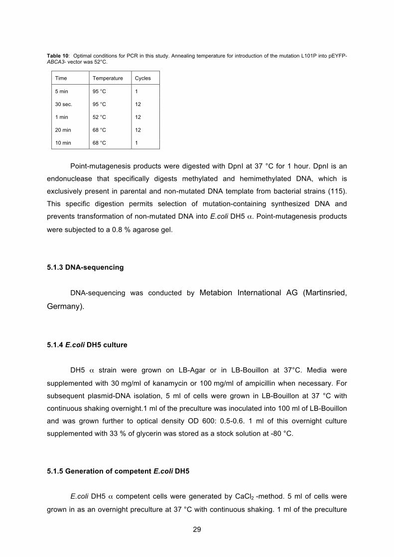
Table 10: Optimal conditions for PCR in this study. Annealing temperature for introduction of the mutation L101P into pEYFP-ABCA3- vector was 52°C.
Time Temperature Cycles
5 min 95 °C 1
30 sec. 95 °C 12
1 min 52 °C 12
20 min 68 °C 12
10 min 68 °C 1
Point-mutagenesis products were digested with DpnI at 37 °C for 1 hour. DpnI is an
endonuclease that specifically digests methylated and hemimethylated DNA, which is
exclusively present in parental and non-mutated DNA template from bacterial strains (115).
This specific digestion permits selection of mutation-containing synthesized DNA and
prevents transformation of non-mutated DNA into E.coli DH5 . Point-mutagenesis products
were subjected to a 0.8 % agarose gel.
5.1.3 DNA-sequencing
DNA-sequencing was conducted by Metabion International AG (Martinsried,
Germany).
5.1.4 E.coli DH5 culture
DH5 strain were grown on LB-Agar or in LB-Bouillon at 37°C. Media were
supplemented with 30 mg/ml of kanamycin or 100 mg/ml of ampicillin when necessary. For
subsequent plasmid-DNA isolation, 5 ml of cells were grown in LB-Bouillon at 37 °C with
continuous shaking overnight.1 ml of the preculture was inoculated into 100 ml of LB-Bouillon
and was grown further to optical density OD 600: 0.5-0.6. 1 ml of this overnight culture
supplemented with 33 % of glycerin was stored as a stock solution at -80 °C.
5.1.5 Generation of competent E.coli DH5
E.coli DH5 competent cells were generated by CaCl2 -method. 5 ml of cells were
grown in as an overnight preculture at 37 °C with continuous shaking. 1 ml of the preculture
30
was inoculated into 100 ml of LB-Bouillon and grown further to optical density OD 600: 0.5-
0.6. For further procedure, DH5 cells were incubated on ice. After 5 min of inoculation on
ice, cells were collected at 4 °C and 5000 xg for 10 min. The cell pellet was resuspended in
40 ml of ice cold Tfb I buffer (100 mM RbCl2, 50 mM MnCl2, 30 mM K-acetat, 10 mM CaCl2-
2H2O, 15% glycerin, pH 5) and incubated on ice for further 30 min. Cells were collected at
4 °C and 5000 xg for 10 min, the pellet was disolved in ice cold Tfb II buffer (10 mM MOPS,
10 mM RbCl2, 75 mM CaCl2-2H2O, 15 % glycerin, pH 6.8), was frozen in liquid nitrogen and
stored at -80 °C.
5.1.6 Transformation in E.coli DH5
First, 15 l of point-mutagenesis products were added to DH5 cells and kept on ice
for 30 min. Second, cells were heat-treated at 42 °C for 2 min and immediately cooled down
on ice for 2 min. Third, cells were inoculated into 900 l of LB medium and grown at 37 °C for
1 hour. Fourth, cells were collected at 5000 xg for 2 min. Cell pellet was dissolved in 100 l
of media and cells were grown on LB agar with supplemented antibiotics at 37 °C overnight.
One sample with not transformed DH5 cells was used as a negative control. When no
colonies were detectable in negative controls, plasmid-DNA of transformed DH5 cells was
isolated, restricted, subjected to 0.8 % agarose gel and sequence was confirmed by
conventional sequencing.
5.1.7 Plasmid-DNA isolation
After transformation of pEYFP-ABCA3 or pUB6-ABCA3-HA vectors into DH5 strain
an overnight culture of DH5 strain was prepared. DNA was isolated out of DH5 strain
culture either with the help of QIAprep Spin Miniprep Kit or with NucleoBond Xtra Midi EF
according to the protocol provided by the manufacturer.
5.1.8 Restriction
Restriction was performed with 500 ng of DNA, 1 U of enzyme/1 g of DNA and 10x
corresponding reaction buffer were used. Mixtures were incubated at 37 °C for a minimum of
three hours. Results were subjected to a 0.8 % agarose gel.

31
5.2. Mamalian cell culture
5.2.1 Media and growth conditions
A549 cells were incubated at 37 °C, 5 % of CO2 and grown in RPMI+ 10% FBS. In
order to keep the lines in culture, cells were regularly split every third day in 1:10 ratio. Cells
were used up to 20 passages. Where necessary, 10 ng/ml of tunicamycin (TM), 25 ng/ml of
TNF- and 0.05 ng/ml, 0.5 ng/ml or 5.0 ng/ml of TGF- were added to media.
5.2.2 Mycoplasma testing
Mycoplasma testing was performed regularly with the help of Venor GEM-
Mycoplasma Detection Kit according to manufacturer´ s recommendations.
5.2.3 Transfection of A549 cells
A549 cells were grown up to 60-70 % confluence. Transfection was performed with
ExGen 500 and optimized based on the protocol provided by the manufacturer (Fermentas,
Burlington, Canada). 48 hours after transfection, immunofluorescence signal was recognized
(Fig.12) and cells were collected for further experiments.
Fig.12: Transfection of A549 cell line with pEYFP-ABCA3-WT.
5.3. Biochemical methods
5.3.1 Whole cell lysates preparation
To prepare whole cell lysates, A549 cells were trypsinized and subsequently
incubated with RIPA- buffer (containing: 0.15M NaCl, 1% Triton-X-100, 0.5% Sodium
32
deoxycholate, 0.1% SDS, 50 mM Tris, 5 mM EDTA, supplied with Complete protease
inhibitor cocktail) for 30 min followed by sonication for 15 sec and incubation on ice for
further 15 min. To remove nuclei and cell debris, samples were centrifuged at 4 °C and
1000 xg for 30 min. Supernatant was frozen at – 80 °C.
5.3.2 Crude membrane preparation
A549 cells were grown to confluence, once washed with PBS, covered with 1 ml of
ice-cold homogenization buffer (PBS supplemented with 1 mM EDTA and Complete
protease inhibitor cocktail from Roche) and subsequently scraped off with a cell scraper on
ice. Cells were broken in a 2 ml glass Potter Elvehjem homogenizer with 30 strokes while
keeping the homogeniser constantly on ice. The homogenised sample was transfered in a
fresh 1.5 ml tube and was sonicated for 30 sec. To remove nuclei and cell debris, samples
were centrifuged at 4 °C and 700 xg for 10 min. The post-nuclear supernatant was
centrifuged at 4 °C and 100.000 xg for one hour to obtain the crude membrane fraction in
form of a pellet. This pellet was resuspended in 20 ml of ice cold resuspension buffer (25 mM
HEPES/NaOH pH 7.0, Complete protease inhibitor cocktail) and stored at -20 °C.
5.3.3 Determination of protein concentration
Protein concentration of isolated membrane fraction and whole cell lysates were
determined by Bradford protein assay with the help of Bio-Rad Protein Assay Kit (Bio-Rad
Laboratories, München, Germany).
5.3.4 Deglycosylation-assay
5 g of crude membrane fraction was incubated either with PNGase F or with Endo H
and corresponding reaction buffers according to protocol provided by the manufacturer
(NewEngland BioLabs, Ipswich, USA). After incubation, proteins were subjected to precast
SDS-polyacrylamide gels.
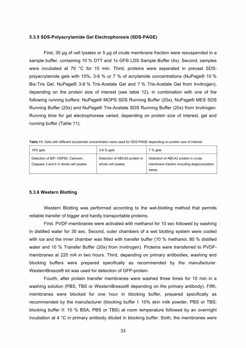
33
5.3.5 SDS-Polyacrylamide Gel Electrophoresis (SDS-PAGE)
First, 30 g of cell lysates or 5 g of crude membrane fraction were resuspended in a
sample buffer, containing 10 % DTT and 1x GF® LDS Sample Buffer (4x). Second, samples
were incubated at 70 °C for 10 min. Third, proteins were separated in precast SDS-
polyacrylamide gels with 10%, 3-8 % or 7 % of acrylamide concentrations (NuPage® 10 %
Bis-Tris Gel, NuPage® 3-8 % Tris-Acetate Gel and 7 % Tris-Acetate Gel from Invitrogen),
depending on the protein size of interest (see table 12), in combination with one of the
following running buffers: NuPage® MOPS SDS Running Buffer (20x), NuPage® MES SDS
Running Buffer (20x) and NuPage® Tris-Acetate SDS Running Buffer (20x) from Invitrogen.
Running time for gel electrophorese varied, depending on protein size of interest, gel and
running buffer (Table 11).
Table 11: Gels with different acrylamide concentration were used for SDS-PAGE depending on protein size of interest.
10% gels 3-8 % gels 7 % gels
Detection of BiP, HSP90, Calnexin,
Caspase 3 and 4 in whole cell lysates
Detection of ABCA3 protein in
whole cell lysates
Detection of ABCA3 protein in crude
membrane fraction including deglycosylation
assay
5.3.6 Western Blotting
Western Blotting was performed according to the wet-blotting method that permits
reliable transfer of bigger and hardly transportable proteins.
First, PVDF-membranes were activated with methanol for 10 sec followed by washing
in distilled water for 30 sec. Second, outer chambers of a wet blotting system were cooled
with ice and the inner chamber was filled with transfer buffer (10 % methanol, 80 % distilled
water and 10 % Transfer Buffer (20x) from Invitrogen). Proteins were transferred to PVDF-
membranes at 220 mA in two hours. Third, depending on primary antibodies, washing and
blocking buffers were prepared specifically as recommended by the manufacturer.
WesternBreeze® kit was used for detection of GFP-protein.
Fourth, after protein transfer membranes were washed three times for 10 min in a
washing solution (PBS, TBS or WesternBreeze® depending on the primary antibody). Fifth,
membranes were blocked for one hour in blocking buffer, prepared specifically as
recommended by the manufacturer (blocking buffer I: 10% skin milk powder, PBS or TBS;
blocking buffer II: 10 % BSA, PBS or TBS) at room temperature followed by an overnight
incubation at 4 °C in primary antibody diluted in blocking buffer. Sixth, the membranes were
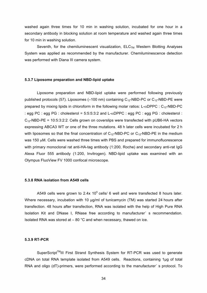
34
washed again three times for 10 min in washing solution, incubated for one hour in a
secondary antibody in blocking solution at room temperature and washed again three times
for 10 min in washing solution.
Seventh, for the chemiluminescent visualization, ELCTM Western Blotting Analyses
System was applied as recommended by the manufacturer. Chemiluminescence detection
was performed with Diana III camera system.
5.3.7 Liposome preparation and NBD-lipid uptake
Liposome preparation and NBD-lipid uptake were performed following previously
published protocols (57). Liposomes ( 100 nm) containing C12-NBD-PC or C12-NBD-PE were
prepared by mixing lipids in chloroform in the following molar ratios: L- DPPC : C12-NBD-PC
: egg PC : egg PG : cholesterol = 5:5:5:3:2 and L- DPPC : egg PC : egg PG : cholesterol :
C12-NBD-PE = 10:5:3:2:2. Cells grown on coverslips were transfected with pUB6-HA vectors
expressing ABCA3 WT or one of the three mutations. 48 h later cells were incubated for 2 h
with liposomes so that the final concentration of C12-NBD-PC or C12-NBD-PE in the medium
was 150 μM. Cells were washed three times with PBS and prepared for immunofluorescence
with primary monoclonal rat anti-HA-tag antibody (1:200, Roche) and secondary anti-rat IgG
Alexa Fluor 555 antibody (1:200, Invitrogen). NBD-lipid uptake was examined with an
Olympus FluoView FV 1000 confocal microscope.
5.3.8 RNA isolation from A549 cells
A549 cells were grown to 2.4x 10⁵ cells/ 6 well and were transfected 8 hours later.
Where necessary, incubation with 10 g/ml of tunicamycin (TM) was started 24 hours after
transfection. 48 hours after transfection, RNA was isolated with the help of High Pure RNA
Isolation Kit and DNase I, RNase free according to manufacturer´ s recommendation.
Isolated RNA was stored at – 80 °C and when necessary, thawed on ice.
5.3.9 RT-PCR
SuperScriptTMIII First Strand Synthesis System for RT-PCR was used to generate
cDNA on total RNA template isolated from A549 cells. Reactions, containing 1 g of total
RNA and oligo (dT)-primers, were performed according to the manufacturer´ s protocol. To
35
exclude genomic DNA contamination of RNA template a negative control without addition of
reverse transcriptase (RT) was performed for each sample. To amplify gene of interest, PCR
was conducted with gene specific primers and 5 l of DNA-product. Table 12 shows optimal
parameters for RT-PCR.
Table 12: Optimal cycling parameters for RT-PCR in this study.
Time Temperature Cycles
5 min 95 °C 1
20 sec. 95 °C 35
20 sec. 65 °C 35
45 sec. 72 °C 35
5 min 72 °C 1
4 °C 1
To obtain the level of basal protein expression, PCR was also performed with a
housekeeping specific primer. PCR products were subjected to a 3% agarose gel. The
results were considered positive and were statistically evaluated, when no PCR-bands
appeared in reactions without addition of RT.
5.3.10 Densitrometric analysis of the intensity of protein and DNA bands
First, chemiluminescence detection of proteins separated by Western Blotting was
performed with Diana III camera system (Raytest). Second, the amount of proteins or the
amount of DNA separated on agarose gels was evaluated by densitrometry with the help of
the software AIDA. The relative band intensity of the protein or DNA band was obtained
through normalization to the intensity of a housekeeping protein or gene.
5.3.11 Immunofluorescence
Cells were grown on coverslips and after 24 hours these cells were transfected either
with pEYFP-ABCA3- or with pUB6-ABCA3-HA- vector. 48 hours after transfection, cells were
fixed and permeabled by methanol incubations at room temperature for 10 min and were
washed three times with D-PBS buffer. Unspecific binding sites were blocked by blocking
buffer (containing D-PBS, 3 % BSA and 10 % FBS) for 30 min. Cells were incubated in
36
primary antibody (Table 6) diluted in blocking buffer for one hour. After incubation, samples
were washed three times with D-PBS and incubated in secondary antibody, either a green-
or red-fluorescent antibody (Table 7), in blocking buffer for 30 min. All further steps were
performed in dark. After incubation, samples were washed again three times and coverslips
were fixed on a microscope slide with Vectashield Hard and Set Mounting Media (Vector
Laboratories, Burlingame, USA), containing DAPI that stains DNA and is a brilliant blue-
fluorescent marker for cell nuclei.
For immunofluorescence analysis samples were evaluated with the help of
Fluorescence microscope Axiovert 135 with filter sets R/B/G and filter set FRET and with the
software AxioVision Release 4.7.1.
5.3.12 FACS analyses
A549 cells were transfected with pEYFP-ABCA3-Wt, -R43L, -R280C or –L101P and
were grown to 70 % confluence. Cells were gated in the FL-1 channel (YFP-positive
population of cells). Apoptosis was determined using different approaches. Early apoptosis
was assayed by 1) measuring intracellular glutathione (GSH) levels with the cell-permeable
monochlorobimane (Sigma) method and 2) by annexin V+/propidium iodide (PI)- staining
(Cy5-conjugated anti-annexin V and PI; BD Pharmingen), and late apoptosis was determined
via intracellular active caspase 3 levels (PE-conjugated anti-active-caspase 3; BD
Pharmingen) in cells permeabilized with the IntraPrep kit (Beckman Coulter, Krefeld,
Germany) according to manufacturer’s protocol. To exclude unspecific binding isotype
controls for caspase 3 and negative controls for glutathione and annexin V/PI staining were
applied. BD FACSCanto II Flow Cytometer was used for the assay and FACSDiva v6.1.3 for
data analyses (BD Bioscience).
5.3.13 Statistical analyses
Statistical analysis was performed by one-way ANOVA and Bonferroni’s test using
GraphPad Prism version 4.0 (GrapPad Software Inc., San Diego, CA). All results were
presented as means +SEM of minimum four experiments and p-values < 0.05 were
considered significant.
37
6. Results
6.1 Creating a model system to analyse the effect of ABCA3 mutations on alveolar
type II cells´ homeostasis
In attempt to elucidate how the overexpression of R43L, R280C and L101P mutant
ABCA3 protein impair homeostasis of human type II pneumocytes, the first part of the study
was concentrated on creation of a plasmid vector carrying WT ABCA3 cDNA, on introduction
of point mutations and on generating a cell system that can be easily transfected with a high
efficiency.
6.1.1 Generation of pUB6-hABCA3 vector and site-directed point mutagenesis
With intention to detect and exclude any effect of YFP-protein expression on
intracellular ABCA3 protein localization, another plasmid vector carrying WT ABCA3 cDNA
was created and subsequent used for transfection into A549 cells and for
immunofluorescence studies. For this purpose, pJet1/hABCA3-HA, carrying HA-tag fused to
C-terminal of the ABCA3 gene, was used as a starting vector from which ABCA3-HA cDNA
was cloned into puB6/lacZ (Fig.13, 14).
Fig.13: Vector map of pUB6-hABCA3-HA-WT
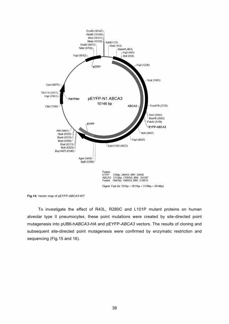
38
Fig.14: Vector map of pEYFP-ABCA3-WT
To investigate the effect of R43L, R280C and L101P mutant proteins on human
alveolar type II pneumocytes, these point mutations were created by site-directed point
mutagenesis into pUB6-hABCA3-HA and pEYFP-ABCA3 vectors. The results of cloning and
subsequent site-directed point mutagenesis were confirmed by enzymatic restriction and
sequencing (Fig.15 and 16).
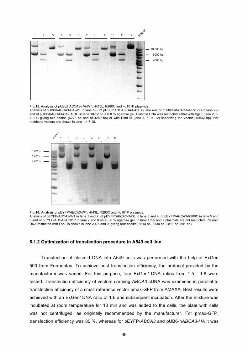
39
Fig.15: Analysis of pUB6/hABCA3-HA-WT, -R43L, R280C and –L101P plasmids. Analysis of pUB6/hABCA3-HA-WT in lane 1-3, of pUB6/hABCA3-HA-R43L in lane 4-6, of pUB6/hABCA3-HA-R280C in lane 7-9 and of pUB6/hABCA3-HA-L101P in lane 10-12 on a 0.8 % agarose gel. Plasmid DNA was restricted either with Bgl II (lane 2, 5, 8, 11) giving two chains (6273 bp and of 4269 bp) or with Hind III (lane 3, 6, 9, 12) linearizing the vector (10542 bp). Not restricted vectors are shown in lane 1,4,7,10.
Fig.16: Analysis of pEYFP/ABCA3-WT, -R43L, R280C and –L101P plasmids. Analysis of pEYFP/ABCA3-WT in lane 1 and 2, of pEYFP/ABCA3-R43L in lane 3 and 4, of pEYFP/ABCA3-R280C in lane 5 and 6 and of pEYFP/ABCA3-L101P in lane 7 and 8 on a 0.8 % agarose gel. In lane 1,3,5 and 7 plasmids are not restricted. Plasmid DNA restricted with Fsp I is shown in lane 2,4,6 and 9, giving four chains (3614 bp, 3130 bp, 2811 bp, 591 bp).
6.1.2 Optimization of transfection procedure in A549 cell line
Transfection of plasmid DNA into A549 cells was performed with the help of ExGen
500 from Fermentas. To achieve best transfection efficiency, the protocol provided by the
manufacturer was varied. For this purpose, four ExGen/ DNA ratios from 1:5 - 1:8 were
tested. Transfection efficiency of vectors carrying ABCA3 cDNA was examined in parallel to
transfection efficiency of a small reference vector pmax-GFP from AMAXA. Best results were
achieved with an ExGen/ DNA ratio of 1:6 and subsequent incubation. After the mixture was
incubated at room temperature for 10 min and was added to the cells, the plate with cells
was not centrifuged, as originally recommended by the manufacturer. For pmax-GFP,
transfection efficiency was 80 %, whereas for pEYFP-ABCA3 and pUB6-hABCA3-HA it was
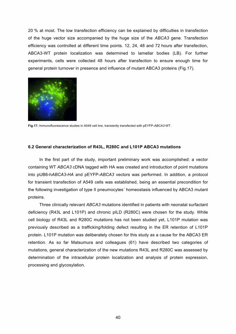
40
20 % at most. The low transfection efficiency can be explained by difficulties in transfection
of the huge vector size accompanied by the huge size of the ABCA3 gene. Transfection
efficiency was controlled at different time points. 12, 24, 48 and 72 hours after transfection,
ABCA3-WT protein localization was determined to lamellar bodies (LB). For further
experiments, cells were collected 48 hours after transfection to ensure enough time for
general protein turnover in presence and influence of mutant ABCA3 proteins (Fig.17).
Fig.17: Immunofluorescence studies in A549 cell line, transiently transfected with pEYFP-ABCA3-WT.
6.2 General characterization of R43L, R280C and L101P ABCA3 mutations
In the first part of the study, important preliminary work was accomplished: a vector
containing WT ABCA3 cDNA tagged with HA was created and introduction of point mutations
into pUB6-hABCA3-HA and pEYFP-ABCA3 vectors was performed. In addition, a protocol
for transient transfection of A549 cells was established, being an essential precondition for
the following investigation of type II pneumocytes´ homeostasis influenced by ABCA3 mutant
proteins.
Three clinically relevant ABCA3 mutations identified in patients with neonatal surfactant
deficiency (R43L and L101P) and chronic pILD (R280C) were chosen for the study. While
cell biology of R43L and R280C mutations has not been studied yet, L101P mutation was
previously described as a trafficking/folding defect resulting in the ER retention of L101P
protein. L101P mutation was deliberately chosen for this study as a cause for the ABCA3 ER
retention. As so far Matsumura and colleagues (61) have described two categories of
mutations, general characterization of the new mutations R43L and R280C was assessed by
determination of the intracellular protein localization and analysis of protein expression,
processing and glycosylation.

41
6.2.1 Localization and trafficking
Previous studies showed localization of the ABCA3 transporter to the limiting
membrane of lamellar bodies (LBs) in lung alveolar type II-cells (41). In order to investigate
ABCA3 protein localization or possibly impaired protein trafficking in A549 cells, staining of
different cell compartments was performed in parallel with staining of the ABCA3 protein.
Following compartments were analysed: First, LBs were stained with the marker LAMP3, an
integral part of membranes of LBs. Second, the ER, important for protein processing and
quality controlling, was marked by calnexin, an ER-bound chaperone that is involved in
quality control of protein folding within the UPR system (see “Introduction”), in order to
elucidate if ABCA3 mutant proteins might be retained in the ER due to processing or folding
defect. Third, as the Golgi apparatus is an integral part of processing, sorting and
modification of proteins delivered from the ER, ABCA3 mutant protein localization to this cell
compartment was assessed as well via the marker GM130. GM130 codes for a protein that
participates in forming and maintaining cis-Golgi and that supports vesicular transport.
Fourth, early endosomes were detected via EEA-1, a protein that binds phosphatidylinositol-
3-phosphate in endosomes membranes. Endosomes are formed via endocytosis and plasma
membrane invagination and transport proteins to lysosomes for subsequent digestion.
To double confirm ABCA3 protein localization in A549 cells and to rule out possible
influence and effect of YFP-expression, colocalization studies were performed in parallel with
pEYFP-ABCA3 vector and pUB6-hABCA3-HA vector. Effects that could have been expected
in case of YFP-ABCA3 expression were protein aggregation, impaired protein trafficking and
protein degradation. The intracellular protein localization of ABCA3 protein was determined
48 hours after transfection. Fluorescence microscopy of A549 cells expressing either type of
ABCA3 protein fusions showed no differences in protein behavior depending on the type of
the vector or the size of the protein tag. YFP-ABCA3 WT protein was completely localized to
LBs (Fig.18). ABCA3 WT protein appeared as a ring-like and dot-like structure in
immunofluorescence (Fig.18). The colocalization of WT with the ER protein calnexin, GM130
and EEA1 was not present (Fig.18B, 19, 20). Similar was observed in the case of R43L
mutant, which gave a vesicular signal that overlapped with LAMP3 fluorescence (Fig.18A)
and showed no colocalization with calnexin, GM130 and EEA1 (Fig.18B, 19, 20). R280C
protein colocalized frequently with LAMP3, however the colocalization was not absolute,
often showing cytoplasmic distribution that overlapped with the fluorescence of calnexin
(Fig.18, A and B). L101P mutation did not show any vesicular signal at all (Fig.18A) but it
presented with a net-like cytoplasmic fluorescence colocalizing with the ER protein calnexin
(Fig.18B). Further immunofluorescence studies showed no localization of L101P mutant
protein with markers for Golgi apparatus or early endosomes (Fig.19, 20).
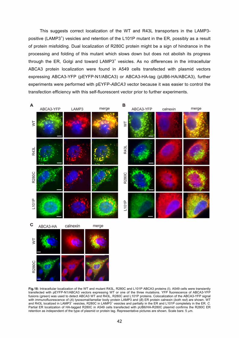
42
This suggests correct localization of the WT and R43L transporters in the LAMP3-
positive (LAMP3+) vesicles and retention of the L101P mutant in the ER, possibly as a result
of protein misfolding. Dual localization of R280C protein might be a sign of hindrance in the
processing and folding of this mutant which slows down but does not abolish its progress
through the ER, Golgi and toward LAMP3+ vesicles. As no differences in the intracellular
ABCA3 protein localization were found in A549 cells transfected with plasmid vectors
expressing ABCA3-YFP (pEYFP-N1/ABCA3) or ABCA3-HA-tag (pUB6-HA/ABCA3), further
experiments were performed with pEYFP-ABCA3 vector because it was easier to control the
transfection efficiency with this self-fluorescent vector prior to further experiments.
Fig.18: Intracellular localization of the WT and mutant R43L, R280C and L101P ABCA3 proteins (I). A549 cells were transiently transfected with pEYFP-N1/ABCA3 vectors expressing WT or one of the three mutations. YFP fluorescence of ABCA3-YFP fusions (green) was used to detect ABCA3 WT and R43L, R280C and L101P proteins. Colocalization of the ABCA3-YFP signal with immunofluorescence of (A) lysosomal/lamellar body protein LAMP3 and (B) ER protein calnexin (both red) are shown. WT and R43L localized in LAMP3
+ vesicles, R280C in LAMP3
+ vesicles and partially in the ER and L101P completely in the ER. C.
Partial ER localization of HA-tagged R280C in A549 cells transfected with pUB6/HA-R280C plasmid confirms the R280C ER retention as independent of the type of plasmid or protein tag. Representative pictures are shown. Scale bars: 5 μm.
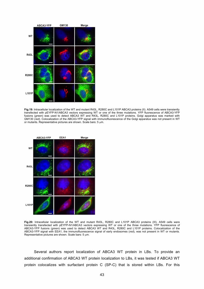
43
Fig.19: Intracellular localization of the WT and mutant R43L, R280C and L101P ABCA3 proteins (II). A549 cells were transiently transfected with pEYFP-N1/ABCA3 vectors expressing WT or one of the three mutations. YFP fluorescence of ABCA3-YFP fusions (green) was used to detect ABCA3 WT and R43L, R280C and L101P proteins. Golgi apparatus was marked with GM130 (red). Colocalization of the ABCA3-YFP signal with immunofluorescence of the Golgi apparatus was not present in WT or mutants. Representative pictures are shown. Scale bars: 5 μm.
Fig.20: Intracellular localization of the WT and mutant R43L, R280C and L101P ABCA3 proteins (III). A549 cells were transiently transfected with pEYFP-N1/ABCA3 vectors expressing WT or one of the three mutations. YFP fluorescence of ABCA3-YFP fusions (green) was used to detect ABCA3 WT and R43L, R280C and L101P proteins. Colocalization of the ABCA3-YFP signal with EEA1, the immunofluorescence signal of early endosomes (red), was not present in WT or mutants. Representative pictures are shown. Scale bars: 5 μm.
Several authors report localization of ABCA3 WT protein in LBs. To provide an
additional confirmation of ABCA3 WT protein localization to LBs, it was tested if ABCA3 WT
protein colocalizes with surfactant protein C (SP-C) that is stored within LBs. For this
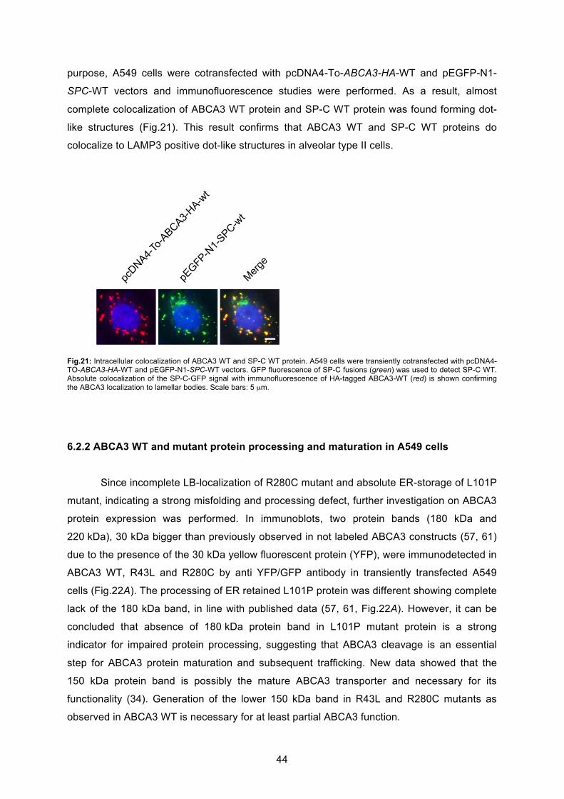
44
purpose, A549 cells were cotransfected with pcDNA4-To-ABCA3-HA-WT and pEGFP-N1-
SPC-WT vectors and immunofluorescence studies were performed. As a result, almost
complete colocalization of ABCA3 WT protein and SP-C WT protein was found forming dot-
like structures (Fig.21). This result confirms that ABCA3 WT and SP-C WT proteins do
colocalize to LAMP3 positive dot-like structures in alveolar type II cells.
Fig.21: Intracellular colocalization of ABCA3 WT and SP-C WT protein. A549 cells were transiently cotransfected with pcDNA4-TO-ABCA3-HA-WT and pEGFP-N1-SPC-WT vectors. GFP fluorescence of SP-C fusions (green) was used to detect SP-C WT. Absolute colocalization of the SP-C-GFP signal with immunofluorescence of HA-tagged ABCA3-WT (red) is shown confirming the ABCA3 localization to lamellar bodies. Scale bars: 5 μm.
6.2.2 ABCA3 WT and mutant protein processing and maturation in A549 cells
Since incomplete LB-localization of R280C mutant and absolute ER-storage of L101P
mutant, indicating a strong misfolding and processing defect, further investigation on ABCA3
protein expression was performed. In immunoblots, two protein bands (180 kDa and
220 kDa), 30 kDa bigger than previously observed in not labeled ABCA3 constructs (57, 61)
due to the presence of the 30 kDa yellow fluorescent protein (YFP), were immunodetected in
ABCA3 WT, R43L and R280C by anti YFP/GFP antibody in transiently transfected A549
cells (Fig.22A). The processing of ER retained L101P protein was different showing complete
lack of the 180 kDa band, in line with published data (57, 61, Fig.22A). However, it can be
concluded that absence of 180 kDa protein band in L101P mutant protein is a strong
indicator for impaired protein processing, suggesting that ABCA3 cleavage is an essential
step for ABCA3 protein maturation and subsequent trafficking. New data showed that the
150 kDa protein band is possibly the mature ABCA3 transporter and necessary for its
functionality (34). Generation of the lower 150 kDa band in R43L and R280C mutants as
observed in ABCA3 WT is necessary for at least partial ABCA3 function.

45
6.2.3 Glycosylation of ABCA3 protein
After Western Blotting analysis of cell lysates from cells expressing ABCA3 protein,
demonstrating that ABCA3 WT, R43L and R280C mutant proteins are expressed as 220 kDa
and 180 kDa forms, whereas 180 kDa protein is not detectable in L101P mutant (Fig.22A),
further investigation on ABCA3 processing was performed.
Glycosylation is an enzymatic process within the ER and Golgi, which links
saccharides to immature proteins to produce glycans. ABCA3 is a glycoprotein that obtains
high-mannose oligosaccharides inside of the ER and complex oligosaccharides by
modification of high-mannose oligosaccharides in the Golgi. Processing of oligosaccharides
and protein progress down the ER-Golgi maturation pathway was analyzed by
deglycosylation of the membrane fractions from the A549 cells expressing WT and three
mutations with two endoglycosidases EndoH (cleaves only high-mannose sugars) and
PNGaseF (cleaves high-mannose and complex sugars) (Fig. 22B). Immunoblotting with anti-
GFP antibody revealed absence of complex sugars in L101P protein that was susceptible to
both enzymes, PNGaseF and EndoH, resulting in both cases in a single shifted
deglycosylated 210 kDa band and no 220 kDa band. Both sugar types were present in WT,
R43L and R280C proteins, as visible by the resistance of a portion of the 220 kDa band to
the EndoH treatment. The 180 kDa band was resistant to deglycosylation (Fig.22B). These
results also additionally confirm the localization studies showing retention of the L101P
mutant in the ER and ability of WT, R43L and R280C to progress further from the ER to the
Golgi.
A B
Fig.22: Processing of the WT and mutant R43L, R280C and L101P ABCA3. (A) Immunodetection with anti-GFP antibody showed two ABCA3 protein bands (180 kDa and 220 kDa) in whole cell lysates of A549 cells expressing WT and R43L and R280C mutations, and only one protein band in the cells expressing L101P mutation. (B) Deglycosylation assay with PNGaseF and EndoH on the membrane fractions from A549 cells transfected with pEYFP-N1/ABCA3 plasmids and subsequent ABCA3-YFP immunodetection with anti-GFP antibody showed presence of high-mannose and complex oligosaccharides in WT, R43L and R280C proteins, as well as only high-mannose and no complex oligosaccharides in the L101P mutant resulting from the L101P ER retention.
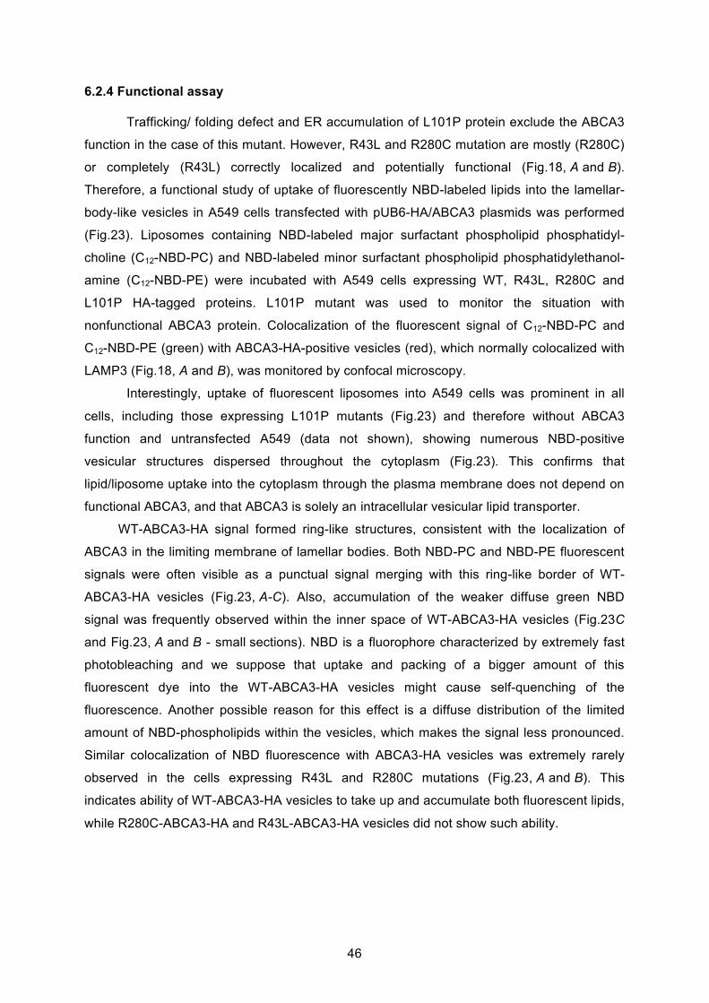
46
6.2.4 Functional assay
Trafficking/ folding defect and ER accumulation of L101P protein exclude the ABCA3
function in the case of this mutant. However, R43L and R280C mutation are mostly (R280C)
or completely (R43L) correctly localized and potentially functional (Fig.18, A and B).
Therefore, a functional study of uptake of fluorescently NBD-labeled lipids into the lamellar-
body-like vesicles in A549 cells transfected with pUB6-HA/ABCA3 plasmids was performed
(Fig.23). Liposomes containing NBD-labeled major surfactant phospholipid phosphatidyl-
choline (C12-NBD-PC) and NBD-labeled minor surfactant phospholipid phosphatidylethanol-
amine (C12-NBD-PE) were incubated with A549 cells expressing WT, R43L, R280C and
L101P HA-tagged proteins. L101P mutant was used to monitor the situation with
nonfunctional ABCA3 protein. Colocalization of the fluorescent signal of C12-NBD-PC and
C12-NBD-PE (green) with ABCA3-HA-positive vesicles (red), which normally colocalized with
LAMP3 (Fig.18, A and B), was monitored by confocal microscopy.
Interestingly, uptake of fluorescent liposomes into A549 cells was prominent in all
cells, including those expressing L101P mutants (Fig.23) and therefore without ABCA3
function and untransfected A549 (data not shown), showing numerous NBD-positive
vesicular structures dispersed throughout the cytoplasm (Fig.23). This confirms that
lipid/liposome uptake into the cytoplasm through the plasma membrane does not depend on
functional ABCA3, and that ABCA3 is solely an intracellular vesicular lipid transporter.
WT-ABCA3-HA signal formed ring-like structures, consistent with the localization of
ABCA3 in the limiting membrane of lamellar bodies. Both NBD-PC and NBD-PE fluorescent
signals were often visible as a punctual signal merging with this ring-like border of WT-
ABCA3-HA vesicles (Fig.23, A-C). Also, accumulation of the weaker diffuse green NBD
signal was frequently observed within the inner space of WT-ABCA3-HA vesicles (Fig.23C
and Fig.23, A and B - small sections). NBD is a fluorophore characterized by extremely fast
photobleaching and we suppose that uptake and packing of a bigger amount of this
fluorescent dye into the WT-ABCA3-HA vesicles might cause self-quenching of the
fluorescence. Another possible reason for this effect is a diffuse distribution of the limited
amount of NBD-phospholipids within the vesicles, which makes the signal less pronounced.
Similar colocalization of NBD fluorescence with ABCA3-HA vesicles was extremely rarely
observed in the cells expressing R43L and R280C mutations (Fig.23, A and B). This
indicates ability of WT-ABCA3-HA vesicles to take up and accumulate both fluorescent lipids,
while R280C-ABCA3-HA and R43L-ABCA3-HA vesicles did not show such ability.
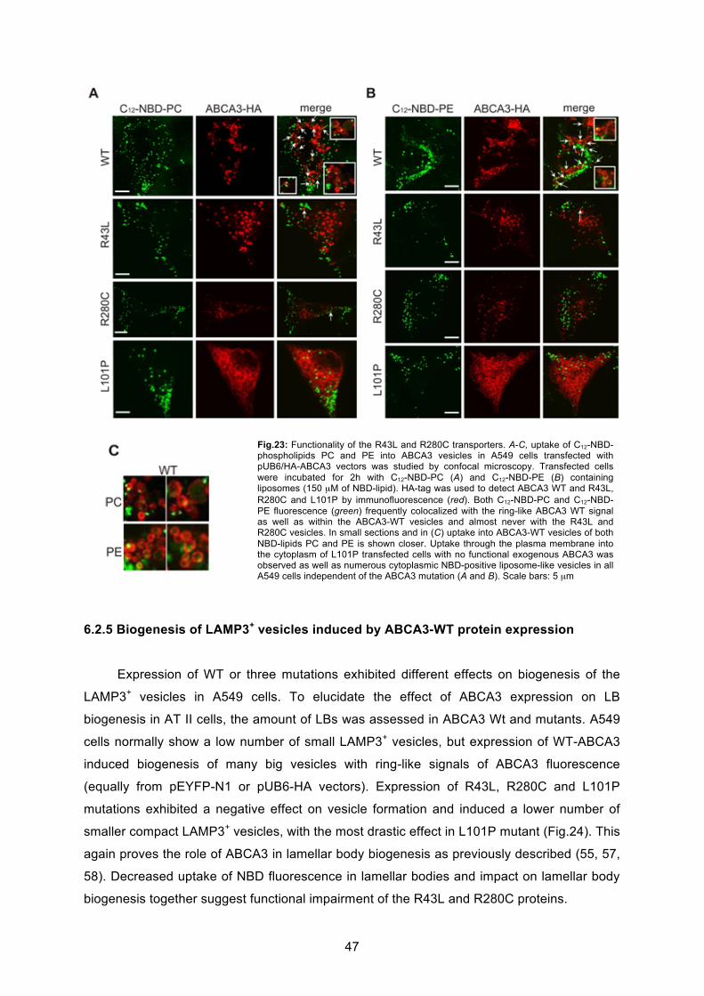
47
6.2.5 Biogenesis of LAMP3+ vesicles induced by ABCA3-WT protein expression
Expression of WT or three mutations exhibited different effects on biogenesis of the
LAMP3+ vesicles in A549 cells. To elucidate the effect of ABCA3 expression on LB
biogenesis in AT II cells, the amount of LBs was assessed in ABCA3 Wt and mutants. A549
cells normally show a low number of small LAMP3+ vesicles, but expression of WT-ABCA3
induced biogenesis of many big vesicles with ring-like signals of ABCA3 fluorescence
(equally from pEYFP-N1 or pUB6-HA vectors). Expression of R43L, R280C and L101P
mutations exhibited a negative effect on vesicle formation and induced a lower number of
smaller compact LAMP3+ vesicles, with the most drastic effect in L101P mutant (Fig.24). This
again proves the role of ABCA3 in lamellar body biogenesis as previously described (55, 57,
58). Decreased uptake of NBD fluorescence in lamellar bodies and impact on lamellar body
biogenesis together suggest functional impairment of the R43L and R280C proteins.
Fig.23: Functionality of the R43L and R280C transporters. A-C, uptake of C12-NBD-phospholipids PC and PE into ABCA3 vesicles in A549 cells transfected with pUB6/HA-ABCA3 vectors was studied by confocal microscopy. Transfected cells were incubated for 2h with C12-NBD-PC (A) and C12-NBD-PE (B) containing liposomes (150 μM of NBD-lipid). HA-tag was used to detect ABCA3 WT and R43L, R280C and L101P by immunofluorescence (red). Both C12-NBD-PC and C12-NBD-PE fluorescence (green) frequently colocalized with the ring-like ABCA3 WT signal as well as within the ABCA3-WT vesicles and almost never with the R43L and R280C vesicles. In small sections and in (C) uptake into ABCA3-WT vesicles of both NBD-lipids PC and PE is shown closer. Uptake through the plasma membrane into the cytoplasm of L101P transfected cells with no functional exogenous ABCA3 was observed as well as numerous cytoplasmic NBD-positive liposome-like vesicles in all A549 cells independent of the ABCA3 mutation (A and B). Scale bars: 5 μm
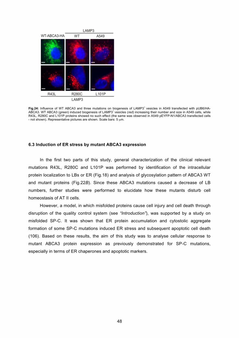
48
Fig.24: Influence of WT ABCA3 and three mutations on biogenesis of LAMP3+
vesicles in A549 transfected with pUB6/HA-ABCA3. WT ABCA3 (green) induced biogenesis of LAMP3
+ vesicles (red) increasing their number and size in A549 cells, while
R43L, R280C and L101P proteins showed no such effect (the same was observed in A549 pEYFP-N1/ABCA3 transfected cells - not shown). Representative pictures are shown. Scale bars: 5 μm.
6.3 Induction of ER stress by mutant ABCA3 expression
In the first two parts of this study, general characterization of the clinical relevant
mutations R43L, R280C and L101P was performed by identification of the intracellular
protein localization to LBs or ER (Fig.18) and analysis of glycosylation pattern of ABCA3 WT
and mutant proteins (Fig.22B). Since these ABCA3 mutations caused a decrease of LB
numbers, further studies were performed to elucidate how these mutants disturb cell
homeostasis of AT II cells.
However, a model, in which misfolded proteins cause cell injury and cell death through
disruption of the quality control system (see “Introduction”), was supported by a study on
misfolded SP-C. It was shown that ER protein accumulation and cytostolic aggregate
formation of some SP-C mutations induced ER stress and subsequent apoptotic cell death
(106). Based on these results, the aim of this study was to analyse cellular response to
mutant ABCA3 protein expression as previously demonstrated for SP-C mutations,
especially in terms of ER chaperones and apoptotic markers.

49
6.3.1 L101P and R280C mutation upregulate ER stress marker BiP
BiP/Grp78 is an essential ER chaperone of Hsp70 family, which assists translocation
of a nascent protein chain into the ER and its subsequent folding. When misfolded proteins
accumulate in the ER, BiP upregulation is one of the first signals of the ER stress and allows
further UPR activation (116). The effect of ABCA3 mutations on chaperones is especially
interesting since until now ER stress-induction has not been investigated in ABCA3 mutants.
Immunoblotting of whole cell lysates from A549 cells expressing ABCA3-WT and
three mutants revealed upregulation of BiP chaperone in the case of the L101P and R280C
mutations in comparison to WT (Fig.25, A and B), caused by complete (L101P) and partial
(R280C) ER retention of these two mutated ABCA3 transporters (Fig.18). No significant BiP
increase was detectable between WT and correctly localized R43L mutation. While treatment
of A549 cells with a potent ER stress-causing antibiotic tunicamycin (TM) caused drastic BiP
upregulation in A549 cells, differences measured between WT and three mutations were
more subtle differences.
A
B
Fig.25: ER stress chaperone BiP. A, A549 cells with ER retained L101P mutant or partially ER retained R280C protein showed upregulation of the immunodetected ER chaperone BiP in comparison to WT and A549 cells. BiP was drastically upregulated after 14h treatment of A549 cells with 10ug/ml of ER stressor tunicamycin (TM). B, intensity of each protein band was densitometrically quantified and normalized to the corresponding ß-actin band. Densitometric quantification of the BiP protein level of four independent experiments, without inclusion of drastic effect of TM treatment, is presented; * p<0.05, * * p<0.01.
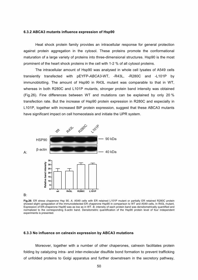
50
6.3.2 ABCA3 mutants influence expression of Hsp90
Heat shock protein family provides an intracellular response for general protection
against protein aggregation in the cytosol. These proteins promote the conformational
maturation of a large variety of proteins into three-dimensional structures. Hsp90 is the most
prominent of the heart shock proteins in the cell with 1-2 % of all cytosol proteins.
The intracellular amount of Hsp90 was analysed in whole cell lysates of A549 cells
transiently transfected with pEYFP-ABCA3-WT, -R43L, -R280C and -L101P by
immunoblotting. The amount of Hsp90 in R43L mutant was comparable to that in WT,
whereas in both R280C and L101P mutants, stronger protein band intensity was obtained
(Fig.26). Fine differences between WT and mutations can be explained by only 20 %
transfection rate. But the increase of Hsp90 protein expression in R280C and especially in
L101P, together with increased BiP protein expression, suggest that these ABCA3 mutants
have significant impact on cell homeostasis and initiate the UPR system.
A:
B:
Fig.26: ER stress chaperone Hsp 90. A, A549 cells with ER retained L101P mutant or partially ER retained R280C protein showed slight upregulation of the immunodetected ER chaperone Hsp90 in comparison to WT and A549 cells. In R43L mutant, Expression of ER-chaperone Hsp90 was as low as in WT. B, intensity of each protein band was densitometrically quantified and normalized to the corresponding ß-actin band. Densitometric quantification of the Hsp90 protein level of four independent experiments is presented.
6.3.3 No influence on calnexin expression by ABCA3 mutations
Moreover, together with a number of other chaperones, calnexin facilitates protein
folding by catalyzing intra- and inter-molecular disulfide bond formation to prevent trafficking
of unfolded proteins to Golgi apparatus and further downstream in the secretory pathway,
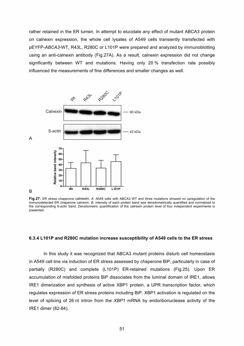
51
rather retained in the ER lumen. In attempt to elucidate any effect of mutant ABCA3 protein
on calnexin expression, the whole cell lysates of A549 cells transiently transfected with
pEYFP-ABCA3-WT, R43L, R280C or L101P were prepared and analyzed by immunoblotting
using an anti-calnexin antibody (Fig.27A). As a result, calnexin expression did not change
significantly between WT and mutations. Having only 20 % transfection rate possibly
influenced the measurements of fine differences and smaller changes as well.
A
B
Fig.27: ER stress chaperone calnexin. A, A549 cells with ABCA3 WT and three mutations showed no upregulation of the immunodetected ER chaperone calnexin. B, intensity of each protein band was densitometrically quantified and normalized to the corresponding ß-actin band. Densitometric quantification of the calnexin protein level of four independent experiments is presented.
6.3.4 L101P and R280C mutation increase susceptibility of A549 cells to the ER stress
In this study it was recognized that ABCA3 mutant proteins disturb cell homeostasis
in A549 cell line via induction of ER stress assessed by chaperone BiP, particularly in case of
partially (R280C) and complete (L101P) ER-retained mutations (Fig.25). Upon ER
accumulation of misfolded proteins BiP dissociates from the luminal domain of IRE1, allows
IRE1 dimerization and synthesis of active XBP1 protein, a UPR transcription factor, which
regulates expression of ER stress proteins including BiP. XBP1 activation is regulated on the
level of splicing of 26 nt intron from the XBP1 mRNA by endoribonuclease activity of the
IRE1 dimer (82-84).

52
To confirm the observations made on the ER chaperone level, splicing of the XBP1
mRNA was assayed by RT-PCR as described by Hybiske and colleagues (117). Two
approaches were applied to analyze XBP1 splicing. In a first experiment, 48 hours after
transfection, A549 cells were analyzed for XBP1 splicing via RT-PCR and in a second
experiment to intensify the stress, transfected cells were additionally incubated with 10 ng/ml
of tunicamycin (TM) for 13 hours.
First, the ratio of spliced (s; 500 bp – 26 bp) and unspliced (u; 500 bp) XBP1 RT-
PCR bands was determined, prior and after the treatment of the PCR products with PstI
restriction endonuclease, as a measuring control. Slower migrating hybrid (h) band of
unspliced and spliced single-stranded DNAs produced during PCR, and thus equally
contributing to unspliced and spliced bands, was observed as well after the long-run
separation on 3 % agarose (84, Fig.28A). Tunicamycin treatment strongly increased the
intensity of the spliced PCR band in A549 cells in comparison to untreated A549 (Fig.28B). In
A549 cells with either WT or one of the three mutations, increase of XBP1 splicing was
measured only in the case of L101P mutation (Fig.28A). Probably because of the robustness
of the method and 20 % transfection rate, finer differences between WT and R43L and
R280C were not observable, and the effect was measurable only in the case of the L101P as
the strongest defect (Fig.28A).
Second, in attempt to imitate that in nature lungs are not exclusively affected by
ABCA3 mutations but also exposed to outside stress factors such as viral infection and
smoking, and to investigate the susceptibility of cells with ABCA3 mutations to external ER
stress-causing agents, transfected cells were exposed to 10 mg/ml of tunicamycin, an
inhibitor of N-linked glycosylation (Fig.28, D and E). The outcome of such double internal
genetic and external tunicamycin pressure on XPB1 splicing was examined. Tunicamycin
treatment strongly induced XBP1 splicing with the comparable effect in A549 and WT cells
(Fig.28D). However, after exposure to tunicamycin XBP1 splicing was considerably more
pronounced in R280C and L101P mutations if compared to A549 cells with WT and R43L
mutations (Fig.28, D and E). Obviously, although XBP1 splicing was measurable only for the
strongest L101P defect under non-stimulated condition, cells with L101P and R280C
mutations were significantly more prone to further elevation of ER stress upon exposure to
an external stressor (Fig.28, D and E).
These results demonstrate that through mimicking outer stress, ABCA3 mutants do
change normal protein cell biology and disturb cell homeostasis of AT II cells more
significantly by raising the level of intracellular stress and susceptibility to it. Furthermore,
these results suggest that presence of outside stress factors has an additional impact on
cellular response to ABCA3 mutants in AT II cells.
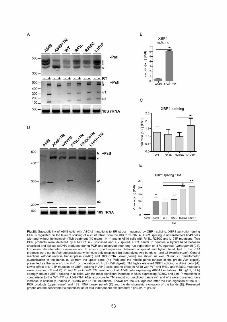
53
A B
C
D
E
Fig.28: Susceptibility of A549 cells with ABCA3 mutations to ER stress measured by XBP1 splicing. XBP1 activation during UPR is regulated on the level of splicing of a 26 nt intron from the XBP1 mRNA. A, XBP1 splicing in untransfected A549 cells with and without tunicamycin (TM) treatment (10 mg/ml, 14 h) and in A549 cells with R43L, R280C and L101P mutations. Two PCR products were detected by RT-PCR: u - unspliced and s - spliced XBP1 bands. h denotes a hybrid band between unspliced and spliced ssDNA produced during PCR and observed after long-run separation on 3 % agarose (upper panel) (21). For easier densitometric evaluation and to ensure good separation between unspliced and hybrid band, half of the PCR products were cut by PstI endonuclease which cuts only unspliced (u) band giving two bands u1 and u2 (middle panel). Control reactions without reverse transcriptase (+/-RT) and 18S rRNA (lower panel) are shown as well. B and C, densitometric quantification of the bands (s, u) from the upper panel (no PstI) and the middle panel (shown in the graph; PstI digest), presented as the ratio s/u (no PstI) or the ration s/u1+u2 (PstI digest). TM highly elevated XBP1 splicing in A549 cells (A). Lower effect of L101P mutation on XBP1 splicing in A549 cells and no effect in A549 with WT and R43L and R280C mutations were observed (B and C). D and E, as in A-C: TM treatment of all A549 cells expressing ABCA3 mutations (10 mg/ml, 14 h) strongly induced XBP1 splicing in all cells, with the most significant increase in A549 expressing R280C and L101P mutations in comparison to the WT+TM or A549+TM. After exposure to TM almost no unspliced bands (u1 and u1) were observed, only increase in spliced (s) bands in R280C and L101P mutations. Shown are the 3 % agarose after the PstI digestion of the RT-PCR products (upper panel) and 18S rRNA (lower panel) (D) and the densitometric evaluation of the bands (E). Presented graphs are the densitometric quantification of four independent experiments; * p<0.05, ** p<0.01.
54
6.4 L101P and to a lesser extent R280C mutation induce apoptosis of A549 cells
Constant accumulation of defect proteins within the ER and activation of the UPR
system affect significantly cell homeostasis: As demonstrated, R280C- and L101P-
expressing cells revealed elevated BiP levels and XBP1 splicing, particularly in the presence
of additional ER stressor, indicating that these mutants perturb homeostasis of ATII cells by
raising the level of the intracellular stress and susceptibility to it (Fig.25, 28). Prolonged and
unresolved ER stress accompanied by constant UPR activation and its failure can lead to
increased degradation of defect proteins, reduced general protein synthesis or initiating of
pro-apoptotic pathway (see “Introduction”). Since it was shown that SP-C mutations,
accumulated in the ER or cytosol, induce ER stress and subsequent apoptotic cell death (94,
106), it was interesting if ABCA3 mutations impair this fatal stress/apoptosis/cell-dysfunction
axis as well. Activation of the apoptotic pathway in cells expressing ABCA3 mutants was
assessed by early and late apoptotic markers: annexin V/PI staining, GSH decrease and
caspase 3 activation.
6.4.1. Annexin V/PI staining
In apoptosis, pro-apoptotic activation of caspase 3 can change characteristics of the
plasma membrane and leads to externalization of phophatidylserines (PS) (97). The exact
mechanism how caspase 3 interacts with phospholipid translocation is not completely
understood to the date. However, the expression of PS on the cell surface of dying cells is a
signal for macrophages to capture these cells for phagocytosis (118). Annexin V specifically
binds to the surface-exposed PS of apoptotic cells and is used as a marker of early
apoptosis. Besides, a marker for late apoptosis and necrosis is propidium iodide (PI) that
binds to double-stranded DNA, but only in non-viable cells that have lost membrane integrity.
In order to experimentally investigate whether both early apoptosis and necrosis might be
induced by ABCA3 mutants, flow cytometry was performed to analyse annexin V staining of
cells expressing ABCA3 defect proteins and to analyse PI staining within these cells.
For this purpose, A549 cells were transiently transfected with pEYFP-ABCA3-WT -
R43L, -R280C or L101P and 48 hours after transfection, samples were prepared for flow
cytometry. Flow cytometry assay of Cy5-coupled Annexin V surface binding showed an
increase in the number of annexin V+/PI- cells in transfected YFP+ cells in the case of R280C
and L101P mutations when compared to WT and R43L indicating early apoptotic state of
those cells (Fig. 30A).
55
6.4.2. GSH decrease
Glutathione is an essential low-molecular-weight thiol and a major component of the
cell antioxidant system. Loss of intracellular GSH is an early hallmark of apoptosis
progression and a part of early changes for generation of a permissive environment for the
activation of apoptotic enzymes (119). For this purpose, A549 cells were transiently
transfected with pEYFP-ABCA3-WT -R43L, -R280C or L101P and 48 hours after
transfection, samples were prepared for flow cytometry. YFP fluorescence was utilized to
determine the population of transfected cells (YFP+) and to measure the apoptotic markers
exclusively in those cells. Intracellular GSH level, measured by flow cytometry of
monochlorobimane binding to GSH and generation of a fluorescent adduct, was significantly
decreased in the YFP+ cells with L101P protein compared to YFP+ WT, and to a lower extent
compared to R43L and to R280C (Fig.30B).
6.4.3. Caspase 3 activation
Pro-apoptotic pathway is initiated by active caspases, a family of highly conserved
proteases present in the cell as inactive precursors and activated through a cascade of
regulated proteolytic cleavages leading to controlled cell death (91, 120). Among 15 known
members in mammalian species, caspase 3 is one of the executioner caspases activated at
the end of the cascade. Therefore, activation of caspase 3 is a late apoptotic marker. For this
purpose, two approaches were applied and active caspase 3 was measured by Western
Blotting as well as by flow cytometry.
First, an increase of caspase 3 activation in cells expressing the ER-retained L101P
mutant in comparison to WT and R43L was observed via flow cytometry assay of intracellular
active caspase 3 (Fig. 30C). R280C values showed steady increase in caspase 3 activation,
however with lower significance than L101P (Fig. 30C).
Second, to confirm this observation, active caspase 3 was additionally measured by
immunoblotting. A549 cells were transiently transfected with pEYFP-ABCA3-WT, -R43L, -
R280C and –L101P and after 48 hours the whole cell lysates were prepared and analyzed by
immunoblotting using an antibody, that detects the pro-form of caspase 3 (32 kDa) as well as
the active forms of caspase 3 (19 and 17 kDa). Cells, expressing R43L, R280C or L101P
mutant proteins, revealed clearly elevated levels of active caspase 3. Especially in L101P
expressing cells the amount of active caspase 3 was significant higher than in WT (Fig.29).
These results confirm data obtained by FACS.
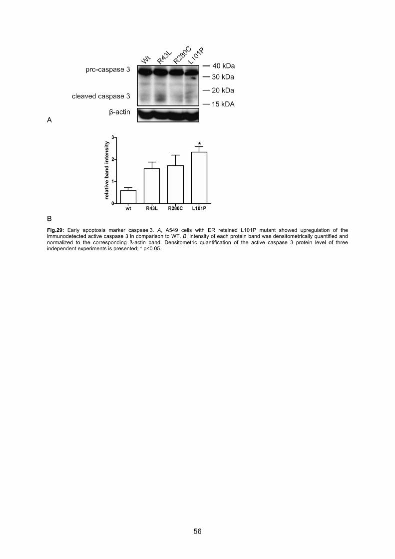
56
A
B
Fig.29: Early apoptosis marker caspase 3. A, A549 cells with ER retained L101P mutant showed upregulation of the immunodetected active caspase 3 in comparison to WT. B, intensity of each protein band was densitometrically quantified and normalized to the corresponding ß-actin band. Densitometric quantification of the active caspase 3 protein level of three independent experiments is presented; * p<0.05.
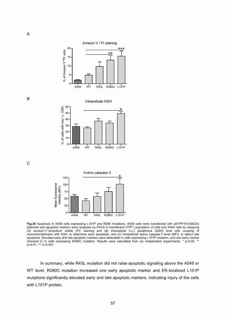
57
A
B
C
Fig.30 Apoptosis in A549 cells expressing L101P and R280 mutations. A549 cells were transfected with pEYFP-N1/ABCA3 plasmids and apoptotic markers were analyses by FACS in transfected (YFP
+) population of cells and A549 cells by assaying
(A) annexin V+/propidium iodide (PI)
- staining and (B) intracellular (i.c.) glutathione (GSH) level with coupling of
monochlorobimane with GSH, to determine early apoptosis, and (C) intracellular active caspase 3 level (MFI), to detect late apoptosis. Elevated early and late apoptotic markers were detectable in cells expressing L101P mutation, and one early marker (Annexin V) in cells expressing R280C mutation. Results were calculated from six independent experiments; * p<0.05, ** p<0.01, *** p<0.001.
In summary, while R43L mutation did not raise apoptotic signaling above the A549 or
WT level, R280C mutation increased one early apoptotic marker and ER-localized L101P
mutations significantly elevated early and late apoptotic markers, indicating injury of the cells
with L101P protein.
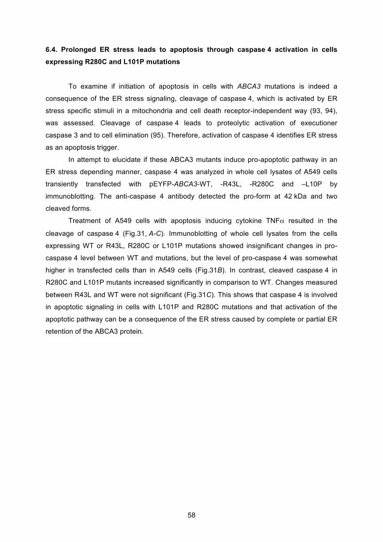
58
6.4. Prolonged ER stress leads to apoptosis through caspase 4 activation in cells
expressing R280C and L101P mutations
To examine if initiation of apoptosis in cells with ABCA3 mutations is indeed a
consequence of the ER stress signaling, cleavage of caspase 4, which is activated by ER
stress specific stimuli in a mitochondria and cell death receptor-independent way (93, 94),
was assessed. Cleavage of caspase 4 leads to proteolytic activation of executioner
caspase 3 and to cell elimination (95). Therefore, activation of caspase 4 identifies ER stress
as an apoptosis trigger.
In attempt to elucidate if these ABCA3 mutants induce pro-apoptotic pathway in an
ER stress depending manner, caspase 4 was analyzed in whole cell lysates of A549 cells
transiently transfected with pEYFP-ABCA3-WT, -R43L, -R280C and –L10P by
immunoblotting. The anti-caspase 4 antibody detected the pro-form at 42 kDa and two
cleaved forms.
Treatment of A549 cells with apoptosis inducing cytokine TNF resulted in the
cleavage of caspase 4 (Fig.31, A-C). Immunoblotting of whole cell lysates from the cells
expressing WT or R43L, R280C or L101P mutations showed insignificant changes in pro-
caspase 4 level between WT and mutations, but the level of pro-caspase 4 was somewhat
higher in transfected cells than in A549 cells (Fig.31B). In contrast, cleaved caspase 4 in
R280C and L101P mutants increased significantly in comparison to WT. Changes measured
between R43L and WT were not significant (Fig.31C). This shows that caspase 4 is involved
in apoptotic signaling in cells with L101P and R280C mutations and that activation of the
apoptotic pathway can be a consequence of the ER stress caused by complete or partial ER
retention of the ABCA3 protein.

59
A
B
C Fig.31: Apoptotic signaling in cells with L101P and R280C mutations is activated by ER stress. A, immunoblotting on whole cell lysates from A549 cells transfected with pEYFP-N1/ABCA3 and densitometric analyses of (B) pro-caspase 4 and (C) cleaved caspase 4 demonstrated increased caspase 4 cleavage in cells expressing L101P and R280C mutations, as well as after TNF treatment of A549 (25 ng/ml, 16 h) in comparison to WT and untreated A549. No significant changes in the pro-caspase 4 level in transfected cells with WT, R43L, R280C and L101P mutations were detected but pro-caspase 4 was slightly increased in transfected cell compared to A549 (B). - unknown unspecific band. Intensity of each protein band was densitometrically quantified and normalized to the corresponding ß-actin band. Six independent experiments were used for densitometric evaluation; * p<0.05, ** p<0.01.
6.5 Epithelial-mesenchymal transition in A549 cells expressing ABCA3 defect proteins
Increasing evidence indicates a connection of fibrogenesis and epithelial apoptosis in
the pathogenesis of interstitial lung disease (103, 107, 108). When lung tissue is injured, the
consequent lung repair requires an additional source of myofibroblasts and fibroblasts (110,
121, see “Introduction”). However, it was discovered that epithelial alveolar type II cells can
undergo epithelial-mesenchymal transition (EMT), thereby changing their morphology and

60
cell characteristics towards myofibroblast like cells and contributing to fibrogenesis via TGF-ß
and cytokine secretion (109, 114).
To the date, it was not yet investigated if alveolar type II cells with defect ABCA3
protein contribute significantly to fibrogenesis in the lungs. Therefore, it was interesting, to
test if AT II cells expressing R43L, R280C and L101P mutants that were identified to cause
unresolved ER stress (Fig.25, 28) and to induce apoptosis (Fig.29-31) undergo EMT in AT II
cells.
6.5.1 Induction of epithelial-mesenchymal transition in A549 cell line
It is well known that some key factors like Tissue Growth factor ß1 (TGF-ß1) are
responsible for transition of epithelial cells to myofibroblasts and fibroblasts and effect cell
characteristics and morphology in vitro (109, 114). In order to characterize and confirm the
transition to mesenchymal phenotype in AT II cells, the effect of TGF-ß1 on A549 cell line
was analzed by two approaches: first, analysis of the mesenchymal marker vimentin and
second, analysis of the epithelial marker E-cadherin.
First, A549 cells were incubated with different concentrations of TGF-ß1 to determine
optimal concentrations of TGF-ß1 to induce EMT in this cell line. For vimentin analysis, A549
cells were cultured in RPMI+ 10 %FBS for 24 hours as usual. Treatment with various
concentrations of TGF-ß1 (0.05 ng/ml –5.0 ng/ml) was performed in serum free RPMI for
additional 24 hours. In whole cell lysates, expression of vimentin was examined by
immnuoblotting. As a result, in not treated as well as in treated A549 cells, vimentin was
detected. Through TGF-ß1 concentrations of 0.05 ng/ml, 0.5 ng/ml and 5.0 ng/ml, there was
an steady increase in vimentin expression in comparison to not treated A549 cells (Fig.32),
whereas vimentin levels were at their highest at 5.0 ng/ml of TGF-ß.
Second, ECAD only appeared in A549 cells that were cultured under standard
conditions (Fig.33). In cell lysates of A549 cells treated with 0.05 and 0.5 ng/ml of TGF-ß1
and cultured in serum free media, ECAD disappeared (Fig.32). These results demonstrate
that EMT can take place in alveolar type II cells and suggest that TGF-ß activated alveolar
type II cells may contribute to pulmonary fibrogenesis.
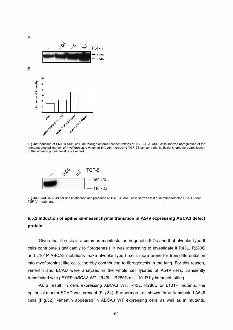
61
A
B
Fig.32: Induction of EMT in A549 cell line through different concentrations of TGF-ß1. A, A549 cells showed upregulation of the immunodetected marker of myofibroblasts vimentin through increasing TGF-ß1 concentrations. B, densitometric quantification of the vimentin protein level is presented.
Fig.33: ECAD in A549 cell line in absence and presence of TGF ß1. A549 cells showed lost of immunodetected ECAD under TGF ß1-treatment.
6.5.2 Induction of epithelial-mesenchymal transition in A549 expressing ABCA3 defect
protein
Given that fibrosis is a common manifestation in genetic ILDs and that alveolar type II
cells contribute significantly to fibrogenesis, it was interesting to investigate if R43L, R280C
and L101P ABCA3 mutations make alveolar type II cells more prone for transdifferentiation
into myofibroblast like cells, thereby contributing to fibrogenesis in the lung. For this reason,
vimentin and ECAD were analyzed in the whole cell lysates of A549 cells, transiently
transfected with pEYFP-ABCA3-WT, -R43L, -R280C or -L101P by immunoblotting.
As a result, in cells expressing ABCA3 WT, R43L, R280C or L101P mutants, the
epithelial marker ECAD was present (Fig.34). Furthermore, as shown for untransfected A549
cells (Fig.32), vimentin appeared in ABCA3 WT expressing cells as well as in mutants.

62
However, vimentin revealed no difference in cells with ABCA3 WT or with the three
mutations (Fig.34). This result can be explained by the low transfection efficiency and less
time for transfected cells to undergo EMT. However, these data are indicators that in stable
transfected cells, alveolar type II cells might undergo EMT in response to disturbed cell
homoeostasis perturbed by ABCA3 defect protein.
A
B
Fig.34: ECAD and vimentin in A549 cells expressing ABCA3 WT or mutant proteins. A, A549 cells showed no differences in immunodetected vimentin and ECAD in WT or the three mutations. B, intensity of each protein band was densitometrically quantified and normalized to the corresponding ß-actin band. Densitometric quantification of the vimentin protein level of three independent experiments is presented.
Considering the fact that alveolar epithelial cells release various profibrotic cytokines
and growth factors (see “Introduction”) thereby contributing to the induction of EMT in
epithelial lung cells and fibrogenesis, it was experimentally tested if ABCA3 mutants induce
EMT in AT II cells under additional stimulation with TGF-ß1.
Therefore, A549 cells were transiently transfected with pEYFP-ABCA3- WT, -R43L, -
R280C or –L101P and 24 hours after transfection, these cells were additionally incubated
with 0.5 ng/ml of TGF-ß1 for further 24 hours. The whole cell lysates were prepared and
analysed via immunoblotting using an anti-vimentin antibody. As a result, vimentin was
slightly increased in cells with R43L and R280C mutations and being at its highest in L101P
mutation (Fig.35).

63
A
B
Fig.35: Marker of myofibroblasts vimentin in A549 cells expressing ABCA3 WT or mutant proteins under additional stimulation with 0.5 ng/ml of TGF-ß1. A, A549 cells with ER retained L101P mutant showed upregulation of the immunodetected marker of myofibroblasts vimentin in comparison to WT. B, intensity of each protein band was densitometrically quantified and normalized to the corresponding ß-actin band. Densitometric quantification of the vimentin protein level of three independent experiments is presented; * p<0.05.
In conclusion, under additional stimulation with TGF-ß1, imitating autocrine or
paracrine cellular stimulation in the lungs, vimentin was upregulated in alveolar type II cells
with ABCA3 defect proteins, especially in A549 cells with ER-retained L101P defect protein.
These results suggest that these mutations push cells toward transdifferentiation into
myofibrobalsts contributing to fibrogenesis in the lungs and pathogenesis of ILD.

64
7. Discussion
ABCA3 mutations cause surfactant deficiency and fatal respiratory distress syndrome
in full-term neonates and chronic ILD of children (21, 22, 63-73). The cellular
pathomechanisms of ABCA3-related chronic ILD are probably complex including influence of
ABCA3 mutations on surfactant homeostasis but possibly also on the fitness and function of
alveolar type II cells as cells for surfactant production, lung repair and immunological defense
(122).
In this study the influence of three ABCA3 mutations, R43L, R280C and L101P, on
intracellular stress and induction of apoptosis in cultured lung epithelial A549 cells was
investigated. All three mutations were found in children with ABCA3-associated lung disease
being either fatal neonatal respiratory distress syndrome (L101P (22) and R43L (28, 48)) or
chronic ILD (R280C (48, own unpublished data) (Table 2). While cell biology of R43L and
R280C mutations was studied here for the first time, L101P mutation was used as a known
example of the trafficking/folding defect leading to the ER retention of ABCA3 with no
information on ER stress (57, 61).
7.1 General characterization of the three ABCA3 mutations R43L, R280C and L101P
The general characterization of R43L, R280C and L101P ABCA3 mutant proteins
included the analysis of the intracellular localization, of protein processing and glycosylation
and an uptake assay of NBD-labeled phospholipids PC and PE. To the date, ABCA3
mutations are either known for a functional defect and for impaired protein trafficking (61).
Improper protein folding, failed trafficking and mislocalization are well recognized in
other diseases associated with mutations in ABC transporters. For example in Tangier
Disease, a genetic HDL- deficiency, caused by defect ABCA1 transporter, the ABCA1
mutations R587W and Q597R are mainly retained in the ER instead of being transported to
the plasma membrane accompanied by impaired processing of oligosaccharides (123).
Moreover, in Cystic Fibrosis caused by mutations in the ABCC7 gene encoding an ABC
transporter, named cystic fibrosis transmembrane conductance regulator (CFTR) (124), the
most common mutant ΔF508 is retained and destroyed in the ER by the ER quality control
system and failed trafficking to plasma membrane (125).
Initial characterization in A549 cells demonstrated that each of the mutations
analyzed in this study affect the ABCA3 transporter in a different way. It was shown the
correct localization of WT and R43L proteins in LAMP3+ vesicles and dual localization of

65
R280C protein in LAMP3+ vesicles and calnexin+ ER compartment (Fig.18-20), indicating
less efficient but not abolished R280C trafficking. Similar dual localization is known in the
case of other ABCA3 mutants as G122S (57). However, L101P mutant protein appeared as
a net-like structure without any vesicle structures and LAMP3 colcoalization but complete
storage in the ER (Fig.18), as previously described (61).
Previous studies have analyzed ABCA3 localization to LBs by using LAMP3 staining
as well (59). In attempt to confirm additionally the localization of ABCA3 WT protein to LBs
and to show normal alveolar type II physiology in A549 cells used in this study, the
colocalization of ABCA3 WT protein and SP-C WT protein, that is stored within LBs, was
demonstrated in A549 cells expressing both proteins simultaneously (Fig.21).
ABCA3 protein expression was analyzed in alveolar type II cells by immunoblotting of
whole cell lysates and of the crude membrane fractions. Previously, ABCA3 protein
expression was described in human and mice tissues as well as in different cell lines. The
appearance of ABCA3 as one or two protein bands by immunoblotting depended on the
species and antibody, which were used (28, 58, 59). But recently, the lower protein band was
recognized as the mature one (34). In this study, when a yellow fluorescent protein (YFP) of
30 kDa was fused to ABCA3 protein, both protein bands, cleaved and noncleaved forms,
were present in cell cultures when ABCA3 WT was overexpressed (Fig.22). Similar
processing was observed for R43L and R280C proteins (Fig.22). Considering the fact of
correct LB localization of R43L mutant protein assessed by immunofluorescence studies, it
was not surprising to find WT-processing and presence of complex oligosaccharides
(Fig.22). In the case of YFP-R280C mutant protein, when dual localization to LAMP3+
vesicles and ER compartment was found, it was interesting to detect similar protein
processing and presence of complex oligosaccharides in this mutation as before in WT and
R43L mutation (Fig.22), confirming their ability to proceed from the ER to the Golgi.
In contrast, L101P protein remained in the ER, having therefore no complex sugars,
and no smaller 180 kDa protein form (Fig.22), as previously published (57, 61). This data
demonstrate a strong processing and glycosylation defect in L101P mutant protein.
In summary, these results suggesting the presence of oligosaccharides in ABCA3 WT
and R43L are consistent with proper immunofluorescence localization studies. However, in
terms of R280C mutation, the similarity of ABCA3 processing and protein glycosylation to
WT and R43L mutation makes a functional defect most likely, being in lane with
immunofluorescence studies when R280C was mainly localized to LBs. In case of L101P
mutant, absent oligosaccharides are consistent with failed trafficking and the ER-localization
demonstrated in this immunofluorescence studies as well as with strong processing
deficiency confirmed through absent cleaved form in Western Blotting. It can be speculated
that L101P mutant increases ER stress due to its processing defect and ER-storage.
66
Following this prediction ER stress was assessed in all mutants.
While ER retention of L101P excludes ABCA3 function, the function of R43L and
R280C transporters was studied additionally. As ABCA3 is involved in lamellar body
biogenesis in lung cells (22, 55, 60), which normally have a low number of compact LAMP3+
vesicles, expression of ABCA3-WT induced biogenesis of LAMP3+ vesicles by increasing
their number and size in A549 cells (59, Fig.24). In addition, ABCA3-WT signal was observed
as ring-like structures consistent with the ABCA3 presence in the limiting membrane of
lamellar bodies (41, Fig.18, 24). In contrast to the WT, expression of ABCA3 mutations,
especially L101P, impaired biogenesis of LAMP3+ vesicles by reducing their number and
size (Fig.24). This can be a consequence of the inability of these mutated transporters to
effectively load lipids into the nascent lamellar bodies.
The uptake assay of NBD-labeled phospholipids PC and PE into ABCA3-HA-positive
vesicles demonstrated frequent overlap of the NBD signal, both PC and PE coupled, with the
ring-like ABCA3-WT fluorescence and its accumulation in the inner space of the WT-ABCA3
vesicles (Fig.23). In the case of R43L and R280C mutations such colocalization was rarely
observed suggesting functional impairment of R43L and R280C proteins. Similar uptake
experiments have been previously described (57). In contrast to those data, which showed
diffuse distribution of NBD-PC and NBD-PE fluorescence throughout the cytoplasm,
numerous distinct NBD-positive vesicles were detected (Fig.23, A and B). The uptake and
number of NBD-vesicles observed in the cytoplasm was similar for both phospholipids in
A549 cells and in transfected A549, and was also independent of the ABCA3 mutation,
including L101P. This confirms that for the liposome/lipid uptake through the plasma
membrane into the cytoplasm ABCA3 function is dispensable and ABCA3 is solely an
intracellular vesicular transporter. Furthermore, we observed an impact of ABCA3 mutations
on the uptake of both PC and PE. This parallels data published in Cheong et al. (2007) (55)
which demonstrated decreased incorporation of radiolabeled PC and PE into the lamellar
bodies and surfactant of the ABCA3 heterozygote mouse, consistent with a role of ABCA3 in
the transport of both phospholipid species. However, this is also in contrast to Cheong et al.
(2006) (57) showing no uptake of NBD-PE into the lysosome-like vesicles in A549 cells.
Therefore, it is questionable if the PE transport through functional ABCA3 actually happens
in vivo or only in in vitro system after exposing cultured A549 cells to 150 μM of NBD lipid.

67
7.2 Induction of ER stress and UPR system by ABCA3 mutants
Continuous overload of the ER compartment with misfolded proteins is harmful for the
cell and leads to activation of the UPR system (126). Misfolding and ER retention of other
lipid ABC transporters is well known (123, 125). The most common mutation F508 of the
ABCC7-transporter, leading to cystic fibrosis, is not only misfolded and therefore retained in
the ER, it also raises ER stress and activates UPR (127). Also in SP-C deficiency, the
mutations exon4 and L188Q, which are retained in the ER or form aggregates in the
cytoplasm, induce UPR activation (94). But also in other organs than the lung, accumulation
of mutant protein in the ER cause toxic effects and cellular injury via ER stress, particularly
via BiP upregulation, as in liver and lung disease caused by 1-AT deficiency (128) and
neuronal degenerating diseases (129).
According to recent in vitro studies on cells expressing mutant ABCA3 proteins it
could have been speculated that dysfunction of the ATP-dependent lipid transporter resulting
in modified composite of surfactant components may give evidence for the mechanism how
mutant ABCA3 proteins disturb cell homeostasis and causes RDS or pILD (56, 59, 60). In
contrast, this study on human lung epithelial cell line A549 overexpressing mutant ABCA3
protein showed previously not examined cellular dysfunction, including raised level of
intracellular stress, susceptibility to it and induction of pro-apoptotic pathway:
Since L101P protein accumulates in the ER, it was not surprising to find significant
increase of the ER stress (Fig.25-28). Upregulation of chaperones is in line with ER
localization and defect protein glycosylation, indicating a strong misfolding defect and a
strong effect on induction of ER stress. Especially, splicing of XBP1 mRNA goes along with
subsequent upregulation of BiP caused by the misfolding defect of L101P mutant protein.
The ER stress caused by R280C mutation was lower. This might be a consequence of a dual
nature of this protein, which could be less harmful for the cell than complete ER retention,
and/or possible dependency of the magnitude of its intracellular effects on small
unmanageable inconsistencies of an experimental system. Correctly localized R43L
mutation, despite its influence on the ABCA3 function and lamellar body biogenesis, had
almost no impact on ER stress under any conditions above the range of the WT values
(Fig.25-28), being in lane with proper localization and processing studies and indicating less
cellular disturbance. However, the low increase of ER stress in cells with R43L or R280C
defect proteins might be due to low transfection efficiency of 20 % and to the robustness of
the method.
Since in nature, lungs are not exclusively affected by mutant ABCA3 protein but also
by environmental conditions such as dust, smoking, infection and hypoxemia that cause

68
additional intracellular stress, ER stress and UPR activation, examined via XBP1 splicing,
were enhanced by exposure of transfected cells to tunicamycin, an ER stressor
(Fig. 28, D and E). In this way the stress pressure imposed on the cells was doubled: 1)
genetic background of the ABCA3 mutations and 2) exposure to the stress-causing agent.
The cells with mutations R280C and L101P, which impair ABCA3 trafficking, were more
prone to further XBP1 splicing than the WT or A549 (Fig.28D). This is interesting if known
that viral infections (e.g. RSV, herpes virus) or cigarette smoke are common outside factors,
which, as tunicamycin, elevate ER stress (130, 131). Also clinical experts report from the
onset of ABCA3-associated ILD in children following exposure to cigarette smoke (68) and
describe a teenage patient with a late onset of ABCA3-related disease with fibrosis following
the beginning of cigarette consumption (71). If ABCA3 mutations raise susceptibility of
alveolar type II cells to external stress, additional exposure to outside stressors as respiratory
viral infections or smoke might contribute to or even trigger genetic ILD.
7.3 Induction of apoptotic cell death by ABCA3 mutations
Constant accumulation of defect proteins within the ER lumen and activation of the
UPR system disturb cell homeostasis, cause cell toxicity and disruption of cell function.
Furthermore, prolonged and unresolved ER can trigger apoptotic cell death (see
“Introduction”). Recently, upon accumulation of mutant SP-C protein in the ER and also in
model systems for other conformational diseases like Parkinson´ s disease, the mechanism
of ER stress and subsequent apoptotic cell death was found in vitro (106, 132). However, in
patients with IPP/IFP alveolar epithelial cells were identified to undergo apoptosis, whereas
apoptosis was absent in control lungs (133, 134).
In this study, the connection between the overexpression of ABCA3 mutations and
induction of apoptotic cell death was analysed by three lines of evidence: activation of
caspase 3, either via immunoblot or via FACS, PI and annexin V staining. Since L101P
protein accumulation in the ER increased stress response elements, early and late apoptosis
markers were also recognized in the cells with this mutation (Fig.29, 30). The ER stress
caused by R280C mutation was lower and therefore surface staining with annexin V, as an
early apoptosis sign, was the only apoptotic marker detected in R280C cells. Correctly
localized R43L mutation, despite its influence on the ABCA3 function and lamellar body
biogenesis, had almost no impact on stress and apoptosis under any conditions above the
range of the WT values (Fig.29, 30).
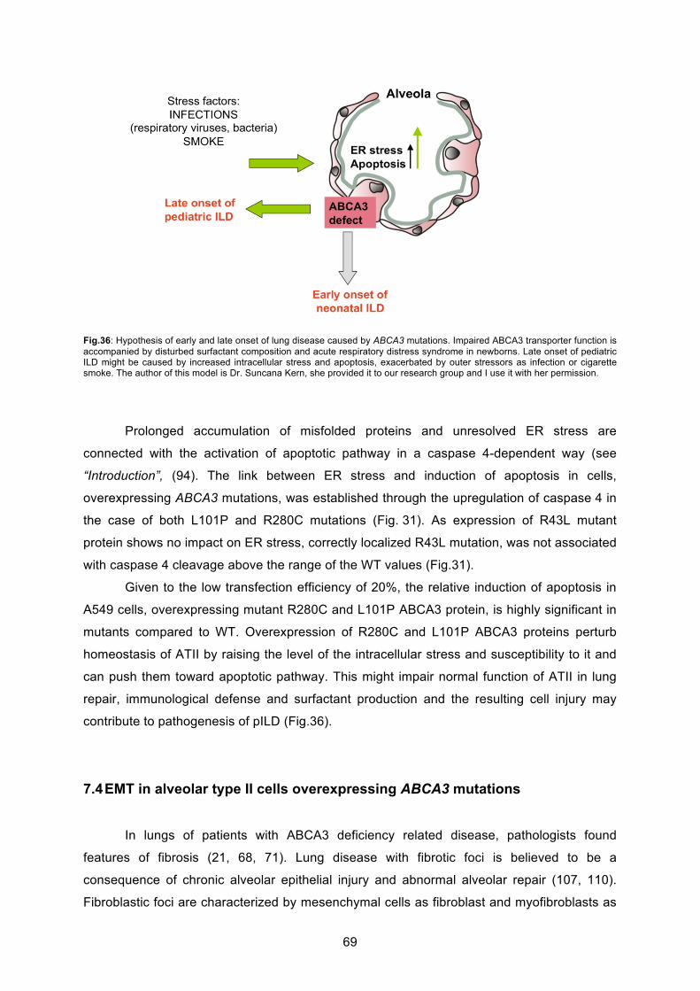
69
Fig.36: Hypothesis of early and late onset of lung disease caused by ABCA3 mutations. Impaired ABCA3 transporter function is accompanied by disturbed surfactant composition and acute respiratory distress syndrome in newborns. Late onset of pediatric ILD might be caused by increased intracellular stress and apoptosis, exacerbated by outer stressors as infection or cigarette smoke. The author of this model is Dr. Suncana Kern, she provided it to our research group and I use it with her permission.
Prolonged accumulation of misfolded proteins and unresolved ER stress are
connected with the activation of apoptotic pathway in a caspase 4-dependent way (see
“Introduction”, (94). The link between ER stress and induction of apoptosis in cells,
overexpressing ABCA3 mutations, was established through the upregulation of caspase 4 in
the case of both L101P and R280C mutations (Fig. 31). As expression of R43L mutant
protein shows no impact on ER stress, correctly localized R43L mutation, was not associated
with caspase 4 cleavage above the range of the WT values (Fig.31).
Given to the low transfection efficiency of 20%, the relative induction of apoptosis in
A549 cells, overexpressing mutant R280C and L101P ABCA3 protein, is highly significant in
mutants compared to WT. Overexpression of R280C and L101P ABCA3 proteins perturb
homeostasis of ATII by raising the level of the intracellular stress and susceptibility to it and
can push them toward apoptotic pathway. This might impair normal function of ATII in lung
repair, immunological defense and surfactant production and the resulting cell injury may
contribute to pathogenesis of pILD (Fig.36).
7.4 EMT in alveolar type II cells overexpressing ABCA3 mutations
In lungs of patients with ABCA3 deficiency related disease, pathologists found
features of fibrosis (21, 68, 71). Lung disease with fibrotic foci is believed to be a
consequence of chronic alveolar epithelial injury and abnormal alveolar repair (107, 110).
Fibroblastic foci are characterized by mesenchymal cells as fibroblast and myofibroblasts as

70
well as by abnormal extracellular proteins (see “Introduction”). However, pathogenesis of
pulmonary fibrosis is still poorly understood, but alveolar type II cells seam to have a key role
in this process (135). Recent studies not only on fibrotic lung disorders, but also on renal and
liver diseases, showed that epithelial cells can undergo EMT and that TGF-ß plays a key role
in transition of epithelial cells into mesenchymal cells (114, 136, 137). Epithelial lung cells
that have undergone EMT may contribute to the development of fibroblastic foci in various
ways: as a new source of fibroblasts or by the activation and migration of fibroblasts and by
the release of cytokines and growth factors. However, the precise mechanism of the
formation of fibroblastic foci and the origin of mesenchymal cells are both incompletely
understood.
In this study, it was demonstrated for the first time that A549 cells overexpressing
ABCA3 mutant proteins, especially L101P cells, gain a mesenchymal phenotype with
increased expression of vimentin, particularly in presence of TGF-ß1 (Fig.34, 35). It can be
speculated that the examined ABCA3 mutations make AT II cells more prone to undergo
EMT. Furthermore, induction of ER stress and apoptosis in these ABCA3 mutations,
particularly the ER-retained L101P mutation, is associated with induction of EMT in AT II.
Role of ER stress and apoptosis in lung disease has also been studied by other groups,
showing that ER stress- and apoptosis- markers occur at sites of fibrosis in alveolar type II
cells (103, 104, 133, 134). These results suggest that ER stress may contribute to lung
fibrosis through induction of EMT in ABCA3 related lung disease. Further experiments are
necessary to support these notions. This mechanism is also believed to underlie in SP-C-
related lung disease (138). SP-C and ABCA3 are both ATII-expressed proteins essential for
surfactant homeostasis and both lead to genetic ILD equally variable in the age of onset,
severity and pathology. Therefore, it is interesting to see that common mechanisms
underlying both types of genetic ILD must exist and this data show that they probably
encompass the ER stress and apoptosis of AT II cells mediating EMT. Therefore, AT II cells
with ABCA3 deficiency may have a critical role for development of fibrosis in pILD (Fig.37).
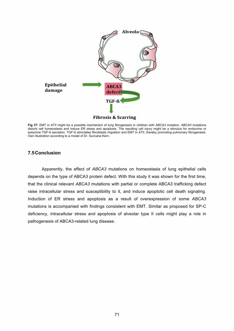
71
Fig 37: EMT in ATII might be a possible mechanism of lung fibrogenesis in children with ABCA3 mutation. ABCA3 mutations disturb cell homeostasis and induce ER stress and apoptosis. The resulting cell injury might be a stimulus for endocrine or autocrine TGF-ß secretion. TGF-ß stimulates fibroblasts migration and EMT in ATII, thereby promoting pulmonary fibrogenesis. Own illustration according to a model of Dr. Suncana Kern.
7.5 Conclusion
Apparently, the effect of ABCA3 mutations on homeostasis of lung epithelial cells
depends on the type of ABCA3 protein defect. With this study it was shown for the first time,
that the clinical relevant ABCA3 mutations with partial or complete ABCA3 trafficking defect
raise intracellular stress and susceptibility to it, and induce apoptotic cell death signaling.
Induction of ER stress and apoptosis as a result of overexpression of some ABCA3
mutations is accompanied with findings consistent with EMT. Similar as proposed for SP-C
deficiency, intracellular stress and apoptosis of alveolar type II cells might play a role in
pathogenesis of ABCA3-related lung disease.
72
7.6 Future directions
This study demonstrated mechanisms, which may contribute to the pathogenesis of
ABCA3 related lung disease thereby raising new questions that should be addressed in
future work.
First, studies on induction of ER stress and apoptosis in ATII cells expressing ABCA3
mutations should be supplemented by further experiments. Studies on the most common
E292V mutation, a type II mutation located to LBs with reduced functional activity, would also
be helpful to confirm findings of ER retained mutation in this study.
Second, more work is necessary to verify if ER stress and apoptosis of AT II cells are
indeed present in (fibrotic) lung tissue of patients with ABCA3 mutations. Studies on lung
tissue from children with ABCA3 mutations could also provide more information on ATII cells
and their role in pathogenesis of pILD, e.g. in terms of morphology, surfactant homeostasis
and cytokine and growth factor release.
Third, exposure to outsides stressor should become a subject of future experiments.
RSV-infection could be a helpful instrument. Further investigations on the precise pathway of
how ABCA3 mutant proteins can lead to fibrogenesis and studies on the role of TGF-ß are
needed.
This might help to understand or even predict the disease course, or consider new
therapy options (as protein rescue with chaperones) depending on the type of the ABCA3
protein defect. Since to date no causative therapy is available for the treatment of pILD, the
role of chemical chaperones should be tested in vitro for ER-retained ABCA3 mutations.

73
8. References
1. Fan LL, Deterding RR, Langston C. Pediatric interstitial lung disease revisited. Pediatric pulmonology.
2004Nov;38(5):369-78. 2. Griese M, Haug M, Brasch F, et al. Incidence and classification of pediatric diffuse parenchymal lung
diseases in Germany. Orphanet Journal of Rare Diseases. 2009;426. 3. Fan LL, Mullen AL, Brugman SM, Inscore SC, Parks DP, White CW. Clinical spectrum of chronic
interstitial lung disease in children. The Journal of pediatrics. 1992Dec;121(6):867-72. 4. Dinwiddie R, Sharief N, Crawford O. Idiopathic interstitial pneumonitis in children: a national survey in
the United Kingdom and Ireland. Pediatric pulmonology. 2002Jul;34(1):23-9. 5. Coultas DB, Zumwalt RE, Black WC, Sobonya RE. The epidemiology of interstitial lung diseases.
American journal of respiratory and critical care medicine. 1994Oct;150(4):967-72. 6. Xaubet A, Ancochea J, Morell F, et al. Report on the incidence of interstitial lung diseases in Spain.
Sarcoidosis vasculitis and diffuse lung diseases official journal of WASOG World Association of Sarcoidosis and Other Granulomatous Disorders. 2004;21(1):64-70.
7. López-Campos JL, Rodríguez-Becerra E. Incidence of interstitial lung diseases in the south of Spain 1998-2000: the RENIA study. European journal of epidemiology. 2004Jan;19(2):155-61.
8. Clement A. Task force on chronic interstitial lung disease in immunocompetent children. The European respiratory journal : official journal of the European Society for Clinical Respiratory Physiology. 2004Oct;24(4):686-97.
9. McFetridge L, McMorrow A, Morrison PJ, Shields MD. Surfactant Metabolism Dysfunction and Childhood Interstitial Lung Disease (chILD). The Ulster medical journal. 2009Jan;78(1):7-9.
10. Deutsch GH, Young LR, Deterding RR, et al. Diffuse Lung Disease in Young Children: Application of a Novel Classification Scheme. American Journal of Respiratory and Critical Care Medicine. 2007;176(11):1120-8.
11. Johansson J, Curstedt T, Robertson B. The proteins of the surfactant system. The European respiratory journal official journal of the European Society for Clinical Respiratory Physiology. 1994;7(2):372-391.
12. Mason RJ, Voelker DR. Regulatory mechanisms of surfactant secretion. Biochimica et Biophysica Acta. 1998;1408(2-3):226-240.
13. Rooney SA. Regulation of surfactant secretion. Comparative biochemistry and physiology Part A Molecular integrative physiology. 2001;129(1):233-243.
14. Andreeva AV, Kutuzov MA, Voyno-Yasenetskaya TA. Regulation of surfactant secretion in alveolar type II cells. American journal of physiology Lung cellular and molecular physiology. 2007;293(2):L259-L271.
15. Schmitz G, Müller G. Structure and function of lamellar bodies, lipid-protein complexes involved in storage and secretion of cellular lipids. Journal Of Lipid Research. 1991;32(10):1539-1570.
16. Thouvenin G, Abou Taam R, Flamein F, et al. Characteristics of disorders associated with genetic mutations of surfactant protein C. Archives of Disease in Childhood. 2010;95(6):449-454.
17. Guillot L, Epaud R, Thouvenin G, et al. New surfactant protein C gene mutations associated with diffuse lung disease. Journal of Medical Genetics. 2009;46(7):490-494.
18. Cameron HS, Somaschini M, Carrera P, et al. A common mutation in the surfactant protein C gene associated with lung disease. The Journal of pediatrics. 2005;146(3):370-375.
19. Nogee LM, Dunbar AE, Wert S, Askin F, Hamvas A, Whitsett JA. Mutations in the surfactant protein C gene associated with interstitial lung disease. Chest. 2002;121(3 Suppl):20S-21S.
20. Nogee LM, Wert SE, Proffit SA, Hull WM, Whitsett JA. Allelic heterogeneity in hereditary surfactant protein B (SP-B) deficiency. American Journal of Respiratory and Critical Care Medicine. 2000;161(3 Pt 1):973-981.
21. Bullard JE, Wert SE, Whitsett JA, Dean M, Nogee LM. ABCA3 mutations associated with pediatric interstitial lung disease. American Journal of Respiratory and Critical Care Medicine. 2005;172(8):1026-1031.
22. Shulenin S, Nogee LM, Annilo T, Wert SE, Whitsett JA, Dean M. ABCA3 gene mutations in newborns with fatal surfactant deficiency. The New England Journal of Medicine. 2004;350(13):1296-1303.
23. Nogee LM, Garnier G, Dietz HC, et al. A mutation in the surfactant protein B gene responsible for fatal neonatal respiratory disease in multiple kindreds.Journal of Clinical Investigation. 1994;93(4):1860-1863.
24. Schuerman FABA, Griese M, Gille JP, Brasch F, Noorduyn LA, Van Kaam AH. Surfactant protein B deficiency caused by a novel mutation involving multiple exons of the SP-B gene. European Journal Of Medical Research. 2008;13(6):281-286.
25. Somaschini M, Wert S, Mangili G, Colombo A, Nogee L. Hereditary surfactant protein B deficiency resulting from a novel mutation. Intensive Care Medicine. 2000;26(1):97-100.

74
26. Tredano M, Griese M, De Blic J, et al. Analysis of 40 sporadic or familial neonatal and pediatric cases with severe unexplained respiratory distress: relationship to SFTPB. American journal of medical genetics Part A. 2003;119A(3):324-339.
27. Garmany TH, Moxley MA, White FV, et al. Surfactant composition and function in patients with ABCA3 mutations. Pediatric Research. 2006;59(6):801-805.
28. Brasch F, Schimanski S, Mühlfeld C, et al. Alteration of the pulmonary surfactant system in full-term infants with hereditary ABCA3 deficiency. American Journal of Respiratory and Critical Care Medicine. 2006;174(5):571-580.
29. Clement A, Nathan N, Epaud R, Fauroux B, Corvol H. Interstitial lung diseases in children. Orphanet Journal of Rare Diseases. 2010;522.
30. Hartl D, Griese M. Interstitial lung disease in children – genetic background and associated phenotypes. Respiratory Research. 2005;6(1):32.
31. Whitsett JA, Wert SE, Weaver TE. Alveolar Surfactant Homeostasis and the Pathogenesis of Pulmonary Disease. Annual Review of Medicine. 2010;61105-119.
32. Dean M, Hamon Y, Chimini G. The human ATP-binding cassette (ABC) transporter superfamily. Journal Of Lipid Research. 2001;42(7):1007-17.
33. Klugbauer N, Hofmann F. Primary structure of a novel ABC transporter with a chromosomal localization on the band encoding the multidrug resistance-associated protein. FEBS Letters. 1996;391(1-2):61-65.
34. Engelbrecht S, Kaltenborn E, Griese M, Kern S. The surfactant lipid transporter ABCA3 is N-terminally cleaved inside LAMP3-positive vesicles.FEBS Letters. 2010;584(20):4306-12.
35. Schmitz G, Kaminski WE. ABC transporters and cholesterol metabolism. Frontiers in bioscience a journal and virtual library. 2001;6D505-D514.
36. Hayden MR, Clee SM, Brooks-Wilson A, Genest J, Attie A, Kastelein JJ. Cholesterol efflux regulatory protein, Tangier disease and familial high-density lipoprotein deficiency. Current Opinion in Lipidology. 2000;11(2):117-122.
37. Tsybovsky Y, Molday RS, Palczewski K. The ATP-binding cassette transporter ABCA4: structural and functional properties and role in retinal disease. Advances in experimental medicine and biology. 2010;703105-125.
38. Dean M. The genetics of ATP-binding cassette transporters. Methods in Enzymology. 2005;400409-429. 39. Oram JF. Tangier disease and ABCA1.Biochimica et Biophysica Acta. 2000;1529(1-3):321-330. 40. Sun H, Nathans J. ABCR: rod photoreceptor-specific ABC transporter responsible for Stargardt disease.
Methods in Enzymology. 2000;315879-897. 41. Mulugeta S, Gray JM, Notarfrancesco KL, et al. Identification of LBM180, a lamellar body limiting
membrane protein of alveolar type II cells, as the ABC transporter protein ABCA3. The Journal of Biological Chemistry. 2002;277(25):22147-22155.
42. Yamano G, Funahashi H, Kawanami O, et al. ABCA3 is a lamellar body membrane protein in human lung alveolar type II cells. FEBS Letters. 2001;508(2):221-225.
43. Stahlman MT, Besnard V, Wert SE, et al. Expression of ABCA3 in developing lung and other tissues. The journal of histochemistry and cytochemistry official journal of the Histochemistry Society. 2007;55(1):71-83.
44. Yoshida I, Ban N, Inagaki N. Expression of ABCA3, a causative gene for fatal surfactant deficiency, is up-regulated by glucocorticoids in lung alveolar type II cells. Biochemical and Biophysical Research Communications. 2004;323(2):547-555.
45. Connors TD, Van Raay TJ, Petry LR, Klinger KW, Landes GM, Burn TC. The cloning of a human ABC gene (ABC3) mapping to chromosome 16p13.3. Genomics. 1997;39(2):231-234.
46. Bruder E, Hofmeister J, Aslanidis C, et al. Ultrastructural and molecular analysis in fatal neonatal interstitial pneumonia caused by a novel ABCA3 mutation. Modern pathology an official journal of the United States and Canadian Academy of Pathology Inc. 2007;20(10):1009-1018.
47. Bullard JE, Wert SE, Nogee LM. ABCA3 deficiency: neonatal respiratory failure and interstitial lung disease. Seminars in Perinatology. 2006;30(6):327-334.
48. Somaschini M, Nogee LM, Sassi I, et al. Unexplained neonatal respiratory distress due to congenital surfactant deficiency. The Journal of pediatrics. 2007;150(6):649-653, 653.e1.
49. Stahlman MT, Gray MP, Falconieri MW, Whitsett JA, Weaver TE. Lamellar body formation in normal and surfactant protein B-deficient fetal mice.Laboratory investigation a journal of technical methods and pathology. 2000;80(3):395-403.
50. DeMello DE, Heyman S, Phelps DS, et al. Ultrastructure of lung in surfactant protein B deficiency. American Journal of Respiratory Cell and Molecular Biology. 1994;11(2):230-239.
51. Brasch F, Griese M, Tredano M, et al. Interstitial lung disease in a baby with a de novo mutation in the SFTPC gene. The European respiratory journal official journal of the European Society for Clinical Respiratory Physiology. 2004;24(1):30-39.
75
52. Hamvas A, Nogee LM, White FV, et al. Progressive lung disease and surfactant dysfunction with a deletion in surfactant protein C gene. American Journal of Respiratory Cell and Molecular Biology. 2004;30(6):771-776.
53. Stevens PA, Pettenazzo A, Brasch F, et al. Nonspecific interstitial pneumonia, alveolar proteinosis, and abnormal proprotein trafficking resulting from a spontaneous mutation in the surfactant protein C gene. Pediatric Research. 2005;57(1):89-98.
54. Fitzgerald ML, Xavier R, Haley KJ, et al. ABCA3 inactivation in mice causes respiratory failure, loss of pulmonary surfactant, and depletion of lung phosphatidylglycerol. Journal Of Lipid Research. 2007;48(3):621-632.
55. Cheong N, Zhang H, Madesh M, et al. ABCA3 is critical for lamellar body biogenesis in vivo. The Journal of Biological Chemistry. 2007;282(33):23811-23817.
56. Hammel M, Michel G, Hoefer C, et al. Targeted inactivation of the murine Abca3 gene leads to respiratory failure in newborns with defective lamellar bodies. Biochemical and Biophysical Research Communications. 2007;359(4):947-951.
57. Cheong N, Madesh M, Gonzales LW, et al. Functional and trafficking defects in ATP binding cassette A3 mutants associated with respiratory distress syndrome. The Journal of Biological Chemistry. 2006;281(14):9791-9800.
58. Nagata K, Yamamoto A, Ban N, et al. Human ABCA3, a product of a responsible gene for abca3 for fatal surfactant deficiency in newborns, exhibits unique ATP hydrolysis activity and generates intracellular multilamellar vesicles. Biochemical and Biophysical Research Communications. 2004;324(1):262-268.
59. Matsumura Y, Sakai H, Sasaki M, Ban N, Inagaki N. ABCA3-mediated choline-phospholipids uptake into intracellular vesicles in A549 cells. FEBS Letters. 2007;581(17):3139-3144.
60. Ban N, Matsumura Y, Sakai H, et al. ABCA3 as a lipid transporter in pulmonary surfactant biogenesis. The Journal of Biological Chemistry. 2007;282(13):9628-9634.
61. Matsumura Y, Ban N, Ueda K, Inagaki N. Characterization and classification of ATP-binding cassette transporter ABCA3 mutants in fatal surfactant deficiency. The Journal of Biological Chemistry. 2006;281(45):34503-34514.
62. Matsumura Y, Ban N, Inagaki N. Aberrant catalytic cycle and impaired lipid transport into intracellular vesicles in ABCA3 mutants associated with nonfatal pediatric interstitial lung disease. American journal of physiology Lung cellular and molecular physiology. 2008;295(4):L698-L707.
63. Prestridge A, Wooldridge J, Deutsch G, et al. Persistent tachypnea and hypoxia in a 3-month-old term infant. The Journal of pediatrics. 2006;149(5):702-706.
64. Saugstad OD, Hansen TWR, Rønnestad A, et al. Novel mutations in the gene encoding ATP binding cassette protein member A3 (ABCA3) resulting in fatal neonatal lung disease. Acta paediatrica. 2007;96(2):185-190.
65. Bullard JE, Nogee LM. Heterozygosity for ABCA3 mutations modifies the severity of lung disease associated with a surfactant protein C gene (SFTPC) mutation. Pediatric Research. 2007;62(2):176-179.
66. Yokota T, Matsumura Y, Ban N, Matsubayashi T, Inagaki N. Heterozygous ABCA3 mutation associated with non-fatal evolution of respiratory distress. European Journal of Pediatrics. 2008;167(6):691-693.
67. Kunig AM, Parker TA, Nogee LM, Abman SH, Kinsella JP. ABCA3 deficiency presenting as persistent pulmonary hypertension of the newborn. The Journal of pediatrics. 2007;151(3):322-324.
68. Doan ML, Guillerman RP, Dishop MK, et al. Clinical, radiological and pathological features of ABCA3 mutations in children. Thorax. 2008;63(4):366-373.
69. Karjalainen MK, Haataja R, Hallman M. Haplotype analysis of ABCA3: association with respiratory distress in very premature infants. Annals of Medicine. 2008;40(1):56-65.
70. Garmany TH, Wambach JA, Heins HB, et al. Population and disease-based prevalence of the common mutations associated with surfactant deficiency. Pediatric Research. 2008;63(6):645-649.
71. Young LR, Nogee LM, Barnett B, Panos RJ, Colby TV, Deutsch GH. Usual interstitial pneumonia in an adolescent with ABCA3 mutations. Chest. 2008;134(1):192-195.
72. Ciantelli M, Ghirri P, Presi S, et al. Fatal respiratory failure in a full-term newborn with two ABCA3 gene mutations: a case report. Journal of perinatology official journal of the California Perinatal Association. 2011;31(1):70-72.
73. Anandarajan M, Paulraj S, Tubman R. ABCA3 Deficiency: an unusual cause of respiratory distress in the newborn. The Ulster medical journal. 2009;78(1):51-52.
74. Anelli T, Sitia R. Protein quality control in the early secretory pathway. the The European Molecular Biology Organization Journal. 2008;27(2):315-327.
75. Ma Y, Hendershot LM. ER chaperone functions during normal and stress conditions. Journal of Chemical Neuroanatomy. 2004;28(1-2):51-65.
76. Ron D, Walter P. Signal integration in the endoplasmic reticulum unfolded protein response. Nat Rev Mol Cell Biol. 2007;8(7):519-529.
76
77. Malhotra JD, Kaufman RJ. The endoplasmic reticulum and the unfolded protein response. Seminars in Cell Developmental Biology. 2007;18(6):716-731.
78. Meusser B, Hirsch C, Jarosch E, Sommer T. ERAD: the long road to destruction. Nature Cell Biology. 2005;7(8):766-772.
79. Oyadomari S, Yun C, Fisher EA, et al. Cotranslocational degradation protects the stressed endoplasmic reticulum from protein overload. Cell. 2006;126(4):727-739.
80. Rao RV, Ellerby HM, Bredesen DE. Coupling endoplasmic reticulum stress to the cell death program. Cell Death and Differentiation. 2004;11(4):372-380.
81. Bertolotti A, Zhang Y, Hendershot LM, Harding HP, Ron D. Dynamic interaction of BiP and ER stress transducers in the unfolded-protein response. Nature Cell Biology. 2000;2(6):326-332.
82. Yoshida H, Matsui T, Yamamoto A, Okada T, Mori K. XBP1 mRNA is induced by ATF6 and spliced by IRE1 in response to ER stress to produce a highly active transcription factor. Cell. 2001;107(7):881-891.
83. Uemura A, Oku M, Mori K, Yoshida H. Unconventional splicing of XBP1 mRNA occurs in the cytoplasm during the mammalian unfolded protein response. Journal of Cell Science. 2009;122(Pt 16):2877-2886.
84. Back SH, Lee K, Vink E, Kaufman RJ. Cytoplasmic IRE1alpha-mediated XBP1 mRNA splicing in the absence of nuclear processing and endoplasmic reticulum stress. The Journal of Biological Chemistry. 2006;281(27):18691-18706.
85. Yoshida H, Oku M, Suzuki M, Mori K. pXBP1(U) encoded in XBP1 pre-mRNA negatively regulates unfolded protein response activator pXBP1(S) in mammalian ER stress response. The Journal of Cell Biology. 2006;172(4):565-575.
86. Yoshida H, Matsui T, Hosokawa N, Kaufman RJ, Nagata K, Mori K. A time-dependent phase shift in the mammalian unfolded protein response. Developmental Cell. 2003;4(2):265-271.
87. Hollien J, Lin JH, Li H, Stevens N, Walter P, Weissman JS. Regulated Ire1-dependent decay of messenger RNAs in mammalian cells. The Journal of Cell Biology. 2009;186(3):323-331.
88. Adachi Y, Yamamoto K, Okada T, Yoshida H, Harada A, Mori K. ATF6 is a transcription factor specializing in the regulation of quality control proteins in the endoplasmic reticulum. Cell Structure and Function. 2008;33(1):75-89.
89. Liu CY, Kaufman RJ. The unfolded protein response. Journal of Cell Science. 2003;116(Pt 10):1861-1862.
90. Tardif KD, Waris G, Siddiqui A. Hepatitis C virus, ER stress, and oxidative stress. Trends in Microbiology. 2005;13(4):159-163.
91. Thornberry NA. Caspases: Enemies Within. Science. 1998;281(5381):1312-1316. 92. Li P, Nijhawan D, Budihardjo I, et al. Cytochrome c and dATP-Dependent Formation of Apaf-1 /
Caspase-9 Complex Initiates an Apoptotic Protease Cascade. Cell. 1997;91479-489. 93. Nakagawa T, Zhu H, Morishima N, et al. Caspase-12 mediates endoplasmic-reticulum-specific apoptosis
and cytotoxicity by amyloid-beta. Nature. 2000;403(6765):98-103. 94. Mulugeta S, Maguire JA, Newitt JL, Russo SJ, Kotorashvili A, Beers MF. Misfolded BRICHOS SP-C
mutant proteins induce apoptosis via caspase-4- and cytochrome c-related mechanisms. American journal of physiology Lung cellular and molecular physiology. 2007;293(3):L720-L729.
95. Hitomi J, Katayama T, Taniguchi M, Honda A, Imaizumi K, Tohyama M. Apoptosis induced by endoplasmic reticulum stress depends on activation of caspase-3 via caspase-12. Neuroscience Letters. 2004;357(2):127-130.
96. Morishima N, Nakanishi K, Takenouchi H, Shibata T, Yasuhiko Y. An endoplasmic reticulum stress-specific caspase cascade in apoptosis. Cytochrome c-independent activation of caspase-9 by caspase-12. The Journal of Biological Chemistry. 2002;277(37):34287-94.
97. Balasubramanian K, Mirnikjoo B, Schroit AJ. Regulated Externalization of Phosphatidylserine at the Cell Surface. Journal of Biological Chemistry. 2007;282(25):18357-18364.
98. Blankenberg FG. Imaging the molecular signatures of apoptosis and injury with radiolabeled annexin V. Proceedings of the American Thoracic Society. 2009;6(5):469-476.
99. Laufer EM, Winkens HM, Corsten MF, Reutelingsperger CPM, Narula J, Hofstra L. PET and SPECT imaging of apoptosis in vulnerable atherosclerotic plaques with radiolabeled Annexin A5. The quarterly journal of nuclear medicine and molecular imaging official publication of the Italian Association of Nuclear Medicine AIMN and the International Association of Radiopharmacology IAR and Section of the Society of. 2009;53(1):26-34.
100. Wu J, Kaufman RJ. From acute ER stress to physiological roles of the Unfolded Protein Response. Cell Death and Differentiation. 2006;13(3):374-384.
101. Marciniak SJ, Ron D. Endoplasmic reticulum stress signaling in disease. Physiological Reviews. 2006;86(4):1133-1149.
77
102. Kim I, Xu W, Reed JC. Cell death and endoplasmic reticulum stress: disease relevance and therapeutic opportunities. Nature Reviews Drug Discovery. 2008;7(12):1013-1030.
103. Korfei M, Ruppert C, Mahavadi P, et al. Epithelial endoplasmic reticulum stress and apoptosis in sporadic idiopathic pulmonary fibrosis. American Journal of Respiratory and Critical Care Medicine. 2008;178(8):838-46.
104. Drakopanagiotakis F, Xifteri A, Polychronopoulos V, Bouros D. Apoptosis in lung injury and fibrosis. The European respiratory journal official journal of the European Society for Clinical Respiratory Physiology. 2008;32(6):1631-1638.
105. Lawson WE, Crossno PF, Polosukhin VV, et al. Endoplasmic reticulum stress in alveolar epithelial cells is prominent in IPF: association with altered surfactant protein processing and herpesvirus infection. American journal of physiology Lung cellular and molecular physiology. 2008;294(6):L1119-L1126.
106. Mulugeta S, Nguyen V, Russo SJ, Muniswamy M, Beers MF. A Surfactant Protein C Precursor Protein BRICHOS Domain Mutation Causes Endoplasmic Reticulum Stress, Proteasome Dysfunction, and Caspase 3 Activation. American Journal of Respiratory Cell and Molecular Biology. 2005;32(6):521-530.
107. Selman M, King TE, Pardo A. Idiopathic pulmonary fibrosis: prevailing and evolving hypotheses about its pathogenesis and implications for therapy. In:Annals of Internal Medicine.2001. p.136-151.
108. Gauldie J, Kolb M, Sime PJ. A new direction in the pathogenesis of idiopathic pulmonary fibrosis? Respiratory Research. 2002;3(1):1.
109. Kim JH, Jang YS, Eom K-S, et al. Transtorming Growth Factor 1 Induces Epithelial-to-Mesenchymal Transition of A549 Cells. 2007;22(5):898-904.
110. Corvol H, Flamein F, Epaud R, Clement A, Guillot L. Lung alveolar epithelium and interstitial lung disease. International Journal of Biochemistry. 2009;41(8-9):1643-51.
111. Kapanci Y, Desmouliere A, Pache JC, Redard M, Gabbiani G. Cytoskeletal protein modulation in pulmonary alveolar myofibroblasts during idiopathic pulmonary fibrosis. Possible role of transforming growth factor beta and tumor necrosis factor alpha. American Journal of Respiratory and Critical Care Medicine. 1995;152(6 Pt 1):2163-2169.
112. Bals R, Hiemstra PS. Innate immunity in the lung: how epithelial cells fight against respiratory pathogens. The European respiratory journal official journal of the European Society for Clinical Respiratory Physiology. 2004;23(2):327-333.
113. Kelly M, Kolb M, Bonniaud P, Gauldie J. Re-evaluation of fibrogenic cytokines in lung fibrosis. Current Pharmaceutical Design. 2003;9(1):39-49.
114. Kasai H, Allen JT, Mason RM, Kamimura T, Zhang Z. TGF- 1 induces human alveolar epithelial to mesenchymal cell transition (EMT). Respiratory Research. 2005;6(1):56.
115. Nelson M, McClelland M. Use of DNA methyltransferase/endonuclease enzyme combinations for megabase mapping of chromosomes. Methods in Enzymology. 1992;216279-303.
116. Buck TM, Wright CM, Brodsky JL. The activities and function of molecular chaperones in the endoplasmic reticulum. Seminars in cell developmental biology. 2007;18(6):751-761.
117. Hybiske K, Fu Z, Schwarzer C, et al. Effects of cystic fibrosis transmembrane conductance regulator and DeltaF508CFTR on inflammatory response, ER stress, and Ca2+ of airway epithelia. American journal of physiology Lung cellular and molecular physiology. 2007;293(5):L1250-L1260.
118. Balasubramanian K, Schroit AJ. Aminophospholipid asymmetry: A matter of life and death. Annual Review of Physiology. 2003;65(1):701-734.
119. Franco R, Schoneveld OJ, Pappa A, Panayiotidis MI. The central role of glutathione in the pathophysiology of human diseases. Archives of Physiology and Biochemistry. 2007;113(4-5):234-258.
120. Kumar S. Caspase function in programmed cell death. Cell Death and Differentiation. 2007;14(1):32-43. 121. Iwano M, Plieth D, Danoff TM, Xue C, Okada H, Neilson EG. Evidence that fibroblasts derive from
epithelium during tissue fibrosis. Journal of Clinical Investigation. 2002;110(3):341-350. 122. Mason RJ. Biology of alveolar type II cells. Respirology Carlton Vic. 2006;11 Suppl(s1):S12-S15. 123. Tanaka AR, Abe-Dohmae S, Ohnishi T, et al. Effects of mutations of ABCA1 in the first extracellular
domain on subcellular trafficking and ATP binding/hydrolysis. The Journal of Biological Chemistry. 2003;278(10):8815-9.
124. Pilewski JM, Frizzell RA. Role of CFTR in airway disease. Physiological Reviews. 1999;79(1 Suppl):S215-S255.
125. Cheng SH, Gregory RJ, Marshall J, et al. Defective intracellular transport and processing of CFTR is the molecular basis of most cystic fibrosis. Cell. 1990;63(4):827-834.
126. Xu C, Bailly-Maitre B, Reed JC. Endoplasmic reticulum stress: cell life and death decisions. Journal of Clinical Investigation. 2005;115(10):2656-2664.
127. Bartoszewski R, Rab A, Jurkuvenaite A, et al. Activation of the unfolded protein response by deltaF508 CFTR. American Journal of Respiratory Cell and Molecular Biology. 2008;39(4):448-457.
78
128. Lawless MW, Greene CM, Mulgrew A, Taggart CC, O Neill SJ, McElvaney NG. Activation of endoplasmic reticulum-specific stress responses associated with the conformational disease Z alpha 1-antitrypsin deficiency. The Journal of Immunology. 2004;172(9):5722-5726.
129. Lindholm D, Wootz H, Korhonen L. ER stress and neurodegenerative diseases. Cell Death and Differentiation. 2006;13(3):385-392.
130. He B. Viruses, endoplasmic reticulum stress, and interferon responses. Cell Death and Differentiation. 2006;13(3):393-403.
131. Jorgensen E, Stinson A, Shan L, Yang J, Gietl D, Albino AP. Cigarette smoke induces endoplasmic reticulum stress and the unfolded protein response in normal and malignant human lung cells. BMC Cancer. 2008;8229.
132. Smith WW, Jiang H, Pei Z, et al. Endoplasmic reticulum stress and mitochondrial cell death pathways mediate A53T mutant alpha-synuclein-induced toxicity. Human Molecular Genetics. 2005;14(24):3801-3811.
133. Barbas-Filho J, Ferreira M, Sesso A, Kairalla R, Carvalho C, Capelozzi V. Evidence of type II pneumocyte apoptosis in the pathogenesis of idiopathic pulmonary fibrosis (IFP)/usual interstitial pneumonia (UIP). Journal of Clinical Pathology. 2001;54(2):132-138.
134. Uhal BD, Joshi I, Hughes WF, Ramos C, Pardo A, Selman M. Alveolar epithelial cell death adjacent to underlying myofibroblasts in advanced fibrotic human lung. American Journal of Physiology. 1998;275(6 Pt 1):L1192-L1199.
135. Selman M, Pardo A. Role of epithelial cells in idiopathic pulmonary fibrosis: from innocent targets to serial killers. Proceedings of the American Thoracic Society. 2006;3(4):364-372.
136. Desmoulière A, Darby IA, Gabbiani G. Normal and pathologic soft tissue remodeling: role of the myofibroblast, with special emphasis on liver and kidney fibrosis. Laboratory investigation a journal of technical methods and pathology. 2003;83(12):1689-1707
137. Marmai C, Sutherland R, Kim K, Dolganov G, Fang X, Kim S, Jiang S, Golden J, Hoopes C, Matthay M, Chapman H, Wolters P. Alveolar Epithelial Cells Express Mesenchymal Proteins in Patients With Idiopathic Pulmonary Fibrosis. Am J Physiol Lung Cell Mol Physiol April 15, 2011 ajplung.00212.2010
138. Zhong Q, Zhou B, Ann D, Minoo P, Liu Y, Banfalvi A, Krishnaveni M, Dubourd M, Demaio L, Willis B, Kim K, duBois R, Crandall E, Beers M and Borok Z. Role of ER Stress in EMT of Alveolar Epithelial Cells: Effects of Misfolded Surfactant Protein. Am. J. Respir. Cell Mol. Biol..2010; 0: 2010- 0347OCv1

79
9. List of tables
Table 1: Overview of ABCA3 mutations examined in previous studies in vitro ...................... 10
Table 2: Characteristics of patients with ABCA3 mutations analyzed in this study................ 14
Table 3: List of primers for point mutagenesis or sequencing................................................ 23
Table 4: List of primers for RT-PCR....................................................................................... 23
Table 5: List of vectors ........................................................................................................... 24
Table 6: List of primary antibodies used for Western Blotting (WB) or immunofluorescence
(IF).......................................................................................................................................... 26
Table 7: List of secondary antibodies used for IF .................................................................. 26
Table 8: List of secondary antibodies used for WB................................................................ 27
Table 9: List of antibodies used for flow cytometry ................................................................ 27
Table 10: Optimal conditions for PCR in this study............................................................... 29
Table 11: Gels with different acrylamide concentration were used for SDS-PAGE depending
on protein size of interest ....................................................................................................... 33
Table 12: Optimal cycling parameters for RT-PCR in this study............................................ 35

80
10. List of figures
Fig.1: Distribution of pediatric ILD in Germany…………………………………………………….3
Fig.2: Classification of diffuse lung disease in children…………………………………………...4
Fig.3: A representative electron micrograph of a lamellar body in A549 cells ......................... 5
Fig.4: Structure of an ABC-transporter..................................................................................... 7
Fig.5: Structure of the ABCA3 transporter in a lipid bilayer membrane ................................... 8
Fig.6: Localization of the ABCA3 mutations R43L, R280C and L101P within the ABCA3
transporter.............................................................................................................................. 13
Fig.7: Schematic representation of the splicing of XBP1 mRNA............................................ 16
Fig.8: Overview of the UPR system and its elements ............................................................ 16
Fig.9: Scheme of the mechanism how ER stress causes apoptotic cell death ...................... 18
Fig.10: Vector map of pEYFP-N1........................................................................................... 25
Fig.11: Vector map of pUB6/V5-His ....................................................................................... 28
Fig.12: Transfection of A549 cell line with pEYFP-ABCA3-WT ............................................. 31
Fig.13: Vector map of pUB6-hABCA3-HA-WT....................................................................... 37
Fig.14: Vector map of pEYFP-ABCA3-WT............................................................................. 38
Fig.15: Analysis of pUB6/hABCA3-HA-WT, -R43L, R280C and –L101P plasmids ............... 39
Fig.16: Analysis of pEYFP/ABCA3-WT, -R43L, R280C and –L101P plasmids ..................... 39
Fig.17: Immunofluorescence studies in A549 cell line, transiently transfected with pEYFP-
ABCA3-WT............................................................................................................................. 40
Fig.18: Intracellular localization of the WT and mutant R43L, R280C and L101P ABCA3
proteins (I) .............................................................................................................................. 42
Fig.19: Intracellular localization of the WT and mutant R43L, R280C and L101P ABCA3
proteins (II) ............................................................................................................................. 43
Fig.20: Intracellular localization of the WT and mutant R43L, R280C and L101P ABCA3
proteins (III) ............................................................................................................................ 43
Fig.21: Intracellular colocalization of ABCA3 WT and SP-C WT protein ............................... 44
Fig.22: Processing of the WT and mutant R43L, R280C and L101P ABCA3........................ 45
81
Fig.23: Functionality of the R43L and R280C transporters .................................................... 47
Fig.24: Influence of WT ABCA3 and three mutations on biogenesis of LAMP3+ vesicles in
A549 transfected with pUB6/HA-ABCA3................................................................................ 48
Fig.25: ER stress chaperone BiP ........................................................................................... 49
Fig.26: ER stress chaperone Hsp 90 ..................................................................................... 50
Fig.27: ER stress chaperone calnexin ................................................................................... 51
Fig.28: Susceptibility of A549 cells with ABCA3 mutations to ER stress measured by XBP1
splicing ................................................................................................................................... 53
Fig.29: Early apoptosis marker caspase 3 ............................................................................. 56
Fig.30 Apoptosis in A549 cells expressing L101P and R280 mutations ................................ 57
Fig.31: Apoptotic signaling in cells with L101P and R280C mutations is activated by ER
stress...................................................................................................................................... 59
Fig.32: Induction of EMT in A549 cell line through different concentrations of TGF-ß1......... 61
Fig.33: ECAD in A549 cell line in absence and presence of TGF ß1 .................................... 61
Fig.34: ECAD and vimentin in A549 cells expressing ABCA3 WT or mutant proteins........... 62
Fig.35: Marker of myofibroblasts vimentin in A549 cells expressing ABCA3 WT or mutant
proteins under additional stimulation with 0.5 ng/ml of TGF-ß1............................................. 63
Fig.36: Hypothesis of early and late onset of lung disease caused by ABCA3 mutations ..... 69
Fig 37: EMT in ATII as a possible mechanism of lung fibrogenesis in children with ABCA3
mutation ................................................................................................................................. 71

82
11. Danksagung
Mein Dank gilt im besonderen meinem Doktorvater Herrn Prof. Dr. Matthias Griese für die
Überlassung des Themas, für die ausgezeichnete Betreuung und die wissenschaftlichen
Anregungen und Hilfestellungen. Darüber hinaus danke ich ihm für seine warmen und
aufbauenden Worte in Borstel, die bis heute in mir nachklingen und mir Mut vor jedem neuen
Vortrag machen.
Des Weiteren möchte ich meinen beiden Betreuern im Labor, Frau Dr. S. Kern und Herr Dr.
M. Woischnik, für ihre Unterstützung und Ausdauer sowohl bei der Lösung der methodischen
Probleme als auch bei der Interpretation der erhobenen Daten, danken, sowie für die Hilfe
bei der Anfertigung von Abbildungen und Zeichnungen.
Besonderer Dank gilt der MTA Frau Stefanie Gruschka für ihre Unterstützung bei der
Umsetzung der Experimente, ihre Hilfsbereitschaft und ihre Freundschaft.
Meiner Mitdoktorandin Eva Kaltenborn danke ich für die gute Zusammenarbeit im Labor und
ihre Hilfsbereitschaft und Ausdauer bei der Erstellung unsere Veröffentlichung. Bei meinen
Mitdoktoranden Steffi Heinrich und Christiane Sparr und unserer technischen Assistentin
Andrea Schams möchte ich mich für die gute Zusammenarbeit bedanken.
Vielen Dank auch an unsere beiden Postdocs Herrn Dr. D. Hartl und Herrn Dr. A. Hector für
die Unterstützung bei der Planung und Ausführung der durchflusszytometrischen Analysen.
Besonderer Dank gilt meiner Familie und im Besonderen meiner Mutter. Ihre Unterstützung
und anhaltende Motivation trugen entscheiden zur Anfertigung der experimentellen Arbeit als
auch zur Fertigstellung dieses Manuskripts bei. Weil sie mich berät und mir mit ihrer
Erfahrung stets zur Seite steht, bin ich heute dort, wo ich sein möchte.
Meinem Freund Stephan danke ich für seine liebevolle Unterstützung und für die Hilfe bei
der Anfertigung der Abbildungen und Tabellen.
83
12. Publication list
Weichert N, Kaltenborn E, Hector A, Woischnik M, Schams A, Holzinger A, Kern S
and Griese M: Some ABCA3 mutations elevate ER stress and initiate apoptosis of
lung epithelial cells. Respiratory Research 2011, 12:4
Posters and presentations
1. Cellular mechanisms of interstitial lung disease in children caused by mutations of the
ABCA3-transporter
Presentation: Herbsttreffen der Sektion Zellbiologie der DGP 2009
2. Nina Weichert, Eva Kaltenborn, Andreas Hector, Markus Woischnik, Suncana
Moslavac, Matthias Griese: How ABCA3 Mutations disturb the homeostasis of type-II
pneumocytes
Posterpresentation: Treffen des Forschungskubus der Dr. von Haunerschen
Kinderklinik 2009
3. Nina Weichert, Eva Kaltenborn, Andreas Hector, Markus Woischnik, Suncana
Moslavac, Matthias Griese: Cellular mechanisms of interstitial lung disease in
children caused by mutations of the ABCA3-transporter
Posterpresentation: DoktaMed 2009 at LMU München
4. Nina Weichert, Eva Kaltenborn, Andreas Hector, Markus Woischnik, Suncana
Moslavac, Matthias Griese: Molekulare und zelluläre Pathomechanismen interstitieller
Lungenerkrankungen verursacht durch Mutationen im ABCA3-Transporter
Posterpresentation: Herbsttreffen der Sektion Zellbiologie der DGP 2008
Award:
Poster Award: Nina Weichert, Eva Kaltenborn, Andreas Hector, Markus Woischnik,
Suncana Moslavac, Matthias Griese: Cellular mechanisms of interstitial lung disease
in children caused by mutations of the ABCA3-transporter
DoktaMed 2009 at LMU München