Embed Size (px)
Citation preview

REVIEW AND PERSPECTIVE
Soft tissue tumors associated with EWSR1 translocation
Salvatore Romeo & Angelo P. Dei Tos
Received: 20 August 2009 /Revised: 7 October 2009 /Accepted: 17 October 2009 /Published online: 20 November 2009# Springer-Verlag 2009
Abstract The Ewing sarcoma breakpoint region 1(EWSR1; also known as EWS) represents one of the mostcommonly involved genes in sarcoma translocations. Infact, it is involved in a broad variety of mesenchymallesions which includes Ewing's sarcoma/peripheral neuro-ectodermal tumor, desmoplastic small round cell tumor,clear cell sarcoma, angiomatoid fibrous histiocytoma,extraskeletal myxoid chondrosarcoma, and a subset ofmyxoid liposarcoma. The fusion products between EWSR1and partners usually results in fusion of the N-terminaltranscription-activating domain of EWSR1 and the C-terminal DNA-binding domain of the fusion partner,eventually generating novel transcription factors. EWSR1rearrangement can be visualized by the means of fluores-cence in situ hybridization (FISH). As soft tissue sarcomasrepresent a diagnostically challenging group, FISH analysisis an extremely useful confirmatory diagnostic tool.However, as in most instances a split-apart approach isused, the results of molecular genetics must be evaluated incontext with morphology.
Keywords EWSR1 gene . Soft tissue tumors .
Sarcoma translocations
Introduction
The Ewing sarcoma breakpoint region 1 (EWSR1; alsoknown as EWS) represents one of the most commonly
involved genes in sarcoma translocations. These structuralchanges result in a fusion product between EWSR1 andseveral distinct partners. EWSR1, as implied by its name,was initially identified in Ewing sarcoma [1, 2] but it isinvolved in a variety of distinct clinicopathological entities.This includes Ewing's sarcoma/peripheral neuroectodermaltumour (ES/PNET), desmoplastic small round cell tumour(DSRCT), clear cell sarcoma of soft tissue (ST-CCS),angiomatoid fibrous histiocytoma (AFH), extraskeletalmyxoid chondrosarcoma (EMCS), and a subset of myxoidliposarcoma (MLPS). An up-to-date list of the knowntranslocations involving the EWSR1 gene and thecorresponding soft tissue tumor types is given in Table 1.EWSR1 rearrangements are also described in tumors otherthan soft tissue tumors: i.e., cutaneous hidradenoma andmucoepidermoid carcinoma of salivary glands [3].
EWSR1 maps on 22q12, and its coding sequenceincludes 17 exons [4]. Related pseudogenes have beenidentified on chromosomes 1(annotation NC_000001.10)and 14(annotation NC_000014.8) [5]. The encoded proteinis made up of 656 amino acids [4]. The N-terminal domain,transcription activation domain, is encoded by exon 1–7and contains a repeated degenerated polypeptide of 7 to 12residues rich in tyrosine, serine, threonine, glycine, andglutamine with consensus SYGQQSX [4]. C-terminalcontains three arginine- and glycine-rich tracts (respectivelyencoded by exons 8–9, 14, and 16) and an RNA-bindingdomain (encoded by exons 11–13) [4]. The sequence of thislatter allows for RNA binding and/or single-strand DNAand is common to the novel TET protein family [6]. Thisfamily includes EWSR1, FUS, TAF15, the fruit fly proteinCAZ, and the zebra fish proteins EWSR1A and EWSR1B[1, 7–10] TET family has a crucial role in sarcoma geneticsas also FUS and TAF15 are involved in fusion proteinformation upon translocation and remarkably with the same
S. Romeo :A. P. Dei Tos (*)Department of Pathology, General Hospital of Treviso,Piazza Ospedale 1,31100 Treviso, Italye-mail: [email protected]
Virchows Arch (2010) 456:219–234DOI 10.1007/s00428-009-0854-3

partners reported for EWSR1 in an exclusive way (respec-tively FUS in ES, AFH, MLPS, and low grade fibromyxoidsarcoma and TAF15 in EMCS) [11].
EWSR1 messenger RNA (mRNA) is ubiquitouslyexpressed with little variation between different tissues, itsexpression is stable throughout cell cycle, and the half-lifeis long. These three features taken together suggest thatEWSR1 is a housekeeping gene [12]. The encoded proteinis mainly located in the nucleus [13] and in the C-terminal,and is present a highly positively charged nuclear localiza-tion signal, again sharing strong similarity with therespective C-terminal sequences of the TET family [13].The role of this protein is still a matter of investigation. Theprocesses in which it is involved are several and includesinteractions with TFIID subunits and RNA polymerase IIcomplex [14], interaction with components of the spliceo-some such as splicing factor1 and the small nuclearribonucleoprotein polypeptide C [15–17], interaction withmitotic spindle and stabilization of microtubules [9, 18],meiosis, with proposed roles in DNA pairing and recom-bination/repair mechanisms, and cellular senescence via itsinteraction with lamin A/C [19].
The formation of fusion products between EWSR1 andpartners usually results in fusion of the N-terminaltranscription activating domain of EWSR1 with removal ofits RNA-binding domain and substitution with the C-terminal DNA binding of the fusion gene partner. Amongthe genes that fuse with EWSR1 are often members of theerythroblastosis virus-transforming sequence (avian ETS)transcription factor family: including FLI1, ERG, ETV1,ETV4, and FEV. The transformation effect of fusion proteinwas thought to be mediated by the abnormal activation ofthe target genes of the fusion partner contributing the DNA-
binding domain, i.e., ETS family member. However, targetgenes have turned out to be distinct from the expectedgenes [20, 21].
The most important practical outcome of the above-mentioned knowledge is that ancillary techniques havebeen developed for recognizing EWSR1 rearrangement intumors. Recent availability of ESWR1 split-apart fluores-cence in situ hybridization (FISH) probes allow diagnosticconfirmation for those tumors in which this gene isrearranged. Furthermore, it is possible to amplify themRNA transcript of the fusion product, too. As mentionedabove, the presence of pseudogenes demands some extracaution in the interpretation of the results of the moleculartechniques [5]. For instance, some probes for FISH mightrecognize also the pseudogenes sequence with subsequentpresence of extra spots. For the same reason, real-timepolymerase chain reaction in the absence of a DNAsetreatment might end up amplifying pseudogene DNA [5].In this review, we will discuss the feature of the soft tissuetumors harboring a translocation involving EWSR1.
Ewing's sarcoma/PNET
Ewing's sarcoma/PNET (also recently referred to with thesomewhat misleading term “Ewing's family of tumor”)refers to a group of sarcomas made up of small blue roundcells showing variable extent of neuroectodermal differen-tiation [22], the vast majority of which shares rearrange-ments of the EWSR1 gene [22]. Soft tissue ES/PNETs arerare neoplasms accounting for about 5% of soft tissuesarcomas in adults and 10–15% in childhood. They canpresent at any age, however, the peak incidence is between
Tumour Translocation Fusion product
Angiomatoid fibrous histiocytoma t(12;22)(q13;q12) EWSR1–ATF1
Clear cell sarcoma t(12;22)(q13;q12) EWSR1–ATF1
t(2;22)(q34;q12) EWSR1–CREB1
Desmoplastic round cell tumor t(11;22)(p13;q12) EWSR1–WT1
t(21;22)(q22;q12) EWSR1–ERG
Extraskeletal myxoid chondrosarcoma t(9;22)(q22;q12) EWSR1–NR4A3
Ewing sarcoma/PNET t(11;22)(q24;q12) EWSR1–FLI1
t(21;22)(q22;q12) EWSR1–ERG
t(20;22)(q13;q12) EWSR1–NFATC2
t(2;22)(q33;q12) EWSR1–FEV
t(7;22)(p22;q12) EWSR1–ETV1
t(17;22)(q12;q12) EWSR1–E1AF
t(2;22)(q31;q12) EWSR1–SP3
t(1;22)(p36.1;q12) EWSR1–ZNF278
t(6;22)(p21;q12) EWSR1–POU5FI
Myxoid–round cell liposarcoma t(12;22)(q13;q12) EWSR1–DDIT3
Table 1 Chromosomal translo-cations involving EWSR1 insarcomas
220 Virchows Arch (2010) 456:219–234

the first and the third decades, with no sex predilection.Most cases occur in the deep soft tissues of the para-vertebral region and proximal portions of the lower andupper extremities. Visceral location, such as kidney [23],pancreas [24], and meninges [25], has been documented.The involvement of a major nerve with subsequentneurologic symptoms is reported in up to one third ofcases, as in the very first example of this entity, describedby Arthur Purdy Stout in 1918 [26]. Examples of ES/PNETinvolving the thoracopulmonary region have been epo-nymically indicated as Askin's tumors [27] and arecurrently regarded as a distinct clinical presentation of thesame tumor entity.
Grossly, ES/PNET is a large multilobulated soft tissuemass with extensive necrosis and/or hemorrhage [22]. Inaxial tumors, frequent osseous involvement makes difficultto determine whether site of origin was in bone or softtissue.
Microscopically, the morphologic picture shows variousdegree of neuroectodermal differentiation along a spectrumranging from ES to PNET. Growth pattern is mainly lobular(Fig. 1a). At the ES end of the spectrum, the neoplasticlobules are composed of small round cells exhibiting roundor ovoid vesicular nuclei, distinct nuclear membrane, smallnucleoli, and poorly defined, scanty cytoplasm. At thePNET end, the neoplastic cells may have more abundant,eosinophilic cytoplasm with discernible nucleoli. Impor-tantly, at this better differentiated end of the spectrum, avariable number of rosettes (from scarce to numerous) canbe detected (Fig. 1b). Most frequently, the rosettes are ofthe Homer–Wright type, similar to those seen in neuroblas-toma, but occasionally Flexner–Wintersteiner rosettes,resembling those ones of ependymomas, are seen. Intra-cytoplasmic glycogen, highlighted by periodic acid Schiffstains, is present in the majority of undifferentiated casesbut only in less than half of ES/PNET-containing rosettes.The mitotic activity tends to be quite variable. Necrosis isalmost always present (Fig. 1c) and can be extensive,sometimes leaving collars of viable tumor cells around therichly ramified capillary network. Occasionally, ES/PNETshows focal atypical features: i.e., presence of spindled orlarge anaplastic tumor cells.
Immunohistochemistry plays a major role in the differ-ential diagnosis of ES/PNET which includes alveolarrhabdomyosarcoma (ARMS), DSRCT, poorly differentiatedsynovial sarcoma (SS), and Merkel cell carcinoma (MCC).CD99 (the product of the MIC2 antigen) certainly repre-sents the most useful marker [28]. Strong CD99 membraneimmunopositivity is usually seen in most ES/PNET(Fig. 1d), independently from the degree of differentiation.However, as with most differentiation markers, CD99 tendsto be very sensitive but not specific. When dealing withround cell neoplasms, it has to be kept in mind that CD99 is
expressed in most lymphoblastic lymphomas, in poorlydifferentiated SS including its round cell variant [29], inMCC, and in a small percentage of DSRCT. CD99immunopositivity evaluated in context with morphologyand along with the results of other pertinent differentiationmarkers helps avoiding most of the diagnostic pitfalls.However, there are still cases that may represent a truechallenge.
As it has been shown recently, ES/PNET can occur asprimary cutaneous neoplasm, raising the problems of thedistinction from MCC [30, 31]. It is important to rememberthat about 20% of ES/PNET does express cytokeratins andthat, on the other hand, MCC can express CD99 inapproximately 30% of cases [31]. Neuroendocrine markerscan be detected in both lesions (of course much moreconsistently in MCC). CK20 immunopositivity favors adiagnosis of MCC, as it is consistently negative in all thecases of ES/PNET tested so far [32].
The presence of neuroectodermal differentiation in ES/PNET can be demonstrated by immunopositivity for Leu7,synaptophysin, S-100 protein, neurofilaments, and chro-mogranin. However, all these markers exhibit high vari-ability eventually proving of scarce diagnostic utility.
ES/PNET has been increasingly reported to occur atvisceral sites. When occurring in the kidney, the maindifferential diagnosis is with the so-called adult variant ofWilms' tumor, particularly when undifferentiated blastemalareas predominate [23]. It has to be stressed that CD99 isusually negative in Wilms' tumor, making immunohisto-chemistry extremely important in the differential diagnosis.As a consequence, most adult type Wilms' tumors havebeen now reclassified as ES/PNET, following eitherimmunohistochemical or molecular genetics analysis [23].
Cytogenetics and molecular genetics findings
The central karyotypic anomaly is a t(11;22) with othervariants found in about 15% of cases: a t(21;22), a t(7;22),a t(17;22) a t(2;22), and a t(6;22). Rare cases of ES/PNETharbor translocation involving FUS in place of EWSR1: i.e.,t(16;21)(p11;q22) with a FUS–ERG fusion gene [33] or t(2;16)(q35;p11) with a FUS–FEV fusion gene [34].
The so-called Ewing's sarcoma translocation is the bestknown example of sarcoma translocation as it was the firstone to be identified [35] as well as the first of which theinvolved genes were cloned [1, 2]. Molecularly, the resultof the t(11;22) is a fusion of the EWSR1 gene with thetranscription factor FLI1 (friend leukemia virus integrationsite 1) on 11q24 belonging to the ETS family. Thismolecular aberration leads to the oncogenic conversion ofthe EWSR1 gene, and the normal function of which is stillpoorly understood. The replacement of the RNA-bindingdomain of the EWSR1 gene with the DNA-binding domain
Virchows Arch (2010) 456:219–234 221

of the FLI1 (or related genes) leads to the formation of anovel transcription factor whose target genes are now beingelucidated. Recent in vitro and ex vivo experiments havebeen extremely beneficial for our understanding on themolecular effect of the fusion transcript expression. Thepossibility to induce expression of the fusion transcript viacell transfection has given some important clues. The mainquestions here were: Which is the originating cell of ESFT?Is the occurrence of the translocation and the subsequentfusion gene transcription necessary and sufficient forneoplastic transformation? Which are the key events inneoplastic transformation? Are there any secondary recurrentevents? And what is their effect on the biological behavior?
In preliminary experiments, transfection of EWS–FLI1construct in normal fibroblasts and TERT-immortalizedfibroblasts was respectively inducing cell death and growtharrest (reviewed in Kovar H 2005) [36, 37]. The transfec-tion in NIH3T3 not only augments tumorigenicity but alsoinduces a phenotype closely resembling ESFT [37]. Taken
together, these results allow concluding that the fusiontranscript is indeed promoting tumorigenicity but only inthe right cellular context, e.g., in NIH3T3 and not in normalfibroblasts [37]. Furthermore, transfection of fusion tran-scripts as for oncogenes transfection induces growth arrestunless there is already another operating mechanism, as it ishappening for instance for RAS transfection [38]. NIH3T3cell lines are known to be already tumorigenic [20]. Insearch of a cell of origin for ESFT, Riggi et al. [39]transfected murine mesenchymal stem cells with EWS–FLI1 transcript, remarkably the cells got tumorigenic,acquired a phenotype closely resembling ESFT, and moreimportantly, with neither apparent structural changes noralteration in ARF and TP53 pathway [39]. When the samegroup, Riggi et al. [40], transfected human mesenchymalstem cells with the EWSR1–FLI1 transcript, they did notget growth arrest, despite an expression profile similar toESFT was triggered, no tumorigenicity was observed,implying that other events are necessary for transformation
Fig. 1 ES/PNET's features: a Growth pattern is usually in solidsheets, b rosettes are often present, and c intratumoral necrosis isalmost always observed. d A strong membranous positivity for CD99
immunohistochemistry is found. e Diagnosis is further corroboratedby presence of rearrangement of EWSR1 gene as shown by interphaseFISH using split-apart probes
222 Virchows Arch (2010) 456:219–234

[40]. These last two experiments show that mesenchymalstem cells are an attractive candidate of originating cells forESFT: (1) they are permissive to translocation transfectionwith no growth arrest, (2) the expression of the fusiontranscript induce a phenotype resembling ESFT, and (3)however, additional events are needed for malignanttransformation. Established cell lines from ESFT allowedaddressing other questions. Silencing of the fusion tran-script results in a loss of the tumorigenicity [41] and aswitch of phenotype toward mesenchymal stem cells [42].These two results, on one hand, underline the importance ofthe fusion transcript expression for the tumorigenicity, andon the other hand, support the putative origin of ESFT frommesenchymal stem cells. Furthermore, culturing of ESFTcell lines acquired random alterations resulting in complexrearrangements [43]. Such rearrangements, although, mayoccur also in vivo and result in selective growth advantageof one clone with malignant progression and ultimatelyworse prognosis.
Recently, the fusion between EWSR1 and a non-ETSfamily member has been reported. NFATC2 is this newpartner, and it functions as a T cell differentiation regulator[44]. Remarkably, NFATC2 shares a sequence recognitionwith the ETS family with possible transcriptional controlvia activating protein complex 1 [44].
The possibility to detect these karyotypic abnormalitiesby the means of chromosome analysis as well as bymolecular genetics on a routine basis has greatly increaseddiagnostic accuracy (Fig. 1e). However, even cytogeneticsdoes not exhibit absolute specificity, as demonstrated by theexistence of exceptional bonafide cases of ARMS, as wellas of polyphenotipic sarcomas bearing a t(11;22) [45]. As aconsequence, any result provided by molecular geneticsshould be evaluated in context with accurate morphologicalevaluation.
Several studies have claimed that molecular geneticsmay also prove useful in providing prognostic parameters.In particular, it seems that the presence of a type 1 EWS–FLI1 fusion (EWS exon 7 is linked in frame with exon 6 ofFLI1) represents an independent positive prognostic factor[46]. However, large prospective EuroEwing trial ischallenging such assumption (JCO in press).
The analysis of cell cycle regulators has also providedpotentially valuable information regarding the prognosis ofthis important family of round cell sarcomas. Although p53mutation and overexpression seem to delineate only a smallsubset of ES/PNET, they show remarkably poor clinicaloutcome [47, 48], and the same effect appears to bedetermined by deletion of the CDKN2A gene [49, 50]. Mostlikely, impairment of cell cycle/apoptosis regulation are lateevents in malignant progression affecting prognosis andresponse to therapy as shown also in other mesenchymaltumors such as gastrointestinal stromal tumors [51].
Prognosis and treatment
The prognosis of the ES/PNET family of neoplasms ispoor; however, using multimodality therapy, whichincludes surgical resection and/or radiation therapy andchemotherapy, long-term survival has increased from lessthan 10% to approximately 30% to 40% [52]. It is acommonly shared opinion that lesions belonging to the lessdifferentiated (ES) end of the spectrum are characterized bybetter response [53]. Unfortunately, such an assumption hasnot been entirely elucidated, with different studies leadingto conflicting results [54, 55]. Moreover, most of cases tendto cluster in-between the extremes of the morphologiccontinuum making histopathologic distinction somewhatarbitrary.
The importance of the IGF1 receptor pathway in thedevelopment of ES/PNET has been recognized for sometime [56, 57]. Targeting such pathway with the anti IGF1receptor monoclonal antibody R1507 is the strategy for asingle-agent clinical trial (www.clinicaltrials.gov) inpatients with recurrent or refractory sarcomas including:ES/PNET, MLPS, EMCS, and DSRCT. Molecules targetingthe fusion transcript are being searched, and promisingresults are coming from targeting the interaction of EWS–FLI1 and RNA helicase A [58]. Targeting tumor metabo-lism, in general, seems also an appealing alternativetherapeutic strategy [59].
Desmoplastic small round cell tumor
Desmoplastic small round cell tumor is a highly malignantpolyphenotipic mesenchymal neoplasm associated with aprominent fibrous stroma [60]. It mainly affects youngadults with peak incidence in the second decade [60]. Malepatients outnumber female patients 4 to 1 [60].
DSRCT most often arise in serous lined surfaces [60].Originally, described as a predominantly intra-abdominaltumor, a broader distribution has emerged gradually thatincludes pleura [61] and tunica vaginalis [62]. Extraserouslocation has been occasionally reported, including parotidgland [63], posterior cranial fossa [64], central nervoussystem [65], bone and soft tissue [66–68], ovary [69],pancreas [70], and kidney [71, 72].
In its most frequent intra-abdominal presentation,DSRCT usually causes abdominal pain associated withascites, abdominal distension, and/or intestinal obstruction.At surgery, the lesion usually presents as a large intra-abdominal mass associated with multiple peritoneal smallerimplants and hematogenous metastases, especially to theliver [73].
Grossly, the neoplastic masses tend to be solid, firm, andmultilobulated with a gray–white occasionally cystic cut
Virchows Arch (2010) 456:219–234 223

surface and most often featuring abundant coagulativenecrosis. Microscopically, at low power, DSRCT is char-acterized by the presence of sharply demarcated clusters ofsmall rounded cells, separated by a hypocellular desmo-plastic stroma (Fig. 2a). However, desmoplasia in rare casesmay be absent [74]. Cellular clusters may vary fromcentrally necrotic large islands to cord-like structures. Intypical cases, tumor cells are usually uniform in size andshape and are characterized by small to medium size, roundto oval shape, hyperchromatic nuclei, inconspicuous nucle-oli, and cytoplasm varying from scanty to abundant(Fig. 2b). Mitotic figures are usually numerous, andprominent necrosis is frequently observed. More rarely,rhabdoid, spindle cell, or even signet-ring cell morphologyhas been reported [75, 76]. Rare cases of DSRCT exhibit astriking epithelial morphology that makes the differentialdiagnosis really challenging. It is also important to mentionthat pseudorosettes can occasionally be observed, makingchallenging the differential diagnosis with ES/PNET.
Immunohistochemistry plays a major role in the differ-ential diagnosis with other small round cell tumors. Thecoexpression of epithelial (keratins, epithelial membraneantigen), myogenic (desmin), and neural/neuronal (neuron-specific enolase, S-100 protein, Leu 7, and synaptophysin)differentiation markers is a helpful diagnostic tool. Cyto-keratin immunoreactivity is observed in the vast majority ofcases (Fig. 2c); in particular when “cocktails” of differentmolecular weight keratins are used. It is worth noting thatall DSRCTs tested so far appear to be negative for bothcytokeratin 20 (usually expressed in Merkel cell carcinoma)and cytokeratin 5 (usually expressed in normal mesotheli-um and mesotheliomas), whereas both MOC31 andBerEP4, which are negative in mesotheliomas, areexpressed in DSRCT [77]. One of the most characteristicimmunohistochemical features of DSRCT is represented bythe expression of desmin (Fig. 2d). A distinctive dot-likepattern of staining with tiny paranuclear globules is oftenobserved. Of course, the expression of myogenic markers
Fig. 2 DSRCT's features: a Solid nests of small round blue cells are surrounded by dense collagenous stroma. b Nuclei are hyperchromatic andcytoplasms of various size. Immunostains for cytrokeratins (c) and desmin (d) is usually positive and helpful for differential diagnosis
224 Virchows Arch (2010) 456:219–234

may mislead to a diagnosis of alveolar rhabdomyosarcoma.However, myogenin is positive in ARMS and negative inDSRCTs. Furthermore, DSRCT also tends to exhibitimmunohistochemical as well as electron microscopicfeatures of neural differentiation. The expression of neuraldifferentiation markers is very variable [78], but bothneuron-specific enolase (NSE) and CD57 have testedpositive in about two thirds of cases. Unfortunately,considering the poor specificity of both markers, thepresence of either NSE or CD57 immunopositivity cannotbe regarded as unequivocal evidence of neural differentia-tion. Other more specific markers of neural/neuronaldifferentiation, such as synaptophysin and chromogranin,have been observed in about 20% of cases. Both MOC31and BerEP4, which are negative in mesotheliomas, areexpressed in DSRCT [77]. Interestingly, placental alkalinephosphatase, a marker for germ cell tumors, was foundexpressed with a paranuclear pattern of staining, similar tothat of desmin expression, in 80% of the cases in a series ofmolecularly confirmed DSRCT [36], but its meaning hasnot yet been elucidated. Immunohistochemical analysis hasalso demonstrated overexpression of growth factors nor-mally repressed by the wild-type WT1, as IGF-IR, PDGF-alpha, and its receptor, PDGFRbeta, latency-associatedpeptide of transforming growth factor beta, CCN2, aconnective tissue growth factor, and IL2/15 receptor beta-chain in the neoplastic cells (the desmoplastic stromaproduces IL2 and IL15). All of these molecules mightcontribute to the development of the typical desmoplasticstroma of this tumor [78–80].
The diagnostic use of anti-WT1 polyclonal antisera hasbeen also suggested. However, WT1 appears to beexpressed in a broad range of neoplasms including Wilms'tumors, renal cell carcinomas, mesotheliomas, and papillaryserous carcinomas. As the differential diagnosis includesES/PNET, it has to be stressed that CD99 immunopositivityis observed in less than 20% of DSRCTs. However, incontrast with the thick membrane pattern observed in ES/PNET, CD99 tends to decorate diffusely the cytoplasm ofthe DSRCT neoplastic cells.
Cytogenetics and molecular genetics
Cytogenetically, DSRCT is characterized by a reciprocaltranslocation, t(11;22)(p13;q12), that was first reported bySawyer in 1992 [81]. At the molecular level, the Wilms'tumor gene (WT1) fuses with the EWS gene resulting in itsoncogenic activation. The fusion protein functions as anovel, aberrant transcription factor that has the ability totransactivate genes that overlap with those normallyregulated by WT1. One of the most interesting targets isrepresented by the platelet-derived growth factor alpha, apowerful fibroblast growth factor probably promoting the
desmoplastic fibroblastic stroma that is typically seen inDSRCT [78]. Recently, two intra-abdominal pediatrictumors expressing EWSR1–WT1 fusion transcript and witha morphology and immunophenotype consistent with adiagnosis of leiomyosarcoma have been reported. Remark-ably, both patients showed good prognosis which raises thepossibility of a new entity showing the same molecularabnormality of DSRCT [82].
The existence of polyphenotypic neoplasms morpholog-ically similar to DSRCT but containing chimeric transcriptsassociated with the ES/PNET category (EWS–FLI1 andEWS–ERG) underlines the importance of strict correlationbetween conventional morphology and genetic analysis[83].
Prognosis and treatment
DSRCT represents an extremely aggressive mesenchymalneoplasm with a dismal prognosis, despite all therapeuticefforts. Even if a modest increase of survival seems to beachieved by high-dose chemotherapy regimens, mostpatients die either of advanced uncontrolled local diseaseor of distant metastases, frequently within 24 months ofdiagnosis. Recent identification of ENT4, whose expressionis related to clinical efficacy of a number of nucleosideanalogs used in cancer chemotherapy, may represent anattractive pathway for targeting chemotherapeutic drugsinto DSRCT [84].
Clear cell sarcoma of soft tissue
ST-CCS, also known as melanoma of soft parts, is a softtissue sarcoma of young adults showing immunohisto-chemical as well ultrastructural features of melanocyticdifferentiation. It usually occurs in adolescents and youngadults, with a peak incidence between the second and thefourth decades. ST-CCS is a slow-growing mass, associatedwith pain or tenderness in half of cases. The lowerextremities are the commonest anatomic site (foot andankle accounting for approximately 40% of cases). It isgenerally deep seated, most often in close proximity totendons and aponeuroses. It has been shown that ST-CCScan involve the gastrointestinal tract wherein it tends toexhibit minimal features of melanocytic differentiation.
Grossly, ST-CCS is usually of small size, well circum-scribed (Fig. 3a) but rarely encapsulated. Pigmented areascan occur. Necrosis (Fig. 3b), hemorrhage, and cysticdegeneration are occasionally seen.
Microscopically, ST-CCS is composed of uniform nestsand/or fascicles of polygonal to spindle cells with abundantclear or eosinophilic cytoplasm (more often, eosinophilicthan clear; Fig. 3c). Nuclei are vesicular, round to ovoid,
Virchows Arch (2010) 456:219–234 225

with prominent nucleoli. Multinucleated giant cells are seenin half of the cases. Melanin pigment can be observed.Morphological variations are represented by presence ofmarked pleomorphism (associated with brisk mitotic activ-ity) microcystic degeneration, and myxoid change of thestroma. Rare cases tend to assume a prominent spindledmorphology. Pleomorphism seems to be more frequent inrecurrent lesions as well as in metastases. ST-CCS showsimmunoreactivity for S100 protein (Fig. 3d), HMB45,MART.1, and MITF-1 in almost all cases, with the notableexception of gastrointestinal (GI) tract cases in which onlyS-100 immunopositivity is most often detected [85, 86].Expression profiling has shown consistent upregulation ofavian erythroblastic leukemia viral oncogene homolog 3,and evaluation of its expression might be useful in clinicalpractice [87, 88].
Malignant melanoma needs to be ruled out on the basisof clinical history and absence of junctional activity [89,90]. Metastatic melanoma can exhibits complete morpho-
logic overlap with ST-CCS, making genetic analysis theonly way to separate them [89, 90].
Cytogenetics and molecular genetics
ST-CCS is characterized by the presence of a specifictranslocation t(12;22)(q13;q12), fusing the EWSR1 genewith the activating transcription factor-1 gene (ATF1) [91,92]. A variant translocation t(2:22)(q34:q12) that seems tobe associated with the GI tract location has been recentlyreported and involves the EWSR1 gene on 22q12 and theCREB1 gene on 2q34 [85]. Both ATF1 and cAMP-responsive element-binding protein (CREB1) belongs tothe basic leucine zipper superfamily of transcription factors.Interestingly, CREB overexpression is associated with theacquisition of metastatic potential by melanoma cells [93].Both EWSR1–ATF1 and EWSR1–CREB1 lead to theformation of chimeric transcripts in which the basic leucinezipper domain is retained. MITF upregulation is probably a
Fig. 3 ST-CCS's features: a Well demarcation is usually present, and b necrosis may occasionally be found. c Polygonal cells with clearcytoplasm are found, d almost always showing diffuse positivity for S100
226 Virchows Arch (2010) 456:219–234

pre-existing characteristic of ST-CCS and not induced byfusion transcripts [91].
Prognosis and treatment
ST-CCS is an aggressive neoplasm, with an overallmortality ranging between 37% and 59%. Recurrencesand metastases can occur even after 10 years. Nodalmetastases are present in about 50% of cases. Also lungand bone metastases are frequent. Size above 5 cm, necrosisand local recurrence are unfavorable prognostic factors. Thefact that multiple histone deacetylase inhibitors suppressMITF expression, it may potentially represent a noveltherapeutic strategy in ST-CCS [94].
EMCS
EMCS is a rare malignant soft tissue tumor of uncertaindifferentiation, representing less than 3% of all soft tissuesarcomas [95]. It usually occurs in adults, with a peakincidence in the sixth decade [95]. A male predominance isreported. Despite the name, there is no convincing evidenceof cartilaginous differentiation in most EMCS [96].
EMCS involves mainly the deep soft tissues of theproximal extremities and limb girdles. Thigh and poplitealfossa are the most commonly involved anatomic sites,followed by trunk, paraspinal region, foot, and head andneck regions. EMCS presents as a slow-growing soft tissue-enlarging mass, often associated with pain and tenderness.Skin ulceration and hemorrhage occur in some cases. Theduration of symptoms spans from a few weeks to severalyears.
Grossly, EMCS is usually large and well demarcated bya fibrous pseudocapsule. It appears multinodular on cutsection, with gelatinous nodules separated by fibrous septa.Hemorrhage, necrosis, and cystic degeneration can bepresent. Highly cellular tumors tend to be fleshy. Micro-scopically, EMCS can be subdivided into two types: aconventional “well differentiated” EMCS and cellular “highgrade” EMCS. Low-grade forms are characterized by amultinodular architecture (Fig. 4a) with fibrous septadelimiting areas filled with strikingly hypovascular myxoid(Fig. 4b) or chondromyxoid stroma. Neoplastic cellspresent eosinophilic, granular to vacuolated cytoplasms,and uniform, round to oval nuclei and are organized incords or clusters, and sometimes in a distinctive filigree orcribriform pattern. An accentuation of cellularity is typical-ly observed at the periphery of the nodules. Ten percent ofcases may contain at least focally neoplastic cells featuringan eosinophilic “rhabdoid” cytoplasm. Mitoses are usuallyscarce. Hemorrhage is common. True cartilage is uncom-mon. Cellular “high grade” EMCS shows increased cellu-
larity associated with minimal myxoid stroma (Fig. 4c).Neoplastic cells are epithelioid and overtly atypical both indiffuse and cribriform areas (Fig. 4d). Mitotic count can behigh in cellular EMCS. EMCS shows immunoreactivity forS100 protein in less than 20% of cases. Neuroendocrinedifferentiation, with chromogranin and synaptophysin im-munoreactivity, has been reported and confirmed by gene-profiling data [97].
Mixed tumor of soft tissue/myoepithelioma/parachor-doma represents the main differential diagnosis. Herein,S100 immunopositivity is generally observed as well as arelatively consistent expression of myoepithelial and epi-thelial differentiation markers, actin, and glial fibrillaryacidic protein. Ductal differentiation can be seen focally.Chordoma exhibits a distinctive midline anatomic location,consistently coexpresses S100 and cytokeratins, and con-tains the characteristic physalipherous cells.
Cytogenetics and molecular genetics
The most frequent translocations observed in EMCS ist(9;22)(q22;q12) fusing EWSR1 with NR4A3 (also knownas CHN) [98, 99]. Also, a t(9;17)(q22;q11) and a t(9;15)(q22;q21) are found, fusing the NR4A3 gene with RBP56and TAF15, respectively. Recent data indicate that theEWSR1/NR4A3 fusion protein may activate the PPARGnuclear receptor gene that may not only play a key role inthe molecular oncogenesis of EMCS but also represents apotential therapeutic target [100].
Prognosis and treatment
EMCS is associated with prolonged survival; however,local recurrences and metastases (usually to the lungs)occur in half of cases, generally more than 10 years afterthe diagnosis. Long survival even in the presence ofmetastases is reported. Large tumor size (in particularlarger than 10 cm) and high-grade morphology (inparticular with pleomorphic and rhabdoid cells) predictpoor outcome. Response to chemotherapy is generally low,and the most effective therapeutic strategy is still repre-sented by aggressive management of localized disease.
Angiomatoid fibrous histiocytoma
Angiomatoid fibrous histiocytoma is an intermediate, rarelymetastasizing soft tissue neoplasm exhibiting a partialmyoid phenotype. It should not to be confused withaneurysmal benign fibrous histiocytoma [101]. It occursmost frequently in children and young adults [101], malesand females being equally affected [101]. Systemic signssuch as pyrexia, anemia, or paraproteinemia are rarely
Virchows Arch (2010) 456:219–234 227

observed and tend to disappear following removal of thelesion.
The most frequently affected sites are represented by thesuperficial soft tissues of the extremities followed by thetrunk and the head and neck regions [101]. Two thirds ofcases occur in areas wherein lymph nodes are normallypresent.
Grossly, AFH is well circumscribed (Fig. 5a) andmultinodular, showing cystic and hemorrhagic areas. Athick fibrous pseudocapsule is usually present at theperiphery of the lesion wherein a lymphoplasmacyticinfiltrate is also often observed. A multinodular prolifer-ation of eosinophilic, “myoid” cells, featuring ovoid,uniform, plump nuclei is most often present (Fig. 5b).Hemorrhagic areas and pseudovascular spaces containinga proteinaceous material can be often observed. Stromaldeposition of hemosiderin is also relatively common. Cellatypia is very rare but it can be detected occasionally(Fig. 5c). Mitotic activity is generally low. Immunohisto-chemically, AFH exhibits desmin positivity in approximately
40% of cases (Fig. 5d). Epithelial membrane antigen andmuscle-specific actin are also positive in less than half ofcases. Differential diagnosis include aneurysmal fibroushistiocytoma (more superficial, composed of a polymorphiccell population and usually desmin-negative), IntranodalKaposi sarcoma may represent a diagnostic alternative whichcan be sorted out by HHSV8 immunodetection.
Cytogenetics and molecular genetics
AFH represents the first example of a mesenchymal lesionof intermediate malignancy to show a rearrangement of theEWSR1 genes [102, 103]. Fascinatingly, AFH is character-ized by the same chromosome translocation observed inST-CCS leading to the formation of a EWSR1–CREB1fusion, and more rarely, of a EWSR1–ATF1 fusion [102,103]. Such distinctive phenotypes, in presence of the samegene rearrangement, may be explained hypothesizing thatdistinct differentiation programs pre-exist in two differentmesenchymal progenitor cells. EWSR1–ATF1/EWSR1–
Fig. 4 EMCS's features: a Multinodular growth is evident, and b nodules are formed by hypovascular myxoid matrix mainly hypocellular. cMore cellular, high-grade variant of EMCS with d epithelioid atypical cells
228 Virchows Arch (2010) 456:219–234

CREB1 independent activation of MITF pathway in ST-CCS strongly supports this view.
Prognosis and treatment
As mentioned, current World Health Organization classifi-cation considers AFH as an intermediated malignancy softtissue lesion, in which local recurrences are observed in lessthan 15% of cases, and metastatic spread to locoregionallymph nodes is being observed in less than 2% of cases.Complete local excision is considered curative.
Myxoid liposarcoma
Myxoid liposarcoma represents the second larger group ofadipocytic malignancies accounting for about 30–35% ofall liposarcomas [104]. Peak incidence is between the thirdand the fifth decade, and both sexes are equally affected[104]. Clinically, myxoid and round cell liposarcoma occur
predominantly in the limbs whereas the retroperitoneallocation is exceptional. Grossly, myxoid liposarcoma ismost often a well-circumscribed, multinodular, gelatinousmass. The presence of hypercellular (round cell) areas mayconfer a fleshy appearance [104].
Microscopically, purely myxoid liposarcoma is com-posed by a hypocellular spindle cell proliferation set in amyxoid background, often featuring mucinous pools(Fig. 6a) [104]. Lipoblasts are most often monovacuolated,small, and tend to cluster around vessels or at the peripheryof the lesion (Fig. 6b) [104]. The most distinctivemorphologic clue is represented by the presence of acapillary network organized in a plexiform pattern (Fig. 6c)[104]. Myxoid/round cell liposarcoma is defined by thepresence of hypercellular areas (that most frequently beginsto form in a perivascular distribution) with an undifferenti-ated round cell component (Fig. 6d) ranging in extentbetween 5% and 80% [105, 106]. Pure round cell lip-osarcoma is a rare neoplasm in which hypercellularity orround cell differentiation account for more than 80% of tumor
Fig. 5 AFH's features: a Multiple nodules with hemorrhagic changes are formed by b a proliferation of eosinophilic cells with plump nuclei. cCell atypia may occasionally be found. d Neoplastic cells are often diffusely positive for desmin
Virchows Arch (2010) 456:219–234 229
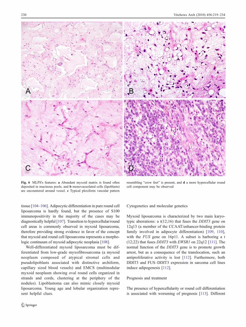
tissue [104–106]. Adipocytic differentiation in pure round cellliposarcoma is hardly found, but the presence of S100immunopositivity in the majority of the cases may bediagnostically helpful [107]. Transition to hypercellular/roundcell areas is commonly observed in myxoid liposarcoma,therefore providing strong evidence in favor of the conceptthat myxoid and round cell liposarcoma represents a morpho-logic continuum of myxoid adipocytic neoplasia [108].
Well-differentiated myxoid liposarcoma must be dif-ferentiated from low-grade myxofibrosarcoma (a myxoidneoplasm composed of atypical stromal cells andpseudolipoblasts associated with distinctive archiform,capillary sized blood vessels) and EMCS (multinodularmyxoid neoplasm showing oval round cells organized instrands and cords, clustering at the periphery of thenodules). Lipoblastoma can also mimic closely myxoidliposarcoma. Young age and lobular organization repre-sent helpful clues.
Cytogenetics and molecular genetics
Myxoid liposarcoma is characterized by two main karyo-typic aberrations: a t(12;16) that fuses the DDIT3 gene on12q13 (a member of the CCAAT/enhancer-binding proteinfamily involved in adipocyte differentiation) [109, 110],with the FUS gene on 16p11. A subset is harboring a t(12;22) that fuses DDIT3 with EWSR1 on 22q12 [111]. Thenormal function of the DDIT3 gene is to promote growtharrest, but as a consequence of the translocation, such anantiproliferative activity is lost [112]. Furthermore, bothDDIT3 and FUS–DDIT3 expression in sarcoma cell linesinduce adipogenesis [112].
Prognosis and treatment
The presence of hypercellularity or round cell differentiationis associated with worsening of prognosis [113]. Different
Fig. 6 MLPS's features: a Abundant myxoid matrix is found oftendeposited in mucinous pools, and b monovacuolated cells (lipoblasts)are encountered around vessel. c Typical plexiform vascular pattern
resembling “crow feet” is present, and d a more hypercellular roundcell component may be observed
230 Virchows Arch (2010) 456:219–234

cutoff values, ranging between 5% and 25%, have been set byindependent studies. A reliable assessment of the percentageof hypercellular areas is difficult to achieve as it may behampered by inadequate sampling as well as by that degree ofsubjectivity that is intrinsic part of morphologic evaluation.For the time being, it appears safer to consider any amount ofhypercellularity as prognostically relevant. Myxoid liposar-coma tends to recur repeatedly and tends to metastasize toboth bone [114] and soft tissue locations including theretroperitoneum. Standard treatment is represented by widesurgical resection. In high-grade lesions, adjuvant therapy(radiotherapy and/or chemotherapy) may be associated. Thedrug trabectidin appears to be a highly promising treatment formetastatic myxoid liposarcoma [115, 116].
Conclusions
Mesenchymal tumors, in strong analogy with hematologicalmalignancies, frequently harbor chromosome translocations.The EWSR1 gene is one of the favorite partners as it isinvolved in several unrelated soft tissue lesions. Interestingly,EWSR1 rearrangement has been recently observed inintermediate malignant tumors such as AFH. Even more,interestingly, the same alterations are observed in ST-CCS, asarcoma phenotypically unrelated to AFH and characterizedby full-blown malignant potential. This is a further exampleof the relative unspecificity of chromosome translocations, asalso emphasized by the occurrence of ALK gene rearrange-ment in both inflammatory myofibroblastic tumor [117] andanaplastic large cell lymphoma [118] as well by theoccurrence of the ETV6–NTRK3 fusion gene in infantilefibrosarcoma [119], mesoblastic nephroma [120], secretorycarcinoma of the breast [121], and in an isolate report acutemyeloid leukemia [122]. Another eloquent example is theoccurrence of ASPSCR1–TEF3 fusion transcript in bothalveolar soft part sarcomas as well as in pediatric forms ofrenal cell carcinoma [123, 124]. This does not hamper at allthe diagnostic value of both cytogenetics and moleculargenetics; however, it underscores the extreme importance ofpairing genetic testing of tumors with proper diagnosticexpertise. In particular, when using the split-apart FISHapproach to EWSR1 gene rearrangement, evaluation of theresults in context with morphology becomes mandatory.
Conflict of interest statement We declare that we have no conflictof interest.
References
1. Delattre O, Zucman J, Plougastel B et al (1992) Gene fusionwith an ETS DNA-binding domain caused by chromosometranslocation in human tumours. Nature 359:162–165
2. Zucman J, Delattre O, Desmaze C et al (1992) Cloning andcharacterization of the Ewing's sarcoma and peripheral neuro-epithelioma t(11;22) translocation breakpoints. Genes Chromo-somes Cancer 5:271–277
3. Moller E, Stenman G, Mandahl N et al (2008) POU5F1,encoding a key regulator of stem cell pluripotency, is fused toEWSR1 in hidradenoma of the skin and mucoepidermoidcarcinoma of the salivary glands. J Pathol 215:78–86
4. Plougastel B, Zucman J, Peter M et al (1993) Genomicstructure of the EWS gene and its relationship to EWSR1, asite of tumor-associated chromosome translocation. Genomics18:609–615
5. Bovée JVMG, Devilee P, Cornelisse CJ et al (1995) Identifica-tion of an EWS-pseudogene using translocation detection by RT-PCR in Ewing's sarcoma. Biochem Biophys Res Commun213:1051–1060
6. Bertolotti A, Lutz Y, Heard DJ et al (1996) hTAF(II)68, a novelRNA/ssDNA-binding protein with homology to the pro-oncoproteins TLS/FUS and EWS is associated with both TFIIDand RNA polymerase II. EMBO J 15:5022–5031
7. Crozat A, Aman P, Mandahl P et al (1993) Fusion of CHOP anovel RNA-binding protein in human myxoid liposarcoma.Nature 363:640–644
8. Morohoshi F, Arai K, Takahashi EI et al (1996) Cloning andmapping of a human RBP56 gene encoding a putative RNAbinding protein similar to FUS/TLS and EWS proteins.Genomics 38:51–57
9. Azuma M, Embree LJ, Sabaawy H et al (2007) Ewing sarcomaprotein ewsr1 maintains mitotic integrity and proneural cellsurvival in the zebrafish embryo. PLoS ONE 2:e979
10. Stolow DT, Haynes SR (1995) Cabeza, a Drosophila geneencoding a novel RNA binding protein, shares homology withEWS and TLS, two genes involved in human sarcoma formation.Nucleic Acids Res 23:835–843
11. Riggi N, Cironi L, Suva ML et al (2007) Sarcomas: genetics,signalling, and cellular origins. Part 1: the fellowship of TET. JPathol 213:4–20
12. Aman P, Panagopoulos I, Lassen C et al (1996) Expressionpatterns of the human sarcoma-associated genes FUS and EWSand the genomic structure of FUS. Genomics 37:1–8
13. Zakaryan RP, Gehring H (2006) Identification and characteriza-tion of the nuclear localization/retention signal in the EWSproto-oncoprotein. J Mol Biol 363:27–38
14. Bertolotti A, Melot T, Acker J et al (1998) EWS, but not EWS-FLI-1, is associated with both TFIID and RNA polymerase II:interactions between two members of the TET family, EWS andhTAFII68, and subunits of TFIID and RNA polymerase IIcomplexes. Mol Cell Biol 18:1489–1497
15. Zhang D, Paley AJ, Childs G (1998) The transcriptionalrepressor ZFM1 interacts with and modulates the ability ofEWS to activate transcription. J Biol Chem 273:18086–18091
16. Knoop LL, Baker SJ (2001) EWS/FLI alters 5′-splice siteselection. J Biol Chem 276:22317–22322
17. Knoop LL, Baker SJ (2000) The splicing factor U1C repressesEWS/FLI-mediated transactivation. J Biol Chem 275:24865–24871
18. Leemann-Zakaryan RP, Pahlich S, Sedda MJ et al (2009)Dynamic subcellular localization of the Ewing sarcoma proto-oncoprotein and its association with and stabilization of micro-tubules. J Mol Biol 386:1–13
19. Li H, Watford W, Li C et al (2007) Ewing sarcoma gene EWS isessential for meiosis and B lymphocyte development. J ClinInvest 117:1314–1323
20. Thompson AD, Teitell MA, Arvand A et al (1999) DivergentEwing's sarcoma EWS/ETS fusions confer a common tumori-genic phenotype on NIH3T3 cells. Oncogene 18:5506–5513
Virchows Arch (2010) 456:219–234 231

21. May WA, Arvand A, Thompson AD et al (1997) EWS/FLI1-induced manic fringe renders NIH 3T3 cells tumorigenic. NatGenet 17:495–497
22. Ushigome S, Machinami R, Sorensen PH (2002) Ewingsarcoma/primitive neuroectodermal tumour (PNET). In: FletcherCDM, Unni KK, Mertens F (eds) World Health Organizationclassification of tumours; pathology & genetics; tumours of softtissue and bone. IARC Presss, Lyon, pp 298–300
23. Parham DM, Roloson GJ, Feely M et al (2001) Primarymalignant neuroepithelial tumors of the kidney: a clinicopatho-logic analysis of 146 adult and pediatric cases from the NationalWilms' Tumor Study Group Pathology Center. Am J Surg Pathol25:133–146
24. Welsch T, Mechtersheimer G, Aulmann S et al (2006) Hugeprimitive neuroectodermal tumor of the pancreas: report of a caseand review of the literature. World J Gastroenterol 12:6070–6073
25. Dedeurwaerdere F, Giannini C, Sciot R et al (2002) Primaryperipheral PNET/Ewing's sarcoma of the dura: a clinicopatho-logic entity distinct from central PNET. Mod Pathol 15:673–678
26. Stout AP (1918) A tumor of the ulnar nerve. Proc N Y PatholSoc 18:2–12
27. Askin FB, Rosai J, Sibley RK et al (1979) Malignant small celltumor of the thoracopulmonary region in childhood: a distinctiveclinicopathologic entity of uncertain histogenesis. Cancer43:2438–2451
28. Ambros IM, Ambros PF, Strehl S et al (1991) MIC2 is a specificmarker for Ewing's sarcoma and peripheral primitive neuro-ectodermal tumors: evidence for a common histogenesis ofEwing's sarcoma and peripheral primitive neuroectodermaltumors from MIC2 expression and specific chromosome aberra-tion. Cancer 67:1886–1893
29. Dei Tos AP, Wadden C, Calonje E et al (1995) Immunohisto-chemical demonstration of glycoprotein p30/32mic2 (CD99) insynovial sarcoma. Appl Immunohistochem 3:168–173
30. Terrier-Lacombe MJ, Guillou L, Chibon F et al (2009) Superficialprimitive Ewing's sarcoma: a clinicopathologic and molecularcytogenetic analysis of 14 cases. Mod Pathol 22:87–94
31. Hasegawa SL, Davison JM, Rutten A et al (1998) Primarycutaneous Ewing's sarcoma: immunophenotypic and molecularcytogenetic evaluation of five cases. Am J Surg Pathol 22:310–318
32. Nicholson SA, McDermott MB, Swanson PE et al (2000) CD99and cytokeratin-20 in small-cell and basaloid tumors of the skin.Appl Immunohistochem Mol Morphol 8:37–41
33. Shing DC, McMullan DJ, Roberts P et al (2003) FUS/ERG genefusions in Ewing's tumors. Cancer Res 63:4568–4576
34. Ng TL, O'Sullivan MJ, Pallen CJ et al (2007) Ewing sarcomawith novel translocation t(2;16) producing an in-frame fusion ofFUS and FEV. J Mol Diagnostics 9:459–463
35. Aurias A, Rimbaut C, Buffe D et al (1983) Chromosomaltranslocations in Ewing's sarcoma. Letter to the editor. N Engl JMed 309:496–498
36. Lessnick SL, Dacwag CS, Golub TR (2002) The Ewing'ssarcoma oncoprotein EWS/FLI induces a p53-dependent growtharrest in primary human fibroblasts. Cancer Cells 1:393–401
37. Kovar H (2005) Context matters: the hen or egg problem inEwing's sarcoma. Semin Cancer Biol 15:189–196
38. Serrano M, Lin AW, McCurrach ME et al (1997) Oncogenic rasprovokes premature cell senescence associated with accumula-tion of p53 and p16INK4a. Cell 88:593–602
39. Riggi N, Cironi L, Provero P et al (2005) Development ofEwing's sarcoma from primary bone marrow-derived mesenchy-mal progenitor cells. Cancer Res 65:11459–11468
40. Riggi N, Suva ML, Suva D et al (2008) EWS-FLI-1 expressiontriggers a Ewing's sarcoma initiation program in primary humanmesenchymal stem cells. Cancer Res 68:2176–2185
41. Prieur A, Tirode F, Cohen P et al (2004) EWS/FLI-1 silencingand gene profiling of Ewing cells reveal downstream oncogenicpathways and a crucial role for repression of insulin-like growthfactor binding protein 3. Mol Cell Biol 24:7275–7283
42. Tirode F, Laud-Duval K, Prieur A et al (2007) Mesenchymalstem cell features of Ewing tumors. Cancer Cells 11:421–429
43. Szuhai K, Ijszenga M, Tanke HJ et al (2006) Molecularcytogenetic characterization of four previously established andtwo newly established Ewing sarcoma cell lines. Cancer GenetCytogenet 166:173–179
44. Szuhai K, Ijszenga M, De JD et al (2009) The NFATc2 gene isinvolved in a novel cloned translocation in a Ewing sarcomavariant that couples its function in immunology to oncology. ClinCancer Res 15:2259–2268
45. Thorner P, Squire J, Chilton-MacNeill S et al (1996) Is the EWS/FLI-1 fusion transcript specific for Ewing sarcoma and periph-eral primitive neuroectodermal tumor? A report of four casesshowing this transcript in a wider range of tumor types. Am JPathol 148:1125–1138
46. Ed A, Kawai A, Healy JH et al (1998) EWS-FL11 fusiontranscript structure is an independent determinant of prognosis inEwing's sarcoma. J Clin Oncol 16:1248–1255
47. Mangham DC, Cannon A, Li XQ et al (1995) p53 over-expression in Ewing's sarcoma/primitive neuroectodermal tu-mour is an uncommon event. J Clin Pathol 48M:M79–M82
48. Ueda Y, Dockhorn-Dworniczak B, Blasius S et al (1993)Analysis of mutant P53 protein in osteosarcomas and othermalignant and benign lesions of bone. J Cancer Res Clin Oncol119:172–178
49. Tsuchiya T, Sekine K, Hinohara S et al (2000) Analysis of thep16INK4, p14ARF, p15, TP53, and MDM2 genes and theirprognostic implications in osteosarcoma and Ewing sarcoma.Cancer Genet Cytogenet 120:91–98
50. Wei G, Antonescu CR, de Alava E et al (2000) Prognosticimpact of INK4A deletion in Ewing sarcoma. Cancer 89:793–799
51. Romeo S, Debiec-Rychter M, Van GM et al (2009) Cell cycle/apoptosis molecule expression correlates with imatinib responsein patients with advanced gastrointestinal stromal tumors. ClinCancer Res 15:4191–4198
52. Zogopoulos G, Teskey L, Sung L et al (2004) Ewing sarcoma:favourable results with combined modality therapy and conser-vative use of radiotherapy. Pediatr Blood Cancer 43:35–39
53. Schmidt D, Herrmann C, Jürgens H et al (1991) Malignantperipheral neuroectodermal tumor and its necessary distinctionfrom Ewing's sarcoma. A report from the Kiel Pediatric TumorRegistry. Cancer 68:2251–2259
54. Parham DM, Hijazi Y, Steinberg SM et al (1999) Neuro-ectodermal differentiation in Ewing's sarcoma family of tumorsdoes not predict tumor behavior. Hum Pathol 30:911–918
55. Terrier Ph, Henry-Amar M, Triche TJ et al (1995) Is neuro-ectodermal differentiation of Ewing's sarcoma of bone associatedwith an unfavourable prognosis? Eur J Cancer 31A:307–314
56. Toretsky JA, Kalebic T, Blakesley V et al (1997) The insulin-likegrowth factor-I receptor is required for EWS/FLI-1 transforma-tion of fibroblasts. J Biol Chem 272:30822–30827
57. Toretsky JA, Steinberg SM, Thakar M et al (2001) Insulin-likegrowth factor type 1 (IGF-1) and IGF binding protein-3 inpatients with Ewing sarcoma family of tumors. Cancer 92:2941–2947
58. Erkizan HV, Kong Y, Merchant M et al (2009) A small moleculeblocking oncogenic protein EWS-FLI1 interaction with RNAhelicase A inhibits growth of Ewing's sarcoma. Nat Med15:750–756
59. Lessnick SL, Dei Tos AP, Sorensen PH et al (2009) Small roundcell sarcomas. Semin Oncol 36:338–346
232 Virchows Arch (2010) 456:219–234

60. Antonescu CR, Gerald W (2002) Desmoplastic small round celltumour. In: Fletcher CDM, Unni KK, Mertens F (eds) WorldHealth Organisation classification of tumours, pathology andgenetics, tumours of soft tissue and bone. pp 216–218
61. Parkash V, Gerald WL, Parma A et al (1995) Desmoplastic smallround cell tumor of the pleura. Am J Surg Pathol 19:659–665
62. Cummings OW, Ulbright TM, Young RH et al (1997) Desmo-plastic small round cell tumors of the paratesticular region. Areport of six cases. Am J Surg Pathol 21:219–225
63. Wolf AN, Ladanyi M, Paull G et al (1999) The expandingclinical spectrum of desmoplastic small round-cell tumor: areport of two cases with molecular confirmation. Hum Pathol30:430–435
64. Tison V, Cerasoli S, Morigi F et al (1996) Intracranial desmo-plastic small-cell tumor. Report of a case. Am J Surg Pathol20:112–117
65. Neder L, Scheithauer BW, Turel KE et al (2009) Desmoplasticsmall round cell tumor of the central nervous system: report of twocases and review of the literature. Virchows Arch 454:431–439
66. Adsay V, Cheng J, Athanasian E et al (1999) Primary desmo-plastic small cell tumor of soft tissues and bone of the hand. AmJ Surg Pathol 23:1408–1413
67. Hamazaki M, Okita H, Hata J et al (2006) Desmoplastic smallcell tumor of soft tissue: molecular variant of EWS-WT1chimeric fusion. Pathol Int 56:543–548
68. Murphy AJ, Bishop K, Pereira C et al (2008) A new molecularvariant of desmoplastic small round cell tumor: significance ofWT1 immunostaining in this entity. Hum Pathol 39:1763–1770
69. Slomovitz BM, Girotra M, Aledo A et al (2000) Desmoplasticsmall round cell tumor with primary ovarian involvement: casereport and review. Gynecol Oncol 79:124–128
70. Bismar TA, Basturk O, Gerald WL et al (2004) Desmoplasticsmall cell tumor in the pancreas. Am J Surg Pathol 28:808–812
71. Su MC, Jeng YM, Chu YC (2004) Desmoplastic small roundcell tumor of the kidney. Am J Surg Pathol 28:1379–1383
72. Wang LL, Perlman EJ, Vujanic GM et al (2007) Desmoplasticsmall round cell tumor of the kidney in childhood. Am J SurgPathol 31:576–584
73. Stuart-Buttle CE, Smart CJ, Pritchard S et al (2008) Desmo-plastic small round cell tumour: a review of literature andtreatment options. Surg Oncol 17:107–112
74. Ordonez NG (1998) Desmoplastic small round cell tumor. I: ahistopathologic study of 39 cases with emphasis on unusualhistological patterns. Am J Surg Pathol 22:1303–1313
75. Gerald WL, Miller HK, Battifora H et al (1991) Intra-abdominaldesmoplastic small round-cell tumor. Report of 19 cases of adistinctive type of high-grade polyphenotypic malignancyaffecting young individuals. Am J Surg Pathol 15:499–513
76. Gerald WL, Ladanyi M, de Alava E et al (1998) Clinical,pathologic, and molecular spectrum of tumors associated with t(11;22)(p13;q12): desmoplastic small round-cell tumor and itsvariants. J Clin Oncol 16:3028–3036
77. Ordonez NG (1998) Desmoplastic small round cell tumor. II: anultrastructural and immunohistochemical study with emphasis onnew immunohistochemical markers. Am J Surg Pathol 22:1314–1327
78. Lee SB, Kolquist KA, Nichols K et al (1997) The EWS-WT1translocation product induces PDGFA in desmoplastic smallround-cell tumour. Na Genet 17:309–313
79. Werner H, Idelman G, Rubinstein M et al (2007) A novel EWS-WT1 gene fusion product in desmoplastic small round cell tumoris a potent transactivator of the insulin-like growth factor-Ireceptor (IGF-IR) gene. Cancer Lett 247:84–90
80. Wong JC, Lee SB, Bell MD et al (2002) Induction of theinterleukin-2/15 receptor beta-chain by the EWS-WT1 translo-cation product. Oncogene 21:2009–2019
81. Sawyer JR, Tryka AF, Lewis JM (1992) A novel reciprocalchromosome translocation t(11;22)(p13;q12) in an intraabdomi-nal desmoplastic small round-cell tumor. Am J Surg Pathol16:411–416
82. Alaggio R, Rosolen A, Sartori F et al (2007) Spindle cell tumorwith EWS-WT1 transcript and a favorable clinical course: avariant of DSCT, a variant of leiomyosarcoma, or a new entity?Report of 2 pediatric cases. Am J Surg Pathol 31:454–459
83. Katz RL, Quezado M, Senderowicz AM et al (1997) An intra-abdominal small round cell neoplasm with features of primitiveneuroectodermal and desmoplastic round cell tumor and a EWS/FLI-1 fusion transcript. Hum Pathol 28:502–509
84. Li H, Smolen GA, Beers LF et al (2008) Adenosine transporterENT4 is a direct target of EWS/WT1 translocation product and ishighly expressed in desmoplastic small round cell tumor. PLoSONE 3:e2353
85. Antonescu CR, Nafa K, Segal NH et al (2006) EWS-CREB1: arecurrent variant fusion in clear cell sarcoma association withgastrointestinal location and absence of melanocytic differentia-tion. Clin Cancer Res 12:5356–5362
86. Zambrano E, Reyes-Mugica M, Franchi A et al (2003) Anosteoclast-rich tumor of the gastrointestinal tract with featuresresembling clear cell sarcoma of soft parts: reports of 6 cases of aGIST simulator. Int J Surg Pathol 11:75–81
87. Schaefer KL, Brachwitz K, Wai DH et al (2004) Expressionprofiling of t(12;22) positive clear cell sarcoma of soft tissue celllines reveals characteristic up-regulation of potential new markergenes including ERBB3. Cancer Res 64:3395–3405
88. Segal NH, Pavlidis P, Noble WS et al (2003) Classification ofclear-cell sarcoma as a subtype of melanoma by genomicprofiling. J Clin Oncol 21:1775–1781
89. Langezaal SM, Graadt van Roggen JF, Cleton-Jansen AM et al(2001) Malignant melanoma is genetically distinct from clearcell sarcoma of tendons and aponeurosis (malignant melanomaof soft parts). Br J Cancer 84:535–538
90. Graadt van Roggen JF, Mooi WJ, Hogendoorn PCW (1998)Clear cell sarcoma of tendons and aponeuroses (malignantmelanoma of soft parts) and cutaneous melanoma: exploringthe histogenetic relationship between these two clinicopatholog-ical entities. J Pathol 186:3–7
91. Antonescu CR, Tschernyavsky SJ, Woodruff JM et al (2002)Molecular diagnosis of clear cell sarcoma: detection of EWS-ATF1 and MITF-M transcripts and histopathological andultrastructural analysis of 12 cases. J Mol Diagnostics 4:44–52
92. Zucman J, Delattre O, Desmaze C et al (1993) EWS and ATF-1gene fusion induced by t(12;22) translocation in malignantmelanoma of soft parts. Nat Genet 4:341–345
93. Xie S, Price JE, Luca M et al (1997) Dominant-negative CREBinhibits tumor growth and metastasis of human melanoma cells.Oncogene 15:2069–2075
94. Yokoyama S, Feige E, Poling LL et al (2008) Pharmacologicsuppression of MITF expression via HDAC inhibitors in themelanocyte lineage. Pigment Cell Melanoma Res 21:457–463
95. Lucas DR, Heim S (2002) Extraskeletal myxoid chondrosar-coma. In: Fletcher CDM, Unni KK, Mertens F (eds) Pathology& genetics. Tumours of soft tissue and bone. IARC Press, Lyon,pp 213–215
96. Aigner T, Oliveira AM, Nascimento AG (2004) Extraskeletalmyxoid chondrosarcomas do not show a chondrocytic pheno-type. Mod Pathol 17:214–221
97. Subramanian S, West RB, Marinelli RJ et al (2005) The geneexpression profile of extraskeletal myxoid chondrosarcoma. JPathol 206:433–444
98. Turc-Carel C, Dal Cin P, Rao U et al (1988) Recurrentbreakpoints at 9q31 and 22q12.2 in extraskeletal myxoidchondrosarcoma. Cancer Genet Cytogenet 30:145–150
Virchows Arch (2010) 456:219–234 233

99. Panagopoulos I, Mertens F, Isaksson M et al (2002) Moleculargenetic characterization of the EWS/CHN and RBP56/CHNfusion genes in extraskeletal myxoid chondrosarcoma. GenesChromosomes Cancer 35:340–352
100. Filion C, Motoi T, Olshen AB et al (2009) The EWSR1/NR4A3fusion protein of extraskeletal myxoid chondrosarcoma activatesthe PPARG nuclear receptor gene. J Pathol 217:83–93
101. Fanburg-Smith JC, Dal Cin P (2002) Angiomatoid fibroushistiocytoma. In: Fletcher CDM, Unni KK, Mertens F (eds)World Health Organisation classification of tumours. Pathologyand genetics of tumours of soft tissue and bone. IARC Press,Lyon, pp 194–195
102. Rossi S, Szuhai K, Ijszenga M et al (2007) EWSR1-CREB1 andEWSR1-ATF1 fusion genes in angiomatoid fibrous histiocy-toma. Clin Cancer Res 13:7322–7328
103. Antonescu CR, Dal CP, Nafa K et al (2007) EWSR1-CREB1 isthe predominant gene fusion in angiomatoid fibrous histiocy-toma. Genes Chromosomes Cancer 46:1051–1060
104. Antonescu CR, Ladanyi M (2002) Myxoid Liposarcoma. In:Fletcher CDM, Unni KK, Mertens F (eds) World HealthOrganisation classification of tumours. Pathology and geneticsof tumours of soft tissue and bone. IARC Press, Lyon, pp 40–43
105. Kilpatrick SE, Doyon J, Choong PFM et al (1996) Theclinicopathologic spectrum of myxoid and round cell liposar-coma. A study of 95 cases. Cancer 77:1450–1458
106. Smith TA, Easley KA, Goldblum JR (1996) Myxoid/round cellliposarcoma of the extremities. A clinicopathologic study of 29cases with particular attention to extent of round cell lip-osarcoma. Am J Surg Pathol 20:171–180
107. Dei Tos AP, Wadden C, Fletcher CDM (1996) S-100 proteinstaining in liposarcoma. Its diagnostic utility in the high grademyxoid (round cell) variant. Appl Immunohistochem 4:95–101
108. Orvieto E, Furlanetto A, Laurino L et al (2001) Myxoid andround cell liposarcoma: a spectrum of myxoid adipocyticneoplasia. Semin Diagn Pathol 18:267–273
109. Tallini G, Rosai J, Fletcher CDM et al (1995) Myxoid lip-osarcoma represents a distinct type of liposarcoma characterisedby the specific cytogenetic translocation t(12; 16)(q13; p11).Mod Pathol 8
110. Knight JC, Renwick PJ, Dal Cin P et al (1995) Translocation t(12;16)(q13;p11) in myxoid liposarcoma and round cell lip-osarcma: molecular and cytogenetic analysis. Cancer Res 55:24–27
111. Panagopoulos I, Hoglund M, Mertens F et al (1996) Fusion ofthe EWS and CHOP genes in myxoid liposarcoma. Oncogene12:489–494
112. Engstrom K, Willen H, Kabjorn-Gustafsson C et al (2006) Themyxoid/round cell liposarcoma fusion oncogene FUS-DDIT3 andthe normal DDIT3 induce a liposarcoma phenotype in transfectedhuman fibrosarcoma cells. Am J Pathol 168:1642–1653
113. Antonescu CR, Tschernyavsky SJ, Decuseara R et al (2001)Prognostic impact of P53 status, TLS-CHOP fusion transcriptstructure, and histological grade in myxoid liposarcoma: amolecular and clinicopathologic study of 82 cases. Clin CancerRes 7:3977–3987
114. Schwab JH, Boland P, Guo T et al (2007) Skeletal metastases inmyxoid liposarcoma: an unusual pattern of distant spread. AnnSurg Oncol 14:1507–1514
115. Grosso F, Jones RL, Demetri GD et al (2007) Efficacy oftrabectedin (ecteinascidin-743) in advanced pretreated myxoidliposarcomas: a retrospective study. Lancet Oncol 8:595–602
116. Forni C, Minuzzo M, Virdis E et al (2009) Trabectedin (ET-743)promotes differentiation in myxoid liposarcoma tumors. MolCancer Ther
117. Lawrence B, Perez-Atayde A, Hibbard MK et al (2000) TPM3-ALK and TPM4-ALK oncogenes in inflammatory myofibro-blastic tumors. Am J Pathol 157:377–384
118. Lamant L, Dastugue N, Pulford K et al (1999) A new fusiongene TPM3-ALK in anaplastic large cell lymphoma created by a(1;2)(q25;p23) translocation. Blood 93:3088–3095
119. Knezevich SR, McFadden DE, Tao W et al (1998) A novelETV6-NTRK3 gene fusion in congenital fibrosarcoma. NatGenet 18:184–187
120. Knezevich SR, Garnett MJ, Pysher TJ et al (1998) ETV6-NTRK3 gene fusions and trisomy 11 establish a histogenetic linkbetween mesoblastic nephroma and congenital fibrosarcoma.Cancer Res 58:5046–5048
121. Tognon C, Knezevich SR, Huntsman D et al (2002) Expressionof the ETV6-NTRK3 gene fusion as a primary event in humansecretory breast carcinoma. Cancer Cells 2:367–376
122. Eguchi M, Eguchi-Ishimae M, Tojo A et al (1999) Fusion ofETV6 to neurotrophin-3 receptor TRKC in acute myeloidleukemia with t(12;15)(p13;q25). Blood 93:1355–1363
123. Argani P, Antonescu CR, Illei PB et al (2001) Primary renalneoplasms with the ASPL-TFE3 gene fusion of alveolar soft partsarcoma: a distinctive tumor entity previously included amongrenal cell carcinomas of children and adolescents. Am J Pathol159:179–192
124. Ladanyi M, Lui MY, Antonescu CR et al (2001) The der(17)t(X;17)(p11;q25) of human alveolar soft part sarcoma fuses theTFE3 transcription factor gene to ASPL, a novel gene at 17q25.Oncogene 20:48–57
234 Virchows Arch (2010) 456:219–234