Embed Size (px)
Citation preview

1
�
�
�
SOFT TISSUE ATTACHMENT TO TITANIUM IMPLANTS COATED WITH GROWTH
FACTORS �
�
�
A report submitted to the University of Adelaide in partial fulfilment of the requirements of the Degree of Doctor of
Clinical Dentistry (Periodontology)
Christopher William BATES BDS (Adel), MClinDent (Pros) (Lond)
�
�

67
Chapter 2.
SOFT TISSUE ATTACHMENT TO TITANIUM IMPLANTS
COATED WITH GROWTH FACTORS
CW BATES1, V MARINO2, N FAZZALARI3, PM BARTOLD4
1Post Graduate Student (Periodontology), School of Dentistry, University of Adelaide.
2Research Assistant, Colgate Australian Clinical Dental Research Centre, School of
Dentistry, University of Adelaide.
3Professor and Director, Bone and Joint Research Laboratory, Institute of Medical and
Vetinary Science, Adelaide.
4Professor of Periodontology, Colgate Australian Clinical Dental Research Centre, School
of Dentistry, University of Adelaide.
68
2.1 ABSTRACT
Background: Peri-implant tissues form a crucial but fragile seal between the oral
environment, the bone and the implant surface. Enhancing the seal formed by the peri-implant
soft tissues at the titanium/connective tissue interface may be an important factor in implant
survival. Additionally, enhancing soft tissue adherence to the implant surface when implants
are placed in dehiscence type defects may mean that simultaneous osseous grafting
procedures will not always be required.
Objective: The aim of this study was to investigate the effect of implant surface
modification with either platelet-derived growth factor (PDGF) or enamel matrix derivative
(EMD) on the connective tissue attachment to moderately roughened titanium implants.
Material and Methods: 18 moderately roughened titanium implants were subcutaneously
implanted into 14 rats. 6 implants each were coated with PDGF and EMD immediately prior
to implantation and 6 implants were left uncoated. The implants were retrieved with a sample
of surrounding tissue at 4 and 8 weeks. The specimens were resin-embedded and sections
viewed under confocal microscopy for collagen autofluorescence and prepared for qualitative
and histomorphometric analysis under light microscopy. ANOVA and t-tests were used to
compare the thickness of fibroblast encapsulation on the implant surface and the depth of
connective tissue penetration onto the implant grooves.
Results: Qualitative analysis under confocal and light microscopy showed encapsulation of
all implants by fibroblasts and good soft tissue integration at the end of 4 and 8 weeks.
Coating of the implants with growth factors did not alter the orientation of fibroblasts and
collagen fibres. Histomorphometric analysis demonstrated that the depth of connective tissue
penetration into the implant grooves was significantly greater for the implants coated with
PDGF at 4 weeks (ANOVA, P value 0.0014). The thickness of the fibroblast encapsulation on
the implant surface was significantly less for the implants coated with PDGF at 8 weeks
(ANOVA, P value 0.0012).

69
Conclusion: Good soft tissue integration can be achieved on a moderately roughened
titanium implant surface. Coating the implant surface with rhPDGF-BB could increase the
speed of soft tissue healing around an implant surface but this increased rate of healing with
rhPDGF-BB coating could also result in a less robust titanium/connective tissue interface.
2.2 INTRODUCTION
Osseointegrated dental implants are transmucosal “masticatory devices” that penetrate the
oral mucosa, with the peri-implant tissues expected to exercise a protective function (Weber
& Cochran 1998). Research and clinical focus in dental implantology in the past two decades
has primarily concentrated on the bone-to-implant interface and the peri-implant mucosa, with
the soft tissue seal around implants investigated to a much lesser degree. Both bone and soft
tissue integration to dental implants are wound healing processes involving several stages of
tissue formation and degradation (Berglundh et al 2003, Abrahamsson et al 2004, Berglundh
et al 2007). Osseointegration is the result of the modelling and remodelling of bone tissue that
occurs after implant placement, whilst the wound healing that occurs following the closure of
mucoperiosteal flaps during implant surgery results in the establishment of a mucosal
attachment (transmucosal attachment) to the implant. The establishment of the mucosal
barrier around the implant is characterised by a gradual shift from a coagulum to granulation
tissue followed by the formation of a barrier epithelium and the formation of connective tissue
(Berglundh et al 2007).
Several studies using animal and human models have investigated the structure and
function of the peri-implant mucosa (Berglundh et al 1991, 1992, 1994, 2003, 2007; Buser et
al 1992, Ericsson et al 1996, 1997; Abrahamsson et al 1996, 1997, 1998, 1999, 2002, 2004;
Berglundh & Lindhe 1996, Cochran et al 1997, Moon et al 1999, Glauser et al 2005,
Schüpbach & Glauser 2007, Welander et al 2007, 2008, Allegrini Jr et al 2008, Nevins et al
2008). In an early study in dogs, Berglundh et al (1991) compared the gingiva around teeth
and the mucosa around two-stage implants (Branemark System®, Nobel Biocare, Götenburg,

70
Sweden). It was found that the peri-implant mucosa consisted of a 2 mm long barrier
epithelium and a zone 1-1.5 mm high where the connective tissue was in direct contact with
the TiO2 layer of the implant. This area was termed a zone of “connective tissue integration”.
Histologically, the peri-implant epithelium and the surrounding connective tissue of dental
implants have similar characteristics to those structures surrounding teeth (Abrahamsson &
Soldini 2006) but differ in terms of the orientation of collagen fibres (Buser et al 1992), the
composition of the connective tissue (Moon et al 1999, Abrahamsson et al 2002), and the
distribution of the vascular system of the peri-implant mucosa (Berglundh et al 1994). The
connective tissue in the zone of integration has a low density of blood vessels but a large
number of fibroblasts and collagen fibres appearing to originate from the periosteum of the
bone crest and extending towards the margin of the soft tissue in a direction parallel to the
long axis of the abutment. More detailed analyses of the soft tissue/implant interface using
transmission electron microscopy found that the zone of connective tissue directly adjacent to
the implant surface has a large number of round and flat-shaped fibroblasts with their long
axes parallel with the implant surface but virtually no blood vessels. Further away from this
zone the number of fibroblasts decreases but there are more collagen fibres and there is an
increase in vascularity (Moon et al 1999, Abrahamsson et al 2002). Berglundh et al (1991)
stated that the main difference between the mesenchymal tissues present at a tooth and at an
implant site is the occurrence of cementum (acellular or cellular) on the root surface.
There is no doubt that the peri-implant soft-tissues form a crucial seal between the oral
environment, the bone and the implant surface (Cochran et al 1994, 1997). The seal is fragile
and due to the absence of periodontal ligament fibres when subjected to bacterial or
mechanical challenge the destruction of peri-implant tissues can be a faster and more
devastating process than in periodontal tissues (Salcetti et al 1997, Maksoud 2003). Thus,
enhancing the seal formed by the peri-implant soft tissues, especially that of the
titanium/connective tissue interface, may be an important factor in implant survival.

71
The titanium/connective tissue interface, certainly for smooth, machined surface dental
implants, lacks a mechanical attachment of inserting collagen fibres, unlike that of periodontal
tissues of teeth. Whether this lack of mechanical attachment differs for roughened surface
implants has not been extensively investigated. A small number of recent in vivo studies have
indicated that microtexturing of the implant can be used to control the soft tissue response
(Glauser et al 2005, Schüpbach & Glauser 2007, Nevins et al 2008). Until recently most
dental implants were designed such that the transmucosal portion of the implant was of a
smooth or polished nature. Recently, these design concepts have changed, with several
implant designs allowing crestal placement and incorporating roughened surfaces into the
coronal portion of the body of the implant, up to the level of the implant-abutment platform
(eg. Nobel Replace, Straumann Bone-level, Astra Osseospeed). Some clinicians have
advocated that roughened surfaces may in fact be conducive to very good soft tissue
adherence in dehiscence type defects and therefore placement of implants into these defects
may not always require osseous grafting procedures to correct these defects (Dragoo, personal
communication).
Surface modification of titanium implants may improve the ability of connective tissue
components in the peri-implant mucosa to attach to the implants. Currently, most dental
implant types incorporate a “roughened” surface as part of their macro-design. Many of these
surfaces are able to absorb proteins and thus act as a reservoir or carrier for attachment
proteins, growth factors and other biological agents which may be of assistance for soft or
hard tissue integration. In vitro studies have shown that epithelial cell adhesion to titanium
surfaces coated with biological agents such as fibronectin, laminin and collagen was enhanced
in comparison with uncoated titanium (Dean et al 1995, Tamura et al 1998, Park et al 1998,
Roessler et al 2001, Nagai et al 2002). However, in a recent study investigating soft tissue
healing around implants in a canine model, it was found that the vertical dimensions of the
epithelial and connective tissue components as well as the composition of the connective

72
tissue zone directly adjacent to the implant were similar for collagen-coated and non-coated
implants at 4 and 8 weeks of healing (Welander et al 2007).
2.2.1 Platelet-Derived Growth Factor (PDGF)
PDGF is a natural protein sequestered by blood platelets and bone matrix secreted locally
during clotting at the site of soft- or hard-tissue injury, stimulating a cascade of events that
leads to the wound healing response. The primary effect of PDGF is that of a mitogen,
initiating cell division. It has been shown, particularly with the PDGF-BB form, to be a potent
stimulator of many types of connective tissue cells, including periodontal ligament
fibroblasts, cementoblasts and osteoblasts (Lynch et al 1989, Piche & Graves 1989, Matsuda
et al 1992, Dennison et al 1994, Boyan et al 1994). Improvement in periodontal wound
healing leading to significant bone, cementum and periodontal ligament regeneration has been
observed after applying PDGF-BB in combination with either insulin-like growth factor-1
(IGF-1), �-tricalcium phosphate (�-TCP) or a bovine derived xenograft (Lynch et al 1989,
1991a, 1991b, 2006; Rutherford et al 1992, 1993; Giannobile et al 1994, 1996; Howell et al
1997, Camelo et al 2003, Nevins et al 2003, 2005, 2007; McGuire et al 2006, Simion et al
2006). These results illustrate the beneficial effects of PDGF on both soft and hard tissue
healing and regeneration. Recently, a growth-factor enhanced matrix (GEM) has become
available for clinical use. This graft material consists of a concentrated solution of pure
recombinant human platelet-derived growth factor (rhPDGF-BB), mixed with an
osteoconductive matrix composed of �-TCP and is marketed as GEM 21S (Osteohealth,
Shirley, New York, USA).
2.2.2 Enamel Matrix Protein Derivatives
The biologic concept behind enamel matrix-induced periodontal regeneration is based on
the discovery that enamel matrix proteins, are not only involved in enamel formation but also
play a key role in the formation of the root and attachment apparatus. Enamel matrix protein

73
derivatives (EMD), of which 90% are amelogenins, are secreted during tooth root
development by Hertwig’s epithelial root sheath and play a crucial role in the formation of
acellular root cementum which is the most important tissue for the insertion of collagen fibres
(Slavkin and Boyde 1975, Slavkin 1976, Slavkin et al 1988, Lindskog 1982a, 1982b;
Lindskog & Hammarstrom 1982, Brookes et al 1995, Fong et al 1996). These proteins are
thought to induce the formation of periodontal attachment during tooth formation and it is
believed that EMD used in periodontal lesions mimic the development of the tooth supporting
apparatus (Hammarström 1997). Emdogain® (Biora AB, Straumann, Malmö, Sweden), a
porcine-derived material, is the only commercially available product using EMD.
Apart from its original use as an agent to enhance and promote regeneration, Emdogain®
has also been reported to be effective in the management of recession defects by enhancing
soft tissue adherence to root surfaces. Its use in these situations may promote collagen
synthesis, the formation of cementum, periodontal ligament and bone and may therefore
increase the width of keratinised tissue (Hägewald et al 2002, McGuire & Nunn 2003,
McGuire & Cochran 2003, Nemcovsky et al 2004, Spahr et al 2005, Castellanos et al 2006,
Moses et al 2006, Sato et al 2006, Shin et al 2007). This finding may be of particular
relevance to the previously-stated opinion of some clinicians that moderately roughened
surfaces may be conducive to good soft tissue adherence and that placement of implants into
dehiscence-type defects may not always require osseous correction. Whether an agent such as
EMD would enhance soft tissue adhesion to exposed implant surfaces in dehiscence type
defects remains to be established.
A number of studies have examined the changes in soft tissue level after implant placement
(Bengazi et al 1996, Grunder 2000, Ekfeldt et al 2003). Despite significant differences in
experimental designs, the majority of studies conclude that gingival recession that varies
between 0.6 mm to 1.5 mm is unavoidable. Whilst multiple factors can influence gingival
recession around transmucosal dental implants, there is little doubt that the low level of
connective tissue attachment to implant surfaces is important (Rompen et al 2006). Various

74
methods have been proposed to improve the quality of the soft tissue interface, including
micro and macro design features of the transmucosal portion of the implant (Glauser et al
2005, Schüpbach & Glauser 2007, Nevins et al 2008).
To date there have been few studies investigating the effect of surface modification with
EMD but none with PDGF on the connective tissue attachment to titanium implants. A
number of studies have investigated the effects of PDGF and EMD on bone healing around
dental implants. Whilst PDGF has been shown to be influential in improving the regeneration
of peri-implant bone (Lynch et al 1991a, Becker et al 1992, Meraw et al 2000), the use of
EMD has not been shown to contribute to the amount of bone-to-implant contact around
titanium implants (Franke Stenport & Johansson 2003, Cangini & Cornelini 2005). Recently,
a pilot study reporting on the effects of autogenous periodontal cell grafts, with and without
the application of EMD, on the implant-connective tissue interface found that an implant-
connective tissue interface morphologically consistent with a periodontal connective tissue
attachment was not observed in sections from any of the implant or autogenous cell grafts
(Craig et al 2006).
2.3 HYPOTHESIS AND AIM
The hypothesis for this study is that surface modification of roughened surface (TiUnite)
titanium implants with PDGF or EMD results in improved bioactivity of the implant surface,
thereby promoting cell attachment and CT formation, which is expected to result in an
improved attachment.
The aim of this study is to investigate if surface modification of roughened surface
(TiUnite) titanium implants with PDGF or EMD has the potential to enhance connective
tissue attachment to titanium implants.

75
2.4 SIGNIFICANCE OF THE AIM
Enhancing the soft tissue seal formed by the peri-implant soft tissues will be of clinical
significance, particularly with regards to preventing recession, enhancing aesthetic outcomes
and even enhancing placement into sites with significant dehiscence defects.
2.5 MATERIAL AND METHODS
2.5.1 Animals
Fourteen female Dark Agouti (DA) rats, each about 6 to 8 weeks old were used. These
were acquired through the Animal Services Division, Institute of Medical and Veterinary
Science (IMVS), Adelaide. The research protocol related to the use of animals in this study
was approved by the animal ethics committees of both the University of Adelaide and the
IMVS.
2.5.2 Implants
Eighteen Branemark System® Mk III Groovy NP (3.3 mmØ x 10 mm) (Nobel Biocare
AB, Göteborg, Sweden) implants were used. Six test implants were coated with enamel
matrix protein derivative (Emdogain®, Biora AB, Straumann, Malmö, Sweden) and 6 test
implants were coated with reconstituted recombinant human platelet-derived growth factor-
BB (rhPDGF-BB, Pepro Tech, Rocky Hill, New Jersey, USA). The 6 control implants were
uncoated. Two of these control implants were used for an initial pilot study to verify the
feasibility of the experimental protocol.
2.5.3 Pilot Study
For the pilot study, two single uncoated implants were surgically placed subcutaneously
into the backs of two rats. One animal was sacrificed at 4 weeks and the other at 8 weeks. The
implants and surrounding tissue were retrieved at the time of sacrifice and then processed for
future analysis. This experiment was conducted to check on the viability of the study, i.e. the

76
ability to get a meaningful sample by ensuring that the animals tolerated the implants and that
there were no ill effects over the time course planned for this study.
2.5.4 Major Experimental Study
For the major experimental study, there were three groups:
Group 1 – Animals with uncoated implants
In this group, two rats each had two uncoated implants surgically implanted, making a total
of four uncoated implants for the group.
Group 2 – Animals with enamel matrix protein derivative (Emdogain®) coated implants
Six implants coated with Emdogain® were placed into four animals. Two rats received two
coated implants and the other two rats had only one coated implant placed into their backs.
Group 3 – Animals with platelet-derived growth factor (rhPDGF-BB) coated implants
This group also had four animals with two rats receiving two rhPDGF-BB coated implants
while the other two had only one coated implant surgically implanted, making a total of six
rhPDGF-BB coated implants.
Animals in each group were sacrificed at 4 weeks and 8 weeks. At the time of sacrifice, the
implants and surrounding tissues were surgically retrieved, processed and analysed. Two
implants were retrieved from Group 1 at each time point while 3 implants were retrieved from
each of the other groups at each time point. A total of 16 implants were used for the major
study.
2.5.5 Preparation of Growth Factors
2.5.5.1 Enamel Matrix Protein Derivative
A commercially available enamel matrix protein derivative (Emdogain® (Lot no.
E1636A), Biora AB, Straumann, Malmö, Sweden) with a concentration of 30 mg/ml in a
propylene glycol alginate carrier was opened at the time of implantation.
77
2.5.5.2 rhPDGF-BB
On the day of implantation, 500μg of recombinant human platelet derived growth factor-
BB (rhPDGF-BB, Pepro Tech, Rocky Hill, New Jersey, USA) was reconstituted in 1.67 ml of
sterile saline in accordance to the manufacturer’s instruction to produce a rhPDGF-BB
concentration of 0.3 mg/ml and stored at 4°C until ready for use. This concentration is the
same as GEM 21S® (Osteohealth, Shirley, New York, USA), a commercially available
rhPDGF-BB used in conjunction with �-TCP in periodontal regenerative therapy.
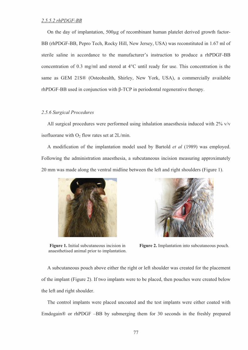
2.5.6 Surgical Procedures
All surgical procedures were performed using inhalation anaesthesia induced with 2% v/v
isofluorane with O2 flow rates set at 2L/min.
A modification of the implantation model used by Bartold et al (1989) was employed.
Following the administration anaesthesia, a subcutaneous incision measuring approximately
20 mm was made along the ventral midline between the left and right shoulders (Figure 1).
Figure 1. Initial subcutaneous incision in anaesthetised animal prior to implantation.
Figure 2. Implantation into subcutaneous pouch.
A subcutaneous pouch above either the right or left shoulder was created for the placement
of the implant (Figure 2). If two implants were to be placed, then pouches were created below
the left and right shoulder.
The control implants were placed uncoated and the test implants were either coated with
Emdogain® or rhPDGF –BB by submerging them for 30 seconds in the freshly prepared

78
growth factor contained within an Ependorf tube (Figure 3) before immediate placement into
the subcutaneous pouches.
Figure 3. Coating of test implant with growth factor.
Figure 4. Implant in euthanized animal prior to retrieval.
After the implants were secured in their positions, the incision was closed using staples and
swabbed with Betadine. Post-operatively the rats were administered 22.7 mg/ml enrofloxacin
(Baytril®, Bayer AG, Leverkusen, Germany) orally for 1 week. The staples were removed 2
weeks after implant placement and the rats were monitored daily and weighed weekly during
the healing period.
2.5.7 Implant Retrieval
The rats were euthanized by CO2 asphyxiation and the implants were located through
implantation records and palpation. For implant retrieval, a similar but larger ventral incision
was made and the implant retrieved with a sample of surrounding tissue (Figure 4). The
retrieved samples were placed in a fixative (10% PBS buffered formalin) for 48 hours prior to
processing into resin blocks.
2.5.8 Resin Embedding
The retrieved implant/tissue biopsies were transferred from the fixative and dehydrated in
serial steps of alcohol concentrations and subsequently embedded in a methyl-methacrylate
resin (Merck Schuchardt OHG, Hohenbrunn, Germany) (Appendix 1 and 2). The resulting
resin embedded implant/tissue blocks were cut using an Isomet slow-speed diamond saw

79
(Beuhler, Illinois, USA) along the long axis of the implant and maximising the volume of
surrounding tissue to obtain two central sections. The distal portions of the implant/tissue
blocks were cut along the same axis to create a resin block with parallel surfaces (Figure 5).
Figure 5. Sectioned implant/tissue resin embedded blocks.
2.5.9 Confocal Laser Scanning Microscopy Analysis
Confocal laser scanning microscopy of the sectioned implant/tissue resin embedded blocks
was carried out using a Leica TCS SP5 Confocal Microscope System (Leica Microsystems,
Heidelberg, Germany) (Clarke 2007). The implant/tissue block sections were viewed using a
20x IMM objective lens (magnification of x200) on the Leica DMI6000B inverted
microscope (Leica Microsystems, Heidelberg, Germany), using the argon-neon laser set at a
power setting of 40% and emitting at a wavelength of 458 nm, allowing confocal laser
scanning microscopic analyses of collagen autofluorescence. The filter cube on the
microscope was set for blue fluorescent light for excitation of the green fluorophores. The
images were captured using the LAS AF software (Leica Microsystems, Heidelberg,
Germany). Scanning speed was set at 400 Hz with a pixel format of 1024 x 1024. The default
pinhole size of 1 Airy and line average of 4x were used.
2.5.10 Thin Sections
Six implant/tissue resin embedded block sections representative of each coated and control
group were selected for thin sectioning. These were further sectioned using an Isomet slow-
speed diamond saw (Beuhler, Illinois, USA) to 100μm sections and mounted with an adhesive

80
on a clear perspex slide. The sections were polished with progressively finer silicon carbide
abrasive discs mounted on a Abramin micro-grinding system (Struers, Denmark) with the
final polish using diamond paste to achieve a specimen thickness of 14-18μm measured by
micrometer (Moore & Wright, Sheffield, England). The sections were re-imaged on confocal
microscopy and then stained with haematoxylin and eosin (H&E).
2.5.11 Histomorphometric Measurements
The thin sections were used for measurement of the thickness of fibrous encapsulation
taken at the apex of the implant threads (Figure 6) and the depth of connective tissue
penetration into the implant grooves (Figure 7). The depth of penetration was measured
perpendicularly from an imaginary line connecting the apices of two adjacent threads to the
maximum depth of tissue penetration into the implant groove.
Figure 6. Measurement of thickness of fibrous encapsulation.
Figure 7. Measurement of depth of connective tissue penetration.
The measurements were carried out at a magnification of x200 in an Olympus BH-2
brightfield microscope (Olympus Optical Co. Ltd., Tokyo, Japan) equipped with an image
system Altra 20 colour camera (Olympus Soft Imaging Solutions, Munster, Germany) and
ANALYSIS imaging software package.

81
2.5.12 Statistical Analysis
Mean values for each variable were calculated for each implant unit. The differences
within the 4 week and 8 week groups of control and coated implants were analysed using a
one-way ANOVA and Bonferroni’s Multiple Comparison Test was used as a post test. The
null hypothesis was rejected at P<0.05. The differences between the control implants at 4
weeks and 8 weeks and the coated implants at 4 weeks and 8 weeks were analysed using
Student’s t-tests. Once again the null hypothesis was rejected at P<0.05. Statistical analysis
was carried out using a Graph Pad Prism 5 for windows statistical software package (Graph
Pad Software Inc, La Jolla, California, USA).
2.6 RESULTS
Healing following implant placement was uneventful for all animals involved in the study.
The incision wounds appeared to have healed by 4 weeks. Although 18 implants were placed
in the rats (including the pilot study), 16 were retrieved. Two implants were not recovered
from two animals at the 8 week time point. One of these lost implants had been coated with
Emdogain® and the other with rhPDGF-BB. This reduced the number for analysis to 5 for
each coated groups at the 8 week time point.
2.6.1 Histological Assessment – Qualitative Analysis at Four weeks
Fibrous encapsulation of the control and growth factors coated implants was evident after 4
weeks. The fibroblast layer adherent to the implants and the surrounding connective tissues
appeared well-organised with little indication of any residual inflammation. The images seen
under confocal microscopy were well correlated with the images seen for the H&E stained
thin sections viewed under light microscopy (Figures 8 - 17).
For the control uncoated implant viewed under confocal microscopy (Figure 8a), collagen
autofluoresecence indicated that the collagen fibres were aligned parallel with the long axis of
the implant, with a high concentration of collagen. The same thin sections, when stained with

82
H&E, showed a dense distinct layer of fibroblasts over the implant threads and suspended
over the implant grooves, surrounded by less dense connective tissue (Figure 8b). The
fibroblasts also appeared to be aligned parallel to the long axis of the implant. A thin (1-3
cells thick) but distinct cellular layer, in intimate contact with the surface of the implant
grooves, that autofluoresced for collagen was evident (Figure 8a).
The collagen fibre orientation and fibroblast alignment observed in the coated implants at 4
weeks was no different to that reported for the uncoated implants when viewed under
confocal and light microscopy. However, from this qualitative histological analysis, there
appeared to be a greater depth of connective tissue penetration into the implant grooves with
the Emdogain® and PDGF coated implants and a thicker dense cellular/fibroblast layer with
the Emdogain® coated implants (Figures 9a, b & c, Figures 10a, b & c).
The presence of an adipose-like cell layer almost devoid of other cell types surrounding the
dense fibroblast layer was a distinctive feature of the rh-PDGF-BB coated implant at 4 weeks
(Figures 10a, b & c).
Figure 8a. Control (uncoated) implant at 4 weeks of healing. Thin section (16μm/unstained),
confocal microscopy, thread 4 (LHS), original magnification x200.
Figure 8b. Control (uncoated) implant at 4 weeks of healing. Thin sections (16μm/H&E stained), light microscopy, threads 3-5 (LHS), original
magnification x 200 (composite image).

83
Figure 9a. Emdogain® coated implant at 4 weeks of healing. Thin section (16μm/unstained),
confocal microscopy, thread 5 (LHS), original magnification x200.
Figure 9b. Emdogain® coated implant at 4 weeks of healing. Thin sections (16μm/H&E stained), light microscopy, threads 4-6 (LHS),
original magnification x 200 (composite image).
Figure 9c. Emdogain® coated implant at 4 weeks of healing. Thin section (16μm/H&E stained), light microscopy, groove 4 (LHS), original magnification x 200 (Close-up image).
84
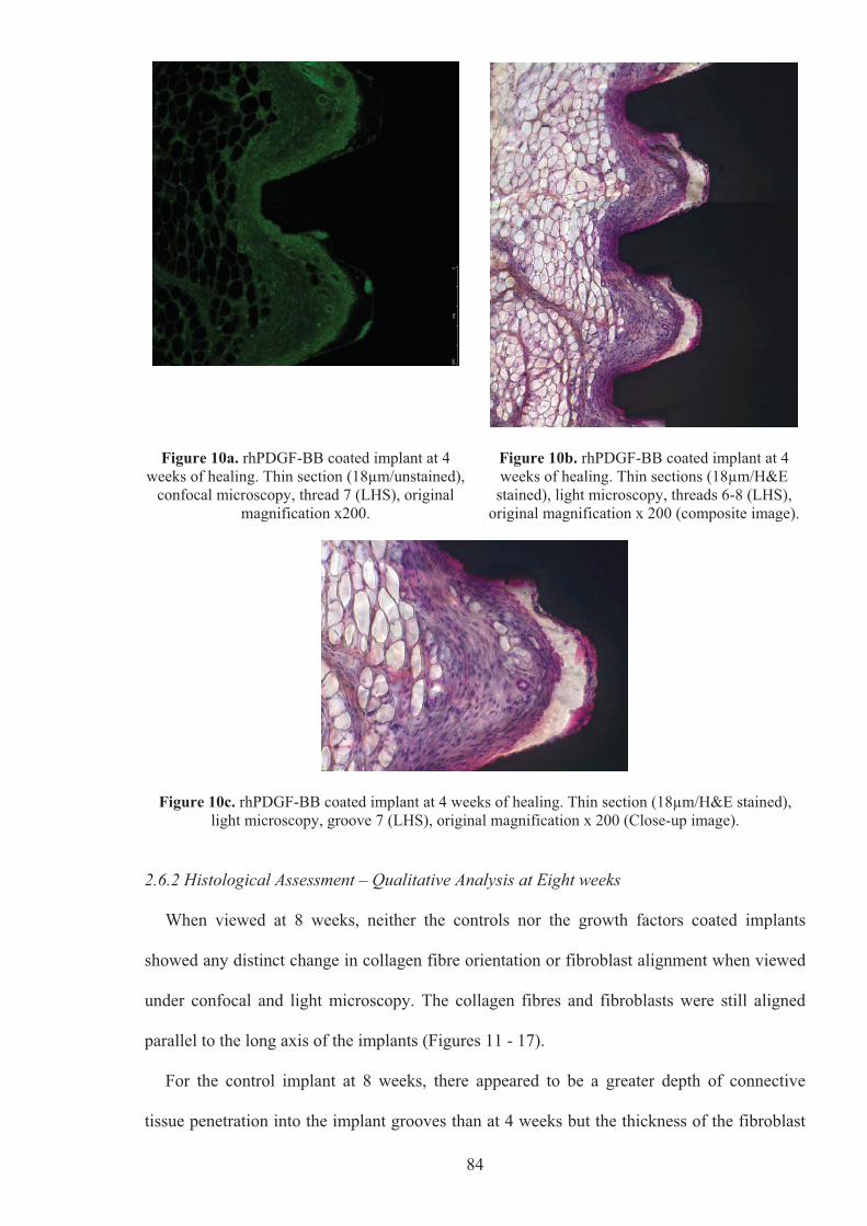
Figure 10a. rhPDGF-BB coated implant at 4 weeks of healing. Thin section (18μm/unstained),
confocal microscopy, thread 7 (LHS), original magnification x200.
Figure 10b. rhPDGF-BB coated implant at 4 weeks of healing. Thin sections (18μm/H&E stained), light microscopy, threads 6-8 (LHS),
original magnification x 200 (composite image).
Figure 10c. rhPDGF-BB coated implant at 4 weeks of healing. Thin section (18μm/H&E stained), light microscopy, groove 7 (LHS), original magnification x 200 (Close-up image).
2.6.2 Histological Assessment – Qualitative Analysis at Eight weeks
When viewed at 8 weeks, neither the controls nor the growth factors coated implants
showed any distinct change in collagen fibre orientation or fibroblast alignment when viewed
under confocal and light microscopy. The collagen fibres and fibroblasts were still aligned
parallel to the long axis of the implants (Figures 11 - 17).
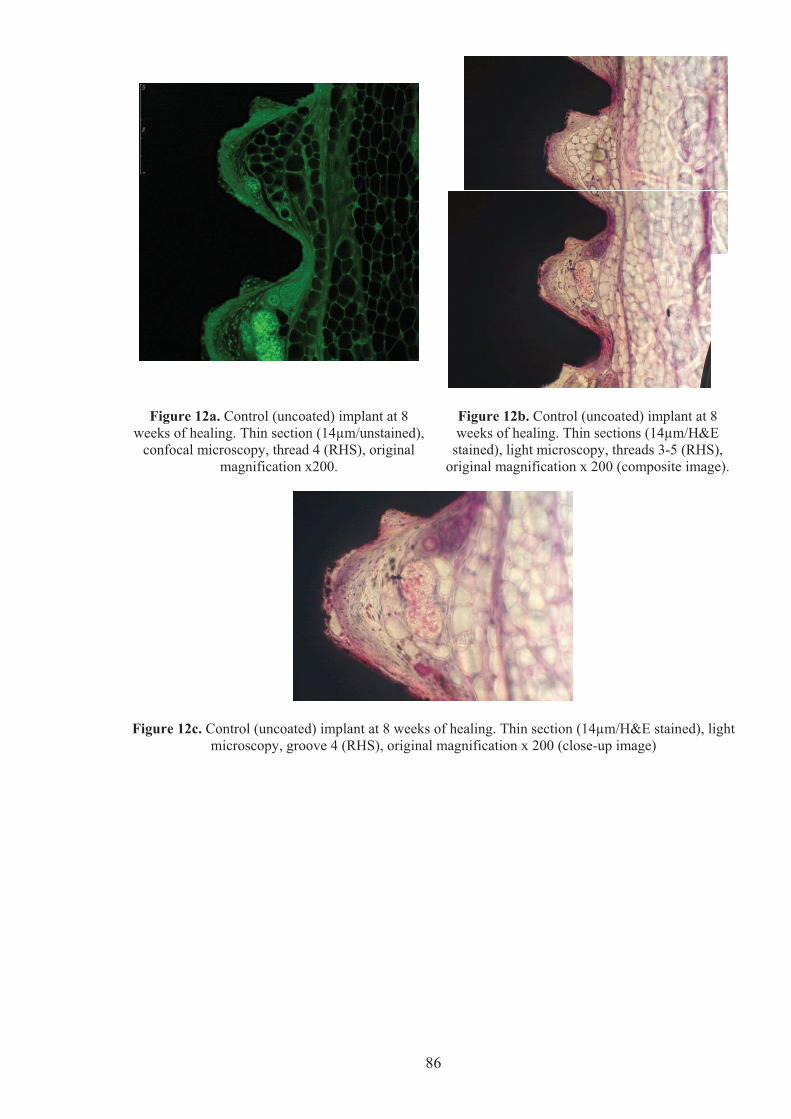
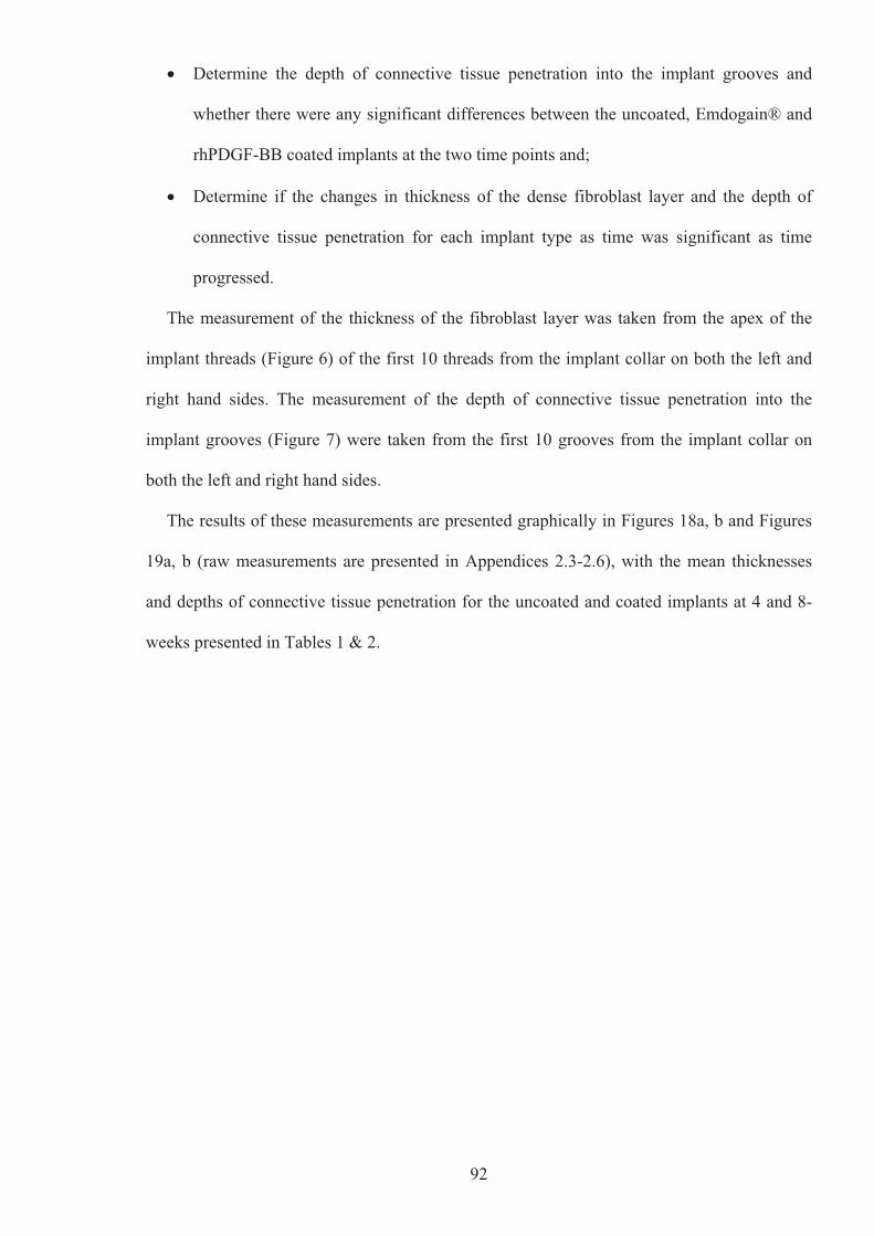
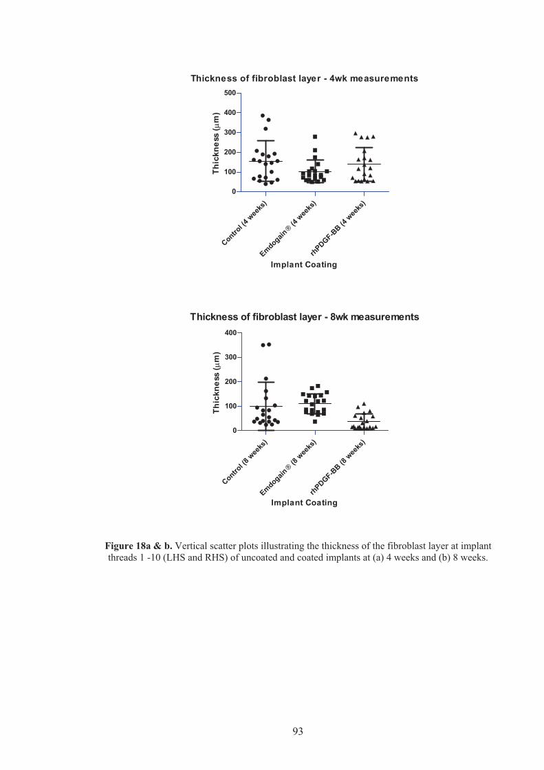
For the control implant at 8 weeks, there appeared to be a greater depth of connective
tissue penetration into the implant grooves than at 4 weeks but the thickness of the fibroblast

85
layer was inconsistent around the implant, with some areas having a thick dense fibroblast
layer (Figures 11a & b) but in other areas, the dense fibroblast layer remained thin (Figures
12a, b & c).
The Emdogain® coated implant at 8 weeks also showed good depth of connective tissue
penetration into the implant grooves, with a good, consistent thickness of the dense fibroblast
layer over the threads and in the grooves of the implant (Figures 13 - 15). This fibroblast layer
appeared more organised and closely-packed together than the Emdogain® coated implant at
4 weeks (Compare Figures 9 to 13 - 15).
The rhPDGF-BB coated implant at 8 weeks also showed good depth of connective tissue
penetration into the implant grooves. However, with the rhPDGF-BB coated implant, the
dense fibroblast layer was consistently thin all around the implant, especially at the implant
threads, surrounded by a pronounced adipose-like tissue response (Figures 16 & 17). The
thickness of the dense fibroblast layer in the rhPDGF-BB coated implants appeared to have
decreased from 4 to 8 weeks (Compare Figures 10 to 16 & 17).
Figure 11a. Control (uncoated) implant at 8 weeks of healing. Thin section (14μm/unstained),
confocal microscopy, thread 8 (LHS), original magnification x200.
Figure 11b. Control (uncoated) implant at 8 weeks of healing. Thin sections (14μm/H&E stained), light microscopy, threads 7-9 (LHS),
original magnification x 200 (composite image).
86
Figure 12a. Control (uncoated) implant at 8 weeks of healing. Thin section (14μm/unstained),
confocal microscopy, thread 4 (RHS), original magnification x200.
Figure 12b. Control (uncoated) implant at 8 weeks of healing. Thin sections (14μm/H&E
stained), light microscopy, threads 3-5 (RHS), original magnification x 200 (composite image).
Figure 12c. Control (uncoated) implant at 8 weeks of healing. Thin section (14μm/H&E stained), light microscopy, groove 4 (RHS), original magnification x 200 (close-up image)

87
Figure 13a. Emdogain® coated implant at 8 weeks of healing. Thin section (14μm/unstained),
confocal microscopy, thread 11 (LHS), original magnification x200.
Figure 13b. Emdogain® coated implant at 8 weeks of healing. Thin sections (14μm/H&E
stained), light microscopy, threads 10-12 (LHS), original magnification x 200 (composite image).
Figure 13c. Emdogain® coated implant at 8 weeks of healing. Thin sections (14μm/H&E stained), light microscopy, groove 10 (LHS), original magnification x 200 (close-up image).

88
Figure 14a. Emdogain® coated implant at 8 weeks of healing. Thin section (14μm/unstained),
confocal microscopy, thread 7 (RHS), original magnification x200.
Figure 14b. Emdogain® coated implant at 8 weeks of healing. Thin sections (14μm/H&E
stained), light microscopy, threads 6-8 (RHS), original magnification x 200 (composite image).

89
Figure 15a. Emdogain® coated implant at 8 weeks of healing. Thin section (14μm/unstained),
confocal microscopy, thread 9 (RHS), original magnification x200.
Figure 15b. Emdogain® coated implant at 8 weeks of healing. Thin sections (14μm/H&E
stained), light microscopy, threads 8-10 (RHS), original magnification x 200 (composite image).
Figure 15c. Emdogain® coated implant at 8 weeks of healing. Thin section (14μm/H&E stained), light microscopy, groove 9 (RHS), original magnification x 200 (close-up image).

90
Figure 16a. rhPDGF-BB coated implant at 8 weeks of healing. Thin section (14μm/unstained),
confocal microscopy, thread 4 (LHS), original magnification x200.
Figure 16b. rhPDGF-BB coated implant at 8 weeks of healing. Thin sections (14μm/H&E stained), light microscopy, threads 3-5 (LHS),
original magnification x 200 (composite image).
Figure 16c. rhPDGF-BB coated implant at 8 weeks of healing. Thin section (14μm/H&E stained), light microscopy, groove 4 (LHS), original magnification x 200 (close-up image).

91
Figure 17a. rhPDGF-BB coated implant at 8 weeks of healing. Thin section (14μm/unstained),
confocal microscopy, thread 2 (LHS), original magnification x200.
Figure 17b. rhPDGF-BB coated implant at 8 weeks of healing. Thin sections (14μm/H&E stained), light microscopy, threads 1-3 (LHS),
original magnification x 200 (composite image).
Figure 17c. rhPDGF-BB coated implant at 8 weeks of healing. Thin section (14μm/H&E stained), light microscopy, groove 2 (LHS), original magnification x 200 (close-up image).
2.6.3 Histomorphometric Measurements
The aims of the histomorphometric measurements were to:
� Determine whether there were any significant differences between the uncoated,
Emdogain® and rhPDGF-BB coated implants in terms of the thickness of the dense
fibroblast layer at the two time points;
92
� Determine the depth of connective tissue penetration into the implant grooves and
whether there were any significant differences between the uncoated, Emdogain® and
rhPDGF-BB coated implants at the two time points and;
� Determine if the changes in thickness of the dense fibroblast layer and the depth of
connective tissue penetration for each implant type as time was significant as time
progressed.
The measurement of the thickness of the fibroblast layer was taken from the apex of the
implant threads (Figure 6) of the first 10 threads from the implant collar on both the left and
right hand sides. The measurement of the depth of connective tissue penetration into the
implant grooves (Figure 7) were taken from the first 10 grooves from the implant collar on
both the left and right hand sides.
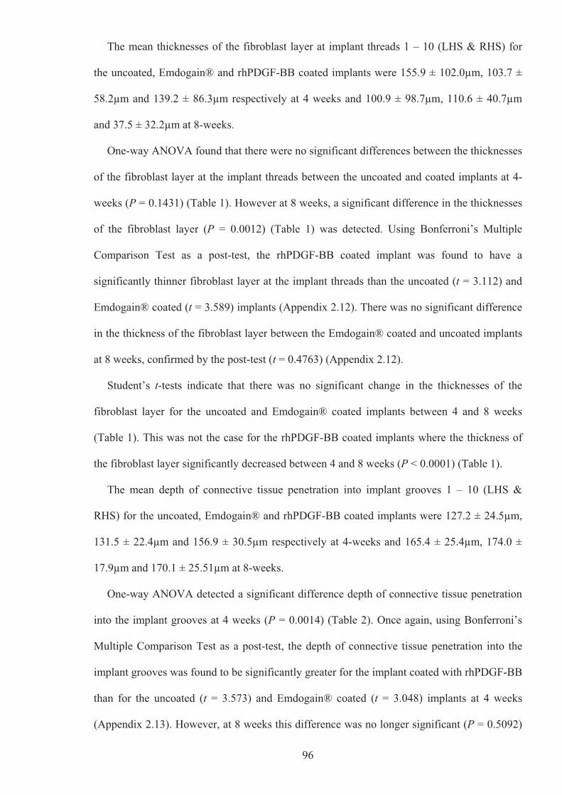
The results of these measurements are presented graphically in Figures 18a, b and Figures
19a, b (raw measurements are presented in Appendices 2.3-2.6), with the mean thicknesses
and depths of connective tissue penetration for the uncoated and coated implants at 4 and 8-
weeks presented in Tables 1 & 2.
93
Thickness of fibroblast layer - 4wk measurements
Control (4
wee
ks)
(4 w
eeks
)
�
Emdogain
rhPDGF-B
B (4 w
eeks
)0
100
200
300
400
500
Implant Coating
Thic
knes
s ( �
m)
Thickness of fibroblast layer - 8wk measurements
Control (8
wee
ks)
(8 w
eeks
)
�
Emdogain
rhPDGF-B
B (8 w
eeks
)0
100
200
300
400
Implant Coating
Thic
knes
s ( �
m)
Figure 18a & b. Vertical scatter plots illustrating the thickness of the fibroblast layer at implant threads 1 -10 (LHS and RHS) of uncoated and coated implants at (a) 4 weeks and (b) 8 weeks.

94
Depth of connective tissue penetration - 4 week measurements
Control (u
ncoate
d) �
Emdogain
rhPDGF-B
B
0
50
100
150
200
250
Implant Coating
Dep
th ( �
m)
Depth of connective tissue penetration - 8 week measurements
Control (u
ncoate
d) �
Emdogain
rhPDGF-B
B 0
50
100
150
200
250
Implant Coating
Dep
th ( �
m)
Figure 19a & b. Vertical scatter plots illustrating the depth of connective tissue penetration at implant grooves 1-10 (LHS and RHS) of the coated and uncoated implants at (a) 4 weeks and (b) 8 weeks.

95
Control (uncoated)
Emdogain� rhPDGF-BB
Number of values 20 20 20 4 Weeks Minimum 39.86 50.17 52.82 Maximum 386.0 278.4 296.3 Mean 155.9 103.7 139.2 Std. Deviation One-way ANOVA P value 0.1431 (ns)
102.2 58.21 86.33
Number of values 20 20 20 8 Weeks Minimum 24.25 36.88 8.310 Maximum 352.5 183.1 110.6 Mean 100.9 110.6 37.54 Std. Deviation One-way ANOVA P value 0.0012 (s)
98.72 40.74 32.24
t-test (4 wks. Versus 8 wks.) P value 0.0912 (ns) 0.06638 (ns) <0.0001 (s)
Table 1. Thickness of fibroblast layer at the implant thread – 4 and 8 week measurements (μm).
Control (uncoated)
Emdogain� rhPDGF-BB
Number of values 20 20 19 4 Weeks Minimum 80.40 95.35 112.6 Maximum 182.4 170.4 194.7 Mean 127.2 131.5 156.9 Std. Deviation One-way ANOVA P value 0.0014 (s)
24.52 22.46 30.53
Number of values 20 20 20 8 Weeks Minimum 131.2 118.3 120.6 Maximum 214.6 198.3 205.0 Mean 165.4 174.0 170.1 Std. Deviation One-way ANOVA P value 0.5092 (ns)
25.44 17.89 25.51
t-test (4 wks. versus 8 wks.) P value
<0.0001 (s) <0.0001 (s) 0.1515 (ns)
Table 2. Depth of connective tissue penetration into the implant grooves – 4 and 8 week
measurements (μm).
96
The mean thicknesses of the fibroblast layer at implant threads 1 – 10 (LHS & RHS) for
the uncoated, Emdogain® and rhPDGF-BB coated implants were 155.9 ± 102.0μm, 103.7 ±
58.2μm and 139.2 ± 86.3μm respectively at 4 weeks and 100.9 ± 98.7μm, 110.6 ± 40.7μm
and 37.5 ± 32.2μm at 8-weeks.
One-way ANOVA found that there were no significant differences between the thicknesses
of the fibroblast layer at the implant threads between the uncoated and coated implants at 4-
weeks (P = 0.1431) (Table 1). However at 8 weeks, a significant difference in the thicknesses
of the fibroblast layer (P = 0.0012) (Table 1) was detected. Using Bonferroni’s Multiple
Comparison Test as a post-test, the rhPDGF-BB coated implant was found to have a
significantly thinner fibroblast layer at the implant threads than the uncoated (t = 3.112) and
Emdogain® coated (t = 3.589) implants (Appendix 2.12). There was no significant difference
in the thickness of the fibroblast layer between the Emdogain® coated and uncoated implants
at 8 weeks, confirmed by the post-test (t = 0.4763) (Appendix 2.12).
Student’s t-tests indicate that there was no significant change in the thicknesses of the
fibroblast layer for the uncoated and Emdogain® coated implants between 4 and 8 weeks
(Table 1). This was not the case for the rhPDGF-BB coated implants where the thickness of
the fibroblast layer significantly decreased between 4 and 8 weeks (P < 0.0001) (Table 1).
The mean depth of connective tissue penetration into implant grooves 1 – 10 (LHS &
RHS) for the uncoated, Emdogain® and rhPDGF-BB coated implants were 127.2 ± 24.5μm,
131.5 ± 22.4μm and 156.9 ± 30.5μm respectively at 4-weeks and 165.4 ± 25.4μm, 174.0 ±
17.9μm and 170.1 ± 25.51μm at 8-weeks.
One-way ANOVA detected a significant difference depth of connective tissue penetration
into the implant grooves at 4 weeks (P = 0.0014) (Table 2). Once again, using Bonferroni’s
Multiple Comparison Test as a post-test, the depth of connective tissue penetration into the
implant grooves was found to be significantly greater for the implant coated with rhPDGF-BB
than for the uncoated (t = 3.573) and Emdogain® coated (t = 3.048) implants at 4 weeks
(Appendix 2.13). However, at 8 weeks this difference was no longer significant (P = 0.5092)

97
(Table 2). Coating the implant with Emdogain® was not found to significantly alter the depth
of connective tissue penetration into the implant grooves over an uncoated implant, confirmed
by the post-test (t = 0.5319) (Appendix 2.14).
Student’s t-tests indicate that the depth of connective tissue penetration into the implant
grooves increased for the uncoated (P < 0.0001) and Emdogain® coated (P < 0.001) implants
between 4- and 8-weeks but remained the same for the rhPDGF-BB coated implants (Table
2).
2.7 DISCUSSION
The adherence of the peri-implant tissues to the implant/abutment surface is crucial to its
function as a barrier between the oral environment and the bone and implant surfaces.
Enhancing this adherence by surface modification with biological agents can serve to improve
implant survival, as well as potentially contribute to an improvement in implant success rates
by preventing recession and improving aesthetic outcomes.
The aim of this study was to investigate the connective tissue attachment to the roughened
surface of (TiUnite) titanium implants and roughened surfaces modified with rhPDGF-BB or
EMD. Although there are distinct differences between gingival and subcutaneous connective
tissues in terms of remodelling, turnover rates and architecture, both of these connective tissue
types contain principally type I and III collagen as the most abundant biochemical component.
As this study investigates connective tissue attachment by examining the collagen fibre
orientation to the implant surface, the use of a subcutaneous murine model, whilst not
mimicking the conditions in the oral cavity as accurately as would a buccal dehiscence model
in a larger animal, does provide an appropriate cost-effective means to test the hypothesis of
this study.
To our knowledge, this is the first study to utilise confocal laser scanning microscopy
(CLSM) and collagen autofluorescence to image connective tissue attachment to titanium
implants. The basic rationale behind CLSM is that illumination of tissues with a short

98
wavelength light from a monochromatic punctiform laser source leads to excitation of
endogenous substances, resulting in the emission of fluorescence light of longer wavelengths.
The resulting emission energy is detected by a spatially filtered optical system, the pinhole,
which filters out light signals from out-of focus planes (Lucchese et al 2008). Amongst the
molecules, called fluorophores, responsible for this tissue autofluorescnce include collagen
(DaCosta et al 2002, 2003). Recent studies investigating the collagen fibre orientation in the
peri-implant mucosa have used a variety of methods including: decalcified ground sections
stained with toluidine blue (Abrahamsson & Cardaropoli 2007, Berglundh et al 2007,
Welander et al 2007, 2008; Nevins et al 2008), decalcified ground sections stained with
methylene blue/Azure II (Schüpbach et al 2007), “fracture technique” sections stained in PAS
and toluidine blue (Welander et al 2007), scanning electron microscopy (Schüpbach et al
2007, Welander et al 2007, Nevins et al 2008, Tete et al 2009), transmission electron
microscopy (Schüpbach et al 2007) and circular polarised light microscopy (Allegrini Jr et al
2008, Tete et al 2009). Collagen fibre orientation in bone around osseointegrated implants
have also been investigated using circular polarised light microscopy with tissue incorporated
fluorescent dyes in human peri-implant bone (Traini et al 2005) and peri-implant bone from
minipigs (Neugebauer et al 2006). These methods, whilst effective and allowing for high
quality imaging of collagen, require complex and time-consuming sample preparation
techniques.
Recently, Lucchese et al (2008) analysed collagen fibre distribution in human crown
dentine using CLSM and found an intense autofluorescence that was ascribed to collagen
fibres in all their samples. In our study, we were able to correlate the collagen
autofluorescence seen in the CLSM images to the fibroblasts observed in the same thin
sections when stained with H&E and viewed under light microscopy. The use of CLSM thus
appears to provide a less time-consuming method of preparing tissue samples and therefore is
a useful auxiliary tool for investigating the presence, distribution and collagen fibre
orientation in the peri-implant soft tissues.

99
In this study, two of the coated implants, one coated with Emdogain® and the other coated
with rhPDGF-BB, were not recovered from two animals at the 8 week time point. The reasons
for this exfoliation are unknown. We suspect that this exfoliation would have occurred early
on in the experiment as healing following implant placement was uneventful for all animals
involved in this study. A distinct encapsulation by a layer of fibroblasts occurred around all
the retrieved implants, regardless of whether the implants were coated or uncoated. This is
similar to what occurs with an osseointegrated implant intraorally, whereby the connective
tissue forms a non-vascularised, circular, scar-like structure around the transmucosal portion
of the implant. The qualitative analysis and histomorphometric measurements of the uncoated
implants indicate that resolution of inflammation and connective tissue formation appeared to
be completed by 4 weeks. However the healing process, which included tissue maturation and
organisation continued between the 4 and 8 week period, as evidenced by the significant
change in depth of connective tissue penetration into the implant grooves. Our observations in
a murine model appear to be consistent with the conclusions made recently by Berglundh et al
(2007). In their investigation of the morphogenesis of the peri-implant mucosa in a canine
model, they concluded that the peri-implant mucosa exhibited minor signs of inflammation
during the first 2 weeks of healing but from 4 weeks, the mucosa was stable and well attached
to the bone. Berglundh et al (2007) further concluded that the soft tissue barrier adjacent to
titanium implants placed in a non-submerged protocol takes about 6 to 8 weeks to establish a
soft tissue barrier with proper dimensions and tissue organisation.
A number of previous in vitro studies have investigated the effect of surface modification
by the coating of titanium with biological agents on epithelial and connective tissue
attachment to titanium surfaces. Dean et al (1995) observed that coating machined, plasma-
sprayed and hydroxyapatite titanium surfaces in vitro with fibronectin and laminin-1, a
component of epithelial cell membranes, enhanced gingival fibroblast and epithelial cell
attachment respectively by about threefold. Tamura et al (1997) observed, also in vitro, that
coating titanium alloy with laminin-5 enhanced gingival epithelial cell attachment and

100
hemidesmosome assembly. Park et al (1998) observed that type IV collagen provided an
excellent substrate for epithelial cell attachment to titanium surfaces and later in vitro studies
have shown that cell adhesion to titanium discs coated with collagen was enhanced compared
with uncoated titanium (Roessler et al 2001, Nagai et al 2002). However, in a recent study
investigating soft tissue healing around implants in a canine model, Welander et al (2007)
found that the vertical dimensions of epithelial and connective tissue components, as well as
the composition of the connective tissue zone directly adjacent to the implant were similar at
collagen-coated and non-coated implants after 4 and 8 weeks of healing.
This study is the first study to investigate the effect of surface modification with the
growth factor PDGF on connective tissue attachment to titanium implants. Recently, a pilot
study conducted on a minipig reported on the effects of autogenous periodontal cell grafts
(periodontal ligament and gingival connective tissue cultures), with and without the
application of EMD, on the implant-connective tissue interface (Craig et al 2006). This pilot
study was based on the hypothesis that a periodontal connective tissue attachment could be
formed on dental implants provided a source of periodontal regeneration competent cells was
present in the wound healing environment and that the application of EMD might aid in the
formation of this attachment. However, in this pilot study, with and without the application of
EMD, an implant-connective tissue interface morphologically consistent with a periodontal
connective tissue attachment was not observed in sections from any of the implant or
autogenous cell grafts (Craig et al 2006).
In this study, surface modification of the TiUnite surface of titanium implants with
Emdogain® or rhPDGF coating was not found to change the orientation of the fibroblasts or
collagen fibres in the encapsulating fibroblast layer. The orientation of the fibroblasts and
collagen fibres when viewed under light microscopy and confocal laser scanning microscopy
respectively appeared parallel to the long axis of the implant. Although reorientation of the
fibroblasts and collagen fibres did not occur, there was good adaptation of the fibroblast layer
onto the TiUnite surface and implant grooves for both the uncoated and growth factor coated
101
implants at the end of the study period. This could indicate a degree of soft tissue integration
onto the TiUnite surface that is more adherent than previously thought.
The mechanical attachment at the titanium/connective tissue interface for roughened
surface implants has not been extensively investigated. Abrahamsson et al (2002) compared
the composition of soft tissue barriers onto implant abutments with a machined surface with
abutments with a dual, thermal acid-etched surface using a canine model over a 6-month
period. It was found that the roughness of the titanium surface did not influence the soft-tissue
attachment that formed on commercially pure titanium in terms of the dimensions of the
epithelial-connective tissue barrier and the composition of the connective tissue attachment.
However, two recent in vivo studies provide evidence that microtexturing of the implant
surface can influence the soft tissue response (Glauser et al 2005, Schüpbach & Glauser
2007). The influence of surface modifications on interactions between the implant surface on
both the junctional epithelium and connective tissue was evaluated in a human study using
one-piece experimental mini-implants (Nobel Biocare AB, Gothenburg, Sweden) with either a
machined surface, acid-etched surface, or a surface with an oxidised and microporous TiO2
layer, essentially a TiUnite surface. A shorter epithelial attachment and a longer connective
tissue seal was observed with the acid-etched and oxidised implants compared to the
machined surface implants (Glauser et al 2005). Furthermore, it was found that with the
machined and acid-etched mini-implants, the adherence of the junctional epithelium to the
implant surface was characterised by a basal lamina and numerous hemidesmosomes but the
interface between the connective tissue and the implant surface was smooth, with collagen
fibres running a course more or less parallel to the implant surface, indicating poor
mechanical resistance. However, with the microtopographically complex oxidised TiUnite
implant surface, the junctional epithelium exhibited attachment by hemidesmosomes together
with mechanical interdigitation of the innermost cell layer with the open pores of the implant
surface, with the connective tissue showing functionally oriented collagen fibrils towards the
implant surface under polarised light microscopy, indicating a less vulnerable seal

102
(Schüpbach & Glauser 2007). Nevins et al (2008), employing a single-stage protocol using
implants with ‘Laser-Lok’ microchannels at the collar (Biohorizons Implant Systems,
Birmingham AL, USA), observed under light microscopy that the junctional epithelial cells
were in close contact with the implant surface and that the microgrooved area of the implants
were covered with connective tissue. Polarized light and scanning electron microscopy of the
microgrooved area showed functionally oriented collagen fibres running toward and attaching
to the grooves of the implant surface (Nevins et al 2008).
In our study, close adaptation of the fibroblast layer onto the TiUnite implant surface was
seen at the implant threads histologically, and a good depth of connective tissue penetration
into the implant grooves was observed for both uncoated and growth factor coated implants at
the end of the study period. Coating the implant with rhPDGF-BB significantly increased the
depth of connective tissue penetration into the implant grooves at 4 weeks over that in the
Emdogain® coated and uncoated implants. Whilst the depth of connective tissue penetration
for the rhPDGF-BB coated implants did not change after 4 weeks, it increased significantly
for the Emdogain® coated and uncoated implants from 4 to 8 weeks. Thus, at 8 weeks all the
uncoated and growth factor implants exhibited similar depths of connective tissue penetrance.
Therefore, coating the TiUnite implant surface with rhPDGF-BB seems to increase the speed
of soft tissue healing, but ultimately the same degree of soft tissue integration occurs around
the TiUnite implant surface regardless of whether it has been coated or not.
Nevins et al (2005) in a large multi-centre, randomized blinded human clinical trial of 180
participants investigated the effectiveness of PDGF-BB with a porous �-tricalcium phosphate
(TCP) matrix. The subjects had at least one interproximal periodontal defect �4 mm after
debridement and were divided into three treatment groups: Group 1 – �-TCP plus 0.3 mg/ml
rhPDGF-BB (GEM 21S); Group 2 – �-TCP plus 0.1 mg/ml rhPDGF-BB; and Group 3 – �-
TCP and buffer alone. At 3 months post surgically, GEM 21S showed a significantly greater
CAL gain than the �-TCP alone but at 6 months although the CAL gain for GEM 21S
continued to be greater than the �-TCP alone, this was found not to be statistically significant.

103
One of the conclusions that made by Nevins et al (2005) was that growth factors such as
rhPDGF-BB have short half-lives and so after a sharp initial increase in clinical attachment
gain, no significant gains are observed in the long-term. The results from our study seem to
support this statement.
On closer inspection, there appears to be a degree of tissue separation between the
fibroblast layer and a thin cellular layer on the implant surface for a large number of the
implant grooves. This thin cellular layer when present appears to be continuous with areas
where the fibroblast layer is in close contact with the implant surface. Although tissues
processed for embedding in acrylic or epoxy resins are considered less prone to artefacts
(Bosshardt et al 2005), due to the concave morphology of the implant grooves, tissue
shrinkage has probably occurred in a number of the implant grooves in our samples and that
the degree of connective tissue penetration into the implant grooves for the coated and
uncoated implant in this study is probably greater than that observed histologically.
Coating of the implants with rhPDGF-BB did not result in any significant difference in
fibroblast thickness at the implant threads when compared to the Emdogain® coated and
uncoated implants at 4 weeks, but the more intense appearance of the collagen
autofluorescence under confocal microscopy could have indicated a higher concentration of
collagen in the dense fibroblast layer adjacent to the implant surface with the rhPDGF-BB
coated implants at this time point (Figure 10a). However, a significant decrease in the
thickness of the fibroblast layer at the implant threads with the rhPDGF-BB coated implant
was evident at the end of the 8 week study period. A noticeable amount of adipose-like tissue
deposited around the rhPDGF-BB coated implants was also observed, both at the 4 week and
8 week implants. Preadipocytes convert to adipocytes through the process of adipogenesis, a
process that is catalysed biochemically by glycerol-3-phosphate dehydrogenase (GPDH)
activity. GDPH is a specific marker for adipogenesis and catalyses the formation of glycerol-3
phosphate, an important step in the synthesis of triacylglycerines (Wise & Green 1979).
PDGF, a mitogen, is known to stimulate the proliferation of human preadipocytes (Hauner et

104
al 1995) but inhibits their differentiation to adipocytes by inhibiting the activity of GDPH
(Hauner et al 1995, Koellensperger et al 2006). Thus, we propose two possible explanations
to explain this phenomenon observed in our study. The first plausible explanation is that the
increased amount of adipose tissue observed is a coincidental artefact from the implants being
surgically implanted in a rat, or part of a rat that simply had more adipose tissue deposits. The
second explanation is that the rhPDGF-BB initially stimulated the proliferation of
preadipocytes in the immediate vicinity of the implant, but because rhPDGF-BB is a short-
lived growth factor (Nevins et al 2005), its inhibitory effects towards preadipocyte
differentiation were also short-lived and over the experimental period these increased
numbers of preadipocytes differentiated to adipocytes.
The thickness of the fibroblast layer on the implant threads of the Emdogain® coated
implants were not significantly different from the uncoated implants throughout the whole
study period. Qualitative analysis of the confocal and light microscopy images however,
indicate a more consistent thickness and density of the fibroblast layer with the Emdogain®
coated implants compared to the uncoated implants. Although there is no statistical
significance between the two, the much smaller standard deviation (s.d.) values at 4 and 8
weeks seen with the Emdogain® coated implants as compared to the uncoated implants for
fibroblast layer thickness help reinforce the observations of the qualitative analysis. One of
the characteristic difference between EMD and rhPDGF-BB is that whilst the half-life of
rhPDGF-BB is short, EMD acts over a long-period of time and further clinical gains have
been observed in the short (3 years) and longer term (5 years) post- operatively (Heijl et al
1997, Heden & Wennstromm 2006). The short duration of this study may have therefore
precluded any significant differences being observed between the Emdogain® coated and
uncoated implants.
In conclusion, this study shows that good soft tissue integration can be achieved on a
moderately roughened TiUnite surface. Surface modification of the TiUnite surface by
coating with rhPDGF-BB could increase the speed of soft tissue healing around the implant

105
surface. However, the increased speed of healing with rhPDGF-BB coating could result in a
less robust titanium/connective tissue interface. The positive influence of implant surface
modification with Emdogain® on soft tissue attachment and maturation around the implant
surface should not be discounted and more research into this area is warranted.

106
2.8 REFERENCES
Abrahamsson I, Berglundh T, Glantz PO, Lindhe J. The mucosal attachment at different
abutments. An experimental study in dogs. J Clin Periodontol 1998; 25: 721-727.
Abrahamsson I, Berglundh T, Lindhe, J. The reaction of the peri-implant tissues to
repeated abutment dis- and reconnection. J Clin Periodontol 1997; 24: 568-572.
Abrahamsson I, Berglundh T, Linder E, Lang NP, Lindhe, J. Early bone formation
adjacent to rough and turned endosseous implant surfaces. An experimental study in the dog.
Clin Oral Implants Res 2004; 15: 381-391.
Abrahamsson I, Berglundh T, Moon IS, Lindhe, J. Peri-implant tissues at submerged and
non-submerged titanium implants. J Clin Periodontol 1999; 26: 600-607.
Abrahamsson I, Berglundh T, Wennstrom J, Lindhe, J. The peri-implant hard and soft
tissues at different implant systems. A comparative study in the dog. Clin Oral Implants Res
1996; 7:212-219.
Abrahamsson I, Cardapoli G. Peri-implant hard and soft tissue integration to dental
implants made of titanium and gold. Clin Oral Implants Res 2007; 18: 269-274.
Abrahamsson I, Soldini C. Probe penetration in periodontal and peri-implant tissues: An
experimental study in the beagle dog. Clin Oral Implants Res 2006; 17: 601-605.
Abrahamsson I, Zitzman NU, Berglundh T, Linder E, Wennrberg A, Lindhe J. The
mucosal attachment to titanium implants with different surface characteristics: an
experimental study in dogs. J Clin Periodontol 2002; 29: 448-455.
Allegrini Jr S, Allegrini MRF, Yoshimoto M, Konig Jr B, Mai R, Fanghenel J, Gedrange
T. Soft tissue integration in the neck area of titanium implants – An animal trial. J Physiol
Pharmacol 2008; 59 (suppl 5): 117-132.
Bartold PM, Hay S, Vernon-Roberts B. Effect of cyclosporine-A on connective tissue
deposition in experimental inflammatory lesions. Matrix 1989; 9: 293-300.
Becker W, Lynch SE, Lekholm U, Becker BE, Caffesse R, Donath K, Sanchez R. A
comparison of ePTFE membranes alone or in combination with platelet-derived growth factor

107
and insulin-like growth factor-1 or demineralized freeze-dried bone in promoting bone
formation around immediate extraction socket implants. J Periodontol 1992; 63: 929-940.
Bengazi F, Wennstrom JL, Lekholm U. Recession of the soft tissue margin at oral
implants. A 2-year longitudinal prospective study. Clin Oral Impl Res 1996; 7: 303-310.
Berglundh T, Abrahamsson I, Lang NP, Lindhe, J. De novo alveolar bone formation
adjacent to endosseous implants. Clin Oral Implants Res 2003; 14: 251-262.
Berglundh T, Abrahamsson I, Welander M, Lang NP, Lindhe, J. Morphogenesis of the
peri-implant mucosa: an experimental study in dogs. Clin Oral Implants Res 2007; 18: 1-8.
Berglundh T, Lindhe J. Dimension of the peri-implant mucosa. Biological width revisited.
J Clin Periodontol 1996; 23: 971-973.
Berglundh T, Lindhe J, Ericsson I, Marinello CP, Liljenberg B, Thomsen P. The soft tissue
barrier at implants and teeth. Clin Oral Implants Res 1991; 2: 81-90.
Berglundh T, Lindhe J, Jonsson K, Ericsson I. The topography of the vascular systems in
the periodontal and peri-implant tissues in the dog. J Clin Periodontol 1994; 21: 189-193.
Berglundh T, Lindhe J, Marinello, CP, Ericsson I, Liljenberg B. Soft tissue reaction to de
novo plaque formation on implants and teeth. Clin Oral Implants Res 1992; 3: 1-8.
Bosshardt DD, Sculean A, Windisch P, Pjetursson BE, Lang NP. Effects of enamel matrix
proteins on tissue formation along the roots of human teeth. J Periodont Res 2005; 40: 158-
167.
Boyan LA, Bhargava G, Nishimura F, Orman R, Price R, Terranova VP. Mitogenic and
chemotactic responses of human periodontal ligament cells to the different isoforms of
platelet-derived growth factor. J Dent Res 1994; 73: 1593-1600.
Buser D, Weber HP, Donath K, Fiorellini JP, Paquette, DW, Williams RC. Soft tissue
reactions to non-submerged unloaded titanium implants in beagle dogs. J Periodontol 1992;
63: 226-236.
Brookes SJ, Robinson C, Kirkham J, Bonass WA. Biochemistry and molecular biology of
amelogenin proteins of developing dental enamel. Arch Oral Biol 1995; 40: 1-4.

108
Camelo M, Nevins ML, Schenk RK, Lynch SE, Nevins M. Periodontal regeneration in
human class II furcations using purified recombinant human platelet-derived growth factor-
BB (rhPDGF-BB) with bone allograft. Int J Periodontics Restorative Dent 2003; 23: 213-225.
Cangini F, Cornelini R. A comparison between enamel matrix derivative and a
bioabsorbable membrane to enhance healing around transmucosal immediate post-extraction
implants. J Periodontol 2005; 76: 1785-1792.
Castellanos A, de la Rosa M, de la Garza M, Caffesse RG. Enamel matrix derivative and
coronal flaps to cover marginal tissue recessions. J Periodontol 2006; 77: 7-14.
Clarke J. Leica TCS SP5 Confocal User Notes. Adelaide Microscopy 2007; June: 1-16.
Cochran DL, Hermann JS, Schenk RK, Higginbottom FL, Buser D. Biologic width around
titanium implants. A histometric analysis of the implantogingival junction around unloaded
and loaded non-submerged implants in the canine mandible. J Periodontol 1997; 68: 186-198.
Cochran DL, Simpson J, Weber HP, Buser D. Attachment and growth of periodontal cells
on smooth and rough titanium. Int J Oral Maxillofac Implants 1994; 9: 289-297.
Craig RC, Kamer A, Kallur SP, Inoue M, Tarnow DP. Effects of periodontal cell grafts and
enamel matrix proteins on the implant-connective tissue interface: A pilot study in the
minipig. J Oral Implantol 2006; 32: 228-236.
Da Costa RS, Andersson H, Wilson BC. Molecular fluorescence excitation-emission
matrices relavant to tissue spectroscopy. Photochem Photobiol 2003; 78: 384-392.
DaCosta RS, Wilson BC, Marcon NE. New Optical technologies for earlier endoscopic
diagnosis of premalignant gastrointestinal lesions. J Gastroenterol Hepatol 2002; 17 (Suppl):
S85-S104.
Dean JW, Culbertson KC, D’Angelo AM. Fibronectin and laminin enhance gingival cell
attachment to dental implant surfaces in vitro. Int J Oral Maxillofac Implants 1995; 10: 721-
728.

109
Dennisson DK, Vallone DR, Pinero GJ, Ritman B, Caffesse RG. Differential effects of
TGF-beta 1 and PDGF on proliferation of periodontal ligament cells and gingival fibroblasts.
J Periodontol 1994; 65: 641-648.
Ekfeldt A, Eriksson A, Johansson LA. Peri-implant mucosal level in patients treated with
implant-supported fixed prostheses: a 1-year follow-up study. Int J Prosthodont 2003; 16:
529-532.
Ericsson I, Nilner K, Klinge B, Glantz PO. Radiographical and histological characteristics
of submerged and nonsubmerged titanium implants. An experimental study in the Labrador
dog. Clin Oral Implants Res 1996; 7: 20-26.
Ericsson I, Randow K, Nilner K, Petersson A. Some clinical and radiographical features of
submerged and non-submerged titanium implants. A 5-year follow-up study. Clin Oral
Implants Res 1997; 8: 422-426.
Fong DC, Slaby I, Hammarström L. Amelin: an enamel-related protein transcribed in the
cells of the epithelial root sheath. J Bone Miner Res 1996; 11: 892-898.
Franke Stenport V, Johansson CB. Enamel matrix derivative and titanium implants. An
experimental pilot study in the rabbit. J Clin Periodontol 2003; 30: 359-363.
Giannoblie WV. Periodontal tissue engineering by growth factors. Bone 1996; 19: 23S-
37S.
Giannoblie WV, Finkleman RD, Lynch SE. Comparison of canine and non-human primate
animal models for periodontal regenerative therapy: results following a single administration
of PDGF/IGF-1. J Periodontol 1994; 65: 1158-1168.
Glauser R, Schüpbach P, Gottlow J, Hammerle CHF. Perimplant soft tissue barrier at
experimental one-piece mini-implants with different surface topography in humans: a light-
microscopic overview and histometric analysis. Clin Impl Dent Relat Res 2005; 7; S44-S51.
Grunder U. Stability of the mucosal topography around single-tooth implants and adjacent
teeth: 1-year results. Int J Periodontics Restorative Dent 2000; 20: 11-17.

110
Hägerwald S, Spahr A, Rompola E, Haller B, Heijl L, Bernimoulin JP. Comparative study
of Emdogain® and coronally advanced flap technique in the treatment of human gingival
recessions: a prospective controlled clinical study. J Clin Periodontol 2002; 29: 35-41.
Hammarström L. Enamel matrix, cementum and regeneration. Journal of Clinical
Periodontology 1997; 24: 658-668.
Hauner H, Rohrig K, Petruschke T. Effects of epidermal growth factor (EGF), platelet-
derived growth factor (PDGF) and fibroblast growth factor (FGF) on human adipocyte
development and function. Eur J Clin Invest 1995; 25: 90-96.
Heden G, Wennstromm J. Five-year follow up of regenerative periodontal therapy with
enamel matrix derivative at sites with angular bone defects. J Periodontol 2006; 77: 295-301.
Heijl L, Heden G, Svardstrom G, Ostgren A. Enamel matrix derivative (Emdogain) in
treatment of intrabony periodontal defects. J Clin Periodontol 1997; 24: 705-714.
Howell TH, Fiorellini JP, Paquette DW, Offenbacher S, Giannobile WV, Lynch SE. A
phase I/II clinical trial to evaluate a combination of recombinant human platelet-derived
growth factor –BB and recombinant human insulin-like growth factor-I in patients with
periodontal disease. J Periodontol 1997; 68: 1186-1193.
Koellensperger E, von Hiemburg D, Markowicz M, Pallua N. Human serum from platelet-
poor plasma for the culture of primary human preadipocytes. Stem Cells 2006; 24: 1218-
1225.
Lindskog S. Formation of intermediate cementum I: Early mineralization of aprismatic
enamel and intermediate cementum. J Craniofac Genet Develop Biol 1982; 2: 147-160.
Lindskog S. Formation of intermediate cementum II: A scanning electron microscopic
study of the epithelial root sheath of Hertwig. J Craniofac Genet Develop Biol 1982; 2: 161-
169.
Lindskog S, Hammarström L. Formation of intermediate cementum III: 3H-proline and 3H-
tryptophan uptake into the epithelial root sheath of Hertwig in vitro. J Craniofac Genet
Develop Biol 1982; 2: 171-177.

111
Lucchese A, Pilolli GP, Petruzzi M, Crincoli V, Scivetti M, Favia G. Analysis of collagen
distribution in human crown dentine by confocal laser scanning microscopy. Ultrastruc Pathol
2008; 32: 107-111.
Lynch SE, Buser D, Hernandez RA, Weber HP, Stich H, Fox CH, Williams RC. Effects of
the platelet-derived growth factor/insulin-like growth factor-1 combination on bone
regeneration around titanium dental implants. Results of a pilot-study in beagle dogs. J
Periodontol 1991a; 62: 710-716.
Lynch SE, de Castilla R, Williams RC, Kiritsy CP, Howell TH, Reddy MS, Antoniades
HN. The effects of a short-term appllication of a combination of platelet-derived and insulin-
like growth factors on periodontal wound healing. J Periodontol 1991b; 62: 458-467.
Lynch SE, Williams RC, Polson AM, Howell TH, Reddy MS, Zappa UE, Antoniades HN.
A combination of platelet-derived and insulin-like growth factors enhances periodontal
regeneration. J Clin Periodontol 1989; 16: 545-548.
Lynch SE, Wisner-Lynch L, Nevins M, Nevins ML. (2006) A new era in periodontal and
periimplant regeneration: use of growth factor enhanced matrices incorporating rhPDGF.
Compend Contin Educ Dent 2006; 27: 672-678.
Maksoud MA. Manipulation of the peri-implant tissues for better maintenance: A
periodontal perspective. J Oral Implantol 2003; 29: 120-123.
McGuire MK, Cochran DL. Evaluation of human recession defects treated with coronally
advanced flaps and either enamel matrix derivative or connective tissue. 2. Histological
evaluation. J Periodontol 2003; 74: 1126-1135.
McGuire MK, Kao RT, Nevins M, Lynch SE. rhPDGF-BB promotes healing of
periodontal defects: 24-month clinical and radiographic observations. Int J Periodontics
Restorative Dent 2006; 26: 223-231.
McGuire MK, Nunn M. Evaluation of human recession defects treated with coronally
advanced flaps and either enamel matrix derivative or connective tissue. 1. Comparison of
clinical parameters. J Periodontol 2003; 74: 1110-1125.

112
Meraw SJ, Reeve CM, Lohse CM, Sioussat TM. Treatment of peri-implant defects with
combination growth factor cement. J Periodontol 2000; 71: 8-13.
Moon I-S, Berglundh T, Abrahamsson I, Linder E, Lindhe J. The barrier between the
keratinized mucosa and the dental implant. An experimental study in the dog. J Clin
Periodontol 1999; 26: 658-663.
Moses O, Artzi Z, Sculean A, Tal H, Kozlovsky A, Romanos GE, Nemcovsky CE.
Comparative study of two root coverage procedures: A 24-month follow-up multi-centre
study. J Periodontol 2006; 77: 195-202.
Nagai M, Hayakawa T, Fukatsu A, Yamamoto M, Fukumoto M, Nagahama F, Mishima H,
Yoshinari M, Nemoto K, Kato T. In vitro study of collagen coating of titanium implants for
initial cell attachment. Dent Mater J 2002; 21: 250-260.
Nemcovsky CE, Artzi Z, Tal H, Kozlovsky A, Moses O. A multicenter comparative study
of two root coverage procedures: coronally advanced flap with addition of enamel matrix
proteins and subepithelial connective tissue graft. J Periodontol 2004; 75: 600-607.
Neugebauer J, Traini T, Thams U, Piatelli A, Zoller JE. Peri-implant bone organization
under immediate loading state. Circular polarised light analyses: A minipig study. J
Periodontol 2006; 77: 152-160.
Nevins, M, Camelo M, Nevins ML, Schenk RK, Lynch SE. Periodontal regeneration in
humans using recombinant human platelet-derived growth factor-BB (rhPDGF-BB) and
allogenic bone. J Periodontol 2003; 74: 1282-1292.
Nevins M, Giannobile WV, McGuire MK, Kao RT, Mellonig JT, Hinrichs JE, McAliister
BS, Murphy KS, McClain PK, Nevins ML, Paquette DW, Han TJ, Reddy MS, Lavin PT,
Genco RJ, Lynch SE. Platelet-derived growth factor stimulates bone fill and rate of
attachment level gain: results of a large multi-center randomized controlled trial. J Periodontol
2005; 76: 2205-2215.

113
Nevins M, Hanratty J, Lynch SE. Clinical results using recombinant human platelet-
derived growth factor and mineralized freeze-dried bone allograft. Int J Periodontics
Restorative Dent 2007; 27: 421-427.
Nevins M, Nevins ML, Camelo M, Boyesen JL, Kim DM. Human histologic evidence of a
connective tissue attachment to a dental implant. Int J Periodontics Restorative Dent 2008; 28:
111-121.
Park JC, Kim HM, Ko J. Effects of extracellular matrix constituents on the attachment of
human oral epithelial cells at the titanium surface. Int J Oral Maxillofac Implants 1998; 13:
826-836.
Piche JE, Graves DT. Study of the growth factor requirements of human bone-derived
cells: a comparison with human fibroblasts. Bone 1989; 10: 131-138.
Roessler S, Born R, Scharnweber D, Worch H, Sewing A, Dard, M. Biomimetic coatings
functionalised with adhesion peptides for dental implants. J Mater Sci Mater Med 2001; 12:
871-877.
Rompen E, Domken O, Degidi M, Pontes AEF, Piatelli A. The effect of material
characteristics, of surface topography and of implant components and connections on soft
tissue integration: A literature review. Clin Oral Implants Res 2006; 17 (Suppl 2): 55-67.
Rutherford RB, Niekrash CE, Kennedy JE, Charette MF. Platelet-derived and insulin-like
growth factors stimulate regeneration of periodontal attachment in monkeys. J Periodontal
Res 1992; 27: 285-290.
Rutherford RB, Ryan ME, Kennedy JE, Tucker MM, Charette MF. Platelet-derived growth
factor and dexamethasone combined with a collagen matrix induce regeneration of the
periodontium in monkeys. J Clin Periodontol 1993; 20: 537-544.
Salcetti JM, Moriarty JD, Cooper LF, Smith FW, Collins JD, Socransky SS, Offenbacher
S. The clinical, microbial and host characteristics of the failing implant. Int J Oral Maxillofac
Implants 1997; 12: 32-42.

114
Sato S, Yamada K, Kato T, Haryu K, Ito K. Treatment of Miller Class III recessions with
enamel matrix derivative (Emdogain®) in combination with subepithelial connective tissue
grafting. Int J Periodontics Restorative Dent 2006; 26: 71-77.
Schüpbach P, Glauser R. The defence architecture of the human peri-implant mucosa: A
histological study. J Prosthet Dent 2007; 97: S15-S25.
Shin SH, Cueva MA, Kerns DG, Hallmon WW, Rivera-Hidalgo F, Nunn ME. A
comparative study of root coverage using acellular dermal matrix with and without enamel
matrix derivative. J Periodontol 2007; 78: 411-421.
Simion M, Rochetta I, Kim D, Nevins M, Fiorellini J. Vertical ridge augmentation by
means of deproteinized bovine bone block and recombinant human platelet-derived growth
factor-BB. A histologic study in the dog model. Int J Periodontics Restorative Dent 2006; 26:
415-423.
Slavkin HC. Towards a cellular and molecular understanding of periodontics:
Cementogenesis revisisted. J Periodontol 1976; 47: 249-255.
Slavkin HC, Boyde A. Cementum: An epithelial secertory product? J Dent Res 1975; 53:
157.
Slavkin HC, Bringas P, Bessem C, Santos V, Nakamura M, Hsu MY, Snead ML,
Zeichner-David M, Fincham AM. Hertwig’s epithelial root sheath differentiation and initial
cementum and bone formation during long-term organ culture of mouse mandibular first
molars using serumless, chemically defined medium. J Periodontal Res 1988; 23: 28-40
Spahr A, Hägewald S, Tsoulfidou F, Rompola E, Heijl L, Bernimoulin JP, Ring C, Sander
S, Haller B. Coverage of Miller class I and II recession defects using enamel matrix proteins
versus coronally repositioned flap technique: a 2-year report. J Periodontol 2005; 76: 1871-
1880.
Tamura RN, Oda D, Quaranta V. Coating of titanium alloy with soluble laminin-5
promotes cell attachment and hemidesmosome assembly in gingival epithelial cells: potential
application to dental implants. J Periodont Res 1997; 32: 287-294.

115
Tete S, Mastranglo F, Bianchi A, Zizzari V, Scarano A. Collagen fiber orientation around
machined titanium and zirconia implant necks. Int J Oral Maxillofac Implants 2009; 24: 52-
58.
Traini T, Degidi M, Caputi S, Strocchi R, Di Iorio D, Piatelli A. Collagen fiber orientation
in human peri-implant bone around immediately loaded and unloaded titanium dental
implants. J Periodontol 2005; 76: 83-89.
Weber HP, Cochran DL. The soft tissue response to osseointegrated dental implants. J
Prosthet Dent 1998; 79: 79-89.
Welander M, Abrahamsson I, Berglundh T. The mucosal barrier at implant abutments of
different materials. Clin Oral Impl Res 2008; 19: 635-641.
Welander M, Abrahamsson I, Linder E, Liljenberg B, Berglundh T. (2007) Soft tissue
healing at titanium implants coated with type I collagen. An experimental study in dogs. J
Clin Periodontol 2007; 34: 452-458.
Wise LS, Green H. Participation of one isozyme of cytosolic glycerol-phosphate
dehydrogenase in the adipose conversion of 3T3 cells. J Biol Chem 1979; 254: 273-275.