Embed Size (px)
Citation preview

SobottaMusclesFlashcards
1st Edition
Arrangement and text by Lars Bräuer
Translation by Christian M. Hammer
142 Flashcards
München
Sobotta_LK_Muscles.indb 1ta_LK_Muscles.indb 1 21.05.2013 16:20:0521.05.2013 16:20:05

All business correspondence should be made with:Elsevier GmbH, Urban & Fischer Verlag, Lektorat Medizinstudium, Hackerbrücke 6, 80335 Munich, Germany, email-address: [email protected]
Anschrift des Autors:Prof. Dr. Lars BräuerInstitute of Anatomy LST IIUniversity Erlangen-NurembergUniversitätsstraße 1991054 ErlangenGermany
Unless otherwise declared – all illustrations are taken from:Sobotta – Atlas of Human AnatomyEdited by F. Paulsen and J. Waschke
Bibliografi sche Information der Deutschen Nationalbibliothek The Deutsche Nationalbibliothek lists this publication in the Deutsche Nationalbibliografi e; detailed bibliographic data are available in the Internet at http://www.d-nb.de/.
All rights reserved1st Edition 2013© Elsevier GmbH, MunichUrban & Fischer Verlag is an imprint of Elsevier GmbH.
13 14 15 16 17 5 4 3 2 1
All rights, including translation, are reserved. No part of this publication may be reproduced, stored in a retrieval system, or transmitted in any other form or by any means, electronic, mechanical, photocopying, recording, or otherwise without the prior written permission of the publisher.
Acquisition Editor: Julia BaierDevelopment Editor: Dr. Andrea BeilmannEditor: Ulrike Kriegel, buchundmehr, Munich; Michael Beall, KleveProduction Manager: Sibylle, Hartl; Renate Hausdorf, buchundmehr, Gräfelfi ngComposed by: abavo GmbH, BuchloeIllustration Design: Nicola Neubauer, PuchheimPrinted and bound by: Print consult, MünchenCover Design: Nicola Neubauer, Puchheim; SpieszDesign, Neu-Ulm
ISBN Print: 978-0-7020-5258-3
Current information by www.elsevier.de and www.elsevier.com
Sobotta_LK_Muscles.indb 2ta_LK_Muscles.indb 2 21.05.2013 16:20:0521.05.2013 16:20:05

How to use the fl ashcards!
The cards are organised thematically to provide excellent preparation for your exams if worked through systematically.
All cards (with a few exceptions) are structured according to the following simple and self-explanatory concept:
• Every card face contains a possible examination question relating to the card’s topic. On the back of the card, the answer is displayed either in the answer box , in a chart or as highlighted image annotation.
• Another way to test yourself is included in the cards that show images with numbered structures. Here you are supposed to name the structures. The solution is on the back of the card.
Many cards also display an information box providing additional information, learning tips (“mnemonics“) or pathological and clinical facts or aspects. Some of these contents are meant to help you memorise certain facts, while others are simply “good to know“.
Of course not all of the questions will be relevant to your examination. They have, however, been asked in a similar or even identical way in past anatomical tests or exams.
Have fun practising!
Sobotta_LK_Muscles.indb 3ta_LK_Muscles.indb 3 21.05.2013 16:20:0521.05.2013 16:20:05

Picture Credits
Numbers in square brackets at the end of the caption of each illustration of the fl ashcards refer to the respective image source.Graphics and illustrations – unless identifi ed otherwise – originate from Sobotta’s Atlas of Human Anatomy 15th Edition, © Elsevier 2011.
[1] Drake, R. L., Vogl, A. W., Mitchell, A.: Gray´s Atlas der Anatomie, Urban & Fischer 2009
Abbreviations
Singular: Plural:A. = Arteria Aa. = ArteriaeLig. = Ligamentum Ligg. = LigamentaM. = Musculus Mm. = MusculiN. = Nervus Nn. = NerviProc. = Processus Procc. = ProcessusR. = Ramus Rr. = RamiV. = Vena Vv. = VenaeVar. = Variation
Sobotta_LK_Muscles.indb 4ta_LK_Muscles.indb 4 21.05.2013 16:20:0521.05.2013 16:20:05

Table of contents
General Anatomy of Muscles 1– 8
Organisational principle of skeletal muscles . . . . . . . . . . . . . . . . . . . . . . . . . . . . . . . . . . . . . . . . . . 1
Single-headed parallel-fi bred muscle . . . . . . . . . . . . . . . . . . . . . . . . . . . . . . . . . . . . . . . . . . . . . . . . . . . . 2
Double-headed parallel-fi bred muscle . . . . . . . . . . . . . . . . . . . . . . . . . . . . . . . . . . . . . . . . . . . . . . . . . . 3
Double-bellied parallel-fi bred muscle . . . . . . . . . . . . . . . . . . . . . . . . . . . . . . . . . . . . . . . . . . . . . . . . . . . 4
Multi-headed fl at muscle . . . . . . . . . . . . . . . . . . . . . . . . . . . . . . . . . . . . . . . . . . . . . . . . . . . . . . . . . . . . . . . . . . 5
Multi-bellied muscle with tendinous intersections. . . . . . . . . . . . . . . . . . . . . . . . . . . . . . . . . . . 6
Unipennate and bipennate muscles . . . . . . . . . . . . . . . . . . . . . . . . . . . . . . . . . . . . . . . . . . . . . . . . . . . . . 7
Architecture of a tendon sheath . . . . . . . . . . . . . . . . . . . . . . . . . . . . . . . . . . . . . . . . . . . . . . . . . . . . . . . . . 8
Head and Neck 9 –38
Facial and masticatory muscles 1–2. . . . . . . . . . . . . . . . . . . . . . . . . . . . . . . . . . . . . . . . . . . . . . . . . . . . . 9–10
M. masseter and M. temporalis . . . . . . . . . . . . . . . . . . . . . . . . . . . . . . . . . . . . . . . . . . . . . . . . . . . . . . . . . 11
M. pterygoideus lateralis and M. pterygoideus medialis . . . . . . . . . . . . . . . . . . . . . . . . . . . 12
Masticatory muscles. . . . . . . . . . . . . . . . . . . . . . . . . . . . . . . . . . . . . . . . . . . . . . . . . . . . . . . . . . . . . . . . . . . . . . . . 13
Temporomandibular joint . . . . . . . . . . . . . . . . . . . . . . . . . . . . . . . . . . . . . . . . . . . . . . . . . . . . . . . . . . . . . . . . . . 14
Deep facial muscles . . . . . . . . . . . . . . . . . . . . . . . . . . . . . . . . . . . . . . . . . . . . . . . . . . . . . . . . . . . . . . . . . . . . . . . . 15
Extrinsic muscles of the tongue 1–2 . . . . . . . . . . . . . . . . . . . . . . . . . . . . . . . . . . . . . . . . . . . . . . . . . . . . 16–17
Intrinsic muscles of the tongue 1–2 . . . . . . . . . . . . . . . . . . . . . . . . . . . . . . . . . . . . . . . . . . . . . . . . . . . . . 18–19
Mouth region and fl oor of the mouth (Diaphragma oris) . . . . . . . . . . . . . . . . . . . . . . . . . . . 20
Muscles of the fl oor of the mouth . . . . . . . . . . . . . . . . . . . . . . . . . . . . . . . . . . . . . . . . . . . . . . . . . . . . . . . 21
Extra-ocular muscles 1–2 . . . . . . . . . . . . . . . . . . . . . . . . . . . . . . . . . . . . . . . . . . . . . . . . . . . . . . . . . . . . . . . . . . 22–23
Facial muscles in the orbital region . . . . . . . . . . . . . . . . . . . . . . . . . . . . . . . . . . . . . . . . . . . . . . . . . . . . . . 24
M. orbicularis oculi . . . . . . . . . . . . . . . . . . . . . . . . . . . . . . . . . . . . . . . . . . . . . . . . . . . . . . . . . . . . . . . . . . . . . . . . . . 25
Muscles of the auricle . . . . . . . . . . . . . . . . . . . . . . . . . . . . . . . . . . . . . . . . . . . . . . . . . . . . . . . . . . . . . . . . . . . . . . 26
M. tensor tympani . . . . . . . . . . . . . . . . . . . . . . . . . . . . . . . . . . . . . . . . . . . . . . . . . . . . . . . . . . . . . . . . . . . . . . . . . . . 27
Platysma and M. sternocleidomastoideus . . . . . . . . . . . . . . . . . . . . . . . . . . . . . . . . . . . . . . . . . . . . . 28
Neck muscles and infrahyoid musculature . . . . . . . . . . . . . . . . . . . . . . . . . . . . . . . . . . . . . . . . . . . . 29
Prevertebral muscles and Mm. scaleni . . . . . . . . . . . . . . . . . . . . . . . . . . . . . . . . . . . . . . . . . . . . . . . . . 30
Pharyngeal muscles . . . . . . . . . . . . . . . . . . . . . . . . . . . . . . . . . . . . . . . . . . . . . . . . . . . . . . . . . . . . . . . . . . . . . . . . 31
Pharynx and parapharyngeal space . . . . . . . . . . . . . . . . . . . . . . . . . . . . . . . . . . . . . . . . . . . . . . . . . . . . . . 32
Laryngeal muscles 1–2 . . . . . . . . . . . . . . . . . . . . . . . . . . . . . . . . . . . . . . . . . . . . . . . . . . . . . . . . . . . . . . . . . . . . . 33–34
Sobotta_LK_Muscles.indb 5ta_LK_Muscles.indb 5 21.05.2013 16:20:0521.05.2013 16:20:05
Outer laryngeal muscles . . . . . . . . . . . . . . . . . . . . . . . . . . . . . . . . . . . . . . . . . . . . . . . . . . . . . . . . . . . . . . . . . . . 35
Larynx 1–3 . . . . . . . . . . . . . . . . . . . . . . . . . . . . . . . . . . . . . . . . . . . . . . . . . . . . . . . . . . . . . . . . . . . . . . . . . . . . . . . . . . . . 36–38
Upper Extremity 39–80
Muscles of shoulder and arm 1–3 . . . . . . . . . . . . . . . . . . . . . . . . . . . . . . . . . . . . . . . . . . . . . . . . . . . . . . . 39–41
M. trapezius . . . . . . . . . . . . . . . . . . . . . . . . . . . . . . . . . . . . . . . . . . . . . . . . . . . . . . . . . . . . . . . . . . . . . . . . . . . . . . . . . . 42
M. levator scapulae and Mm. rhomboidei . . . . . . . . . . . . . . . . . . . . . . . . . . . . . . . . . . . . . . . . . . . . . 43
M. latissimus dorsi . . . . . . . . . . . . . . . . . . . . . . . . . . . . . . . . . . . . . . . . . . . . . . . . . . . . . . . . . . . . . . . . . . . . . . . . . . 44
M. serratus anterior . . . . . . . . . . . . . . . . . . . . . . . . . . . . . . . . . . . . . . . . . . . . . . . . . . . . . . . . . . . . . . . . . . . . . . . . . 45
M. pectoralis major . . . . . . . . . . . . . . . . . . . . . . . . . . . . . . . . . . . . . . . . . . . . . . . . . . . . . . . . . . . . . . . . . . . . . . . . . 46
M. pectoralis minor and M. subclavius . . . . . . . . . . . . . . . . . . . . . . . . . . . . . . . . . . . . . . . . . . . . . . . . . 47
Muscles of the rotator cuff 1–3 . . . . . . . . . . . . . . . . . . . . . . . . . . . . . . . . . . . . . . . . . . . . . . . . . . . . . . . . . . 48–50
M. supraspinatus . . . . . . . . . . . . . . . . . . . . . . . . . . . . . . . . . . . . . . . . . . . . . . . . . . . . . . . . . . . . . . . . . . . . . . . . . . . . 51
Axillary spaces 1–2 . . . . . . . . . . . . . . . . . . . . . . . . . . . . . . . . . . . . . . . . . . . . . . . . . . . . . . . . . . . . . . . . . . . . . . . . . . 52–53
Muscles of the upper arm 1–3 . . . . . . . . . . . . . . . . . . . . . . . . . . . . . . . . . . . . . . . . . . . . . . . . . . . . . . . . . . . 54, 56–57
M. biceps brachii . . . . . . . . . . . . . . . . . . . . . . . . . . . . . . . . . . . . . . . . . . . . . . . . . . . . . . . . . . . . . . . . . . . . . . . . . . . . 55
M. brachialis . . . . . . . . . . . . . . . . . . . . . . . . . . . . . . . . . . . . . . . . . . . . . . . . . . . . . . . . . . . . . . . . . . . . . . . . . . . . . . . . . . 58
M. triceps brachii . . . . . . . . . . . . . . . . . . . . . . . . . . . . . . . . . . . . . . . . . . . . . . . . . . . . . . . . . . . . . . . . . . . . . . . . . . . . 59
Triceps slit . . . . . . . . . . . . . . . . . . . . . . . . . . . . . . . . . . . . . . . . . . . . . . . . . . . . . . . . . . . . . . . . . . . . . . . . . . . . . . . . . . . . . 60
Superfi cial layer of the ventral muscles of the forearm . . . . . . . . . . . . . . . . . . . . . . . . . . . . . 61
Superfi cial layer of the forearm fl exors . . . . . . . . . . . . . . . . . . . . . . . . . . . . . . . . . . . . . . . . . . . . . . . . . 62
Middle and deep layer of the forearm fl exors . . . . . . . . . . . . . . . . . . . . . . . . . . . . . . . . . . . . . . . . . 63
Deepest layer of the forearm fl exors . . . . . . . . . . . . . . . . . . . . . . . . . . . . . . . . . . . . . . . . . . . . . . . . . . . . 64
Middle layer of the ventral muscles of the forearm . . . . . . . . . . . . . . . . . . . . . . . . . . . . . . . . . . 65
Deep and deepest layer of the ventral muscles of the forearm . . . . . . . . . . . . . . . . . . . 66
Superfi cial layer of the dorsal muscles of the forearm 1–3 . . . . . . . . . . . . . . . . . . . . . . . . 67–69
Deep layer of the dorsal muscles of the forearm 1–3 . . . . . . . . . . . . . . . . . . . . . . . . . . . . . . . 70–72
Deep layer of the dorsal muscles of the forearm, M. supinator . . . . . . . . . . . . . . . . . . 73
Dorsum of the hand. . . . . . . . . . . . . . . . . . . . . . . . . . . . . . . . . . . . . . . . . . . . . . . . . . . . . . . . . . . . . . . . . . . . . . . . . 74
Osseofi brous tunnels of the dorsal hand . . . . . . . . . . . . . . . . . . . . . . . . . . . . . . . . . . . . . . . . . . . . . . 75
Palm of the hand. . . . . . . . . . . . . . . . . . . . . . . . . . . . . . . . . . . . . . . . . . . . . . . . . . . . . . . . . . . . . . . . . . . . . . . . . . . . . 76
Intermediate layer of muscles of the palmar hand 1–2 . . . . . . . . . . . . . . . . . . . . . . . . . . . . . 77–78
Tendon sheaths of the palmar hand . . . . . . . . . . . . . . . . . . . . . . . . . . . . . . . . . . . . . . . . . . . . . . . . . . . . . 79
Deep layer of muscles of the palmar hand . . . . . . . . . . . . . . . . . . . . . . . . . . . . . . . . . . . . . . . . . . . . 80
Sobotta_LK_Muscles.indb 6ta_LK_Muscles.indb 6 21.05.2013 16:20:0521.05.2013 16:20:05

Trunk 81–110
Muscles of the trunk and shoulder girdle . . . . . . . . . . . . . . . . . . . . . . . . . . . . . . . . . . . . . . . . . . . . . . 81
Deep layer of the trunk-shoulder girdle muscles 1–2 . . . . . . . . . . . . . . . . . . . . . . . . . . . . . . . 82–83
Superfi cial layer of the deep muscles of the back . . . . . . . . . . . . . . . . . . . . . . . . . . . . . . . . . . . 84
Muscles of back and neck 1–2 . . . . . . . . . . . . . . . . . . . . . . . . . . . . . . . . . . . . . . . . . . . . . . . . . . . . . . . . . . . 85–86
Deep layer of back muscles of the lower thoracic and lumbar vertebral column 87
Short muscles of the neck . . . . . . . . . . . . . . . . . . . . . . . . . . . . . . . . . . . . . . . . . . . . . . . . . . . . . . . . . . . . . . . . 88
Muscles of back and neck . . . . . . . . . . . . . . . . . . . . . . . . . . . . . . . . . . . . . . . . . . . . . . . . . . . . . . . . . . . . . . . . . 89
Muscles of the thoracic and abdominal wall . . . . . . . . . . . . . . . . . . . . . . . . . . . . . . . . . . . . . . . . . . 90
Superfi cial and middle layer of the abdominal muscles . . . . . . . . . . . . . . . . . . . . . . . . . . . . . 91
Middle layer of the abdominal muscles . . . . . . . . . . . . . . . . . . . . . . . . . . . . . . . . . . . . . . . . . . . . . . . . 92
Deep layer of the abdominal muscles . . . . . . . . . . . . . . . . . . . . . . . . . . . . . . . . . . . . . . . . . . . . . . . . . . 93
Architecture of the rectus sheath . . . . . . . . . . . . . . . . . . . . . . . . . . . . . . . . . . . . . . . . . . . . . . . . . . . . . . . . 94
Posterior wall of the thoracic cavity . . . . . . . . . . . . . . . . . . . . . . . . . . . . . . . . . . . . . . . . . . . . . . . . . . . . . 95
Anterior wall of the thoracic cavity . . . . . . . . . . . . . . . . . . . . . . . . . . . . . . . . . . . . . . . . . . . . . . . . . . . . . . 96
Diaphragm and muscles of the abdominal wall . . . . . . . . . . . . . . . . . . . . . . . . . . . . . . . . . . . . . . . 97
M. psoas major and M. quadratus lumborum . . . . . . . . . . . . . . . . . . . . . . . . . . . . . . . . . . . . . . . . . 98
Diaphragm . . . . . . . . . . . . . . . . . . . . . . . . . . . . . . . . . . . . . . . . . . . . . . . . . . . . . . . . . . . . . . . . . . . . . . . . . . . . . . . . . . . . 99
Diaphragm and diaphragmatic apertures . . . . . . . . . . . . . . . . . . . . . . . . . . . . . . . . . . . . . . . . . . . . . . . 100
Muscles of the male pelvic fl oor . . . . . . . . . . . . . . . . . . . . . . . . . . . . . . . . . . . . . . . . . . . . . . . . . . . . . . . . . 101
M. bulbospongiosus and M. ischiocavernosus in men . . . . . . . . . . . . . . . . . . . . . . . . . . . . . 102
Mm. transversi perinei . . . . . . . . . . . . . . . . . . . . . . . . . . . . . . . . . . . . . . . . . . . . . . . . . . . . . . . . . . . . . . . . . . . . . 103
M. sphincter ani externus and M. levator ani . . . . . . . . . . . . . . . . . . . . . . . . . . . . . . . . . . . . . . . . . 104
Female perineal region . . . . . . . . . . . . . . . . . . . . . . . . . . . . . . . . . . . . . . . . . . . . . . . . . . . . . . . . . . . . . . . . . . . . . 105
Female pelvic fl oor 1–2. . . . . . . . . . . . . . . . . . . . . . . . . . . . . . . . . . . . . . . . . . . . . . . . . . . . . . . . . . . . . . . . . . . . . 106–107
Male pelvis . . . . . . . . . . . . . . . . . . . . . . . . . . . . . . . . . . . . . . . . . . . . . . . . . . . . . . . . . . . . . . . . . . . . . . . . . . . . . . . . . . . . 108
Male pelvis (CT) . . . . . . . . . . . . . . . . . . . . . . . . . . . . . . . . . . . . . . . . . . . . . . . . . . . . . . . . . . . . . . . . . . . . . . . . . . . . . . 109
Female pelvis . . . . . . . . . . . . . . . . . . . . . . . . . . . . . . . . . . . . . . . . . . . . . . . . . . . . . . . . . . . . . . . . . . . . . . . . . . . . . . . . . 110
Sobotta_LK_Muscles.indb 7ta_LK_Muscles.indb 7 21.05.2013 16:20:0521.05.2013 16:20:05

Lower Extremity 111–142
Ventral muscles of hip and leg . . . . . . . . . . . . . . . . . . . . . . . . . . . . . . . . . . . . . . . . . . . . . . . . . . . . . . . . . . . . 111
Dorsal muscles of hip and leg . . . . . . . . . . . . . . . . . . . . . . . . . . . . . . . . . . . . . . . . . . . . . . . . . . . . . . . . . . . . 112
Muscles of hip and thigh. . . . . . . . . . . . . . . . . . . . . . . . . . . . . . . . . . . . . . . . . . . . . . . . . . . . . . . . . . . . . . . . . . . 113
M. quadriceps femoris . . . . . . . . . . . . . . . . . . . . . . . . . . . . . . . . . . . . . . . . . . . . . . . . . . . . . . . . . . . . . . . . . . . . . 114
M. tensor fasciae latae and M. sartorius . . . . . . . . . . . . . . . . . . . . . . . . . . . . . . . . . . . . . . . . . . . . . . . 115
Mm. adductores . . . . . . . . . . . . . . . . . . . . . . . . . . . . . . . . . . . . . . . . . . . . . . . . . . . . . . . . . . . . . . . . . . . . . . . . . . . . . 116
Ventral and deep medial muscles of the thigh 1–2 . . . . . . . . . . . . . . . . . . . . . . . . . . . . . . . . . . 117–118
M. iliopsoas . . . . . . . . . . . . . . . . . . . . . . . . . . . . . . . . . . . . . . . . . . . . . . . . . . . . . . . . . . . . . . . . . . . . . . . . . . . . . . . . . . . 119
M. gluteus maximus . . . . . . . . . . . . . . . . . . . . . . . . . . . . . . . . . . . . . . . . . . . . . . . . . . . . . . . . . . . . . . . . . . . . . . . . 120
M. gluteus medius and M. obturatorius externus . . . . . . . . . . . . . . . . . . . . . . . . . . . . . . . . . . . . 121
Dorsal muscles of thigh and hip . . . . . . . . . . . . . . . . . . . . . . . . . . . . . . . . . . . . . . . . . . . . . . . . . . . . . . . . . . 122
Pelvitrochanteric muscles . . . . . . . . . . . . . . . . . . . . . . . . . . . . . . . . . . . . . . . . . . . . . . . . . . . . . . . . . . . . . . . . . 123
Ischiocrural muscles . . . . . . . . . . . . . . . . . . . . . . . . . . . . . . . . . . . . . . . . . . . . . . . . . . . . . . . . . . . . . . . . . . . . . . . . 124
Deep dorsal muscles of hip and thigh . . . . . . . . . . . . . . . . . . . . . . . . . . . . . . . . . . . . . . . . . . . . . . . . . . 125
Muscles in the region of the knee joint . . . . . . . . . . . . . . . . . . . . . . . . . . . . . . . . . . . . . . . . . . . . . . . . 126
Ventral and lateral muscles of the lower leg . . . . . . . . . . . . . . . . . . . . . . . . . . . . . . . . . . . . . . . . . . . 127
Ventral muscles of the lower leg . . . . . . . . . . . . . . . . . . . . . . . . . . . . . . . . . . . . . . . . . . . . . . . . . . . . . . . . . 128
Muscles of the lower leg and foot . . . . . . . . . . . . . . . . . . . . . . . . . . . . . . . . . . . . . . . . . . . . . . . . . . . . . . . 129
Lateral muscles of the lower leg . . . . . . . . . . . . . . . . . . . . . . . . . . . . . . . . . . . . . . . . . . . . . . . . . . . . . . . . . 130
Superfi cial layer of the dorsal lower leg muscles . . . . . . . . . . . . . . . . . . . . . . . . . . . . . . . . . . . . . 131
Dorsal muscles of the lower leg 1–2 . . . . . . . . . . . . . . . . . . . . . . . . . . . . . . . . . . . . . . . . . . . . . . . . . . . . 132, 134
Deep layer of the dorsal muscles of the lower leg . . . . . . . . . . . . . . . . . . . . . . . . . . . . . . . . . . . 133
Synovial sheaths of the foot 1–2 . . . . . . . . . . . . . . . . . . . . . . . . . . . . . . . . . . . . . . . . . . . . . . . . . . . . . . . . . 135–136
Muscles of the dorsum of the foot . . . . . . . . . . . . . . . . . . . . . . . . . . . . . . . . . . . . . . . . . . . . . . . . . . . . . . 137
Plantar aponeurosis . . . . . . . . . . . . . . . . . . . . . . . . . . . . . . . . . . . . . . . . . . . . . . . . . . . . . . . . . . . . . . . . . . . . . . . . . 138
Superfi cial layer of plantar muscles . . . . . . . . . . . . . . . . . . . . . . . . . . . . . . . . . . . . . . . . . . . . . . . . . . . . . 139
Middle layer of plantar muscles . . . . . . . . . . . . . . . . . . . . . . . . . . . . . . . . . . . . . . . . . . . . . . . . . . . . . . . . . . 140
Deep and deepest layers of plantar muscles. . . . . . . . . . . . . . . . . . . . . . . . . . . . . . . . . . . . . . . . . . 141
Mm. interossei dorsales and plantares . . . . . . . . . . . . . . . . . . . . . . . . . . . . . . . . . . . . . . . . . . . . . . . . . 142
Sobotta_LK_Muscles.indb 8ta_LK_Muscles.indb 8 21.05.2013 16:20:0521.05.2013 16:20:05

1 s
agitt
al p
lane
2 m
idsa
gitt
al p
lane
3 fr
onta
l pla
ne
4 tr
ansv
erse
of h
oriz
onta
l pla
ne5
sag
ittal
axi
s6
tran
sver
se p
lane
5 lo
ngitu
dina
l or
vert
ical
axi
s
51
5
7
72
55
5
6
66
55
5
4
6
3
7
Axes and planes
© E
lsev
ier
Gm
bH
Sobotta_LK_Muscles.indb 9ta_LK_Muscles.indb 9 21.05.2013 16:20:0521.05.2013 16:20:05

Main Planes
median (sagittal) plane symmetry plane, divides the body into two equal halves
sagittal plane runs parallel to the median (sagittal) plane
transverse plane any cross-sectional plane of the body
frontal plane parallel to the forehead
Main Axes
sagittal axis is positioned perpendicular to transverse and longitudinal axis
transverse axis is positioned perpendicular to longitudinal and sagittal axis
longitudinal or vertical axis
is positioned perpendicular to sagittal and transverse axis
Radiological Section Planes
Radiological Terms Anatomical Terms
sagittal section sagittal plane
coronal section frontal plane
axial section transverse plane
Radiology terminology in imaging procedures (computed tomography and magnetic resonance imaging) defi nes the three main anatomical planes as sections with their own nomenclature.
Direction of Movement
extension stretching of the torso or the extremities
fl exion bending of the torso or the extremities
abduction moving extremities away from the torso
adduction moving extremities towards the torso
elevation lifting of arms above the horizontal plane
rotation turning extremities inwards and outwards around a longitudinal axis
circumduction spinning motion
© E
lsev
ier
Gm
bH
Sobotta_LK_Muscles.indb 10ta_LK_Muscles.indb 10 21.05.2013 16:20:0621.05.2013 16:20:06
cran
ial
(= s
uper
ior)
late
ral
med
ial
radi
al
ulna
r caud
al(=
infe
rior)
fibul
ar
tibia
l
plan
tar
dors
al
Line
a ax
illar
ispo
ster
ior
cran
ial
caud
al prox
imal
dist
al
Line
asc
apul
aris
Line
a pa
rave
rteb
ralis
Line
a m
edia
napo
ster
ior
Line
a m
edia
naan
terio
r
Line
a st
erna
lis
Line
a pa
rast
erna
lis
Line
am
edio
clav
icul
aris
Line
a ax
illar
isan
terio
r
palm
ar
dors
al
dist
al
prox
imal
dist
al
prox
imal
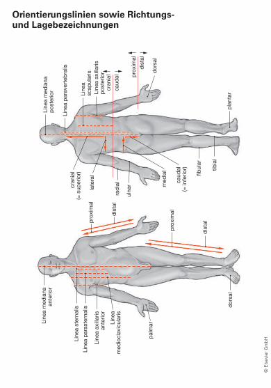
Orientierungslinien sowie Richtungs- und Lagebezeichnungen
© E
lsev
ier
Gm
bH
Sobotta_LK_Muscles.indb 11ta_LK_Muscles.indb 11 21.05.2013 16:20:0621.05.2013 16:20:06

Terms of Direction and Positioning of Body Parts
cranial or superior towards the head
caudal or inferior towards the sacrum
anterior or ventral towards the front or abdomen
posterior or dorsal towards the back
lateral sideways, away from the midline
medial centered, towards the midline
median or medianus within the median plane
intermedial positioned in between
central towards the interior of the body
peripheral towards the body surface
profundus located deeply
superfi cial or superfi cialis located superfi cially
external or externus located externally
internal or internus located internally
apical pointed or belonging to the tip
basal pointed towards the base
dexter right
sinister left
proximal towards the torso
distal towards the end of the limbs
ulnar towards the ulna
radial towards the radius
tibial towards the tibia
fi bular towards the fi bula
volar or palmar towards the palm of the hand
plantar towards the sole of the foot
dorsal (extremities) towards the back (dorsum) of the hand or the foot
frontal towards the forehead
rostral (literally translated: „towards the beak“) towards the mouth or tip of the nose (exclusively used for directional and positional information related to the head)
© E
lsev
ier
Gm
bH
Sobotta_LK_Muscles.indb 12ta_LK_Muscles.indb 12 21.05.2013 16:20:0621.05.2013 16:20:06

M. brachialis.
Defi ne the “lever arm” of a muscle.
Organisational principle of skeletal muscles 1
© E
lsev
ier
Gm
bH
(Origo)
Fascia
Caput
Venter
(Insertio)
Line of forceof the muscle
Virtual lever armof the muscle
Axis of rotation of the joint
Tendo
Sobotta_LK_Muscles.indb 13ta_LK_Muscles.indb 13 21.05.2013 16:20:0621.05.2013 16:20:06

Organisational principle of skeletal muscles 1
© E
lsev
ier
Gm
bH
The amount of force a muscle transfers to a joint depends on the length of the lever arm involved. The perpendicular distance of the joint’s rotation axis from the muscle’s line of action represents the lever arm of the muscle.As the length of the lever arm varies depending on the relative joint position, it is also called the virtual lever arm.
Normally, skeletal muscles link two bones and move one bone relative to the other. Originally, the idea was to refer to a muscle’s attachment site on the fi xed bone as its origin (Punctum fi xum or Orgio) and to the site on the moved bone as its inser-tion (Punctum mobile or Insertio). However, as bones are moved relative to each other, the proximal attachment site is simply defi ned as origin and the distal one as insertion.
Sobotta_LK_Muscles.indb 14ta_LK_Muscles.indb 14 21.05.2013 16:20:0621.05.2013 16:20:06