Embed Size (px)
Citation preview

Smart 3D-FISH: Automation of Distance Analysisin Nuclei of Interphase Cells by Image Processing
Michael Gu�e,2 C�edric Messaoudi,1 Jian Sheng Sun,2 and Thomas Boudier1*1 UMR 168 CNRS-Institut Curie, Paris, France
2 USM 0503, UMR 5153 CNRS-MNHN, U 565 INSERM, D�epartement ‘‘R�egulations, D�eveloppement et Diversit�e Mol�eculaire,’’Mus�eum National d’Histoire Naturelle, Paris, France
Received 20 August 2004; Revision Received 3 June 2005; Accepted 7 June 2005
Background: Detection of fluorescent probes by fluores-cence in situ hybridization in cells with preserved three-dimensional nuclear structures (3D-FISH) is useful forstudying the organization of chromatin and localization ofgenes in interphase nuclei. Fast and reliable measure-ments of the relative positioning of fluorescent spots spe-cific to subchromosomal regions and genes wouldimprove understanding of cell structure and function.Methods: 3D-FISH protocol, confocal microscopy, anddigital image analysis were used.Results: New software (Smart 3D-FISH) has been devel-oped to automate the process of spot segmentation anddistance measurements in images from 3D-FISH experi-ments. It can handle any number of fluorescent spots andincorporate images of 40,6-diamino-2-phenylindole coun-terstained nuclei to measure the relative positioning of
spot loci in the nucleus and inter-spot distance. Resultsfrom a pilot experiment using Smart 3D-FISH on ENL,MLL, and AF4 genes in two lymphoblastic cell lines weresatisfactory and consistent with data published in the lit-erature.Conclusion: Smart 3D-FISH should greatly facilitateimage processing and analysis of 3D-FISH images by pro-viding a useful tool to overcome the laborious task ofimage segmentation based on user-defined parameters anddecrease subjectivity in data analysis. It is available as a setof plugins for ImageJ software. q 2005 Wiley-Liss, Inc.
Key terms: fluorescence; confocal microscopy; three-dimensional fluorescence in situ hybridization; image pro-cessing; segmentation; ImageJ
Cell imaging techniques are integral to successful lifescience studies and, at the dawn of the post-genomesequencing era, techniques that permit the accurate locali-zation of genes and their products within cells and tissueswill be in ever-increasing demand. The rapidly developingfield of cell imaging techniques has greatly contributed tothe current understanding of cell structure and function.Fluorescence in situ hybridization (FISH) is used to studythe organization and positioning of chromosomes, sub-chromosomal regions, and/or specific sequences such asgenes or RNA in cellular preparations by hybridization ofprobes with complementary sequences in cells. It isimportant that after fixation of cells on glass slides, theyretain, as near as possible, their in vivo morphology (1–3).A protocol of cell fixation based on paraformaldehyde hasbeen developed to perform FISH in the three-dimension-ally (3D) preserved structure of nuclei in cells in whichthe relative position of chromatin and genes are quite wellconserved (4). This technique, named 3D-FISH, hasopened the way to study the organization of the chroma-tin in interphase nuclei.
Individual chromosomes have been shown to occupydiscrete regions in interphase nuclei, called chromosometerritories (CTs) (5,6). The global position of chromo-
somes in interphase nuclei seems to be conservedthrough cell division (7) and even along evolution (8,9).This localization depends on the size of the chromosomes(10) and the density of genes carried by the chromosomes(11). Further, observations have shown that the spatialorganization of the genome is specific to cell lineage (12).Recently, several investigations have provided evidence
that CTs are not randomly distributed but are partiallyoriented in cell nuclei (13). However, the precise organi-zation of CTs, in particular the positioning of genes insideCTs, is not fully understood. This organization maydepend on functional processes such as transcription,replication, repair, and recombination processes.Relative positions of genes inside nuclei and their
dynamics in interphase cells could play a major role inchromosomal rearrangements, i.e., reciprocal transloca-tion or inversion. For instance, some studies about chro-matin organization in interphase nuclei have indicated
*Correspondence to: Thomas Boudier, UMR 168 CNRS-Institut Curie,
11 rue Pierre et Marie Curie, 75005 Paris, France.
E-mail: [email protected] online 4 August 2005 in Wiley InterScience (www.interscience.
wiley.com).
DOI: 10.1002/cyto.a.20170
q 2005 International Society for Analytical Cytology Cytometry Part A 67A:18–26 (2005)

that the spatial proximity of genes could be a key factor inthe induction of translocations (14–18). Further, the differ-ences in relative position of CTs in different cell lineagescould contribute to the large variety of translocations,especially in hematologic cancer (19).
To further address this relation, the spatial distributionof CTs, or intergenic distances between genes potentiallyinvolved in chromosomal translocations, must be accu-rately analyzed. The 3D-FISH technique allows one to pro-duce 3D images where real gene-to-gene distances can bemeasured with better precision than with classic 2D-FISHimages (20). In a 2D-FISH experiment, such a task is quitestraightforward, but it often, if not always, leads to erro-neous data due to the localization of spots (genes) on dif-ferent focal planes (2D projection). In a 3D-FISH experi-ment, the analysis is a heavy workload and complex taskfor the biologist. Obtaining reliable data involves multiplesteps: visual inspection for selecting a region of interest,manual segmentation, and detection of the center of spotscorresponding to the localization of genes. Because hun-dreds of image stacks need to be analyzed for reaching sta-tistical significance, the analysis is very time consuming.Further, and importantly, it has the bias of the user’s sub-jectivity and is not well reproducible or reliable.
The goal of the present work is to provide an user-friendly software to improve the quality of rather highthroughput analysis of 3D-FISH images and the subse-quent positioning of genes and measurement of intergenicdistances. With the help of this software, named Smart3D-FISH, image stacks corresponding to hundreds of cellscan be automatically analyzed overnight on a desktopcomputer. The software can be used on any FISH studies,including 2D studies. It can analyze images with a virtuallyunlimited number of color channels (probes). The resultsare saved as text files that can be directly incorporatedinto standard spreadsheet software. In the first part, theprocedure of image processing and software aredescribed. The second part deals with the validation ofthese procedures and software in a 3D-FISH experimentinvolving three genes (three color probes) and 40,6-dia-mino-2-phenylindole (DAPI) counterstained nuclei.
This new software has been successfully applied in a3D-FISH experiment aimed at measuring the 3D radius dis-tribution of genes involved in acute leukemia, namelyMLL, AF4, and ENL, in two lymphoblastic cell lines. Smart3D-FISH is available as a set of plug-ins for the image analy-sis ImageJ software (21). It can be freely downloaded fromhttp://www.snv.jussieu.fr/�wboudier/softs.html.
MATERIALS AND METHODSCell Culture and Karyotype
Two human cell lines were chosen due to their neardiploid karyotype. A human B-cell precursor leukemia(NALM-6) was kindly provided by Lagneaux L. (Brussels,Belgium) with the karyotype 46(43-47)<2n>XY,t(5;12)(q33.2;p13.2); and we purchased a human B lym-phoblastoid cell line (IM-9, DSMZ Braunschweig, Ger-many) that has an apparently normal karyotype 46(44-
46)<2n>XX. These two lymphoblastic cell lines were cul-tured in RPMI-1640 medium supplemented with 10% fetalcalf serum, 1% l-glutamine, in the absence of antibiotics, at37�C in a humidified atmosphere containing 5% CO2.
DNA Probes
Bacterial artificial chromosome/phage artificial chromo-some (BAC/PAC) clones were obtained from the Resourcesfor Molecular Cytogenetics (RMC) database (http://www.biologia.uniba.it/rmc/) in Italy: CTD-217A21 for theMLL gene (22) and RP11-476C8 for the AF4 gene. RP11-2344B19 for the ENL gene was purchased from Invitrogen(Carlsbad, CA, USA). Clones were grown in LB mediumwith appropriate antibiotics: kanamycin (50 lg/ml) for PACclones or chloramphenicol (12.5 lg/ml) for BAC clones.DNA extraction was carried out according to the protocolsin the RMC database. The probes of MLL, AF4, and ENL
genes were labeled by nick translation by incorporatingfluorophore-tagged nucleotides dUTP-Alexa 488 (MolecularProbes Europe, Netherlands), dUTP-Cy5, and dUTP-Cy3(Amersham, Buckinghamshire, UK), respectively. Four hun-dred nanograms of labeled probes, 10 lg of human cot-1-DNA, and 5 lg of salmon sperm DNA were mixed in med-ium containing 50% deionized formamide, 23 standard sal-ine citrate (SSC), 10% dextran sulfate, and 0.5 M of sodiumphosphate dibasic:sodium phosphate monobasic (5:1).
3D-FISH Experiment
A dense cell suspension in 13 phosphate buffer saline(PBS) was applied to slides coated with poly-L-lysine for10 min to allow cell adhesion. Cells were then fixed inbuffered 4% paraformaldehyde in 13 PBS for 10 min topreserve the native 3D structure of the nuclei and thenwashed 3 times for 5 min in 13 PBS. During all proce-dures, air drying was carefully avoided. Cells were per-meabilized with 0.5% Triton X-100 and 0.5% saponin in13 PBS for 15 min. After a bath in 20% glycerol in 13 PBSfor 20 min at room temperature, cells were freeze-thawedby briefly dipping the slides three times in liquid nitrogen.Cells were treated with 400 lg/ml of RNase A (RocheDiagnostics, Myelan, France) for 15 min at 37�C. After a 5-min bath in 0.1 N HCl, cells were washed in 23 SSC for 5min and incubated in 50% formaldehyde and 23 SSC, pH7.5, for 1 h. The probe preparation was then droppedonto slides. Cells and probes were simultaneous dena-tured at 75�C for 8 min. Cells were incubated overnight at37�C in a humidified chamber. Post-hybridization washeswere performed a first time in 50% formaldehyde and 23SSC, pH 7.2, at 42�C, 3 times for 15 min and a second timein 0.13 SSC, pH 7.2, at 60�C, 3 times for 15 min. Nucleiwere counterstained with DAPI at 0.2 lg/ml. Slides weremounted in Vectashield medium.
Image Acquisition and Measurement
Confocal microscopy was carried out using a TCS con-focal imaging system (Leica Instruments, Heidelberg, Ger-many) equipped with a 633 objective. For Alexa 488,Cy3, and Cy5 excitations, an argon-krypton ion laser and
19SMART 3D-FISH: SOFTWARE FOR IMAGE PROCESSING OF 3D-FISH

a helium-neon ion laser were adjusted to 488, 568, and647 nm, respectively. A biphotonic device was used forDAPI excitation. For each optical section, four fluores-cence images were acquired in a sequential mode (i.e.,Cy5, Cy3, Alexa 488, and DAPI). The confocal pinholewas adjusted to allow a minimum field depth. The focusstep between sections was generally 0.35 lm (which cor-responds to the optimal optical resolution) and the XY
pixelization was set to 100 nm. Focal series were then pro-cessed to produce a single composite image file (stack).Typically, a stack of 40 confocal planes was acquired.
Total DNA is counterstained by DAPI dye. Due to thebiphotonic acquisition mode on the microscope, imagesfrom the DAPI channel are shifted. To register them withother images from other color channels, DAPI images weretranslated. The XY translation was then implemented toautomatically shift the area of interest for the DAPI channel.
The radius of each nucleus was estimated according toa spherical approximation by measuring the volume ofthe nucleus, counterstained by DAPI, in 3D-FISH images.The distance of genes to the nuclear center was then mea-sured and expressed as a percentage of the nuclear radius.Statistical analyses were performed using Student’s t testwith an a value of 0.05.
Image Processing
All algorithms were implemented in JAVA and can be runas plugins on the free multiplatform ImageJ software. A pos-sible procedure for 3D processing is to apply 2D processingin a slice-by-slice manner. However, slice-by-slice processingtends to remove weak signals that could be considered pix-els of noise in the 2D slice but that are part of a signal in 3Dimages. Hence, all implemented procedures were adaptedto work with voxels (i.e., incorporating X, Y, and Z direc-tions) in 3D images. The implemented algorithms can workwith real 3D color images. A 3D color image is consideredas a set of unlimited numbers of 3D gray-level images (eachcorresponding to a different color channel). For a set ofimages less than or equal to three channels, the standardred/green/blue (RGB) color code is used for display. Formore than three colors, each 3D gray-level image is distribu-ted into at least one red, green, or blue channel; a 3D RGBimage is then calculated by summing the contribution of allthe 3D single gray-level images.
Noise Filtering and Spot Segmentation
Figure 1 shows the flow chart of the overall algorithm.The first step in processing the images is to remove back-ground noise. The median filter is well suited for salt-and-pepper noise; for a general reference on image processing,see Russ (23) and Castleman (24). This filter, adapted to3D images, is used with a neighborhood of radius equal to2 pixels. Then a 3D Top Hat filter is applied to enhancethe spots against the background and decrease the level ofbackground noise. This filter is defined by: Top Hat(Im) 5Im 2 Max7(Min7(Im)), as previously described (25). Thesecond step is the segmentation of the spots. The centralpixels inside each spot are determined to correspond topixels whose value is greater than 99.95% of the histo-
gram, i.e., with a value greater than v (v is defined as99.95% of all pixel values in the range [0 2 v]). This valueof 99.95% of the histogram was computed by estimatingthe volume of one spot and considering that a stack couldcomprise up to four spots.More precisely, the pixel whose intensity is maximal in
the 3D stack, but greater than 99.95% of the histogram, isdetected. This pixel is a seed from where a spot will besegmented. A local threshold is computed correspondingto mean 1 sigma of the pixels belonging to three lines inthe three directions (X, Y, Z) passing through the detectedcenter of the spot. The seed is extended, by 3D connectiv-ity, to adjacent pixels whose value is greater than this localthreshold to form an object. If the final segmented objectis touching one border of the image in the X-Y plane, thenit is removed. These procedures (detection of maximumpixel and 3D connectivity) are applied until no moreunsegmented pixels have a value greater than 99.95% ofthe histogram. The segmentation is performed on all thedifferent stacks except for the DAPI channel.For the DAPI channel, a special segmentation is automa-
tically performed. The radius of the first median filterapplied to reduce the noise is increased to a radius of4 pixels to obtain a more homogeneous signal. The stackis then thresholded using the ‘‘Isodata’’ algorithm (26).For spots and DAPI segmentation, a 3D mathematical
morphologic closing procedure is applied followed by a 3Dmathematical morphologic opening procedure (radius 2 forgene images, radius 4 for nucleus DAPI images) to removevery small objects, fill holes, and make shape look morecompact.
Analysis and Validation of Segmentation
A first analysis of the validity of the segmentation isdone after the application of the median filter (Fig. 1). Ifthe computed signal-to-noise ratio (SNR; maximum 2minimum value of gray level in the image stack) is lowerthan a threshold fixed by the user, further measurementswill be discarded into an invalidated spots file. A secondanalysis of the validity of the segmentation is based on thevolume and number of objects. The list of segmentedobjects is analyzed to detect whether or not the segmen-ted objects correspond to gene spots. Segmented objectswith volumes smaller than the minimal volume (minV1),fixed by the user, are removed. In the same way, objectswith volumes larger than the maximum volume (maxV1)are removed. They are often due to an incorrect experi-mental procedure or acquisition mode. Then, if the num-ber of spots exceeds the value fixed by the user (two inthe present work), the segmentation is considered invalidand further measurements will be performed, but theresults will be redirected into the invalidated spots file.In all cases, results of measurements are saved as text
files. Two text files are created; the first text file will storethe results for correct segmentation, and the other one willstore the results for incorrect segmentation, i.e., where oneparameter (SNR, volume and number of spots) does notcorrespond to the values fixed by the user. In this last case,annotations will indicate the nature of the problem.
20 GU�E ET AL.

Spot Separation Procedure
Figure 2 illustrates the spot separation procedure. Themathematical morphologic procedures may not be suffi-cient to separate two closely located spots. Close spotsmay be merged into one big spot with a volume roughlytwice the average volume of one spot. The user can fixthe minimum volume that may correspond to a mergedspot (minV2). The slices of the stack containing the objectare projected along the Z axis onto a 2D plane to find localmaxima that may correspond to the center of the twomerged spots. The obtained projection is then smoothed
so that local maxima are more easily detected. If two or
more local maxima are encountered, they are separated
into two clusters using a k-means algorithm. If the center
of the two clusters are farther apart more than a fixed dis-
tance, corresponding approximately to the diameter of
one spot (dist), the object can be separated into two smal-
ler objects. The centers of these two clusters are the new
x and y coordinates. The z value of the two new objects is
determined as the center of the primary segmented object
along the Z axis for the x and y coordinates of each new
spot. Pixels of the primary big object are then resegmen-
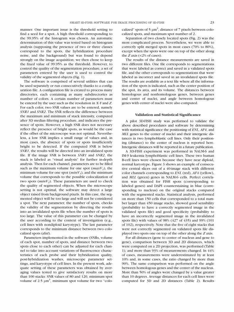
FIG. 1. Flow chart of Smart 3D-FISH software. Threshold values corresponding to signal-to-noise ratio (SNR1, SNR2), spot volume (minV1, minV2,maxV1), and number of spots are set by the user and can be saved in a configuration file. A 3D median filter is applied to original images before SNR calcula-tion. If SNR is between 0 and SNR1 [0;SNR1[, the results will be directed into the invalidated spots file (#). If the SNR is between SNR1 and SNR2[SNR1;SNR2], the results will be labeled as ‘‘visual analysis’’ for further in-depth analysis. A 3D Top Hat filter is then performed on the filtered stack. The seg-mentation process is validated by checking the volume and number of objects. If the volume of an object is smaller than the minimum spot size (minV1),the object will be discarded (not shown on flow chart). If the volume is between the minimum size of two colocalized spots (minV2) and the maximumvolume of one spot (maxV1), the process of separation will be run. If this process cannot find two colocalized spots, the original object is labeled as a ‘‘largespot’’ in the final results. If the volume of an object is larger than the maximum volume (maxV1), this object is discarded from the analysis but will be dis-played on the final segmented image for visual control, and the results will be directed into the invalidated spots file. The number of objects is then checkedto finalize the segmentation analysis process. The invalidated spots file contains all results where the image SNR is lower than the SNR1 value or the volumeof one spot is larger than maxV1 or the number of spots is larger than the limit value fixed by the user (maxN). All segmented image stacks are saved forfurther visual examination.
21SMART 3D-FISH: SOFTWARE FOR IMAGE PROCESSING OF 3D-FISH

ted according to their distance to the center of these twonew objects. If the primary object cannot be split intotwo objects, it is kept in the list of segmented objects butwill be annotated as ‘‘large spot.’’
RESULTSImage Processing and Software Description
From the confocal microscope, a stack is obtainedwhere cells are present for all channels (corresponding toprobe colors). An integrated utility was developed to help
biologists select an area of interest delimiting one cell andsave all the various channels in different directories.Stacks corresponding to ENL, AF4, and MLL genes weresaved on the red, yellow, and green channels, respectively.An additional blue channel for DAPI counterstaining ofnuclei was also used to determine the relative positioningof different probes to the center of nucleus.The segmentation procedure was inspired by detection
of microcalcification in mammograms using a RH-maximaprocedure (27), with the exception that all the proce-dures were performed in 3D and not in a slice-by-slice
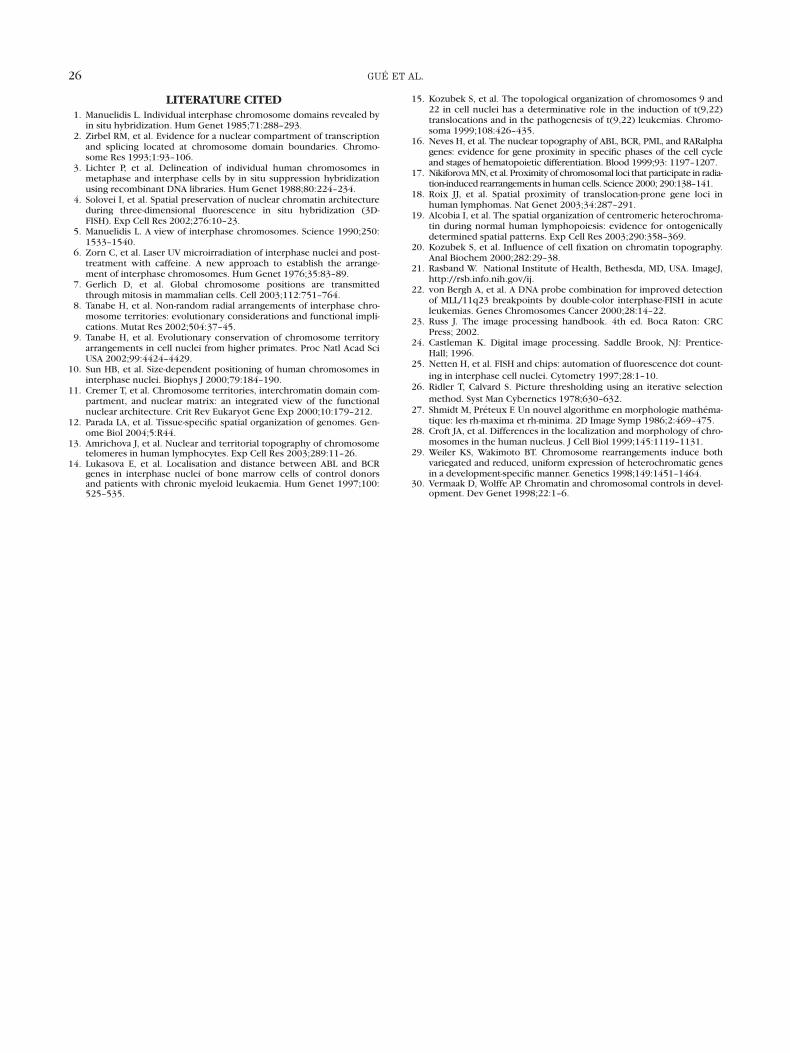
FIG. 2. Spot separation process. A: 2D projection along the Y axis of the segmented image stack. White arrow indicates one object whose volume isbetween minV2 and maxV1 (Fig. 1) before the spot separation process. B: The same 2D projection proves that the object detected as one object is, in rea-lity, composed of two colocalized spots. C: To distinguish these two spots on the segmented image, a 2D projection is performed along the Z axis toenhance the signal corresponding to each spot. Then local maxima are detected to find XY coordinates of each spot. If two local maxima are detectedfarther apart than the dist (see text), the two spots are separated. D: On the segmented stack, Z coordinates of each new spot are then detected along theZ axis (from XY coordinates previously detected) as the middle of the segmented object.
22 GU�E ET AL.
manner. One important issue is the threshold setting tofind a seed for a spot. A high threshold corresponding tothe 99.95% of the histogram was chosen. An automaticdetermination of this value was tested based on histogramanalysis (supposing the presence of two or three classescorrespond to the spots, the hybridization procedurenoise, and the background) but was found to dependstrongly on the image acquisition; we then chose to keepthe fixed value of 99.95% as the threshold. However, tocontrol the quality of the segmentation procedure, a set ofparameters entered by the user is used to control thevalidity of the segmented objects (Fig. 1).
The software is comprised of several utilities that canbe used separately or run consecutively thanks to a config-uration file. A configuration file is created to process manydirectories, each containing as many subdirectories asnumber of colors. A certain number of parameters mustbe entered by the user such as the resolution in X-Y and Z.For each color, two SNR values are to be entered, namelySNR1 and SNR2. The SNR reflects the difference betweenthe maximum and minimum of stack intensity, computedafter 3D median filtering procedure, and indicates the pre-sence of spots. However, a high SNR does not necessaryreflect the presence of bright spots, as would be the caseif the offset of the microscope was not optimal. Neverthe-less, a low SNR implies a small range of values and, inmost cases, the absence of spots or spots insufficientlybright to be detected. If the computed SNR is belowSNR1, the results will be directed into an invalidated spotsfile. If the image SNR is between SNR1 and SNR2, thestack is labeled as ‘‘visual analysis’’ for further in-depthanalysis. Then for each channel, parameters are to be filledsuch as the maximum volume for one spot (maxV1), theminimum volume for one spot (minV1), and the minimumvolume that corresponds to the possible colocalization oftwo spots (minV2). These parameters are used to checkthe quality of segmented objects. When the microscopesetting is not optimal, the software may detect a largeobject raised from background pixels. In this case, the seg-mented object will be too large and will not be considereda spot. The next parameter, the number of spots, checksthe validity of the segmentation by directing the resultsinto an invalidated spots file when the number of spots istoo large. The value of this parameter can be changed bythe user according to the context of investigation (e.g.,cell lines with nondiploid karyotype). The last parametercorresponds to the minimum distance between two colo-calized spots (dist).
Parameters implemented in the software (SNRs, volumeof each spot, number of spots, and distance between twospots close to each other) can be adjusted for each chan-nel to take into account variations of fluorescence charac-teristics of each probe and their hybridization quality,post-hybridization washes, microscope parameter set-tings, and karyotype of cell lines. In the present work, ade-quate setting of these parameters was obtained by aver-aging values tested to give satisfactory results on morethan 100 stacks: SNR values of 80 and 120, minimum spotvolume of 2.5 lm3, minimum spot volume for two ‘‘colo-
calized’’ spots of 5 lm3, distance of 7 pixels between colo-calized spots, and maximum spot number of 2.Separation of two closely located spots (Fig. 2) was the
most complicated process. Nevertheless, we were able tocorrectly split merged spots in most cases (70% to 80%),except when the spots were one on top of the other alongthe Z axis (<2% of cases).The results of the distance measurements are saved in
two different files. One file corresponds to segmentationsthat were labeled as correct and saved in a validated spotsfile, and the other corresponds to segmentations that werelabeled as incorrect and saved in an invalidated spots file.The results are available as a text file where all the informa-tion of the spots is indicated, such as the center position ofthe spot, its area, and its volume. The distances betweenhomologous and nonhomologous genes, between genesand center of nuclei, and angle between homologousgenes with center of nuclei were also computed.
Validation and Statistical Significance
A pilot 3D-FISH study was performed to validate theabove described procedures and software by determiningwith statistical significance the positioning of ENL, AF4, andMLL genes to the center of nuclei and their intergenic dis-tances in two lymphoblastic cell lines. Only their position-ing (distance) to the center of nucleus is reported here.Intergenic distances will be reported in a future publication.A 3D-FISH experiment was performed in NALM-6 and
IM-9 leukemia lymphoblastic cell lines. These two humanB-cell lines were chosen because they have near diploid,normal karyotype. Figure 3 shows an example of consecu-tive central slices out of a 40-image stack with differentcolor channels corresponding to ENL (red), AF4 (yellow),and MLL (green) genes in NALM-6 cells. Perfect correla-tion was obtained for FISH spots (corresponding tolabeled genes) and DAPI counterstaining in blue (corre-sponding to nucleus) on the original stacks comparedwith the segmented stacks. Statistical analysis, performedon more than 150 cells that corresponded to a total num-ber larger than 450 image stacks, showed good sensibility(probability to have a correctly segmented image in thevalidated spots file) and good specificity (probability tohave an incorrectly segmented image in the invalidatedspots file) with values of 98% (427 of 435) and 99% (101of 102), respectively. Note that the five of eight stacks thatwere not correctly segmented on validated spots file dis-played two spots one on top of the other along the Z axis.For all distances (gene to center of nucleus and gene to
gene), comparison between 3D and 2D distances, whichwere computed on a 2D projection, was performed (Table1), and more than 93% of measurements changed. In 44%of cases, measurements were underestimated by at least10% and, in some cases, the ratio changed by more than90%. The same comparison was performed on the anglebetween homologous genes and the center of the nucleus.More than 50% of angles were changed by a value greaterthan 10 degrees. Average distances for each cell lines werecomputed for 3D and 2D distances (Table 2). Results
23SMART 3D-FISH: SOFTWARE FOR IMAGE PROCESSING OF 3D-FISH

showed significative differences between 3D and 2Dgene-to-center measurements in both cell lines. Distribu-tion of 3D distances between loci of each gene (MLL, AF4,and ENL) with the center of nucleus in NALM-6 and IM-9cell lines was analyzed. Results are shown in Figure 4 andTable 2. Radius distribution of the AF4 and ENL genes wassignificantly similar in the two cell lines (P < 0.0001).However, a small difference in radius distribution wasnoted for the MLL gene. It was noted that the distributionalong the nucleus radius showed that the ENL gene has amore central nucleus localization than the MLL and AF4
genes. The AF4 gene appears to be more peripheral.
DISCUSSION
Visual comparison between segmented image stacksand original images stacks showed no difference in posi-tion, size, and number of spots. These observations vali-dated the segmentation procedure. The procedure of spotseparation implemented in this software is quite complexbut seems to give quite a satisfactory outcome. However,it strongly depends on the minimal distance between twospots (dist). The separation of two spots along the Z axiscould be implemented by projecting candidate spots ontoxy and yz planes. However, due to the relative small num-ber of colocalized spots in this position and the optical dis-tortion along the Z direction, this procedure was notimplemented. The interest of this separation could be tostudy the position of all genes through the cell cycle, tak-ing into account newly replicated spots. The comparisonbetween 3D and 2D measurements showed an underesti-mation of distances in 2D condition, which could lead toerroneous interpretation. Further, thanks to real 3D mea-surements, exceptional events have a better probability tobe detected than on 2D projections.Our 3D-FISH experiments show that the ENL gene loca-
lized on chromosome 19 occupies the most centralnuclear localization, whereas the AF4 gene (on chromo-some 4) appears to be more peripheral, probably near thenuclear membrane. The MLL gene (on chromosome 11)
FIG. 3. Example of four-color image stack from a 3D-FISH experi-ment. Distance between slices was 0.35 lm. The leftmost column showsimages corresponding to DAPI counterstaining of nucleus. The second,third, and fourth columns show images corresponding to hybridizationsignals of two the loci of the ENL gene (19p13.3), AF4 gene (4q21), andMLL gene (11q23), respectively. The rightmost column, named ‘‘segmen-ted and merged,’’ displays the combination of all segmented images.
<
Table 1Ratio Between 2D and 3D Measurements for G-G and
G-C Distances*
2D/3D ratio
1–0.9 (1) 0.9–0.5 0.5–0.1 0.1–0
G-C (n5 2,034) 54.5 (5.8) 37.8 7.6 0.1G-G (n5 4,068) 55.5 (6.45) 36.6 7.6 0.1
*G-C, gene-to-nuclear center distances; G-G, gene-to-gene dis-tances. Percentage of measurements, for which ratio is equal to1, is presented in parentheses.
24 GU�E ET AL.

has an intermediate localization. These observationsreflect dynamics of loci inside CTs. They are consistentwith previous studies showing that chromosomes 19 ininterphase nuclei are more central (28). The relative posi-tioning of chromosomes in nuclei is organized accordingto their size (10): the smaller chromosomes (e.g., chromo-somes 19) are more centrally localized than the largerones (e.g., chromosome 4). No significant difference inradial distribution was observed for the AF4 and ENL
genes between these two lymphoblastics cell lines. Asexpected, these results corroborated a relative conserva-
tion of CT organization for a given cell line (12). However,the radial distribution of the MLL gene seems to be slightlydifferent on these two lymphoblastic cell lines. The MLL
gene seems to be more centrally located in the NALM-6than in the IM-9 cell line. NALM-6 is a human B-cell precur-sor in contrast to the IM-9 cell line. Cell differentiationmay cause a subtle difference in nuclear organization oftheMLL gene, as has been shown for other genes in the lit-erature (29,30).In conclusion, it seems that the image processing proce-
dures and parameter settings implemented in the softwareallow satisfactory image data processing and analysis.Other 3D-FISH experiments carried out in different celllines with additional probes are also satisfactory (data notshown). Smart 3D-FISH is new software that has beendeveloped to automate the process of spot segmentationand perform distance measurements in 3D-FISH. It canhandle virtually any number of spots and color channelsfor intergenic distance measurements. It can also incorpo-rate the images from DAPI channel (total DNA of nucleus)to measure distances of genes to a nuclear center. A 3D-FISH experiment carried out on ENL, MLL, and AF4 genesin two lymphoblastic cell lines has shown self-consistentdata and corroborated previously reported data in the lit-erature concerning the organization of chromosomesinside nuclei. Visual comparison of original image stackswith segmented image stacks by Smart 3D-FISH providedsatisfactory detection of all spots, with a sensitivity of98%. The software is user friendly and robust in use. Smart3D-FISH is available as a set of plug-ins for ImageJ softwareat http://www.snv.jussieu.fr/~wboudier/softs. html.It should greatly facilitate image processing and analysis
by providing a useful tool to overcome the laborious taskof 3D image measurements based on user-defined para-meters and decrease subjectivity in data analysis.
ACKNOWLEDGMENTS
The authors thank Ma€ıt�e Coppey and ChristopheChamot at the Institut Jacques Monod (Paris, France)for providing access and assistance to confocal micro-scopy facility. Thanks also go to Jean Soulier (HospitalSaint Louis, Paris, France) and Micka€el Durand-Dubieffor providing assistance in the FISH technique, Mar-riono Rocchi (University of Bari, Italy) for providingBAC/PAC FISH clones, Laurence Lagneaux (Belgium) forproviding NALM-6 cell line, and Louise Anderson forreading the manuscript.
Table 22D Versus 3D Radial Distances* for the AF4, MLL, and ENL Genes in Two Cell Lines
3D 2D P
IM-9 NALM-6 IM-9 NALM-6 IM-9 NALM-6
AF4 83.26 1.5 81.7 6 1.3 74.46 1.9 68.9 6 1.8 0.00041 <0.0001MLL 74.76 1.6 71.9 6 1.7 67.66 1.9 60.3 6 2.1 0.004 <0.0001ENL 61.26 1.6 62.9 6 1.6 55.86 1.7 55.0 6 1.8 0.025 0.001
*Values are expressed as average distances (percentage of nuclear radius) 6 standard error. Student’s t test was applied with a 5 0.05.
FIG. 4. Radial distribution of gene-to-nuclear center distances for theAF4, MLL, and ENL genes (from top to bottom) in NALM-6 (black bars)and IM-9 (gray bars) lymphoblastic cell lines. Distances are expressed as apercentage of the nuclear radius.
25SMART 3D-FISH: SOFTWARE FOR IMAGE PROCESSING OF 3D-FISH
LITERATURE CITED1. Manuelidis L. Individual interphase chromosome domains revealed by
in situ hybridization. Hum Genet 1985;71:288–293.2. Zirbel RM, et al. Evidence for a nuclear compartment of transcription
and splicing located at chromosome domain boundaries. Chromo-some Res 1993;1:93–106.
3. Lichter P, et al. Delineation of individual human chromosomes inmetaphase and interphase cells by in situ suppression hybridizationusing recombinant DNA libraries. Hum Genet 1988;80:224–234.
4. Solovei I, et al. Spatial preservation of nuclear chromatin architectureduring three-dimensional fluorescence in situ hybridization (3D-FISH). Exp Cell Res 2002;276:10–23.
5. Manuelidis L. A view of interphase chromosomes. Science 1990;250:1533–1540.
6. Zorn C, et al. Laser UV microirradiation of interphase nuclei and post-treatment with caffeine. A new approach to establish the arrange-ment of interphase chromosomes. Hum Genet 1976;35:83–89.
7. Gerlich D, et al. Global chromosome positions are transmittedthrough mitosis in mammalian cells. Cell 2003;112:751–764.
8. Tanabe H, et al. Non-random radial arrangements of interphase chro-mosome territories: evolutionary considerations and functional impli-cations. Mutat Res 2002;504:37–45.
9. Tanabe H, et al. Evolutionary conservation of chromosome territoryarrangements in cell nuclei from higher primates. Proc Natl Acad SciUSA 2002;99:4424–4429.
10. Sun HB, et al. Size-dependent positioning of human chromosomes ininterphase nuclei. Biophys J 2000;79:184–190.
11. Cremer T, et al. Chromosome territories, interchromatin domain com-partment, and nuclear matrix: an integrated view of the functionalnuclear architecture. Crit Rev Eukaryot Gene Exp 2000;10:179–212.
12. Parada LA, et al. Tissue-specific spatial organization of genomes. Gen-ome Biol 2004;5:R44.
13. Amrichova J, et al. Nuclear and territorial topography of chromosometelomeres in human lymphocytes. Exp Cell Res 2003;289:11–26.
14. Lukasova E, et al. Localisation and distance between ABL and BCRgenes in interphase nuclei of bone marrow cells of control donorsand patients with chronic myeloid leukaemia. Hum Genet 1997;100:525–535.
15. Kozubek S, et al. The topological organization of chromosomes 9 and22 in cell nuclei has a determinative role in the induction of t(9,22)translocations and in the pathogenesis of t(9,22) leukemias. Chromo-soma 1999;108:426–435.
16. Neves H, et al. The nuclear topography of ABL, BCR, PML, and RARalphagenes: evidence for gene proximity in specific phases of the cell cycleand stages of hematopoietic differentiation. Blood 1999;93: 1197–1207.
17. NikiforovaMN, et al. Proximity of chromosomal loci that participate in radia-tion-induced rearrangements in human cells. Science 2000; 290:138–141.
18. Roix JJ, et al. Spatial proximity of translocation-prone gene loci inhuman lymphomas. Nat Genet 2003;34:287–291.
19. Alcobia I, et al. The spatial organization of centromeric heterochroma-tin during normal human lymphopoiesis: evidence for ontogenicallydetermined spatial patterns. Exp Cell Res 2003;290:358–369.
20. Kozubek S, et al. Influence of cell fixation on chromatin topography.Anal Biochem 2000;282:29–38.
21. Rasband W. National Institute of Health, Bethesda, MD, USA. ImageJ,http://rsb.info.nih.gov/ij.
22. von Bergh A, et al. A DNA probe combination for improved detectionof MLL/11q23 breakpoints by double-color interphase-FISH in acuteleukemias. Genes Chromosomes Cancer 2000;28:14–22.
23. Russ J. The image processing handbook. 4th ed. Boca Raton: CRCPress; 2002.
24. Castleman K. Digital image processing. Saddle Brook, NJ: Prentice-Hall; 1996.
25. Netten H, et al. FISH and chips: automation of fluorescence dot count-
ing in interphase cell nuclei. Cytometry 1997;28:1–10.26. Ridler T, Calvard S. Picture thresholding using an iterative selection
method. Syst Man Cybernetics 1978;630–632.27. Shmidt M, Pr�eteux F. Un nouvel algorithme en morphologie mathema-
tique: les rh-maxima et rh-minima. 2D Image Symp 1986;2:469–475.28. Croft JA, et al. Differences in the localization and morphology of chro-
mosomes in the human nucleus. J Cell Biol 1999;145:1119–1131.29. Weiler KS, Wakimoto BT. Chromosome rearrangements induce both
variegated and reduced, uniform expression of heterochromatic genesin a development-specific manner. Genetics 1998;149:1451–1464.
30. Vermaak D, Wolffe AP. Chromatin and chromosomal controls in devel-opment. Dev Genet 1998;22:1–6.
26 GU�E ET AL.