Embed Size (px)
Citation preview

Small molecules that bind the inner core of gp41and inhibit HIV envelope-mediated fusionGary Frey†, Sophia Rits-Volloch‡§, X.-Q. Zhang¶, Robert T. Schooley¶, Bing Chen‡, and Stephen C. Harrison†‡§�
†Department of Biological Chemistry and Molecular Pharmacology, Harvard Medical School, 250 Longwood Avenue, Boston, MA 02115;‡Laboratory of Molecular Medicine and §Howard Hughes Medical Institute, Children’s Hospital, Boston, MA 02115; and ¶Division ofInfectious Diseases, University of California, San Diego, CA 92023
Edited by Malcolm A. Martin, National Institutes of Health, Bethesda, MD, and approved July 24, 2006 (received for review February 8, 2006)
HIV-1 enters cells by membrane fusion, mediated by the trimericviral envelope glycoprotein gp160, which is processed by a singleproteolytic cleavage into stably associated gp120 and gp41. Thegp120�gp41 trimer can be triggered to undergo an irreversibleconformational change. Using a protein-based assay designed tomimic the gp41 conformational change, we screened for smallmolecules that prevent the formation of postfusion gp41. Severalcompounds were identified. One set of structurally related mole-cules inhibited formation of a postfusion-like assembly with an IC50
of �5 �M. The compounds also inhibited envelope-mediatedmembrane fusion in both cell–cell fusion and viral infectivityassays. Thus, our screen identifies effective fusion inhibitors.Tested against a panel of envelope proteins from primary HIV-1isolates, the compounds inhibited fusion across a broad range ofclades, including both M and T tropic strains. They bind in a highlyconserved, hydrophobic pocket on the inner core of the gp41trimer, a region previously identified as a potential inhibitor site.
viral entry � antiviral drugs � high-throughput screen
Enveloped viruses such as HIV and influenza virus enter cellsby membrane fusion (1). The gp160 envelope glycoprotein of
HIV-1, its fusion protein, is synthesized as a single polypeptidechain with a C-terminal membrane anchor, and it requires aprocessing cleavage (to gp120 and gp41) for activation (2, 3). Thefusion peptide, a glycine-rich, hydrophobic sequence requiredfor interaction with the target-cell membrane, lies near the newlycreated N terminus of gp41 (4). Like other class 1 fusionproteins, such as influenza virus hemagglutinin, gp160 is ahomotrimer, and its fusion-promoting fragment, gp41, is alsotrimeric (1).
The cleaved, mature trimer of gp120�gp41 can be triggered byreceptor binding to undergo an irreversible conformationalchange (5–9). In effect, cleavage of the gp160 precursor producesa metastable conformation, but the barrier to its rearrangementis high. The image of a spring-loaded device has been used todescribe this situation, and the triggering event then correspondsto releasing the catch on the spring (10).
The structure of the gp41 ectodomain in the postfusionconformation is a trimer of �-helical hairpins (11, 12). Its innercore is an �-helical coiled coil formed by N-terminal segmentsof the three polypeptide chains; the helices of its outer layer arethe C-terminal segments of those chains. The 30-residue loopthat connects the inner core and outer-layer helices [also called‘‘heptad repeats 1 and 2’’ (HR1 and HR2, respectively)] is absentfrom the crystal structures; sequence conservation suggests thatit resembles the loop seen in the ectodomain of the transmem-brane (TM) subunit from Moloney murine leukemia virus(MoMuLV) and in the Gp2 ectodomain of Ebola virus (13, 14).A noteworthy feature of the contact between each outer-layerhelix and the inner core is a hydrophobic pocket on the latter,containing residues Leu-57, Trp-60, and Lys-63, which receivesthe side chains of Trp-117, Trp-120, and Ile-124.
The sequence of molecular events during the fusion-promoting conformational rearrangement (refolding) of gp120�
gp41 is thought to be as follows (Fig. 1) (1). The gp120 fragmentdissociates, allowing the gp41 fragment to rearrange (2). Aninitial reorganization of the gp41 polypeptide chain pulls thefusion peptide from a buried position and projects it against thetarget-cell membrane. The result of this process is probably aconformation with significant half-life, the so called ‘‘prefusionintermediate’’ (3). In a subsequent step, each fusion-promotingfragment folds back on itself, probably by zippering up the outerlayer along the surface of the inner core. This step bringstogether the transmembrane (TM) segment at the C terminus ofthe fusion-promoting fragment, anchored in the viral bilayer,and the fusion peptide at the N terminus, bound to the target-cellmembrane. The two membranes are thus induced to approacheach other and ultimately to merge.
Even before the structure of the postfusion gp41 trimer wasknown, screening of peptides derived from gp160 had identifiedfusion inhibitors (15–19). The gp41 structure showed that someof these peptides represent inner-core segments and that otherscover all or part of the outer helix (11, 12). The presumedmechanism of inhibition by the latter is the binding of the peptideto the inner-core portion of the prefusion intermediate, pre-venting completion of the fusion-promoting conformationalrearrangement. The inhibitory peptide known as T-20 (now alicensed drug with the generic name enfuvirtide) contains res-idues 127–162 (16, 17, 20). Its sequence includes most, but notall, of the outer-layer helix and an additional tryptophan-richregion whose function is still unknown. It does not includeTrp-117, Trp-120, or Ile-124, residues that would occupy thehydrophobic pocket near positions Trp-60 and Lys-63 on theinner core. Evidence suggests that this pocket would be a suitablesite for an inhibitory drug (21). It was targeted by Eckert et al.(22) in their design of a 16-residue cyclic D peptide, which docksagainst the pocket and also inhibits fusion and viral infectivity.Ferrer et al. (23) used the pocket to select nonnatural bindingelements tethered to a peptide derived from the outer helix. Theconjugate was shown to inhibit envelope-mediated cell fusion.Molecular docking simulations against the pocket have also beenperformed (24). A quite different sort of fusion inhibitor is asingle-chain model for five of the six helical segments in gp41,which presumably traps an incipient outer-layer helix (25).
Can small molecules, with more conventional drug-like prop-erties, also inhibit the fusion-promoting refolding of gp41? Thisquestion is a particular case of the more generally debated issue:can small molecules block formation of protein–protein inter-faces? Several small-molecule respiratory syncytial virus (RSV)inhibitors target the inner core of the F1 protein, a related class1 fusion protein (26). We have found a set of compounds that
Conflict of interest statement: No conflicts declared.
This paper was submitted directly (Track II) to the PNAS office.
Freely available online through the PNAS open access option.
Abbreviations: HR, heptad repeat; SIV, simian immunodeficiency virus.
�To whom correspondence should be addressed. E-mail: [email protected].
© 2006 by The National Academy of Sciences of the USA
13938–13943 � PNAS � September 19, 2006 � vol. 103 � no. 38 www.pnas.org�cgi�doi�10.1073�pnas.0601036103

interact with the gp41 inner core by using a screen based onblocking the association of outer-layer peptides with the innercore. These molecules reduce gp41-mediated cell–cell fusionand inhibit HIV-1 infectivity. We have narrowed down the targetof binding to a hydrophobic pocket on the gp41 inner core. Thispocket is highly conserved among the different clades of groupM. Consistent with this conservation, we have shown that ourcompounds are active against a panel of HIV-1 primary isolatesthat includes both M and T tropic strains from different cladesbut are substantially less active against divergent strains fromgroup O and simian immunodeficiency virus (SIV).
Results and DiscussionWe designed and expressed a soluble, single-chain proteinconstruct we call gp41-5 to provide a suitable target for ahigh-throughput assay (Fig. 2). This strategy takes advantage ofthe unusually simple structure of gp41 in its postfusion state, atrimer of hairpins, which can be linked into a single, covalentpolypeptide that folds into a six-helix bundle. A related construct(called ‘‘five-helix’’) was used as a fusion inhibitor, in studiesmentioned above (25). Our gp41-5 contains three inner-coresegments (residues 35–70), alternating with two outer-layersegments (residues 117–150). Short linkers connect the seg-ments. After folding, the molecule contains five of the helicespresent in the six-helix bundle. It is relatively soluble, and it bindswith high-affinity peptides that contain part or all of thesequence in the sixth helix. Moreover, molecules of any sort thatcan bind the groove exposed by the absence of the sixth HRregion should also be able to bind the prehairpin intermediate.
A peptide with the sequence of the missing outer helix (C38,residues 117–154) was labeled at its N terminus with fluorescein.Binding of the labeled peptide (C38*) to gp41-5 was monitoredby fluorescence polarization (Fig. 2C). The assay is sensitive,enabling us to perform high-throughput screens. We screenedseveral libraries of small molecules (Mr � 500), searching forcompounds that interfered with the binding of C38*. As apositive control, C38 (6 �M) was added to one well on each plate.Of the 34,800 compounds screened, four (5M030, 5M038,5M041, and S2986; Fig. 3A) completely blocked C38* binding togp41-5 under our screening conditions (library compound con-centration of 40 �M; C38* � 10 nM). Three contained a2,4-bis(trif luoromethyl)[1,2,4] triazolo[4,3-a][1,8] naphthyridinering, with different substituents in the 9 position. Follow-upanalysis of structure activity relationships revealed two addi-tional compounds structurally similar to 5M038 (Fig. 3A). Onlycompounds 5M038 and 5M030 had sufficient solubility andinhibitory activity to determine accurate IC50 values in ourfluorescence polarization assay (�5 and 9 �M, respectively; Fig.3B). Compounds 5M041 and 6K061 also had significant inhib-itory activity (30% and 40% inhibition, respectively, at 7.5 �M).Compound 6M007, which contains a similar ring system with theexception of a trif luoromethyl substituent in the 6 position ratherthan the 2 and 4 positions, showed no activity in the fluorescencepolarization assay.
To determine the effects of these compounds on gp41-mediated membrane fusion, we used a cell–cell fusion assay (23,27). Compound toxicities were initially determined by usingtrypan blue staining. In addition, to control for nonspecific
Fig. 1. Model for fusion by the HIV envelope glycoprotein. Binding of gp120 to CD4 and coreceptor (first panel) triggers a conformational change that releasesthe grip of gp120 (red) on gp41 (blue). The latter extends (second panel) so that its fusion peptide (green) inserts into the target-cell membrane (green bar attop). As the outer-layer region of gp41 (dark blue) zips up along the outside of the inner layer (light blue: a three-chain, �-helical coiled coil), the viral membrane(tan) and cell membrane (green) are drawn together (third and fourth panels). It is this step that is inhibited by T-20�enfuvirtide. The formation of a hemifusionstalk (fourth panel) and a fusion pore (fifth panel) completes the membrane-fusion reaction.
Fig. 2. Association of an outer-layer peptide with gp41-5 as a screening assay. (A) The sequence of a single-chain polypeptide that can fold into a model forfive of the six helices in the postfusion form of the gp41 ectodomain. (B) Diagram illustrating that a fluoresceinated, outer-layer peptide can bind to gp41-5.N, N terminus; C, C terminus. (C) Fluorescence anisotropy binding curve for the association of the outer-layer peptide with gp41-5. The fraction bound, f, is plottedas a function of gp41-5 concentration (in nM), at a constant concentration of peptide (5 nM).
Frey et al. PNAS � September 19, 2006 � vol. 103 � no. 38 � 13939
BIO
CHEM
ISTR
Y

effects of added compound, we prepared doubly transfected cellswith plasmids encoding T7 polymerase and luciferase. Com-pounds 5M038 and 5M041 strongly inhibited gp41-mediatedmembrane fusion. At their solubility limits (90 �M for 5M038and 50 �M for 5M041), both compounds reduced the level ofcell–cell fusion by �80% (Fig. 4). Although compound 6M007had little activity in the fluorescence polarization assay, it was asactive as 5M038 against envelope-mediated membrane fusion.We discuss below this difference in assay response. Compounds6K061 and 5M030 were too toxic to be tested properly.
We determined the effects on HIV infectivity of the series ofsmall molecules just described, by exposing peripheral bloodmononuclear cells (PBMCs) to HIV-1 (isolate 2076) in thepresence and absence of each compound. When cells werepretreated with a compound before HIV infection, compounds5M038 and 5M041 blocked infection with IC50 values of 19 and18 �M, respectively (Table 1). Compound 6M007 had a weaker
effect, showing 50% inhibition at a concentration of �30 �M. Todemonstrate that, like enfuvirtide, 5M038 can inhibit the cell-to-cell spread of virus, we compared the two directly over a 7-dayperiod, as shown in Fig. 5. At a concentration as low as 30 �M,5M038 nearly completely suppressed the appearance of p24antigen over the course of the experiment.
How do these molecules interact with gp41? Proton NMRexperiments show that 5M038 gives significantly broadenedproton resonances in the presence of gp41-5 (data not shown).To use this property to pinpoint the location of binding, wesynthesized a set of three peptides containing 17-residue seg-ments of the gp41 inner core attached to a 29-residue trimer-
Fig. 3. Results of high-throughput screen. (A) Compounds identified asstrong inhibitors of outer-layer peptide binding (5M038, 5M030, 5M041, andS2986), as well as compounds in the chemical library related to compound5M038. (B) Inhibition of peptide binding for five of the compounds shown inA. The fraction bound, �, is plotted as a function of inhibitor concentration.Symbols correspond to those beneath the various compounds in A. The curvesshow optimal sigmoidal fits to the data for 5M030 and 5M038, the only twocompounds that were sufficiently soluble to yield reliable plots. CompoundS2986 had an IC50 of �5 �M in this assay (data not shown).
Fig. 4. Inhibition of cell–cell fusion by 5M038 (A), 5M041 (B), and 6M007 (C).The fractional degree of fusion, determined by the luciferase assay describedin Materials and Methods, is plotted as a function of inhibitor concentration(diamonds). Squares show an experiment in which luciferase was expresseddirectly in the target cells, independent of fusion, as a control for toxicity andother side effects of the inhibitors. Curves are fractional inhibition, normal-ized for these toxicity effects. All points are the average of three experiments(error bars).
13940 � www.pnas.org�cgi�doi�10.1073�pnas.0601036103 Frey et al.
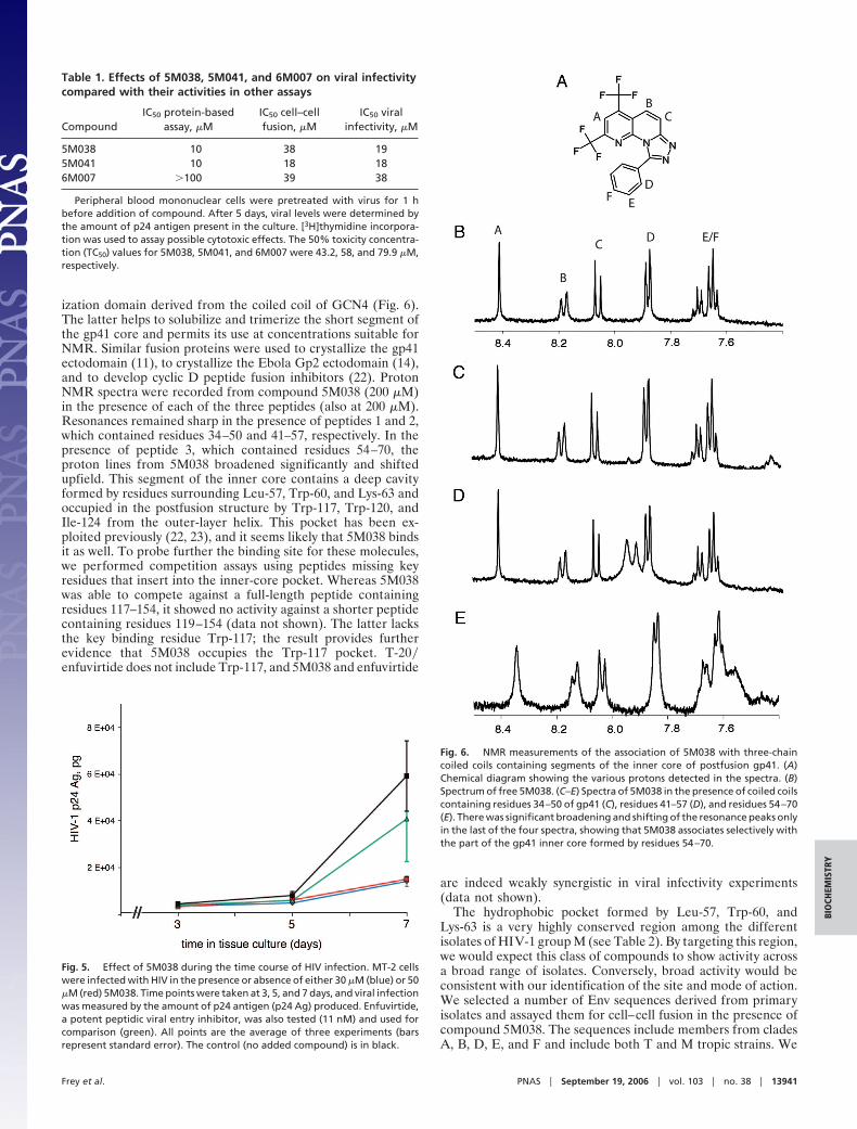
ization domain derived from the coiled coil of GCN4 (Fig. 6).The latter helps to solubilize and trimerize the short segment ofthe gp41 core and permits its use at concentrations suitable forNMR. Similar fusion proteins were used to crystallize the gp41ectodomain (11), to crystallize the Ebola Gp2 ectodomain (14),and to develop cyclic D peptide fusion inhibitors (22). ProtonNMR spectra were recorded from compound 5M038 (200 �M)in the presence of each of the three peptides (also at 200 �M).Resonances remained sharp in the presence of peptides 1 and 2,which contained residues 34–50 and 41–57, respectively. In thepresence of peptide 3, which contained residues 54–70, theproton lines from 5M038 broadened significantly and shiftedupfield. This segment of the inner core contains a deep cavityformed by residues surrounding Leu-57, Trp-60, and Lys-63 andoccupied in the postfusion structure by Trp-117, Trp-120, andIle-124 from the outer-layer helix. This pocket has been ex-ploited previously (22, 23), and it seems likely that 5M038 bindsit as well. To probe further the binding site for these molecules,we performed competition assays using peptides missing keyresidues that insert into the inner-core pocket. Whereas 5M038was able to compete against a full-length peptide containingresidues 117–154, it showed no activity against a shorter peptidecontaining residues 119–154 (data not shown). The latter lacksthe key binding residue Trp-117; the result provides furtherevidence that 5M038 occupies the Trp-117 pocket. T-20�enfuvirtide does not include Trp-117, and 5M038 and enfuvirtide
are indeed weakly synergistic in viral infectivity experiments(data not shown).
The hydrophobic pocket formed by Leu-57, Trp-60, andLys-63 is a very highly conserved region among the differentisolates of HIV-1 group M (see Table 2). By targeting this region,we would expect this class of compounds to show activity acrossa broad range of isolates. Conversely, broad activity would beconsistent with our identification of the site and mode of action.We selected a number of Env sequences derived from primaryisolates and assayed them for cell–cell fusion in the presence ofcompound 5M038. The sequences include members from cladesA, B, D, E, and F and include both T and M tropic strains. We
Table 1. Effects of 5M038, 5M041, and 6M007 on viral infectivitycompared with their activities in other assays
CompoundIC50 protein-based
assay, �MIC50 cell–cellfusion, �M
IC50 viralinfectivity, �M
5M038 10 38 195M041 10 18 186M007 �100 39 38
Peripheral blood mononuclear cells were pretreated with virus for 1 hbefore addition of compound. After 5 days, viral levels were determined bythe amount of p24 antigen present in the culture. [3H]thymidine incorpora-tion was used to assay possible cytotoxic effects. The 50% toxicity concentra-tion (TC50) values for 5M038, 5M041, and 6M007 were 43.2, 58, and 79.9 �M,respectively.
Fig. 5. Effect of 5M038 during the time course of HIV infection. MT-2 cellswere infected with HIV in the presence or absence of either 30 �M (blue) or 50�M (red) 5M038. Time points were taken at 3, 5, and 7 days, and viral infectionwas measured by the amount of p24 antigen (p24 Ag) produced. Enfuvirtide,a potent peptidic viral entry inhibitor, was also tested (11 nM) and used forcomparison (green). All points are the average of three experiments (barsrepresent standard error). The control (no added compound) is in black.
Fig. 6. NMR measurements of the association of 5M038 with three-chaincoiled coils containing segments of the inner core of postfusion gp41. (A)Chemical diagram showing the various protons detected in the spectra. (B)Spectrum of free 5M038. (C–E) Spectra of 5M038 in the presence of coiled coilscontaining residues 34–50 of gp41 (C), residues 41–57 (D), and residues 54–70(E). There was significant broadening and shifting of the resonance peaks onlyin the last of the four spectra, showing that 5M038 associates selectively withthe part of the gp41 inner core formed by residues 54–70.
Frey et al. PNAS � September 19, 2006 � vol. 103 � no. 38 � 13941
BIO
CHEM
ISTR
Y

also included two divergent strains, BCF03 from group O andSIV mac32H. The compound was active against all of the groupM isolates tested while exhibiting much weaker activity towardthe group O strain (only 40% inhibition at 100 �M) and SIV(35% inhibition at 100 �M).
We used the fluorescence polarization assay to examine otherinhibitors that target gp41. A peptide corresponding to the partof T-20 contained in gp41-5 (i.e., residues 127–154) and apeptidic entry inhibitor (23) both failed to compete with C38 atconcentrations up to 100 and 150 �M, respectively, under ourassay conditions ([*C38] � 5 nM), even though the peptidicentry inhibitor blocks cell–cell fusion. These results are similarto our observations with 6M007. The in vitro f luorescencepolarization assay is an equilibrium measurement; the inhibitionof fusion (cell–cell or virus–cell) is probably a kinetic one.Moreover, there are three potential sites per envelope trimer inthe cellular assays, but only one in our gp41-5 measurement. Forboth these reasons, competitive inhibition of the binding of theouter helix to gp41-5 should be a more stringent criterion ofinteraction than inhibition of fusion, and the spectrum ofpotential small-molecule fusion inhibitors may be broader thanthe range detected in our present assay. The limited solubility ofsome of the compounds, such as 6M007, and its variation withbuffer, ionic strength, or other factors may also account fordifferences between cell-based and protein-based assays.
The likelihood of finding small, drug-like compounds that canblock protein association is currently a subject of considerabledebate. The coiled coil is a relatively special kind of proteininterface, but our results suggest that suitably designed, sensitive,structure-based assays can indeed detect such interference andthat a screen of relatively modest extent can uncover compoundswith IC50 values in the low micromolar range. Understandingproperly how compounds such as 5M038 inhibit C38 associationwith gp41-5 and, hence, how they interfere with viral fusion willrequire structures of suitable bound complexes. Although effortsto cocrystallize 5M038 with gp41 have thus far failed, it is alreadyclear that a structure-based assay like the one described here canlead to detection of refolding inhibitors in chemical libraries.
Materials and MethodsGp41-5 Cloning, Expression, and Purification. We have expressed asingle-chain model for five of the six gp41 helices (Fig. 2 A). Theconstruct contains residues 35–70 of HR1 and residues 117–150of HR2 from HXB2. The amino acid sequence for HR1 isSGIVQQQNNLLRAIEAQQHLLQLTVWGIKQLQARIL,and the sequence for HR2 is WMEWDREINNYTSLIH-SLIEESQNQQEKNEQELL. The C terminus of each of the firsttwo inner-core segments is connected to the N terminus of thesucceeding outer-layer helix by the 6-residue linker SGGRGG.The C terminus of each outer-layer helix is connected to the
N-terminus of the succeeding inner-core segment by the 6-res-idue linker GGKGGS. The DNA fragment encoding gp41-5 wassubcloned into the expression vector pRSET (Invitrogen, Carls-bad, CA) and transformed into Escherichia coli cells BL21DE3�pUBS. For purification, cell pellets were dissolved in cold(4°C) glacial acetic acid and incubated on ice for 30 min. Celldebris was removed by centrifugation (39,000 � g, 30 min). Thesupernatant was diluted to 10% acetic acid with deionized waterand loaded onto a reversed-phase C18 column (Vydac, Hesperia,CA). The column was eluted with an acetonitrile gradient(30–90%). The protein eluted at 50% acetonitrile; it was �90%pure as judged by SDS�PAGE.
Gp41-5 Refolding. Lyophilized protein was dissolved in 6 Mguanidine HCl at a concentration of 1 mg�ml and dialyzedsuccessively against 100 mM glycine, pH 3.5, and PBS, pH 7.4.The precipitate was removed by centrifugation, and the protein(�98% pure as judged by SDS�PAGE) was used without furtherpurification.
Peptide Synthesis. All peptides were synthesized by using Fmocchemistry on PAL (PE Biosystems, Warrington, U.K.) supportsby using an Applied Biosystems (Foster City, CA) model 431peptide synthesizer. Peptides were cleaved by using reagent R[TFA�thioanisole�1,2-ethanedithiol (EDT)�anisole, 90:5:3:2],precipitated into cold diethyl ether and purified by reversed-phase C18 HPLC (0.1% TFA�acetonitrile gradient). All pep-tides were characterized by electrospray mass spectrometry atthe Mass Spectrometry Facility of the Department of Chemistryand Chemical Biology at Harvard University (Cambridge, MA).Labeling of peptides at the N terminus was achieved as follows.Synthetic peptide, still attached to the resin and with side chainsprotected but N terminus deprotected, was suspended in a smallvolume of NMP. 5-FAM (Molecular Probes, Carlsbad, CA) wasdissolved in NMP and added to the peptide suspension followedby the addition of 10 �l of 4-methyl morpholine. The reactionwas allowed to proceed under slow stirring for �2 days. Cleavageand deprotection of labeled peptides were performed as de-scribed above.
High-Throughput Screening. All screens were performed at theHarvard Medical School Institute for Chemistry and ChemicalBiology (ICCB). Gp41-5 at a concentration of 7.4 nM inPBS�Tween 20 was loaded into 384-well microtiter plates (30-�lvolume per well), followed by the transfer of 0.1 �l of compound(5 mg�ml in DMSO). After incubation for 1 h at room temper-ature, f luorescein-labeled C38 was added to each well (finalconcentration of 5–10 nM). After a 90-min incubation period atroom temperature, f luorescence polarization measurementswere recorded on an Analyst HTS (Molecular Devices, Sunny-vale, CA). A 12-h time point revealed no major differences fromthe 90-min time point. Compounds that appeared to be activewere retested independently. Commercial libraries screenedincluded those from CEREP (Redmond, WA), Maybridge(Trevillet, Tintagel, Cornwall, U.K.), Bionet (Ryan Scientific,Mount Pleasant, SC), and ChemBridge (San Diego, CA). Thehits described in this work were from Bionet (the 5M series) andCEREP (S2986).
Cell–Cell Fusion Assay. Assays were performed as described pre-viously (23, 27) with the following modifications. Target andeffector cells were mixed briefly at equal concentrations beforetransfer to 96-well plates. All inhibitors were dissolved in DMSOand diluted 100-fold in the final assay. After the addition ofinhibitor, cells were incubated at 30°C for 6 h, a time pointpreviously determined to be in the linear range of the assay.Assays were stopped by aspiration of media followed by addition
Table 2. Effect of 5M038 on cell–cell fusion mediated byenvelope glycoproteins from various HIV and SIV isolates
Isolate IC50, �M Clade Coreceptor Pocket sequence
HXB2 38 B X4 LTVWGIKQLQARIL
92ug024.2 30 D X4 LTVWGIKQLQARVL
93th976.10 40 E Unknown LTVWGIKQLQARVL
JRFL 40 B R5 LTVWGIKQLQARVL
93br029.2 50 F R5 LTVWGITQLQARIL
92ug037.8 50 A R5 LTVWGIKQLQARVL
92us716.6 70 B R5 LTVWGIKQLQARVL
BCF03 �100 Group O Unknown LSVWGIRQLRARLQ
SIV mac32H �100 SIV — LTVWGTKNLQTRVT
The pocket sequence is the amino acid sequence of residues 57–70 (HXB2numbering) on the inner-core helix of gp41.
13942 � www.pnas.org�cgi�doi�10.1073�pnas.0601036103 Frey et al.

of reporter lysis buffer (Promega, Madison, WI) and transfer to�20°C.
Viral Infectivity Assay. Peripheral blood mononuclear cells (PB-MCs) were exposed to HIV-1 isolate 2076 [1,000 tissue culture50% infective dose (TCID50) per 106 cells] for 1 h. Cells werewashed once with medium, and 0.1 ml of HIV-infected PBMCs(2 � 105 cells) were added to each well of a 96-flat-well cell platefollowed by the addition of compound. The final concentrationof DMSO in each well was 0.5%. Supernatants were harvestedafter 5 days and assayed for HIV-1 p24 antigen production. Fortime course assays, 106 MT-2 cells were cultured in 1 ml of RPMImedium 1640 with 10% FCS in individual wells of a 24-well plate.In selected wells, 5M038 or enfuvirtide was added to each wellto achieve final concentrations of 30 or 50 �M for 5M038 or 50ng�ml (11 nM) enfuvirtide. Each control or experimental con-dition was conducted in triplicate. After 1 h, 500 TCID50HIV-IIIB was added, and cells were incubated for 7 days at 37°Cin 5% CO2. Supernatant fluid (100 �l) was removed from eachwell on days 3, 5, and 7 for HIV-1 p24 antigen quantification. Themedium was replaced with culture medium with the appropriateconcentrations of inhibitor. HIV-1 p24 antigen was quantified byusing kits according to the manufacturer’s instructions (Perkin–Elmer, Wellesley, MA).
Gp41 Chimeras. All chimeras were synthesized by using Fmocchemistry and purified as described above. The sequence for pep-tide 1 was Ac-RMKQIEDKIEEIESKQKKIENEIARIKKLLSQ-IVQQQNNLLRAIEA-NH2. The sequence for peptide 2 wasAc-RMKQIEDKIEEIESKQKKIENEIARIKKLQNNLLRA-IEAQQHLLQL-NH2. The sequence for peptide 3 (IQN17) was
Ac-RMKQIEDKIEEIESKQKKIENEIARIKKLLQLTVWG-IKQLQARIL-NH2. All peptides were analyzed by CD spectros-copy and analytical ultracentrifugation. Peptides 1 and 3 were fullyhelical; peptide 2 was �60% helical. Peptides 1 and 3 gave apparentmolecular weights consistent with a trimer, whereas peptide 2 gavea slightly lower apparent molecular weight (11,000 as comparedwith the expected 16,782). We interpreted this last result to indicatea mixture of monomer and trimer. Size exclusion chromatographysupported this interpretation.
Proton NMR Experiments. All assays were performed on a Varian(Palo Alto, CA) 400-MHz spectrophotometer with a tripleresonance probe. Samples were prepared in PBS�2H2O. Com-pound stocks were prepared in DMSO-d6 at 10 mg�ml (�26mM) and diluted in PBS�2H2O to a final concentration of 200�M (final DMSO, �1%). Spectra were in the presence orabsence of protein, also at 200 �M (based on Mr of trimer).
We thank Genfa Zhou (FusoGen Pharmaceuticals, Tianjin, China), whodesigned the original gp41-5 construct; Rebecca Ward, who facilitated allinitial contacts with the Harvard Medical School Institute for Chemistryand Cell Biology; Tim Strassmeier and Daniel Oprian (Brandeis Uni-versity, Waltham, MA) for help in the early stages of this work; BeatriceHahn (University of Alabama, Birmingham, AL) for DNA encodingenvelopes from a panel of HIV and SIV isolates; Caroline Shamu foradvice on screening; and Jon Clardy for comments on the manuscript.The screens were carried out at the Harvard Medical School Institute forChemistry and Cell Biology. This work was supported by Ruth L.Kirschstein National Research Service Award AI52859 (to G.F.), Na-tional Institutes of Health Program Project Grant GM39589 (to S.C.H.),and Centers for AIDS Research Grant AI36214 (to X.-Q.Z. and R.T.S.).S.C.H. is an Investigator of the Howard Hughes Medical Institute.
1. Harrison SC (2005) Adv Virus Res 64:231–261.2. Allan JS, Coligan JE, Barin F, McLane MF, Sodroski JG, Rosen CA, Haseltine
WA, Lee TH, Essex M (1985) Science 228:1091–1094.3. Veronese FD, DeVico AL, Copeland TD, Oroszlan S, Gallo RC, Sarngadharan
MG (1985) Science 229:1402–1405.4. Gallaher WR (1987) Cell 50:327–328.5. Moore JP, McKeating JA, Weiss RA, Sattentau QJ (1990) Science 250:1139–
1142.6. Hart TK, Kirsh R, Ellens H, Sweet RW, Lambert DM, Petteway SR, Jr, Leary
J, Bugelski PJ (1991) Proc Natl Acad Sci USA 88:2189–2193.7. Sattentau QJ, Moore JP (1991) J Exp Med 174:407–415.8. Furuta RA, Wild CT, Weng Y, Weiss CD (1998) Nat Struct Biol 5:276–279.9. Kowalski M, Potz J, Basiripour L, Dorfman T, Goh WC, Terwilliger E, Dayton
A, Rosen C, Haseltine W, Sodroski J (1987) Science 237:1351–1355.10. Carr CM, Kim PS (1993) Cell 73:823–832.11. Weissenhorn W, Dessen A, Harrison SC, Skehel JJ, Wiley DC (1997) Nature
387:426–430.12. Chan DC, Fass D, Berger JM, Kim PS (1997) Cell 89:263–273.13. Fass D, Harrison SC, Kim PS (1996) Nat Struct Biol 3:465–469.14. Weissenhorn W, Carfi A, Lee KH, Skehel JJ, Wiley DC (1998) Mol Cell
2:605–616.
15. Wild C, Oas T, McDanal C, Bolognesi D, Matthews T (1992) Proc Natl AcadSci USA 89:10537–10541.
16. Wild C, Greenwell T, Matthews T (1993) AIDS Res Hum Retroviruses 9:1051–1053.17. Wild CT, Shugars DC, Greenwell TK, McDanal CB, Matthews TJ (1994) Proc
Natl Acad Sci USA 91:9770–9774.18. Jiang S, Lin K, Strick N, Neurath AR (1993) Biochem Biophys Res Commun
195:533–538.19. Jiang S, Lin K, Strick N, Neurath AR (1993) Nature 365:113 (lett).20. Kilby JM, Eron JJ (2003) N Engl J Med 348:2228–2238.21. Chan DC, Chutkowski CT, Kim PS (1998) Proc Natl Acad Sci USA 95:15613–
15617.22. Eckert DM, Malashkevich VN, Hong LH, Carr PA, Kim PS (1999) Cell
99:103–115.23. Ferrer M, Kapoor TM, Strassmaier T, Weissenhorn W, Skehel JJ, Oprian D,
Schreiber SL, Wiley DC, Harrison SC (1999) Nat Struct Biol 6:953–960.24. Debnath AK, Radigan L, Jiang S (1999) J Med Chem 42:3203–3209.25. Root MJ, Kay MS, Kim PS (2001) Science 291:884–888.26. Cianci C, Langley DR, Dischino DD, Sun Y, Yu KL, Stanley A, Roach J, Li
Z, Dalterio R, Colonno R, et al. (2004) Proc Natl Acad Sci USA 101:15046–15051.
27. Nussbaum O, Broder CC, Berger EA (1994) J Virol 68:5411–5422.
Frey et al. PNAS � September 19, 2006 � vol. 103 � no. 38 � 13943
BIO
CHEM
ISTR
Y


![Panasonic Tc-21fx30l 29fx30l Chassis-gp41 [ET]](https://img.dokumen.tips/doc/110x75/54800066b37959a22b8b5909/panasonic-tc-21fx30l-29fx30l-chassis-gp41-et.jpg)