Embed Size (px)
Citation preview

SMALL MOLECULE INHIBITORS OF THE SARS-COV NSP15
ENDORIBONUCLEASE, MECHANISM
OF ACTION AND INSIGHT INTO
CORONAVIRUS INFECTION
A Thesis
by
JOANNA MARIA ORTIZ ALCANTARA
Submitted to the Office of Graduate Studies of Texas A&M University
in partial fulfillment of the requirements for the degree of
MASTER OF SCIENCE
May 2009
Major Subject: Biochemistry

SMALL MOLECULE INHIBITORS OF THE SARS-COV NSP15
ENDORIBONUCLEASE, MECHANISM
OF ACTION AND INSIGHT INTO
CORONAVIRUS INFECTION
A Thesis
by
JOANNA MARIA ORTIZ ALCANTARA
Submitted to the Office of Graduate Studies of Texas A&M University
in partial fulfillment of the requirements for the degree of
MASTER OF SCIENCE
Approved by:
Chair of Committee, Cheng C. Kao Committee Members, Andreas K. H. Holzenburg Julian L. Leibowitz Ryland F. Young Head of Department, Gregory D. Reinhart
May 2009
Major Subject: Biochemistry

iii
ABSTRACT
Small Molecule Inhibitors of the SARS-CoV Nsp15 Endoribonuclease, Mechanism of
Action and Insight into Coronavirus Infection. (May 2009)
Joanna Maria Ortiz Alcantara, B.S., National Autonomous University of Mexico
Chair of Advisory Committee: Dr. Cheng C. Kao
The Severe Acute Respiratory Syndrome (SARS) virus encodes several unusual
RNA processing enzymes, including Nsp15, an endoribonuclease that preferentially
cleaves 3’ of uridylates through a Ribonuclease A-like mechanism. Crystal structures of
Nsp15 confirmed that the Nsp15 active site is structurally similar to that of Ribonuclease
A. These similarities and our molecular docking analysis lead us to hypothesize that
previously characterized Ribonuclease A inhibitors will also inhibit the SARS-CoV
Nsp15. Benzopurpurin B, C-467929, C-473872, N-36711, N-65828, N-103018 and
Congo red were tested for effects on Nsp15 endoribonuclease activity. A real-time
fluorescence assay revealed that the IC50 values for inhibiting Nsp15 were between 0.2
µM and 40 µM. Benzopurpurin B, C-473872, and Congo red are competitive inhibitors,
according to kinetic studies and were demonstrated to bind SARS-CoV Nsp15 by a
differential scanning fluorimetry assay. Benzopurpurin B also inhibited the Nsp15
orthologs from two other coronaviruses: mouse hepatitis virus (MHV) and infectious
bronchitis virus. The three compounds reduced infectivity of MHV in L2 cells by 8 to 26
fold. The more effective drugs also caused a decrease in MHV RNA accumulation.

iv
DEDICATION
To my Mom and Dad, Cecilia and Bernardo, for their love, guidance and support,
for their encouragement to be better everyday and for their wise words.
To my sisters, Ivette and Nelly for being such good friends and for their
geniality, for all the good times we spend together.
To Hector, for being the light of my life, making everyday so shiny, because with
you around everything makes sense and there is always hope, for sharing with me all
those laughs and tears, for all the moments you were here and you were not and for
being so patient. I love you.

v
ACKNOWLEDGEMENTS
I would like to thank my committee chair, Dr. Kao, and my committee members,
Dr. Holzenburg, Dr. Leibowitz, and Dr. Young, for their support and guidance
throughout the course of this research.
I extend my gratitude to my colleagues at Texas A & M University, specially
Kanchan Bhardwaj, Satheesh Palaninathan, Lillian Li Yi and Will Snee and everybody
in Kao’s lab who were willing to share their knowledge and experience to support my
research. I extend my gratitude to the Biochemistry & Biophysics faculty and staff for
making my time here a great and rich experience. I also gratefully acknowledge a
National Science Foundation supplemental grant to develop the differential fluorimetry
assay, and the NIAID which partially provided the funding for this research.
Thanks to my mom, dad and sisters for their encouragement, to Hector for his
patience and love and Adriana Perea, Catalina Velez and Hermes Reyes for their
friendship.

vi
NOMENCLATURE
SARS-CoV Severe Acute Respiratory Syndrome Coronavirus
Nsp15 Nonstructural Protein 15
RNase A Ribonuclease A
MHV Mouse Hepatitis Virus
IBV Infectious Bronchitis Virus

vii
TABLE OF CONTENTS
Page
ABSTRACT .............................................................................................................. iii
DEDICATION ........................................................................................................... iv
ACKNOWLEDGEMENTS ....................................................................................... v
NOMENCLATURE .................................................................................................. vi
TABLE OF CONTENTS .......................................................................................... vii
LIST OF FIGURES ................................................................................................... ix
INTRODUCTION ..................................................................................................... 1 Biology of Coronavirus Infection .................................................................. 2 The SARS-CoV Nsp15 Endoribonuclease .................................................... 10 OBJECTIVES ............................................................................................................ 18
MATERIALS AND METHODS .............................................................................. 19
Reagents ......................................................................................................... 19 Molecular Modeling ...................................................................................... 19 Endoribonuclease Assays .............................................................................. 20 Differential Scanning Fluorimetry ................................................................. 21 Plaque Formation Assays .............................................................................. 21 Labeling of Viral RNAs ................................................................................ 22 RESULTS .................................................................................................................. 24
Computational Docking of RNase A and Angiogenin Inhibitors .................. 24 Effects of the Inhibitors on sNsp15 Endoribonuclease Activity ................... 26 Mechanism of Action of Select Inhibitors ..................................................... 27 The MHV and IBV Nsp15 Activity are Inhibited by Benzopurpurin B in vitro ............................................................................... 30 Inhibition of MHV Infection in Cultured Cells ............................................. 32 DISCUSSION ............................................................................................................ 34

viii
Page
CONCLUSIONS ....................................................................................................... 37
REFERENCES .......................................................................................................... 39
VITA .......................................................................................................................... 45

ix
LIST OF FIGURES
FIGURE Page
1 Domain organization and proteolytic processing of SARS-CoV replicase proteins ................................................................ 5 2 Genome organization and RNA synthesis of SARS-CoV .......................... 7 3 Mechanism of action of Ribonuclease A .................................................... 13 4 Molecular docking of UMP into the Nsp15 active site .............................. 15 5 Small molecule inhibitors tested for effects on Nsp15 ............................... 25 6 Effects of the small molecule inhibitors on the SARS-CoV Nsp15 endoribonuclease activity ................................................................ 27
7 Mechanism of inhibition of Nsp15 by select inhibitors ............................. 29 8 Inhibition of RNA cleavage in vitro in other coronavirus Nsp15 orthologs .......................................................................................... 31
9 Effects of select Nsp15 inhibitors on MHV plaque formation in mouse L2 cells ........................................................................................ 33

1
INTRODUCTION
The Severe Acute Respiratory Syndrome (SARS) outbreak emerged in
Southeastern China in 2002 and eventually spread to 30 countries by 2003. SARS was
characterized by an atypical pneumonia transmitted primarily by contact with infectious
respiratory droplets or fomites and it was responsible for 8096 confirmed cases with a
mortality rate of about 10% (WHO). SARS is caused by the SARS coronavirus (SARS-
CoV) (1, 2, 3). Horseshoe bats are the natural reservoir of SARS-CoV like viruses but
they have been isolated also from palm civets and raccoon dogs from wild animal
markets in China, suggesting that these mammals served as an amplification host and
could be the source of infection in humans (4, 5, 6). The existence of a natural reservoir
raises the possibility of a reemergence of SARS. The treatment used during the
epidemic consisted of corticoid steroids and ribavirin (1, 2, 3). In hindsight, some of
these treatments had severe side effects and minimal efficacy (1, 2, 3). Currently there
are no approved anti-virals against SARS (7), and specific therapeutic options will be
needed to contain future outbreaks.
SARS is a member of the group II Coronavirus within the order Nidovirales (8).
Coronaviruses are enveloped and their positive-strand RNA genomes are the largest
known, they are of interest in their mechanism of gene expression and replication. In this
section, a general overview of the biology of coronaviruses will be presented, from their
____________ This thesis follows the style of The Journal of Biological Chemistry.

2
taxonomy, morphology and genome organization to their viral replication cycle,
including a summary of the proposed RNA synthesis and processing mechanisms and
the proteins involved (for reviews of coronaviruses, see references 9-11).
Biology of Coronavirus Infection
Taxonomy
The Coronaviridae family of enveloped RNA viruses is formed by two genera,
the coronaviruses and the toroviruses, which together with the arteriviruses and
roniviruses form the order Nidovirales. Members of the coronavirus family are divided
in three groups, originally based on antigenic relationships, later by comparisons of the
sequences of the entire viral genomes (12). Coronaviruses cause respiratory or intestinal
infections. Most of the group 1 and group 2 coronaviruses infect mammals, and human
coronaviruses are classified in these groups. Group 3 viruses have avian hosts only.
Mouse hepatitis virus (MHV), human coronavirus OC43, and the bovine coronavirus are
members of group 2, of which SARS-CoV according to rooted phylogenetic trees
together with bat SARS-CoV form group 2b, and the infectious bronchitis virus is
member of group 3 (8, 13).
Virion morphology
Coronaviruses are spherical, with virions of about 80-120 nm projecting club-
like, petal shaped surface spikes of 17-20 nm from the lipid envelope, and helical
nucleocapsids according to negatively stained electron micrographs (14, 15).

3
Genome organization
Coronaviruses have nonsegmented, single-stranded positive RNA genomes of
about 30 kb, with 5’ caps and 3’ poly(A) tails; the largest among all RNA viruses. The
SARS-CoV genome contains ~29730 nucleotides excluding the 3’ poly(A) tail. The 5’
and the 3’ untranslated regions contain 265 and 342 nucleotides respectively. The
genome is predicted to have 14 functional open reading frames (ORFs). Two large, 5’-
terminal ORFs 1a and 1b form the replicase gene that encodes 16 nonstructural proteins
including those required for viral RNA synthesis. The other twelve ORFs encode the
structural proteins S, M, N and E and eight accessory proteins (16, 17, 18).
Viral replication cycle and virion assembly
Coronaviruses entry into target cells initiate with binding of the viral S (Spike)
protein to cellular receptors. Trimers of the Spike protein decorate the virion surface like
the spikes in a crown and inspired the name for this group of viruses. The Spike protein
is heavily glycosylated and have three domains, the N-terminal S1, which is conserved
and the C-terminal transmembrane and cytoplasmic S2 subdomains. The minimal
binding domain of the SARS-CoV S protein was mapped to the residues 318-510 of S1
and antibodies specific for this subunit neutralize SARS-CoV infection (19, 20).
Aminopeptidase N, a zinc metalloprotease is the receptor of several group 1
coronaviruses (21); carcinoembryonic antigen-related cell adhesion molecules are
cellular receptors of MHV from group 2 (22) and the angiotensin-converting enzyme 2
(ACE2), a metalloprotease expressed in the lung, intestine, liver, heart, vascular

4
endothelium, testis and kidney is the functional cellular receptor of SARS-CoV (23, 24).
S1 binds to the cellular receptor triggering conformational changes that will co-localize
the fusion peptide upstream of the two heptad repeats of S2 to the transmembrane
domain to facilitate fusion of the viral and cellular membranes. SARS-CoV membrane
fusion is activated within endosomes by cathepsin L proteolysis (25, 26).
Following the fusion, the viral genome RNA is released into the cytoplasm of the
infected cell, the mechanism involved in the RNA uncoating is not well understood.
Genome expression of the SARS-CoV starts with the cap-dependent translation of the
genomic RNA, mRNA 1, producing a 4382 amino-acid protein called polyprotein 1
(pp1a) encoded by ORF 1a; then the 7073-residue polyprotein 1ab (pp1ab) encoded by
ORFs 1a and b sequences is produced by ribosomal frameshifting. This occurs into the -
1 reading frame, just upstream of the ORF1a translation stop codon mediated by the
signal of the slippery sequence 13392UUUAAAC13398 and a downstream RNA pseudoknot
structure. Polyproteins pp1a and pp1ab are processed by viral proteinases into Nsp1-16
to form the viral replicase-transcriptase multi-protein complex. In general, coronaviruses
process the replicative polyproteins at the N-proximal regions at three sites using two
papain-like proteases PL1pro and PL2pro. SARS-CoV uses only one papain-like protease,
a PL2pro orthologue with narrow substrate specificity, containing a putative Zn-finger
structure required for the proteolytic activity (18, 27, 28). The central and C-terminal
regions of pp1a and pp1b are processed by the chymotrypsin-like protease 3CL pro, also
called the coronavirus main protease, Mpro , that releases the RdRp and helicase, key

5
replicative functions. The SARS-CoV 3CL pro cleaves pp1a and pp1ab at 11 sites, as
illustrated in Fig. 1 (18, 29, 30).
FIGURE 1. Domain organization and proteolytic processing of SARS-CoV replicase proteins. Taken from Ziehbur J. (2004) Curr. Opin. Microbiol. (31).
The viral replicase-transcriptase complex mediates the viral genome replication
and the transcription of a nested set of eight subgenomic (sg) mRNAs, both require the
synthesis of negative-strand intermediates. All the sg mRNAs have a 72-nucleotide, 5’-
terminal leader sequence identical to the 5’-end of the genome, and they are generated
by discontinuous synthesis rather than full-length RNA transcription as supported by UV
transcription mapping experiments in MHV-infected cells in which the size of a given
mRNA was correlated to the target size for ultraviolet light irradiation for the synthesis
of that mRNA (32) and principally by mixed infection experiments, in which the

6
reassortment of leader sequences was observed in the mRNAs produced in cells infected
with two different strains of MHV (33). The discontinuous synthesis of RNA depends on
transcription-regulating sequences TRSs, cis-active RNA elements with the common
core sequence ACGAAC for the SARS-CoV (18). There is currently consensus in the
field about discontinuous transcription occurring during the synthesis of minus RNA
strands. In the leader-primed transcription model the leader RNA is produced first, then
transferred to the full-length minus strand, to the complementary TRS (34). In a second
model, the nascent minus strand synthesis is paused at the TRSs and translocated to the
leader TRS located downstream of the 5’-leader sequence on the genomic RNA, to
continue the transcription. The minus-strand RNAs containing the anti-leader sequence
are then used as templates for continuous plus-strand synthesis of sg mRNAs (35, 36,
37). Complementary base-pairing and protein-protein interactions keep the 5’ end of the
genome close to the site of minus-strand synthesis in both models. The negative-strand
discontinuous transcription model is currently gaining favored status (37). The finding in
infected cells of subgenomic minus strand RNAs with antileader sequence, subgenomic
replicative-intermediate RNAs active for transcription for each mRNA (38, 39, 40) as
well as the analysis of RNA synthesis using equine arterivirus full-length infectious
cDNA in which mutations were introduced in the leader or body copies of TRS
performed support this model (41, 42, 43, 44). The SARS-CoV structural proteins S, M,
N and E, and eight additional proteins with unknown functions are predicted to be
encoded in RNAs 2-9. mRNAs 2, 4, 5 and 6 are functionally monocistronic, mRNAs 3,
7, 8 and 9 bicistronic, Fig. 2. (8, 18).

7
FIGURE 2. Genome organization and RNA synthesis of SARS-CoV. Taken from Ziehbur J. (2004) Curr. Opin Microbiol. (31).
Coronaviruses likely attach the replication machinery to the membrane of
autophagosomes forming double-membrane vesicles (9). The membrane bound
structural proteins M, S, and E are inserted into the endoplasmic reticulum ER, then they
transit to the endoplasmic reticulum-Golgi intermediate compartment ERGIC. When
viral genomic RNA and structural proteins are accumulated, the N protein, the most
abundantly expressed viral protein, encapsidate the progeny genomes forming
nucleocapsids that interact with the membrane-bound proteins and bud into the ERGIC
to form virions. Progeny virions are transported to the plasma membrane in vesicles or
Golgi sacs not yet clearly defined to be exported out of the infected cell (9).

8
Proteins involved in RNA synthesis and processing
The roles of many of the nonstructural proteins that assemble a membrane-
associated replicase-transcriptase complex are not completely elucidated. However,
several have novel functions that are not found in smaller positive-strand RNA viruses
(8). Included in these are proteins Nsp8, a putative RNA primase for Nsp12, the RNA-
dependent RNA polymerase; Nsp14, a 3’ to 5’ exoribonuclease that may serve
proofreading functions; Nsp15, an endoribonuclease, and Nsp16 a putative
methyltransferase that acts on the ribose 2’ hydroxyl (45, 46, 47, 48).
Nsp12 is the coronavirus RNA-dependent RNA polymerase (RdRp), it is
predicted to have the fingers, palm and thumb domains characteristic of other viral and
reverse transcriptases, and unique to coronaviruses a very large amino-terminal domain
essential for activity and containing the segment responsible for the association of the
RdRp with intracellular membranes. RdRp has been shown to associate with Mpro, Nsp8
and Nsp9 (49, 50). Nsp8 is a putative second RdRp unique to coronaviruses with
specificity for the internal 5’-(G/U)CC-3’ trinucleotides on RNA templates to start the
synthesis of complementary oligonucleotides of less than 6 nucleotides with low fidelity.
It is proposed to produce primers for the Nsp12 RdRp (45). In this regard, Nsp8 is highly
novel in acting as a RNA primase for a viral RNA-dependent RNA polymerase.
Nsp13 is a helicase with multiple activities according to studies in HCoV-229E
and SARS-CoV. It binds single and double-stranded RNA and DNA. It has NTPase and
dNTPase activities, that are template-dependent, probably to provide the energy to
translocate along the template. It unwinds both DNA and RNA duplexes and is more

9
processive in a 5’-to-3’ direction, suggesting a role in the preparation of the template for
RNA synthesis by the RdRp complex. It has RNA 5’-triphosphatase activity, proposed
to participate in the 5’ cap synthesis on coronavirus RNA (51, 52, 53, 54).
Nsp14 has exoribonuclease activity. It has been shown to act on the 3’ to 5’
direction on single and double-stranded RNA and requires divalent metal ions for its
activity. It does not hydrolyze ribose-2’-O-methylated RNA or DNA substrates. Its
ribonucleolytic activity is regulated by binding to the dsRNA. It is involved in viral
RNA synthesis. Active site mutants of the human CoV 229E Nsp14 are defective in viral
sgRNA synthesis, they present reduced genome replication and no viable virus can be
recovered (46).
Nsp15, the topic of this research, is an uridylate-specific endoribonuclease that
will be described in detail later.
Nsp16 is a putative 2’-O-methyltransferase (2’-O-MT), activity that is speculated
to participate in RNA capping or in negative-strand RNA synthesis (8, 48).
Nsp14-16, with ExoN, Nendo U and 2’-O-MT activities respectively are located
in a single protein block in pp1ab just next to the RdRp and helicase, suggesting that
they may have concerted activities. Snijder et al. (8) proposed a parallel to two cellular
RNA processing pathways. In one of them, Xendo U and ExoN participate in the
cleavage of pre-mRNA to produce U16 and U86 intron encode small nucleolar RNAs
(snoRNAs) that could be used in subsequent rRNA processing mediated by 2’-O-
methylation and the 2’-O-MT activity. In the second, an ADP-ribose 1’’-phosphatase
(ADRP) and a cyclic phosphodiesterase (CPD) present only in some group 2

10
coronaviruses but not SARS would be involved, in comparison to their cellular
homologs that mediate the ADP-ribose 1’, 2’ cyclic phosphate processing to produce
mature tRNA. However, the roles of the viral enzymes in a given pathway as well as
their biological substrates are still elusive. It is interesting that although the ExoN,
NendoU, 2’-O-MT and ADRP activities are conserved in all coronaviruses, just the
NendoU is conserved in arteriviruses whose genomes are smaller, suggesting that given
the extremely large genome of the coronaviruses, extra activities are required for RNA
replication and transcription similar to DNA organisms but unprecedented for RNA
viruses; as an example, it has been speculated that ExoN could have a role in RNA
proofreading and repair, activities not seen in RNA viruses, as well as recombination.
These predictions still need to be proved experimentally (8).
The SARS-CoV Nsp15 Endoribonuclease
This section will summarize the features of the SARS-CoV Nsp15, required for
its enzymatic activity, and the structural properties required for oligomerization and
substrate recognition.
Nsp15 has endoribonuclease activity. It cleaves at RNA uridylates, stimulated by
Mn2+ as a cofactor, which could induce a conformational change in the protein according
to measurement of changes in tryptophan fluorescence (47, 48). Later it was
demonstrated that the cleavage occurs specifically at 3’ of uridylates, preferentially
unpaired, with higher affinity for substrates with consecutive uridylate bases, and in the
proper sequence context it can also cleave 3’ of cytidylates, and Mn2+ enhance RNA

11
binding (55). Upon cleavage of RNA molecules a 2’-3’ cyclic phosphate ended molecule
is produced (48, 55). It has been named Nendo U and it is a genetic marker of
nidoviruses that is not present in other RNA viruses (8, 48). It has similarities to Xendo
U, an endoribonuclease from Xenopus laevis that participates in the processing of pre-
mRNA to produce small nucleolar RNAs. Xendo U is also uridylate-specific, Mn2+
dependent and generates products with 2’-3’ cyclic phosphate ends; besides the
functional aspects both endoribonucleases have sequence similarities (8, 56, 57). Nsp15
plays a role in viral replication and transcription as demonstrated in a study with a
Human CoV 229E active site mutant in which viral RNA synthesis was abolished (48).
Furthermore, in mutational studies in the Arterivirus Nsp15 ortholog, several active site
mutants showed reduced plaque formation and virus titers (58). Similar observations
were reported for MHV A59 (59). Nsp15 is not able to cleave 2’-O-ribose methylated
RNA, suggesting a functional cooperation with Nsp14, a putative 2’-O-ribose
methyltransferase. (48).
The WT SARS-CoV Nsp15 was found to be in equilibrium between monomers,
trimers and hexamers in solution. Only the hexameric form is enzymatically active (60).
Transmission electron microscopy demonstrated that Nsp15 assembles into a hexameric
structure formed by a dimer of trimers interacting end-to end, forming a narrow tunnel in
the center and several channels. It is also the hexameric form that binds to RNA. The
RNA binding to the outside of the hexamer, interacting with both trimers, was suggested
by a low-resolution co-crystal of Nsp15 and RNA (55, 60). Crystal structures with one
subunit in the asymmetric unit have been solved for SARS-CoV Nsp15 and its MHV

12
ortholog (61, 62). Six catalytic sites are exposed at the surface when a hexamer is
assembled. The three catalytic residues His-234, His-249, Lys-289 located in the C-
terminal domain of the SARS-CoV Nsp15 resemble the catalytic triad His-12, Lys-41
and His-119 found in Ribonuclease A and although superimposition of tertiary structures
was impossible, the catalytic residues Lys-His-His of both enzymes were superimposed
successfully (61), supporting previous mutational studies in which the residues His-234,
His-249, Lys-289 and Asp-272 of SARS-CoV Nsp15 (60), and the corresponding
residues His-His-Lys in the MHV ortholog (62) were required for ribonucleolytic
activity, proving to have a direct role in catalysis, as it was suggested because they are
highly conserved among members of the Xendo U family and coronavirus orthologs (8).
Furthermore, mechanistic similarities between RNase A and Nsp15 are also conceivable,
in RNase A, His-12 and His-119 have general base and general acid functions, and the
pentacovalent intermediate is stabilized by the interaction of the phosphate moiety with
Lys-41. In SARS-Co-V Nsp15 the position of the two catalytic histidines suggest they
can accept and donate protons, and this is supported by the fact that Nsp15 cannot cleave
DNA or 2’-O-methylated RNA where there is no 2’-OH to be deprotonated by His-249;
for both enzymes 2’-3’ cyclic phosphate molecules are produced, see Fig. 3 (48, 55, 61,
63).

13
FIGURE 3. Mechanism of action of Ribonuclease A. Taken from Roberts G. et al. (1969) PNAS (63).
Of note the C-terminal tail of Nsp15 folds towards the active site suggesting that
residues at that location could be important for substrate recognition and specificity (61).
The elucidation of the structure of a SARS-CoV active-site mutant with six
subunits in the asymmetric unit led to the prediction of residues required for subunit
interaction, specificity in substrate recognition and RNA binding (64). The N-terminal
residue E3, previously demonstrated to be involved in oligomerization, plays a role in

14
trimer-trimer interactions according to the crystal structure (60, 64). Residues N52, I26
and I27, were also predicted to participate in trimer-trimer interactions. Interestingly, the
mutants E3A, N52A, I26, 27A expected to form trimers eluted as monomers in gel
filtration, suggesting that trimers are unstable intermediates. Consistent with hexamers
being required for endoribonuclease activity, Nsp15 with mutations that prevented
proper trimer-trimer interaction had endoribonuclease activity to less than 1% of WT.
D39, R90, E266, participate in intratrimer interactions, and mutations in these residues
in Nsp15 result in proteins that elute as monomers and show a significant reduction in
endoribonuclease activity to less than 1% of WT. A N163A mutant, also expected to be
affected on interactions between trimers, was found to form monomers and hexamers
with nearly equal distribution, and to have 29% of the activity of WT (21). A molecular
docking analysis with UMP revealed that some active site residues located in the C-
terminal tail could play a role in specificity and substrate recognition. S293, Y342, L345
and P343 were predicted to be important. Previously Ricagno et al. (61) observed that
superimposing RNase A and Nsp15 catalytic triads, the positions of S293 and Y342 of
Nsp15 were comparable to those of T45 and F120 of RNase A that participate in
pyrimidine specificity by forming hydrogen bonds and van der Waals interactions with
the base respectively. In the docking model, S293 could form H-bonds with the uracil,
and the aromatic ring of Y342 could stack against the uracil while P343 and L345 are
above it narrowing the pocket. The catalytic residues K289, H234 and H249 are
positioned to interact with the ribose 2’ OH and the phosphodiester group as illustrated
in Fig. 4 (64).
15
FIGURE 4. Molecular docking of UMP into the Nsp15 active site. Taken from Bhardwaj, et al. (2008) J. Biol. Chem. (64).
S293, L345, P343 and Y342 were demonstrated to be important for catalysis
since Nsp15 mutants of this residues showed either a reduction in endoribonuclease
activity in comparison to WT, or inactivity (Y342A), while no alterations in the
oligomerization state were observed. The role of S293 in specificity was established in a
study in which mutants to A, N or T were constructed. All the mutants formed hexamers.
In this study, S293T showed very low cleavage activity against cytidylates and
uridylates in comparison to WT, S293N cleaved cytidylates and uridylates with strong
preference for uridylates comparable to WT, while S293A presented similar rates of
cleavage of cytidylates and uridylates and was not able to cleave purines, demonstrating
that this residue is important to specifically recognize the substrate (64). P343 and L345
16
are also important for substrate recognition according to observations made in mutants
of these residues that would have a wider pocket or a more flexible active site. P343G
was showed to have reduced catalytic activity in comparison to WT and preference to
cleave uridylates over cytidylates. L345G had reduced cleavage activity, with nearly
equal preference for uridylates and cytidylates and interestingly it was able to cleave
purines, confirming that this residue should sterically block the access of a purine in the
WT catalytic pocket (64). In the same study, mutants H18A, T47A,N28/29A, N73A and
K256A presented decreased RNA binding as well as the active site mutants Y342A and
L345G, suggesting that these residues contribute to RNA binding (64).
Ribonucleases are classified in two main groups. The ribonucleases in the first
group include RNase H and RNase III, they depend on two metal ions, one to activate
the substrate, the second to stabilize the transition state; they cleave 5’ of the cognate
phosphodiester. The second group formed by members of the RNase A family and
RNase T1 are independent of metal ions and cleave at 3’ of the cognate phosphodiester.
Nsp15 shares characteristics of both groups, its activity is enhanced by Mn2+ and its
mechanism of cleavage is similar to that of RNase A; it together with Xendo U define a
novel family of endoribonucleases, endo U (65, 66, 67, 68).
Nendo U is a promising target for developing antiviral drugs, given that is widely
conserved among nidoviruses but absent in other RNA viruses, and it plays an important
role in viral RNA synthesis.
Compounds Benzopurpurin B, C-473872, C-467929, N-306711, N-103019 and
N-65828 were previously described as inhibitors binding to the active site of angiogenin

17
and RNase A, by Kao, R. Y., et al. (69) and Jenkins, J. L., et al. (70). Congo red is an
analog of Benzopurpurin B. The structural and mechanistic similarities between SARS-
CoV Nsp15 and RNase A suggested that those compounds might inhibit the
endoribonuclease activity of coronavirus Nsp15.
18
OBJECTIVES
The present study is intended to determine whether small-molecule inhibitors of
RNase A and Angiogenin, an RNase A–like enzyme, can affect enzymatic activity of the
SARS-CoV Nsp15 endoribonuclease (sNsp15) in vitro and coronavirus infection in
cultured cells.
Specific aim 1: To investigate whether the selected small-molecule inhibitors of
RNase A and Angiogenin are also able to inhibit the endoribonuclease activity sNsp15
and to establish their mechanism of inhibition. A real-time fluorescence assay was used
to study the efficacy of the compounds on the activity of purified sNsp15 in vitro. A gel-
based assay was used to confirm the results. To demonstrate binding of the compounds
to sNsp15 differential scanning fluorimetry assays were performed. The mechanism of
inhibition of the compounds was analyzed using kinetic studies.
Specific aim 2: To test the effects of the selected small-molecule compounds on
Nsp15 orthologs of other coronaviruses. The activity of the compounds on highly
purified recombinant MHV and IBV Nsp15 orthologs was established by the real-time
fluorescence assay.
Specific aim 3: To examine the effect of the compounds on MHV infection in
cultured cells and to investigate the effect of the compounds on viral RNA synthesis.
Plaque assays were performed in L2 cells for MHV. Agarose gel electrophoresis of viral
RNAs metabolically labeled in the presence of actinomycin D was used to study the
effect of the compounds on the production of genomic and sub-genomic MHV RNAs.

19
MATERIALS AND METHODS
Reagents
SARS and MHV Nsp15 His-tagged at the N termini, and IBV Nsp15 with an
His-tag at both termini were expressed in E. coli and purified by metal ion affinity
chromatography and Mono Q ion exchange chromatography as previously described
(47). The proteins were stored in a buffer containing 50 mM Tris (pH 7.9), 300 mM
NaCl, 1 mM dithiothreitol and 50% (v/v) glycerol at -20 °C. The protein concentrations
were quantified by absorbance at 280 nm. Compounds N-306711, N-103019 and N-
65828 were obtained from the National Cancer Institute. C-473872 and C-467929 were
purchased from ChemBridge Corp. Benzopurpurin B and Congo red were purchased
from Sigma-Aldrich (St. Louis, MO).
Molecular Modeling
The molecular docking program Dock 6.0 was used to execute flexible docking
of the energy minimized inhibitor into the wild type sNsp15 crystal structure (PDB ID
2H85, (61)), which was kept rigid. A set of spheres that represent the negative image of
the binding pocket was defined within the 10-Å radius of the sNSP15 catalytic-site
residues H249, H234, K289, and Y342 to adopt the sphere-matching algorithm;
incremental construction (anchor-and-grow method) was used to allocate the flexible
conformations for the ligand. The automatic matching mode was used with 20
configurations per ligand building cycle. Interaction between the ligand and the receptor
20
was evaluated by the grid score (a combination of van der Waals and electrostatic
components) followed by visual inspection.
Whether the inhibitor candidate could bind the MHV Nsp15 protein crystal
structure (PDB ID 2GTH, (62)) over sNsp15 was analyzed by superimposing the latter
docked with Benzopurpurin B over the former; for IBV a homology model was
generated using Swiss-Model server (http://swissmodel.expasy.-
org/SWISSMODEL.html) and superimposed on sNsp15 docked with Benzopurpurin B.
The figures were prepared using UCSF Chimera (http://www.cgl.ucsf.edu/chimera/).
Endoribonuclease Assays
A real time endoribonuclease assay was performed as previously described (55).
The assay used a substrate named rU from Integrated DNA Technologies, Inc.
(Coralville, Iowa) whose fluorescence is quenched until cleavage at the uridylate.
Fluorescence was monitored in a Fluorostar Optima (BMG Inc.) at excitation and
emission wavelengths of 492 and 518 nm, respectively.
A gel-based RNA cleavage assay is used to confirm endoribonuclease activity
and was performed as described previously (47). The 16-nt oligoribonucleotide substrate
(GAAGCGAAACCCUAAG; Dharmacon Inc.) was labeled at the 5’ end with [γ-32P]-
ATP and T4 polynucleotide kinase. Each reaction contained 10,000 cpm radiolabeled
RNA substrate at a final concentration of 1 µM and 26 nM Nsp15 in Buffer T (50 mM
Tris-HCl [pH 7.5], 50 mM KCl, 1 mM dithiothreitol and 5 mM MnCl2). The reactions
were incubated at 30 °C for 30 min and terminated by adding the gel-loading buffer

21
containing 90% (v/v) formamide. Products were separated by electrophoresis in 7.5 M
urea, 20% (w/v) polyacrylamide gels. Gels were wrapped in plastic, and radiolabeled
bands were quantified using a PhosphorImager (Molecular Dynamics).
Differential Scanning Fluorimetry
Differential scanning fluorimetry was performed in an Eppendorf Mastercycler
EP realplex machine. Each sample was prepared in a total volume of 50 µL containing
solutions of SARS-CoV Nsp15 at 2.5 µM final concentration, SYPRO orange
(Molecular probes) at 2.5 X final concentration, and inhibitor in buffer T (100 mM Tris
[pH 7.0], 50 mM KCl and 5 mM MnCl2). The 96-well plate containing all of the samples
was heated at a rate of 1.0 °C/min, from 25 to 95 °C, and the fluorescence intensity was
measured with Ex/Em wavelengths of 470/550 nm. The Tm values were calculated by
obtaining the maximum of the first derivative using Kaleidagraph (71). Each sample
was tested in triplicate, and the results were duplicated in at least two independent
assays.
Plaque Formation Assays
Mouse L2 cells were grown in DMEM with 10% serum medium in 6-well cell
culture plates at 37 °C and 3% CO2 for 48 h or until 100% confluent. MHV A59
dilutions (10-1 to 10-6) were prepared in DMEM without serum. Virus inoculum and
inhibitor were incubated 15 min at 4 °C. Cells were infected and incubated 1 h at room
temperature then covered with 3 mL of a mixture 1:1 of 2X DMEM 2% serum and 1.6

22
% agarose (equilibrated to 45 °C). Plates were incubated at 37 °C and 3% CO2 for 48 h,
stained with a 1% crystal violet solution and the number of plaques formed was used to
calculate the pfu/mL. Each inhibitor was tested at 100 µM in triplicate.
Labeling of Viral RNAs
L2 cells (2.25 x 10⁵ per well) were seeded in 12-well plates and incubated at 37
°C in CO2 incubator for 12 h. Cells were infected with MHV A59 at an MOI of 1 in the
presence or absence of 100 µM inhibitor and further incubated for 6 h, washed twice
with phosphate-free DMEM, fed with DMEM supplemented with 2 % dialyzed fetal
bovine serum and actinomycin D (10 µg/ml) and incubated at 37 °C in CO2 incubator.
After a 15-min. incubation, the medium was replaced with phosphate-free DMEM
supplemented with 2% dialyzed serum, 10 µg/ml actinomycin D and 200 µCi/ml 32PO4.
Cultures were further incubated at 37 °C in CO2 incubator for 5.5 h. The radiolabeled
cultures were washed twice with ice-cold phosphate-buffered saline and RNA was
extracted using an RNeasy mini kit (Qiagen). Purified RNA was mixed with
formaldehyde gel-loading buffer containing ethidium bromide, incubated at 65 °C for 15
min, chilled on ice and loaded onto a 1% formaldehyde-agarose gel. Electrophoresis was
carried out at 100 V for 6 h. Following electrophoresis, the gel was illuminated with UV
light and the image was captured with a BioDoc-It imaging system, and the relative
amounts of 28S rRNA bands were determined by densitometry. The gel was then fixed

23
with 70 % methanol for 30 min, dried over vacuum and exposed to a PhosphorImager
screen for quantification using Molecular Dynamics software.

24
RESULTS
Computational Docking of RNase A and Angiogenin Inhibitors
Compounds active against Ribonuclease A (70) and against Angiogenin (69),
which have an Ribonuclease A active site, were selected for analysis against the SARS-
CoV Nsp15 protein. The structures of the compounds are shown in Fig. 5A and B. We
first formed a collaboration with Dr. S. Palanitathan of the Sacchettini laboratory to
perform computational docking of the compounds into the structure of sNsp15 (PDB
2H85). The goal of this study was to determine whether the compounds are sterically
and electronically compatible with the active site structure of sNsp15. The results in this
section are from Dr. S. Palanitathan.
Small-molecule compounds N-65828, its analog Benzopurpurin B as well as two
analogs of the compound C-181431, C-473872 and C-467929, and the compounds N-
306711 and N-103019 were all predicted to bind to the enzyme active site by
computational docking (Fig. 5C and 1D). All these inhibitors are predicted to interact
with the active site groove formed by the residues Y342, H234, H249 and K289 and two
cavities at either side of the active site. The docking scores were better for the
Ribonuclease A inhibitors (Benzopurpurin B, C-473872, and C-467929) than for the
Angiogenin inhibitors (Fig. 5E). In fact, the RNase A inhibitors were predicted to fully
utilize the active site and the associated cavities present on either side of it. These
results prompted us to also analyze the docking of Congo red, which is chemically
similar to Benzopurpurin B except that it lacks the two methyl groups in the inner two
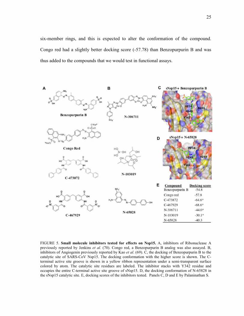
25
six-member rings, and this is expected to alter the conformation of the compound.
Congo red had a slightly better docking score (-57.78) than Benzopurpurin B and was
thus added to the compounds that we would test in functional assays.
FIGURE 5. Small molecule inhibitors tested for effects on Nsp15. A, inhibitors of Ribonuclease A previously reported by Jenkins et al. (70). Congo red, a Benzopurpurin B analog was also assayed. B, inhibitors of Angiogenin previously reported by Kao et al. (69). C, the docking of Benzopurpurin B to the catalytic site of SARS-CoV Nsp15. The docking conformation with the higher score is shown. The C-terminal active site groove is shown in a yellow ribbon representation under a semi-transparent surface colored by atom. The catalytic site residues are labeled. The inhibitor stacks with Y342 residue and occupies the entire C-terminal active site groove of sNsp15. D, the docking conformation of N-65828 in the sNsp15 catalytic site. E, docking scores of the inhibitors tested. Panels C, D and E by Palaninathan S.

26
Effects of the Inhibitors on sNsp15 Endoribonuclease Activity
A real-time fluorescence assay and highly purified recombinant sNsp15 protein
were used to examine the efficacy of the inhibitors against sNsp15 in vitro (55). All of
the compounds showed a concentration-dependent inhibition of sNsp15
endoribonuclease activity and an examination of the output of such an assay performed
with Benzopurpurin B is shown in Fig. 6A.
To confirm that the real time cleavage results are illustrative of an inhibition of
RNA cleavage activity, the effects of the compounds on the cleavage of a previously
characterized 16-nt RNA in a gel-based assay was performed. The results were found to
be consistent with those from the real time assay (Fig. 6B). The remainder of the results
presented will be from the real time assay.
The concentration of inhibitors needed to reduce sNsp15 endoribonuclease
activity by 50 and 90% (IC50 and IC90, respectively), were determined for each of the
compounds used in the molecular docking reaction. Inhibitor titration experiments with
Benzopurpurin B and Congo red are shown in Fig. 6C and a summary of all of the IC50
and IC90s are shown in Fig. 6D. Of all the compounds tested, Benzopurpurin B was the
best inhibitor, with IC50 and IC90 of 0.2 and 0.9 µM, respectively. We also note that
while functional examination of the effects of the inhibitors on endoribonuclease
activities confirmed that the Ribonuclease A inhibitors were more effective than the
Angiogenin inhibitors, the relative ranking of the results from two assays did not
correspond.

27
FIGURE 6. Effects of the small molecule inhibitors on the SARS-CoV Nsp15 endoribonuclease activity. A, representative results of a real-time endoribonuclease assay showing the inhibitory effect of Benzopurpurin B at different concentrations on the sNsp15 activity. The slope of the change in fluorescence was used to determine the rate of cleavage by Nsp15. B, a demonstration of the results from a gel-based RNA cleavage assay in presence of increasing concentrations of Benzopurpurin B. C, Benzopurpurin B and Congo red titrations plots using the real-time endoribonuclease assay. Concentration dependent inhibition of the substrate cleavage by sNsp15 is shown. D, summary of the IC50 and IC90 values for the small molecule compounds tested. Mechanism of Action of Select Inhibitors
To demonstrate that the compounds could bind to the sNsp15, we tested for
changes in Nsp15 in response to temperature. The assays were performed using
differential scanning fluorimetry in the presence of the dye SYPRO Orange, which binds
to the hydrophobic regions of a protein that are exposed by temperature-induced
unfolding. The transition curves are complex, as would be expected by the unfolding of
an oligomeric protein, however, a major peak could be readily identified (Fig. 7A). The
temperature where there is a maximal transition in this peak will be designated the

28
Tmapp. All samples were tested in triplicate and in a buffer capable of supporting
enzymatic activity that included 5 mM Mn2+, which we observed to help in smoothing
the changes in transition, likely by affecting sNsp15 conformation, as previously
demonstrated by Bhardwaj et al. (2004), (47). The sNsp15 protein had a Tmapp at 49 oC
in the absence of ligand. In the presence of Benzopurpurin B at 1, 3 and 5 µM final
concentration, the Tmapp values were 49, 55 and 56°C respectively (Fig. 7B). A reaction
performed in the absence of sNsp15, but in the presence of these concentrations of
Benzopurpurin B or other inhibitors tested did not change the fluorescence of SYPRO
orange. The change in the Tmapp provides evidence for the interaction between the
compounds and sNsp15. Compound C-473872 at 5, 10 and 20 µM also increased the
Tmapp values in a concentration-dependent manner (Fig. 7B). Unexpectedly, while
Congo red did cause a concentration-dependent change, there was decrease in the Tmapp.
Given that the inhibitors were originally active site inhibitors of Ribonuclease A
and Angiogenin, it is likely that they will act as competitive inhibitors of sNsp15. The
inhibitory effects of Benzopurpurin B, Congo red, and C-473872 were tested at several
concentrations, and the results from a minimum of three independent analyses were
averaged and analyzed by a double reciprocal plot. The results are compatible with
inhibition of SARS-CoV Nsp15 by a competitive mechanism (Fig. 7C). According to
this mechanism the compounds are able to bind to the free enzyme active site, competing
with the substrate. Because not all of the plots crossed at the zero in the axes, these
results do not rule out the presence of additional ligand binding sites or that all of the
active sites bind to the inhibitors in the same manner. In fact, crystal structure of the

29
Nsp15 revealed that there is a significant difference in the active sites of the six subunits
of Nsp15 and that the difference is particularly distinct between the two trimers of
Nsp15.
FIGURE 7. Mechanism of inhibition of Nsp15 by select inhibitors. A, first derivative of the differential scanning fluorimetry assays for C-473872. A peak for each curve is obtained at the Tm. A shift of the Tm values corresponding to each peak is observed in the presence of increasing concentrations of compound. B, summary of the Tm data obtained by differential scanning fluorimetry in the presence of Benzopurpurin B (BP), Congo red (CR) and C-473872. A shift in the Tm values is indicative of binding to the compound to sNsp15. C, double reciprocal plot analysis for Benzopurpurin B, Congo red and C-473872. A change in the slope of the plots upon increasing inhibitor concentrations is characteristic of a competitive mechanism of inhibition.

30
The MHV and IBV Nsp15 Activity are Inhibited by Benzopurpurin B in vitro
Coronaviruses can be important pathogens of animals as well as humans (9, 10,
11). Elucidation of the structure of the MHV Nsp15 (mNsp15) and molecular modeling
revealed that the active site structure is highly similar to that of the SARS-CoV Nsp15
(62, 64, Fig. 8A). We wanted to determine whether an inhibitor active against the SARS-
CoV Nsp15 will have similar effects on the Nsp15 proteins of other coronaviruses.
Superimposition of sNsp15 docked with Benzopurpurin B on top of mNsp15 showed
that Benzopurpurin-like inhibitors could be accommodated within the MHV Nsp15
active site (Fig. 8A); the flexibility of the C-terminal active site can further augment the
binding. When tested for effects on RNA cleavage by the MHV Nsp15 protein
expressed in E. coli, Benzopurpurin B was found to have an IC50 and IC90 of 0.4 and 0.9
µM, highly similar to the values obtained with the SARS-CoV Nsp15 (Fig. 8B).
To extend the analysis further, we tested the effects of Benzopurpurin B on the
Nsp15 ortholog of infectious bronchitis virus (IBV), iNsp15, which was demonstrated by
Bhardwaj et al. (2004) (47) to specifically cleave uridylates. Since the iNsp15 structure
has not been determined, we threaded its sequence into the crystal structure of the
SARS-CoV. The active site of the protein was essentially comparable to that of the
SARS-CoV and MHV orthologs and could accommodate Benzopurpurin B (Fig. 8C). A
prediction of this analysis is that the iNsp15 will also be inhibited by the compounds
effective on the MHV and SARS-CoV Nsp15. Indeed, the iNsp15 was inhibited by
Benzopurpurin B in a concentration-dependent manner and with IC50 and IC90 values
similar to that of the MHV and SARS-CoV Nsp15 enzymes (Fig. 8D). These results

31
suggest that the Nsp15 orthologs from these three coronaviruses likely have highly
similar active site pockets. Furthermore, it should be possible to use model
coronaviruses to study the effects of these drugs on viral infection under conditions that
do not require BSL3 containment.
FIGURE 8. Inhibition of RNA cleavage in vitro in other coronavirus Nsp15 orthologs. A, a model for the binding of Benzopurpurin B in the MHV Nsp15 crystal structure (magenta ribbon and grey sticks; PDB ID 2gth). Althought the catalytic site residues are identical, the nearby residues are not conserved between sNsp15 and mNsp15, non-conserved residues are shown in parenthesis. Perhaps the flexibility of the C-terminal active site residues should enable the inhibitor binding to mNsp15 as well. B, effects of Benzopurpurin B on the MHV Nsp15 ortholog. The enzyme is inhibited in a concentration dependent manner. C, a model of the IBV Nsp15 ortholog. The active site residues in the IBV Nsp15 homology model are well conserved with the SARS-CoV Nsp15. D, Effects of benzopurpurin B on endoribonuclease activity of the IBV Nsp15 ortholog using the real-time fluorescence assay.

32
Inhibition of MHV Infection in Cultured Cells
The effect of the compounds on MHV replication was investigated in plaque
formation assays using mouse L2 cells. The cells were plated in six-well cell culture
plates and grown until 100% confluence then infected with different dilutions of MHV
A59 and the inhibitor at a final concentration of 100 µM. After a 1 h period to allow the
infection to initiate, the cells were covered with a mixture of medium and agarose and
incubated for two days, when the plaques were stained and scored. In three independent
experiments, Congo red reproducibly showed the most inhibition of MHV plaque
formation, with the mean reduction of plaque formation being 26-fold. Compound C-
473872 caused an 11-fold reduction, and Benzopurpurin B, despite being the best
inhibitor in vitro, resulted in an 8-fold reduction. None of the compounds had any
obvious effect on the shape or viability of the cells when present at 100 µM (Fig. 9A, 9B
and data not shown).
To confirm and extend the analysis of the compounds on MHV infection, Dr.
Kanchan Bhardwaj examined MHV RNA production in the presence of Benzopurpurin
B, Congo red and C-473872. Genomic and subgenomic RNAs were reduced in the
presence of the drugs, with C-473872 being the most effective and Benzopurpurin B
having only a minimal effect in cells. Furthermore, both the genomic and subgenomic
MHV RNA levels were uniformly affected (Fig. 9C).

33
FIGURE 9. Effects of select Nsp15 inhibitors on MHV plaque formation in mouse L2 cells. A, representative plaque assay results in presence of Congo red and C-473872 at 100 uM, or controls such as the solvent DMSO. B, summary of the effects of the small-molecule compounds on MHV replication in L2 cells. The number of PFU/mL obtained and the fold reduction observed in three independent assays are reported. C, effects of the compounds on MHV RNA accumulation. The identities of the RNAs are shown to the left of the gel image. The rRNAs are intended to serve as a loading control. The result in panel C was obtained by Bhardwaj K.

34
DISCUSSION
The development of both vaccines and anti-virals specific against coronavirus
infections is currently a need. The Nsp15 endoribonuclease is a genetic marker for
coronaviruses and is an unusual enzyme for RNA viruses. Based on the observation that
Nsp15 has an Ribonuclease A-like active site and a mechanism for RNA cleavage
identical to Ribonuclease A (61, 64) we tested previously identified inhibitors of
Ribonuclease A and Angiogenin, a Ribonuclease A-like enzyme.
Computational docking into the structure of sNsp15 predicted that small-
molecule inhibitors of Angiogenin and RNase A could bind to the Nsp15 active site.
Functionally, three Angiogenin and RNase A inhibitors, C-473872, C-467929,
Benzopurpurin B, and the structurally related Congo red, were found to be potent
inhibitors of the sNsp15 endoribonuclease activity in vitro. To put this into context,
more than 50 candidates with high probability of docking into the sNsp15 active site
were tested previously and no compounds were found to have IC50 values better than 50
µM (Bhardwaj et al. unpublished data). Surprisingly, four inhibitors out of seven with
IC50s of better than 10 µM were found. The results add a layer of functional relevance to
the claims that sNsp15 has an RNase A-like catalytic pocket and cleavage mechanism
for the Nsp15 of different coronaviruses (61, 64).
Benzopurpurin B, Congo red, and C-473872 were able to bind sNsp15 since
they induce a concentration-dependent shift of the Tm of sNsp15. Benzopurpurin B and
C-473872 induce an increase in the sNsp15 Tm that corresponds to a stabilizing effect of

35
the compounds. Interestingly, Congo red induces the Tm to decrease, suggesting that it
destabilized the protein. Since Benzopurpurin B and C-473872 are active site inhibitors
of RNase A and Angiogenin (69, 70), and Congo red is likely to be one as well, and the
three compounds could bind to the sNsp15 active site according to computational
docking models, it is logical to assume that these compounds are competitive inhibitors.
The inhibition curves suggest this as well. However, at the present time, we cannot rule
out the presence of additional binding sites for these compounds in sNsp15.
Benzopurpurin B showed to inhibit the MHV and IBV Nsp15 orthologs similarly
to sNsp15, suggesting that the Nsp15 orthologs from these three coronaviruses likely
have highly similar active site pockets, which supports previous structural and
mutational studies of the mNsp15 (59, 62).
Importantly, Congo red and C-473872 reduced the infection of MHV in cultured
cells by several fold. The structures of these compounds should be considered as leads
for subsequent development of coronavirus inhibitors. The best inhibitor in biochemical
assays, Benzopurpurin B, was not particularly effective in virus-infected cells. This is
likely due to pharmacological properties of Benzopurpurin B, like half-life in solution or
in the cells or the ability to enter cells, features that we have not pursued in this work.
Notably, the fact that Benzopurpurin B differed from Congo red only by the presence of
two methyl groups indicates that small modifications to Congo red or Benzopurpurin B
could have dramatic effects on the efficacy of these compounds in cells.
Congo red is also a promising lead compound in the imaging and treatment of
amyloid protein plaques (72, 73). Its ability to interact with nucleotide-binding enzymes

36
explains in part its efficacy to inhibit RNase A and Nsp15 (74). Congo red and its
derivatives when administered near the time of infection have been shown to delay the
onset of clinical disease in scrapie infected hamsters (75). However, Congo red is
potentially toxic due to its degradation into the carcinogenic compound benzidine and
several derivatives have been tested for anti-amyloid properties (76). These compounds
would be of interest for testing its efficacy to treat coronavirus infections.
The compounds can also serve as new tools to analyze coronavirus infection. A
proposed role for Nsp15 is that it may participate in the processing of the RNAs needed
to form coronavirus subgenomic RNAs (8). However, there is no direct evidence for
this in MHV. The compounds induced a reduction in the MHV RNA levels affecting
uniformly genomic and subgenomic RNAs. In fact, mutational analysis of the mNsp15
resulted in a general decrease in all MHV RNAs and an approximately one log decrease
in MHV virion production. If Nsp15 does have a direct effect on subgenomic RNA
production, the effects are sufficiently pleiotropic to affect all MHV RNAs (59). The
effects of the inhibitors on MHV RNA production are consistent with those from
previous mutational analyses (59). More judicious application of Congo red and C-
473872 at different stages of coronavirus infection in cultured cells could allow better
insight into how Nsp15 contributes to this process.

37
CONCLUSIONS
The Nsp15 endoribonuclease is a genetic marker for coronaviruses and is an
unusual enzyme for RNA viruses which makes it a promising target for new antiviral
therapies.
Nsp15 active site and mechanism similarities to Ribonuclease A (61, 64) led us
to test previously identified inhibitors of Ribonuclease A and Angiogenin, a
Ribonuclease A-like enzyme.
Computational docking into the structure of sNsp15 predicted that small-
molecule inhibitors of Angiogenin and RNase A could bind to the Nsp15 active site.
Three Angiogenin and RNase A inhibitors, C-473872, C-467929, Benzopurpurin B, and
the structurally related Congo red, were found to be potent inhibitors of the sNsp15
endoribonuclease activity in vitro with IC50 of less than 10 µM.
Benzopurpurin B, Congo red, and C-473872 were able to bind sNsp15 since they
induce a concentration-dependent shift of the Tm of sNsp15. As predicted by the docking
models they are active site inhibitors of sNsp15 according to double reciprocal analysis.
Benzopurpurin B inhibited the MHV and IBV Nsp15 orthologs similarly to
sNsp15, suggesting that the Nsp15 orthologs from these three coronaviruses likely have
highly similar active site pockets, which supports previous structural and mutational
studies of the mNsp15.

38
Congo red and C-473872 reduced the infection of MHV in cultured cells by
several fold. The structures of these compounds should be considered as leads for
subsequent development of coronavirus inhibitors.
The compounds induced a reduction in the MHV RNA levels affecting uniformly
genomic and subgenomic RNas.
The compounds can also serve as new tools to analyze coronavirus infection.

39
REFERENCES
1. Peiris, J. S., Yuen, K. Y., Osterhaus, A. D., and Stohr, K. (2003) N. Engl. J. Med. 349, 2431-2441
2. Poutanen, S. M., Low, D. E., Henry, B., Finkelstein, S., Rose, D., Green, K.,
Tellier, R., Draker, R., Adachi, D., Ayers, M., Chan, A. K., Skowronski, D.M., Salit, I., Simor, A. E., Slutsky, A. S., Doyle, P. W., Krajden, M., Petric, M., Brunham, R. C., and McGeer, A. J. (2004) N. Engl. J. Med. 348, 1995-2005
3. Christian, M. D., Poutanen, S. M., Loutfy, M. R., Muller, M. P., and Low, D. E.
(2004) Clin. Infect. Dis. 38, 1420-1427
4. Guan, Y., Zheng, B. J., He, Y. Q., Liu, X. L., Zhuang, Z. X., Cheung, C. L., Luo, S. W., Li, P. H., Zhang, L. J., Guan, Y. J., Butt, K. M., Wong, K. L., Chan, K.W., Lim, W., Shortridge, K. F., Yuen, K. Y., Peiris, J. S., and Poon, L. L. (2003) Science 302, 276-278
5. Song, H. D., Tu, C. C., Zhang, G. W., Wang, S. Y., Zheng, K., Lei, L. C., Chen,
Q. X., Gao, Y. W., Zhou, H. Q., Xiang, H., Zheng, H. J., Chern, S. W., Cheng, F., Pan, C. M., Xuan, H., Chen, S. J., Luo, H. M., Zhou, D. H., Liu, Y. F., He, J. F., Qin, P. Z., Li, L. H., Ren, Y. Q., Liang, W. J., Yu, Y. D., Anderson, L., Wang, M., Xu, R. H., Wu, X. W., Zheng, H. Y., Chen, J. D., Liang, G., Gao, Y., Liao, M., Fang, L., Jiang, L. Y., Li, H., Chen, F., Di, B., He, L. J., Lin, J. Y., Tong, S., Kong, X., Du, L., Hao, P., Tang, H., Bernini, A., Yu, X. J., Spiga, O., Guo, Z. M., Pan, H. Y., He, W. Z., Manuguerra, J. C., Fontanet, A., Danchin, A., Niccolai, N., Li, Y. X., Wu, C. I., and Zhao, G. P. (2005) Proc. Natl. Acad. Sci. U. S. A. 102, 2430-2435
6. Li, W., Shi, Z., Yu, M., Ren, W., Smith, C., Epstein, J. H., Wang, H., Crameri,
G., Hu, Z., Zhang, H., Zhang, J., McEachern, J., Field, H., Daszak, P., Eaton, B. T., Zhang, S., and Wang, L. F. (2005) Science 310, 670-679
7. Cheng, V. C., Lau, S. K., Woo, P. C., and Yuen, K. Y. (2007) Clin. Microbiol.
Rev. 20, 660-694
8. Snijder, E. J., Bredenbeek, P. J., Dobbe, J. C., Thiel, V., Ziebuhr, J., Poon, L. L., Guan, Y., Rozanov, M., Spaan, W. J., and Gorbalenya, A. E. (2003) J. Mol. Biol. 331, 991-1004
9. Masters, P. S. (2006) Adv. Virus Res. 66, 193-292
10. Lai, M. M., and Cavanagh, D. (1997) Adv. Virus Res. 48, 1-100

40
11. Spaan, W., Cavanagh, D., and Horzinek, M. C. (1988) J. Gen. Virol. 69, 2939-2952
12. Gorbalenya, A. E., Snijder, E. J., and Spaan, W. J. (2004) J. Virol. 78, 7863-7866
13. Lau, S. K., Woo, P. C., Li, K. S., Huang, Y., Tsoi, H. W., Wong, B. H., Wong, S. S., Leung, S. Y., Chan, K. H., and Yuen, K. Y. (2005) Proc. Natl. Acad. Sci. U. S. A. 102, 14040-14045
14. McIntosh, K. (1974) Curr. Top. Microbiol. Imunol. 63, 85-129
15. Macnaughton, M. R., Davies, H. A., and Nermut, M. V. (1978) J. Gen. Virol. 39, 545-549
16. Marra, M. A., Jones, S. J., Astell, C. R., Holt, R. A., Brooks-Wilson, A.,
Butterfield, Y. S., Khattra, J., Asano, J. K., Barber, S. A., Chan, S. Y., Cloutier, A., Coughlin, S. M., Freeman, D., Girn, N., Griffith, O. L., Leach, S. R., Mayo, M., McDonald, H., Montgomery, S. B., Pandoh, P. K., Petrescu, A. S., Robertson, A. G., Schein, J. E., Siddiqui, A., Smailus, D. E., Stott, J. M., Yang, G. S., Plummer, F., Andonov, A., Artsob, H., Bastien, N., Bernard, K., Booth, T. F., Bowness, D., Czub, M., Drebot, M., Fernando, L., Flick, R., Garbutt, M., Gray, M., Grolla, A., Jones, S., Feldmann, H., Meyers, A., Kabani, A., Li, Y., Normand, S., Stroher, U., Tipples, G. A., Tyler, S., Vogrig, R., Ward, D., Watson, B., Brunham, R. C., Krajden, M., Petric, M., Skowronski, D. M., Upton, C., and Roper, R. L. (2003) Science 300, 1399-1404
17. Rota, P. A., Oberste, M. S., Monroe, S. S., Nix, W. A., Campagnoli, R.,
Icenogle, J. P., Penaranda, S., Bankamp, B., Maher, K., Chen, M. H., Tong, S., Tamin, A., Lowe, L., Frace, M., DeRisi, J. L., Chen, Q., Wang, D., Erdman, D. D., Peret, T. C., Burns, C., Ksiazek, T. G., Rollin, P. E., Sanchez, A., Liffick, S., Holloway, B., Limor, J., McCaustland, K., Olsen-Rasmussen, M., Fouchier, R., Gunther, S., Osterhaus, A. D., Drosten, C., Pallansch, M. A., Anderson, L. J., and Bellini, W. J. (2003) Science 300, 1394-1399
18. Thiel, V., Ivanov, K. A., Putics, A., Hertzig, T., Schelle, B., Bayer, S.,
Weissbrich, B., Snijder, E. J., Rabenau, H., Doerr, H. W., Gorbalenya, A. E., and Ziebuhr, J. (2003) J. Gen. Virol. 84, 2305-2315
19. Wong, S. K., Li, W., Moore, M. J., Choe, H., and Farzan, M. (2004) J. Biol.
Chem. 279, 3197-3201
20. Sui, J., Li, W., Murakami, A., Tamin, A., Matthews, L. J., Wong, S. K., Moore, M. J., Tallarico, A. S., Olurinde, M., Choe, H., Anderson, L. J., Bellini, W. J.,

41
Farzan, M., and Marasco, W. A. (2004) Proc. Natl. Acad. Sci. U. S. A. 101, 2536-2541
21. Yeager, C. L., Ashmun, R. A., Williams, R. K., Cardellichio, C. B., Shapiro, L.
H., Look, A. T., and Holmes, K. V. (1992) Nature 357, 420-422 22. Williams, R. K., Jiang, G. S., and Holmes, K. V. (1991) Proc. Natl. Acad. Sci. U.
S. A. 88, 5533-5536
23. Li, W., Moore, M. J., Vasilieva, N., Sui, J., Wong, S. K., Berne, M. A., Somasundaran, M., Sullivan, J. L., Luzuriaga, K., Greenough, T. C., Choe, H., and Farzan, M. (2003) Nature 426, 450-454
24. Hamming, I., Timens, W., Bulthuis, M. L., Lely, A. T., Navis, G. J., and van
Goor, H. (2004) J.Pathol. 203, 631-637
25. Liu, S., Xiao, G., Chen, Y., He, Y., Niu, J., Escalante, C. R., Xiong, H., Farmar, J., Debnath, A. K., Tien, P., and Jiang, S. (2004) Lancet 363, 938-947
26. Bosch, B. J., Martina, B. E., Van Der Zee, R., Lepault, J., Haijema, B. J.,
Versluis, C., Heck, A. J., De Groot, R., Osterhaus, A. D., and Rottier, P. J. (2004) Proc. Natl. Acad. Sci. U. S. A. 101, 8455-8460
27. Herold, J., Siddell, S. G., and Gorbanleya, A. E. (1999) J. Biol. Chem. 274,
14918-14925 28. Tijms, M. A., van Dinten, F. C., Gorbanleya, A. E., and Snijder, E. J. (2001)
Proc. Natl. Acad. Sci. U. S. A. 98, 1889-1894
29. Ziebuhr, J. (2005) Curr. Top. Microbiol. Immunol. 287, 57-94
30. Ziebuhr, J., Snijder, E. J., and Gorbalenya, A. E. (2000) J. Gen. Virol. 81, 853-879
31. Ziebuhr, J. (2004) Curr. Opin. Microbiol. 7, 412-419
32. Jacobs, L., Spaan, W. J. M., Horzinek M. C., and van der Zeijst, B. A. M. (1981)
J. Virol. 39, 401-406
33. Makino, S., Stohlman, S. A., and Lai, M. M. C. (1986) Proc. Natl. Acad. Sci. U. S. A. 83, 4204-4208
34. Lai, M. M. C. (1986) BioEssays 5, 257-260

42
35. Zuniga, S., Sola, I., Alonso, S., and Enjuanes, L. (2004) J. Virol. 78, 980-994
36. Sawiki, S. G., and Sawiki, D. L. (1998) Adv. Exp. Med. Biol. 440, 215-219
37. Sawiki, S. G., and Sawiki, D. L. (2005) Curr. Top. Microbiol. Immunol. 287, 31-55
38. Sawiki, S. G., and Sawiki, D. L. (1990) J. Virol. 64, 1050-1056
39. Sethna, P., Hofmann, M., and Brian, D. (1991) J. Virol. 65, 320-325
40. Sethna, P., Hung, S., and Brian, D. (1989) Proc. Natl. Acad. Sci. U. S. A. 86,
5626-5630
41. Pasternak, A. O., van der Born, E., Spaan, W. J. M., and Snijder, E. J. (2001) EMBO J. 20, 7220-7228
42. Pasternak, A. O., van der Born, E., Spaan, W. J. M., and Snijder, E. J. (2003) J.
Virol. 77, 1175-1183
43. Pasternak, A. O., Spaan, W. J. M., and Snijder, E. J. (2003) J. Virol. 78, 8102-8113
44. van Marle, G., Dobbe, J. C., Gultyaev, A. P., Luytjes, Spaan, W. J. M., and
Snijder, E. J. (1999) Proc. Natl. Acad. Sci. U. S. A. 96, 12056-12061
45. Imbert, I., Guillemot, J. C., Bourhis, J. M., Bussetta, C., Coutard, B., Egloff, M. P., Ferron, F., Gorbalenya, A. E., and Canard, B. (2006) EMBO J. 25, 4933-4942
46. Minskaia, E., Hertzig, T., Gorbalenya, A. E., Campanacci, V., Cambillau, C.,
Canard, B., and Ziebuhr, J. (2006) Proc. Natl. Acad. Sci. U. S. A. 103, 5108-5113
47. Bhardwaj, K., Guarino, L., and Kao, C. C. (2004) J. Virol. 78, 12218-12224
48. Ivanov, K. A., Hertzig, T., Rozanov, M., Bayer, S., Thiel, V., Gorbalenya, A. E., and Ziebuhr, J. (2004) Proc. Natl. Acad. Sci. U. S. A. 101, 12694-12699
49. Brockway, S. M., Clay, C. T., Lu, X. T., and Denison, M. R. (2003) J. Virol. 77,
10515-10527
50. Cheng, A., Zhang, W., Xie, Y., Jiang, W., Arnold, E., Sarafianos, S. G., and Ding, J. (2005) Virology 335, 165-176

43
51. Ivanov, K. A., Thiel, V., Dobbe, J. C., van der Meer, Y., Snijder, E. J., and Ziebuhr, J. (2004) J. Virol. 78, 5619-5632
52. Ivanov, K. A., and Ziebuhr, J. (2004) J. Virol. 78, 7833-7838
53. Tanner, J. A., Watt, R. M., Chai, Y. B., Lu, L. Y., Lin, M. C., Peiris, J. S., Poon,
L. L., Kung, H. F., and Huang, J. D. (2003) J. Biol. Chem. 278, 39578-39582 54. Seybert, A., Hegyi, A., Siddell, S. G., and Ziebuhr, J. (2000) RNA 6, 1056-1068
55. Bhardwaj, K., Sun, J., Holzenburg, A., Guarino, L. A., and Kao, C. C. (2006) J.
Mol. Biol. 361, 243-256
56. Caffarelli, E., Maggi, L., Fatica, A., Jiricny, J., and Bozzoni, I. (1997) Biochem. Biophys. Res. Commun. 233, 514-517
57. Laneve, P., Altieri, F., Fiori, M. E., Scaloni, A., Bozzoni, I., and Caffarelli, E.
(2003) J. Biol. Chem. 278, 13026-13032
58. Posthuma, C. C., Nedialkova, D. D., Zevenhoven-Dobbe, J. C., Blokhuis, J. H., Gorbalenya, A. E., and Snijder, E. J. (2006) J. Virol. 80, 1653-1661
59. Kang, H., Bhardwaj, K., Li, Y., Palaninathan, S., Sacchettini, J., Guarino, L.,
Leibowitz, J. L., and Kao, C. C. (2007) J. Virol. 81, 13587-13597
60. Guarino, L. A., Bhardwaj, K., Dong, W., Sun, J., Holzenburg, A., and Kao, C. (2005) J. Mol. Biol. 353, 1106-1117
61. Ricagno, S., Egloff, M. P., Ulferts, R., Coutard, B., Nurizzo, D., Campanacci, V.,
Cambillau, C., Ziebuhr, J., and Canard, B. (2006) Proc. Natl. Acad. Sci .U. S. A. 103, 11892-11897
62. Xu, X., Zhai, Y., Sun, F., Lou, Z., Su, D., Xu, Y., Zhang, R., Joachimiak, A.,
Zhang, X. C., Bartlam, M., and Rao, Z. (2006) J.Virol. 80, 7909-7917
63. Roberts, G., Dennis, E. A., Meadows, D. H., Cohen, J. S., and Jardentzky, O. Proc. Natl. Acad. Sci .U. S. A. 62, 1151-1158
64. Bhardwaj, K., Palaninathan, S., Alcantara, J. M., Yi, L. L., Guarino, L.,
Sacchettini, J. C., and Kao, C. C. (2008) J. Biol. Chem. 283, 3655-3664
65. Saida, F., Uzan, M., and Bontems, F. (2003) Nucleic Acids Res. 31, 2751-2758
66. Deshpande, R. A., and Shankar, V. (2002) Crit. Rev. Microbiol. 28, 79-122

44
67. Gioia, U., Laneve, P., Dlakic, M., Arceci, M., Bozzoni, I., and Caffarelli, E.
(2005) J. Biol. Chem. 280, 18996-19002
68. Nowotny, M., Gaidamakov, S. A., Crouch,R.J., and Yang, W. (2005) Cell 121, 1005-1016
69. Kao, R.Y., Jenkins, J. L., Olson, K. A., Key, M. E., Fett, J. W., and Shapiro, R.
(2002) Proc. Natl. Acad. Sci. U. S. A. 99, 10066-10071
70. Jenkins, J. L., and Shapiro, R. (2003) Biochemistry 42, 6674-6687
71. Niesen, F. H., Berglund, H., and Vedadi, M. (2007) Nature Protocols 2, 2212-2221
72. Furumoto, S., Okamura, N., Iwata, R., Yanai, K., Arai, H., and Kudo,Y. (2007)
Curr. Top. Med. Chem. 7, 1773-1789
73. Wenn, S., Lekishvili, T.,Loeschner, C., Sellarajah, S., Prelli, F., Wisniewski, T., Gilber, I. H., and Brown, D. R. (2007) J. Virol. 81, 10729-10741
74. Edwards, R. A., and Woody, R. W. (1979) Biochemistry 18, 5197-5204
75. Ingrosso, L, Ladogana, A., and Pocchiari, M. (1995) J. Virol. 69, 506-508
76. Rudyk, H., Vasiljevic, S., Hennion, R. M., Birkett, C. R., Hope, J., and Gilbert, I. H. (2000) J. Gen. Virol. 81, 1155-1164.

45
VITA
Name: Joanna Maria Ortiz Alcantara
Address: 2128 TAMU, Texas A&M University, College Station, TX 77843-2128 Email Address: [email protected] Education: B.S., Pharmaceutical and Biological Chemistry, National Autonomous University of Mexico, 2006
M.S., Biochemistry, Texas A&M University, 2009