Embed Size (px)
Citation preview

Small Fiber Neuropathy: Answering the Burning
QuestionsEzekiel Fink, MD
Clinical Instructor, UCLA Department of Neurology

Introduction Central vs peripheral nervous system Peripheral nervous system
Types of nerves Motor nerves Sensory nerves Mixed nerves
Examples Median nerve
Types of pathology Mononeuropathy Generalized neuropathy
Small fiber nerves

Introduction Peripheral neuropathy is among
the most common disorders evaluated by neurologists.
Most peripheral neuropathies affect all fiber sizes. Etiology of these types of peripheral
neuropathies is quite broad
Few peripheral neuropathies are associated with pure or predominantly small-fiber involvement.

Types of Nerve Fibers
Peripheral nerve contains myelinated and unmyelinatedfibers, which subserve both somatic and autonomic functions.
Peripheral nerve fibers can be classified according to size, which correlates with the degree of myelination. Large nerve fibers are heavily myelinated
Mediates: motor strength vibratory and touch sensation.
Medium-sized fibers are myelinated and carry information to muscle spindles.
Small fibers include lightly myelinated and unmyelinated fibers Innervates
Mediate pain, thermal sensation, and autonomic function.
Small fiber neuropathy results from selective impairment of small myelinated fibers.

Small Fiber Nerves All large fibers (6 to 12 µm axonal diameter range)
are myelinated, and hence, fastly conducting. Small fibers are both myelinated and
unmyelinated. Myelinated fibers have a bimodal distribution with peak
axonal diameters at 4 µm and 10 µm for small and large myelinated fibers, respectively.
Small myelinated fibers outnumber large fibers, with estimates placing the total percentage of small fibers between 55 and 68% of all myelinated fibers.
Furthermore, unmyelinated fibers (peak axonal diameter of 1 to 1.2 µm) outnumber myelinated fibers by a ratio of 4:1.
The combination of very small axonal diameters and the absence of myelin result in unmyelinated fibers conducting very slowly, in the range of 1 to 2 ms.
What is Small Fiber Neuropathy? Small fiber neuropathy is a disorder of the peripheral
nerves that affects: small somatic fibers autonomic fibers or both
As small fibers subserve autonomic function, as well as thermal and pain sensations, small fiber neuropathy presents with: autonomic dysfunction lack of thermal and pain sensations neuropathic pain dysesthesias a combination of the above symptoms

Small Fiber Neuropathy Small fiber neuropathy can present in all
age groups. Inherited neuropathies with small-fiber
involvement are more common in younger age groups.
Acquired and idiopathic cases are more often seen in the elderly.
Depending on the underlying pathogenesis, small fiber neuropathy may or may not progress to also affect large fibers.
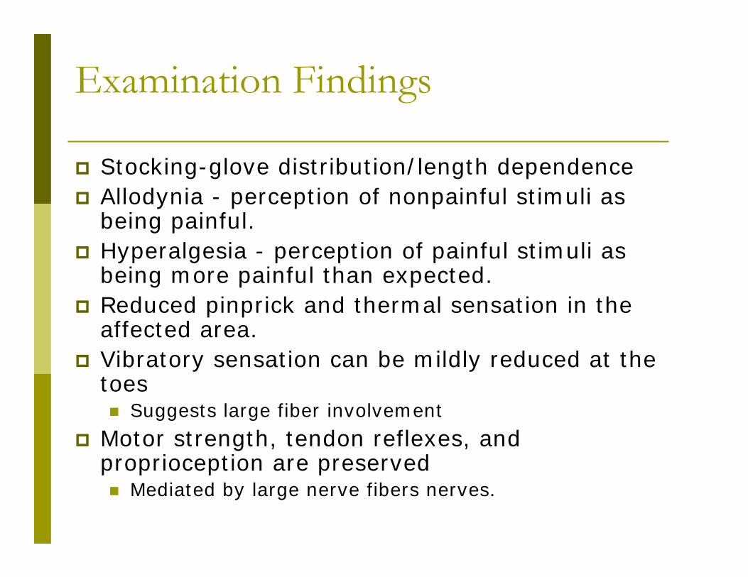
Examination Findings
Stocking-glove distribution/length dependence Allodynia - perception of nonpainful stimuli as
being painful. Hyperalgesia - perception of painful stimuli as
being more painful than expected. Reduced pinprick and thermal sensation in the
affected area. Vibratory sensation can be mildly reduced at the
toes Suggests large fiber involvement
Motor strength, tendon reflexes, and proprioception are preserved Mediated by large nerve fibers nerves.

Sensory Symptoms of Small Fiber Neuropathy Damage to or loss of small somatic nerve fibers results in:
pain, burning, tingling, or numbness typically affects the limbs in a distal-to proximal gradient In some cases, it can follow a non-length-dependent distribution in which
symptoms may be manifested predominantly in the arms, face, or trunk. Patient description:
Symptoms may be mild initially complain of vague discomfort in one or both feet
The most common symptom is burning pain in the feet The common syndrome of painful burning feet is most often caused by a sensory
neuropathy with small-fiber involvement, accounting for more than 90% of such cases Other description of pain; stabbing, aching pains, electric shock-like, pins-and-needles
sensations, or cramping of the feet and calves. Symptoms are usually worse at night and often affect sleep.
Other sensory complaints: wooden quality in their feet numbness in their toes walking on pebbles, sand, or golf balls.
Some patients say that their feet have become so exquisitely tender that they cannot bear having the bed sheets touch them, and so they sleep with their feet uncovered.

Small Fiber Neuropathy: Clinical Picture Most small fiber neuropathy begin distally and evolve slowly in length-
dependent (i.e., dying back) fashion similar to other peripheral neuropathies
A minority of patients present with a non-length dependent pattern and may represent a neuronopathy.
Small fiber neuropathy commonly presents with both positive and negative symptoms of pain and temperature fibers. Patients complain of numbness and are found to have reduced or absent pain
and temperature sensations distally Often, painful dysesthesias including burning and lancinating pains occur as well. These positive symptoms are believed to result from spontaneous activity of
damaged small fibers. Frequently, dysesthesia awaken patients at night or are triggered by
touching the bed sheet. In addition to sensory symptoms, autonomic symptoms may occur. Patients with pure small-fiber involvement display normal large fiber
function. Normal muscle bulk and strength Normal muscle stretch reflexes Normal large fiber sensory function (i.e., vibration, proprioception)

Autonomic Findings
Autonomic signs may be discovered when evaluating a patient with a small fiber neuropathy or may represent the principal symptoms bringing the patient to medical attention.
When autonomic fibers are affected, patients may experience: dry eyes dry mouth orthostatic dizziness constipation bladder incontinence sexual dysfunction trouble sweating orthostatic hypotension skin changes
atrophic, dry, shiny, discolored, or mildly edematous
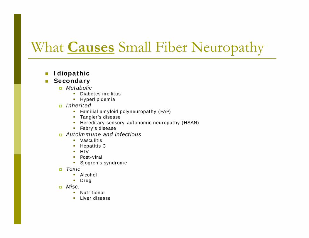
What Causes Small Fiber Neuropathy Idiopathic Secondary
Metabolic Diabetes mellitus Hyperlipidemia
Inherited Familial amyloid polyneuropathy (FAP) Tangier’s disease Hereditary sensory-autonomic neuropathy (HSAN) Fabry’s disease
Autoimmune and infectious Vasculitis Hepatitis C HIV Post-viral Sjogren’s syndrome
Toxic Alcohol Drug
Misc. Nutritional Liver disease

Glucose Dysregulation Glucose dysmetabolism (diabetes and impaired glucose
tolerance) is the most common identifiable associated condition present in about one-third of patients with painful sensory
neuropathy Present in nearly half of those with otherwise idiopathic small fiber
neuropathy dose-response relationship intermittent hyperglycemia
Research findings strongly suggest that even mild abnormalities of glucose metabolism (“prediabetes”) is a risk factor for small fiber neuropathy may represent the earliest stage of diabetic neuropathy
Several recent studies have found a high prevalence of impaired glucose tolerance in patients with sensory peripheral neuropathy rate of up to 42% in cases initially thought to be idiopathic
compared with 14% in the general population small fibers are often involved early and predispose
the feet to injuries and subsequent diabetic ulceration

Idiopathic Small Fiber Neuropathy
One of the more common causes of small fiber neuropathy is idiopathic sensory neuropathy.
Idiopathic sensory neuropathy is a common clinical problem
Often frustrating to both the physician and patient. Neurologic examination is typically normal. Even with extensive testing, no identifiable cause is found.
It usually affects adults older than age 60 and presents with a slowly progressive course of numbness and burning pain of the feet.
Diagnosis is based on exclusion after the condition has persisted for more than three months.
The pathogenesis is thought to be postinfectious or immune mediated.

Hyperlipidemia Hyperlipidemia is a common metabolic
disorder, sometimes associated with small fiber neuropathy.
Hypertriglyceridemia typically occurs in middle age and when associated with a small fiber neuropathy, presents with mild, painful, distal sensory peripheral neuropathy with no autonomic involvement.
Symptoms can resolve with lowering serum lipid concentration.

Hereditary Causes
Amyloidosis
Tangier disease
Fabry disease
Hereditary sensory autonomic neuropathy

Amyloidosis Both familial and acquired types of amyloidosis exist
amyloid protein is deposited in different tissues, including peripheral nerve.
Familial amyloid polyneuropathy (FAP) amyloid deposition occurs from an abnormal circulating
prealbumin (transthyretin). Acquired: primary systemic amyloidosis
amyloid is composed of a variable portion of a monoclonal light chain
peripheral neuropathy is reported in 15% of patients. Small fiber dysfunction occurs early in the disease
as the disease progresses, all fibers become involved to produce a severe sensorimotor peripheral neuropathy.
Mechanism of Injury The mechanism of the nerve injury is not completely understood. Whether the mechanism is nerve ischemia or inflammatory
changes remains unclear

Hereditary Sensory Autonomic Neuropathy (HSAN) Hereditary Sensory Autonomic Neuropathy, Type 1
autosomal dominant disorder Known chomosomal defect
Symptoms become prominent in the second to fourth decade Patients have a loss of pain and thermal sensations in the distal lower extremities
Can result in Charcot joints, ulcers and infections Hereditary Sensory Autonomic Neuropathy, Type
Autosomal recessive disorder Unknown chromosomal linkage
patients are usually affected in early life with severe loss of pain and thermal sensation Small and large sensory fibers are affected.
Patients are prone to significant foot and leg ulcers, finger mutilation, and paronychia. Autonomic dysfunction
Impaired sweating, sphincter disturbance, and impotence. Hereditary Sensory Autonomic Neuropathy, Type 3
Autosomal recessive disorder, typically seen in Ashkenazi Jews. Known chromosomal defect
Clinical signs present very early in life with impaired lacrimation, poor sucking, and failure to thrive. Physical findings
Fungiform papillae on the tongue are absent corneal insensitivity hyporeflexia, decreased pain sensation kyphoscoliosis.
Emotional stimuli can result in autonomic crisis with hypertension and marked sweating. Autonomic findings: postural hypotension and disordered sweating are common.
Fatal cardiac arrhythmias can occur. Other types of HSAN

Tangier’s disease Tangier’s disease (hereditary high-density lipoprotein
deficiency) autosomal recessive disorder characterized by:
a relapsing and remitting asymmetric neuropathy a syringomyelic-like syndrome a slowly progressive symmetric neuropathy.
Initially, it may manifest as extensive loss of pain and temperature sensation.
Intrinsic hand and bifacial muscle weakness may occur. Orange colored, enlarged, lobulated tonsils are characteristic. Serum lipoprotein studies demonstrate reduced cholesterol,
reduced or absent high-density lipoproteins (HDL) and elevated triglyceride levels.

Fabry’s disease Fabry’s disease (hereditary alpha-galactosidase A deficiency)
X-linked complex multi-system clinical syndrome, which includes
central nervous system cardiac metabolic gastrointestinal
It is characterized by severe burning pain in the extremities (80 to 90% of affected males) Pain is usually associated with paresthesia, and is difficult to treat. The mechanism of the pain is unknown, but may be related to
glycophospholipid accumulation in the dorsal root ganglia and subsequent cell death.
Anhidrosis or hypohidrosis heat intolerance reduced tear and saliva production.
Skin findings
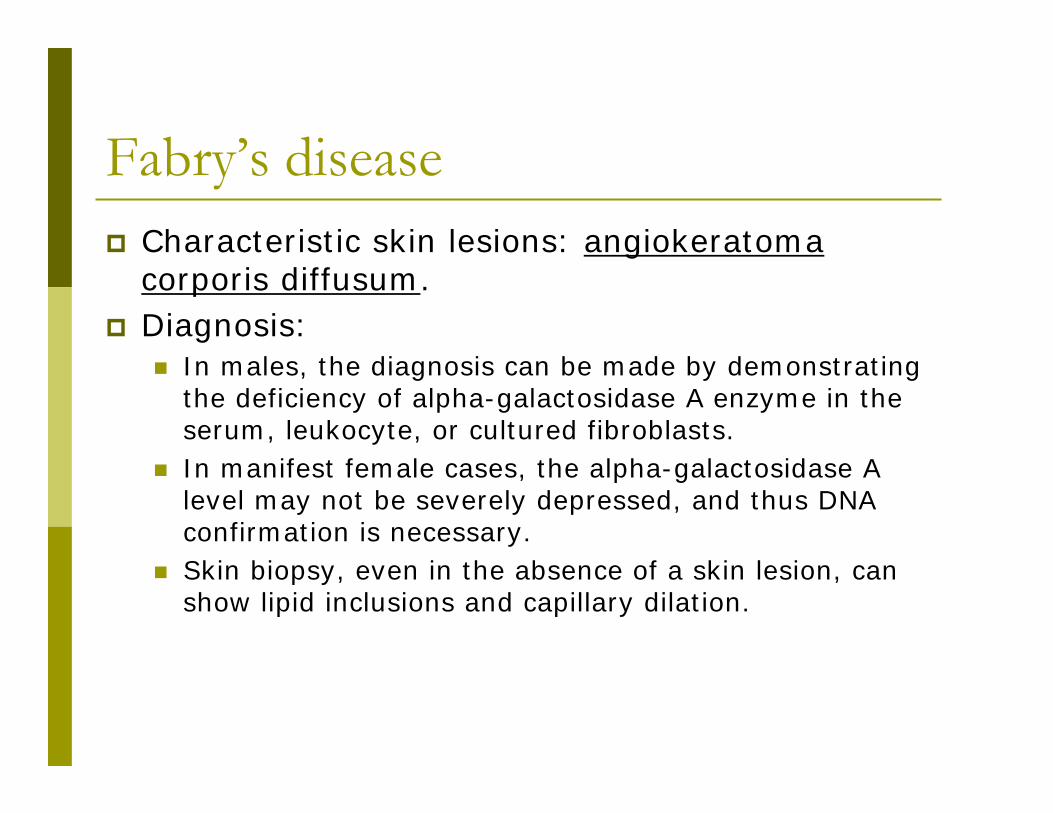
Fabry’s disease Characteristic skin lesions: angiokeratoma
corporis diffusum. Diagnosis:
In males, the diagnosis can be made by demonstrating the deficiency of alpha-galactosidase A enzyme in the serum, leukocyte, or cultured fibroblasts.
In manifest female cases, the alpha-galactosidase A level may not be severely depressed, and thus DNA confirmation is necessary.
Skin biopsy, even in the absence of a skin lesion, can show lipid inclusions and capillary dilation.

Alcohol
Alcohol often results in a painful peripheral neuropathy. In a large series of alcoholic patients, 26% had sensory
neuropathy, and in 25% of these had predominant involvement of small fibers.
May be caused by nutritional deficiency vs direct toxic effect of alcohol In alcoholic patients with a normal nutritional status and
thiamine level, painful burning neuropathy does occur, implicating a direct toxic effect.
On nerve biopsy, severely reduced density of small myelinated and unmyelinated fibers is found.
Patients also often demonstrate signs of autonomic dysfunction: urinary retention Constipation impaired sweating orthostatic hypotension

Toxic
A number of medicines result in peripheral neuropathies, many with predominantly small-fiber dysfunction. Antineoplastic drugs such as cisplatin, vinca alkaloids
(Vincristine), and taxol (Paclitaxel) are neurotoxic Patients can develop peripheral neuropathic symptoms.
Antibiotic metronidazole has been associated with small fiber neuropathy in
certain instances Antiretroviral:Zalcitabine, didanosine, and stauvudine
associated with SFN. Arsenic, thallium, gold, perhexiline, and disulfiram may result
in sensorimotor neuropathies with an early phase of SFN. Other neurotoxic agents
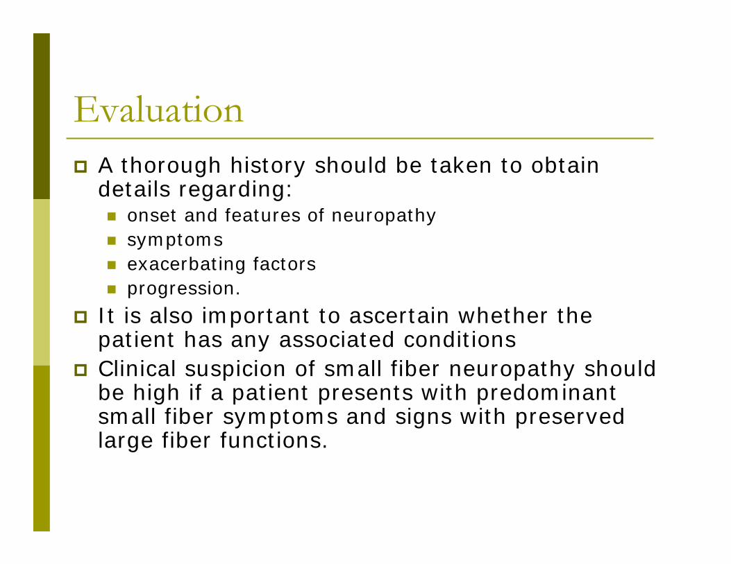
Evaluation A thorough history should be taken to obtain
details regarding: onset and features of neuropathy symptoms exacerbating factors progression.
It is also important to ascertain whether the patient has any associated conditions
Clinical suspicion of small fiber neuropathy should be high if a patient presents with predominant small fiber symptoms and signs with preserved large fiber functions.

Electrodiagnostics
Small fiber neuropathies are challenging to diagnose
Nerve conduction studies and electromyography (EMG) play a key role in diagnosing most peripheral neuropathies assess the function of large nerve fibers only are normal in small fiber neuropathy. if the results of these tests are normal, specialized studies
are needed to evaluate small fibers. Although several tests are available to evaluate
somatic and autonomic small fibers, two have the highest diagnostic efficiency for small fiber neuropathy skin biopsy, to evaluate intraepidermal nerve fiber density quantitative sudomotor axon reflex testing (QSART), to
assess sudomotor autonomic function.

Neurodiagnostic Skin Biopsy Assessment of intraepidermal nerve fiber
density (IENF) is a novel method in directly assessing cutaneous small fibers via a skin biopsy.
Skin biopsy is a minimally invasive procedure in which a small punch biopsy specimen is taken from several areas of the body
The technique is relatively easy, repeatable at different sites, and minimally invasive. Biopsy performed by infiltrating the skin with local
anesthetic and a 3-mm punch biopsy is obtained from different locations.

Neurodiagnostic Skin Biopsy
Biopsy specimens are immunostained using an antibody against protein gene product 9.5, which is a panaxonal marker. Small nerve fibers in the epidermis are counted
under a microscope, and intraepithelial nerve fiber densities are calculated and compared with established normative values.
The diagnosis of small fiber neuropathy can be established if the intraepidermal nerve fiber density is lower than normal.

Neurodiagnostic Skin Biopsy: Accurate IENF correlates well with the loss of unmyelinated
and small myelinated nerve fibers as quantitatedin sural nerve biopsies.
As in most length dependent neuropathies, IENF loss is more prominent distally in chronic SFNs rarely a more diffuse proximal and distal loss may be
seen (e.g., postinfectious acute SFN). It has been estimated that small-fiber density is
reduced below the fifth percentile in approximately 80% to 90% of SFN patients.
In three different series of patients with painful SFNs the incidence of a reduced IENF varied between 81% to 87%, compared to: an abnormal QST in 60% to 72% an abnormal QSART in 59% to 80%.

Neurodiagnostic Skin Biopsy: A Good Tool A good tool
it is more sensitive than quantitative sensory testing
It is more sensitive and less invasive than sural nerve biopsy
Intraepidermal nerve fiber density also correlates well with a variety of measures of severity of HIV distal sensory neuropathy and thus may be used to measure the severity and treatment response of small fiber neuropathy.
Skin biopsy with IENF can also easily be done in children in whom traditional sural nerve biopsy is usually avoided. Cases of SFN in children have been demonstrated in
which the symptoms had previously been thought secondary to a psychiatric disorder after a normal physical examination and electrophysiologic testing.

Examples of Skin Biopsy Results
Normal Small Fiber Neuropathy
Source:http://www.therapath.com/
Quantitative Sudomotor Axon Reflex Testing (QSART)
QSART is an autonomic study that measures sweat output reflects the function of a certain population of small fiber
nerves. Electrodes are placed on the arms and legs to record the volume
of sweat produced. The output is compared with normative values.
One prospective study showed that 67 (72.8%) of 92 patients with painful feet had abnormal results on QSART, ie, low sweat output.
A retrospective study found that 77 (62%) of 125 patients with clinical features of distal small fiber neuropathy had a length-dependent pattern of QSART abnormalities.

Laboratory Work-up Basic laboratory tests to find the cause
Once the diagnosis of small fiber neuropathy is established, thenext important step is to order a battery of laboratory tests tosearch for an underlying cause. OGTT Serum protein electrophoresis/urine
immunoelectrophoresis DNA testing (eg FAP, HSAN) Serum lipids Liver function tests HCV antibody cryoglobulins, anti-Hu antibody, HIV Consider testing lyme serology are Other tests

Special laboratory tests in special cases Celiac sprue
If there is a history of gastrointestinal symptoms or herpetiform-like rash, then testing for gliadin antibody and tissue transglutaminaseantibodies as well as small-bowel biopsy may be pursued to evaluate for celiac sprue.
Infectious diseases serologic tests for HIV or hepatitis C should be ordered if the patient
has risk factors. If there is a significant family history, further genetic testing
should be considered. Lip biopsy or bone marrow biopsy should be considered if clinical
suspicion is high for Sjögren disease, seronegative siccasyndrome, or amyloidosis.
The serum ACE level has a low sensitivity and specificity; therefore, if sarcoid is suspected clinically, additional confirmatory testing, such as computed tomography of the chest, should be ordered despite a normal ACE value.

Treatment Treatment of small fiber neuropathy
should target the underlying cause and neuropathic pain.
Cause-specific treatment is a key in preventing small fiber neuropathy or slowing its progression.
It has also been reported that treatment of sarcoidosis, autoimmune diseases, and celiac disease improved the symptoms of small fiber neuropathy resulting from these condition
Diabetes
The keys to controlling the complications of diabetes are Glucose control, weight control, and regular exercise
The Diabetic Prevention Program, a study in 3,234 people with prediabetes, found that diet and exercise were more effective than metformin (Glucophage) in preventing full-blown diabetes.
Since impaired glucose tolerance neuropathy may represent the earliest stage of diabetic neuropathy, the neuropathy at this stage may be reversible with lifestyle intervention and improvement ofimpaired glucose tolerance.
This concept is supported by a 3-year study in 31 people, which showed that lifestyle intervention significantly improved impaired glucose tolerance, reduced the body mass index, and lowered total serum cholesterol levels.
Changes in these metabolic variables were accompanied by significant improvement of neuropathy as evidenced by significantly increased intraepidermal nerve fiber density, increased foot sweat volume, and decreased neuropathic pain.
Treatment Medication
Anti-neuropathic medications Antidepressants Antiepileptics
Opiates, tramadol Topicals
TENS unit Neuromodulation Alternative therapies for small fiber
neuropathy, such as meditation, yoga, and acupuncture, have yet to be studied.
Prognosis Most patients with small fiber neuropathy experience a slowly
progressive course, with symptoms and signs spreading proximally over time.
In one study, only 13% of 124 patients with small fiber neuropathy showed evidence of large-fiber involvement over a 2-year period. None went on to develop Charcot joints, foot ulcers, weakness, or
sensory ataxia, as is often seen in patients with long-standing or severe large fiber neuropathy.
Neuropathic pain worsened in 30% and resolved spontaneously in 11%.
Most patients with small fiber neuropathy require chronic pain management.
Treatment of the underlying cause is important and can improve the prognosis.
The overall progression of small fiber neuropathy appears to be slow. Longitudinal studies required to define the progression of disease

Take Home Points for Patients The typical course of small fiber
neuropathy is often benign often little or no weakness develops with
these symptoms. Try to identify underlying cause and treat
it. There are multiple treatment options for
the symptoms of pain.

THANK YOU
The Neuropathy Association Martha Chandley Charles Moore
MANY OTHERS!!!

Disclosure This lecture was made possible with an
educational grant by Therapath.
THANK YOU!!

Bibliography
Midroni G, Milbao JM. Biopsy Diagnosis of Peripheral Neuropathy. Boston, MA: Butterworth Heinemann; 1995. Holland NR. Idiopathic painful sensory neuropathy. J Clin Neuromusc Dis. 2001; 2: 211–220. Holland NR, Crawford TO, Hauer P, et al. Small-fiber sensory neuropathies: clinical course and neuropathology of idiopathic cases.
Ann Neurol. 1998; 44: 47–59. Sivak M, Ochoa J. Positive manifestations of nerve fiber dysfunction: clinical, electrophysiologic, and pathologic correlates. In:
Brown WF, Bolton CF, eds. Clinical Electromyography. Boston, MA: Butterworth; 1987: 3–30. Giuliani MJ, Stewart JD, Low PA. Distal Small Fiber Neuropathy. Clin Autonomic Disorders. 2nd ed. Philadelphia: Lippincott-Raven;
1997: 699–714. Singleton JR, Smith AG, Bromberg MB. Painful sensory polyneuropathy associated with impaired glucose tolerance. Muscle Nerve.
2001; 24 (9): 1225–1228. Novella SP, Inzucchi SE, Goldstein JM. The frequency of undiagnosed diabetes and impaired glucose tolerance in patients with
idiopathic sensory neuropathy. Muscle Nerve. 2001; 24 (9): 1229–1231. Ewing DJ, Boland O, Neilson JM, et al. Autonomic neuropathy, QT interval lengthening, and unexpected deaths in male diabetic
patients. Diabetologia. 1991; 34: 182–185. Greene DA, Stevens MJ, Feldman EL. Diabetic neuropathy: scope of the syndrome. Am J Med. 1999; 107 (2B): 2S–8S. Apfel SC, Schwartz S, Adornato BT, et al. Efficacy and safety of recombinant human nerve growth factor in patients with diabetic
polyneuropathy: a randomized controlled trial. rhNGF. Clinical Investigator Group. JAMA. 2000; 284: 2215–2221. McManis PG, Windebank AJ, Kiziltan M. Neuropathy associated with hyperlipidemia. Neurology. 1994; 44: 2185–2186. Thomas PK, King RH. Peripheral nerve changes in amyloid neuropathy. Brain. 1974; 97: 395–406. Authier FJ, Pawlotsky JM, Viard JP, et al. High incidence of hepatitis C virus infection in patients with cryoglobulinemic
neuropathy. Ann Neurol. 1993; 34: 749–750. Garcia-Bragado F, Fernandez JM, Navarro C, et al. Peripheral neuropathy in essential mixed cryoglobulinemia. Arch
Neurol. 1988; 45: 1210–1214. McArthur JC, Yiannoutsos C, Simpson DM, et al. A phase II trial of nerve growth factor for sensory neuropathy
associated with HIV infection. AIDS Clinical Trials Group Team 291. Neurology. 2000; 54: 1080–1088. Al-Shekhlee A, Chelimsky TC, Preston DC. Review: Small Fiber Neuropathy. Neurologist. 2002 Jul;8(4):237-53.

Bibliography
Gertz MA, Kyle RA, Thibodeau SN. Familial amyloidosis: a study of 52 North American-born patients examined during a 30-year period. Mayo ClinProc. 1992; 67: 428–440.
Reilly MM, Staunton H. Peripheral nerve amyloidosis. Brain Pathol. 1996; 6: 163–177. Dyck PJ, Lambert EH. Dissociated sensation in amylidosis. Compound action potential, quantitative histologic and teased-fiber, and electron
microscopic studies of sural nerve biopsies. Arch Neurol. 1969; 20: 490–507. Lockman LA, Hunninghake DB, Krivit W, et al. Relief of pain of Fabry’s disease by diphenylhydantoin. Neurology. 1973; 23: 871–875. Grewal RP. Psychiatric disorders in patients with Fabry’s disease. Int J Psychiatry Med. 1993; 23: 307–312. Cable WJ, Kolodny EH, Adams RD. Fabry disease: impaired autonomic function. Neurology. 1982; 32: 498–502. 36s. Kornreich R, Astrin KH, Desnick RJ. Amplification of human polymorphic sites in the X-chromosomal region q21.33 to q24:DXS17, DXS87,
DXS287, and alpha-galactosidase-A. Genomics. 1992; 13: 70–74. Lacomis D, Giuliani MJ, Steen V, et al.. Small fiber neuropathy and vasculitis. Arthritis Rheum. 1997; 40: 1173–1177. Gordon SC. Extrahepatic manifestations of hepatitis C. Dig Dis. 1996; 14: 157–168. Temble J, Ferrer JM, Sevilla MT, et al. Neurologic complications associated with hepatitis C virus infection. Neurology 1999; 53: 861–864. So YT, Holtzman DM, Abrams DI, et al. Peripheral neuropathy associated with acquired immunodeficiency syndrome. Prevalence and clinical features
from a population-based survey. Arch Neurol. 1988; 45: 945–948. Snider WD, Simpson DM, Nielsen S, et al. Neurological complications of acquired immune deficiency syndrome: analysis of 50 patients. Ann Neurol.
1983; 14: 403–418. Miller RG, Parry GJ, Pfaeffl W, et al. The spectrum of peripheral neuropathy associated with ARC and AIDS. Muscle Nerve. 1988; 11: 857–863. Fuller GN, Jacobs JM, Guiloff RJ. Association of painful peripheral neuropathy in AIDS with cytomegalovirus infection. Lancet. 1989; 2: 937–941. Moore RD, Wong WM, Keruly JC, et al. Incidence of neuropathy in HIV-infected patients on monotherapy versus those on combination therapy with
didanosine, stavudine and hydroxyurea. AIDS. 2000; 14 (3): 273–278. Blum AS, Dal Pan GJ, Feinberg J, et al. Low-dose zalcitabine-related toxic neuropathy: frequency, natural history, and risk factors. Neurology. 1996;
46: 999–1003. Bennett JL, Mahalingam R, Wellish MC, et al. Epstein-Barr virus–associated acute autonomic neuropathy. Ann Neurol. 1996; 40: 453–455. Fujii N, Tabira T, Shibasaki H, et al. Acute autonomic and sensory neuropathy associated with elevated Epstein-Barr virus antibody titre. J Neurol
Neurosurg Psychiatry. 1982; 45: 656–657. Suarez GA, Fealey RD, Camilleri M, et al. Idiopathic autonomic neuropathy: clinical, neurophysiologic, and follow-up studies on 27 patients.
Neurology. 1994; 44: 1675–1682. Victor M. Polyneuropathy due to nutritional deficiency and alcoholism. In: Dyke PJ, Thomas PK, Lambert EH, et al, eds. Peipheral Neuropathy. 2nd
ed. Philadelphia: WB Saunders; 1984: 2133–2161. Koike H, Mori K, Misu K, et al. Painful alcoholic polyneuropathy with predominant small-fiber loss and normal thiamine status. Neurology. 2001; 56:
1727–1732.