Embed Size (px)
Citation preview

17December 13, 2010 CLINICAL SMALL ANIMAL
KELLY BOWLTBVM&S, MRCVS
ED FRIENDBVetMed, CertSAS, DipECVS, MRCVS
KATE MURPHYBVSc(Hons), DSAM, DipECVIM-CA, PGCert(HE), MRCVS
consider, in the second of their two-part article,the differing approaches to dealing with extrahepatic and intrahepatic variants
PORTOSYSTEMIC SHUNT SURGERY AND PO CARE IN CATS AND DOGSFOLLOWING diagnosis and appropriate stabilisation of dogs and cats with portosys-temic shunts (PSS), as detailed in part one of this article (VT40.45), the authors gener-ally recommend surgery.
The aim of surgical treatment
is to occlude the shunting vessel
(preferably completely at surgery
or gradually over time) so that
blood fl ow is diverted entirely
through the liver and the animal
can be slowly weaned off medi-
cal management.
Certain situations may be
infl uenced by both patient and
owner factors, where surgery
is not recommended and the
patient continues on long-term
medical management, as dis-
cussed in the previous article.
Finding the shunt (extrahepatic)Before surgery, the nature of
the shunt (intrahepatic or ext-
rahepatic) should be confi rmed
wherever possible using ultra-
sound, mesenteric portovenog-
raphy, computed tomography
or magnetic resonance imaging.
The shun t ing ves se l i s
approached via a ventral midline
laparotomy, taking particular
care when entering the abdo-
men because, rarely, shunts can
be located in the falciform liga-
ment (Brockman, 1998). In the
fi rst instance, exploration of the
caudal vena cava (CVC) is advis-
able and this can be achieved
by performing the duodenal
manoeuvre, followed by the
colonic manoeuvre. In a normal
dog, no vessels should enter the
CVC between the right renal/
phrenicoabdominal and hepatic
veins, so any large vessels identi-
fi ed in this region may be a shunt
(Figure 1).
Shunts can be very large
(15mm or larger) and the blood
fl ow within the shunt and the
CVC in this region will be tur-
bulent. Occasionally, CVC dila-
tion can also be seen at the
level of shunt.
If a vessel is not visualised
using this technique, explora-
tion of the epiploic foramen
may be rewarding. This can
be performed by opening the
two leaves of the omentum
and following them dorsally.
The boundaries of the epiploic
foramen include the CVC (dor-
sally), the hepatic artery and
portal vein (ventrally), and the
celiac artery (caudally).
A porto-azygous vessel is usu-
ally identifi ed at surgery as a ves-
sel that heads in a craniodorsal
direction arising from a contribu-
tory of the hepatic portal vein.
Exploration of the diaphragmatic
crura and oesophageal hiatus
should also be performed, by
retracting the liver and stomach
to the right to aid visualisation.
Very rarely, more than one
shunt may be present. Shunts
have been described associated
with the phrenic vein, left colic
vein or umbilical vein remnant.
A full abdominal exploration is,
therefore, strongly advised in
every case. If no shunt can be
found, intravenous mesenteric
continued overleaf
Figure 3. An example of intestinal cyanosis and pancreatic oedema/cyanosis (arrow) consistent with portal hypertension seen after complete occlusion of an extrahepatic PSS by Rummel tourniquet in a cat.
Figure 1 (above). Extrahepatic portocaval shunt (solid arrow). Acquired shunts can be seen in up to 20 per cent of dogs with congenital PSS and are secondary to chronic portal hypertension. Figure 2 (below). Rummel tourniquet placement around the extrahepatic PSS allows the opportunity to assess for changes consistent with portal hypertension.
VT40.49 InCopy.indd 17 03/12/2010 11:45

18 Veterinary Times
portovenography is recom-
mended, as is a liver biopsy to
rule out microvascular dysplasia
(hypoplasia of the portal vein).
Finding the shunt (intrahepatic)Intrahepatic shunts are very
challenging to locate because
they are usually completely sur-
rounded by hepatic parenchyma.
In a similar fashion to extrahe-
patic PSS, the hepatic and portal
vein branches associated with
the shunt may be identifi able as
dilated vessels containing turbu-
lent blood fl ow.
To minimise extensive paren-
chymal dissection (which may
cause profuse haemorrhage,
particularly in patients that may
have coagulation problems),
surgeons have attempted to
identify the shunts by palpat-
ing the aneurysms associated
with them through the liver
lobes (Breznock, 1983), per-
forming intraoperative ultra-
sonography (Wrigley, 1983) or
catheterising the shunt via the
portal vein (Tobias, 1996). Shunt
location can be confirmed by
digital occlusion of the vessel and
observation of changes in portal
pressure or visceral appearance.
Shunt occlusion (extraheptic)Once the shunt has been
located, the authors place a
Rummel tourniquet (Figure 2) to achieve complete occlu-
sion for five minutes, while
carefully observing for signs of
portal hypertension. Grossly,
changes suggestive of this include
intestinal cyanosis, increased
intestinal peristalsis, pancreatic
cyanosis/oedema (Figure 3)
and increased mesenteric vascu-
lar pulsations.
Pressure measurements
should be carefully assessed
while completely occluding the
shunt: central venous pressure
should not decrease by greater
than 7mmHg, arterial blood
pressure should be limited to
changes of 5mmHg or less and
the heart rate should not dra-
matically change. Changes in
excess of this mean that com-
plete ligation is not possible at
this stage because the risk of
portal hyperten-
sion is too great.
S o m e s u r -
g e o n s a l s o
measure portal
pressure v ia a
je juna l venous
catheter to assess
f o r hype r ten -
s i o n . N o r m a l
portal pressure
i s 6mmHg to
10mmHg and animals with PSS
have pressure of 0mmHg to
8mmHg. Post-ligation pressure
should be limited to between
12mmHg and 17mmHg, with a
maximum change of 6mmHg to
7mmHg (Martin, 1987; Swalec,
1991; Tobias, 2003).
If gross clinical changes and
pressure measurements are
acceptable, the Rummel tour-
niquet can be removed and a
ligature placed (the authors use
polypropylene) to achieve com-
plete ligation of the shunt, but
only 40 per cent to 68 per cent
of dogs and cats can tolerate this
(Swalec, 1990; Tobias, 2003).
Partial ligation is possible and
this may be performed with
suture, followed by complete
occlusion at a subsequent laparot-
omy. In the authors’ experience,
owners are often reluctant to
proceed with a second proce-
dure and, for this reason, we
recommend the use of ameroid
constrictors or cellophane band-
ing where complete ligation is not
possible. These techniques cause
gradual occlusion
and fewer post-
operative com-
plications (Tobias,
2003), and reduce
the need for sec-
ond surgery.
A m e r o i d
c o n s t r i c t o r s
are casein rings
encased in stain-
less steel (Figure 4). The casein absorbs fl uid from
the body and swells inwards,
compressing the shunt placed
within the lumen over four to
five weeks, although this can
be highly variable (Vogt, 1996;
Monnet, 2005). Rings are avail-
able in a variety of sizes and
should be selected so that the
ring fi ts snugly around the shunt,
causing minimal occlusion when
first placed. Ideally, dissection
around the shunt should be kept
to a minimum to reduce move-
ment of the heavy ring, which
otherwise may cause kinking
and, therefore, premature occlu-
sion of the shunt. For this reason,
placement as close as possible to
the vena cava is also advised.
Folded strips of cellophane
bands are easy to place and
cause gradual occlusion of the
shunt over eight weeks by incit-
ing a fi brous reaction (Youmans,
1998). In the same manner as
the ameroid constrictors, they
should not cause any occlusion
when fi rst placed, and security
of the band is achieved using
vascular clips (Figures 5 and 6).
Particularly large shunts may not
occlude completely after cel-
lophane banding.
Following shunt attenuation, a
liver biopsy should be obtained
to assess for other hepatic dis-
ease, such as copper storage
disease. The clinician should be
aware that the histopathological
changes seen in a liver biopsy
from an animal with PSS will be
similar to that from an animal
with microvascular dysplasia/
hypoplasia of the portal vein.
Biopsy alone cannot differentiate
between the two conditions and
a gross shunt must be excluded
by visualisation, contrast studies
or advanced imaging.
Shunt occlusion (intrahepatic)Complete ligation of intrahepatic
shunts is only possible in 27 per
cent of cases, but the shunts
may also be occluded with cel-
lophane or ameroid constrictors.
Because the shunts are often dif-
fi cult to locate and dissect, treat-
ment often consists of ligation of
the portal vein or hepatic vein
associated with the shunt. For
intrahepatic shunts, the authors
prefer to use transvenous coil
embolisation, which has also
been described for extrahepatic
PSS (Leveille, 2003; Schneider,
2005; Bussadori, 2008).
For this technique, a guide
wire, under fl uoroscopic guid-
ance, is passed from the jugular
vein, via the cranial vena cava
and right atrium, into the caudal
vena cava. A catheter is advanced
over the guide wire (Figure 7)
and contrast angiography is per-
formed to confi rm the location
of the shunt, using fl uoroscopy
CLINICAL SMALL ANIMAL
Figure 4. An ameroid constrictor.
Figure 5 (above). The shunt shown in Figure 1 with a folded cellophane band (open arrow) placed around the extrahepatic PSS (solid arrow). Figure 6 (below). The cellophane is secured by two vascular clips (arrow).
Figure 7 (above). Intraoperative ventrodorsal fl uoroscopy of the liver (left). On the right, the same image undergoes digital subtraction, and a guide wire has been passed into the caudal vena cava (arrow). Heart (H), diaphragm (D) and colon (C).
Figure 8. Intraoperative digital subtraction angiography showing the caudal vena cava (CVC), left hepatic vein (LHV) and left divisional shunting vessel. The guide wire is identifi ed by the arrow.
� PORTOSYSTEMIC SHUNT SURGERY AND PO CARE IN CATS AND DOGS – from page 17
Figure 9. The ensheathed stent is fed along the guide wire and removal of the blue cover deploys the self-expanding stent. Radiopaque markers in the stent catheter aid its accurate placement across the shunt entrance and the shunt can be recaptured and moved within the CVC so long as it has not been completely deployed.
“Following shunt attenuation, a liver
biopsy should be obtained to
assess for other hepatic disease, such as copper
storage disease.”
Outer metal ring
Inner casein ring
Metal plug
VT40.49 InCopy.indd 18 03/12/2010 11:49

19December 13, 2010 CLINICAL SMALL ANIMALand digital subtraction (Figure 8).
The diameter of the caudal
vena cava is measured and an
appropriately sized, ensheathed,
self-expanding nitinol stent (Fig-ure 9) is advanced over the
guide wire and deployed under
fl uoroscopic guidance so that it
covers the vessel entry point into
the CVC – the left hepatic vein in
this case (Figure 10).
Accurate placement is con-
firmed by repeat angiography,
which is especially important
because dilation of the CVC
in the region of the shunt is
not uncommon
and may lead to
incorrect s tent
positioning.
A cobra-tipped
catheter is passed
down the guide
wire, through the
interstices of the
stent in to the
shunt, and is used
to pass thrombo-
genic coils in to it (Figure 11).
The stent prevents migration
of the coils into the CVC and the
catheter tip also allows meas-
urement of portal pressures.
Coils are added until the portal
pressures increase by 7mmHg
to 14mmHg and, in the authors’
experience, three to fi ve coils are
required to achieve this.
Postoperative careFollowing surgery, animals should
be constantly monitored in the
intensive care unit for at least 24
hours, recording demeanour,
heart rate, pulse quality, respira-
tory rate, blood pressure and
urinary output.
Postoperative complications
include hypoglycaemia, pro-
longed anaesthetic recovery,
haemorrhage (surgical or gas-
trointestinal haemorrhage), sei-
zures or portal hypertension,
all of which are reported to be
common in up to 75 per cent of
cats (Kyles, 2002).
Signs of portal hypertension
include gastrointestinal haem-
orrhage, abdominal pain and
ascites, but this is uncommonly
seen with techniques that cause
gradual occlusion.
Other parameters measured
at the authors’ institution include
blood gas analysis, electrolytes
and glucose, and appropriate
treatment, such as potassium
or glucose-spiked fl uids, is pro-
vided as required and assessed
frequently in the immediate
postoperative period.
It is particularly important to
assess pain levels closely because
increased pain may be an indi-
cation of portal hypertension
or pancreatitis. Patients recovering
from PSS surgery are particularly
challenging in this respect because
they respond unpredictably to
analgesia as a result of their slowed
liver metabolism.
For this reason, full opioid
analgesia – administered as
required – may be more advis-
able that a static standard protocol
of one dose every four hours.
The development of hepatic
encephalopathy may also be mis-
interpreted as dysphoria or pain,
so careful monitoring by experi-
enced personnel is essential.
Ideally, PSS patients should
require no long-term medical
management following successful
surgical attenuation of the shunt.
If animals are otherwise clinically
well, they should be reviewed
after one month to allow meas-
urement of serum biochemical
(particularly bile acids) parame-
ters. At this stage, antibiotics may
be stopped if results are normal.
After two months of satisfac-
tory progression,
lactulose may be
t i t r a ted down
a n d s t o p p e d .
After three to
four months, the
gradual introduc-
t ion of a nor-
mal diet may be
considered. It is
important to per-
form dynamic bile
acid assessment at this stage.
Mortality rates for patients
treated surgically are illustrated
in Table 1. Good-to-excellent
outcomes can be expected in up
to 94 per cent of dogs undergo-
ing surgery for extrahepatic PSS,
regardless of the method used.
Where the shunt is intrahe-
patic, the same results may be
expected for 70 to 89 per cent of
dogs receiving ameroid constric-
tors, 76 to 100 per cent under-
going complete occlusion and 50
per cent undergoing cellophane
band placement (Berent, 2009).
In cats, an excellent long-term
outcome is less guaranteed,
being seen in up to 75 per cent
of cats undergoing ligation or
ameroid constrictor placement,
and 80 per cent receiving cel-
lophane bands.
AcknowledgementsEd Friend acknowledges the col-
laboration with Jackie Demetriou
for the interventional radiol-
ogy technique described here,
and the mentorship of Ally-
son Berent, Chick Weisse and
Jeff Solomon.
References and further readingBerent A C and Tobias K M (2009).
Portosystemic vascular anomalies,
Veterinary Clinics of North America
Small Animal Practice 39: 513-541.
Breznock E M, Berger B, Pendray
D, Wagner S, Manley P, Whiting
TABLE 1. Mortality rates for patients undergoing surgery for PSS (Berent, 2009)
Dogs (%)
Cats (%)
Extrahepatic PSS
Complete ligation
2 to 32 0 to 4
Ameroid 7 0 to 4
Cellophane 6-9 0 to 23
Intrahepatic PSS
Complete ligation
6 to 23
Ameroid 0 to 9
Cellophane 27
Coil embolisation
Less than 3
Figure 10. Suitable placement of the coil over the entry point of the shunt into the CVC. Figure 11. An example of intraoperative deployment of a thrombogenic coil into the left hepatic vein. The stent in the CVC is indicated by an arrow.
P, Hornof W and West D (1983).
Surgical manipulation of intrahepatic
portocaval shunts in dogs, Journal
of the American Veterinary Medicine
Association 182: 798-805.
Brockman D J, Brown D C and Holt
D E (1998). Unusual congenital por-
tosystemic communication resulting
from persistence of the extrahepatic
umbilical vein, Journal of Small Animal
Practice 39: 244–248.
Bussadori R, Bussadori C, Millan L,
Costilla S, Rodriguez-Altonaga J A,
Orden MA and Gonzalo-Orden J M
(2007). Transvenous coil embolisation
for the treatment of single congenital
portosystemic shunts in six dogs, The
Veterinary Journal 176: 221-226.
Kyles A E, Hardie E M, Mehl M and
Gregory C (2002). Evaluation of
ameroid ring constrictors for the
management of single extrahepatic
portosystemic shunts in cats: 23 cases
1996-2001), Journal of the American
Veterinary Medical Association 220:
1,341-1,347.
Leveille R, Johnson S and Birchard S
(2003). Transvenous coil embolisa-
tion of portosystemic shunts in dogs, continued overleaf
Veterinary Radiology and Ultrasonogra-
phy 33: 32-36.
Martin R A and Freeman L E (1987).
Identifi cation and surgical manage-
ment of portosystemic shunts in the
dog and cat, Seminars in Veterinary
Medicine and Surgery, Small Animal
2: 302-306.
Monnet E and Rosenberg A (2005).
Effect of protein concentration on
rate of closure of ameroid constric-
tors in vitro, American Journal of
Veterinary Research 66: 1,337-1,340.
Schneider M, Plassmann M and
Rabuer K (2005). Coil embolisation
of portosystemic shunts in com-
parison with conventional therapies,
“Good-to-excellent outcomes can be expected in up to
94 per cent of dogs undergoing surgery
for extrahepatic PSS, regardless of the method used.”
Diaphragm
Cranial
CaudalCVC
Left hepatic vein and shunt
VT40.49 InCopy.indd 19 03/12/2010 11:50
20 Veterinary Times
BEATING DEMONS, SAVING LIVES: PLEDGE DONATIONS TO THE VBF
JOEL DUDLEYVeterinary Times reporter
talks to the national coordinator of the Veterinary Surgeons’ Health Support Programme about the life-changing effects its assistance can have
THIS Christmas, the Veterinary Benevolent Fund (VBF), the profession’s own charity, is seeking donations to support the work of the Veterinary Surgeons’ Health Support Pro-gramme (VSHSP).
The programme provides
support and treatment for vet-
erinary surgeons, nurses and
students who suffer as a result
of mental health and addiction
problems. The service works
alongside the other VBF support
services – Vet Helpline and the
Vetlife website.
Rory O’Connor has been the
national coordinator of VSHSP
for the past two years. Rory
has many years of experience
as a mental health nurse, which
offered him specialist training in
treating addictions. He also has an
undergraduate degree in psychol-
ogy and an MSc in addictions.
Over his career, Rory has
engaged with hundreds of doc-
tors, dentists, veterinary sur-
geons, nurses and students to
help them overcome addictions
or cope with mental health dif-
fi culties – and keep their careers.
In his spare time, Rory is a long-
distance runner and he plans to
run next year’s London Mara-
thon to raise money to support
VSHSP service users.
Idealism � What kind of person does
the VSHSP typically treat?Early in my career, I learned that
many vets resort to chemicals
for relief from emotional turmoil.
Vets, along with dentists and
doctors, are often idealists, per-
fectionists and work-addicted
in the name of healing every-
one else, but they don’t always
know how to handle their own
personal and emotional lives.
Sometimes this is a by-product of
the obsessive-compulsive traits
engendered by the education
and training process of medicine
and veterinary science. � How do you identify who
needs help from the pro-gramme and how do you go about engaging them?Identification is typically a tel-
ephone call to my offi ce from
someone associated with the
p e r s o n w h o
needs help – usu-
ally from a con-
cerned fellow vet,
nurse or other
personal contact,
such as the prac-
tice manager or
a family member.
Firstly, I verify
the ident i f i ca -
tion. I make the
necessary checks
to ensure that
there’s no malice
connected to the
report. I check
with members of
the community,
talk with peers and do a lot
of research before I approach
someone to offer help. � How do people react when
you get in touch with them? Those who a re severe l y
impaired by their addiction or
mental health problems are so
protected by their denial systems
that frequently they don’t recog-
nise that they need support.
We have to deliver that help
to them – sometimes we have
to keep pushing before they
accept help. � How do you treat people?
First and foremost, the pro-
gramme is anonymous and con-
fi dential. That is crucial. When
vets, nurses and students are
made aware of how veterinary
support works, many start calling
for help voluntarily.
Each treatment is tailored
according to the patient’s needs
and responses. The more seri-
ously addicted often need at
least four months away from the
workplace and in treatment. For
those vets who cannot afford it,
VSHSP will consider funding a
combined inpa-
tient and residen-
tial programme at
a well-respected
clinic. While they
are in treatment,
we can work with
one of our local
volunteers to try
to ensure that
their practice and
affairs are looked
a f t e r b y c o l -
leagues, friends
and peers. Eve-
ryone around the
affl icted person is
usually very sup-
portive, but I also
work with the practice if any
feelings of hostility, anger, fear
or other strong emotions are
present. In a small business, the
practical fall-out needs careful
management.
Some don’t need to go into
inpatient treatment, but can
benefi t from counselling. Often, I
am able to provide this myself by
telephone. Vets and nurses often
need counsellors with personali-
ties at least as strong as theirs.
� What happens after treat-ment is complete?I maintain regular contact and,
where necessary, we have some
practices that will provide work
experience to those who need
to rebuild their confi dence.
In the case of those with
mental health diffi culties, I work
to ensure they are adequately
supported and in touch with their
local NHS mental health team. � Do you report vets or
nurses to the RCVS? Certainly not. Contact with the
RCVS is only at the request,
and with the written agree-
ment, of the individual involved
with VSHSP.
Although you will be encour-
aged to give all your details,
you don’t have to give them to
receive support – although they
will be needed for referrals on
to specialist services. It may take
time to build a trusting, thera-
peutic and effective relationship. � What is the success rate of
cases at the VSHSP?I defi ne success as returning to
work with no further complaints
or, in the case of addiction, no
relapses, and I would estimate
our success rate at about 80 per
cent. Part of our success can be
attributed to tailoring treatment
to each person according to
his or her needs. I don’t think
I will ever stop getting excited
over seeing the remarkable
transformation when someone
conquers an addiction or eating
disorder and receives relief from
suffering depression.
I get to see them not only
functioning again in the profes-
sion they have chosen, but often
functioning better than they had
done previously. At this time of
year, it is especially moving to see
their families heal too.
Real-life case study“A few years ago, I was in a
hopeless state with severe para-
lysing depression and anxiety,
and was unable to carry out
even basic daily tasks, let alone
work as a vet.
“It seemed any return to
‘normality’ was impossible. I was
also oblivious to the role drug
use had played in my demise,
and continued to use my drug of
choice throughout my descent,
believing it was helping me to
cope. After all, my family and
‘friends’ were also regular drug
users and seemed okay. I was
convinced there was something
else fundamentally wrong with
me to end up in this state.
“As my condition worsened,
I was really struggling to keep it
together on the work front and
eventually realised I couldn’t. My
head was always jumbled and I
couldn’t make decisions or be
objective about anything. Pretty
soon I was jobless, on income
support and on a cocktail of
antidepressants, antipsychotics
and sleeping pills prescribed by
my GP. My call to the VSHSP was
to ultimately turn my life around.
“It helped me to get and stay
clean, arranged for me to ‘see
practice’ in a friendly environment
as a stepping stone to returning to
the workplace and, as I began to
work again, it found me a mentor
to support me in the early days.
Today I have my life back – a new
and better life.”
How readers can support the VSHSPOnline donations can be made
at www.justgiving.com/vbf or
cheques made out to VBF can
be addressed to the administra-
tor at 7 Mansfi eld Street, London
W1G 9NQ. � You can also become a mem-
ber of the VBF by telephoning
020 7908 6385 or emailing
[email protected] � Contact the VSHSP by
telephoning Rory on 07946
634220 or emailling VSHSP@
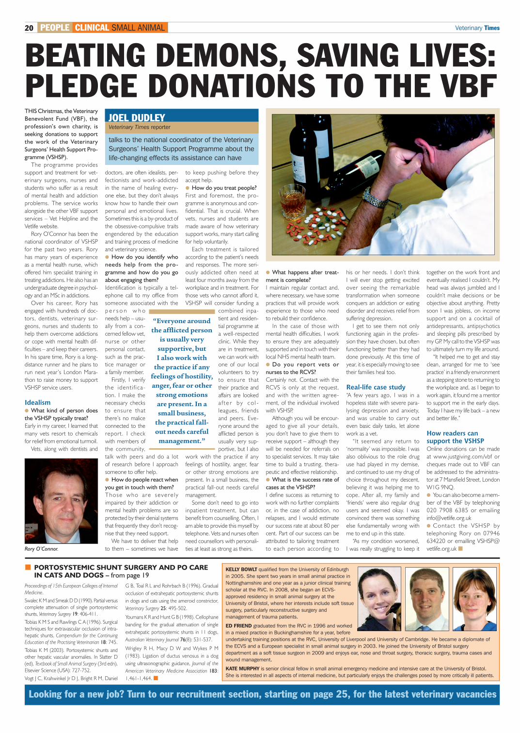
vetlife.org.uk �Rory O’Connor.
“Everyone around the affl icted person
is usually very supportive, but I also work with
the practice if any feelings of hostility, anger, fear or other
strong emotions are present. In a small business,
the practical fall-out needs careful
management.”
PEOPLE CLINICAL SMALL ANIMAL
Proceedings of 15th European Colleges of Internal
Medicine.
Swalec K M and Smeak D D (1990). Partial versus
complete attenuation of single portosystemic
shunts, Veterinary Surgery 19: 406-411.
Tobias K M S and Rawlings C A (1996). Surgical
techniques for extravascular occlusion of intra-
hepatic shunts, Compendium for the Continuing
Education of the Practising Veterinarian 18: 745.
Tobias K M (2003). Portosystemic shunts and
other hepatic vascular anomalies. In Slatter D
(ed), Textbook of Small Animal Surgery (3rd edn),
Elsevier Science (USA): 727-752.
Vogt J C, Krahwinkel Jr D J, Bright R M, Daniel
G B, Toal R L and Rohrbach B (1996). Gradual
occlusion of extrahepatic portosystemic shunts
in dogs and cats using the ameroid constrictor,
Veterinary Surgery 25: 495-502.
Youmans K R and Hunt G B (1998). Cellophane
banding for the gradual attenuation of single
extrahepatic portosystemic shunts in 11 dogs,
Australian Veterinary Journal 76(8): 531-537.
Wrigley R H, Macy D W and Wykes P M
(1983). Ligation of ductus venosus in a dog
using ultrasonographic guidance, Journal of the
American Veterinary Medicine Association 183:
1,461-1,464. �
KELLY BOWLT qualifi ed from the University of Edinburgh in 2005. She spent two years in small animal practice in Nottinghamshire and one year as a junior clinical training scholar at the RVC. In 2008, she began an ECVS-approved residency in small animal surgery at the University of Bristol, where her interests include soft tissue surgery, particularly reconstructive surgery and management of trauma patients.
ED FRIEND graduated from the RVC in 1996 and worked in a mixed practice in Buckinghamshire for a year, before undertaking training positions at the RVC, University of Liverpool and University of Cambridge. He became a diplomate of the ECVS and a European specialist in small animal surgery in 2003. He joined the University of Bristol surgery department as a soft tissue surgeon in 2009 and enjoys ear, nose and throat surgery, thoracic surgery, trauma cases and wound management.
KATE MURPHY is senior clinical fellow in small animal emergency medicine and intensive care at the University of Bristol. She is interested in all aspects of internal medicine, but particularly enjoys the challenges posed by more critically ill patients.
Looking for a new job? Turn to our recruitment section, starting on page 25, for the latest veterinary vacancies
� PORTOSYSTEMIC SHUNT SURGERY AND PO CARE IN CATS AND DOGS – from page 19
VT40.49 InCopy.indd 20 03/12/2010 11:53