Embed Size (px)
Citation preview

SKIN MANIFESTATIONS OF
SYSTEMIC DISEASES
Assistant Prof Dr. Thamir A. Kubaisi
DERMATOLOGIST

Skin act as a mirror for internal organs…

Skin problems associated with diabetes mellitus

ABOUT 30% OF PATIENTS WITH DIABETES
MELLITUS WILL EXPERIENCE A SKIN PROBLEM.
THE MOST COMMON SKIN DISEASES ARE
* CANDIDIASIS,
* IMPETIGO
* AND RECURRENT BOIL.
Assistant Prof Dr. Thamir A. Kubaisi

Specific skin conditions associated with DM:
1- Diabetic dermopathy (shin spots): light brown or reddish, oval or round,
slightly depressed scaly patches most often appearing on the shins.
Cause: Microangiopathy and neuropathy
Recent research suggests that there is a significant correlation between the presence of these lesions and other complications of diabetes, such as retinopathy, nephropathy and neuropathy

2- Diabetic bullae: blister-like lesions that occur spontaneously (for unknown cause)
on the feet and hands.
Treatmennt: maintains its clean, by topical antiseptic

3- diabetic foot ulcer (Neuropathy +/or Microangiopathy)

4- Nicrobiosis lipoidica diabeticorum Typical lesions occur on the pretibial skin, and begin
as a firm, dull-red papule or plaque.
which enlarges radially to become a yellowish,
atrophic plaque with an erythematous edge.
The surface is often glazed in appearance, with
prominent telangiectatic vessels.
Lesions are usually symptomless.
Assistant Prof Dr. Thamir A. Kubaisi

5- Stiff (tighty)skin (ex. Dupuytren's contracture )
6- Chronic pruritus (due to dry skin & sweating
disturbances.
Anogenital pruritus may be due to secondary infection
with candidiasis or haemolytic streptococci )
7- Xanthoma. (fat accumulations in the skin) If DM
associated with hyperlipidemia

SKIN SIGNS OF CHRONIC LIVER FAILURE
• Jaundice – yellow skin and eyes
• Pigmentations
• Guttate hypomelanosis-like lesions – white patches
• Bleeding into skin or mucosa – red or purple haemorrhages, petechiae, purpura or ecchymoses (bruises
• Spider angiomas – also called spider nevus or spider telangiectasis
• Atrophic, thin (Paper money-like) skin & stria, thread-like blood vessels (telangiectasia).
• Palmar erythema – red palms
• Caput medusa – distended veins radiating from the umbilicus
Assistant Prof Dr. Thamir A. Kubaisi

• Severe pruritus cause scratch marks and prurigo nodularis.
• Dry skin.
• Nail changes: – clubbing, flat and brittle nails, opaque white nails (Terry nails), parallel white lines.
• Loss of armpit and pubic hair – due to hormonal changes
• Regional variations in peripheral circulation, e.g., forearm warmer than calf
• Xanthoma
• Pruritus
• Acneform eruption

Pigmentation
• Anaemia presenting as pallor is an early and common sign in
renal failure, resulting from reduced erythropoiesis and
increased haemolysis.
• A muddy brown hyperpigmentation develops in many cases,
attributed to retention of chromogens and deposition of melanin,
Skin problems associated with ranal disease

• Uraemic neuropathy affects (60%)patients with renal failure or on
long-term haemodialysis.
• Up to 40% of patients with renal failure may develop
gynaecomastia
• Calcinosis cutis may occur in 1% of patients with end-stage renal
disease
• Perforating disorders. as acquired reactive perforating
collagenosis, Kyrle’s disease or perforating folliculitis
Skin problems associated with ranal disease

Skin problems associated with ranal disease
Uraemic pruritus (renal itch) is the most common skin problem
Pruritus, or itch, is a common problem for patients with chronic
renal failure. It affects about one-third of patients on dialysis and
is more common with haemodialysis than continuous ambulatory
peritoneal dialysis (CAPD)
Uraemic pruritus is characterized by daily bouts of itching that
tend to worsen at night and may prevent sleep. The skin may
appear normal or dry (xerosis), with few scratch marks.

Uraemic pruritus is thought to be due to a combination of factors including:
Dry skin.
Reduced sweating
Abnormal metabolism of calcium and phosphorus / raised parathroid hormone
Accumulation of toxins and histamine
Nerves excitation
Systemic inflammation
Co-existing medical problems, particularly diabetes and liver disease

TREATMENT OF URAEMIC PRURITUS
• Cleanser and Emollients
• UVB is the mainstay of treatment
• Antihistamine & systemic steroid generally not effective
• Gabapentin.
• Naloxone
• Activated charcoal
• Thalidomide
• Cholestyramine
• Ondansetron.
• Kidney transplantation usually results in resolution of uraemic pruritus.
Assistant Prof Dr. Thamir A. Kubaisi
Skin tumors in a transplant recipients are at greatly
increased risk of developing skin cancers, particularly
squamous cell carcinoma

SKIN PROBLEMS ASSOCIATED WITH THYROID DISEASE
THYROTOXICOSIS RESULTS IN AN INCREASE IN METABOLIC
RATE. THIS MAY RESULT IN:
• Smooth, moist, warm skin
• Flushing of face and hands
• Overgrown nails (acropachy, clubbing), with seperation on nail from the nail bed (onycholysis)
• Fine soft thinned scalp hair
• Generalized itching
• Urticaria (chronic)
• Increased skin pigmentation
• Pretibial myxoedema
• Grave's disease can be associated with other autoimmune skin conditions, including vitiligo.

PRETIBIAL MYXOEDEMA • Pretibial myxoedema affects 5% of patients with Grave's disease. It may
appear before, during or after the thyrotoxic state and is sometimes associated
with an under-active thyroid
• Pretibial myxoedema presents with a swollen and lumpy appearance over the
shins and sometimes affects the feet.
• The skin may be pink or purple, with prominent hair follicles. This is known as
‘peau d'orange’ (orange-peel) appearance .
• Pretibial myxoedema is a form of diffuse mucinosis.
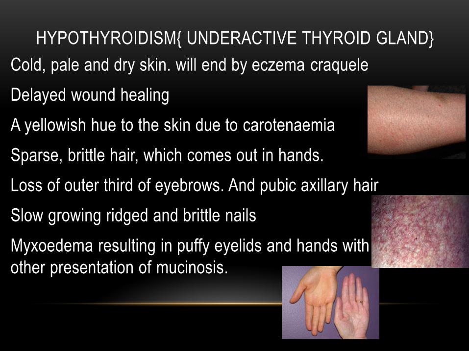
HYPOTHYROIDISM{ UNDERACTIVE THYROID GLAND}
• Cold, pale and dry skin. will end by eczema craquele
• Delayed wound healing
• A yellowish hue to the skin due to carotenaemia
• Sparse, brittle hair, which comes out in hands.
• Loss of outer third of eyebrows. And pubic axillary hair
• Slow growing ridged and brittle nails
• Myxoedema resulting in puffy eyelids and hands with
other presentation of mucinosis.

Assistant Prof Dr. Thamir A. Kubaisi
Zinc
Is an essential trace elements of the diet. Zinc in human
milk is more absorbable than that from infant formulas or cow's
milk, hence the later onset of acrodermatitis enteropathica in
breast-fed babies if compared to formula-fed babies.
Zinc is also found in meat, shellfish.
Zinc is needed to assist metalloenzymes that are involved in
many cellular processes throughout the body. These include the
production of anti-inflammatory agents (cytokines and
antioxidants) and the normal functioning of the brain.

ZINC DEFICIENCY CHARACTERIZED BY 5
DS:
DERMATOLOGICAL FEATURES (DERMATITIS AROUND THE
MOUTH AND/OR ANUS, ALLOPECIA ),
DIARHEA,
DEVELOPMENTAL IMPAIRMENTS,
DEPRESSION,
DEATH.

Zn deficiency are classified according to the causes
in to 3 group: 1- Primary
a- acrodermatitis enteropathica: autosomal recessive inherited
disease
b- Endemic (nutritional) : As in Chronic Zn deficiency, low Zn
diet (high fiber diet, grains bread), clay eating habit and
hookworm infestations
2- Secondary Zn deficiency (Acquired) as in :
Alcoholism, GI disorders, anti metabolic drugs, renal disorders,
premature infants, anorexia nervosa.
3- Marginal Zn deficiency : There are marginal Zn intake, but
still adequate to prevent overt clinical manifestation. (Infant were
failure to thrive)
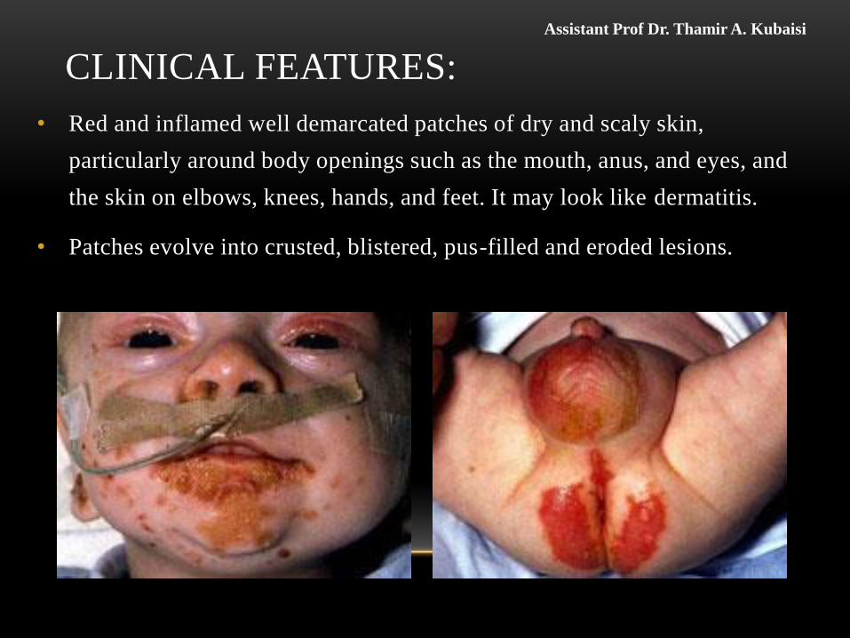
CLINICAL FEATURES:
• Red and inflamed well demarcated patches of dry and scaly skin,
particularly around body openings such as the mouth, anus, and eyes, and
the skin on elbows, knees, hands, and feet. It may look like dermatitis.
• Patches evolve into crusted, blistered, pus-filled and eroded lesions.
Assistant Prof Dr. Thamir A. Kubaisi

•Skin around nails becomes inflamed (paronychia) and there may
be nail ridging.

•Diffuse hair loss on the scalp, eyebrows and, eyelashes.
•Secondary infection with candida or staphylococcus aureus.
•Red glossy tongue and mouth ulcers.
•Impaired wound healing
•In chronic cases: generalized alopecia, depressions, nail
dystrophy,

OTHER FEATURES OF ACRODERMATITIS
ENTEROPATHICA INCLUDE:
• Conjunctivitis
• Sensitivity to light
• Loss of appetite
• Diarrhoea, mild or severe
• Irritability
• Depressed mood
• Growth failure

DIAGNOSIS
1. Serum/plasma zinc levels confirm the diagnosis (normal levels are 10.7 - 23.0 µmol/L)
2. Reduced urinary zinc excretion
3. Blood count may reveal anaemia
4. Therapeutic diagnosis (oral Zn give dramatic response)

TREATMENT
• The inherited form of acrodermatitis enteropathica was
usually fatal until the role of zinc was discovered in 1973.
• It should be treated with 1 mg/kg body weight of oral zinc
supplementation per day for life.
• Zinc gluconate is better tolerated than zinc sulfate.
• Zinc can be given during pregnancy.
• After zinc replacement, the diarrhoea ceases and irritability
and depression of mood improve within 24 hrs. Skin lesions
heal within 1-2 weeks
Assistant Prof Dr. Thamir A. Kubaisi

•Secondary bacterial and/or fungal infection of lesions
require appropriate antibiotic therapy.
•Although zinc is usually non-toxic, high doses for a long
period can result in gastrointestinal symptoms, dizziness
and anaemia.
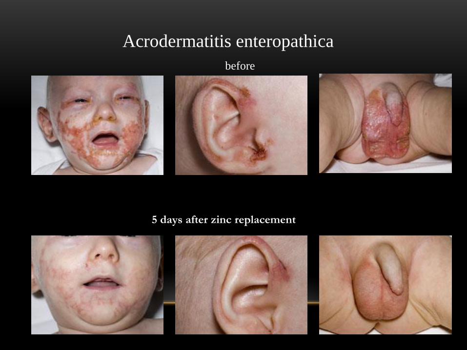
Acrodermatitis enteropathica
before
5 days after zinc replacement

THE CLINICAL FEATURES OF IRON DEFICIENCY ANAEMIA
• Itching
• Nails become brittle and fragile and develop vertical stripes. Nail
plate changes result in koilonychia-spoon shaped nails.
• Patients develop angular cheiliti, a condition in which painful
cracks appear at the corners of the mouth.

•The tongue may become swollen and smooth and
develop a burning sensation.
•Dryness of the mouth and throat making it difficult to
swallow.
•Hair becomes dry, brittle and dull. Increased hair
shedding may be noticed.
•Skin is abnormally pale if the patient is anaemic.
•Predispose to bacterial and fungal infections such as
impetigo, boil and candidiasis.
THE CLINICAL FEATURES OF IRON DEFICIENCY ANAEMIA

PELLAGRA
Pellagra caused by vitamin B3 (niacin) deficiency has the 4 Ds:
• Dermatitis(Photodermatitis)
• Dementia
• Diarrhea
• Death (Within 2-3 years if misdiagnose)

• The first sign is reddened skin with superficial scaling in
areas exposed to sunlight, heat & friction. This may resemble
severe sunburn.
• The rash is usually symmetrical with a clear edge between
affected and unaffected skin (necklace sign).
• An improvement in primary pellagra should be seen within two
days of starting treatment (intravenous or oral niacin or
nicotinamide)