Embed Size (px)
Citation preview

1
SKILLED MOVEMENTS IN AUTISM: A REVIEW OF CURRENT LITERATURE AND TWO CASE STUDIES
By
KATHLEEN A. BERGER
A THESIS PRESENTED TO THE GRADUATE SCHOOL OF THE UNIVERSITY OF FLORIDA IN PARTIAL FULFILLMENT
OF THE REQUIREMENTS FOR THE DEGREE OF MASTER OF SCIENCE
UNIVERSITY OF FLORIDA
2010
2
© 2010 Kathleen A. Berger

3
To my family

4
ACKNOWLEDGMENTS
I would like to thank my committee, Keith White, Donald Stehouwer and Keith
Berg, for their time, advice and encouragement.
5
TABLE OF CONTENTS page
ACKNOWLEDGMENTS .................................................................................................. 4
LIST OF TABLES ............................................................................................................ 7
LIST OF FIGURES .......................................................................................................... 8
LIST OF ABBREVIATIONS ............................................................................................. 9
ABSTRACT ................................................................................................................... 10
NEUROBIOLOGICAL FINDINGS IN AUTISM RELEVANT TO MOTOR BEHAVIORS ........................................................................................................... 12
What is Autism? ...................................................................................................... 12 Neurobiological findings in Autism .......................................................................... 14
The Cerebellum ................................................................................................ 14 Structural Imaging ...................................................................................... 14 Functional Imaging and Behavioral relationship to Cerebellar
differences .............................................................................................. 17 Putative behavioral consequences of damage to the cerebellum .............. 17
The Cerebral Cortex ......................................................................................... 21 Brain Growth and Macrostructure .............................................................. 21 Microstructure ............................................................................................ 23 What is the minicolumn? ............................................................................ 24 How should we interpret this increased number but decreased volume
of minicolumns? ...................................................................................... 24 Connectivity between brain regions ........................................................... 25
The Striatum ..................................................................................................... 26
IMITATION AND PRAXIS IN ASD................................................................................. 36
Motor Overview ....................................................................................................... 36 Imitation .................................................................................................................. 37 Praxis beyond Imitation........................................................................................... 39
Mapping findings onto fcMRI studies ................................................................ 43 Motor Learning and The third core feature of autism ........................................ 44
CASE STUDY: ANALYSIS OF THE FIRST USE OF AN EARLY PRAXIS SKILL ........ 47
Methods and Results .............................................................................................. 48 ASD Participant ................................................................................................ 48 Procedure ......................................................................................................... 48
Movement trajectory directions .................................................................. 49 Variance of movement timing..................................................................... 51

6
Errors ......................................................................................................... 52 Discussion .............................................................................................................. 53 Limitations, conclusion and future research directions ........................................... 55
CASE STUDY: TEACHING THE FINE MOTOR SKILLS OF BUTTONING AND SNAPPING ............................................................................................................. 58
Methods .................................................................................................................. 58 Participant ........................................................................................................ 58 Procedure ......................................................................................................... 59
Results .................................................................................................................... 61 Discussion and Conclusion ..................................................................................... 62
QUALITATIVE DESCRIPTION OF VIDEOS FROM CHAPTER 3 ................................ 65
LIST OF REFERENCES ............................................................................................... 70
BIOGRAPHICAL SKETCH ............................................................................................ 80
7
LIST OF TABLES
Table page Table 1-1. Imaging studies of the cerebellum ................................................................ 31
Table 1-2. Connectivity studies ..................................................................................... 32
Table 1-3. Structural imaging studies of the striatum .................................................... 34
Table 1-4. Brain-repetitive behavior correlations (Reproduced from Langen et al., 2009) .................................................................................................................. 35
Table 3-1. Distribution of trajectory directions (degrees) ............................................... 55
Table 3-2. Distribution of change in trajectory directions (degrees) .............................. 56
Table 3-3. Timing ratios. Ratio of mouth to bowl/bowl to the mouth: movement durations: ............................................................................................................ 56

8
LIST OF FIGURES
Figure page Figure 2-1. What and why of grasping in ASD .............................................................. 46
Figure 3-1. Typical child movement trajectory directions. .............................................. 57
Figure 3-2. ASD child movement trajectory directions................................................... 57
Figure 4-1. Snapping and buttoning ability while dressing self ...................................... 64
Figure 4-2. Whole hand and pincer grasp ..................................................................... 64
Figure A-1. Wrist flexion and extension ......................................................................... 68
Figure A-2. Tool use error ............................................................................................. 69
Figure A-3. Repetitive movements in a typical child. ..................................................... 69
9
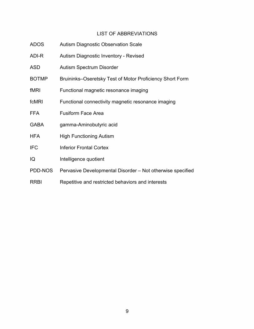
LIST OF ABBREVIATIONS
ADOS Autism Diagnostic Observation Scale ADI-R Autism Diagnostic Inventory - Revised ASD Autism Spectrum Disorder BOTMP Bruininks–Oseretsky Test of Motor Proficiency Short Form fMRI Functional magnetic resonance imaging fcMRI Functional connectivity magnetic resonance imaging FFA Fusiform Face Area GABA gamma-Aminobutyric acid HFA High Functioning Autism IFC Inferior Frontal Cortex IQ Intelligence quotient PDD-NOS Pervasive Developmental Disorder – Not otherwise specified RRBI Repetitive and restricted behaviors and interests

10
Abstract of Thesis Presented to the Graduate School of the University of Florida in Partial Fulfillment of the Requirements for the Degree of Master of Science
SKILLED MOVEMENTS IN AUTISM: A REVIEW OF CURRENT LITERATURE AND
TWO CASE STUDIES
By
Kathleen A. Berger
May 2010
Chair: Keith White Major: Psychology
Autism is a developmental disorder that is diagnosed behaviorally from deficits in
social interaction, communication and repetitive and restricted patterns of behavior.
This thesis documents changes in our understanding of these behaviors: early theory
proposed that autism was caused by the lack of a strong attachment between the child
and his mother or father, yet today it is accepted that autism is a complex genetic
disorder affecting brain development in which environment also plays an important role.
To do so this paper reviews the neurobiological and clinical findings of motor
deficits in autism and then presents two case studies. The first case study is a detailed
analysis of the first time a child with autism is able to eat with a spoon independently
contrasted with a typical child also engaging in eating with a fork for the first time. The
second study describes the process of teaching a child with autism a fine motor skill.
Specifically, the child is taught how to manipulate clothing fasteners such as a button
and snap. The second case study was informed in part by the kinematic analysis of the
first case study.
Motor aspects of the third diagnostic component for autism, restricted and
repetitive behaviors and interests, have traditionally been described as purposeless
11
mannerisms such as hand flapping or rocking. However, purposeful movements
conducted by children with autism can show similar characteristics of repetitive
behaviors such as invariance in timing and pattern. For instance, the first case study
describes invariance during a purposeful motor skill (i.e., eating with a spoon). The
second study shows that it is highly effective to teach a child with autism to button a
button or snap a clothing fastener specifically in the pattern he will use when doing the
activity. This child had been working in occupational therapy for the prior two years on
this skill, but not directly in the pattern he would use. The child learned how to button
and snap effectively using this method. This paper presents evidence in support of
expanding the definition of restricted and repetitive behavior in ASD to include
purposeful, learned skills.

12
CHAPTER 1 NEUROBIOLOGICAL FINDINGS IN AUTISM RELEVANT TO MOTOR BEHAVIORS
Chapter 1 begins with a review of current diagnostic criterion for autism followed
by a review of the neuroanatomical findings in the cerebellum, the cerebral cortex and
the basal ganglia. There is evidence to support that the cerebellum may be involved
early on in autism and that neuroanatomical differences observed in the cerebral cortex
and the basal ganglia may be due to developmental changes in response to the early
cerebellar differences (Bauman and Kemper 2005), so this is the order in which these
regions are presented. Three firm findings in the brain of people with autism include:
decreased numbers of Purkinje cells in post mortem studies and volumetric differences
in vivo (imaging) in the cerebellum; enlargement of the brain in the first few years of life
and volumetric differences in the striatum of the basal ganglia. Additionally, these
structural and functional differences have also been putatatively correlated with
behavioral measures.
What is Autism?
In the American Psychiatric Association Diagnostic and Statistical Manual of
Mental Disorders, fourth edition (DSM IV, Filipek et al., 1999), autism is but one of
several categories that fall under the umbrella disorder of pervasive developmental
disorders (PDD). Autism, Asperger's syndrome and PDD- Not Otherwise Specified
(PDD-NOS) are classified as autism spectrum disorders (ASD), although PDD also
includes Rett's syndrome and childhood disintegrative disorder. Inclusion criteria for
ASD are impairments in (1) social and (2) communication abilities (the first two
domains), and (3) the presence of repetitive and stereotyped behaviors. Exclusion is by
other diagnosis such as Rett’s syndrome. The PDD-NOS diagnosis is used when
13
autistic symptoms are present, but the individual does not meet the full criterion cutoff
for autism. Added criteria for categories of PDD relate to developmental progression:
e.g. Asperger's syndrome is differentiated from autism in that language appeared to
develop normally up until the age of three. The diagnosis of childhood disintegrative
disorder is indicated when a child develops normally up until at least 2 years of age,
followed by a rapid regression with autistic symptoms. Numerous attempts have been
made to improve reliable diagnosis of ASD (Mayes et al., 2009). Current ‘gold
standards’ for ASD diagnosis are behavioral inventories; the Autism Diagnostic
Inventory- Revised and the Autism Diagnostic Observation Schedule (Gotham et al.,
2009; Lord et al., 1994).
Given that the three core symptom domains of ASD inclusion criteria are generally
derived from clinical judgment, several recent studies have used factor analysis as a
means to inform whether ASD is best characterized by the current three domains or
whether different domains would be a better fit. For example, Georgiades et al. (2007)
found the best fit for three, but different, domains: (1) inflexible language behaviors, (2)
impaired social communication, and (3) repetitive sensory and motor behaviors.
Alternatively, Snow et al. (2009) concluded that a two domain model of (1) social and
communication problems, and (2) repetitive and restrictive behaviors and interests
(RRBI) were the best fit. Further still, Happe (2008) suggests that autism defined by
DSM-IV domains may be a ‘fractionable triad’ with differing genes responsible for each
domain.
Further research is certainly needed before making any firm conclusions, but the
above examples illustrate the uncertain and evolving nature of ASD diagnosis.
14
Diagnostic uncertainty is a recognized challenge for the field. It has been theorized that
people with autism suffer from a lack of "central coherence", the cognitive ability to bind
together a jumble of separate features into a single, coherent object or concept (Frith,
1989). Ironically, the field of autism research all too often seems like a fragmented
tapestry stitched from different analytical threads and theoretical patterns (Belmonte et
al., 2004).
Neurobiological findings in Autism
Abnormalities have been reported in virtually every part of the autistic brain
(Stanfield et al., 2008). The present review will focus on areas with particular import to
motor function, namely, cerebellum, cerebral cortex (particularly frontal and parietal
lobes), and striatum in the basal ganglia. For these regions findings in autism include:
early brain overgrowth followed by slowed or arrested overall brain growth, but
particularly frontal lobes (Redcay & Courchesne, 2005); differing connectivity and
activation patterns (Muller, Pierce, Ambrose, Allen, & Courchesne, 2001; Just,
Cherkassky, Keller, & Minshew, 2004; Kana, Keller, Cherkassky, Minshew, & Just,
2006; Mizuno, Villalobos, Davies, Dahl, & Muller, 2006); micro structural differences
(Casanova, Buxhoeveden, Switala, & Roy, 2002; Vargas, Nascimbene, Krishnan,
Zimmerman, & Pardo, 2005; Casanova et al., 2006); and volumetric differences of the
cerebellum (Pierce & Courchesne 2001) and of the striatum (Hollander et al. 2005).
The Cerebellum
Structural Imaging
Guided by post mortem studies of autism brains evidencing abnormally decreased
numbers of Purkinje cells, one of the earliest imaging finding was hypoplasia
(decreased size relative to typically developing, normal brains) in ASD of vermal lobules

15
VI and VII in the cerebellum (Courchesne, Yeung-Courchesne, Press, Hesselink, &
Jernigan, 1988). Figure 1-1, below, shows a midsagittal section of the cerebellum with
areas VI and VII of the vermis smaller in the participants with ASD (A) as compared to
the typical participants (B). Comparisons of these panels can be aided by looking at the
pons and brainstem, which appear relatively equal in size. The small triangular regions
outlined in black are labeled I-V and VI-VII. Comparing the two panels, areas I-V
appear roughly equal in size whereas area VI and VII appear smaller in the autism brain
(A), as compared to the typical brain (B).
While Courchesne et al. (1988) had found hypoplasia of vermal lobules VI and VII
in their original study, subsequent work by their lab showed one subgroup with
hypoplasia and a second subgroup with hyperplasia of these lobules when compared to
controls (Courchesne et al., 1994). In contrast, using volumetric measurement, Hardan,
Minshew, Harenski, & Keshavan (2001) reported that in non-mentally retarded
adolescents and adults with ASD the cerebellar volumes were greater than controls, but
no significant difference for the vermis areas were noted. In fact, when reading through
the literature pertaining to cerebellar differences in autism, one can easily get confused.
For example in a review article, Belmonte (2004) says, "MRI morphometry reveals
hypoplasia of the cerebellar vermis and hemispheres and autopsy studies report
reductions in the number of Purkinje Cells." Yet, from a recent meta-analysis, Stanfield
et al. (2008) reports of firm findings: increased size of the cerebellum overall and some
evidence for decreased size of vermal lobules VI and VII and possibly of areas VIII-X.
The reasons for such confusion may stem from diagnostic uncertainty or from
common comorbidities with autism such as low IQ. While Courchesne et al. (1988)
16
found hypoplasia of vermal lobules VI and VII in their study, in their subsequent study
(Courchesne et al., 1994) the majority of participants exhibiting hypo- or hyper-plasia of
vermal lobules VI and VII were those with verbal IQs less than 70. In Hardan et al.
(2001), where no significant differences were found in the vermis, the ASD participants
were of average IQ. Indeed, Piven et al. (1992) hypothesized that volumetric
differences in the cerebellum might be secondary to IQ differences between ASD and
comparison groups, and found no specific differences in areas of the neocerebellum
when controlling for IQ. Again in 1997 Piven, Saliba, Bailey, & Arndt (1997) found larger
cerebellar volumes in the autism group, but after controlling for total brain volume and
performance IQ; there was no difference from controls.
The question whether IQ should be used as a control variable in such studies
has been debated. Courchesne, Townsend, & Saitoh, 1994 argue that low IQ measures
are a part of the autistic syndrome, while others maintain that by not controlling for IQ
any observed abnormalities will be confounded (Piven et al., 1997) because cerebellar
abnormalities are also observed in non-ASD disorders associated with mental
retardation. Accordingly, Kaufman et al. (2003) compared cerebellar vermis size in
children with Fragile X, Down’s syndrome and autism, and additionally within Fragile X
and Down’s syndrome groups, contrasted participants who did or did not have the co-
morbid diagnosis of autism. Findings indicated differences in cerebellar vermis across
all groups, but hypoplasia of vermal lobes VI and VII as the sole abnormality was
specific to the idiopathic autism group.
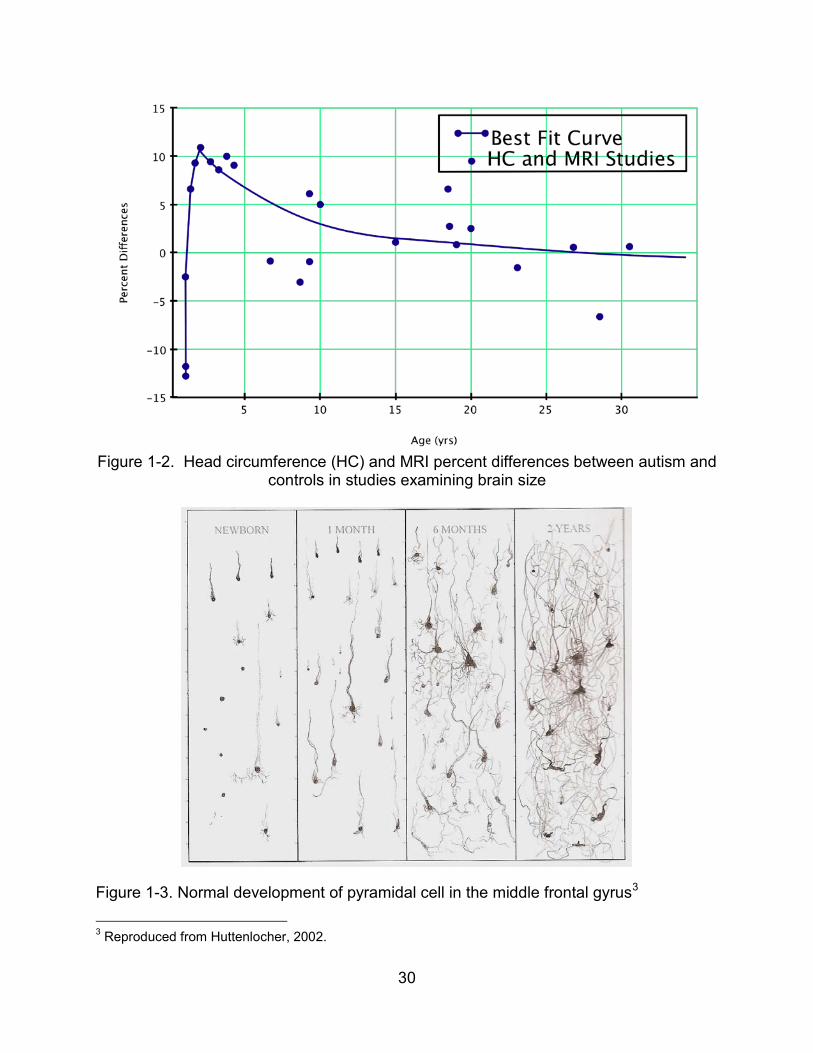
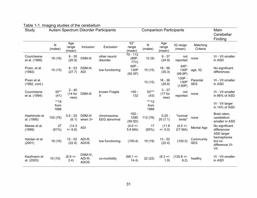
Table 1-1 outlines study participants, parameters and findings for structural
imaging of the cerebellum. When participants included those with mental retardation,

17
and the participants were at younger ages, vermal size differences were generally
found. When participants were older and when total brain volumes and IQs were
controlled, no significant vermal size difference emerged.
Functional Imaging and Behavioral relationship to Cerebellar differences
Further investigations into the cerebellum have included examining the
relationship between behavior and size differences in the cerebellum, and neural activity
through fMRI.
Belmonte et al. (2004) propose that decreased inhibition from the Purkinje cells of
the cerebellum during early development would lead to different activity dependent
neural activity, which might explain findings of abnormal individual mapping in and
overgrowth of the frontal lobes in ASD. Referencing their fMRI findings of abnormally
low cerebellar activation during a selective attention task, and abnormally high
cerebellar activation during a simple motor task in persons with ASD, Belmonte et al.
(2003) argue:
Both of these functional abnormalities correlate significantly with reduced size of cerebellar subregions, and it seems likely that this structure–function correspondence extends to the microscopic level and in particular to the reduction in Purkinje cell numbers. Such a reduction would release the deep cerebellar nuclei from inhibition, producing abnormally strong physical connectivity and potentially abnormally weak computational connectivity along the cerebello-thalamocortical circuit. This altered pattern of cortical excitation may produce aberrant activity-dependent patterning and may thus be related to findings of abnormal individual variability in cortical maps for motor function (Muller et al., 2001) and face processing (Pierce et al., 2001) and to abnormal overgrowth in frontal lobes (Carper and Courchesne, 2000).” Belmont et al. (2004), pg. 9229
Putative behavioral consequences of damage to the cerebellum
Whereas science has long viewed function of the cerebellum as largely motor,
(Middleton & Strick, 2000) recent imaging, behavioral and neuroanatomical studies
18
indicate there may be a role for the cerebellum in cognitive functions (Strick, Dunn &
Fiez, 2009). People who suffer from a stroke that involves the anterior lobules (I-V) of
the cerebellum present with the motor symptoms associated with cerebellar damage -
gait ataxia, dysmetria, oculomotor abnormalities and dysarthria (Schmahmann &
Pandya, 2008). Alternatively, lesions of the posterior lobe of the cerebellum, which
includes areas VI and VII,1
The CCAS is characterized by deficits in executive function, visual spatial performance, linguistic processing and affective dysregulation. Executive impairments include deficits in working memory, motor or ideational set shifting, and perseveration. Verbal fluency may be impaired to the point of telegraphic speech or mutism. Visuospatial disintegration impairs attempts to draw or copy a diagram, conceptualization of figures can be disorganized, and some patients display simultanagnosia. Anomia, agrammatic speech and abnormal syntactic structure are observed, with abnormal prosody and occasionally high pitched, hypophonic whining. (p.1052)
presents in what is known as Cerebellar Cognitive Affect
Syndrome (CCAS):
Schmahmann & Pandya (2008) further have put forward the dysmetria of thought
theory that the cerebellum is involved with automatizing and optimizes cognition as well
as motor processes:
We have proposed that the cerebellum plays an essential role in automatization and optimizing behavior around a homeostatic baseline according to context; that the cerebellum modulates cognition and emotion in the same way that it coordinates motor control; and that disruption of the neural circuitry linking the cerebellum with the association and paralimbic cerebral regions prevents the cerebellar modulation of functions subserved by the affected subsystems, thereby impairing the regulation of movement, cognition and emotion. This loss of the ‘‘cerebellumizing’’2
1 Area VI of the cerebellum demonstrates connections to the premotor cortex.
of behavior leads not only to gait and appendicular ataxia, dysarthria and oculomotor abnormalities when the motor cerebellum is involved, but also to the various
2 The loss of regulation around a baseline homeostasis.

19
aspects of the cerebellar cognitive affective syndrome when the cognitive and limbic cerebellar regions are damaged. (p. 1054)
While much of the above discussion relates to adult lesion studies, it is noteworthy
that children having posterior fossa tumor resections, which removed areas VI and VII
of the vermis, behavioral delays 'reminiscent' of autism such as mutism and language
deficits were observed (Riva & Giorgi, 2000).
Middleton & Strick state that the cerebellum may be as specialized, or
topographically organized as the cerebral cortex, and that to categorize a patient as a
'cerebellar patient' may be a bad descriptor as just as patients with cerebral cortex focal
damage have specific loss of function, so might patients with damage to specific areas
of the cerebellum have a specific loss of function as to the area of the cerebellum
damage. For example, specific areas of the dentate nucleus are shown to connect to
areas of the prefrontal cortex and these areas of the prefrontal cortex in turn connect to
the same area of the cerebellum, forming a closed loop. Damage to an area in the
cerebellum that connects to Brodman area 46 would likely present as a cognitive deficit,
whereas damage to a different area that connects to the primary motor area in the
cerebrum would present as a motor deficit.
Though the above citation seems to link the cerebellum, an area implicated
possibly at prenatal to early postnatal times, very clearly with symptoms associated with
autism, it should be noted that there continues to be debate as to whether the
cerebellum is involved in cognitive processing at all (Glickstein, 2007; Strick et al. 2009),
and to what extent the cerebellum is affected in autism (Stanfield et al., 2008).
Guided by previous work observing decreased exploration in children with autism,
and noting a study that observed decreased exploratory behavior, as compared to a
20
control, in a guinea pig strain that had abnormalities of cerebellar lobules Vi and VII
(Caston et al. 1998), Pierce and Courchesne (2001) examined the relationship between
area of vermal areas VI and VII in the cerebellum and behavioral measures of persons
with autism in a visuospatial exploration task. Decreased size of the cerebellum was
linked to decreased exploration as well as with increased repetitive behaviors in children
with autism (Pierce & Courchesne, 2001). Though no relationship was found between
IQ and exploratory behavior, the authors suggest it is certainly possible this would affect
the results and the measure used, the non-verbal portion of the Leiter, may not have
been comprehensive enough to detect the relationship between IQ and exploratory
behavior.
Although hypoplasia of the cerebellar vermis, particularly areas VI and VII, has
been correlated to the decreased exploratory behavior seen in people with autism, the
exact nature and import of this abnormality is still not clear and much work still needs to
be done. (Stanfield et al., 2008) In post mortem cases, the most robust finding has been
a decrease in number of Purkinje cells in the cerebellum. Also, the reduced Purkinje
cell numbers have been found in autism cases with seizure disorders, with children as
well as adults, and with differing medications. In post mortem cases, though, reduced
Purkinje cell number has not been confined to the posterior vermis (Bauman & Kemper,
2005). To add to the confusing findings, though some investigators (Bauman & Kemper,
1996) have observed a lack of glial cell hyperplasia and propose that, as glial cell
hyperplasia is usually seen in children with Purkinje cell loss at older ages secondary to
ischemia and inflammation, this implicates an early developmental timeframe for the
decrease in Purkinje cell numbers, others (Bailey et al., 1998), have found modest glial
21
hyperplasia and suggest that if the cerebellar cortex develops normally, then the
difference in Purkinje cell numbers may not occur in the prenatal timeframe suggested
by Bauman and Kemper.
Alternatively and interestingly though, a recent study investigating the
phenomenon of prism adaptation in individuals with autism indicated that prism
adaptation was not affected in individuals with autism (Larson et al., 2008). Lesions
that involved areas VI and VII did not affect prism adaptation in the monkeys in this
study (Baizer, Kralj-Hans, & Glickstein, 1999). While Larson’s (2008) null finding does
not confirm abnormalities specific to areas VI and VII in the cerebellum, it would be
consistent with negative findings in areas outside of these lobules that affect prism
adaptation.
In summary, the cerebellum appears to an area of the brain that is malformed in
autism and may affect neural connections through experience and developmental
plasticity. Further research is certainly needed before we can draw strong conclusions
as to the certainty and consequences of cerebellar malformations.
The Cerebral Cortex
Early post mortem studies did not reveal structural differences in the cerebral
cortex in autism, but recent imaging and newer stereologic techniques have revealed
developmental brain growth differences in individuals with autism as well as micro
structural differences.
Brain Growth and Macrostructure
Other recent research in autism has focused on the finding that there appears to
be an early overgrowth of the brain in the first few years of life (up until about 4 years of
age) followed by a period of arrested growth when compared to controls. Though
22
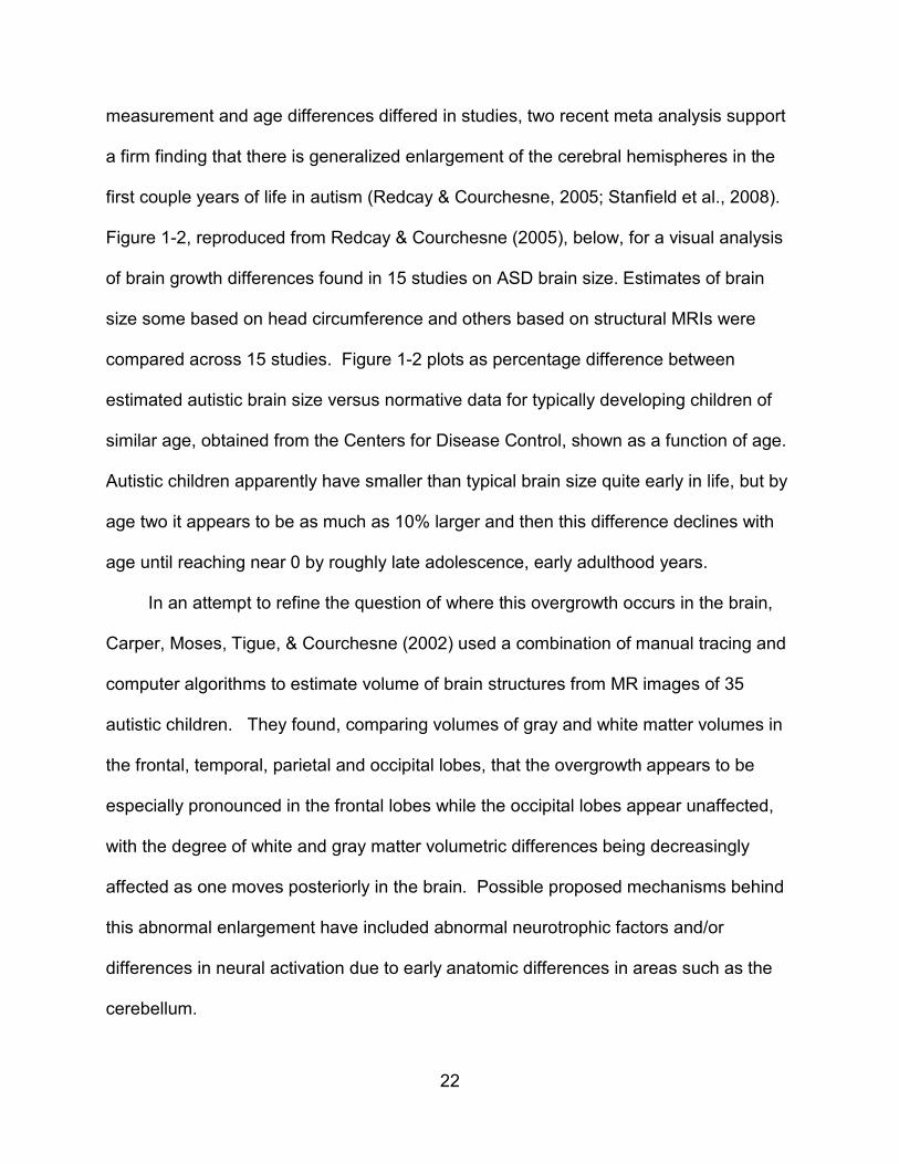
measurement and age differences differed in studies, two recent meta analysis support
a firm finding that there is generalized enlargement of the cerebral hemispheres in the
first couple years of life in autism (Redcay & Courchesne, 2005; Stanfield et al., 2008).
Figure 1-2, reproduced from Redcay & Courchesne (2005), below, for a visual analysis
of brain growth differences found in 15 studies on ASD brain size. Estimates of brain
size some based on head circumference and others based on structural MRIs were
compared across 15 studies. Figure 1-2 plots as percentage difference between
estimated autistic brain size versus normative data for typically developing children of
similar age, obtained from the Centers for Disease Control, shown as a function of age.
Autistic children apparently have smaller than typical brain size quite early in life, but by
age two it appears to be as much as 10% larger and then this difference declines with
age until reaching near 0 by roughly late adolescence, early adulthood years.
In an attempt to refine the question of where this overgrowth occurs in the brain,
Carper, Moses, Tigue, & Courchesne (2002) used a combination of manual tracing and
computer algorithms to estimate volume of brain structures from MR images of 35
autistic children. They found, comparing volumes of gray and white matter volumes in
the frontal, temporal, parietal and occipital lobes, that the overgrowth appears to be
especially pronounced in the frontal lobes while the occipital lobes appear unaffected,
with the degree of white and gray matter volumetric differences being decreasingly
affected as one moves posteriorly in the brain. Possible proposed mechanisms behind
this abnormal enlargement have included abnormal neurotrophic factors and/or
differences in neural activation due to early anatomic differences in areas such as the
cerebellum.
23
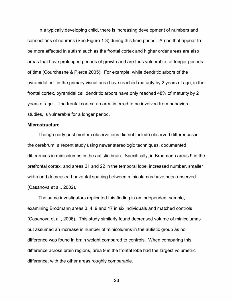
In a typically developing child, there is increasing development of numbers and
connections of neurons (See Figure 1-3) during this time period. Areas that appear to
be more affected in autism such as the frontal cortex and higher order areas are also
areas that have prolonged periods of growth and are thus vulnerable for longer periods
of time (Courchesne & Pierce 2005). For example, while dendritic arbors of the
pyramidal cell in the primary visual area have reached maturity by 2 years of age, in the
frontal cortex, pyramidal cell dendritic arbors have only reached 48% of maturity by 2
years of age. The frontal cortex, an area inferred to be involved from behavioral
studies, is vulnerable for a longer period.
Microstructure
Though early post mortem observations did not include observed differences in
the cerebrum, a recent study using newer stereologic techniques, documented
differences in minicolumns in the autistic brain. Specifically, in Brodmann areas 9 in the
prefrontal cortex, and areas 21 and 22 in the temporal lobe, increased number, smaller
width and decreased horizontal spacing between minicolumns have been observed
(Casanova et al., 2002).
The same investigators replicated this finding in an independent sample,
examining Brodmann areas 3, 4, 9 and 17 in six individuals and matched controls
(Casanova et al., 2006). This study similarly found decreased volume of minicolumns
but assumed an increase in number of minicolumns in the autistic group as no
difference was found in brain weight compared to controls. When comparing this
difference across brain regions, area 9 in the frontal lobe had the largest volumetric
difference, with the other areas roughly comparable.

24
What is the minicolumn?
The minicolumnar circuit is an evolutionarily and ontogenetically conserved template adapted in the various cortical areas according to their specific developmental and functional requirements. The minicolumnar core comprises radially oriented arrays of pyramidal projection neurons. At the core and periphery of the minicolumn, combinations of GABAergic interneurons provide for a diversity of signaling properties that serve to dynamically modulate pyramidal cell inputs and outputs that perform area and task-specific information processing needs. (Casanova et al., 2006 p. 287)
This cytoarchitecture, and knowledge from animal studies of the visual system, has
been the basis for theoretical models of information processing in cognitive psychology
(Roelfsema 2006). From studies in monkeys, we know that certain visual stimuli are
preferentially dependent on feedback, feed forward or lateral inhibitory neural
processing. A recent study by Vandebrouke, Scholte, van England, Lamme & Kemner
(2008) examined visual processing in autism based on these models (see further
mention below).
How should we interpret this increased number but decreased volume of minicolumns?
One possibility raised in the Casanova et al. (2006) study was that increased
minicolumn number might be a general indicator of mental retardation. But, Casonova
et al. cite Buxhoeveden et al. (2002), which evidences normal minicolumn width with
smaller brain volume in individuals with Down's syndrome, a disorder strongly
associated with mental retardation. Further, many clinical investigations typically have
participants who are high functioning and two recent studies evidenced information
processing differences that would be consistent with observed minicolumn differences
(see above). More specifically, lateral inhibition has been hypothesized to be aberrant

25
due to the decrease in neuropil space between minicolumns, where lateral inhibitory
neurons reside, in individuals with ASD.
Bertone, Mottron, Jelenic & Faubert (2005) and Vandenbroucke, Scholte, van
England, Lamme & Kemner (2008) concluded that lateral inhibition was aberrant in
visual processing whereas feedback or recurrent processes were unaffected or
enhanced. Vandenbroucke, Scholte, van England, Lamme & Kemner (2009) used
different visual stimuli to assess accuracy at detecting differences in feedback
processing (detecting surface from background) as compared to visual stimuli to assess
horizontal inhibitory influences (detecting boundaries where two different visual
orientations meet). Both controls and participants with autism were comparable in
discriminating stimuli reliant on feedback processing, whereas the autistic participants
scored lower on visual discrimination tasks relying more on lateral inhibitory neural
connections. The authors suggest that visual aberrancy in the participants with autism
is "probably caused by impaired interactions through horizontal connections in lower
visual areas." And that "malfunctioning of horizontal connections is possibly a more
general deficit underlying several symptoms of autism."
Connectivity between brain regions
With the advent of techniques to study connectivity to different brain regions,
functional connectivity studies using fMRI, fcMRI, been used to study differing
connectivity in neural tracts in people with autism. (Just et al., 2004; Kana et al., 2006;
Mizuno et al., 2006) Functional connectivity studies use the temporal correlations of
activation patterns from fMRI measures in different areas of the brain. One of the more
recent models proposed based on some of these findings is increased local
overconnectivity with decreased long distance connectivity, resulting in reduced or

26
aberrant information transfer. (Belmonte et al., 2004; Courchesne & Pierce, 2005)
Belmonte et al. (2004) suggest what one would see in this type of network is amplified
neural response to sensory stimuli, whether attended or unattended and decreased
synchrony between areas of the brain that integrate sensory information. Belmonte
cites (Belmonte & Allen, 2000; Belmonte & Yurgelun-Todd, 2003), two studies (EEG
and fMRI) that show a pattern of decreased activation in integrative regions in the brain
(medial temporal and prefrontal) with decreased synchrony of these regions with
sensory regions.
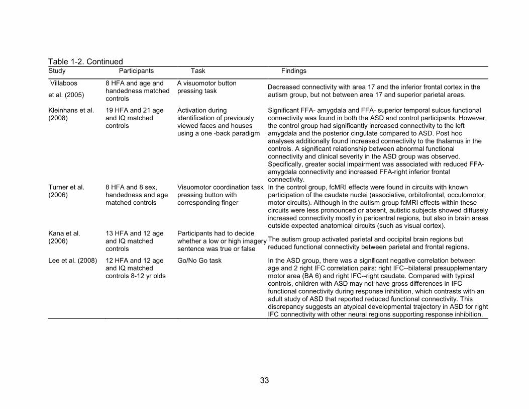
Table 1-2 summarizes imaging studies investigating connectivity in people with
ASD. Generally, there appears to be different connectivity in the autism brain with
evidence for relatively decreased connectivity to the frontal lobes, (Villalobos, Mizuno,
Dahl, Kemmotsu & Muller, 2005; Lee et al., 2008, Turner, Frost, Linsenbardt, McIlroy, &
Muller, 2006) but more intact connectivity as one moves posteriorly in the brain
(Kleinhans et al., 2008, Just et al., 2004, Villaboos et al., 2005). One recent study
investigated connectivity within known corticostriatal loops (Turner et al., 2006). While
the control group demonstrated connectivity between the caudate and associative,
orbitofrontal, occulomotor and motor regions of the frontal cortex, the autism group
showed decreased effects in these regions with increased activation, mostly in
pericentral regions, but also in areas not expected such as the visual cortex.
The Striatum
Recent imaging studies in people with autism have evidenced a relationship
between the size of the striatum and the restricted and repetitive behaviors and
interests (RRBI) domain of autism. Though the studies appear conflicting in that at
times there seems to be a positive correlation to the RRBI domain and other times a

27
negative correlation, this may be due to the type of assessment and the type of
repetitive behavior. Langen et al. (2009) suggests that this may be that higher order
'cognitive' behavior appears to be negatively correlated with caudate size, whereas
lower order 'motor' behavior tends to be positively correlated with the striatal size,
especially the caudate, but further research is needed to confirm this hypothesis. Table
1-3, below, summarizes findings from structural imaging studies of the striatum.
The striatum, comprised of the putamen and caudate, is the major input nucleus of
the basal ganglia, with major inputs from frontal areas of the cerebral cortex. Over the
last several years there has been increasing evidence supporting a role for the basal
ganglia in sharpening the selection of actions and intentions, while suppressing
competing actions or intentions. (Mink, 1996; Middleton & Strick, 2000) Additionally,
differences in frontal-striatal- thalamic circuitry have been associated with repetitive and
restricted behaviors in animal models (Lewis, Tanimura, Lee, & Bodfish, 2007) and in
volumetric studies of the striatum (Sears et al., 1999; Hollander et al., 2005; Langen et
al., 2009). Different connectivity has also been found between the frontal cortex and
the striatum in an fcMRI study with high functioning adults and adolescents with autism
(Turner et al., 2006).
Guided by findings of striatal volumetric differences in persons with obsessive-
compulsive disorder (OCD) and Tourette's syndrome, disorders with overlapping
repetitive behaviors, Hollander et al. (2005) compared 17 adults with autism with 17
controls for difference in putamen and caudate volumes. The right caudate and
putamen were larger in the autism group and repetitive behaviors, particularly higher
level repetitive behaviors were positively correlated with increased volumes. As the use

28
of neuroleptics has been associated with increased volume of basal ganglia structures,
Langen, Durston, Staal, Palmen, & van Engeland (2007) compared caudate volume in
high-functioning individuals with autism with and without neuroleptics use. The volume
difference was significant for individuals with autism as compared to controls in both
medication and medication naive groups. The difference also remained significant after
correction for total brain volume.
Although the Hollander et al. (2005) study evidenced differences in the striatum,
the participants in this study were adults so developmental changes could not be
detected. A recent large (n= 99 autism participants and n=88 controls) cross sectional
study examined volumetric differences in the striatum. (Langen et al., 2009) Whereas
caudate volume decreased as age increased for typically developing children, for the
children with autism, caudate volume, greatest in the right caudate head, increased with
age. These investigators also found a negative correlation for volume of the caudate
and the behavioral diagnostic category of insistence on sameness, and this was more
apparent in younger subjects.
A comparison to prior findings though revealed apparently conflicting results. For
example, (Hollander et al., 2005) found a positive correlation of the size of the right
caudate and higher order repetitive behavior. Sears et al. (1999) found a negative
correlation between size of caudate and ritualistic patterns, but a positive correlation
with complex mannerisms. Similarly, Rojas et al. found a positive correlation between
the caudate and repetitive behaviors. Langen et al., (2009) suggest the findings are not
as contradictory as they appear as lower order repetitive behaviors (e.g. complex
mannerisms) are associated with increased caudate volume, whereas higher order

29
ritualistic behaviors are negatively associated with caudate volume (at least in three of
four studies), so for example in Rojas 2006, the entire category of repetitive behaviors
were positively correlated, rather than analyzing individual categories of repetitive
behaviors. Table 1-4 compares correlations in different studies with repetitive behaviors
listed in the ADI-R.
To summarize, findings implicate an association between the size of the caudate
and the behavioral category of repetitive behavior in people with autism. Though the
exact nature of the association is not clear, there is evidence that an increased size of
the caudate is related to decrease in ritualistic behaviors, but may be related to an
increase in complex mannerisms.
Figure 1-1. Area of cerebellum lobules
30
Figure 1-2. Head circumference (HC) and MRI percent differences between autism and
controls in studies examining brain size
Figure 1-3. Normal development of pyramidal cell in the middle frontal gyrus3
3 Reproduced from Huttenlocher, 2002.
31
Table 1-1. Imaging studies of the cerebellum Study Autism Spectrum Disorder Participants Comparison Participants Main
Cerebellar Finding
N (males)
Age range
(mean) Inclusion Exclusion
IQ* range
(mean)
N (males)
Age range
(mean)
IQ range (mean)
Matching Criteria
Courchesne et al. (1988) 18 (16) 6 - 30
(20.9) DSM-III other neurol. disorder
70 - 112 (88P, 77V)
12 (9) 9 - 37 (24.9)
not reported none VI - VII smaller
in ASD
Piven, et al. (1992) 15 (15) 8 - 53
(27.7) DSM-III, ADI low functioning
60P - 130P
(92.5P) 15 (15) 18 - 56
(30.3)
64P- 130P
(99.9P) age, IQ No significant
differences
Piven et al. (1992, cont.) 15 (15) 18 - 36
(28.8)
130P - 130P
(130P)
Parental SES
VI - VII smaller in ASD
Courchesne et al. (1994)
50** (41)
2 - 40 (14 for
new) DSM-III known Fragile
X <50 -
132 53*** (43)
3 - 37 (17 for
new)
not reported none VI - VII smaller
in 86% of ASD
**18 from 1988
***12 from 1988
VI - VII larger in 14% of ASD
Hashimoto et al. (1995) 102 (76) 0.5 - 20
(6.1) DSM-III when 3+
chromosome, EEG abnormal
10D - 129D
(59.5D) 112 (76) 0.25 -
20 (7.1) "normal
limits" none Brain stem, cerebellum smaller in ASD
Manes et al. (1999)
27 (81%)
(14.3 +/- 6.8) ADI (4.6 +/-
5.6 MA) 17
(65%) (11.8
+/- 5.0) (4.5 +/-
2/7 MA) Mental Age No significant differences
Hardan et al. (2001) 16 (16) 12 - 52
(22.4) ADI-R, ADOS low functioning (100.4) 19 (19) 13 - 52
(22.4) (100.5) Community SES
ASD larger hemispheres but no difference VI-VII
Kaufmann et al. (2003) 10 (10) (6.9 +/-
2.4)
DSM-IV, ADI-R, ADOS
co-morbidity (66,1 +/- 14.4) 22 (22) (8.3 +/-
1.9) (120.8 +/-
9.2) healthy VI - VII smaller in ASD

32
Table 1-1. Continued
Study Autism Spectrum Disorder Participants Comparison Participants Main Cerebellar Finding
Kaufmann et al. (2003, cont.)
16 (16) (7.0 +/- 1.8)
ASD plus Down's (20.1 +/-
6.9) 11 (11) (7.2 +/- 2.1)
(41.2 +/- 9.1) Down's only VI - VII smaller
both groups
Kaufmann et al. (2003, cont.)
13 (13) (5.7 +/- 2.1)
ASD plus Fragile X (46.0 +/-
15.0) 9 (9) (5.3 +/- 1.1)
(56.0 +/- 15.2)
Fragile X only
VI - VII larger co-morbid ASD
***Note: P denotes Performance IQ, V denotes Verbal IQ, D denotes Development IQ, MA denotes Mental Age, else Full-Scale IQ. * 18 autism participants were from the 1988 study. ** 12 were from the 1988 study *** ICA = Intracranial area
Table 1-2. Connectivity studies Study Participants Task Findings
Just et al. (2004) 17 HFA and 17 Controls
Reading an active or passive sentence and then answering as to the agent or recipient of the action
The autism group produced reliably more activation than the control group in Wernicke’s (left laterosuperior temporal) area and reliably less activation than the control group in Broca’s (left inferior frontal gyrus) area. Furthermore, the functional connectivity between the various participating cortical areas was consistently lower for the autistic than the control participants.
Kashino et al. (2005)
14 HFA and 14 healthy normal controls
N-back working memory task with letters
The control group demonstrated more activation in the left than the right parietal regions, whereas the autism group showed more right lateralized activation in the prefrontal and parietal regions. The autism group also had more activation than the control group in the posterior regions including inferior temporal and occipital regions. The analysis of functional connectivity yielded similar patterns for the two groups with different hemispheric correlations. The temporal profile of the activity in the prefrontal regions was more correlated with the left parietal regions for the control group, whereas it was more correlated with the right parietal regions for the autism group.
33
Table 1-2. Continued Study Participants Task Findings
Villaboos
et al. (2005)
8 HFA and age and handedness matched controls
A visuomotor button pressing task Decreased connectivity with area 17 and the inferior frontal cortex in the
autism group, but not between area 17 and superior parietal areas.
Kleinhans et al. (2008)
19 HFA and 21 age and IQ matched controls
Activation during identification of previously viewed faces and houses using a one -back paradigm
Significant FFA- amygdala and FFA- superior temporal sulcus functional connectivity was found in both the ASD and control participants. However, the control group had significantly increased connectivity to the left amygdala and the posterior cingulate compared to ASD. Post hoc analyses additionally found increased connectivity to the thalamus in the controls. A significant relationship between abnormal functional connectivity and clinical severity in the ASD group was observed. Specifically, greater social impairment was associated with reduced FFA- amygdala connectivity and increased FFA-right inferior frontal connectivity.
Turner et al. (2006)
8 HFA and 8 sex, handedness and age matched controls
Visuomotor coordination task pressing button with corresponding finger
In the control group, fcMRI effects were found in circuits with known participation of the caudate nuclei (associative, orbitofrontal, occulomotor, motor circuits). Although in the autism group fcMRI effects within these circuits were less pronounced or absent, autistic subjects showed diffusely increased connectivity mostly in pericentral regions, but also in brain areas outside expected anatomical circuits (such as visual cortex).
Kana et al. (2006)
13 HFA and 12 age and IQ matched controls
Participants had to decide whether a low or high imagery sentence was true or false
The autism group activated parietal and occipital brain regions but reduced functional connectivity between parietal and frontal regions.
Lee et al. (2008) 12 HFA and 12 age and IQ matched controls 8-12 yr olds
Go/No Go task In the ASD group, there was a signi!"#$%&$'(#%)*'&"+,,'-#%)+$&.'%/''$&age and 2 right IFC correlation pairs: right IFC--bilateral presupplementary motor area (BA 6) and right IFC--right caudate. Compared with typical controls, children with ASD may not have gross differences in IFC functional connectivity during response inhibition, which contrasts with an adult study of ASD that reported reduced functional connectivity. This discrepancy suggests an atypical developmental trajectory in ASD for right IFC connectivity with other neural regions supporting response inhibition.

34
Table 1-2. Continued Study Participants Task Findings
Mostofsky et al. 2009
13 HFA and 13 age, sex and IQ matched peers; 8-12 yrs old
Appositional finger tapping task The autism group showed significantly less connectivity in motor circuits than typically developing controls.
Table 1-3. Structural imaging studies of the striatum Study Participants Findings
Sears et al., 1999 35 ASD: 12-29 years old; IQ mean = 91 (19.8) and 36 healthy controls: 20.1 (3.8)4
Size of the caudate was larger in ASD group; Using scores from the ADI-R, increased size was related to increased repetitive behaviors of complex mannerisms, compulsions/rituals and difficulties with minor changes in routine.
; IQ mean = 102.1 (12.8)
In an independent sample from a prior study with 15 HFA and 20 controls the authors reproduced the increased caudate size in ASD.
Hollander et al., 2005 17 ASD: 28.39 (11.26) years of age, 97.12 (25.36) IQ; 17 healthy controls: 29.4 (9.08) years of age, 111.5 (14.25) IQ
ASD group had significantly increased size of right caudate. Also, the size of the right caudate and the total putamen volumes were positively correlated with repetitive behaviors; particularly the higher order behaviors.
Rojas et al. 2006 24 ASD: 20.79 (10.58) years of age, 94.75 (20.64) IQ; 22 healthy controls: 21.41 (10.91) years of age, 118.74 (11.18) IQ.
They compared ADI category of RRBI and caudate size was significantly positively correlated. Other brain regions associated with social and communicative deficits and symptom severity were also significantly correlated.
Langen et al., 2007 Two independent samples of medication naïve subjects. Sample 1: 21 HFA and 21 age, IQ, SES, height, weight, gender and handedness controls. Age: 11.2 (2.18) Sample 2: 21 HFA and 21 age, IQ, SES, height, weight, gender and handedness controls. Age: 20.08 (3.01)
The caudate was enlarged in both autism groups. Previous findings of significant correlation with RRBI domain of the ADI-R was not replicated but the authors suggest that this might have been due to low incidence and variability in this domain in this sample.
4 The mean and standard deviation were only reported in this study for the control group.

35
Table 1-3. Continued Study Participants Findings
Langen et al., 2009 99 HFA 12.89 (4.4.5) years of age, 107.59
(13.56) IQ and 89 Healthy Controls 12.36 (4.70) years of age, 109.99 (12.81) IQ.
Whereas the caudate decreased in size with development in the control group, the caudate increased in size for the ASD group. A significant negative correlation was noted with the category of insistence of sameness and size of the caudate and this effect tended to decrease with age.
Table 1-4. Brain-repetitive behavior correlations (Reproduced from Langen et al., 2009) ADI-R Items Langen et al.
(2009) n=88 Sears et al.(1999) n=35
Hollander et al. (2005) n=12
Rojas et al. (2006) n=24
Repetitive Use of Objects Repetitive motor behavior (NS) Insistence on sameness (- ) a
n.s. Low order NS
Repetitive and stereotyped behavior domain - a
Hand and Finger Mannerisms n.s. Other ComplexMannerisms/Stereotyped Body Movements
Lower order +
Resistance to Trivial Changes in the Environment
n.s. N/A
Difficulties with Minor Changes in Routine
High order - b
N/A
Compulsions and Rituals Circumscribed interests (NS)
High order - b
High order +a
Circumscribed Interests n.s. Unusual Preoccupations n.s. Unusual attachments to objects n.s. N/A aSignificant correlation with caudate volume (p _ .05). bSignificant correlation with caudate volume (p _ .01)
36
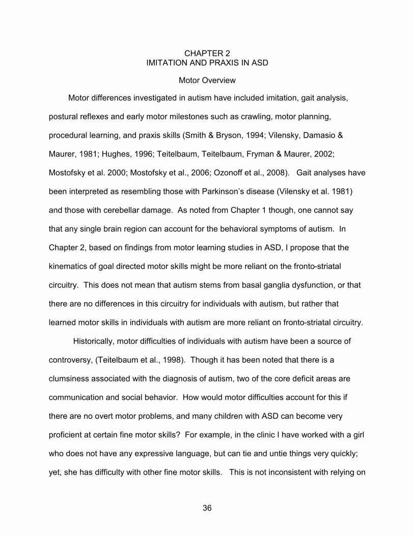
CHAPTER 2 IMITATION AND PRAXIS IN ASD
Motor Overview
Motor differences investigated in autism have included imitation, gait analysis,
postural reflexes and early motor milestones such as crawling, motor planning,
procedural learning, and praxis skills (Smith & Bryson, 1994; Vilensky, Damasio &
Maurer, 1981; Hughes, 1996; Teitelbaum, Teitelbaum, Fryman & Maurer, 2002;
Mostofsky et al. 2000; Mostofsky et al., 2006; Ozonoff et al., 2008). Gait analyses have
been interpreted as resembling those with Parkinson’s disease (Vilensky et al. 1981)
and those with cerebellar damage. As noted from Chapter 1 though, one cannot say
that any single brain region can account for the behavioral symptoms of autism. In
Chapter 2, based on findings from motor learning studies in ASD, I propose that the
kinematics of goal directed motor skills might be more reliant on the fronto-striatal
circuitry. This does not mean that autism stems from basal ganglia dysfunction, or that
there are no differences in this circuitry for individuals with autism, but rather that
learned motor skills in individuals with autism are more reliant on fronto-striatal circuitry.
Historically, motor difficulties of individuals with autism have been a source of
controversy, (Teitelbaum et al., 1998). Though it has been noted that there is a
clumsiness associated with the diagnosis of autism, two of the core deficit areas are
communication and social behavior. How would motor difficulties account for this if
there are no overt motor problems, and many children with ASD can become very
proficient at certain fine motor skills? For example, in the clinic I have worked with a girl
who does not have any expressive language, but can tie and untie things very quickly;
yet, she has difficulty with other fine motor skills. This is not inconsistent with relying on

37
fronto-striatal circuitry for learned motor skills, as fronto-striatal circuitry is thought to be
more active for overlearned motor skills (Doyon & Carrier, 2009). Additionally, in an
fMRI study, fronto-striatal circuitry has also been noted to be preferentially active in a
voluntary eye movement task in people with ASD (Takarae et al., 2007).
One area of motor ability that has been a focus of extensive review in autism has
been imitative abilities, as it was thought that this might underlie communication and
social difficulties. Chapter 2 will begin by reviewing studies of imitation: major findings
and generated hypotheses. Imitation is one aspect of the broader skill of praxis, the
ability to learn and perform higher-level motor skills. More recently there has been more
focus on praxis deficits in ASD, and findings from these studies will be discussed next,
followed by a discussion relating these findings to neuroanatomical findings.
Imitation
Imitation difficulties were noted very early on in autism research:
A mother described the inability of one 21-month-old child to make pat-a-cake simply from watching her. The only way he could learn the game was to have the mother hold his hands and put them through the appropriate movements. (Ritvo & Provence, 1953)
The idea that deficits in imitation might be part of a more global deficiency in self-other
mapping led to studying imitation in autism (Rogers & Pennington, 1991), and a recent
review of studies investigating imitation deficits in autism (Williams, Whiten & Singh,
2004) found the following:
imitation tasks that were meaningful were preferentially helpful to participants with autism, with this effect being more apparent with older participants.
reversal errors are common in participants with autism.
imitation of actions with objects produced less group differences in studies than non-meaningful gestures.

38
Williams et al. (2004) discusses six hypotheses proposed for these deficits, and
proposes that existing evidence is inconsistent for the first three:
(1) A deficit in representational or symbolic functioning (Curcio, 1978).
Williams et al. (2004) suggest that if this were the case, then meaningful imitation would
not be easier for the autism group. Smith and Bryson however argue that evidence is
not compelling for the meaningful/non-meaningful distinction from the studies cited
(Smith & Bryson 2007). For example Williams cites Rogers et al. (1996) as supporting
this hypothesis. But, the control group outperformed the ASD group on 3 out of 4 non-
meaningful tasks, but only 1 out of 4 of the meaningful tasks.
(2) Poor engagement in the experimental tasks by the autism group
(Trevarthen & Aitken, 2001). If this were the case, then the participants with autism
would be equally impaired on imitation tasks and this was not the case.
(3) A long-term deficit in social interaction that leads to less practiced motor
skills (Tantum, 1991). Group differences appear to decrease as age increases. If this
hypothesis were to be correct, we should expect differences to increase.
(4) A dyspraxic problem (Jones & Prior, 1985). Williams et al. (2004) argue
against this hypothesis as Green et al. found that children with Asperger's did worse
than the control group of individuals with dyspraxia. And, individuals with autism do
better with imitation of meaningful gestures.
(5) A disorder of action representation (Smith & Bryson, 1994). Williams et al.
(2004) cites that while Bartak, Rutter & Cox 1975 showed the autism group showed less
understanding and expression than a control group with language disorder, Smith and
Bryson (1994) found no group difference in the recognition of postures and sequences.
39
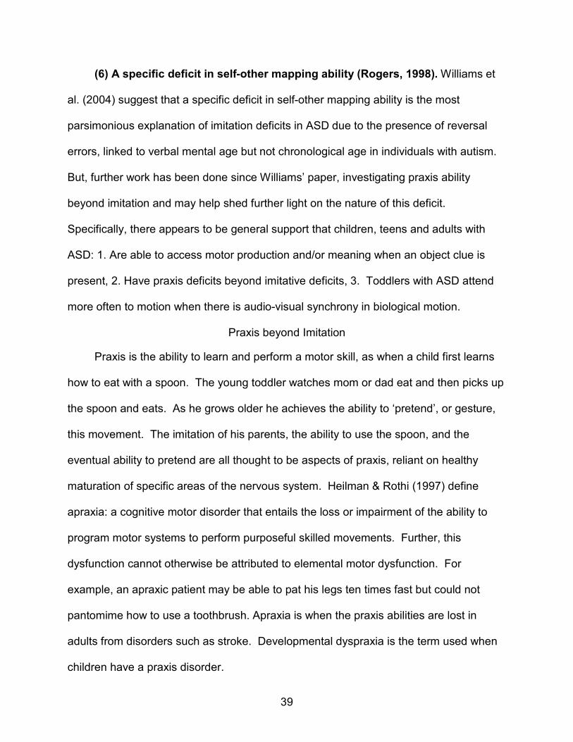
(6) A specific deficit in self-other mapping ability (Rogers, 1998). Williams et
al. (2004) suggest that a specific deficit in self-other mapping ability is the most
parsimonious explanation of imitation deficits in ASD due to the presence of reversal
errors, linked to verbal mental age but not chronological age in individuals with autism.
But, further work has been done since Williams’ paper, investigating praxis ability
beyond imitation and may help shed further light on the nature of this deficit.
Specifically, there appears to be general support that children, teens and adults with
ASD: 1. Are able to access motor production and/or meaning when an object clue is
present, 2. Have praxis deficits beyond imitative deficits, 3. Toddlers with ASD attend
more often to motion when there is audio-visual synchrony in biological motion.
Praxis beyond Imitation
Praxis is the ability to learn and perform a motor skill, as when a child first learns
how to eat with a spoon. The young toddler watches mom or dad eat and then picks up
the spoon and eats. As he grows older he achieves the ability to ‘pretend’, or gesture,
this movement. The imitation of his parents, the ability to use the spoon, and the
eventual ability to pretend are all thought to be aspects of praxis, reliant on healthy
maturation of specific areas of the nervous system. Heilman & Rothi (1997) define
apraxia: a cognitive motor disorder that entails the loss or impairment of the ability to
program motor systems to perform purposeful skilled movements. Further, this
dysfunction cannot otherwise be attributed to elemental motor dysfunction. For
example, an apraxic patient may be able to pat his legs ten times fast but could not
pantomime how to use a toothbrush. Apraxia is when the praxis abilities are lost in
adults from disorders such as stroke. Developmental dyspraxia is the term used when
children have a praxis disorder.

40
In an adult this disorder is acquired, usually through some identifiable lesion that
occurs from a stroke, e.g. While adults with acquired apraxia once were able to perform
these skilled movements, children affected with praxis deficits are hindered in
progressing through this normal developmental period, which may in turn affect
development of typical neural pathways. In children with developmental disabilities,
praxis difficulties are more difficult to determine than in adults with acquired praxis
difficulties – are there difficulties because the child has attentional or cognitive deficits
that prevent initial learning of the motor skill, or is it a failure in the neural networks that
are needed for these skills. Tests that measure recognition as compared to production
of gestures aim to tease out the nature of praxis difficulties.
Though there is no standardized test available for praxis, typical tests include
items such as whether subjects can perform a gesture such as waving goodbye when
asked verbally; by visual imitation; from the cue of a picture of the tool e.g. seeing a
picture of a hammer and gesturing this motion or by demonstrating how you would use
a tool. Trained raters then score whether there are spatial, timing or other type of errors.
Additionally, picture cards may be used to assess correct recognition.
Several studies since the Williams article reviewing imitation have provided
evidence for a dyspraxic component in children with autism beyond imitative abilities. In
2006, Mostofsky et al. compared praxis in children with autism with an age and IQ
matched control group. The participants with autism demonstrated more praxis errors
than the control group and this was interpreted as meaning that the praxis deficits in
persons with autism are not restricted to deficits in imitation (Mostofsky et al., 2006). In
a separate paper (Dziuk et al., 2007) used hierarchical regression and determined that

41
after controlling for age and IQ while basic motor abilities were predictive of praxis
scores, after controlling for basic motor performance, praxis errors were a significant
predictor of autism severity. Though it could be reasonably argued that if imitation were
the primary deficit, and the skill was never learned correctly that one would expect to
see general deficits in praxis.
Dewey et al. (2007) similarly looked at praxis in individuals with autism. Control
groups were children with developmental coordination disorder (DCD), children with
DCD and Attention Hyperactivity Deficit Disorder (ADHD) and children with just ADHD.
While all participants performed poorly on the Bruininks–Oseretsky Test of Motor
Proficiency Short Form (BOTMP), a standardized test of motor proficiency, the group
with autism had significantly more praxis errors. An ANCOVA with age and IQ included
as covariates revealed that the children with autism scored significantly lower than all of
the other groups on both gestures to verbal command and imitation. When gender and
motor skill measures from the BOTMP was added as a covariate, the difference
remained significant.
Praxis gestures can be categorized as either gestures that use tools or social and
communicative gestures. Smith & Bryson (2007) evaluated social and communicative
praxis gestures and pantomimed object use gestures in children with autism, a
language impaired group and a typically developing group, matched on verbal abilities
and sex. These investigators used different input modalities (verbal request and
pictures) to further investigate neural pathways that might be affected in autism.
These investigators found:
Children with autism had a significantly more difficult time imitating unconventional use of objects.

42
Though children with autism demonstrated understanding of the gestures, through the use of pictures, they had a more difficult time with production of gestures through imitation or gesture when verbally requested than the control groups.
All of the children with autism passed a control task of performing the gesture with the actual object.
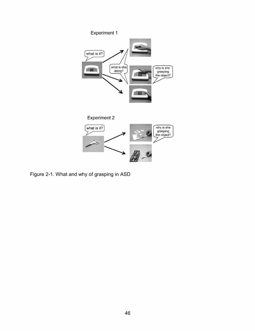
Mentioned in the beginning of this chapter, a relevant recent study (Boria et al.,
2009) assessed whether children with autism's could detect information about the goal
of an act (e.g. to grasp a cup) and the intention behind the act (e.g. to drink from the
cup). Boria et al. compared high functioning children with autism with typically
developing controls in a task that asked, based on a picture of an object hand
interaction, what an individual was doing and why. The children with autism showed
that they could gather the intent of an action through information from the objects
represented, but not from the motor action. For example, if the task was to determine
why the person was picking up the phone (see figure 2-1 (Boria et al., 2009)) but the
information had to be determined by the way the person was grasping the object, the
person with autism could not make the determination of why the person was grasping
the phone. Alternatively, if the picture had object clues, such as either a container or a
paper that had been partially cut, the person with autism could determine the reason for
the grasp.
Interestingly, in the Mostofsky et al. study (2006), the persons with autism had an
easier time demonstrating pantomimed actions when shown a picture of a tool. These
investigators attributed it to a practice effect as this task was presented last. In Smith &
Bryson (2007) the participants with autism were able to perform all tasks accurately
when given the object.

43
In Dowell, Mahone, & Mostofsky (2009) the investigators compared persons with
autism to typically developing controls on: 1. A basic motor measure 2. A postural
knowledge test of gestures with tools, and communicative gestures 3. A praxis test.
The autism group performed comparably to the control group on the postural knowledge
of gestures that included tools, but the autism group performed significantly worse on
postural knowledge of gestures that were communicative. In preliminary results from a
study in our lab with children with autism, they performed comparable on tool or
communicative gestures if an object was present (e.g. if there was a doll to wave
goodbye to). The results of these studies suggest that neural pathways that rely on
object identification to access motor representations are intact in ASD, whereas
pathways that rely on biological motion alone are impaired in ASD.
Mapping findings onto fcMRI studies
While a theory of underconnectivity in autism has been proposed, (Hughes, 2007)
two pertinent fcMRI studies indicate that the certain pathways may be intact or even
enhanced (Villalobos et al., 2005; Mizuno et al., 2006). Using a visuomotor task to
assess thalamocortical function, Mizuno et al. (2006) evidenced increased
thalamocortical connections to the left insula, right postcentral and middle frontal
regions. Villaboos et al. (2005) examined functional connectivity of the dorsal stream of
the visual system, based on mirror neuron dysfunction hypothesis of autism, and found
intact connectivity with superior parietal regions but significantly decreased connectivity
to inferior frontal area 44.
Possibly applicable to these connectivity differences in autism, (Klin, Lin, Gorrindo,
Ramsay, & Jones, 2009) reported on a serendipitous finding that while infants with
autism did not show the preference for biological motion associated with typically

44
developing infants, they did show a preference for viewing motion with audiovisual
synchrony. For example, the infant with autism looked for longer periods at a person
playing ‘pat a cake’ with clapping sounds and visual point lights coming together but not
to a person walking. In the control group, the typical infants did not show such a
preference. One possible explanation for this preferential attention to audiovisual
synchronies could be reliant on ‘ bottom up’ processing, or thalamocortical activity in
children with autism.
To summarize the findings in the imitation and praxis literature in autism:
Children with ASD are preferentially helped in motor production tasks when an object is present
Children with HFA preferentially are able to detect meaning from object clues over biological motion cues, at least for hand grasp
Toddlers with autism show a preference for attending to visual auditory synchrony over biological motion alone
Praxis deficits in ASD are present beyond imitative deficits
Motor Learning and The third core feature of autism
Though the above studies focus on motor learning as it pertains to social and
communicative deficits in autistic persons, other work has focused on the third domain,
repetitive and restricted behaviors and interests (RRBI).
As noted in Chapter 1, RRBI has been correlated with volumetric differences in the
striatum. Investigations of the role of the basal ganglia, and its connections to the
frontal lobes, or fronto-striatal connections, in normal motor learning indicate that this
neural pathway is critical for the maintenance and retrieval of over-learned motor skills,
whereas cortico-cerebellar circuitry is more active in motor adaptation or in early stages
of motor learning. (Doyon et al., 2009) Additionally, there is considered agreement that
45
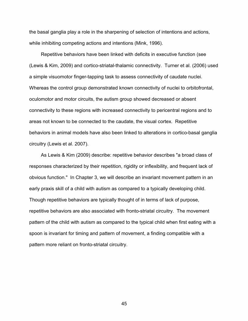
the basal ganglia play a role in the sharpening of selection of intentions and actions,
while inhibiting competing actions and intentions (Mink, 1996).
Repetitive behaviors have been linked with deficits in executive function (see
(Lewis & Kim, 2009) and cortico-striatal-thalamic connectivity. Turner et al. (2006) used
a simple visuomotor finger-tapping task to assess connectivity of caudate nuclei.
Whereas the control group demonstrated known connectivity of nuclei to orbitofrontal,
oculomotor and motor circuits, the autism group showed decreased or absent
connectivity to these regions with increased connectivity to pericentral regions and to
areas not known to be connected to the caudate, the visual cortex. Repetitive
behaviors in animal models have also been linked to alterations in cortico-basal ganglia
circuitry (Lewis et al. 2007).
As Lewis & Kim (2009) describe: repetitive behavior describes "a broad class of
responses characterized by their repetition, rigidity or inflexibility, and frequent lack of
obvious function." In Chapter 3, we will describe an invariant movement pattern in an
early praxis skill of a child with autism as compared to a typically developing child.
Though repetitive behaviors are typically thought of in terms of lack of purpose,
repetitive behaviors are also associated with fronto-striatal circuitry. The movement
pattern of the child with autism as compared to the typical child when first eating with a
spoon is invariant for timing and pattern of movement, a finding compatible with a
pattern more reliant on fronto-striatal circuitry.
46
Figure 2-1. What and why of grasping in ASD

47
CHAPTER 3 CASE STUDY: ANALYSIS OF THE FIRST USE OF AN EARLY PRAXIS SKILL
Chapter 3 presents the kinematic analysis of an early skilled movement in a child
with autism as compared to a typically developing child. In a recent retrospective study,
(Gernsbacher, Sauer, Geye, Schweigert, & Hillgoldsmith, 2008) demonstrated that early
oral motor and manual praxis abilities in children with autism are correlated with verbal
skills at a later age. To our knowledge, however, the kinematics of the first use of a
skilled movement in a child with autism has not been studied previously. A detailed
examination of a praxis skill execution when the child is first performing the skill
independently precedes the complication of compensatory mechanisms that come into
play later. Eating with a utensil develops in the typical child in the second year of life,
thought to be a reflection of cortical maturation and experience (Bundy, Lane & Murray,
2002; Luria 1980). Higher-level skills such as eating with a spoon have been noted to
be impaired in autism, although the exact nature of this impairment has not been clearly
defined (Mostofsky, et al., 2006; Dewey et al., 2007, Gernsbacher et al., 2008).
The present study compares the movement profiles for two children, one with
autism and one typically developing, each child having been captured on home video
eating with a utensil for their first time. Qualitative observations concerning movements
made by the child with autism included that the movements appeared highly
stereotyped; that is, neither the spatial pattern nor timing pattern of the movement
varied as much as was the case for the typical child. While the movement appeared
highly stereotyped, there were two types of errors that disrupted this patterned
movement. Sometimes, the child with autism missed the bowl he was eating from and
had to correct the movement trajectory, and at other times, the spoon turned in his hand
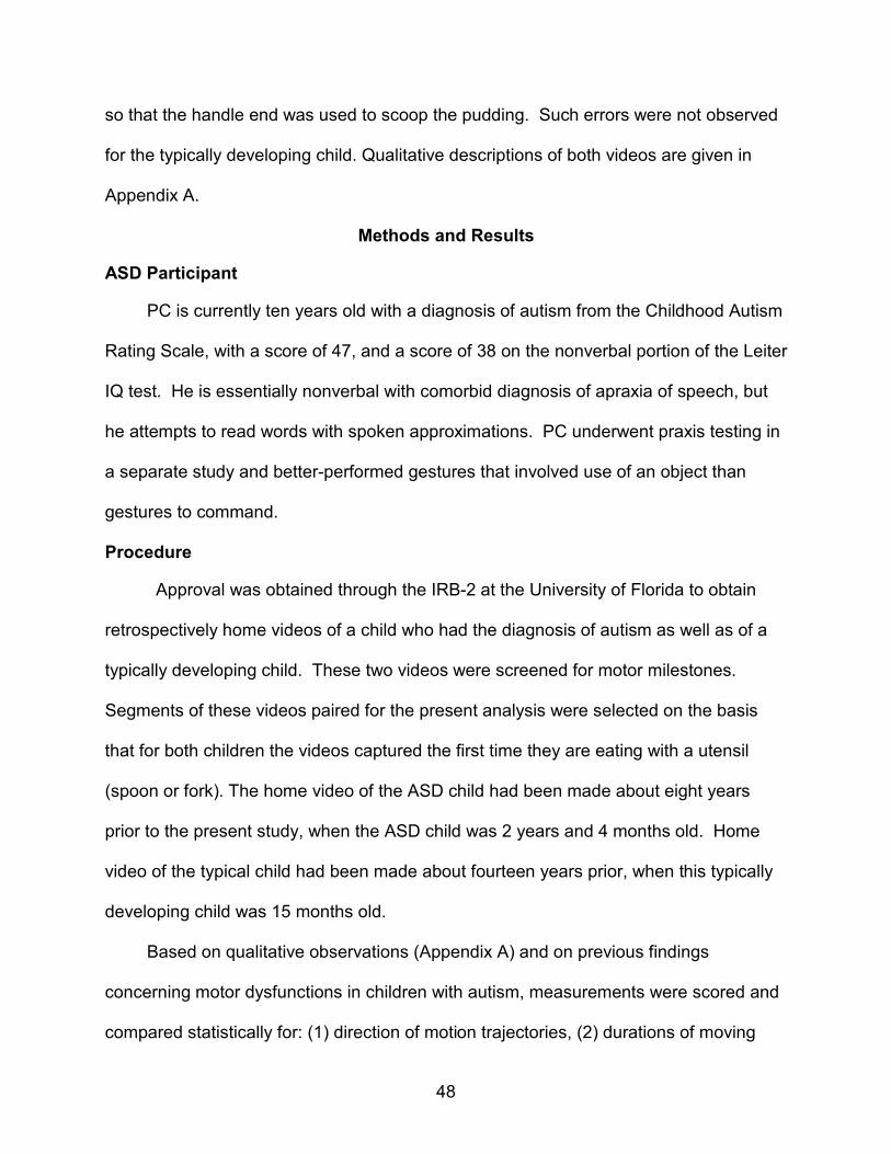
48
so that the handle end was used to scoop the pudding. Such errors were not observed
for the typically developing child. Qualitative descriptions of both videos are given in
Appendix A.
Methods and Results
ASD Participant
PC is currently ten years old with a diagnosis of autism from the Childhood Autism
Rating Scale, with a score of 47, and a score of 38 on the nonverbal portion of the Leiter
IQ test. He is essentially nonverbal with comorbid diagnosis of apraxia of speech, but
he attempts to read words with spoken approximations. PC underwent praxis testing in
a separate study and better-performed gestures that involved use of an object than
gestures to command.
Procedure
Approval was obtained through the IRB-2 at the University of Florida to obtain
retrospectively home videos of a child who had the diagnosis of autism as well as of a
typically developing child. These two videos were screened for motor milestones.
Segments of these videos paired for the present analysis were selected on the basis
that for both children the videos captured the first time they are eating with a utensil
(spoon or fork). The home video of the ASD child had been made about eight years
prior to the present study, when the ASD child was 2 years and 4 months old. Home
video of the typical child had been made about fourteen years prior, when this typically
developing child was 15 months old.
Based on qualitative observations (Appendix A) and on previous findings
concerning motor dysfunctions in children with autism, measurements were scored and
compared statistically for: (1) direction of motion trajectories, (2) durations of moving
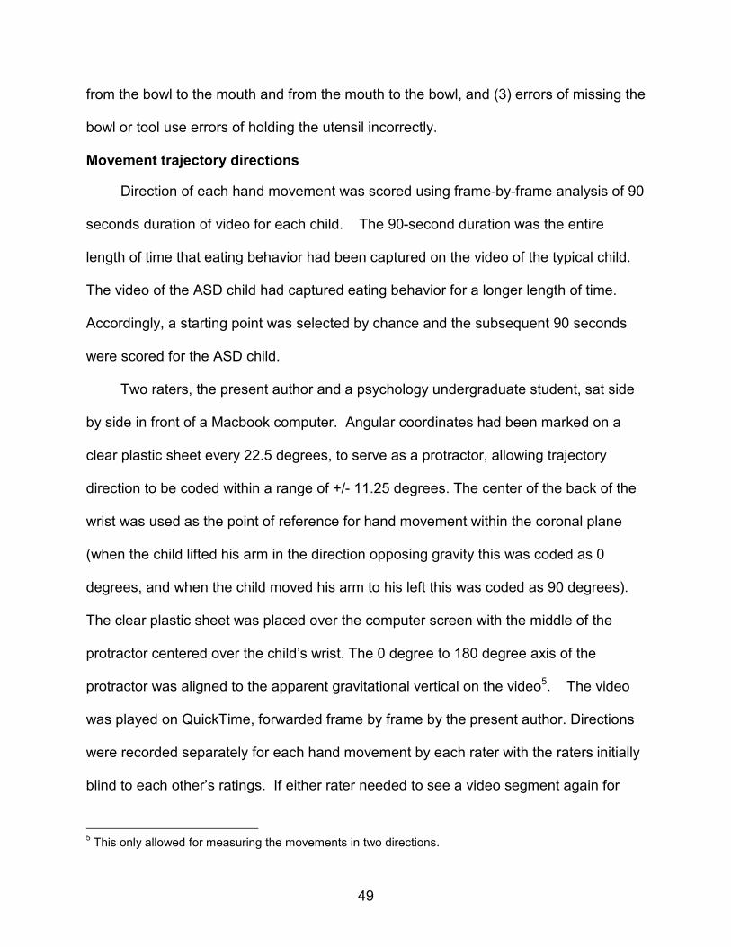
49
from the bowl to the mouth and from the mouth to the bowl, and (3) errors of missing the
bowl or tool use errors of holding the utensil incorrectly.
Movement trajectory directions
Direction of each hand movement was scored using frame-by-frame analysis of 90
seconds duration of video for each child. The 90-second duration was the entire
length of time that eating behavior had been captured on the video of the typical child.
The video of the ASD child had captured eating behavior for a longer length of time.
Accordingly, a starting point was selected by chance and the subsequent 90 seconds
were scored for the ASD child.
Two raters, the present author and a psychology undergraduate student, sat side
by side in front of a Macbook computer. Angular coordinates had been marked on a
clear plastic sheet every 22.5 degrees, to serve as a protractor, allowing trajectory
direction to be coded within a range of +/- 11.25 degrees. The center of the back of the
wrist was used as the point of reference for hand movement within the coronal plane
(when the child lifted his arm in the direction opposing gravity this was coded as 0
degrees, and when the child moved his arm to his left this was coded as 90 degrees).
The clear plastic sheet was placed over the computer screen with the middle of the
protractor centered over the child’s wrist. The 0 degree to 180 degree axis of the
protractor was aligned to the apparent gravitational vertical on the video5
5 This only allowed for measuring the movements in two directions.
. The video
was played on QuickTime, forwarded frame by frame by the present author. Directions
were recorded separately for each hand movement by each rater with the raters initially
blind to each other’s ratings. If either rater needed to see a video segment again for
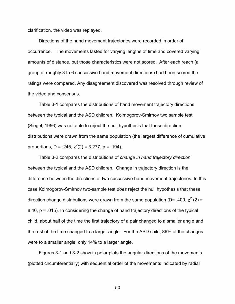
50
clarification, the video was replayed.
Directions of the hand movement trajectories were recorded in order of
occurrence. The movements lasted for varying lengths of time and covered varying
amounts of distance, but those characteristics were not scored. After each reach (a
group of roughly 3 to 6 successive hand movement directions) had been scored the
ratings were compared. Any disagreement discovered was resolved through review of
the video and consensus.
Table 3-1 compares the distributions of hand movement trajectory directions
between the typical and the ASD children. Kolmogorov-Smirnov two sample test
(Siegel, 1956) was not able to reject the null hypothesis that these direction
distributions were drawn from the same population (the largest difference of cumulative
proportions, D = .245, 02(2) = 3.277, p = .194).
Table 3-2 compares the distributions of change in hand trajectory direction
between the typical and the ASD children. Change in trajectory direction is the
difference between the directions of two successive hand movement trajectories. In this
case Kolmogorov-Smirnov two-sample test does reject the null hypothesis that these
direction change distributions were drawn from the same population (D= .400, 02 (2) =
8.40, p = .015). In considering the change of hand trajectory directions of the typical
child, about half of the time the first trajectory of a pair changed to a smaller angle and
the rest of the time changed to a larger angle. For the ASD child, 86% of the changes
were to a smaller angle, only 14% to a larger angle.
Figures 3-1 and 3-2 show in polar plots the angular directions of the movements
(plotted circumferentially) with sequential order of the movements indicated by radial

51
distance (earlier movements plotted farther from the center). The plot for the typical
child does not have any remarkable pattern, but the plot for the ASD child shows a clear
spiral-like pattern. The plot for the ASD child also shows a relatively high density of
movement directions along the -45 deg to 135 deg oblique, although the trajectory
directions are distributed diversely across most directions.
Variance of movement timing
The duration to move the utensil from the bowl to the mouth and the duration to
move the utensil from the mouth to the bowl were measured using frame-by-frame
analysis. For these measurements the individual hand movements previously scored
were not taken into account. In general, more than one hand movement direction
accompanied a particular utensil movement. Utensil movements interrupted by the child
being distracted were set aside from analysis. The ratio of utensil movement durations
was calculated to account for any differences between children in distances from their
bowl to their mouth.
The time to move the utensil from mouth to bowl was initiated at the frame when
the utensil came out of the mouth. The end point for this movement was the frame
when the utensil stopped going forward into the bowl. For the bowl to mouth utensil
movement duration, the time was initiated at the frame when the utensil began an
upward movement until the end point frame when the mouth closed around the utensil.
Table 3-3 shows the ratios of mouth to bowl: bowl to mouth utensil movement durations.
To assess the hypothesis that the timing ratio was less variant for the child with autism,
the absolute value of the difference of mouth to bowl/bowl to the mouth – bowl to the
mouth/mouth to bowl was calculated. Using a one tailed, group variance comparison
test in Stata, version 8, for the absolute value between ratio differences, the null
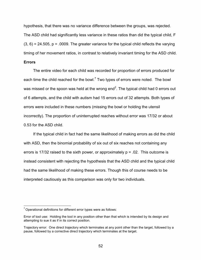
52
hypothesis, that there was no variance difference between the groups, was rejected.
The ASD child had significantly less variance in these ratios than did the typical child, F
(3, 6) = 24.505, p = .0009. The greater variance for the typical child reflects the varying
timing of her movement ratios, in contrast to relatively invariant timing for the ASD child.
Errors
The entire video for each child was recorded for proportion of errors produced for
each time the child reached for the bowl.7 Two types of errors were noted. The bowl
was missed or the spoon was held at the wrong end2
If the typical child in fact had the same likelihood of making errors as did the child
with ASD, then the binomial probability of six out of six reaches not containing any
errors is 17/32 raised to the sixth power, or approximately p = .02. This outcome is
instead consistent with rejecting the hypothesis that the ASD child and the typical child
had the same likelihood of making these errors. Though this of course needs to be
interpreted cautiously as this comparison was only for two individuals.
. The typical child had 0 errors out
of 6 attempts, and the child with autism had 15 errors out of 32 attempts. Both types of
errors were included in these numbers (missing the bowl or holding the utensil
incorrectly). The proportion of uninterrupted reaches without error was 17/32 or about
0.53 for the ASD child.
7 Operational definitions for different error types were as follows:
Error of tool use: Holding the tool in any position other than that which is intended by its design and attempting to sue it as if in its correct position.
Trajectory error: One direct trajectory which terminates at any point other than the target, followed by a pause, followed by a corrective direct trajectory which terminates at the target.

53
Discussion
Difficulties with motor planning have been noted in children with autism spectrum
disorders. The exact nature of early motor skill acquisition is understudied in this
population, unfortunately. The present case study demonstrates highly invariant timing
and trajectory directions in the movements of child with autism compared to a typical
child, when each first ate using a utensil. Also, trajectory errors that prevented reaching
the target, and tool use errors were present for the child with autism, but not for the
typically developing child.
The repetitive behavior hallmark for diagnosing individuals with autism has been
described as "a broad class of responses characterized by their repetition, rigidity or
inflexibility, and frequent lack of obvious function" (Lewis & Kim, 2009, p. 114, emphasis
added). The present study shows that an inflexible pattern also occurs for a functional
motor skill in a child with autism. Repetitive behaviors have been associated with fronto-
striatal circuitry such as increased size of the caudate associated with complex
mannerisms in autism. In animal research, striatal putamen neurons encoding for
muscle pattern and direction have been found, but not those encoding for velocity,
amplitude, force, position, or acceleration (Mink, 1996). Consistent with a limited
capacity to encode variations of movements It is possible that if the child with autism
described in this paper had been unusually reliant on fronto-striatal circuitry for first
using the praxis skill presently studied, then the relative invariance of his movements
would not be surprising. It was somewhat surprising how clearly a pattern of sequential
dependencies in movement directions emerged.
A recent study by (Fabbri-Destro et al., 2009) examined the motor kinematics of
intentional reaching in typical children compared to children with autism. In typical
54
individuals, the kinematics of reach changed according to the goal of the movement.
For example, when the goal was to place a pencil in a container; for the typical children
movement initiation speed was guided by the size of the target container. A typical child
will accelerate the initial movement to pick up the pencil for a smaller target container
but ASD participants did not change their movement speeds relative to the target.
Fabbri-Destro et al. (2009) suggest that individuals with ASD program their movements
independent of the target’s sensory characteristics. However, an alternative
explanation could be that goal oriented movements are less able to be varied in their
timing for individuals with autism. Mostofsky et al. (2006) compared children and teens
with high functioning autism to a typical control group on a serial reaction time task.
The ASD group did not show the characteristic decline in reaction times across blocks
typically seen in this paradigm, and seen in their control participants. The reaction
times (and therefore the movement times) of ASD participants did not differ between
blocks. Mostofsky et al. (2006) attributed this finding to a procedural learning deficit in
ASD. Again, however, as with Fabbri-Destro et al. (2009) and as with the present study,
the observations were that ASD movements were invariant for timing.
Typical treatment interventions available for occupational and physical therapists
to treat praxis difficulties in children with autism include a “bottom up” approach of
sensory integration (Schaaf & Miller, 2005), as well as newer techniques such as the
interactive metronome and music therapy techniques that use rhythm to assist with the
child's sense of internal timing and rhythmicity of a movement (Burpee et al., 2001;
Sabado, 2008; Cosper, Lee, Peters, & Bishop, 2009; LaGasse, 2009) Although frequent
referrals are made for remediation of fine motor skills (Watling, Deitz, Kanny, &

55
McLaughlin, 1999), little empirical research addresses the effectiveness of therapeutic
interventions (Dawson & Watling, 2000).
If a child with autism has difficulty with motor planning and is highly reliant on
movements that are difficult to vary for timing and pattern, then one could infer that
interventions capitalizing on repetition of a pattern and rhythmic timing could be
particularly helpful in teaching a new motor skill. This conjecture is tested in Chapter 4.
Limitations, conclusion and future research directions
The major limitations of the present study are that it (1) only examined one child
with ASD and one typical child, (2) only examined one praxis skill, and (3) the home
videos analyzed were of short durations, and were created under uncontrolled
conditions analyzed retrospectively. It could nevertheless be demonstrated that first
use of the praxis skill of eating with a utensil is highly regular for timing and pattern in
the child with ASD as compared to a typical child.
Table 3-1. Distribution of trajectory directions (degrees) Trajectory Typical child ASD child Cumulative (deg) Cumulative Cumulative Proportion Bin Count Proportion Count Proportion Difference
0 1 0.028 6 0.273 0.245 45 3 0.083 7 0.318 0.235 90 12 0.333 8 0.364 0.030 135 17 0.472 13 0.591 0.119 180 20 0.556 14 0.636 0.081 225 21 0.583 14 0.636 0.053 270 25 0.694 17 0.773 0.078 315 33 0.917 22 1.000 0.083 360 36 1.000 22 1.000 0.000

56
Table 3-2. Distribution of change in trajectory directions (degrees) Trajectory Typical child ASD child Cumulative Change Cumulative Cumulative Proportion Bin Count Proportion Count Proportion Difference ‐180 5 0.143 3 0.143 0.000 ‐135 11 0.314 8 0.381 0.067 ‐90 15 0.429 9 0.429 0.000 ‐45 16 0.457 18 0.857 0.400 45 20 0.571 18 0.857 0.286 90 22 0.629 19 0.905 0.276 135 28 0.800 20 0.952 0.152 180 35 1.000 21 1.000 0.000
Table 3-3. Timing ratios. Ratio of mouth to bowl/bowl to the mouth: movement
durations6
:
Typical Autism
3.909091 .9527778
1.020734 .8222812
1.975309 1.126374
1.830189 1.098093
1.074906
1.588
1.069697
6 The number of trials for the typical child was different for timing ratio trials and error trials as timing ratios were only used for movements where the typical child did not pause when taking the spoon out of her mouth. At times she paused and looked at the camera or smiled at Dad off screen. Such discontinuous movements were not included for calculating the movement timing ratios.
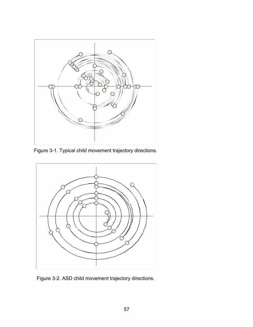
57
Figure 3-1. Typical child movement trajectory directions.
Figure 3-2. ASD child movement trajectory directions.
58
CHAPTER 4 CASE STUDY: TEACHING THE FINE MOTOR SKILLS OF BUTTONING AND
SNAPPING
The author of this paper is a licensed physical therapist. After finishing the study
described in Chapter 3, a former client was referred to work on the fine motor skills of
buttoning and snapping clothing fasteners. According to the mother, this child had been
unsuccessful at reaching independence with these skills from previous therapies carried
out for the preceding year or more.
A functional training program was designed to teach buttoning and snapping that
focused on motor patterns and movement timing patterns specific to these skills.
Additional parameters of the program included intensive training; positive behavioral
and visual supports; sensory-motor activities for attention and arousal support; and
traditional strengthening exercises. Baseline data were obtained for a two-week period
prior to beginning the training program.
Methods
Participant
This child, LC, is a seven-year-old boy with a history of cytomegalovirus infection
in utero with subsequent brain damage and diagnosis of autism on the Autism
Diagnostic Observation Scale (ADOS) at age 5. LC is verbal. Though his language is
marked by frequent repetition of dialogue from favorite videos, he is able to answer
questions appropriately with yes or no statements. In school and in therapy, visual and
positive behavioral supports had reportedly worked well for LC.
At the time of this study, LC had been working on the goal of buttoning and
snapping in school based and private occupational therapy for 30-45 minute sessions
two times per week for more than one year. LC had been sporadically successful in
59
buttoning and snapping on a doll or on apparatus that he accessed on a tabletop,
however he lacked independence with snaps and buttons for dressing himself.
Procedure
Rehabilitation studies have demonstrated that there is increased benefit from
intensive therapy following a stroke (Kwakkel et al., 2004; Kwakkel, 2006). Additionally,
there is evidence that intensive intervention on a daily basis can improve abilities in
individuals with autism (Lovaas, 1987). We set up a program using 70-minute daily
treatment periods for 5 days per week over 4 weeks.
Before beginning the session, the tasks were written down on a visual schedule
(Dettmer, Simpson, Myles, & Ganz, 2000) so that LC could anticipate what came next in
the session. Verbal praise and encouragement were used liberally throughout the
session to promote LC's engagement (Horner, 2000). Verbal encouragement was
guided by a technique, the rapid prompting method, used with children with autism
(Mukhopadhyay, 2003). Activity breaks further promoted LC's engagement. Brief
periods of swinging have been proposed to increase resting muscle tone temporarily
and to increase baseline arousal (Burpee et al., 2001). While there are few studies
examining these effects in children with autism (Baranek, 2002; Gardner, 2005), many
children with autism love to swing, and LC enjoyed swinging. Choices for an activity
break included swinging or running. Aerobic exercise may increase attentional focus in
children with autism (Baranek, 2002) and may increase expression of brain derived
neurotrophic factor (Cotman & Engesser-Cesar, 2002). Intermittent 5 minute breaks of
fast swinging or running (every 20 minutes) took place between strengthening exercise
and functional fine motor training. LC chose the activity for each break.
60
Hand strength exercises consisted of 10-15 repetitions per set for 3 sets of whole
hand squeezes, and of individual finger flexions and extensions. These exercises were
followed by deep tissue massage to fingers and hands because massage has been
shown to improve time on task in children with autism (Xerri, Coq, Merzenich, &
Jenkins, 1996). Alternately touching each finger to thumb was then done 5 times per
hand.
Practice of buttoning and snapping was done on pants LC wore, for buttoning Lee
cotton shorts that had a round wooden button 3/4-inch diameter and for snapping, Lee
brand blue jeans that had a ½ inch diameter metal snap. These same pants were used
for all assessments for buttoning and snapping. At the beginning of treatment physical
assistance was given as needed. To promote modest generalization of the skill (Haring
et al., 1987), training was also conducted on clothing of a doll, which had 1-inch
diameter round buttons and ½ inch diameter snaps. Skills were trained using two sizes
of buttons and two kinds of snaps. Repetitions were for 3 sets of 10 repetitions,
alternating on doll and on self. During the practice sessions verbal encouragement and
physical assist was given.
Whole hand grasp and pincer grasp strengths were measured using a hand
dynamometer. To assess whole hand strength, LC was instructed to squeeze the hand
dynamometer as hard as he could and hold this for a count of five. In order to assess
pincer grasp, the therapist stabilized the four non-index fingers and LC was instructed to
pinch the ball between his thumb and index finger for a count of five. Also, the number
of times LC could snap or button his pants during a 30 sec testing epoch were recorded.
Four observations of each of these pre-treatment measures were obtained, on the
61
Wednesday and Saturday for two weeks prior to beginning treatment on the Saturday of
the fourth pre-treatment measures. Three measures were then obtained after two,
three and four weeks of treatment, all obtained on the last day of that treatment week.
Three post-treatment measures were obtained at one month, two months, and fifteen
months following the end of treatment. No therapy was given at these follow-up visits,
although verbal encouragement was given to engage LC in the measurement tasks.
Results
Pre-treatment LC was unable to button or snap pants he wore during any of the
four test epochs, but during treatment and out to 15 months follow-up he could. Figure
4-1 shows the average number of successful button or snap executions made during
test epochs, to compare pre-treatment baselines to combined treatment and measures
taken after treatment concluded.
Neither skill could be executed at baseline, but both skills could be executed at
least once during every test epoch from week 3 of treatment through post treatment
measures. Student's t tests for the differences between means at baseline versus at
treatment and follow-up combined revealed significant differences (button, t(8) = -
8.3152, p = 0.0000 snap, t(8) = -4.8913, p = .0006).
LC also improved on test epochs made using the doll. During pre-treatment
baseline test epochs, zero buttons and zero snaps were successfully fastened.
Combined across treatment and follow-up observations, mean number of buttons
fastened on the doll per 30 sec test epoch increased to 1.77 (standard deviation 0.97)
and mean number of snaps fastened on the doll increased to 4.8 (standard deviation
0.44), Student's t tests for the differences between means at baseline versus at

62
treatment and follow up combined revealed significant differences (button, t(8) = -5.488,
p = .0003; snap, t(8) = -34.9937, p = .0000
Both left hand pincer grasp and both whole hand grasp strengths showed
modestly increased pressure on a hand dynamometer, comparing baseline to the
course of treatment and follow-ups. Figure 4-2 shows the average pressures of pincer
or whole hand grasps of each hand made during test epochs, to compare pre-treatment
baselines to combined treatment and follow-up observations. Student's t tests for the
differences between means at baseline versus at treatment and follow up revealed
significant differences (left pincer grasp t(7.75) = -2.455, p = .02; left whole hand grasp
t(10.95) = -2.347, p = .02; right whole hand grasp t(11) = -3.8651, p = .0013)7
Discussion and Conclusion
There
was a non-significant trend for improvement of right pincer grasp strength (t(3.8) =-
1.9692 p = .06).
Many occupational and physical therapists receive referrals for remediation of fine
motor skills with children with autism, but there is little empirical work addressing
optimum treatment efficacy. The child participating in this study had previously been
treated twice weekly for a year leading to limited success on tabletop activities of
buttoning and snapping but no success in dressing himself. During the present
treatment and up to 15 months follow-up, this child can now button and snap clothing
fasteners to dress himself. The present training focus was on few skills trained in an
intensive manner for a shorter period rather than at a lower frequency for a longer
7 Satterthwaite's degrees of freedom are estimated to account for unequal variances between groups.
63
period. It may be especially effective with children with autism to train the specific motor
pattern and motor timing that the skill will need to be performed.
The present study is limited as a case study of a single participant. More frequent
observations of the research data would have supported more robust statistical
analyses, for example, to rule out spontaneous recovery of hand function. Further
research is needed with more participants and more detailed observations before,
during, and as follow-ups to treatment to reveal optimal treatment parameters.

64
Figure 4-1. Snapping and buttoning ability while dressing self
Figure 4-2. Whole hand and pincer grasp
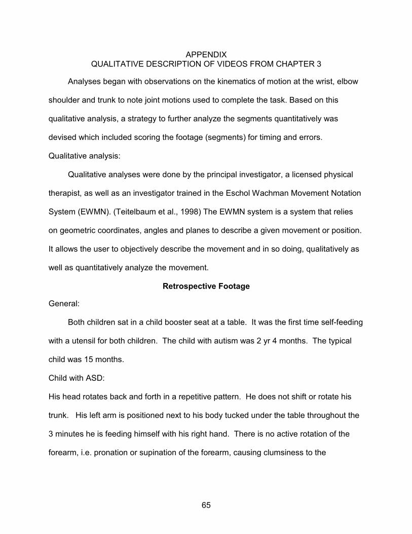
65
APPENDIX QUALITATIVE DESCRIPTION OF VIDEOS FROM CHAPTER 3
Analyses began with observations on the kinematics of motion at the wrist, elbow
shoulder and trunk to note joint motions used to complete the task. Based on this
qualitative analysis, a strategy to further analyze the segments quantitatively was
devised which included scoring the footage (segments) for timing and errors.
Qualitative analysis:
Qualitative analyses were done by the principal investigator, a licensed physical
therapist, as well as an investigator trained in the Eschol Wachman Movement Notation
System (EWMN). (Teitelbaum et al., 1998) The EWMN system is a system that relies
on geometric coordinates, angles and planes to describe a given movement or position.
It allows the user to objectively describe the movement and in so doing, qualitatively as
well as quantitatively analyze the movement.
Retrospective Footage
General:
Both children sat in a child booster seat at a table. It was the first time self-feeding
with a utensil for both children. The child with autism was 2 yr 4 months. The typical
child was 15 months.
Child with ASD:
His head rotates back and forth in a repetitive pattern. He does not shift or rotate his
trunk. His left arm is positioned next to his body tucked under the table throughout the
3 minutes he is feeding himself with his right hand. There is no active rotation of the
forearm, i.e. pronation or supination of the forearm, causing clumsiness to the

66
movement. Instead, the child uses flexion and extension of the wrist to scoop the
pudding. (See Figure A-1).
At times when the child is not looking directly at the bowl, he aims the spoon
towards the bowl but misses and has to correct the trajectory. He never misses when
he is looking at the bowl. At other times, the spoon shifts in his hand when he scoops
the pudding off the spoon and, ‘stuck’ in the movement pattern, he aims the handle end
of the spoon into the bowl. Mom, sitting next to him, switches the spoon to the correct
way. See Figure A-2.
The Typical Child
This child uses rotation of the forearm, synergistic with flexion and extension of the
wrist, to scoop her food. When she moves her arm, the trunk shifts and rotates to
support her movement. The rotational movements of the wrist allow for flexibility in the
manipulation of the spoon when scooping the food or bringing it towards her mouth.
Though she does have repetitive movements such as shaking the fork (see Figure A-3),
as is typical in normal development (Thelen, 1979), her movement when she is eating is
not repetitive but manipulated in order to reach her desired target.
Follow-up Footage
As the typically developing child is a relative of the investigator, and the child with
autism attends a therapy camp directed by the investigator, recent footage was
obtained of the participants eating with a spoon. The length and quality of this for the
child with autism was not sufficient for quantitative analysis so a qualitative description
was done.
The typical child is presently 17 and the child with autism 10. In a five-minute
segment of eating, the child with autism does not make errors with the spoon, but

67
continues to lack the rotational movements of the forearm. Instead he compensates by
rotating the spoon in his mouth. The pattern is less apparent as the child takes his
spoon out of his lunch bag, eats with it, then puts it away, then later takes it out again.
The typical child does not use the repetitive movements seen at her first time of eating.
Her movements continue to be synergistic with rotational movements of the forearm the
primary range used to manipulate the bringing the spoon to her mouth.
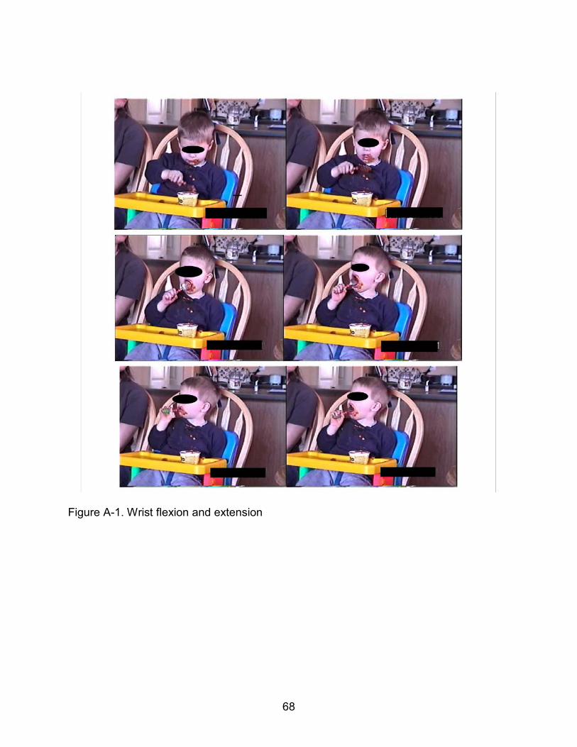
68
Figure A-1. Wrist flexion and extension
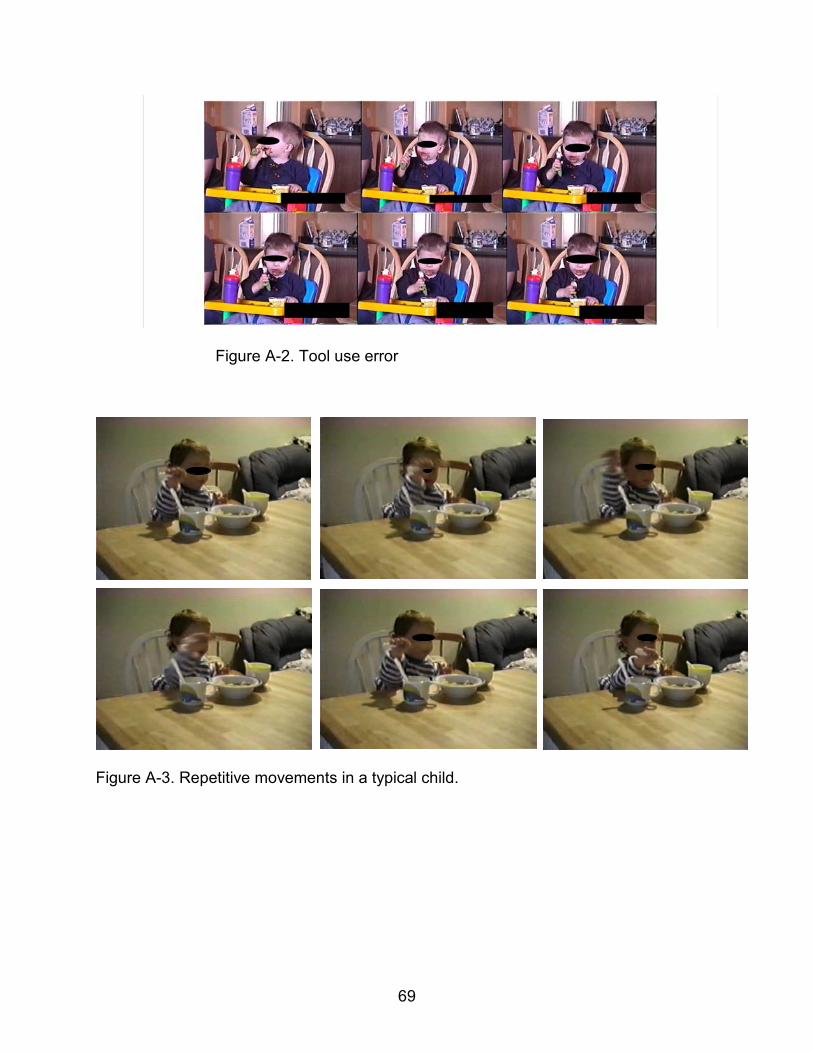
69
Figure A-2. Tool use error
Figure A-3. Repetitive movements in a typical child.
70
LIST OF REFERENCES
Amaral, D. G., Schumann, C. M., & Nordahl, C. W. (2008). Neuroanatomy of autism. Trends in Neurosciences, 31(3), 137-145.
Bailey, A., Luthert, P., Dean, A., Harding, B., Janota, I., Montgomery, M. et al. (1998). A clinicopathological study of autism. Brain, 121(5), 889.
Baizer, J. S., Kralj-Hans, I., & Glickstein, M. (1999). Cerebellar lesions and prism adaptation in macaque monkeys. Journal of neurophysiology, 81(4), 1960.
Baranek, G. T. (2002). Efficacy of sensory and motor interventions for children with autism. Journal of Autism and Developmental Disorders, 32(5), 397-422.
Bartak, L., Rutter, M., & Cox, A. (1975). A comparative study of infantile autism and specific development receptive language disorder. I. The children. British Journal of Psychiatry,126, 127–145.
Bauman, M. L., & Kemper, T. L. (1996). Observations on the Purkinje cells in the cerebellar vermis in autism. Journal of Neuropathology & Experimental Neurology, 55(5), 613.
Bauman, M. L., & Kemper, T. L. (2005). Neuroanatomic observations of the brain in autism: a review and future directions. International Journal of Developmental Neuroscience, 23(2-3), 183-187.
Belmonte, M. (2000). Abnormal attention in autism shown by steady-state visual evoked potentials. Autism, 4(3), 269-285.
Belmonte, M. K., Allen, G., Beckel-Mitchener, A., Boulanger, L. M., Carper, R. A., & Webb, S. J. (2004). Autism and abnormal development of brain connectivity. Journal of Neuroscience, 24(42), 9228-9231.
Belmonte, M. K., & Yurgelun-Todd, D. A. (2003). Functional anatomy of impaired selective attention and compensatory processing in autism. Cognitive Brain Research, 17(3), 651-664.
Bertone, A., Mottron, L., Jelenic, P., & Faubert, J. (2005). Enhanced and diminished visuo-spatial information processing in autism depends on stimulus complexity. Brain, 128(10), 2430-2441.
Boria, S., Cattaneo, L., Sparaci, L., Sinigaglia, C., Santelli, G., & Rizzolatti, G. (2009). Intention Understanding in Autism. PLoS ONE, 4(5).
Brooks, V. B. (1986). The Neural Basis of Motor Control. New York, NY: Oxford University Press

71
Burpee, J., DeJean, V., Frick, S., Kawar, M., Koomar, J., & Murphy Fisher, D. (2001). Theoretical and clinical perspectives on the Interactive Metronome (IM): a view from clinical occupational therapy practice. The American Journal of Occupational Therapy, 2, 163-166.
Bundy, A., Lane, S., & Murray, E. (2002). Sensory integration: theory and practice. Philadelphia: F. A. Davis Company
Buxhoeveden, D., Fobbs, A; Roy, E; Casanova, M. (2002). Quantitative comparison of radial cell columns in children with Down's syndrome and controls. Journal of Intellectual Disability Research, 46(1), 76-81.
Carper, R. A., & Courchesne, E. (2000). Inverse correlation between frontal lobe and cerebellum sizes in children with autism. Brain, 123(4), 836.
Carper, R. A., Moses, P., Tigue, Z. D., & Courchesne, E. (2002). Cerebral lobes in autism: early hyperplasia and abnormal age effects. Neuroimage, 16(4), 1038-1051.
Casanova, M. F., Buxhoeveden, D. P., Switala, A. E., & Roy, E. (2002). Minicolumnar pathology in autism. Neurology, 58(3), 428-432.
Casanova, M. F., van Kooten, I. A. J., Switala, A. E., van Engeland, H., Heinsen, H., Steinbusch, H. W. M. et al. (2006). Minicolumnar abnormalities in autism. Acta neuropathologica, 112(3), 287-303.
Caston, J., Yon, E., Mellier, D., Godfrey, H. P., Delhaye-bouchaud, N., & Mariani, J. (1998). An animal model of autism: behavioural studies in the GS guinea-pig. Eur J Neurosci, 10(8), 2677-2684.
Cosper, S. M., Lee, G. P., Peters, S. B., & Bishop, E. (2009). Metronome training in children with attention deficit and developmental coordination. International Journal of Rehabilitation Research.
Cotman, C. W., & Engesser-Cesar, C. (2002). Exercise enhances and protects brain function. Exercise and sport sciences reviews, 30(2), 75.
Courchesne, E., & Pierce, K. (2005). Why the frontal cortex in autism might be talking only to itself: local over-connectivity but long-distance disconnection. Current Opinion in Neurobiology, 15(2), 225-230.
Courchesne, E., Saitoh, O., Yeung-Courchesne, R., Press, G. A., Lincoln, A. J., Haas, R. H. (1994). Abnormality of cerebellar vermian lobules VI and VII in patients with infantile autism: identification of hypoplastic and hyperplastic subgroups with MR imaging. American Journal of Roentgenology, 162(1), 123.
Courchesne, E., Townsend, J., & Saitoh, O. (1994). The brain in infantile autism: posterior fossa structures are abnormal. Neurology, 44(2), 214-223.

72
Courchesne, E., Yeung-Courchesne, R., Press, G. A., Hesselink, J. R., & Jernigan, T. L. (1988). Hypoplasia of cerebellar vermal lobules VI and VII in autism. New England Journal of Medicine, 318(21), 1349.
Curcio, F. (1978). Sensorimotor functioning and communication in mute autistic children. Journal of Autism and Developmental Disorders, 8(3), 281-292.
Dawson, G., & Watling, R. (2000). Interventions to facilitate auditory, visual, and motor integration in autism: A review of the evidence. Journal of Autism and Developmental Disorders, 30(5), 415-421.
Dettmer, S., Simpson, R. L., Myles, B. S., & Ganz, J. B. (2000). The use of visual supports to facilitate transitions of students with autism. Focus on Autism and Other Developmental Disabilities, 15(3), 163.
Dewey, D. Cantell, M. & Crawford, S. G. (2007). Motor and gestural performance in children with autism spectrum disorders, developmental coordination disorder, and/or attention deficit hyperactivity disorder. Journal of the International Neuropsychological Society, 13(02), 246-256.
Doyon, J., Bellec,, P., Amsel, R., Penhune, V., Monchi, O.,Carrier, J., & Benali, H. (2009). Contributions of the basal ganglia and functionally related brain structures to motor learning. Behavioral Brain Research, 199, 61-75.
Dowell, L. R., Mahone, E. M., & Mostofsky, S. H. (2009). Associations of postural knowledge and basic motor skill with dyspraxia in autism: Implication for abnormalities in distributed connectivity and motor learning. Neuropsychology, 23(5), 563.
Dziuk, M. A., Larson, J. C., Apostu, A., Mahone, E. M., Denckla, M. B., & Mostofsky, S. H. (2007). Dyspraxia in autism: association with motor, social, and communicative deficits. Developmental Medicine and Child Neurology, 49(10), 734-739.
Fabbri-Destro, M., Cattaneo, L., Boria, S., & Rizzolatti, G. (2009). Planning actions in autism. Exp Brain Res, 192(3), 521-525.
Filipek, P. A., Accardo, P. J., Baranek, G. T., Cook, E. H., Dawson, G., Gordon, B. et al. (1999). The screening and diagnosis of autistic spectrum disorders. Journal of Autism and Developmental Disorders, 29(6), 439-484.
Frith, U. (1989). A new look at language and communication in autism. Br J Disord Commun, 24(2), 123-150.
Gardner, S. (2005). The Effects and Benefits of Sensory Integration Therapy on Children with Autism. Master’s Thesis. University of Wisconsin, Stout.
Georgiades, S., Szatmari, P., Zwaigenbaum, L., Duku, E., Bryson, S., Roberts, W. et al. (2007). Structure of the autism symptom phenotype: A proposed multidimensional model. Journal of Amer Academy of Child & Adolescent Psychiatry, 46(2), 188.

73
Gernsbacher, M., Sauer, E., Geye, H., Schweigert, E., & Hillgoldsmith, H. (2008). Infant and toddler oral-and manual-motor skills predict later speech fluency in autism. Journal of Child Psychology and Psychiatry, 49(1), 43-50.
Glickstein, M. (2007). What does the cerebellum really do? Current Biology, 17(19), 824-827.
Gotham, K., Pickles, A., & Lord, C. (2009). Standardizing ADOS scores for a measure of severity in autism spectrum disorders. J Autism Dev Disord, 39(5), 693-705.
Happe, F., & Ronald, A. (2008). The 'fractionable autism triad': a review of evidence from behavioural, genetic, cognitive and neural research. Neuropsychol Rev, 18(4), 287-304.
Hardan, A. Y., Minshew, N. J., Harenski, K., & Keshavan, M. S. (2001). Posterior fossa magnetic resonance imaging in autism. Journal of Amer Academy of Child & Adolescent Psychiatry, 40(6), 666.
Haring, T. G., Kennedy, C. H., Adams, M. J., & Pitts-Conway, V. (1987). Teaching generalization of purchasing skills across community settings to autistic youth using videotape modeling. J Appl Behav Anal, 20(1), 89-96.
Hashimoto, T., Tayama, M., Murakawa, K., Yoshimoto, T., Miyazaki, M., Harada, M. et al. (1995). Development of the brainstem and cerebellum in autistic patients. Journal of Autism and Developmental Disorders, 25(1), 1-18.
Heilman, K. M., Gonzalez Rothi, L.J. (1997). Apraxia: The neuropsychology of action. East Sussex, UK: Psychology Press.
Hollander, E., Anagnostou, E., Chaplin, W., Esposito, K., Haznedar, M. M., Licalzi, E. et al. (2005). Striatal volume on magnetic resonance imaging and repetitive behaviors in autism. Biological Psychiatry, 58(3), 226-232.
Horner, R. H. (2000). Positive behavior supports. Focus on Autism and Other Developmental Disabilities, 15(2), 97.
Hughes, C. (1996). Brief report: Planning problems in autism at the level of motor control. Journal of Autism and Developmental Disorders, 26(1), 99-107.
Hughes, J. R. (2007). Autism: The first firm finding= underconnectivity? Epilepsy and Behavior, 11(1), 20-24.
Huttenlocher, P. R. (2002). Neural plasticity: The effects of environment on the development of the cerebral cortex. Cambridge, MA: Harvard University Press.
Jones, V., & Prior, M. (1985). Motor imitation abilities and neurological signs in autistic children. Journal of Autism and Developmental Disorders, 15(1), 37-46.
74
Just, M. A., Cherkassky, V. L., Keller, T. A., & Minshew, N. J. (2004). Cortical activation and synchronization during sentence comprehension in high-functioning autism: evidence of underconnectivity. Brain, 127(8), 1811-1821.
Kana, R. K., Keller, T. A., Cherkassky, V. L., Minshew, N. J., & Just, M. A. (2006). Sentence comprehension in autism: thinking in pictures with decreased functional connectivity. Brain, 129(9), 2484-2493.
Kaufmann, W. E., Cooper, K. L., Mostofsky, S. H., et al. (2003). Specificity of cerebellar vermian abnormalities in autism: a quantitative magnetic resonance imaging study. Journal of Child Neurology, 18(7), 463-470.
Kleinhans, N. M., Richards, T., Sterling, L., Stegbauer, K. C., Mahurin, R., Johnson, L. C. et al. (2008). Abnormal functional connectivity in autism spectrum disorders during face processing. Brain, 131, 1000-1012.
Klin, A., Lin, D. J., Gorrindo, P., Ramsay, G., & Jones, W. (2009). Two-year-olds with autism orient to non-social contingencies rather than biological motion. Nature, 459(7244), 257(7) 257-264.
Koshino, H., Carpenter, P. A., Minshew, N. J., Cherkassky, V. L., Keller, T. A., & Just, M. A. (2005). Functional connectivity in an fMRI working memory task in high-functioning autism. Neuroimage, 24(3), 810-821.
Kwakkel, G. (2006). Impact of intensity of practice after stroke: issues for consideration. Disability & Rehabilitation, 28(13-14), 823-830.
Kwakkel, G., van Peppen, R., Wagenaar, R. C., Dauphinee, S. W., Richards, C., Ashburn, A. et al. (2004). Effects of augmented exercise therapy time after stroke. Stroke, 35(11), 2529-2539.
LaGasse, A. B. (2009). “Oromotor kinematics of speech In Children and the Effect of an External Rhythmic Auditory Stimulus”. PhD Dissertation. University of Kansas.
Langen, M., Durston, S., Staal, W. G., Palmen, S., & van Engeland, H. (2007). Caudate nucleus is enlarged in high-functioning medication-naive subjects with autism. Biological psychiatry, 62(3), 262-266.
Langen, M., Schnack, H. G., Nederveen, H., Bos, D., Lahuis, B. E., de Jonge, M. V. et al. (2009). Changes in the Developmental Trajectories of Striatum in Autism. Biological Psychiatry. 66, 327-333.
Larson, J., Bastian, A., Donchin, O., Shadmehr, R., & Mostofsky, S. H. (2008). Acquisition of internal models of motor tasks in children with autism. Brain, 131(11), 2894-2903.

75
Lee, P. S., Yerys, B. E., Della Rosa, A., Foss-Feig, J., Barnes, K. A., & James, J. D. (2009). Functional connectivity of the inferior frontal cortex changes with age in children with autism spectrum disorders: a fcMRI study of response inhibition. Cereb Cortex, 19(8).
Lewis, M., & Kim, S. J. (2009). The pathophysiology of restricted repetitive behavior. Journal of Neurodevelopmental Disorders, 1(2), 114-132.
Lewis, M. H., & Bodfish, J. W. (1998). Repetitive behavior disorders in autism. Mental Retardation and Developmental Disabilities Research Reviews, 4(2), 80-89.
Lewis, M. H., Tanimura, Y., Lee, L. W., & Bodfish, J. W. (2007). Animal models of restricted repetitive behavior in autism. Behavioural brain research, 176(1), 66-74.
Lord, C., Rutter, M., & Le Couteur, A. (1994). Autism Diagnostic Interview-Revised: a revised version of a diagnostic interview for caregivers of individuals with possible pervasive developmental disorders. J Autism Dev Disord, 24(5), 659-685.
Lovaas, O. I. (1987). Behavioral treatment and normal educational and intellectual functioning in young autistic children. Journal of consulting and clinical psychology, 55(1), 3-9.
Luria, A. (1980). Higher Cortical Functions of man. New York: Basic Books.
Manes, F., Piven, J., Vrancic, D., Nanclares, V., Plebst, C., & Starkstein, S. E. (1999). An MRI study of the corpus callosum and cerebellum in mentally retarded autistic individuals. Journal of Neuropsychiatry and Clinical Neurosciences, 11(4)(4), 470-474.
Mayes, S. D., Calhoun, S. L., Murray, M. J., Morrow, J. D., Yurich, K. K., Mahr, F. et al. (2009). Comparison of Scores on the Checklist for Autism Spectrum Disorder, Childhood Autism Rating Scale, and Gilliam Asperger's Disorder Scale for Children with Low Functioning Autism, High Functioning Autism, Asperger's Disorder, ADHD, and Typical Development. J Autism Dev Disord.
Middleton, F. A., & Strick, P. L. (2000). Basal ganglia and cerebellar loops: motor and cognitive circuits. Brain Research Reviews, 31(2-3), 236-250.
Mink, J. W. (1996). The basal ganglia: focused selection and inhibition of competing motor programs. Progress in Neurobiology, 50(4), 381-425.
Mizuno, A., Villalobos, M. E., Davies, M. M., Dahl, B. C., & Muller, R. A. (2006). Partially enhanced thalamocortical functional connectivity in autism. Brain research, 1104(1), 160-174.
Mostofsky, S. H., Dubey, P., Jerath, V. K., Jansiewicz, E. M., Goldberg, M. C., & Denckla, M. B. (2006). Developmental dyspraxia is not limited to imitation in children with autism spectrum disorders. Journal of the International Neuropsychological Society, 12(03), 314-326.
76
Mostofsky, S. H., Goldberg, M. C., Landa, R. J., & Denckla, M. B. (2000). Evidence for a deficit in procedural learning in children and adolescents with autism: Implications for cerebellar contribution. Journal of the International Neuropsychological Society, 6(07), 752-759.
Mostofsky, S., Powell, S., Simmonds, D., Goldberg, M., Caffo, B. and Pekar, J. (2009). Decreased connectivity and cerebellar activity in autism during motor task performance. Brain, 132(9).
Mukhopadhyay, S. (2003) ‘Rapid Prompting Method’, lecture, Pasadena, CA, August. [Video-recording, produced by Helping Autism through Learning and Outreach].
Muller, R. A., Pierce, K., Ambrose, J. B., Allen, G., & Courchesne, E. (2001). Atypical patterns of cerebral motor activation in autism: a functional magnetic resonance study. Biological Psychiatry, 49(8), 665-676.
Ozonoff, S., Young, G. S., Goldring, S., Greiss-Hess, L., Herrera, A. M., Steele, J. et al. (2008). Gross motor development, movement abnormalities, and early identification of autism. Journal of Autism and Developmental Disorders, 38(4), 644-656.
Pierce, K., & Courchesne, E. (2001). Evidence for a cerebellar role in reduced exploration and stereotyped behavior in autism. Biological psychiatry, 49(8), 655-664.
Piven, J., Nehme, E., Simon, J., Barta, P., Pearlson, G., & Folstein, S. E. (1992). Magnetic resonance imaging in autism: measurement of the cerebellum, pons, and fourth ventricle. Biological Psychiatry, 31(5), 491-504.
Piven, J., Saliba, K., Bailey, J., & Arndt, S. (1997). An MRI study of autism: the cerebellum revisited. Neurology, 49(2), 546-551.
Redcay, E., & Courchesne, E. (2005). When is the brain enlarged in autism? A meta-analysis of all brain size reports. Biological Psychiatry, 58(1), 1-9.
Ritvo, S., & Provence, S. (1953). Form perception and imitation in some autistic children: Diagnostic findings and their contextual interpretation. Psychoanalytic Study of the Child, 8, 155-161.
Riva, D., & Giorgi, C. (2000). The cerebellum contributes to higher functions during development: evidence from a series of children surgically treated for posterior fossa tumours. Brain, 123(5), 1051.
Roelfsema, P. R. (2006). Cortical algorithms for perceptual grouping. Annu. Rev. Neurosci, 29, 203-227.
Rogers, S. J. (1998). Empirically supported comprehensive treatments for young children with autism. Journal of Clinical Child & Adolescent Psychology, 27(2), 168-179.
77
Rogers, S. J., & Pennington, B. F. (1991). A theoretical approach to the deficits in infantile autism. Development and Psychopathology, 3(02), 137-162.
Rojas, D. C., Peterson, E., Winterrowd, E., Reite, M. L., Rogers, S. J., & Tregellas, J. R. (2006). Regional gray matter volumetric changes in autism associated with social and repetitive behavior symptoms. BMC Psychiatry, 6, 56.
Sabado, J., Fuller, D. (2008). A Preliminary Study of the Effects of Interactive Metronome Training on the Language Skills of an Adolescent Female With a Language Learning Disorder. Contemporary Issues in Communicaiton Science and Disorders. 35, 65-71.
Schaaf, R. C., & Miller, L. J. (2005). Occupational therapy using a sensory integrative approach for children with developmental disabilities. Mental Retardation and Developmental Disabilities Research Reviews, 11(2), 143-148.
Schmahmann, J. D., & Pandya, D. N. (2008). Disconnection syndromes of basal ganglia, thalamus, and cerebrocerebellar systems. Cortex, 44(8), 1037-1066.
Sears, L. L., Vest, C., Mohamed, S., Bailey, J., Ranson, B. J., & Piven, J. (1999). An MRI study of the basal ganglia in autism: New trends in developmental neuroimaging in psychiatry. Progress in Neuro-psychopharmacology & Biological Psychiatry, 23(4), 613-624.
Siegel, S. (1956). Nonparametric statistics for the behavioral sciences. (xvii). McGraw-Hill.
Smith, I. M., & Bryson, S. E. (1994). Imitation and action in autism: A critical review. Psychological Bulletin, 116, 259-259.
Smith, I., M., & Bryson, S., E. (2007). Gesture imitation in autism: II. Symbolic gestures and pantomimed object use. Cognitive Neuropsychology, 24(7), 679-700.
Snow, A. V., Lecavalier, L., & Houts, C. (2009). The structure of the Autism Diagnostic Interview-Revised: diagnostic and phenotypic implications. Journal of Child Psychology and Psychiatry, 50(6), 734-742.
Stanfield, A. C., McIntosh, A. M., Spencer, M. D., Philip, R., Gaur, S., & Lawrie, S. M. (2008). Towards a neuroanatomy of autism: A systematic review and meta-analysis of structural magnetic resonance imaging studies. European Psychiatry, 23(4), 289-299.
Strick, P. L., Dum, R.P., & Fiez, J. (2009). Cerebellum and Nonmotor Function. Annual Reviews, 32, 413-434.
Tantum, D. (1991). Asperger Syndrome in adulthood. Cambridge: Cambridge University Press.
78
Teitelbaum, P., Teitelbaum, O., Nye, J., Fryman, J., & Maurer, R. G. (1998). Movement analysis in infancy may be useful for early diagnosis of autism. Proc Natl Acad Sci U S A, 95(23), 13982-13987.
Teitelbaum, P., Teitelbaum, O. B., Fryman, J., & Maurer, R. (2002). Infantile reflexes gone astray in autism. Journal of Developmental and Learning Disorders, 6, 15-22.
Thelen, E. (1979). Rhythmical stereotypies in normal human infants. Anim Behav, 27(Pt 3), 699-715.
Trevarthen, C., & Aitken, K. J. (2001). Infant intersubjectivity: Research, theory, and clinical applications. The Journal of Child Psychology and Psychiatry and Allied Disciplines, 42(01), 3-48.
Turner, K. C., Frost, L., Linsenbardt, D., McIlroy, J. R., & Muller, R. A. (2006). Atypically diffuse functional connectivity between caudate nuclei and cerebral cortex in autism. Behav Brain Funct, 2, 34-46.
Vandenbroucke, M., Scholte, H., Van Engeland, H., Lamme, V., & Kemner, C. (2008). A neural substrate for atypical low-level visual processing in autism spectrum disorder. Brain, 131(4), 1013.
Vandenbroucke, M., Scholte, H., Van Engeland, H., Lamme, V., & Kemner, C. (2009). A New Approach to the study of detail perception in Autism Spectrum Disorder (ASD): Investigating visual feedforward, horizontal and feedback Processing. Vision Research, 49, 1006-1016.
Vargas, D. L., Nascimbene, C., Krishnan, C., Zimmerman, A. W., & Pardo, C. A. (2005). Neuroglial activation and neuroinflammation in the brain of patients with autism. Annals of Neurology, 57(1), 67-81.
Vilensky, J. A., Damasio, A. R., & Maurer, R. G. (1981). Gait disturbances in patients with autistic behavior: a preliminary study. Archives of Neurology, 38(10), 646.
Villalobos, M. E., Mizuno, A., Dahl, B. C., Kemmotsu, N., & Muller, R. A. (2005). Reduced functional connectivity between V1 and inferior frontal cortex associated with visuomotor performance in autism. Neuroimage, 25(3), 916-925.
Watling, R., Deitz, J., Kanny, E. M., & McLaughlin, J. F. (1999) Current practice of occupational therapy for children with autism. J The American journal of occupational therapy: official publication of the American Occupational Therapy Association, 53(5), 498-505.
Williams, J. H. G., Whiten, A., & Singh, T. (2004). A systematic review of action imitation in autistic spectrum disorder. Journal of Autism and Developmental Disorders, 34(3), 285-299.

79
Xerri, C., Coq, J. O., Merzenich, M. M., & Jenkins, W. M. (1996). Experience-induced plasticity of cutaneous maps in the primary somatosensory cortex of adult monkeys and rats. Journal of Physiology-Paris, 90(3-4), 277-287.

80
BIOGRAPHICAL SKETCH
Kathleen A. Berger, graduated with her BS in physical therapy in 1984 from
Northwestern University. Ms Berger is the director and founder of Kris’ Camp/Therapy
Intensive Programs, Inc, a therapy intensive, respite program for children with autism
and their families. In the fall of 2006 Kathleen entered the psychology doctoral program
at the University of Florida, with her primary research interest in movement differences
in autism.