Embed Size (px)
Citation preview

8566
Abstract. – OBJECTIVE: Muscle injury tends to heal with incomplete functional recovery. Among the growth factors released in the physio-patho-logical response of muscle lesion, the Insulin-like Growth-Factor-1 (IGF-1) results in an engine fac-tor of the reparation program. The therapeutic use of growth factors has been exploited to improve healing. As IGF-1 is a primary mediator of the ef-fects of growth hormone (GH), we exploited its systemic administration to muscle recovery in a rat model of muscle injury.
MATERIALS AND METHODS: Monolateral le-sion of the longissimus dorsi muscle of rats was performed. Animals were divided into 5 groups: four groups for histological studies and serum hormone dosage, whilst the fifth group represent-ed the uninjured control. Rat GH was intraperito-neally administered after 24h from the surgical lesion at three different concentrations (0.1, 0.2, 0.4 mg/kg). At 3 days from surgery, immunohis-tochemical and histological analyses evaluated the expression of MyoD and Myogenin, and the presence of neovascularization and inflammation, respectively. After 2 months, we analyzed the presence of muscle regeneration and fibrosis.
RESULTS: The treatment with GH resulted in a significant increase in neovascularization and Myogenin expression at 24h from injury in com-parison with the control. This suggested speed up biological recovery times. After two-months, a dose-dependent increase of the connective com-ponent was observed.
CONCLUSIONS: The potential effect of GH on muscle repair and regeneration, through the ac-tivation of satellite cells already demonstrated in vitro, was confirmed in this in vivo experimental approach. This study sheds light on the role of growth factors in damage repair mechanisms to find an appropriate biological treatment for mus-cle injury. Key Words:
Muscle regeneration, Muscle injury, GH, IGF-1, Sat-ellite cells.
Introduction
Muscle injury is very common, representing one-third of all injuries in sports. Today, not many well-established treatments for muscle damages are present, and most of them are performed con-servatively. The return of the players to training and matches in the shortest time possible is an oc-currence that might sometimes cause a re-injury. Moreover, current standard treatments for muscle injury are unsatisfactory, and complications such as muscle atrophy, contracture, and fibrotic scar formation at the site of wound may lead to sub-optimal clinical outcomes. In this respect, regen-erative medicine approaches have the potential to play a major role in muscle rehabilitation, en-hancing the healing process with growth factors1. Platelet-Rich Plasma (PRP) is a pool of growth factor that include: platelet-derived growth fac-tor (PDGF), epidermal growth factor (EGF), in-sulin-like growth factor-1 (IGF-I), transforming growth factor-beta (TGF β-I), vascular endothe-lial growth factor (VEGF), hepatocyte growth factor (HGF), fibroblast growth factor (FGF), stromal-derived growth factor-alpha (SDF-1α), tumor necrosis factor-alpha (TNF α) and others2. At present, the treatment of the patients and ath-letes with platelet-based applications is permitted and regulated by the Food and Drug Administra-tion (FDA) and the World Anti-Doping Agency (WADA), and from 2011 the use of autologous PRP is also allowed in competitive sports3. De-spite, PRP treatment has been studied and used in various musculoskeletal disorders2,4-7, two sys-tematic reviews8,9 showed uncertainty about the real effectiveness of PRP injections in muscu-loskeletal injuries. Moreover, two clinical stud-ies evidenced the lack of PRP efficacy after in-
European Review for Medical and Pharmacological Sciences 2020; 24: 8566-8572
M. CIANFORLINI1, M. GRASSI2, V. COPPA2, S. MANZOTTI2, F. ORLANDO3, M. MATTIOLI-BELMONTE2, A. GIGANTE2
1Orthopedic Division, Ospedale Carlo Urbani Jesi (AN), Italy2Department of Clinical and Molecular Sciences, Università Politecnica delle Marche, Ancona, Italy3Centro di Tecnologie Avanzate nell’Invecchiamento, IRCCS-INRCA, Ancona, Italy
Corresponding Author: Marco Grassi, MD; e-mail: [email protected]
Skeletal muscle repair in a rat muscle injury model: the role of growth hormone (GH) injection

GH in muscle repair: an in vivo study
8567
jections in patients with acute hamstring muscle injuries10,11. The concentration of growth factors in PRP varies according to individual variability, method of preparations, and storage; otherwise, these bias affects clinical outcomes. The knowl-edge of the individual effect of growth factors could enable the development of the best clinical treatment. Indeed, the growth factors contained in PRP have all been well characterized in terms of inhibitory or acceleratory differentiation pa-rameters using C2C12 murine myoblast cell line in vitro experimental investigations. As far as IGF-1 is concerned, it has been demonstrated its capability to stimulate C2C12 murine myoblasts proliferative response in the first 24-36 h of treat-ment, followed by an increase in myogenic differ-entiation12. Moreover, IGF-1 overexpression was shown to induce a significant increase in mouse muscle mass and enhance its potential regenera-tive acting on satellite cells13. IGF-1 binds to its muscular receptor (IGF-1-R) activating sever-al intracellular pathways (CaMK, PI3K, mito-gen-activated protein kinase) and transcription factors among which the muscle-specific MyoD and Myogenin. These signalings produce the proliferation and differentiation of satellite cells and “muscle-derived stem cell-like population” a new cellular line different from the myogenic and mesenchymal line, expressing marker for the hematopoietic lineage such as CD34 and Sca-1, recently re-named telocytes14,15. These cells are located near capillaries and are activated in case of muscle damage. In muscle tissue regeneration, in addition to the local myogenic stem cells, cir-culating cells (Sca-1+) that arise from the bone marrow are also involved16. The latter are recruit-ed through local signals (chemokine) whose pro-duction is induced by IGF-1.
The clinical use of a single growth factor/cytokine is often expensive, it can be difficult to replicate in physiologically relevant quanti-ties and it needs to be approved by the FDA. For these reasons, in this experimental study, we used a systemic administration of Growth Hormone (GH) in a rat model of muscle injury, to evalu-ate the potential effect of IGF-1 on the activation of satellite cells for muscle repair and regenera-tion17,18. GH induces the synthesis of IGF-1 in the liver generating a systemic hormone (cIGF-1), and in other tissues, including muscle (mIGF-1)16. The relevance of GH on muscle biology is evi-denced by the comparison of its role in two op-posite pathological conditions like GH-deficiency and acromegaly. GH hypertrophic induction in
GH-deficiency, or in situations where the stim-ulus is temporally reduced, has a positive effect on the skeletal muscle, while protracted high GH blood level (i.e., acromegaly) shows a pathologi-cal role inducing myopathy19. Therapeutic use of GH in humans is contraindicated in the case of neoplastic pathologies, renal failure or hypersen-sitivity reactions. The most common side effects to treatment are myalgia, arthralgia, widespread paraesthesia and injection site problems.
The purpose of this in vivo experimental study was the assessment of the GH effects on muscle injury in an animal model. Morphological, immu-nohistochemical, and histomorphometric analy-ses were performed to test the efficacy and safety of this therapeutic approach and to evaluate a pos-sible relationship between GH concentrations and muscle regeneration.
Materials and methods
Animal ModelThe study was performed in 34 male Wistar
rats (340 ± 40 g/BW) between 6 and 8 weeks of age. The rats were inbred; therefore, they could be considered genetically identical. Rats were housed at 21-24°C and maintained on a 12-h light/dark cy-cle. Water and food were given ad libitum during the experiments. The policies and procedures of their use and maintenance were in accordance with those detailed by the directive no. 86/609/CEE re-garding animal care and experimental usage. The rats were randomly assigned to 5 groups according to the treatment (Table I). All groups except the “uninjured group” (E) sustained a unilateral lesion of longissimus dorsi muscle in a controlled man-ner5,20, followed by their designated treatments. Animals were maintained with a normal diet and no forced exercise was induced. Rat GH (rGH) used for treatment (rGH B-9; biopotency, 1.9 IU/mg) was obtained through the NIDDK Rat Pitu-itary Hormone Distribution Program (Rockville, MD, USA). GH was administered intraperitoneally subsequently to disinfection of the injection site at 24h from surgical lesion.
Groups A, B, C, D were used for the histo-morphological evaluation (both time points) and dosage of the GH serum level (48h). Groups E un-derwent only histological analyses.
Four animals for each group (A, B, C, D) were euthanized by intraperitoneal injection of an over-dose of ketamine (75 mg/kg) and xylazine (10 mg/kg) at 2 and 60 days from surgery, respectively.

M. Cianforlini, M. Grassi, V. Coppa, S. Manzotti, F. Orlando, M. Mattioli-Belmonte, A. Gigante
8568
GH serum level was evaluated by E-EL-R0029 (Labome® Princeton, NJ, USA; range 0.313-200 ng/ml, sensibility 0.18 ng/ml) at 24 h from the in-jection, through intracardiac blood sampling after euthanasia. To overcome possible experimental limitations, we used inbred animals that lived in the same environmental conditions, and blood sampling was performed at the same time to re-spect the circadian rhythm of the hormone.
Surgical Procedure of Skeletal Muscle Injury
Rats were anesthetized by an intramuscular injection of a mixture of ketamine (40 mg/kg, i.m.) and xylazine (5 mg/kg, i.m.) before surgi-cal procedure and placed in ventral decubitus on a warm pad (38.5°C) by the fixation of tail and extremities with adhesive strips. The electric scis-sors were used to shave off the hair on the back surface of mice. Unilateral cutaneous incision (3 cm in length) was performed in the paravertebral region. Then, muscular tear lesion (0.7x 0.3 cm) was performed on the longissimus dorsi muscle using a standard pincer technique, in a controlled manner5,20. This muscle was chosen because its position prevents rats from interfering (i.e., biting or scratching) with the surgical treatment (these factors could interfere with the biological re-sponse through an increase in inflammation).
Histological AnalysesThe experimental sites were dissected and the
collected samples were fixed in formaldehyde 4%, embedded in paraffin, sectioned using a Cryotome (5 µm) to perform histological and immunohisto-chemical analysis. For light microscopy, sections were stained with Hematoxylin-Eosin (E.E) and Sirius Red staining. For immunohistochemistry slides (Menzel-Gläser, Braunschweig, Germa-ny) were used. Dewaxing, rehydration, and anti-gen unmasking were performed with EnVision™ FLEX Target Retrieval Solution High pH (Dako,
Carpinteria, CA, USA) by PT Module (Lab Vision Corporation, CA, USA). Endogenous peroxidase activity was quenched by incubating the sections in 3% H2O2 for 10’ at Room Temperature (RT). Sections were then incubated with the monoclonal antibodies anti-MyoD (5.8A) and anti-Myogen-in (5FD) (both from Santa Cruz Biotechnology, Santa Cruz, CA, USA) diluted 1:150 in Antibody Diluent with Background Reducing Components (Dako) for 1h at RT in a humidified atmosphere. The reaction was visualized with LSAB®Plus System-HRP DAB+ kit (Dako, Carpinteria, CA, USA). Sections were counterstained with May-er hematoxylin (Bio-Optica SpA, Milan, Italy). Negative control was represented by primary an-tibody untreated sections. The reaction was ex-amined with a light microscope (Nikon Eclipse 600). Each sample was evaluated in a blinded manner by three experienced observers (SM, MB and FO). The blinded examiner considered three fields for each section for a total of 5 sections for each lesion. All sections were evaluated at 10X magnification using a semiquantitative score, considering the following parameters: neovascu-larization, inflammation, fibrosis, and muscle re-generation (Table II). In the case of disagreement between observers, AG reviewed the samples and an undisputed score was made in agreement with all observers. The presence of metaplastic zones, calcifications, and heterotopic ossifications was further evaluated by histological analysis.
Histomorphometric EvaluationComputerized morphometric analysis was
performed with a Leica Q5OOMC Image Anal-ysis System (Leica Leitz DMRBE, Cambridge, UK) and the associated software was used. The camera images were digitized and modified in a binary way to make them suitable for measure-ment. The considered parameters were: MyoD, Myogenin, fibrosis and muscle regeneration. Af-ter standard filtration procedures for background
Table I. Experimental planning
GROUPS Lesion rGH No. of Sacrifice from surgery animals 2 days 60 days A Yes No treatment 8 4 4B Yes 0.1 mg/kg 8 4 4C Yes 0.2 mg/kg 8 4 4D Yes 0.4 mg/kg 8 4 4E No No treatment 2 2 /
GH in muscle repair: an in vivo study
8569
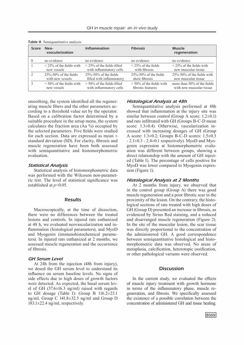
smoothing, the system identified all the regener-ating muscle fibers and the other parameters ac-cording to a threshold value set by the operator. Based on a calibration factor determined by a suitable procedure in the setup menu, the system calculates the fraction area (Aa %) occupied by the selected parameters. Five fields were studied for each section. Data are expressed as mean ± standard deviation (SD). For clarity, fibrosis and muscle regeneration have been both assessed with semiquantitative and histomorphometric evaluation.
Statistical AnalysisStatistical analysis of histomorphometric data
was performed with the Wilcoxon non-paramet-ric test. The level of statistical significance was established at p<0.05.
Results
Macroscopically, at the time of dissection, there were no differences between the treated lesions and controls. In injured rats euthanized at 48 h, we evaluated neovascularization and in-flammation (histological parameters), and MyoD and Myogenin (immunohistochemical parame-ters). In injured rats euthanized at 2 months, we assessed muscle regeneration and the occurrence of fibrosis.
GH Serum LevelAt 24h from the injection (48h from injury),
we dosed the GH serum level to understand its influence on serum baseline levels. No signs of side effects due to high doses of growth factors were detected. As expected, the basal serum lev-el of GH (37.6±16.3 ng/ml) raised with regards to GH dosage (Table I): Group B 116.2±23.1 ng/ml, Group C 141.8±32.5 ng/ml and Group D 183.1±22.4 ng/ml, respectively.
Histological Analysis at 48hSemiquantitative analysis performed at 48h
showed that inflammation at the injury site was similar between control (Group A score: 1.2±0.1) and rats infiltrated with GH (Groups B-C-D mean score: 1.3±0.4). Otherwise, vascularization in-creased with increasing dosages of GH (Group A score: 1.3±0.2; Groups B-C-D scores: 1.5±0.3 - 2.1±0.3 - 2.4±0.1 respectively). MyoD and Myo-genin expression at histomorphometric evalu-ation was different between groups, showing a direct relationship with the amount of GH inject-ed (Table I). The percentage of cells positive for MyoD was lower compared to Myogenin expres-sion (Figure 1).
Histological Analysis at 2 MonthsAt 2 months from injury, we observed that
in the control group (Group A) there was good muscle regeneration and a poor fibrotic scar in the proximity of the lesion. On the contrary, the histo-logical sections of rats treated with high doses of GH (Group D) presented an increase in fibrosis, as evidenced by Sirius Red staining, and a reduced and disarranged muscle regeneration (Figure 2). In the site of the muscular lesion, the scar tissue was directly proportional to the concentration of the administered GH. A good correspondence between semiquantitative histological and histo-morphometric data was observed. No areas of metaplasia, calcification, heterotopic ossification, or other pathological variants were observed.
Discussion
In the current study, we evaluated the effects of muscle injury treatment with growth hormone in terms of the inflammatory phase, muscle re-generation, and fibrosis. We specifically assessed the existence of a possible correlation between the concentration of administered GH and tissue healing.
Table II. Semiquantitative analysis.
Score Neo- Inflammation Fibrosis Muscle vascularization regeneration 0 no evidence no evidence no evidence no evidence 1 < 25% of the fields with < 25% of the fields filled < 25% of the fields < 25% of the fields with new vessels with inflammatory cells with fibrosis new muscular tissue2 25%-50% of the fields 25%-50% of the fields 25%-50% of the fields 25%-50% of the fields with with new vessels filled with inflammatory show fibrosis new muscular tissue3 > 50% of the fields with > 50% of the fields filled > 50% of the fields with more than 50% of the fields new vessels with inflammatory cells fibrotic features with new muscular tissue

M. Cianforlini, M. Grassi, V. Coppa, S. Manzotti, F. Orlando, M. Mattioli-Belmonte, A. Gigante
8570
Immunohistochemical detection of MyoD did not detect significant differences between treated groups and controls (group A). On the contrary, the immunohistochemical staining for Myogen-in showed an expression increase in the active phase of muscle regeneration, which was high-
er in treated animals in comparison to controls (group A) and directly proportional to GH con-centration. This evidence could be explained as Myogenin is expressed later than MyoD in the physiological process of muscle healing21. The lack of significant variations detected in Myo D
Figure 1. A, Representative images of MyoD and Myogenin immunohistochemical detection at 48h from injury in the differently treated groups (Scale bar 20 µm). B, Graph representing histomorphometric evaluation of the area occupied by positive cells after GH treatment.
Figure 2. A, Representative histological sections of lesion after 2 month in Group A and D showing muscle regeneration (H&E) and fibrosis (Sirius Red); B, Graph depict semiquantitative evaluation of muscle regeneration and fibrosis 2 months from injury. (A: no treatment; B: GH 0.1 mg/kg, group C: GH 0.2 mg/kg, group D: GH 0.4 mg/kg group); C, Histogram of histomorphometric comparison between group A and D: *p<0.05.

GH in muscle repair: an in vivo study
8571
expression between treated and untreated lesions could be ascribable to the fact that at our time point (i.e., 48h) the healing process of the control group is situated in the so-called ascending phase of “MyoD curve”, while the treated groups are in the descending ones21. We can, therefore, hypoth-esize that GH administration fastened the muscle healing process.
Ferrari et al22 had already demonstrated that muscle cell growth promoted by GH is mainly mediated by IGF-1: the administration of GH in wild-type rats increased muscle mass and the size of muscle fibers, while no effects were present in IGF-1-R knockouts rats. During muscle regenera-tion, IGF-1 supported satellite cell mobilization, function, and proliferation under pathological conditions13. The enhanced expression of mIGF-1, the local isoform of IGF-1, accelerates regen-erative processes after skeletal muscle injury in a mouse model, creating a qualitative environment capable of efficiently support an appropriate tis-sue repair23. The role of IGF-1 in muscle regen-eration was confirmed by a research performed on MDX (Duchenne muscular dystrophy model) mice. In the latter, the IGF-1 gene was over-ex-pressed by gene modification, bringing a benefit to muscle regenerative capacity24.
In our investigation, we found that the in-crease in muscle regeneration is also associated with a rise of fibrous connective tissue close to the muscle injury, as evidenced by Sirius Red staining. This could be related to the route of GH administration (i.e., through the peritone-um): since IGF-1 receptors are expressed on fibroblasts, the presence of exuberant connec-tive tissue, directly proportional to the admin-istered GH concentration is conceivable. There-fore, GH action is ubiquitous and increased the amount of muscle tissue as well as connective tissues of endomysium and perimysium. To our knowledge, this is the first research that considers the use of GH/IGF-1 for muscle re-pair and regeneration17,18,25,26. There are sever-al limitations to the current study that warrant discussion. First, the surgical procedure is not universally recognized. On the other hand, this surgically-induced lesion determines with accuracy the same type of damage in all rats and could mimic the human skeletal muscle lesions. Second, to observe the effects of the IGF-1 more clearly possible, we used very high GH concentrations compared to basal serum values. Further studies should clarify the best hormone concentration.
Conclusions
The present study showed the in vivo potential effect of IGF-1 on muscle repair and regeneration, through the activation of satellite cells as already demonstrated in vitro studies. These outcomes confirm the good results already obtained by the use of PRP for the muscle injury treatment. It also revealed that muscle repair with hyperplasia of the surrounding connective tissues is dependent on GH and that GH administration fastened the muscle healing process. These findings may con-tribute to the development of new regenerative approaches to facilitate the healing process and to reduce scar tissue formation in muscle injury.
Statement of InterestsThe Authors declare that they have no conflict of interests and none financial support
Ethical Review Committee StatementThe investigation has been performed in accordance with the policies and procedures detailed by the directive No. 86/609/CEE regarding animal care and experimental usage.
References
1) PassiPieri Ja, Christ GJ. The potential of combination therapeutics for more complete repair of volumetric muscle loss injuries: the role of exogenous growth factors and/or progenitor cells in implantable skel-etal muscle tissue engineering technologies. Cells Tissues Organs 2016; 202: 202-213.
2) CruCiani M, FranChini M, MenGoli C, Marano G, Pati i, Masiello F, ProFili s, VeroPaluMbo e, PuPella s, VaGlio s, liuMbruno GM. Platelet-rich plasma for sports-related muscle, tendon and ligament injuries: An umbrella review. Blood Transfusion 2019; 17: 465-478.
3) Scully D, Matsakas A. Current insights into the potential misuse of platelet-based applications for doping in sports. Int J Sports Med 2019; 40: 427-433.
4) MCClure MJ, GarG K, siMPson DG, ryan JJ, sell sa, bowlin Gl, eriCKsen JJ. the influence of plate-let-rich plasma on myogenic differentiation. J Tissue Eng Regen Med 2016; 10: E239-E249.
5) GiGante a, Del torto M, Manzotti s, CianForlini M, busilaCChi a, DaViDson Pa, GreCo F, Mattioli-belMon-te M. Platelet rich fibrin matrix effects on skeletal muscle lesions: an experimental study. J Biol Regul Homeost Agents 2012; 26: 475-484.
6) zhanG Xl, shi KQ, Jia Pt, JianG lh, liu yh, Chen X, zhou zy, li yX, wanG ls. Effects of platelet-rich plasma on angiogenesis and osteogenesis-asso-ciated factors in rabbits with avascular necrosis of the femoral head. Eur Rev Med Pharmacol Sci 2018; 22: 2143-2152.

M. Cianforlini, M. Grassi, V. Coppa, S. Manzotti, F. Orlando, M. Mattioli-Belmonte, A. Gigante
8572
7) a haMiD Ms, MohaMeD ali Mr, yusoF a, GeorGe J, lee lPC. Platelet-rich plasma injections for the treatment of hamstring injuries. Am J Sports Med 2014; 42: 2410-2418.
8) sheth u, siMunoViC n, Klein G, Fu F, einhorn ta, sCheMitsCh e, ayeni or, bhanDari M. Efficacy of au-tologous platelet-rich plasma use for orthopaedic indications: a meta-analysis. J Bone Surg Am 2012; 94: 298-307.
9) Moraes Vy, lenza M, taMaoKi MJ, FaloPPa F, belloti JC. Platelet rich therapies for musculoskeletal soft-tissue injuries. Cochrane Database Syst Rev 2012; 2014(4): CD010071.
10) haMilton b, tol Jl, alMusa e, bouKarrouM s, eirale C, FarooQ a, whiteley r, Chalabi h. Platelet-rich plasma does not enhance return to play in ham-string injuries: a randomised controlled trial. Br J Sports Med 2015; 49: 943-950.
11) reurinK G, GouDswaarD GJ, Moen Mh, weir a, Verhaar Jan, bierMa-zeinstra sMa, Maas M, tol Jl. Rationale, secondary outcome scores and 1-year follow-up of a randomised trial of platelet-rich plasma injections in acute hamstring muscle inju-ry: the Dutch Hamstring Injection Therapy study. Br J Sports Med 2015; 49: 1206-1212.
12) Duan C, ren h, Gao s. Insulin-like growth factors (IGFs), IGF receptors, and IGF-binding proteins: Roles in skeletal muscle growth and differentia-tion. Gen Comp Endocrinol 2010; 167: 344-351.
13) bentzinGer CF, wanG yX, ruDniCKi Ma. Building muscle: molecular regulation of myogenesis. Cold Spring Harb Perspect Biol 2012; 4: a008342.
14) PoPesCu lM, Manole e, Şerboiu Cs, Manole CG, suCiu lC, GherGhiCeanu M, PoPesCu bo. Identifica-tion of telocytes in skeletal muscle interstitium: implication for muscle regeneration. J Cell Mol Med 2011; 15: 1379-1392.
15) sun w, yu wy, yu DJ, zhao tl, wu lJ, han wy. The effects of recombinant human growth hormone (rHGH) on survival of slender narrow pedicle flap and expressions of vascular endo-thelial growth factor (VEGF) and classification determinant 34 (CD34). Eur Rev Med Pharmacol Sci 2018; 3: 771-777.
16) Velloso CP. Regulation of muscle mass by growth hormone and IGF-I. Br J Pharmacol 2008; 22: 557-568.
17) MourKioti F, rosenthal n. IGF-1, inflammation and stem cells: interactions during muscle regenera-tion. Trends Immunol 2005; 26: 535-542.
18) Pelosi l, GiaCinti C, narDis C, borsellino G, rizzuto e, niColetti C, wannenes F, battistini l, rosenthal n, Molinaro M, Musar a. Local expression of IGF‐1 accelerates muscle regeneration by rapidly mod-ulating inflammatory cytokines and chemokines. FASEB J 2007; 21: 1393-1402.
19) he Js, lian Cw, zhou hw, lin XF, yanG hC, ye Xl, zhu sb. The correlation of leptin/leptin receptor gene polymorphism and insulin-like growth factor. Eur Rev Med Pharmacol Sci 2016; 17: 3642-3647.
20) CianForlini M, Mattioli-belMonte M, Manzotti s, Chiu-razzi e, Piani M, orlanDo F, ProVinCiali M, GiGante a. Effect of platelet rich plasma concentration on skel-etal muscle regeneration: an experimental study. J Biol Regul Homeost Agents 2015; 29: 47-55.
21) olGuín hC, PisConti a. Marking the tempo for myogenesis: Pax7 and the regulation of muscle stem cell fate decisions. J Cell Mol Med 2012; 16: 1013-1025.
22) Ferrari G, stornaiuolo a, MaVilio F. Failure to correct murine muscular dystrophy. Nature 2001; 411: 1014-1015.
23) ForCina l, Miano C, sCiCChitano b, Musarò a. Sig-nals from the Niche: insights into the role of IGF-1 and IL-6 in modulating skeletal muscle fibrosis. Cells 2019; 11: 232.
24) rutherForD oM, Jones Da, rounD JM, PreeCe Ma. Changes in skeletal muscle after discontinuation of growth hormone treatment in young adults with hypopituitarism. Acta Paediatr Scand Suppl 1989; 356: 61-63.
25) honDa h, abe s, ishiDa r, watanabe y, iwanuMa o, saKiyaMa K, iDe y. Expression of HGF and IGF-1 during regeneration of masseter muscle in mdx mice. J Muscle Res Cell Motil 2010; 31: 71-77.
26) Chen b, shan t. The role of satellite and other functional cell types in muscle repair and regen-eration. J Muscle Res Cell Motil 2019; 40: 1-8.