Embed Size (px)
Citation preview

106 Copyright © 2015 Korean Dementia Association
INTRODUCTION
Alzheimer’s disease (AD) is the most common form of neu-rodegenerative dementia, mainly characterized by progressive
cognitive decline.1 Cholinesterase inhibitors (ChEI) are effec-tive in AD treatment, however, clinical responses to ChEI th-erapy are variable. Some of patients improve cognitively or behaviorally in response to ChEI therapy while others deteri-
Single Photon Emission Computerized Tomography and Neuropsychological Tests That Predict a Good Response
to Donepezil Therapy for Alzheimer’s Disease
Dong-Eun Kim,1* Ari Chong,2* Ho-Chun Song,3 Seong-Min Choi,1 Kyung Wook Kang,1 Jung-Min Ha,2 Ja-Hae Kim,3 Tae-Hoon Kim,4 Gwang-Woo Jeong,4 Kyung Won Park,5 Mony J. de Leon,6 Byeong C. Kim1
Departments of 1Neurology, 3Nuclear Medicine, and 4Radiology, Chonnam National University Medical School, Gwangju, Korea 2Department of Nuclear Medicine, Chosun University Hospital, Gwangju, Korea
5Department of Neurology, Dong-A University College of Medicine, Busan, Korea 6Department of Psychiatry, New York University Medical Center, New York, NY, USA
Background and Purpose Cholinesterase inhibitors (ChEIs) are effective in Alzheimer’s disease (AD) treatment. The aim of this study is 1) to find neuropsychological factors that affect the functional response to ChEI therapy and 2) to determine whether regional cerebral blood flow (rCBF) pretreatment predicts a cognitive change in response to ChEI.Methods We prospectively recruited 32 patients diagnosed with probable AD and treated them with donepezil, a ChEI, over one year. The patients were divided into stable (s-AD) and declined (d-AD) AD groups, based on changes in Korean version of Mini-Mental State Exami-nation (K-MMSE) scores. Patients were assessed using the Alzheimer’s Disease Co-operative Study-Activities of Daily Living (ADCS-ADL) and Seoul Neuropsychologic Screening Battery, as well as brain single photon emission computerized tomography (SPECT) at baseline and last medical evaluations. The predictors of therapeutic responses were analyzed using general linear models.Results Based on their cognitive function changes, AD patients were classified into two groups: s-AD (n=14, annual change in K-MMSE score <0.9), or d-AD (n=18, annual change in K-MMSE score ≥0.9). The s-AD at baseline showed significantly better ADCS-ADL function (p=0.04) and had a tendency to preserve frontal function compared to the d-AD group. Global Statistical Parametric Mapping analysis re-vealed no significant decrease of rCBF between baseline and follow-up SPECT, in either the s-AD or the d-AD groups. However, on regional perfusion analysis of baseline SPECT, the d-AD group demonstrated perfusion deficits in the supramarginal gyrus, inferior occipital gyrus, and rolandic operculum compared with the s-AD group.Conclusions Highly preserved ADCS-ADLs predicted a better improvement in MMSE scores in response to ChEI therapy and a more positive functional response in the group with preserved frontal function. rCBF provided hints to the variable response to donepezil therapy with ChEI treatment.Key Words Alzheimer’s disease, acetylcholinesterase inhibitor, regional cerebral blood flow, single photon emission computerized tomog-
raphy, Korean version of Mini-Mental State Examination.
Received: August 9, 2015 Revised: September 10, 2015 Accepted: September 10, 2015Correspondence: Byeong C. Kim, MD, PhD, Department of Neurology, Chonnam National University Medical School and Hospital, 42 Jebong-ro, Dong-gu, Gwangju 61469, Korea Tel: +82-62-220-6123, Fax: +82-62-228-3461, E-mail: [email protected]*These authors contributed equally to this work.
cc This is an Open Access article distributed under the terms of the Creative Commons Attribution Non-Commercial License (http://creativecommons.org/licenses/by-nc/3.0) which permits unrestricted non-commercial use, distribution, and reproduction in any medium, provided the ori-ginal work is properly cited.
DNDPrint ISSN 1738-1495 / On-line ISSN 2384-0757Dement Neurocogn Disord 2015;14(3):106-113 / http://dx.doi.org/10.12779/dnd.2015.14.3.106
ORIGINAL ARTICLE

www.dnd.or.kr 107
DNDorate progressively.2 The heterogeneity of AD may be associ-ated to the response to ChEI treatment. The short-term cog-nitive response to ChEI therapy is reported to be heterogene-ous among individuals with AD. A better cognitive response to treatment was observed in patients with a fast disease pro-gression rate before the treatment3 in patients who were more cognitively impaired,4-6 male,7 and patients taking larger doses of ChEIs;8 however, these results were inconclusive9 and long-term cognitive responses were not assessed.10
Recently, functional brain imaging using positron emission tomography (PET) and single photon emission computeriz-ed tomography (SPECT) may reflect heterogeneity within the AD population.11 While PET has a greater spatial resolution than SPECT, SPECT is a clinically simpler and more feasible technique. Therefore, a possible complementary strategy could be one of patient sub-grouping through a combination of functional and neuropsychological measures. The identifica-tion of subgroups that have a good response to ChEI therapy may provide valuable prognostic information to clinicians and social service providers, as well as increased knowledge regarding patient and family counseling concerning treat-ment effects. The aim of this study was 1) to find neuropsy-chological factor that influences the functional response to donepezil therapy, and 2) to determine whether regional ce-rebral blood flow (rCBF) pretreatment predicts a cognitive ch-ange in response to donepezil treatment.
METHODS
PatientsAll new diagnoses of dementia resulting from probable AD
were based on the National Institute of Neurological Com-municative Diseases and Stroke-Alzheimer’s Disease and Re-lated Disorders Association criteria,12 with the additional re-quirement of a minimal severity based on the Korean version of Mini-Mental State Examination (K-MMSE)13 score. K-MMSE scores range between 14 points and 26 points, and a decreas-ed cerebral perfusion compatible with AD in brain SPECT.
Among 37 patients, five patients dropped out after baseline assessment due to an incomplete evaluation at endpoint. We analyzed 32 patients in this study (20 females; 12 males). As a reference, none of them had a history of cerebrovascular ev-ents. All subjects were then examined by experienced neuro-logists and received a full clinical assessment which included standard dementia screening with K-MMSE,13 routine blood tests with CBC, biochemistry, the thyroid function test, chest X-ray, cranial CT and/or MR scanning to rule out cerebrovas-cular disease or other space occupying lesions. All the patients received a fixed dosage of donepezil (10 mg/day) following a
3–6 week induction period (5 mg/day) and tolerated medi-cation without serious adverse effects. All participants or their guardians gave written informed consent before partici-pating in the study, which was approved by the Institutional Review Board of Chonnam National University Hospital.
Neuropsychological tests and evaluation of cognitive impairment
Neuropsychological tests including the K-MMSE, Clinical Dementia Rating (CDR)14 and Seoul Neuropsychological Sc-reening Battery15 including measures of attention, language and associated function, visuospatial function, memory and frontal executive function were performed for each patient upon each hospital visit. Among them, digit span (forward and backward), the Korean version of the Boston Naming Test (K-BNT), calculation, ideomotor praxis, the Rey-Oster-rieth Complex Figure Test (RCFT) (copying, immediate and 20-min delayed recall and recognition), the Seoul Verbal Le-arning Test (SVLT: three learning-immediate recall trials of a 12 item list, a 20-min delayed recall trial for the 12 items), con-trasting program test/go-no-go test, a test of semantic fluen-cy and letter-phonemic fluency (the Controlled Oral Word As-sociation Test, or COWAT), Stroop test (Color Word Stroop Test or CWST; correct number of responses for word reading and naming the color of the font for 112 items during a two-min period) were adopted for this study. Moreover, two be-havioral scales were included in the battery: the Instrumental Activity of Daily Living16 and Alzheimer’s Disease Co-opera-tive Study-Activities of Daily Living (ADCS-ADL).17 A sub-group analysis of ADL was conducted with three subscales derived from 23 items in the ADCS-ADL scale.
The severity and progression of cognitive impairment were assessed using the K-MMSE.13 The K-MMSEs of baseline and follow up evaluations were analyzed to calculate the an-nual K-MMSE change [(baseline-follow up K-MMSE score)/year]. The average annual change in K-MMSE score was -1.51±0.42 (mean±SD). Doody et al.18 report that the mean annual MMSE score change in untreated AD patients is -3.7±4.6 (mean±SD). Based on this data, we divided the sub-jects into s-AD and d-AD, where the cut-off value of annual K-MMSE score was less than 0.9 (Fig. 1).19
Brain SPECT imagingEach patient underwent two Tc-99m ethylcysteinate dimer
(ECD) brain SPECTs at baseline and endpoint evaluations for the cerebral perfusion assessment. The SPECT data was acquired using a dual-headed SMV DST-XLi gamma camera (GE Medical systems, Milwaukee, WI, USA) with a high-resolution collimator, setting the energy photo-peak at 140

Dong-Eun Kim et al.SPECT and Neuropsychological Tests Predict the Response to Treatment in AD
108 Dement Neurocogn Disord 2015;14(3):106-113
keV, with a 20% symmetric window and an 180° acquisition arc. Imaging started 5 min after intravenous injection of 740 MBq (20 mCi) of Tc-99m ECD. The SPECT acquisition was undertaken in 32 steps (64 projections) and each collection step counts for 40 s. Reconstruction of the images was per-formed by filtered backprojection using a Butterworth filter. Matrix size and slice thickness of the SPECT images were 128×128 mm and 3.38 mm, respectively.
Image analysis and statistical analysisSPECT data were analyzed using Statistical Parametric Map-
ping software (SPM, Wellcome Department of Imaging Neu-roscience University College London, UK; http://www.fil.ion.ucl.ac.uk/spm/software/spm/). Prior to statistical analysis, datasets were spatially normalized and smoothed with an iso-tropic Gaussian kernel of FWHM 12 mm. Proportional scal-ing was used for global intensity normalization fitting to a lin-ear statistical model. For global SPM analysis, applications re-turning p<0.001 were considered to be significantly different.
We compared differences in rCBF between the two subgr-oups at baseline and follow up using an independent sample t-test, and between the baseline and follow-up in each sub-group using paired t-test. For regional perfusion analysis, the spatially normalized and smoothed SPECT images of each subject were compared with a normative reference database generated from the SPECT scans of 12 elderly healthy volun-teers (mean age 65±5.5). Each AD patient scan was compar-ed with those from healthy controls for global activity using mean scaling procedures. Z scores [Z=(meansubject-meandatabase)/ SDdatabase] were calculated, voxel by voxel, at a threshold of
p≤0.01 (1-sided) corresponding to Z ≥2.33.20-22 Z scores are reported in absolute values, and high Z scores are indicative of reduced blood perfusion in the scan of patients relative to the control mean.23 Z score maps obtained with this method were analyzed for regional blood perfusion deficits. Regional blood perfusion deficits, which are represented by the per-centage of significant pixels of each anatomical area with the following equation [Activity (%)=(number of significant pix-els/total number of pixels in a given anatomical area)×100], were measured using Functional and Anatomical Labeling of Brain Activation (FALBA) software.24,25 FALBA software is used to identify and quantify the local sensitivity of brain per-fusion deficits in each anatomical and/or functional area. One-hundred percent activity in an anatomical area indicates the highest sensitivity, whereas 0% activity indicates no sensitivity.
Those two groups were compared in age, sex, follow-up interval, education and initial K-MMSE scores. Statistical an-alysis was performed using SPSS statistics software (version 19.0, SPSS Inc., Chicago, IL, USA). An independent sample t-test was used to compare the two groups in terms of clini-cal characteristics. Statistical significance for these tests was set at p<0.05.
RESULTS
Demographic, clinical characteristics and neuropsychological assessment
The mean delay between the baseline and the follow-up neuropsychological tests were 14.91±3.87 months. The mean delay between the baseline brain SPECT and follow-up brain SPECT was 14.50±5.07 months. The mean delay between the baseline neuropsychological tests and baseline brain SPECT was 7.63±10.54 days (range -7 to 27). The mean delay be-tween the follow-up neuropsychological tests and follow-up brain SPECT was 0.66±8.00 days (range -21 to 29).
Table 1 shows the demographic and general cognitive fea-tures of s-AD and d-AD groups based on their response to ChEI treatment at baseline and follow up. Fourteen (43.8%) of the 32 patients were categorized as s-AD, whereas 18 (56.2%) were d-AD. No significant differences in the two groups were found in terms of age, sex, education, apolipoprotein E e4 sta-tus or duration of treatment. At baseline, there were also no significant differences in K-MMSE, CDR, Global Deteriora-tion Scales, Barthel Activities of Daily Living scores between two groups, however, the s-AD group showed significantly better ADCS-ADL functions (p=0.04) than the d-AD group (Table 1).
Table 2 shows the results of the neuropsychological tests in the two groups. At baseline, no significant differences in the two groups were found (p>0.05), however, the s-AD group pre-
Fig. 1. The change from baseline of the K-MMSE score per year for AD patients (n=32). Patients with at least a 0.9 score decrease in K-MMSE score per year are categorized in the AD decline group (d-AD, n=18), whereas the others are categorized in the stable AD group (s-AD, n=14). AD: Alzheimer’s disease, K-MMSE: Korean version of Mini-Mental State Examination.
30
25
20
15
10
5
0
Cum
ulat
ive n
umbe
r of A
D p
atie
nts
-6 -4 -2 0 2 4
Change from baseline of K-MMSE score per year
K-MMSE change per year
Declined Stable

www.dnd.or.kr 109
DNDsented higher performances on Go-no-Go (p=0.14), imme-diate SVLT (p=0.18), immediate RCFT (p=0.26), and K-BNT (p=0.26) than the d-AD group.
Brain SPECTSPM analysis revealed that the baseline SPECT of enrolled
patients showed lower perfusion than the elderly healthy vol-unteers used for comparison (FDR<0.005) (Fig. 2). At base-
Table 1. Baseline demographic and clinical characteristics of patients with Alzheimer’s disease (AD)
Characteristic Total (n=32) Stable (n=14) Declined (n=18) p value*Age, years 69.9±8.4 69.5±7.7 70.2±9.2 0.83 Education (years) 6.5±5.3 6.3±5.0 6.7±5.6 0.86Gender, n (%), men 12 (37.5) 4 (33.3) 8 (44.4) 0.37APOE e4 carriers, n (%) 16 (50.0) 6 (42.9) 10 (55.6) 0.48K-MMSE score, points 20.71±4.59 20.21±5.34 21.11±4.03 0.59CDR 0.78±0.46 0.86±0.13 0.72±0.39 0.42CDR sum of boxes 4.07±2.60 4.29±3.21 3.92±2.26 0.71GDS 4.20±3.07 3.75±0.75 4.50±3.93 0.52B-ADL 19.39±2.40 19.38±2.22 19.39±2.60 0.99I-ADL 8.67±3.78 9.00±2.83 8.38±4.54 0.62ADCS-ADL 59.49±6.11 63.00±5.07 57.60±5.91 0.04NPI 4.82±5.24 4.83±5.24 4.81±5.42 0.99
*p value testing using t test and χ2 test for clinical characteristics is the significant difference of baseline data (p<0.05, adjusted for gender, age, and edu-cation) between s-AD and d-AD.ADCS-ADL: Alzheimer’s Disease Co-operative Study-Activities of Daily Living, APOE: apolipoprotein E, B-ADL: Barthel Activities of Daily Living, CDR: Clinical Dementia Rating, GDS: Global Deterioration Scales, I-ADL: Instrumental Activities of Daily Living, K-MMSE: Korean version of Mini-Mental State Examination, n: number, NPI: neuropsychiatric inventory.
Table 2. Baseline neuropsychological data of patients with Alzheimer’s disease (AD)
Tests Total (n=32) Stable (n=14) Declined (n=18) p value*Attention
Digit span, forwardDigit span, backward
4.90±1.372.74±1.23
4.91±0.942.60±1.17
4.89±1.602.82±1.29
0.970.66
Language function K-BNT, short formCalculation
7.50±3.048.00±3.23
8.44±3.098.00±3.19
7.00±2.978.00±3.34
0.261.00
Visuospatial function RCFT copy 25.79±9.70 23.50±12.06 27.17±8.13 0.38
Memory SVLT, immediate recallSVLT, delayed recallSVLT, recognitionRCFT, immediate recallRCFT, delayed recallRCFT, recognition
10.82±3.430.44±1.05
15.15±2.465.69±3.982.88±2.97
14.78±1.91
11.91±3.110.40±0.84
15.55±2.465.80±2.754.20±3.17
14.88±2.10
10.12±3.410.47±1.18
14.88±2.505.63±4.782.06±2.72
14.73±1.87
0.180.610.420.260.390.65
Frontal/executive function Go-no-GoContrast programCOWAT, animalsCOWAT, supermarketCOWAT, phonemicStroop, word readingStroop, color reading
11.24±6.9313.20±8.53
8.96±3.517.58±3.02
15.24±9.9791.37±27.4345.96±23.53
13.33±6.1214.42±8.12
8.56±3.407.67±4.36
16.00±9.9791.91±24.4046.73±16.18
9.76±7.2512.39±8.93
9.20±3.677.53±2.03
14.70±10.4791.00±30.1145.36±28.63
0.140.400.820.870.870.460.94
*s-AD and d-AD groups were analyzed via t and Mann-Whitney U statistical tests at baseline data (p<0.05, adjusted for gender, age, and education).COWAT: Controlled Oral Word Association Test, K-BNT: Korean version Boston Naming Test, RCFT: Rey-Osterrieth Complex Figure Test, SVLT: Seoul Verbal Learning Test.

Dong-Eun Kim et al.SPECT and Neuropsychological Tests Predict the Response to Treatment in AD
110 Dement Neurocogn Disord 2015;14(3):106-113
line (Fig. 2A) and follow up (Fig. 2B), the s-AD group showed decreased rCBF compared to normal controls. At baseline (Fig. 2C) and follow up (Fig. 2D), the d-AD group revealed the same pattern in the similar cerebrocortical regions, such as the superior frontal gyrus, supplementary motor area, inferi-or frontal gyrus, superior temporal gyrus, supramarginal gy-rus, precuneus, inferior occipital gyrus, and rolandic opercu-lum (Fig. 2).
In the group comparison using global SPM analysis, there was no statistical difference of rCBF at baseline and follow up between s-AD and d-AD groups. In the global SPM longitu-dinal analysis, there was no statistical difference of rCBF be-tween baseline and follow up brain SPECT in each group (no figure). However, with regional blood perfusion analysis (FALBA software), the group comparison at baseline revealed
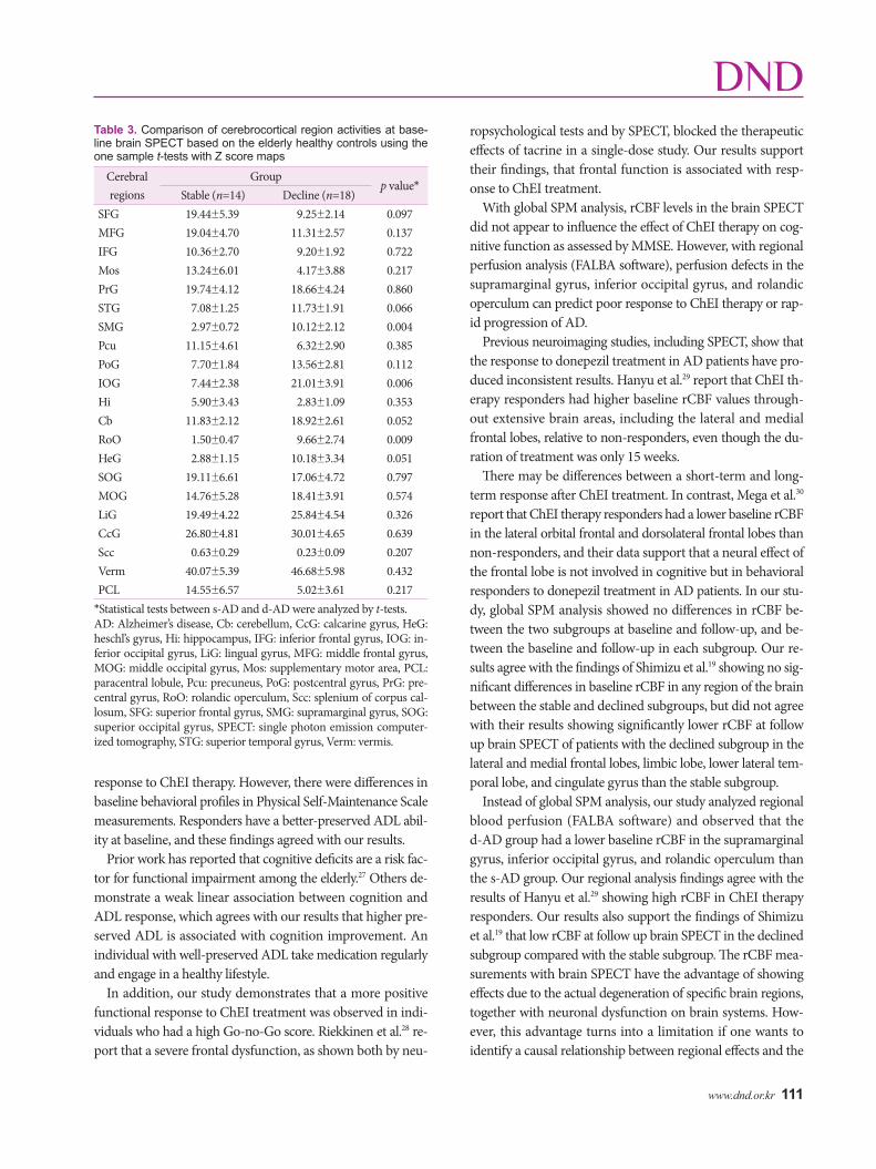
a significantly higher perfusion deficit for the d-AD group in the supramarginal gyrus, inferior occipital gyrus, and rolandic operculum (Table 3).
DISCUSSION
We found that the higher preserved ADCS-ADLs predict-ed a better improvement in the MMSE scores in response to ChEI therapy. An increased positive functional response to ChEI was observed in Go-no-Go, immediate SVLT, immedi-ate RCFT and short form K-BNT. So far, many studies have not examined the relationship of neuropsychological tests and response to ChEI therapy.
Wattmo et al.26 reported that better-preserved cognition and more impaired ADL ability implied an increased functional
A
B
C
D
Superior Anterior R-lateral Posterior L-lateral
Fig. 2. Global Statistical Parametric Mapping software analysis showing a relative decrease in the regional cerebral blood flow of brain sin-gle photon emission computerized tomography in the stable Alzheimer’s disease (AD) group (s-AD) at baseline (A) and follow up (B); and in the declined AD group (d-AD) at baseline (C) and follow up (D) compared to elderly healthy controls (n=12). We used a threshold Z-score ≥2.33 (which corresponding to a p-value ≤0.01). The color of the outer contour corresponds to a Z score of 0–10.
www.dnd.or.kr 111
DND
response to ChEI therapy. However, there were differences in baseline behavioral profiles in Physical Self-Maintenance Scale measurements. Responders have a better-preserved ADL abil-ity at baseline, and these findings agreed with our results.
Prior work has reported that cognitive deficits are a risk fac-tor for functional impairment among the elderly.27 Others de-monstrate a weak linear association between cognition and ADL response, which agrees with our results that higher pre-served ADL is associated with cognition improvement. An individual with well-preserved ADL take medication regularly and engage in a healthy lifestyle.
In addition, our study demonstrates that a more positive functional response to ChEI treatment was observed in indi-viduals who had a high Go-no-Go score. Riekkinen et al.28 re-port that a severe frontal dysfunction, as shown both by neu-
ropsychological tests and by SPECT, blocked the therapeutic effects of tacrine in a single-dose study. Our results support their findings, that frontal function is associated with resp-onse to ChEI treatment.
With global SPM analysis, rCBF levels in the brain SPECT did not appear to influence the effect of ChEI therapy on cog-nitive function as assessed by MMSE. However, with regional perfusion analysis (FALBA software), perfusion defects in the supramarginal gyrus, inferior occipital gyrus, and rolandic operculum can predict poor response to ChEI therapy or rap-id progression of AD.
Previous neuroimaging studies, including SPECT, show that the response to donepezil treatment in AD patients have pro-duced inconsistent results. Hanyu et al.29 report that ChEI th-erapy responders had higher baseline rCBF values through-out extensive brain areas, including the lateral and medial frontal lobes, relative to non-responders, even though the du-ration of treatment was only 15 weeks.
There may be differences between a short-term and long-term response after ChEI treatment. In contrast, Mega et al.30 report that ChEI therapy responders had a lower baseline rCBF in the lateral orbital frontal and dorsolateral frontal lobes than non-responders, and their data support that a neural effect of the frontal lobe is not involved in cognitive but in behavioral responders to donepezil treatment in AD patients. In our stu-dy, global SPM analysis showed no differences in rCBF be-tween the two subgroups at baseline and follow-up, and be-tween the baseline and follow-up in each subgroup. Our re-sults agree with the findings of Shimizu et al.19 showing no sig-nificant differences in baseline rCBF in any region of the brain between the stable and declined subgroups, but did not agree with their results showing significantly lower rCBF at follow up brain SPECT of patients with the declined subgroup in the lateral and medial frontal lobes, limbic lobe, lower lateral tem-poral lobe, and cingulate gyrus than the stable subgroup.
Instead of global SPM analysis, our study analyzed regional blood perfusion (FALBA software) and observed that the d-AD group had a lower baseline rCBF in the supramarginal gyrus, inferior occipital gyrus, and rolandic operculum than the s-AD group. Our regional analysis findings agree with the results of Hanyu et al.29 showing high rCBF in ChEI therapy responders. Our results also support the findings of Shimizu et al.19 that low rCBF at follow up brain SPECT in the declined subgroup compared with the stable subgroup. The rCBF mea-surements with brain SPECT have the advantage of showing effects due to the actual degeneration of specific brain regions, together with neuronal dysfunction on brain systems. How-ever, this advantage turns into a limitation if one wants to identify a causal relationship between regional effects and the
Table 3. Comparison of cerebrocortical region activities at base-line brain SPECT based on the elderly healthy controls using the one sample t-tests with Z score maps
Cerebral regions
Groupp value*
Stable (n=14) Decline (n=18)SFG 19.44±5.39 9.25±2.14 0.097MFG 19.04±4.70 11.31±2.57 0.137IFG 10.36±2.70 9.20±1.92 0.722Mos 13.24±6.01 4.17±3.88 0.217PrG 19.74±4.12 18.66±4.24 0.860STG 7.08±1.25 11.73±1.91 0.066SMG 2.97±0.72 10.12±2.12 0.004Pcu 11.15±4.61 6.32±2.90 0.385PoG 7.70±1.84 13.56±2.81 0.112IOG 7.44±2.38 21.01±3.91 0.006Hi 5.90±3.43 2.83±1.09 0.353Cb 11.83±2.12 18.92±2.61 0.052RoO 1.50±0.47 9.66±2.74 0.009HeG 2.88±1.15 10.18±3.34 0.051SOG 19.11±6.61 17.06±4.72 0.797MOG 14.76±5.28 18.41±3.91 0.574LiG 19.49±4.22 25.84±4.54 0.326CcG 26.80±4.81 30.01±4.65 0.639Scc 0.63±0.29 0.23±0.09 0.207Verm 40.07±5.39 46.68±5.98 0.432PCL 14.55±6.57 5.02±3.61 0.217
*Statistical tests between s-AD and d-AD were analyzed by t-tests.AD: Alzheimer’s disease, Cb: cerebellum, CcG: calcarine gyrus, HeG: heschl’s gyrus, Hi: hippocampus, IFG: inferior frontal gyrus, IOG: in-ferior occipital gyrus, LiG: lingual gyrus, MFG: middle frontal gyrus, MOG: middle occipital gyrus, Mos: supplementary motor area, PCL: paracentral lobule, Pcu: precuneus, PoG: postcentral gyrus, PrG: pre-central gyrus, RoO: rolandic operculum, Scc: splenium of corpus cal-losum, SFG: superior frontal gyrus, SMG: supramarginal gyrus, SOG: superior occipital gyrus, SPECT: single photon emission computer-ized tomography, STG: superior temporal gyrus, Verm: vermis.

Dong-Eun Kim et al.SPECT and Neuropsychological Tests Predict the Response to Treatment in AD
112 Dement Neurocogn Disord 2015;14(3):106-113
response to the therapy, as it is impossible to distinguish rCBF decreases between the two groups diagnosed AD already us-ing global SPM analysis.
There are several limitations in this study. First, the number of subjects was small. Second, we were unable to create a con-trol AD group that did not take a ChEI medication. Thus, we do not know how spontaneous progress of AD affected the change of rCBF, nor do we know the effect of donepezil on the blood perfusion in healthy subjects after a 1-year treatment. Third, we measured clinical outcomes using the K-MMSE. Although K-MMSE has the advantages of being both easy to administer and is not time-consuming, however, this test was not designed to assess subtle changes in cognition.31
In conclusion, we found that the higher ADCS-ADLs pre-dicted a better improvement in the K-MMSE scores in resp-onse to ChEI therapy and a more positive functional response in the group with preserved frontal function. Furthermore, the regional perfusion analysis of brain SPECT, instead of global SPM analysis, provided hints to the variable response to the therapy with ChEI treatment.
Conflicts of InterestThe authors have no financial conflicts of interest.
AcknowledgementsThis study was supported by grants from the Chonnam National Universi-ty Hospital (CRI 10007-1) and the Brain Research Program through the National Research Foundation of Korea, funded by the Ministry of Sci-ence, ICT & Future Planning (NRF-2014M3C7A1046041).
REFERENCES1. Venneri A. Imaging treatment effects in Alzheimer’s disease. Magn
Reson Imaging 2007;25:953-968.2. Rogers SL, Farlow MR, Doody RS, Mohs R, Friedhoff LT. A 24-
week, double-blind, placebo-controlled trial of donepezil in patients with Alzheimer’s disease. Donepezil Study Group. Neurology 1998; 50:136-145.
3. Farlow MR, Hake A, Messina J, Hartman R, Veach J, Anand R. Re-sponse of patients with Alzheimer disease to rivastigmine treatment is predicted by the rate of disease progression. Arch Neurol 2001;58: 417-422.
4. Van Der Putt R, Dineen C, Janes D, Series H, McShane R. Effective-ness of acetylcholinesterase inhibitors: diagnosis and severity as pre-dictors of response in routine practice. Int J Geriatr Psychiatry 2006; 21:755-760.
5. Wattmo C, Hansson O, Wallin AK, Londos E, Minthon L. Predicting long-term cognitive outcome with new regression models in donepe-zil-treated Alzheimer patients in a naturalistic setting. Dement Geriatr Cogn Disord 2008;26:203-211.
6. Feldman HH, Jacova C. Predicting response to acetylcholinesterase inhibitor treatment in Alzheimer disease: has the time come? Nat Clin Pract Neurol 2009;5:128-129.
7. Wattmo C, Wallin AK, Londos E, Minthon L. Predictors of long-term cognitive outcome in Alzheimer’s disease. Alzheimers Res Ther 2011; 3:23.
8. Ritchie CW, Ames D, Clayton T, Lai R. Metaanalysis of randomized
trials of the efficacy and safety of donepezil, galantamine, and rivastig-mine for the treatment of Alzheimer disease. Am J Geriatr Psychiatry 2004;12:358-369.
9. Saumier D, Murtha S, Bergman H, Phillips N, Whitehead V, Chertkow H. Cognitive predictors of donepezil therapy response in Alzheimer disease. Dement Geriatr Cogn Disord 2007;24:28-35.
10. Molinuevo JL, Berthier ML, Rami L. Donepezil provides greater ben-efits in mild compared to moderate Alzheimer’s disease: implications for early diagnosis and treatment. Arch Gerontol Geriatr 2011;52:18-22.
11. Waldemar G, Bruhn P, Kristensen M, Johnsen A, Paulson OB, Las-sen NA. Heterogeneity of neocortical cerebral blood flow deficits in dementia of the Alzheimer type: a [99mTc]-d,l-HMPAO SPECT study. J Neurol Neurosurg Psychiatry 1994;57:285-295.
12. McKhann G, Drachman D, Folstein M, Katzman R, Price D, Stadlan EM. Clinical diagnosis of Alzheimer’s disease: report of the NINCDS-ADRDA Work Group under the auspices of Department of Health and Human Services Task Force on Alzheimer’s Disease. Neurology 1984; 34:939-944.
13. Folstein MF, Folstein SE, McHugh PR. “Mini-mental state”. A practi-cal method for grading the cognitive state of patients for the clinician. J Psychiatr Res 1975;12:189-198.
14. Hughes CP, Berg L, Danziger WL, Coben LA, Martin RL. A new clinical scale for the staging of dementia. Br J Psychiatry 1982;140: 566-572.
15. Kang YW, Na DL. Seoul neuropsychological screening battery. Incheon: Human Brain Research & Consulting Co., 2003.
16. Lawton MP, Brody EM. Assessment of older people: self-maintain-ing and instrumental activities of daily living. Gerontologist 1969;9: 179-186.
17. Galasko D, Bennett D, Sano M, Ernesto C, Thomas R, Grundman M, et al. An inventory to assess activities of daily living for clinical trials in Alzheimer’s disease. The Alzheimer’s Disease Cooperative Study. Alzheimer Dis Assoc Disord 1997;11 Suppl 2:S33-S39.
18. Doody RS, Dunn JK, Clark CM, Farlow M, Foster NL, Liao T, et al. Chronic donepezil treatment is associated with slowed cognitive de-cline in Alzheimer’s disease. Dement Geriatr Cogn Disord 2001;12: 295-300.
19. Shimizu S, Hanyu H, Iwamoto T, Koizumi K, Abe K. SPECT follow-up study of cerebral blood flow changes during Donepezil therapy in patients with Alzheimer’s disease. J Neuroimaging 2006;16:16-23.
20. Mosconi L, Tsui WH, Pupi A, De Santi S, Drzezga A, Minoshima S, et al. (18)F-FDG PET database of longitudinally confirmed healthy elderly individuals improves detection of mild cognitive impairment and Alzheimer’s disease. J Nucl Med 2007;48:1129-1134.
21. Mosconi L, Tsui WH, Herholz K, Pupi A, Drzezga A, Lucignani G, et al. Multicenter standardized 18F-FDG PET diagnosis of mild cogni-tive impairment, Alzheimer’s disease, and other dementias. J Nucl Med 2008;49:390-398.
22. Kim EJ, Kim BC, Kim SJ, Jung DS, Sin JS, Yoon YJ, et al. Clinical staging of semantic dementia in an FDG-PET study using FTLD-CDR. Dement Geriatr Cogn Disord 2012;34:300-306.
23. Baker JG, Williams AJ, Wack DS, Miletich RS. Correlation of cogni-tion and SPECT perfusion: easy Z score and SPM analysis of a pilot sample with cerebral small vessel disease. Dement Geriatr Cogn Dis-ord 2013;36:290-299.
24. Jeong GW, Park K, Youn G, Kang HK, Kim HJ, Seo JJ, et al. Assess-ment of cerebrocortical regions associated with sexual arousal in pre-menopausal and menopausal women by using BOLD-based func-tional MRI. J Sex Med 2005;2:645-651.
25. Kim TH, Jeong GW, Baek HS, Kim GW, Sundaram T, Kang HK, et al. Human brain activation in response to visual stimulation with ru-ral and urban scenery pictures: a functional magnetic resonance im-aging study. Sci Total Environ 2010;408:2600-2607.

www.dnd.or.kr 113
DND26. Wattmo C, Wallin ÅK, Londos E, Minthon L. Long-term outcome
and prediction models of activities of daily living in Alzheimer disease with cholinesterase inhibitor treatment. Alzheimer Dis Assoc Disord 2011;25:63-72.
27. Barberger-Gateau P, Fabrigoule C. Disability and cognitive impair-ment in the elderly. Disabil Rehabil 1997;19:175-193.
28. Riekkinen P Jr, Riekkinen M, Soininen H, Kuikka J, Laakso M, Riek-kinen P Sr. Frontal dysfunction blocks the therapeutic effect of THA on attention in Alzheimer’s disease. Neuroreport 1997;8:1845-1849.
29. Hanyu H, Shimizu T, Tanaka Y, Takasaki M, Koizumi K, Abe K. Re-gional cerebral blood flow patterns and response to donepezil treat-
ment in patients with Alzheimer’s disease. Dement Geriatr Cogn Dis-ord 2003;15:177-182.
30. Mega MS, Dinov ID, Lee L, O’Connor SM, Masterman DM, Wilen B, et al. Orbital and dorsolateral frontal perfusion defect associated with behavioral response to cholinesterase inhibitor therapy in Alzheim-er’s disease. J Neuropsychiatry Clin Neurosci 2000;12:209-218.
31. Takeda A, Loveman E, Clegg A, Kirby J, Picot J, Payne E, et al. A sys-tematic review of the clinical effectiveness of donepezil, rivastigmine and galantamine on cognition, quality of life and adverse events in Al-zheimer’s disease. Int J Geriatr Psychiatry 2006;21:17-28.