Embed Size (px)
Citation preview

Single nucleotide polymorphisms alter kinaseanchoring and the subcellular targeting ofA-kinase anchoring proteinsF. Donelson Smitha, Mitchell H. Omara, Patrick J. Nygrena, Joseph Soughayera,1, Naoto Hoshib, Ho-Tak Laua,Calvin G. Snydera, Tess C. Branonc,d,e, Debapriya Ghoshf, Lorene K. Langeberga, Alice Y. Tingc,d,e, Luis F. Santanag,Shao-En Onga, Manuel F. Navedof, and John D. Scotta,2
aDepartment of Pharmacology, University of Washington, Seattle, WA 98195; bDepartment of Pharmacology, University of California, Irvine, CA 92697;cDepartment of Genetics, Stanford University, Stanford, CA 94305; dDepartment of Biology, Stanford University, Stanford, CA 94305; eDepartment ofChemistry, Stanford University, Stanford, CA 94305; fDepartment of Pharmacology, University of California, Davis, CA 95616; and gDepartment ofPhysiology & Membrane Biology, University of California, Davis, CA 95616
Edited by Melanie H. Cobb, University of Texas Southwestern Medical Center, Dallas, TX, and approved October 23, 2018 (received for review September27, 2018)
A-kinase anchoring proteins (AKAPs) shape second-messenger sig-naling responses by constraining protein kinase A (PKA) at preciseintracellular locations. A defining feature of AKAPs is a helical regionthat binds to regulatory subunits (RII) of PKA. Mining patient-derived databases has identified 42 nonsynonymous SNPs in thePKA-anchoring helices of five AKAPs. Solid-phase RII binding assaysconfirmed that 21 of these amino acid substitutions disrupt PKAanchoring. The most deleterious side-chain modifications are situ-ated toward C-termini of AKAP helices. More extensive analysis wasconducted on a valine-to-methionine variant in the PKA-anchoringhelix of AKAP18. Molecular modeling indicates that additionaldensity provided by methionine at position 282 in the AKAP18γ iso-form deflects the pitch of the helical anchoring surface outward by6.6°. Fluorescence polarization measurements show that this subtletopological change reduces RII-binding affinity 8.8-fold and impairscAMP responsive potentiation of L-type Ca2+ currents in situ. Live-cell imaging of AKAP18γ V282M-GFP adducts led to the unexpecteddiscovery that loss of PKA anchoring promotes nuclear accumulationof this polymorphic variant. Targeting proceeds via a mechanismwhereby association with the PKA holoenzyme masks a polybasicnuclear localization signal on the anchoring protein. This led to thediscovery of AKAP18e: an exclusively nuclear isoform that lacks aPKA-anchoring helix. Enzyme-mediated proximity-proteomics revealthat compartment-selective variants of AKAP18 associate with dis-tinct binding partners. Thus, naturally occurring PKA-anchoring-defective AKAP variants not only perturb dissemination of localsecond-messenger responses, but also may influence the intracellu-lar distribution of certain AKAP18 isoforms.
AKAPs | PKA | proximity labeling | kinase anchoring | nucleus
The recent explosion of deep-sequencing data from diversecohorts of patients reveals the impact of genetic variation
within human populations (1). Nucleotide changes that accumulateover multiple generations are considered inert or “passenger” mu-tations that shape us as individuals. Conversely, acute genetic lesionsthat alter the activity of key enzymes or structural proteins are oftenlinked to disease. Such pathological lesions are particularly preva-lent in cell-signaling enzymes, such as ubiquitin ligases and proteinkinases that guide the passage of chemical signals throughout thecell (2, 3). Consequently, mining of databases that chart thesemutations can be combined with high-resolution structural analysisto inform the development of drugs that combat chronic disorders,such as Parkinson’s disease and various cancers (4).Early examples of this precision-medicine approach include the
identification of genetically modified forms of the BCR-Abl andB-RAF kinases that drive cancers, including chronic myelogenousleukemia, melanoma, and hepatocellular carcinoma (5, 6). Thesubsequent success of groundbreaking drugs, such as imatinib
(Gleevec) and sorafenib, have transformed this innovative drug-discovery strategy into lucrative multinational ventures. The cur-rent kinase inhibitor landscape includes 243 small molecules inclinical trials for a variety of indications (7). However, manipu-lating complex cellular events is often more involved than simplytargeting individual signaling enzymes. This is exemplified by ev-idence that many kinase cascades are constrained by scaffoldingand anchoring proteins (8–10). These organizational elementsensure that signals are transmitted to precise sites within the cell(11). Hence, genetic lesions that disrupt protein kinase anchoringhave been linked to various pathological responses and may rep-resent untapped therapeutic targets (12–14).A-kinase anchoring proteins (AKAPs) are prototypic kinase-
targeting factors (15, 16). This structurally diverse group of proteinswas initially discovered on the basis of solid-phase interaction withradiolabeled RII subunits of the cAMP-dependent protein kinase(PKA) (17). Subsequent interaction-cloning, proteomic, and bio-informatic approaches have identified 39 human genes that en-code in excess of 60 AKAP isoforms (11) (Fig. 1A). AKAPs were
Significance
Dissemination of chemical information throughout the cell is afundamental biological process with clinical relevance. Pathologicalchanges in local signaling enzyme activity are linked to diseases,including schizophrenia, Alzheimer’s disease, cardiac arrhythmias,and seizures. Mining of patient datasets has uncovered geneticvariation in A-kinase anchoring proteins (AKAPs) that promotesmislocalization of protein kinase A (PKA). We investigate 42 SNPsin AKAPs that interrupt association with PKA to impact local cAMPsignaling. The most detrimental variants are situated within thehydrophobic face of a conserved helical region on the AKAP that isessential for kinase anchoring. An unexpected outcome is thediscovery of an alternative targeting mechanism for AKAPs thatutilizes the intact PKA holoenzymes as cytoplasmic “anchors.”
Author contributions: F.D.S., M.H.O., J.S., N.H., L.F.S., M.F.N., and J.D.S. designed research;F.D.S., M.H.O., J.S., N.H., H.-T.L., C.G.S., D.G., and M.F.N. performed research; T.C.B., A.Y.T.,and S.-E.O. contributed new reagents/analytic tools; F.D.S., M.H.O., P.J.N., J.S., N.H.,H.-T.L., M.F.N., and J.D.S. analyzed data; P.J.N. performed structural modeling; andF.D.S., L.K.L., and J.D.S. wrote the paper.
The authors declare no conflict of interest.
This article is a PNAS Direct Submission.
Published under the PNAS license.1Present address: IonOptix, Westwood, MA 02090.2To whom correspondence should be addressed. Email: [email protected].
This article contains supporting information online at www.pnas.org/lookup/suppl/doi:10.1073/pnas.1816614115/-/DCSupplemental.
Published online November 19, 2018.
www.pnas.org/cgi/doi/10.1073/pnas.1816614115 PNAS | vol. 115 | no. 49 | E11465–E11474
BIOCH
EMISTR
Y
Dow
nloa
ded
at U
NIV
ER
SIT
Y O
F W
AS
HIN
GT
ON
on
Nov
embe
r 8,
201
9

I
A
B C D
E G H
F
J
Fig. 1. Identification and analysis of polymorphisms in PKA-anchoring domains of AKAPs. (A) Schematic of the human chromosomal locations for genesencoding 39 experimentally validated AKAPs. AKAPs harboring nonsynonymous SNPs in the PKA-anchoring helices that are investigated in this study arehighlighted in red. (B–E and H) Peptide SPOT arrays containing WT and variant PKA binding helices were constructed and probed by RII overlay. (Upper) Solid-phase RII binding was assessed by far-Western blotting with biotin-RII and detected by neutravidin-HRP and enhanced chemoluminescence. (Lower) Den-sitometric analysis of RII binding was normalized to WT signal. (B) Peptide array and amalgamated densitometric data for analysis of the AKAP-Lbc (AKAP13)RII-binding site. Densitometric analysis of RII binding was normalized to WT signal. Variants that reduce binding to RII are highlighted in blue. (C) Screeningand analysis of peptide arrays representing SNP sequences for PKA binding site 1 in AKAP220 (AKAP11). (D) Screening and analysis of peptide arrays rep-resenting SNP sequences for the PKA binding site 2 in AKAP220 (AKAP11). (E) Screening and analysis of peptide arrays representing SNP sequences for thePKA binding site in AKAP79 (AKAP5). (F) Molecular modeling of the AKAP79 amphipathic helix (PDB ID code 2H9R) showing threonine 395 at the second turnof the helix (blue). (G) Immune complexes of either WT AKAP79 or AKAP79 T395P were probed for coprecipitation of PKA RII and C subunits. Introduction ofthe helix-breaking proline residue disrupts the AKAP79–PKA interaction. (H) Screening and analysis of peptide arrays representing SNP sequences for the PKAbinding in AKAPKL (AKAP2). (I) Modeling of the AKAP-KL amphipathic helix based on coordinates for AKAP-IS (PDB ID code 2IZX), showing the residuesA659 and A666 (blue). (J) Immune complexes of either WT AKAP-KL or AKAP-KL A659S, A659T, or A666P variants were probed for coprecipitation of PKA RIIand C subunits. Mutation of either position drastically impairs PKA binding. IP, immunoprecipitation
E11466 | www.pnas.org/cgi/doi/10.1073/pnas.1816614115 Smith et al.
Dow
nloa
ded
at U
NIV
ER
SIT
Y O
F W
AS
HIN
GT
ON
on
Nov
embe
r 8,
201
9

originally named colloquially or on the basis of their molecularweight, but the systematic cataloging of anchoring proteins hasbeen complicated by a HUGO nomenclature that only recognizes15 AKAP genes (Fig. 1A). Nonetheless, a ubiquitous and definingcharacteristic of the AKAP family is a 14- to 18-residue helicalregion that binds with high affinity to PKA holoenzymes that arecomposed of a regulatory (R) subunit dimer and two catalytic (C)subunits (18). X-ray crystallographic analyses have shown that thehydrophobic face of this amphipathic helix slots into a bindinggroove formed by the docking and dimerization domain (D/D) ofR subunits (19, 20). Negative-stain electron microscopy and 3Dreconstructions show that intact hetero-pentameric AKAP-RII2-C2assemblies adopt a range of flexible tripartite configurations (21,22). This is because intrinsically disordered regions between the D/D domains and cAMP-binding sites on each regulatory subunitpermit a 150- to 200-Å radius of motion to the associated catalyticsubunits (21, 23). Such flexibility within anchored PKA holoenzymecomplexes enables precise orientation of the catalytic subunit to-ward substrates (11, 24). Recent data advances this model, showingthat intact, active, and anchored PKA holoenzymes are the func-tional signaling units inside cells (22). This “signaling island” con-cept radically changes the view of how AKAP complexes operateand indicates that protein phosphorylation is more regionally con-fined than previously appreciated (22).As our understanding of how signaling enzyme complexes
operate becomes more refined, it is evident that perturbation ofAKAP–PKA interactions is linked to disease (11, 14). Scrutiny ofbig datasets has uncovered genetic variation in AKAPs thatcorrelate with susceptibility to diseases, such as schizophrenia,Alzheimer’s, certain cardiac arrhythmias, and the onset of febrileseizures (12, 14, 25, 26). In this report we systematically in-vestigate the properties of polymorphic variants in AKAPs thatelicit single amino acid changes in RII-anchoring helices. Un-expected outcomes of this endeavor include the discovery of analternative AKAP-targeting mechanism that utilizes the intactPKA holoenzyme as a cytoplasmic anchor and the identificationof an AKAP18 variant that does not function as an AKAP.
ResultsSNPs in AKAP Helices Alter PKA Anchoring. Genome-wide associa-tion surveys have identified point mutations and SNPs in genesencoding AKAPs that are linked to a variety of neurological andcardiac diseases (reviewed in ref. 14). The National Center forBiotechnology Information (NCBI) SNP (https://www.ncbi.nlm.nih.gov/SNP/index.html) and Variation Viewer databases (https://www.ncbi.nlm.nih.gov/variation/view/) were used to identify non-synonymous nucleotide changes that introduce missense aminoacids within the R subunit binding helices from a variety ofAKAPs (Fig. 1A, red). This information was used to create pep-tide SPOT arrays that encompass native and variant PKA-bindingsequences from five different anchoring proteins (Fig. 1A andSI Appendix, Table S1). Solid-phase RII-binding (RII-overlay)assessed whether these amino acid substitutions affected PKAanchoring (Fig. 1B). Peptides containing prolines were included inthe peptide array as negative controls (SI Appendix, Table S1).Prior studies have shown that substitution with proline disruptsthe structural integrity of PKA-anchoring helices (18, 27).Densitometric analyses of multiple SPOT arrays revealed that
amino acid substitution within PKA-anchoring helices influencesRII interaction (Fig. 1 B–J). Nine naturally occurring variants inAKAP-Lbc (AKAP13) were tested (Fig. 1B). Single amino acidsubstitutions at four positions (R1245H, I1248V, S1253R, andR1254C) had no effect on RII-binding (Fig. 1B). However, threeside-chain variants toward the C terminus of the PKA-anchoringhelix (V1256M, D1257V, and E1261V) dramatically reducedsolid-phase interaction with RII (Fig. 1B). Two other substitu-tions (E1261K and K1264E) had more modest effects (Fig. 1B).
Next we investigated SNPs in the vesicular and membrane-associated anchoring protein AKAP220 (AKAP11). This an-choring protein encodes two PKA-anchoring helices with dif-ferent affinities for PKA holoenzymes (28, 29). Substitutionstoward the C terminus of site 1 exhibited reduced affinity forRII, whereas amino acid changes throughout helix 2 adverselyaffected PKA anchoring (Fig. 1 C and D). Interestingly, sub-stitution of serine or valine at position 1648 enhanced solid-phase binding of RII in site 2 of AKAP220 (Fig. 1D). Thissuggests that side chains that constrain the initial turns of helicesare positive PKA-anchoring determinants.Further support for this concept was provided by analysis of the
neuronal anchoring protein AKAP79 (AKAP5) (30, 31). ThreeSNP variants in AKAP79 were investigated, but only substitutionof Thr-395 with Pro affected RII-binding (Fig. 1E). Molecularmodeling based upon the NMR structure of this region revealsthat position 395 lies within the first turn of the anchoring helix(32) (Fig. 1F). Proline substitution prevents formation of theα-helix. Further validation was provided when this variant wasintroduced into full-length AKAP79 and expressed in HEK293cells. Western blotting of WT AKAP79 immune complexes con-firmed cofractionation of PKA regulatory and catalytic subunits(Fig. 1G, Upper three panels, lane 2). Cofractionation of PKAsubunits was not detected in cells expressing the AKAP79-T395Pvariant (Fig. 1G, Upper three panels, lane 3).AKAP-KLs (AKAP2) are a family of alternately spliced AKAP
isoforms that organize kidney and lung second-messenger sig-naling events (33). AKAP-KL also sustains PKA regulation oflens aquaporin-0 water channels (34). Disruption of AKAP-KL–mediated PKA anchoring in the lens is implicated in pathologicalcataract formation (34). Peptide array analysis identified severalmissense variants of AKAP-KL that disrupted RII-binding (Fig.1H). The most profound effects were observed in SNP variantsthat contained prolines at positions 664 and 666 in AKAP-KL.However, significant loss of PKA anchoring was also observedwhen positions 659 and 667 were replaced with bulkier sidechains (Fig. 1H). We focused our biochemical analyses on twosites in the AKAP-KL helix that yielded severely impaired PKA-anchoring variants (Fig. 1I). Further validation of this result wasprovided when the A659S, A659T, and A666P variants wereintroduced into full-length AKAP-KL and expressed in HEK293cells. Coprecipitation of PKA subunits was severely reduced ornot detected in AKAP-KL immune complexes isolated from cellsexpressing each of the aforementioned SNP variants (Fig. 1J,Upper two panels, lanes 3–5). Control experiments with WTAKAP-KL immune complexes confirmed cofractionation ofPKA regulatory and catalytic subunits (Fig. 1J, Upper two panels,lane 2). Collectively, the data presented in Fig. 1 show thatnaturally occurring variants on the hydrophobic face of RII-binding helices, and particularly those located in C-terminalturns, can have profound effects on PKA anchoring.
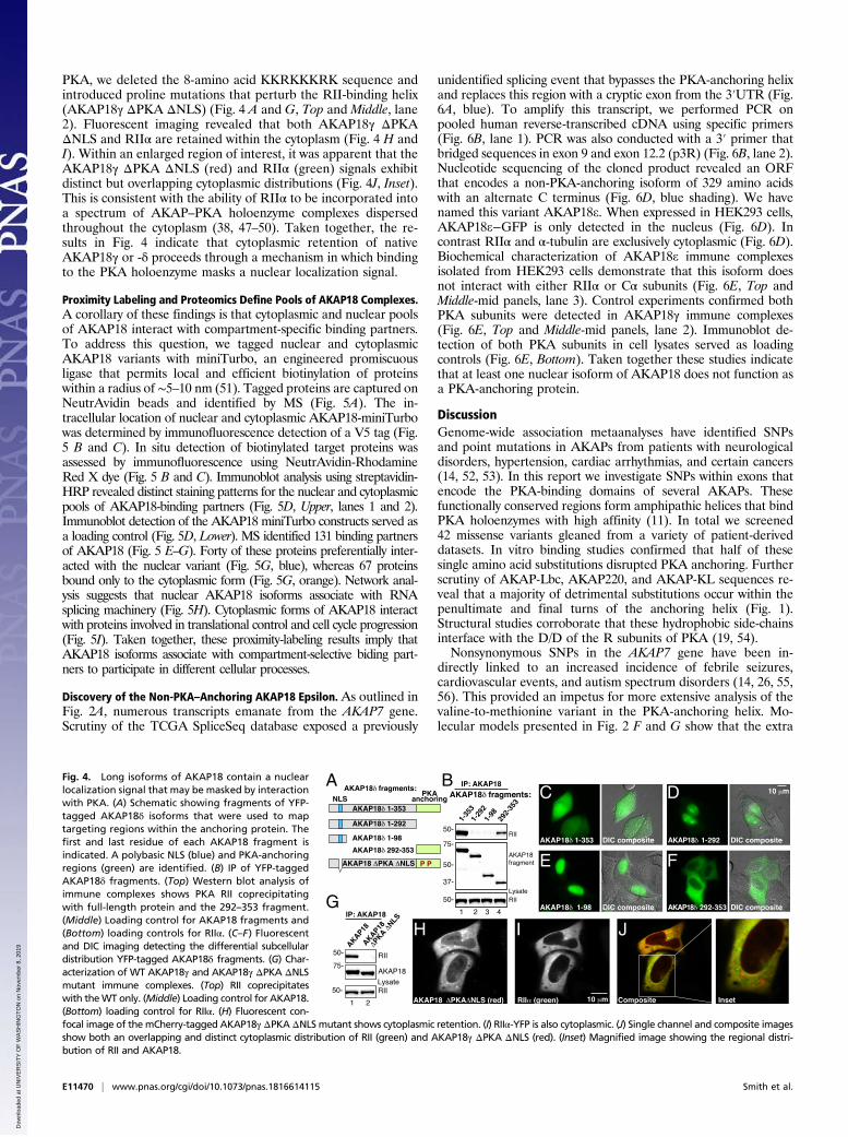
Characterization of AKAP18 Helix-Disrupting SNPs. Alternativesplicing of the AKAP7 gene drives tissue-specific expression ofdifferent AKAP18 protein products that range in size from 15 to42 kDa (35–37) (Fig. 2 A and B). The low molecular weight α-and β-isoforms of AKAP18 couple PKA to a variety of ionchannels in neurons, whereas the longer δ- and γ-isoforms havebeen implicated in modulation of excitation contraction couplingin the heart, water homeostasis in kidney collecting ducts, androles in differentiated adipocytes (35, 38, 39) (Fig. 2B). Poly-morphisms in AKAP18 have also been linked to an increasedincidence of febrile seizures (26). The identification of a clusterof SNPs within the PKA-anchoring helix between residues282 and 286 of AKAP18γ provided the impetus for more in-depth investigation (Fig. 2C). Peptide array analysis of SNP se-quences at positions 282, 283, and 286 revealed that conservativeside-chain substitutions disrupted RII-binding (Fig. 2C). Control
Smith et al. PNAS | vol. 115 | no. 49 | E11467
BIOCH
EMISTR
Y
Dow
nloa
ded
at U
NIV
ER
SIT
Y O
F W
AS
HIN
GT
ON
on
Nov
embe
r 8,
201
9

experiments confirmed that PKA anchoring was also abolishedin the context of the double-mutation AKAP18γ S278/L281P(Fig. 2C). In contrast, the AKAP18γ Arg280Ser (R280S) variantappeared to strengthen the interaction with RII (Fig. 2C). Morestringent cell-based validation of AKAP18γ variants involvedexpression of the full-length proteins in HEK293 cells. Westernblot analysis of AKAP18 immune complexes was used to detectcofractionation of PKA subunits (Fig. 2D, Upper panels). Allvariants at positions 282–286 of AKAP18γ were impaired in theirability to anchor PKA (Fig. 2D, lanes 4–6). In comparison, PKAsubunits remained associated with the AKAP18γ R280S variantinside cells (Fig. 2D, lane 3). Secondary structure predictionalgorithms place residue 280 of AKAP18γ on the hydrophilicface of the AKAP helix, a region that does not directly contactthe RII dimer (40) (Fig. 2E). Conversely, side chains betweenpositions 282–286 of AKAP18γ are located on the hydrophobicface of the helix (19, 40) (Fig. 2E). This substructure slots into abinding pocket formed by a four-helix bundle on the docking anddimerization domain of the R subunits (Fig. 2F, blue helix). Theimpaired binding in the V282M mutant provided us a uniqueopportunity to study this intermediate phenotype.Molecular modeling of the AKAP18–RIIα interface using
PDB coordinates from published structures predicts that theV282M substitution generates a 6.6° upward displacement of theanchoring helix in relation to the binding pocket on RIIα (Fig.
2C, red helix). More detailed examination of the V282M–RIIαinterface reveals that the added bulk and length of the methio-nine side-chain sterically hinders association with the AKAP-binding pocket on RIIα (Fig. 2G). In silico studies using theAutoDock program (41) computed a simulated free energy ofbinding of −4.54 kcal/mol (estimated Kd = 468 μM) for AKAP18V282M compared with −10.58 kcal/mol (estimated Kd =17.4 nM) for WT. Fluorescence polarization experiments cal-culated an IC50 of 283 ± 28 nM (n = 4) for AKAP18V282Mpeptide–RIIα interaction (Fig. 2H, red) compared with an IC50of 32 ± 3 nM (n = 4) for the native sequence (Fig. 2H, blue). Thedifferences in predicted and observed binding-affinity changesmay be a consequence of AutoDock forcing all side chains intorigid conformations (41). Collectively, these data indicate thatAKAP18 variants harboring the V282M substitution are severelyimpaired in their ability to anchor PKA holoenzymes.Because the kinase-anchoring helix is conserved in most splice
variants of AKAP18, we introduced the corresponding mutation(V37M) into the α-isoform. This membrane-associated anchoringprotein mediates PKA-dependent modulation of ion channels inexcitable cells (42). Fluorescent imaging confirmed that GFP-tagged AKAP18α V37M is retained at the plasma membranewhen expressed in HEK293 cells (Fig. 2 I and J). Coexpression ofAKAP18α V37M with L-type Ca2+ channel subunits permittedwhole-cell electrophysiological analysis of local PKA modulation
A B C
DE F G
H I
J
M N
K
L
Fig. 2. Polymorphisms in AKAP18 isoforms (AKAP7)alter PKA binding and regulation of ion channels. (A)Schematic of AKAP7 gene organization depictingexon–intron structure and alternatively spliced iso-forms. (B) IP-RII overlay analysis show differentialexpression of distinct AKAP18 isoforms in 3T3-L1adipocytes and cultured hippocampal neurons. (C)Peptide array and amalgamated densitometric datafor analysis of the AKAP18 (AKAP7) RII-binding site.Densitometric analysis of RII binding was normalizedto WT signal. Variants that reduce binding to RIIare highlighted in blue. (D) AKAP18 SNP variants(denoted above each lane) were immunoprecipitatedand copurification of PKA regulatory (Upper) andcatalytic (Upper-mid) identified by Western blotting.Expression levels of each protein are indicated in theLower three panels. (E) Model of AKAP18 PKA bind-ing helix highlighting selected residues (red asterisk)on either face of the amphipathic helix. (F) Structuralmodel shows WT AKAP18 helix (blue) and AKAP18V282M helix (red) in complex with the RII D/D domain(gray). The V282M helix (red) is oriented at a 6.6°angle from WT. (G) En face and expanded view ofthe AKAP18 V282M helix shows displacement of themutant peptide by bulky methionine side chain(red). (H) Fluorescence polarization data shows AKAP18WT and V282M peptide displacement of a fluorescentAKAP-IS peptide from RII. The IC50 values for each areindicated. (I) Fluorescent and DIC images of HeLacells expressing WT AKAP18α-GFP. (J) Fluorescent andDIC images of HeLa cells expressing AKAP18α V37M-GFP. (Scale bar: I–J, 10 μm.) (K) Whole-cell electrophysi-ological recording of cAMP-potentiated Ca2+ currentin HEK293 cells expressing the L-type Ca2+ channeland either AKAP18α WT or the AKAP18α V37Mvariant. (L) Quantification of peak current potenti-ation. Data are presented as mean ± SEM. *P <0.001 by unpaired Student’s t test. (M) Fluo4-AMmeasurement of cAMP-potentiated Ca2+ current inmouse aortic smooth muscle cells expressing eitherWT AKAP18α (blue) or the AKAP18α V37M variant(red). Calcium accumulation in response to de-polarization was measured before and after brief forskolin stimulation. (N ) Quantification of the change in fluorescence response to K+-induced de-polarization. Data are presented as mean ± SEM. *P < 0.001 by unpaired Student’s t test.
E11468 | www.pnas.org/cgi/doi/10.1073/pnas.1816614115 Smith et al.
Dow
nloa
ded
at U
NIV
ER
SIT
Y O
F W
AS
HIN
GT
ON
on
Nov
embe
r 8,
201
9

of calcium currents (36, 43). Stepwise depolarization of cells wasused to generate a pulse of Ca2+ current. In cells expressing WTAKAP18α, application of cell-soluble cAMP elicits a 1.5-fold in-crease in Ca2+ current (n = 10) (Fig. 2 K and L, blue). Thissecond-messenger response was abolished in cells expressing theAKAP18α V37M variant (Fig. 2 K and L, red).Further substantiation in a more physiologically relevant sys-
tem was possible by measuring depolarization-induced changesin Ca2+ flux in unpassaged cultured mouse aortic smooth-musclecells expressing AKAP18α or AKAP18α V37M. Global Ca2+
influx through calcium channels was measured using the fluo-rescent calcium indicator dye acetoxymethyl-ester (AM) Fluo-4(Fluo4-AM) upon depolarization with 60 mM potassium. In cellsexpressing AKAP18α, a brief pulse of forskolin to activate PKAenhanced Fluo4-AM fluorescence twofold (n = 7) (Fig. 2 M andN, blue). Importantly, this response was absent in cells expressingthe AKAP18α V37M variant (Fig. 2 M and N, red). Taken to-gether, these experiments suggest that naturally occurring vari-ants in the RII-binding helix of AKAP18 isoforms uncouplecAMP-responsive events at the plasma membrane.
AKAP18 Long Isoforms Transit to the Nucleus in a PKA-DependentManner. Long isoforms of AKAP18 encode targeting sequencesthat direct the anchoring protein to a variety of other subcellularlocations (37, 44). For example, AKAP18γ and -δ are seques-tered at the sarcoplasmic reticulum through protein–protein in-teractions with SERCA2 calcium reuptake channels, whereas inkidney collecting ducts, AKAP18δ is a component of intracellularvesicles (38, 39). However, these static targeting interactions do notfully reflect the complexity of AKAP18 retention in the cytoplasm.Surprisingly, application of a cell-soluble PKA-anchoring disruptorpeptide (stHt-31) elicited the nuclear accumulation of AKAP18γover a time course of 40 min (Fig. 3 A and B). In contrast, AKAP18γremained cytoplasmic upon application of the control peptide stHt-31-P (Fig. 3C). These results raised the intriguing possibility ofwhether or not association with the PKA holoenzyme influencesthe cytoplasmic retention of AKAP18γ or -δ.
Leptomycin B (LMB) is a bacterial metabolite that blocksCRM1-dependent nuclear export in mammalian cells. Thiscompound traps proteins that have the capacity to translocateinto the nucleus (45). Hence, we reasoned that the diminishedcapacity of AKAP18γ V282M to anchor PKA would render thisvariant more prone to nuclear translocation. Live-cell imaging ofHeLa cells treated with LMB (20 nM) monitored nuclear ac-cumulation of GFP-tagged AKAP18γ adducts (Fig. 3 D and E).Nuclear accumulation rates were calculated by quantifyingfluorescent intensity ratios between the nucleus and cytoplasmover a time course of 40 min (Fig. 3F). Minimal movement of theWT anchoring protein was detected (Fig. 3 D and F, blue) (n =33 cells). In contrast, nuclear accumulation of AKAP18γ V282Mwas pronounced (Fig. 3 G and H, red) (n = 58 cells) and oc-curred at a rate that is 3.75 times faster than the WT anchoringprotein (Fig. 3F). Considering these data in light of the experi-mental evidence presented in Fig. 2, we propose that a reducedaffinity for PKA contributes to the transit of AKAP18γ V282Minto the nucleus. Independent support for this hypothesis wasprovided by evidence that AKAP18γ S278P/L281P, a PKAbinding-null mutant, was exclusively detected in the nucleus (Fig.3 G and H). Control experiments confirmed that WT AKAP18γassociates with PKA, and is retained in the cytoplasm underthese conditions (Fig. 3 G and H, Upper).The long isoforms of AKAP18 (γ and δ) encode a putative
nuclear localization signal (NLS) within exon 3 of the AKAP7gene (46). Therefore, a potential explanation for the results inFig. 3 is that PKA anchoring obscures this NLS. To test thishypothesis, a family of deletion constructs was generated whereregions of the AKAP18δ were fused to YFP (Fig. 4A). In situinteraction with PKA was confirmed by coprecipitation of RII(Fig. 4B). The subcellular location of each YFP-tagged fragmentwas established by fluorescence microscopy (Fig. 4 C–F). Dif-ferential interference contrast (DIC) microscopy outlined cellcontours (Fig. 4 C–F). These studies reveal that residues 1–98 ofthe anchoring protein are necessary and sufficient for nucleartargeting (Fig. 4E). To further demonstrate that the NLS issufficient to direct AKAP18 into the nucleus in in the absence of
A B
C
D
E
F G H
Fig. 3. Association with the PKA holoenzyme isnecessary for cytoplasmic retention of AKAP18 longforms. (A) Schematic depicting displacement of PKAfrom AKAP18γ by the cell-soluble PKA-anchoringdisruptor peptide (stHt-31, orange) and the effecton subcellular targeting of AKAP18 long isoforms.Selected time points are shown from 0 to 40 min. (Band C) Live-cell imaging of cells expressing AKAP18γ-YFP following application of (B) stHt-31 or (C) theproline control peptide, stHt-31-P. (D and E) Live-cellimaging of cells expressing (D) WT AKAP18γ-GFP or(E) V282M AKAP18γ-GFP. Cells were treated withLMB and subcellular distribution was recorded over40 min. (F) Amalgamated data from multiple ex-periments in D and E. Changes in the ratio of nuclear/cytoplasmic fluorescent signal WT AKAP18γ-GFP (n =33 cells) and V282M AKAP18γ-GFP (n = 58 cells). (G) ThePKA-binding deficient mutant AKAP18γ S278P/L281P(AKAP18γ ΔPKA) is unable coprecipitate RII or interactwith it by solid-phase overlay. (H) Fluorescent confocalimages of HeLa cells expressing RIIα-YFP (green) and(Lower) mCherry-AKAP18γ (red) or (Bottom) the PKA-deficient AKAP18γ mutant.
Smith et al. PNAS | vol. 115 | no. 49 | E11469
BIOCH
EMISTR
Y
Dow
nloa
ded
at U
NIV
ER
SIT
Y O
F W
AS
HIN
GT
ON
on
Nov
embe
r 8,
201
9
PKA, we deleted the 8-amino acid KKRKKKRK sequence andintroduced proline mutations that perturb the RII-binding helix(AKAP18γ ΔPKA ΔNLS) (Fig. 4 A and G, Top and Middle, lane2). Fluorescent imaging revealed that both AKAP18γ ΔPKAΔNLS and RIIα are retained within the cytoplasm (Fig. 4 H andI). Within an enlarged region of interest, it was apparent that theAKAP18γ ΔPKA ΔNLS (red) and RIIα (green) signals exhibitdistinct but overlapping cytoplasmic distributions (Fig. 4J, Inset).This is consistent with the ability of RIIα to be incorporated intoa spectrum of AKAP–PKA holoenzyme complexes dispersedthroughout the cytoplasm (38, 47–50). Taken together, the re-sults in Fig. 4 indicate that cytoplasmic retention of nativeAKAP18γ or -δ proceeds through a mechanism in which bindingto the PKA holoenzyme masks a nuclear localization signal.
Proximity Labeling and Proteomics Define Pools of AKAP18 Complexes.A corollary of these findings is that cytoplasmic and nuclear poolsof AKAP18 interact with compartment-specific binding partners.To address this question, we tagged nuclear and cytoplasmicAKAP18 variants with miniTurbo, an engineered promiscuousligase that permits local and efficient biotinylation of proteinswithin a radius of ∼5–10 nm (51). Tagged proteins are captured onNeutrAvidin beads and identified by MS (Fig. 5A). The in-tracellular location of nuclear and cytoplasmic AKAP18-miniTurbowas determined by immunofluorescence detection of a V5 tag (Fig.5 B and C). In situ detection of biotinylated target proteins wasassessed by immunofluorescence using NeutrAvidin-RhodamineRed X dye (Fig. 5 B and C). Immunoblot analysis using streptavidin-HRP revealed distinct staining patterns for the nuclear and cytoplasmicpools of AKAP18-binding partners (Fig. 5D, Upper, lanes 1 and 2).Immunoblot detection of the AKAP18 miniTurbo constructs served asa loading control (Fig. 5D, Lower). MS identified 131 binding partnersof AKAP18 (Fig. 5 E–G). Forty of these proteins preferentially inter-acted with the nuclear variant (Fig. 5G, blue), whereas 67 proteinsbound only to the cytoplasmic form (Fig. 5G, orange). Network anal-ysis suggests that nuclear AKAP18 isoforms associate with RNAsplicing machinery (Fig. 5H). Cytoplasmic forms of AKAP18 interactwith proteins involved in translational control and cell cycle progression(Fig. 5I). Taken together, these proximity-labeling results imply thatAKAP18 isoforms associate with compartment-selective biding part-ners to participate in different cellular processes.
Discovery of the Non-PKA–Anchoring AKAP18 Epsilon. As outlined inFig. 2A, numerous transcripts emanate from the AKAP7 gene.Scrutiny of the TCGA SpliceSeq database exposed a previously
unidentified splicing event that bypasses the PKA-anchoring helixand replaces this region with a cryptic exon from the 3′UTR (Fig.6A, blue). To amplify this transcript, we performed PCR onpooled human reverse-transcribed cDNA using specific primers(Fig. 6B, lane 1). PCR was also conducted with a 3′ primer thatbridged sequences in exon 9 and exon 12.2 (p3R) (Fig. 6B, lane 2).Nucleotide sequencing of the cloned product revealed an ORFthat encodes a non-PKA-anchoring isoform of 329 amino acidswith an alternate C terminus (Fig. 6D, blue shading). We havenamed this variant AKAP18e. When expressed in HEK293 cells,AKAP18e−GFP is only detected in the nucleus (Fig. 6D). Incontrast RIIα and α-tubulin are exclusively cytoplasmic (Fig. 6D).Biochemical characterization of AKAP18e immune complexesisolated from HEK293 cells demonstrate that this isoform doesnot interact with either RIIα or Cα subunits (Fig. 6E, Top andMiddle-mid panels, lane 3). Control experiments confirmed bothPKA subunits were detected in AKAP18γ immune complexes(Fig. 6E, Top and Middle-mid panels, lane 2). Immunoblot de-tection of both PKA subunits in cell lysates served as loadingcontrols (Fig. 6E, Bottom). Taken together these studies indicatethat at least one nuclear isoform of AKAP18 does not function asa PKA-anchoring protein.
DiscussionGenome-wide association metaanalyses have identified SNPsand point mutations in AKAPs from patients with neurologicaldisorders, hypertension, cardiac arrhythmias, and certain cancers(14, 52, 53). In this report we investigate SNPs within exons thatencode the PKA-binding domains of several AKAPs. Thesefunctionally conserved regions form amphipathic helices that bindPKA holoenzymes with high affinity (11). In total we screened42 missense variants gleaned from a variety of patient-deriveddatasets. In vitro binding studies confirmed that half of thesesingle amino acid substitutions disrupted PKA anchoring. Furtherscrutiny of AKAP-Lbc, AKAP220, and AKAP-KL sequences re-veal that a majority of detrimental substitutions occur within thepenultimate and final turns of the anchoring helix (Fig. 1).Structural studies corroborate that these hydrophobic side-chainsinterface with the D/D of the R subunits of PKA (19, 54).Nonsynonymous SNPs in the AKAP7 gene have been in-
directly linked to an increased incidence of febrile seizures,cardiovascular events, and autism spectrum disorders (14, 26, 55,56). This provided an impetus for more extensive analysis of thevaline-to-methionine variant in the PKA-anchoring helix. Mo-lecular models presented in Fig. 2 F and G show that the extra
A B
G
C D
E F
H I J
Fig. 4. Long isoforms of AKAP18 contain a nuclearlocalization signal that may be masked by interactionwith PKA. (A) Schematic showing fragments of YFP-tagged AKAP18δ isoforms that were used to maptargeting regions within the anchoring protein. Thefirst and last residue of each AKAP18 fragment isindicated. A polybasic NLS (blue) and PKA-anchoringregions (green) are identified. (B) IP of YFP-taggedAKAP18δ fragments. (Top) Western blot analysis ofimmune complexes shows PKA RII coprecipitatingwith full-length protein and the 292–353 fragment.(Middle) Loading control for AKAP18 fragments and(Bottom) loading controls for RIIα. (C–F) Fluorescentand DIC imaging detecting the differential subcellulardistribution YFP-tagged AKAP18δ fragments. (G) Char-acterization of WT AKAP18γ and AKAP18γ ΔPKA ΔNLSmutant immune complexes. (Top) RII coprecipitateswith theWT only. (Middle) Loading control for AKAP18.(Bottom) loading control for RIIα. (H) Fluorescent con-focal image of themCherry-tagged AKAP18γ ΔPKA ΔNLSmutant shows cytoplasmic retention. (I) RIIα-YFP is also cytoplasmic. (J) Single channel and composite imagesshow both an overlapping and distinct cytoplasmic distribution of RII (green) and AKAP18γ ΔPKA ΔNLS (red). (Inset) Magnified image showing the regional distri-bution of RII and AKAP18.
E11470 | www.pnas.org/cgi/doi/10.1073/pnas.1816614115 Smith et al.
Dow
nloa
ded
at U
NIV
ER
SIT
Y O
F W
AS
HIN
GT
ON
on
Nov
embe
r 8,
201
9

A
B
C
D
E FG
H I
Fig. 5. Proximity labeling and proteomic identification of compartment-selective AKAP18 interactors. (A) Schematic depicting the miniTurbo-based bio-tinylation/MS experiments. Biotin is shown as dark-blue circles. (Left) Upon biotin addition, miniTurbo biotinylates only proximal proteins. (Center) Biotinylatedproteins are isolated with streptavidin pulldown and (Right) identified by MS. (B and C) Fluorescence microscopy depicting the subcellular location of (B) nuclearand (C) cytoplasmic AKAP18-miniTurbo variants and detection of compartment specific biotinylated proteins. (D) Western blot analysis of biotinylated proteinsin cell lysates shows distinct and compartment-specific labeling patterns. (E and F) MS results plotted as log difference of experimental intensity minus controlintensity plotted against the negative log of the P value. Selected nuclear (dark-blue) and cytoplasmic (tan) AKAP18 interacting partners are indicated. (G) Lists ofstatistically significant target proteins in nuclear and cytosolic preparations. Of 131 identified proteins, 40 were selectively associated with the nuclear variant,whereas 67 preferentially bound to the cytoplasmic variant. (H and I) STRING database-generated network depictions of compartment-specific proteins. Geneontology process annotation and false-discovery rates (FDR) are listed for the highlighted nodes. Putative biological functions for each subnetwork are indicated.
Smith et al. PNAS | vol. 115 | no. 49 | E11471
BIOCH
EMISTR
Y
Dow
nloa
ded
at U
NIV
ER
SIT
Y O
F W
AS
HIN
GT
ON
on
Nov
embe
r 8,
201
9

molecular density of a methionine at position 282 in AKAP18γdeflects the helical pitch of the hydrophobic anchoring surfaceupward by 6.6°. This relatively modest topological flaw decreasesthe binding affinity for RII by 8.8-fold and impairs PKA-mediated potentiation of L-type Ca2+ currents, as shown inFig. 2H. Because alternative splicing of the AKAP7 gene gen-erates multiple isoforms, Met-282 polymorphic variants mightnegatively impact a certain local PKA signaling events. Theseinclude modulation of neuronal sodium channels, regulation ofSERCA2-mediated calcium reuptake in the heart, and aquaporin-2–mediated control of water homeostasis in the kidney (38, 39, 57).The physiological significance to this concept is underscored by recentevidence that intact and anchored PKA holoenzymes are the pre-dominant cytoplasmic signaling units and are sequestered within 200–400 Å of their preferred substrates (21, 22). Hence, AKAP variantsthat impact the intracellular targeting of PKA holoenzymes have thecapacity to alter the dissemination of second-messenger signals.Although polymorphic variation within AKAPs has the po-
tential to uncouple local cAMP signaling, there is little in-formation about the allelic frequency or prevalence in specificpopulations. In addition, scrutiny of the cBioPortal cancer ge-nomics data interface does not reveal any clear pattern ofAKAP-anchoring helix mutations that correlate with cancers.Perhaps the best-documented example of genetic variationcomes from analysis of a nonsynonymous SNP that promotes anisoleucine to valine substitution at position 646 within the PKA-anchoring domain of D-AKAP-2 (AKAP10) (12, 58). This variantis associated with cardiac dysfunction in aging populations ofEuropean-American individuals that display a shortened electro-cardiogram PR interval (12). The D-AKAP2 I646V variant is alsoassociated with increased basal heart rate (58). Because basal heartrate correlates inversely with lifespan, the altered PKA-anchoringproperties of D-AKAP2 may be linked to adverse pathologicaloutcomes (59). However, the downstream signaling pathways af-fected by this modified kinase-anchoring event remain obscure. Invitro biochemical studies show that the D-AKAP2 646V variantbinds to type I PKA holoenzyme with threefold higher affinity thanD-AKAP2 646I, yet this variation has no effect on type II PKAanchoring (12). The physiological ramifications of these relativelysmall changes in affinity may be limited as D-AKAP2 exhibits astrong binding preference for type II PKA holoenzymes (60, 61).Furthermore, the porcine, murine, and chicken orthologs ofD-AKAP2 have a valine at the corresponding position in thePKA-anchoring helix (12). While structural analyses of theseD-AKAP2 orthologs are scant, it seems that the Ile to Val sub-stitution can be tolerated with minimal effects on PKA anchoring.This raises the issue of whether or not silent variation in AKAP
helices is a more general phenomenon. Data presented in Fig. 1Findicate that a silent change in the final turn of the AKAP79 helix(Ile-406 to Val) has no apparent effect on RII-binding. This mayrepresent another example of evolutionary drift, as valine naturallyoccurs at this position in the mouse, rat, and macaque orthologs ofAKAP79. Thus, malleability within amphipathic helices can ac-commodate structurally conserved variants or passenger mutationswithout any substantial impact on PKA anchoring.AKAPs are sequestered within defined subcellular locations
by specialized targeting motifs that interact with membranes ororganelles (62–65). The α- and β-isoforms of AKAP18 wereoriginally identified as membrane proximal lipid-modified PKA-anchoring proteins that facilitate cAMP-responsive modulationof ion channels (36, 66). In contrast the longer γ- and δ-isoformsof AKAP18 are retained within the cytoplasm through differenttargeting mechanisms (37, 39). Studies presented in Figs. 3–5argue that interaction with the PKA holoenzyme influences thecytoplasmic distribution of AKAP18 long isoforms. This uniquemode of AKAP targeting is supported by time-lapse imagingexperiments in Fig. 3B, showing that application of PKA anchoring-disruptor peptides facilitates nuclear accumulation of AKAP18γ.This may be an AKAP18-selective phenomenon, because otheranchoring proteins remain firmly attached to their targeting sitesupon displacement of PKA. For example, delivery of anchoringinhibitor peptides has no effect on the mitochondrial location ofD-AKAP-1 and OPA1 or the membrane tethering of AKAP79/150and Gravin (47, 63, 67, 68). However, one discriminating featuremay be the relatively small size of AKAP18, which is within themolecular dimensions of particles (30–60 kDa) that diffuse throughthe nuclear pore (69). With this in mind, we took advantage ofthe reduced PKA-anchoring affinity of the AKAP18γ V282Mvariant to show that the rate of nuclear accumulation is enhanced3.8-fold compared with WT in the presence of LMB. Thus, par-adoxically we can propose that intact PKA holoenzymes serveas a cytoplasmic anchor for certain long isoforms of AKAP18.Molecular explanations for this alternative targeting mechanisminclude the dimensions of the AKAP18-RII2-C2 pentameric com-plex (∼230,000 kDa), which is too large to transit the nuclear poreand that R subunit dimers are only detected in the cytoplasmiccompartment (22). In contrast, free catalytic subunits only transitinto the nucleus in response to pharmacological regimens that pro-mote supraphysiological accumulation of cAMP or in adrenal ade-nomas where pathological Cα mutants are excluded from PKAholoenzymes (70–72).Our demonstration of AKAP18γ nuclear shuttling and the
subsequent discovery of AKAP18e further expands our view ofAKAP biology. For example, PKA anchoring contributes to the
A
B C
D
EFig. 6. Identification of an AKAP18 isoform thatlacks a PKA binding helix. (A) AKAP7 intron–exonmap as adapted from TCGA SpliceSeq. The start sitesfor the δ- and γ-isoforms are noted, as are the α- andβ-isoform-specific exons. The RII anchoring site(green) is encoded in exon 12.1 and the alternate12.2 exon (blue) are presented. The locations of theprimers used for RT-PCR are marked. (B) RT-PCR frompooled human cDNA samples using primers againstpredicted transcripts and novel exon junctions pro-duces the expected products where interveningexons are spliced out. (C ) Sequencing chromato-gram of the exon 9–12.2 splice junction shows an in-frame protein sequence corresponding to a distinctC terminus. (D) Fluorescent confocal images show-ing nuclear localization of EmGFP-tagged AKAP18eand cytoplasmic labeling of RIIα-V5 and α-tubulin.(E) Immune complexes of either WT AKAP18γ orAKAP18e were immunoprecipitated and copur-ification of PKA catalytic (Upper) and RII (Upper-mid) subunits were identified by Western blotting. Expression levels of both AKAP18 forms and GFP areindicated in the Lower three panels.
E11472 | www.pnas.org/cgi/doi/10.1073/pnas.1816614115 Smith et al.
Dow
nloa
ded
at U
NIV
ER
SIT
Y O
F W
AS
HIN
GT
ON
on
Nov
embe
r 8,
201
9

cytoplasmic retention of AKAP18 by masking a classic NLS thatis located between residues 52–59. Furthermore, data in Fig. 6characterizing the AKAP18e isoform that lacks a PKA-anchoringhelix, infer that this splice variant utilizes this NLS as its soletargeting signal. In both cases this would permit direct transportthrough the nuclear pore by targeting factors, such as RanGTPase and importin (73). In addition, enzyme-catalyzed proximity-dependent proteomic screens substantiate that cytoplasmic andnuclear AKAP18s coordinate distinct interactomes. Network anal-yses presented in Fig. 5 H and I argue that cytoplasmic AKAP18variants interface with RNA transport and translation machinery,whereas their nuclear counterparts facilitate signaling to spliceo-somal components. Although individual nuclear functions forAKAP18γ, -δ, and -e have yet to be ascribed, X-ray crystallographyidentified a common 2′-3′ phosphoesterase-like fold within eachisoform (74, 75). One notable feature was the detection of 5′-AMPembedded deep within a grove of this shared domain. This deg-radation product of cAMP is coordinated by two His-X-Thr mo-tifs that are hallmarks of the 2H-phosphoesterase–like fold (74).Postulated roles for this nucleotide–protein interaction includeenergy sensing or as an auxiliary nuclear localization element akinto ligand mediated targeting of steroid hormone receptors (74).Another important concept investigated in this study is that non-PKA–anchoring products can be transcribed from AKAP genes.On the surface, this notion may seem counterintuitive to the AKAPmodel (15, 76). However, omission of the PKA-anchoring helixcould favor formation of AKAP18 macromolecular assemblies thatrespond to different activation signals (11, 43). This latter issuemay have confounded analysis of AKAP18 knockout mice, as onlyan exon encoding the PKA-anchoring region was ablated from theAKAP7 gene (77).
MethodsSequence Analysis and Variant Identification. The variants described here werecaptured at several different points in time as new SNPs were deposited intothe NCBI dbSNP and NCBI Variation Viewer databases.
Peptide Array Synthesis. SPOT arrays were generated as described previously (60).
Live Cell Imaging. HeLa cells expressing AKAP18-GFP constructs were washedin HBSS, mounted in a Ludin chamber (Life Imaging Services), and imaged
using a Leica AS-MDW workstation. For nuclear trapping experiments, LMB(Calbiochem) was added to 20 nM and images were captured every 5 min forat least 90 min.
Structural Modeling with AutoDock. AutoDock calculations were carried outusing AutoDock Tools 1.5.6, according to the methods described in ref. 42.
Electrophysiology. HEK293 cells were transfected with cDNAs encoding theα1c and β2a subunits of cardiac L-type Ca2+ channels (36) and WT or V37MAKAP18α-GFP. Whole-cell recordings were performed using an Axopatch200B patch-clamp amplifier (Axon Instruments). L-type calcium channelcurrents were evoked step depolarization (200 ms) from a holding potentialof –80 mV to +10 mV.
Calcium Imaging.MaleWT C57BL/6J mice of 5 to 8 wk of age were euthanizedwith sodium pentobarbital (250 mg/kg; intraperitoneally), as approved bythe University of California, Davis Animal Care and Use Committee (protocol#20321). Mouse aorta were dissected, washed, and digested in DMEM so-lution containing 2.2 mg/mL of collagenase type 2 (Worthington). Unpas-saged arterial myocytes were seeded on glass coverslips coated withlaminin and kept in an incubator at 37 °C with 5% CO2 for 7–10 d beforetransfection. Transfected arterial myocytes were loaded with Fluo4-AM for20 min at room temperature. Images were collected using an Andorspinning-disk confocal system coupled to an Olympus iX-81 inverted mi-croscope equipped with an Olympus 40× oil immersion lens (NA = 0.75).
Proximity Labeling and Mass Spectrometry. The cDNA for miniTurbo (51) wasfused to 3′ end of AKAP18γ cDNAs and tested for expression in HEK293T cells.Transfected cells were incubated in media containing 50 μM biotin for 1 h.After lysate preparation and biotinylated target capture, proteins were re-duced and alkylated digested using Lys-C (Wako) followed by further trypsindigestion. Peptides were analyzed by nanoLC-MS on an Orbitrap Fusion LumosTribrid Mass Spectrometer. Protein network prediction and gene ontologyanalysis were performed using STRING database version 10.5 (78). Networkswere visualized using Cytoscape v3.6.1 software.
Complete methods can be found in SI Appendix.
ACKNOWLEDGMENTS. We thank Darren L. Beene and Laura Gabrovsek fortechnical assistance; current members of the J.D.S. laboratory for thought-ful ideas and critical discussions; and Melanie Milnes for administrativesupport. This work was supported by National Institutes of Health Grants5R01DK105542 and 1R01DK119192 (to J.D.S.) and R01HL098200 andR01HL121059 (to M.F.N.).
1. Lek M, et al.; Exome Aggregation Consortium (2016) Analysis of protein-coding ge-
netic variation in 60,706 humans. Nature 536:285–291.2. Lemmon MA, Schlessinger J (2010) Cell signaling by receptor tyrosine kinases. Cell
141:1117–1134.3. Grabbe C, Dikic I (2008) Cell biology. Going global on ubiquitin. Science 322:872–873.4. Glusman G, et al. (2017) Mapping genetic variations to three-dimensional protein
structures to enhance variant interpretation: A proposed framework. Genome Med 9:
113.5. Davies H, et al. (2002) Mutations of the BRAF gene in human cancer. Nature 417:
949–954.6. Druker BJ, et al. (1996) Effects of a selective inhibitor of the Abl tyrosine kinase on the
growth of Bcr-Abl positive cells. Nat Med 2:561–566.7. Klaeger S, et al. (2017) The target landscape of clinical kinase drugs. Science 358:
eaan4368.8. Lemmon MA (2008) Membrane recognition by phospholipid-binding domains. Nat
Rev Mol Cell Biol 9:99–111.9. Scott JD, Pawson T (2009) Cell signaling in space and time: Where proteins come
together and when they’re apart. Science 326:1220–1224.10. Good MC, Zalatan JG, Lim WA (2011) Scaffold proteins: Hubs for controlling the flow
of cellular information. Science 332:680–686.11. Langeberg LK, Scott JD (2015) Signalling scaffolds and local organization of cellular
behaviour. Nat Rev Mol Cell Biol 16:232–244.12. Kammerer S, et al. (2003) Amino acid variant in the kinase binding domain of dual-
specific A kinase-anchoring protein 2: A disease susceptibility polymorphism. Proc
Natl Acad Sci USA 100:4066–4071.13. Esseltine JL, Scott JD (2013) AKAP signaling complexes: Pointing towards the next
generation of therapeutic targets? Trends Pharmacol Sci 34:648–655.14. Suryavanshi SV, Jadhav SM, McConnell BK (2018) Polymorphisms/mutations in A-kinase
anchoring proteins (AKAPs): Role in the cardiovascular system. J Cardiovasc Dev Dis 5:E7.15. Scott JD, Dessauer CW, Taskén K (2013) Creating order from chaos: Cellular regulation
by kinase anchoring. Annu Rev Pharmacol Toxicol 53:187–210.
16. Musheshe N, Schmidt M, Zaccolo M (2018) cAMP: From long-range second messengerto nanodomain signalling. Trends Pharmacol Sci 39:209–222.
17. Scott JD, Carr DW (1992) Subcellular localization of the type II cAMP-dependentprotein kinase. News Physiol Sci 7:143–148.
18. Carr DW, et al. (1991) Interaction of the regulatory subunit (RII) of cAMP-dependentprotein kinase with RII-anchoring proteins occurs through an amphipathic helixbinding motif. J Biol Chem 266:14188–14192.
19. Gold MG, et al. (2006) Molecular basis of AKAP specificity for PKA regulatory sub-units. Mol Cell 24:383–395.
20. Kinderman FS, et al. (2006) A dynamic mechanism for AKAP binding to RII isoforms ofcAMP-dependent protein kinase. Mol Cell 24:397–408.
21. Smith FD, et al. (2013) Intrinsic disorder within an AKAP-protein kinase A complexguides local substrate phosphorylation. eLife 2:e01319.
22. Smith FD, et al. (2017) Local protein kinase A action proceeds through intact holo-enzymes. Science 356:1288–1293.
23. Nygren PJ, et al. (2017) Intrinsic disorder within AKAP79 fine-tunes anchored phos-phatase activity toward substrates and drug sensitivity. eLife 6:e30872.
24. Taylor SS, Ilouz R, Zhang P, Kornev AP (2012) Assembly of allosteric macromolecularswitches: Lessons from PKA. Nat Rev Mol Cell Biol 13:646–658.
25. Logue MW, et al.; Alzheimer Disease Genetics Consortium (2014) Two rareAKAP9 variants are associated with Alzheimer’s disease in African Americans.Alzheimers Dement 10:609–618.e611.
26. Nabbout R, et al. (2002) A locus for simple pure febrile seizures maps to chromosome6q22-q24. Brain 125:2668–2680.
27. Means CK, et al. (2011) An entirely specific type I A-kinase anchoring protein that cansequester two molecules of protein kinase A at mitochondria. Proc Natl Acad Sci USA108:E1227–E1235.
28. Whiting JL, et al. (2015) Protein kinase A opposes the phosphorylation-dependentrecruitment of glycogen synthase kinase 3β to A-kinase anchoring protein 220. J BiolChem 290:19445–19457.
29. Whiting JL, et al. (2016) AKAP220 manages apical actin networks that coordinate aqua-porin-2 location and renal water reabsorption. Proc Natl Acad Sci USA 113:E4328–E4337.
Smith et al. PNAS | vol. 115 | no. 49 | E11473
BIOCH
EMISTR
Y
Dow
nloa
ded
at U
NIV
ER
SIT
Y O
F W
AS
HIN
GT
ON
on
Nov
embe
r 8,
201
9

30. Carr DW, Stofko-Hahn RE, Fraser IDC, Cone RD, Scott JD (1992) Localization of thecAMP-dependent protein kinase to the postsynaptic densities by A-kinase anchoringproteins. Characterization of AKAP 79. J Biol Chem 267:16816–16823.
31. Klauck TM, et al. (1996) Coordination of three signaling enzymes by AKAP79, amammalian scaffold protein. Science 271:1589–1592.
32. Newlon MG, et al. (2001) A novel mechanism of PKA anchoring revealed by solutionstructures of anchoring complexes. EMBO J 20:1651–1662.
33. Dong F, Feldmesser M, Casadevall A, Rubin CS (1998) Molecular characterization of acDNA that encodes six isoforms of a novel murine A kinase anchor protein. J BiolChem 273:6533–6541.
34. Gold MG, et al. (2012) AKAP2 anchors PKA with aquaporin-0 to support ocular lenstransparency. EMBO Mol Med 4:15–26.
35. Gray PC, Tibbs VC, Catterall WA, Murphy BJ (1997) Identification of a 15-kDa cAMP-dependent protein kinase-anchoring protein associated with skeletal muscle L-typecalcium channels. J Biol Chem 272:6297–6302.
36. Fraser ID, et al. (1998) A novel lipid-anchored A-kinase anchoring protein facilitatescAMP-responsive membrane events. EMBO J 17:2261–2272.
37. Trotter KW, et al. (1999) Alternative splicing regulates the subcellular localization ofA-kinase anchoring protein 18 isoforms. J Cell Biol 147:1481–1492.
38. Lygren B, et al. (2007) AKAP complex regulates Ca2+ re-uptake into heart sarco-plasmic reticulum. EMBO Rep 8:1061–1067.
39. Henn V, et al. (2004) Identification of a novel A-kinase anchoring protein 18 isoformand evidence for its role in the vasopressin-induced aquaporin-2 shuttle in renalprincipal cells. J Biol Chem 279:26654–26665.
40. Götz F, et al. (2016) AKAP18:PKA-RIIα structure reveals crucial anchor points forrecognition of regulatory subunits of PKA. Biochem J 473:1881–1894.
41. Forli S, et al. (2016) Computational protein-ligand docking and virtual drug screeningwith the AutoDock suite. Nat Protoc 11:905–919.
42. Fraser ID, Scott JD (1999) Modulation of ion channels: A “current” view of AKAPs.Neuron 23:423–426.
43. Hoshi N, Langeberg LK, Gould CM, Newton AC, Scott JD (2010) Interaction withAKAP79 modifies the cellular pharmacology of PKC. Mol Cell 37:541–550.
44. Johnson KR, Nicodemus-Johnson J, Carnegie GK, Danziger RS (2012) Molecular evo-lution of A-kinase anchoring protein (AKAP)-7: Implications in comparative PKAcompartmentalization. BMC Evol Biol 12:125.
45. Kudo N, et al. (1999) Leptomycin B inactivates CRM1/exportin 1 by covalent modifi-cation at a cysteine residue in the central conserved region. Proc Natl Acad Sci USA 96:9112–9117.
46. Brown RL, August SL, Williams CJ, Moss SB (2003) AKAP7gamma is a nuclear RI-binding AKAP. Biochem Biophys Res Commun 306:394–401.
47. Pidoux G, et al. (2011) Optic atrophy 1 is an A-kinase anchoring protein on lipiddroplets that mediates adrenergic control of lipolysis. EMBO J 30:4371–4386.
48. Diviani D, Langeberg LK, Doxsey SJ, Scott JD (2000) Pericentrin anchors protein kinaseA at the centrosome through a newly identified RII-binding domain. Curr Biol 10:417–420.
49. Smith FD, et al. (2010) AKAP-Lbc enhances cyclic AMP control of the ERK1/2 cascade.Nat Cell Biol 12:1242–1249.
50. Marx SO, et al. (2002) Requirement of a macromolecular signaling complex for betaadrenergic receptor modulation of the KCNQ1-KCNE1 potassium channel. Science295:496–499.
51. Branon TC, et al. (2018) Efficient proximity labeling in living cells and organisms withTurboID. Nat Biotechnol 36:880–887.
52. Reggi E, Diviani D (2017) The role of A-kinase anchoring proteins in cancer devel-opment. Cell Signal 40:143–155.
53. Li Y, Chen L, Kass RS, Dessauer CW (2012) The A-kinase anchoring protein Yotiaofacilitates complex formation between adenylyl cyclase type 9 and the IKs potassiumchannel in heart. J Biol Chem 287:29815–29824.
54. Banky P, et al. (2000) Isoform-specific differences between the type Ialpha and IIalphacyclic AMP-dependent protein kinase anchoring domains revealed by solution NMR.J Biol Chem 275:35146–35152.
55. Ramirez AH, et al. (2013) Novel rare variants in congenital cardiac arrhythmia genes
are frequent in drug-induced torsades de pointes. Pharmacogenomics J 13:325–329.56. Poelmans G, Franke B, Pauls DL, Glennon JC, Buitelaar JK (2013) AKAPs integrate
genetic findings for autism spectrum disorders. Transl Psychiatry 3:e270.57. Catterall WA (2010) Ion channel voltage sensors: Structure, function, and patho-
physiology. Neuron 67:915–928.58. Tingley WG, et al. (2007) Gene-trapped mouse embryonic stem cell-derived cardiac
myocytes and human genetics implicate AKAP10 in heart rhythm regulation. Proc
Natl Acad Sci USA 104:8461–8466.59. Neumann SA, et al. (2009) AKAP10 (I646V) functional polymorphism predicts heart
rate and heart rate variability in apparently healthy, middle-aged European-Ameri-
cans. Psychophysiology 46:466–472.60. Herberg FW, Maleszka A, Eide T, Vossebein L, Tasken K (2000) Analysis of A-kinase
anchoring protein (AKAP) interaction with protein kinase A (PKA) regulatory sub-
units: PKA isoform specificity in AKAP binding. J Mol Biol 298:329–339.61. Alto NM, et al. (2003) Bioinformatic design of A-kinase anchoring protein-in silico: A
potent and selective peptide antagonist of type II protein kinase A anchoring. Proc
Natl Acad Sci USA 100:4445–4450.62. Dell’Acqua ML, Faux MC, Thorburn J, Thorburn A, Scott JD (1998) Membrane-
targeting sequences on AKAP79 bind phosphatidylinositol-4, 5-bisphosphate. EMBO
J 17:2246–2260.63. Huang LJ, Durick K, Weiner JA, Chun J, Taylor SS (1997) Identification of a novel
protein kinase A anchoring protein that binds both type I and type II regulatory
subunits. J Biol Chem 272:8057–8064.64. Hehnly H, et al. (2015) A mitotic kinase scaffold depleted in testicular seminomas
impacts spindle orientation in germ line stem cells. eLife 4:e09384.65. Dodge-Kafka KL, et al. (2005) The protein kinase A anchoring protein mAKAP coor-
dinates two integrated cAMP effector pathways. Nature 437:574–578.66. Gray PC, et al. (1998) Primary structure and function of an A kinase anchoring protein
associated with calcium channels. Neuron 20:1017–1026.67. Nauert JB, Klauck TM, Langeberg LK, Scott JD (1997) Gravin, an autoantigen recog-
nized by serum from myasthenia gravis patients, is a kinase scaffold protein. Curr Biol
7:52–62.68. Snyder EM, et al. (2005) Role for A kinase-anchoring proteins (AKAPS) in glutamate
receptor trafficking and long term synaptic depression. J Biol Chem 280:16962–16968.69. Timney BL, et al. (2016) Simple rules for passive diffusion through the nuclear pore
complex. J Cell Biol 215:57–76.70. Koschinski A, Zaccolo M (2017) Activation of PKA in cell requires higher concentration
of cAMP than in vitro: Implications for compartmentalization of cAMP signalling. Sci
Rep 7:14090.71. Berthon AS, Szarek E, Stratakis CA (2015) PRKACA: The catalytic subunit of protein
kinase A and adrenocortical tumors. Front Cell Dev Biol 3:26.72. Mo GC, et al. (2017) Genetically encoded biosensors for visualizing live-cell bio-
chemical activity at super-resolution. Nat Methods 14:427–434.73. Rout MP, Aitchison JD (2001) The nuclear pore complex as a transport machine. J Biol
Chem 276:16593–16596.74. Gold MG, Smith FD, Scott JD, Barford D (2008) AKAP18 contains a phosphoesterase
domain that binds AMP. J Mol Biol 375:1329–1343.75. Bjerregaard-Andersen K, Østensen E, Scott JD, Taskén K, Morth JP (2016) Malonate in
the nucleotide-binding site traps human AKAP18γ/δ in a novel conformational state.
Acta Crystallogr F Struct Biol Commun 72:591–597.76. Wong W, Scott JD (2004) AKAP signalling complexes: Focal points in space and time.
Nat Rev Mol Cell Biol 5:959–970.77. Jones BW, et al. (2012) Cardiomyocytes from AKAP7 knockout mice respond normally
to adrenergic stimulation. Proc Natl Acad Sci USA 109:17099–17104.78. Szklarczyk D, et al. (2017) The STRING database in 2017: Quality-controlled protein–
protein association networks, made broadly accessible. Nucleic Acids Res 45:D362–D368.
E11474 | www.pnas.org/cgi/doi/10.1073/pnas.1816614115 Smith et al.
Dow
nloa
ded
at U
NIV
ER
SIT
Y O
F W
AS
HIN
GT
ON
on
Nov
embe
r 8,
201
9