Embed Size (px)
Citation preview

Obstet Gynecol Clin N Am
Simulation in Obstetrics and Gynecology
Roxane Gardner, MD, MPH, FACOGa,b,c,*,Daniel B. Raemer, PhDb,c,d
aDepartment of Obstetrics, Gynecology, Brigham and Women’s Hospital,
75 Francis St., Boston, MA 02115, USAbHarvard Medical School, 25 Shattuck Street, Boston, MA 02115, USA
cCenter for Medical Simulation, 65 Landsdowne St., Cambridge, MA 02139, USAdDepartment of Anesthesia and Critical Care, Massachusetts General Hospital,
55 Fruit Street, Boston, MA 02114, USA
The days of learning ‘‘by trial and error’’ or ‘‘see one, do one, teach one’’are passing as the leading approaches to the acquisition of health care–related knowledge, skills, and abilities and to the provision of clinical careto the surgical or obstetric patient. Simulation is a practical and safeapproach to the acquisition and maintenance of task-oriented and behav-ioral skills across the spectrum of medical specialties, including obstetricsand gynecology. The idea of practicing on inanimate objects before humanbeings dates back to antiquity. However, the idea of systematically embed-ding simulation within the fabric of a graduate or postgraduate medicalcurriculum or of using this technique as an integral part of professionalcertification or credentialing programs is relatively new. Since the 1990s,the profession of obstetrics and gynecology has developed a greater appreci-ation of the value of simulation and major steps are being taken towardincorporating this technique into specialty-specific training, evaluation,and credentialing programs. This article provides an overview of simulatorsand simulation in health care and describes the scope of their current useand anticipated applications within the specialty of obstetrics and gynecology.
35 (2008) 97–127
Overview of simulators and simulation in health care
A ‘‘simulator’’ is a generic term referring to a physical object, device,situation, or environment where a task or a series of tasks can be realistically
* Corresponding author. Center for Medical Simulation, 65 Landsdowne St., Cambridge,
MA 02139.
E-mail address: [email protected] (R. Gardner).
0889-8545/08/$ - see front matter � 2008 Elsevier Inc. All rights reserved.
doi:10.1016/j.ogc.2007.12.008 obgyn.theclinics.com

98 GARDNER & RAEMER
and dynamically represented [1,2]. Simulation typically involves the use ofone or more simulators for educating, training, or evaluating learnersfrom across the spectrum of experience from novice to veteran [3]. Depend-ing on the educational goals and objectives of the curriculum, some or allportions of a routine or critical event can be reenacted using a combinationof verbal role playing, standardized characters or actors, devices, manne-quins, or environments. Full immersion medical simulation is when a com-plex set of tasks takes place in a re-created, realistic health care setting inwhich clinicians interact with each other and care for standardized ormannequin patients.
Simulator taxonomy
Simulators in health care range from simple objects or training devices totechnologically advancedmechanical or haptic systems representing a patientor clinical work environment. Simulators are sometimes distinguished fromtraining devices. For example, Good and Gravenstein [1] reserved the term‘‘anesthesia simulator’’ for systems that mimic patients and realisticallyportray the anesthesia environment. Regardless of their level of sophistica-tion or fidelity, training devices or ‘‘part-task trainers’’ are important forintroducing learners to key components of a clinical procedure and for refin-ing or assessing procedural technique. Part-task trainers replicate a body partor internal organ and are used to practice a clinical task, technique, orprocedure. A model-driven simulator is typically a full-size mannequinthat resembles and responds physiologically like a human being to medicalinterventions. Virtual reality (VR) simulators are computer based, havingsoftware designed to re-create a real-world, three-dimensional environmentthat may be confined to a computer screen display. VR simulators may beaugmented by tools, known as haptics, that facilitate various sensory andtactile aspects of the real-world experience. Simulators, including part-tasktrainers, have been classified by such categories as capability, fidelity, userfeedback, and cost (Table 1) [4]. Cost ranges from less than $100 for part-task trainers to well over $100,000 for VR or haptic simulators. Recognizing
Table 1
A simulator taxonomy
Simulator capability
Part-task
trainer
Instructor-driven
simulator
Model-driven
simulator
Computer
screen–based
simulator
Virtual reality/
haptic simulator
Fidelity Low Intermediate High Low to high Intermediate
to high
User
feedback
Nil Nil to some Yes Yes Yes
Cost Low
to moderate
Moderate High Moderate
to high
Very high

99SIMULATION IN OBSTETRICS AND GYNECOLOGY
the lack of a standardized taxonomy, Cumin andMerry [5] recently proposeda schema for classifying anesthesia simulators by their attributes, includingthose related to (1) use for teaching (knowledge, cognitive skills, psychomotorskills), (2) user interaction (hardware-based, computer-based, VR-based),and (3) simulated physiology (none, script-controlled, model-controlled). Itis not yet known if their schema will be widely adopted by anesthesiology inparticular and health care in general. However, as Gaba [6] noted in 1997,no single classification system will be devoid of overlap and shades of gray.
Human patient simulation
The first reported computer-controlled patient simulator, SimOne, wascreated by Denson and Abrahamson [7] in the late 1960s. SimOne, modeledafter a 6-ft tall male weighing 195 lb, was designed to be interactive andgeared toward training anesthesiologists. Denson and Abrahamson [7]designed a system for students to learn necessary manual and decision-making skills before anesthetizing real patients. Their simulator, clearlyahead of its time, did not attain widespread use and was largely forgotten.Human patient simulators resurfaced when Gaba and DeAnda [8] createdan interactive, comprehensive mannequin-based anesthesia simulation inthe late 1980s. The Comprehensive Anesthesia Simulation Environment(CASE) was designed to facilitate assessment of anesthesiologists’ technicaland behavioral skills. Gaba, Schwid, Howard and colleagues [8–10] appre-ciated the role of simulation-based training in non–health care industriesand likened human patient simulation to cockpit simulation, an experientiallearning environment used in aviation for professional education and train-ing. Medical simulation was seen as a means to augment didactic instruc-tion, providing an out-of-the-chair and hands-on experience in a safeenvironment without harming real patients. The practice of anesthesia-re-lated procedural and behavioral skills for better managing routine and crit-ical clinical events could safely take place in such an environment. Theaviation and nuclear industries were among the first to confront the problemof human errors as contributing factors in accidents and to address the needfor various skilled professionals to learn to work together better andcommunicate more effectively [11]. Crew resource management (CRM)embodied the aviation industry’s approach to optimizing teamwork behav-iors, solving problems, and improving situation awareness for better errorrecognition, management, and recovery [12]. Gaba and associates [13] adap-ted aviation CRM to anesthesia in 1989, calling it anesthesia crisis resourcemanagement (ACRM). The hands-on simulation experience with CASE wasfollowed by reflective debriefing, guided discussions about what went well,what did not go well, and how principles of ACRM could assist in bettermanaging future simulated or real clinical events. The original CASE systemhas since been replaced by more technologically advanced mannequins,firmly grounding human patient simulation within the field of anesthesia

100 GARDNER & RAEMER
for training, evaluation, and research. Human patient simulation has spreadfrom anesthesiology into a number of health care specialties and domains,such as emergency medicine [14,15], critical care medicine [16,17], neonato-logy [18], obstetrics [19,20], invasive cardiology [21], nursing [22,23], andgraduate and postgraduate medical education [24,25].
Fidelity and realism
Some degree of simulator and simulation fidelity is required to engage par-ticipants in a learning or evaluation activity. Physical fidelity, the degree towhich a simulator looks and feels like the real thing; conceptual fidelity, thedegree to which a simulation behaves appropriately; and emotional fidelity,the degree to which a simulation draws the participant into the situation,are all required in some measure to achieve engagement [26]. Attaininga high degree of realism is but one route to this end. For example, practicingan injection with a syringe, needle, and an orange does not have much real-ism, but has sufficient physical, conceptual, and emotional fidelity to engagethe novice. Depending on the purpose of the simulation, be it task training orteamwork practice, the precise recipe for physical, conceptual, and emotionalfidelity differs and is a matter of debate [27].Moreover, fidelity is not a qualitypossessed exclusively by the simulator and simulation. Trainees involved insimulation have a vital role in the perception of fidelity and realism. Theymust recognize that simulators are proxies for the real item and that simu-lated scenarios take the place of or represent what has happened or couldhappen in the real world. Simulation participants do not ‘‘suspend theirdisbelief’’ so much as they agree to believe and behave as if the situationwere real [28,29]. This agreement is facilitated by the design of the curriculum,the expertise of the instructors and trainees, the fidelity of the simulator, andthe realism of the environment or system. Dieckmann and colleagues [29]regard the ‘‘as-if’’ concept as the cornerstone of effective simulation. Thechoice of simulator and how much realism is necessary to engage the partic-ipant for purposes of education, evaluation, or research depends on the goalsand objectives of the task and the curriculum and the expertise of the instruc-tors and participants [27,30]. Successful engagement of the participant doesnot hinge entirely upon the precision with which a simulator or simulationreplicates reality. The educator’s knowledge of the subject matter, the simu-lators, and their attributes facilitates the process by which a simulation canbest achieve the goals and objectives of the curriculum. Application of simu-lators in health care simulations may take place in centers designated for suchpurposedso-called centers for medical simulationdor within contextuallyrelevant health care settingsdso-called ‘‘in situ’’ simulation.
Medical simulation centers
Centers dedicated for the purpose of medical simulation initially focusedon the specialty of anesthesia and were established during the early 1990s in
101SIMULATION IN OBSTETRICS AND GYNECOLOGY
North America and Europe. Among the first in North America were theCenter forMedical Simulation of HarvardMedical School in Boston,Massa-chusetts [31]; the Peter M. Winter Institute for Simulation Education andResearch of the University of PittsburghMedical Center in Pittsburgh, Penn-sylvania [32]; the University of Rochester in Rochester, New York [33]; theVeterans Affairs, Palo Alto Simulation Center of Stanford University SchoolofMedicine in PaloAlto, California [34]; and theCanadian SimulationCenterfor Human Performance and Crisis Management Training of SunnybrookHealth Science Center, Toronto, Ontario, Canada [35]. Among the firstmedical simulation centers established in Europe were the Swiss Center forMedical Simulation of the University Hospital in Basel, Switzerland [36];the Danish Institute for Medical Simulation, Herlev University Hospital, inCopenhagen, Denmark [37]; and the Belgium Anesthesia Simulation Centrein Brussels, Belgium [38]. Since then, hundreds more have been establishedworldwide at various universities, hospitals, nursing schools, small colleges,technical colleges, and community colleges. Expanding beyond the domainof anesthesia, simulation programs are now used for procedural and behav-ioral skills training, performance evaluation, and competency assessmentacross the spectrum of specialties and disciplines. Simulation programs arealso employed in technology research, development, and device testing.Simulation-based training programs in obstetrics and gynecology are amongthose offered in medical simulation centers worldwide.
Simulation in obstetrics
Obstetrical simulation is the reenactment of routine or critical clinicalevents involving a woman who is pregnant or recently delivered and herfetus or newborn for procedural or behavioral skills training, practice, eval-uation, or research. The overall goal of obstetric simulation is to improvethe quality and safety of care for women and newborns [4].
History of obstetric simulators
The use of small wax or wooden figures to illustrate reproductive processesof childbirth dates back to the ninth century [39]. Buck [40] reviewed thedevelopment of simulators in medical education and reported that obstetricmannequin torsos were among the earliest examples of simulators used inthe history of medicine. Known then as ‘‘phantoms,’’ such obstetric simula-tors were developed in the 1600s as a way to teach midwives how to bettermanage difficulties of childbirth. Father and son surgeon-accouchers,Gregoire the elder and the younger of Paris, developed an obstetric simulatormade of wicker and used this and a dead child for simulating normal andabnormal processes of childbirth to teach midwives during the 1700s. SirWilliam Smellie, the father of British midwifery, refined the Gregoireapproach by using a pelvis fashioned from human bones covered by leather,

102 GARDNER & RAEMER
a mannequin fetus made of wood and rubber and complete with articulatinglimbs, and a placentamade of leather [41]. Around the same time, Sir RichardManningham, another strong proponent of practicing obstetric maneuverswith phantoms, fabricated a glass machine for simulating childbirth andshowing midwives in London the maneuvers of the fetus as it passed throughthe birth canal [39]. Madame du Coudray, midwife in the court of King LouisXV, continued the use of childbirth simulators for training midwives ofFrance [42]. She was known in the 1700s for creating ‘‘the Machine,’’ ananatomically correct, life-size mannequin birthing pelvis, made of wicker,flesh-colored fabric, and leather and padded with sponges, and mannequinbabies, made of cloth (Fig. 1). Her mannequins were highly regarded for theirlifelike appearance and she traveled with them throughout the French coun-tryside, teaching village midwives how to deliver babies and perform maneu-vers for managing childbirth-related complications. The phantoms or‘‘machines’’ of the 1600s and 1700s are best classified as part-task trainers.
The use of obstetric phantoms for teaching obstetrics continued throughthe 1800s and 1900s. Professor B.S. Schulze, Director of the UniversityWomen’s Clinic in Jena, Germany, during the 1890s, modified obstetricphantoms by creating interchangeable pelvic floors and sacral promontoriesto better simulate pelvic anatomy for teaching clinical pelvimetry (Fig. 2)[43]. Dougal [44] of Manchester, England, was a strong proponent in theearly 1900s of using lectures in combination with practical hands-on experi-ence with mannequins for teaching obstetrics. Concerned by the high cost ofobstetric phantoms, he commissioned the creation of simple, inexpensiveglazed earthenware obstetric ‘‘basins’’ to simulate a female pelvis. He usedthese in combination with stillborn fetuses and their placentas to teach
Fig. 1. ‘‘The Machine’’ obstetrical simulator of Madame du Coudray. (Courtesy of the Musee
de Flaubert, Rouen, France; with permission.)

Fig. 2. Obstetric phantom (Courtesy of Schultes Medacta, Herten, Germany; with permission.)
103SIMULATION IN OBSTETRICS AND GYNECOLOGY
obstetric maneuvers. Transparent models resurfaced through the work ofWakerlin and Whitacre [45] were inspired by the University of Illinois’‘‘greater than life-size transparent model of a pregnant woman’’ at term.They were avid proponents of transparent mannequins for teaching normallabor and operative delivery and collaborated to create a transparent, plasticfemale abdominal-pelvic torso modeled on the anatomy of a typical Euro-pean female. In 1947, Eloesser [46], a thoracic surgeon of San Francisco,California, described how he modified this simulator by outfitting the trans-parent plastic pelvic canal and abdominal cavity with a rubberized abdom-inal wall and external genitals. His goal was to create a phantom that waslightweight, inexpensive, and easy for a midwife-instructor to transport inmedically remote or underserved areas around the world.
A range of obstetric part-task trainers has since been created for training insuch procedures as determining cervical dilation, repairing episiotomies, andapplying forceps. The transition from the use of obstetric birthing pelvises tothe use of realistic, full-size interactive birthing simulators took place duringthe 1970s. During this time, Knapp and Eades developed amechanical femalebirthing system outfitted with an electro-pneumatic device capable of gener-ating sufficient fluid pressure to push out a mannequin baby and simulatevaginal birth [47]. This device did not gain traction in the obstetric arenaand, like SimOne, was not commercially produced. Eggert, Eggert, andVallejo took a different approach in the 1990s by installing amotorized mech-anism that pushes a life-size mannequin baby out of the pelvis for simulatingvaginal delivery [48]. They outfitted their life-size female birthing mannequinwith a self-contained, indwelling, audible, fetal heart tone simulator. Nowknown as Noelle, this high-fidelity, human patient mannequin was patentedas a ‘‘computerized education system for teaching patient care’’ (Fig. 3).

Fig. 3. Noelle S575 (Courtesy of Gaumard Scientific, Miami, FL; with permission).
104 GARDNER & RAEMER
Current obstetric simulators
Currently available obstetric simulators range from part-task trainers tohigh-fidelity life-size female mannequins, situations, and environments forrealistically representing obstetric events. Table 2 displays select features ofcommercially available obstetric simulators. High-fidelity birthing simulatorscurrently available are equipped with motor-driven mechanics that move themannequin fetus out of the birth canal. The most technologically advancedmodels are outfitted with wireless computer-based software that allow forremote control. Low- and high-fidelity simulators are useful for teachingand practice, depending on the goals and objectives of the curriculum. Alow-fidelity birthing pelvis can be paired with a high-fidelity adult-sizemannequin to enhance the capability or achieve the desired effect neededfor an obstetric scenario. A birthing pelvis can also be held by a live personclose to her own body so that the human and mannequin seem as one. Thepairing of simulators with other simulators or with humans creates so-called‘‘hybrid simulators,’’ useful for more realistically simulating a patient ora clinical environment (Fig. 4). Hybrid simulation techniques can augmentrealism at little to no extra cost. Such techniques are especially useful whereresources or storage capabilities are limited or where portability is essential.
Current use of obstetric simulation
Much has been written about the use of obstetric simulators since their in-troduction during the 1600s. Since Eloesser’s [46] article on the transparentphantom in 1947, most of the published literature involving obstetric simula-tion has focused on acquisition and training of procedural skills. For example,Burd,Motew, and Bieniarz [49] in 1972 described a simulator they created forteaching how to perform fetal scalp sampling. Many articles have since beenwritten describing the creation or use of a variety of obstetric simulators forteaching such skills as assessing cervical dilation [50]; performing ultra-sound-guided amniocentesis [51,52]; using forceps [53,54]; determining fetal
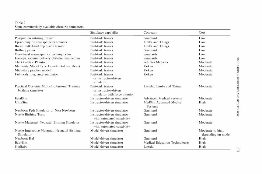
Table 2
Some commercially available obstetric simulators
Simulator capability Company Cost
Postpartum suturing trainer Part-task trainer Gaumard Low
Episiotomy or anal sphincter trainers Part-task trainer Limbs and Things Low
Breast milk hand expression trainer Part-task trainer Limbs and Things Low
Birthing pelvis Part-task trainer Gaumard Low
Obstetrical mannequin or birthing pelvis Part-task trainer Simulaids Low
Forceps, vacuum delivery obstetric mannequin Part-task trainer Simulaids Low
The Obstetric Phantom Part-task trainer Schultes Medacta Moderate
Maternity Model Type 1 (with fetal heartbeat) Part-task trainer Koken Moderate
Midwifery practice model Part-task trainer Koken Moderate
Full-body pregnancy simulator Part-task trainer
or instructor-driven
simulator
Koken Moderate
Practical Obstetric Multi-Professional Training
birthing simulator
Part-task trainer
or instructor-driven
simulator with force monitor
Laerdal; Limbs and Things Moderate
FetalSim Instructor-driven simulator Advanced Medical Systems Moderate
UltraSim Instructor-driven simulator MedSim Advanced Medical
Systems
High
Newborn Pedi Simulator or Nita Newborn Instructor-driven simulator Gaumard Moderate
Noelle Birthing Torso Instructor-driven simulator
with automated capability
Gaumard Moderate
Noelle Maternal, Neonatal Birthing Simulator Instructor-driven simulator
with automated capability
Gaumard Moderate
Noelle Interactive Maternal, Neonatal Birthing
Simulator
Model-driven simulator Gaumard Moderate to high,
depending on model
Newborn Hal Model-driven simulator Gaumard High
BabySim Model-driven simulator Medical Education Technologies High
SimBaby Model-driven simulator Laerdal High 105
SIM
ULATIO
NIN
OBSTETRIC
SAND
GYNECOLOGY

Fig. 4. Example of a hybrid technique that pairs a low-fidelity obstetric simulator with an actor
to simulate shoulder dystocia. (Courtesy of Brigham and Women’s Hospital, Boston, MA; with
permission.)
106 GARDNER & RAEMER
station [55]; conducting breech birth [56]; managing shoulder dystocia [57–59]; managing obstetric emergencies and trauma [60–63]; managing the ob-stetric airway [64,65]; performing intubations [64,65]; and inserting epiduralcatheters [66]. Since 2004, obstetric simulation–based research has been in-creasingly used to address issues related to teamwork [61–63], team perfor-mance [67–69], the identification of clinical errors [70–73], the reduction ofclinical risks [74,75], and the improvement of clinical outcomes [76–78].
Shoulder dystocia
A number of articles have been published in the obstetric literatureinvolving simulation related to research about, training for, and managementof shoulder dystocia. The earliest research in this area was led byGonik, Allenand colleagues, [79–81] in the late 1980s and early 1990s. To study the appliedpressure or force on the brachial plexus, Gonik and colleagues [82] in 2003used a ‘‘computer software crash dummy’’ modified with a female pelvisand a mannequin fetus and outfitted with a spring device to represent the bra-chial plexus. They discerned that stretch on the brachial plexus varied withthe degree of force applied, the position of the pelvis, and the position ofthe fetal head within the pelvis. They also found that the McRobert’s maneu-ver reduced stretch of the brachial plexus. Deering and colleagues [58] in 2004reported on the positive impact of using simulation for teaching residents themaneuvers for managing shoulder dystocia and for promoting best practice

107SIMULATION IN OBSTETRICS AND GYNECOLOGY
for residents in medical record documentation of such clinical events [83].Kim and colleagues [84] and Gurewitsch and associates [85], with Allenand colleagues, created a biofidelic maternal birthing simulator they havesince used in research involving various aspects of shoulder dystocia. In2005, they compared the force applied on the brachial plexus during McRo-bert’s maneuver with that of the Rubin’s maneuver and found less force wasgenerated with Rubin’s maneuver [85]. In 2007, Allen and colleagues [86] dis-cerned greater stretch on the posterior brachial plexus was generated duringsecond stage of a simulated routine vaginal delivery compared with one com-plicated by shoulder dystocia, but before the application of clinician-appliedtraction. They concluded that even though the fetal posterior brachial plexusmay stretch as it traverses the pelvis during the second stage of labor, clini-cians should aim to minimize applied traction, especially lateral traction, inall deliveries to reduce the risk of brachial plexus injury. Crofts and colleagues[87] in 2005 discussed the use of a new birthing simulator they helped developfor training in shoulder dystocia. After the training program, none of thetrainees applied greater than 100 N of traction, a degree of force beyondwhich is associated with fetal injury. In 2006, they presented results of a ran-domized controlled trial of simulation-based training in Bristol, UnitedKing-dom, involving shoulder dystocia scenarios with the Practical ObstetricMulti-Professional Training (PROMPT) birthing simulator (Fig. 5) [57].They compared training with a high-fidelity mannequin with force monitor-ing to training with a low-fidelity mannequin and found that all training im-proved performance of basic maneuvers (P ¼ .002), the achievement of
Fig. 5. The PROMPT birthing simulator. (Courtesy of Limbs and Things, Bristol, United
Kingdom; with permission.)

108 GARDNER & RAEMER
successful deliveries (P!.001), and communications with the patient(P!.001). High-fidelity simulation with force monitoring led to more suc-cessful deliveries (P ¼ .002), lower applied force (P ¼ .006), and shorterhead-to-body delivery (P ¼ .004). This study underscores the importanceof training for managing shoulder dystocia and demonstrates how force mon-itoring in simulated vaginal births heightens clinician awareness of what theycan potentially generate in the process of managing birth complicated byshoulder dystocia. In 2007, Crofts and colleagues [88] described the use ofPROMPT in a standardized shoulder dystocia scenario to assess the force ap-plied by obstetricians andmidwives to the fetal neck. They found awide rangeof variation in the pattern and degree of applied force, ranging from 6 N tomore than 250 N, and over two thirds of study participants exceeded 100N, an amount of force considered excessive. While the force applied duringsimulated shoulder dystocia may not exactly represent what occurs in realcases, this study reiterates the value of educating clinicians about the degreeof force they are capable of generating, and reinforcing the importance of ac-curately using maneuvers to successfully achieve a safe delivery. In a separatestudy using the PROMPTmannequin, Crofts and colleagues [89] assessed re-tention of skills at 6 and 12 months after obstetric providers attended a struc-tured training program on shoulder dystocia. They found a high percentage(O80%) of participants, including those who had failed to successfullydeliver the mannequin baby before training, were able to successfully deliverthe mannequin baby at 6 months and 12 months after training. This studysuggests that annual training is likely appropriate for those who demon-strated proficiency before training. For others, more frequent trainingsessions may be warranted to reinforce such skills.
Fetal station and the use of forceps
In 2005, Dupuis and colleagues [55] conducted a prospective, randomizedtrial for assessing reliability in determining fetal station. They constructeda laboratory birthing simulator consisting of a female pelvis and amannequinfetus with an anatomically correct fetal skull. They then compared the assess-ment of fetal station, ranging from �5 to þ5, and engagement conducted byresidents and attending physicians. They found that 88% of residents and67% of attendings misdiagnosed ‘‘high’’ fetal station, and about 12% ofboth groups incorrectly classified engagement of the fetal head. In view ofthese findings, Dupuis and colleagues advocated simulator-based trainingas a way to improve skills for determining fetal station and engagement. In2006, Dupuis and colleagues [53] combined computer screen–based or virtualcapabilities with a birthing pelvis and equipped forceps with spatial locationsensors to teach and assess forceps application. These sensors made it possibleto monitor forceps blade trajectory in a simulated operative vaginal delivery,and to compare forceps application by attendings and residents. Forcepsblade trajectory was excellent, very good, or good in 92% of cases involving

109SIMULATION IN OBSTETRICS AND GYNECOLOGY
senior obstetricians and in 38% of cases involving junior obstetricians(P!.001). Dupuis and colleagues concluded that simulation provides a safeway to acquire and practice skills in forceps application before trying it onreal patients, and can be used to certify skills in the use of forceps. Moreauand colleagues [90], including Dupuis, are now using forceps trajectory pat-terns created by experienced obstetricians as templates for training residents.
Virtual reality and haptic simulation
Applications of VR and haptic simulation to the field of obstetrics are fewbut have increased over the past decade. Three articles published in the 1990sand three since 2002 focused on VR simulation in obstetrics [91–96]. In 2002,Letterie [94] assessed the use of VR in a variety of non–health care and healthcare industries, exploring potential applications in obstetrics and gynecology.He concluded that VR environments could assist residents and medicalstudents in surgical skills training and in developing better conversing skillswith patients. In 2004, Obst and colleagues [95] created a virtual obstetricenvironment with feedback mechanisms embedded in the simulator to assistlearners in acquiring skills for managing normal and complicated deliveries.In 2005, Lapeer [96] of the United Kingdom assessed the feasibility of usingVR technology to create a mechanical model augmented by haptic feedbackfor simulating forceps delivery. He demonstrated that such a device couldfacilitate skill acquisition and performance of forceps application. His find-ings align well with those of Dupuis’ forceps-related research in France.
Structured simulation-based training programs
Several multidisciplinary obstetric skills training programs have beenestablished in the United Kingdom, the United States, and Canada. Theseprograms include Managing Obstetric Emergencies and Trauma [62] andMultidisciplinary Obstetric Simulated Emergency Scenarios (MOSES) [68]of the United Kingdom; the Advanced Life Support in Obstetrics [97–99]of the American Academy of Family Physicians in the United States; andAdvances in Labor and Risk Management [100] and Managing ObstetricalRisk Efficiently [101] of the Society of Obstetricians and Gynecologists ofCanada. These procedural skills and team training courses have been offeredin some cases for over a decade and have generally been well received byclinicians in their respective areas.
Leaders in obstetrics and gynecology and simulation researchers withinthe military medical corps have long been proponents of simulation-basedtraining in obstetrics and gynecology. Macedonia and colleagues [20]described the integral role that medical simulation has in the training andpractice of obstetrics and gynecology, highlighting aspects of their obstetricskills simulation curriculum at the National Capital Area Medical Simula-tion Center of the Uniformed Services University of the Health Sciencesin Bethesda, Maryland. More recently, members of the Madigan Army

110 GARDNER & RAEMER
Medical Center announced the development of a mobile obstetrics simula-tor, Simulator for High Acuity Deliveries, to facilitate training for managingobstetric emergencies [102]. Plans are in place to deploy these units to mili-tary treatment facilities to help obstetric clinicians maintain and updateclinical skills for managing high-acuity, low-frequency perinatal events.
Reducing risk
Several articles published since 2005 target the use of obstetric simulationfor identifying clinical error, reducing clinical risk, and improving clinicaloutcomes. Cioffi and colleagues [77] conducted a pilot study using simulatedscenarios for teaching clinical decision-making to midwives. The studyshowed a positive effect of this approach on clinical decision making in sim-ulated clinical settings. The investigators noted that translating this effect intothe real world setting was inconclusive. Draycott and colleagues [78] assem-bled a retrospective cohort of births between 1998 and 2003, and investigatedwhether simulation-based training in Bristol, UnitedKingdom,made a differ-ence in perinatal outcomes after clinicians attended a day-long simulation-based training session for managing obstetric emergencies. They comparedpretraining (1998–1999) to posttraining (2001–2003) outcomes for singleton,cephalic term births at tertiary care and teaching hospitals. They found that 5-minute Apgar scores of less than six decreased from 86.6 to 44.6 per 10,000births (P!.001) and hypoxic-ischemic encephalopathy decreased froma rate of 27.3 to 13.6 per 10,000 births (P ¼ .032). Theirs is the first studywhereby an obstetric simulation-based educational program has been associ-ated with improved perinatal outcomes.
Error identification and management
Simulation can assist in identifying recurrent pitfalls in managing obstet-ric emergencies. Maslovitz and colleagues [72] in 2007 described usingsimulation to identify mistakes in obstetric management. They observedteam performance of residents and midwives during simulated obstetricemergencies, such as eclampsia, hemorrhage, shoulder dystocia, and breechbirth. The most common errors involved delay in transport to the operatingroom (82%), lack of familiarity with medications for treating obstetrichemorrhage (82%), poor techniques in using cardiopulmonary resuscitation(80%), and inadequate documentation of shoulder dystocia (80%). Theyacknowledged that although simulation is useful for training, the transferof skills acquired in simulated emergencies to managing real clinical eventsis uncertain and remains an important area of research.
Teamwork, team performance
Simulation facilitates multidisciplinary team training and improves teamperformance in obstetric emergencies and trauma as demonstrated by Freeth

111SIMULATION IN OBSTETRICS AND GYNECOLOGY
and colleagues [68] in 2006. MOSES, launched in the United Kingdom asa day-long program, aimed at improving multidisciplinary team performancevia lectures, workshops, and skills training sessions, concluding with a post-course evaluation. While not yet proven, this program is expected to reduceby 25% the occurrence of harmful adverse events in obstetrics and gyneco-logy that result in litigation.
Tools for evaluating team performance in simulated obstetric events arethe subjects of much research. Scavone and colleagues [103] in 2006 devel-oped and piloted a scoring system for assessing the performance of anesthesiaresidents during emergency cesarean delivery. They found the scoring instru-ment useful and the simulator contextually valid and reliable. Morgan andcolleagues [70] in 2007 investigated tools for evaluating performance ofmultidisciplinary obstetric teams during simulated obstetric emergencies.They concluded that obstetric-domain–specific behavioral markers andassessment tools should be developed instead of using or modifying existingtools, such as the Human Factors Rating Scale and the Global Rating Scale.
Several investigators have recently explored the question of whethersimulation offers advantages over a traditional didactic approach. Jude andcolleagues [69] in 2006 compared third-year medical students who receivedsimulator-based training in vaginal delivery to those who received traditionalinstruction. They found that students with simulated experiences expressedgreater confidence in their own abilities to assist or attempt vaginal deliveryin real clinical settings. Ohlinger and colleagues [104] reported that videosimulation was a useful methodology for teaching effective communicationand improving teamwork among perinatal care providers during deliveries.Birch and colleagues [71] in 2007 compared lecture-based methodologywith a simulation-based approach and with a combined lecture- and simula-tion-based approach for teaching teams to manage postpartum hemorrhage.Six multidisciplinary teams, randomized to one of these three methods, alldemonstrated improved fund of knowledge and skill performance. However,teams trained with simulation demonstrated sustained improvement in clini-cal management, interdisciplinary communication, and self-confidence whentested 3 months later. Teams taught by simulation also improved their inter-disciplinary communication skills compared with those taught exclusively bylecture. Although not powered for statistical significance, this study indicatesthat simulation-based training offers advantages over traditional lecture-onlymethodology. It remains to be seen if such improvements are long-lasting andhow frequently simulation-based team-training coursework should berepeated to maintain clinical proficiency.
Simulation in gynecology
Simulation in gynecology involves the reenactment of routine or criticalgynecologic events involving women across the lifespan for procedural orbehavioral skills training, practice, evaluation, or research. As such, the full

112 GARDNER & RAEMER
spectrum of verbal role playing, standardized characters or actors, devices,mannequins, and environments can be used alone or in combination toachieve the desired educational goals and objectives of a curriculum. Thefocus here will be confined to simulation involving surgery and hospital-basedcare of women with reproductive or post-reproductive age-related gyneco-logic conditions. However, much of what will be addressed can be modifiedor adapted to reflect routine and critical events and simulation environmentstypical of the primary care or outpatient settings. Simulation targeting thefemale newborn and pediatric age groups will not be addressed, nor willsimulation involving use of animals or cadavers.
Gynecological simulators
The history of gynecology simulators dovetails with that of obstetricsas small wax or wooden figures have been used since antiquity for illustrat-ing reproductive processes, contraceptive techniques, and other gynecologicconditions that women experience [39]. A number of objects or more elab-orate part-task trainers have been developed for training in and practicingof procedures and surgical techniques or for examining the female breastand pelvis. These objects and trainers include suture trainers; trainingdevices for proper placement and positioning of barrier, subcutaneous,and intrauterine contraceptives; and devices for practicing placement ofperiurethral slings. Pelvic ExamSim (Medical Education Technologies,Sarasota, Florida) is an example of an elaborate part-task trainer equippedwith sensors and computer-based software that feeds back information tothe learner about his or her performance [105,106]. High-fidelity, physiolog-ically interactive, life-size human female mannequins are available and canbe used for simulating gynecologic surgery scenarios in a simulated orreal operating room environment. However, mannequin technology cur-rently available is inadequate for realistically simulating open laparotomyinvolving major abdominal and pelvic organs, such as a benign or radicalhysterectomy, an oophorectomy, or major vaginal surgery, such as hysterec-tomy, fistula repair, or vaginal vault suspensions. Gynecology-related videosimulation or VR, computer screen–based or haptic systems currently offergreater opportunities for such purposes [107]. Few gynecology-oriented,total immersion VR-haptic environments exist and are primarily used inresearch. Hysteroscopic and laparoscopic simulators are best classified aspart-task trainers ranging in fidelity from simple box trainers, or ‘‘physicalsimulators,’’ to hybrid mechanical-virtual or haptic systems.
History of minimally invasive surgery simulators
The acceptance and integration of laparoscopy as a credible technique forabdominal and pelvic surgery triggered growth in the number and variety ofminimally invasive gynecologic simulators. The first endoscopy may haveoccurred in Greece during the time of Hippocrates. However, not until

113SIMULATION IN OBSTETRICS AND GYNECOLOGY
1806 was an instrument created instrument that could be inserted into thebody for visualizing internal organs. This was the invention of PhillipBozzini of Germany. His idea, although never tested on humans, was ulti-mately reintroduced and accepted by physician-surgeons in the late 1800s[108]. Visualization of the stomach and urethra was first accomplished.Then visualization of the organs of the abdomen and thorax was madepossible when Kelling of Germany created the technique of pneumoperito-neum in the early 1900s [109]. The technique was adapted for use in gyne-cology in the late 1930s by Telinde and in the early 1940s by Palmershortly after introduction of the Veress needle for creating a pnuemoperito-neum. Semm [110], a gynecologist in Germany, invented an automatic insuf-flator in the 1960s, a device that the American medical community embracedfor its simplicity and safety features. Semm used the term pelviscopy todescribe his surgical procedure. The American Association of GynecologicalLaparoscopists was founded in 1971, but it was not until 1981 that theAmerican Board of Obstetrics and Gynecology mandated that laparoscopytraining be included in residency training programs. Semm [111] created thefirst laparoscopy training device in 1985 for colleagues to practice theirsurgical techniques. His ‘‘pelvi-trainer’’ had a clear cover that permittednovices to directly view their techniques. An opaque cover could be usedin place of the clear cover. A video screen was later added to the systemfor more realistic simulation of the laparoscopic procedure. Applicationof VR technology was initially proposed by Satava [112] in 1993, but wasslow to be adopted and integrated into surgical training programs. VR tech-nology is now commercially available and is an integral component ofadvanced minimally invasive surgical simulators. Gallagher and colleagues[113] defined VR as a ‘‘computer-generated representation of an environ-ment allowing sensory interaction,’’ giving an impression of realism. Theynoted in 2005 that the two most likely reasons for delayed adoption ofVR technology in surgical simulation included the lack of solid scientificproof supporting its use for skills training and the lack of knowledge ofhow best to incorporate simulation within a surgical training program.
Current use of gynecologic simulators
Minimally invasive gynecologic surgery simulators should be able todifferentiate between the experienced clinician and the novice and to discernimprovement with successive use [114,115]. Ideally, such a simulator shouldbe affordable and user-friendly as with a simple box trainer or physicalsimulator. Physical simulators are mannequin torsos or similar objects thatcan be placed on a table or platform and that can accommodate the insertionof laparoscopic instruments and the operation of such instruments to grasp ormanipulate small objects within the simulator resembling or representinginternal organs. Physical simulators permit the use of the same or similarinstruments and camera equipment employed in real operating rooms and

114 GARDNER & RAEMER
give learners the opportunity to perform surgical gestures similar to thoseused in real cases, providing realistic depth perception and tactile feedbackto the student. The original VR simulators were expensive and not equippedto provide the depth perception and tactile feedback typical of real cases.Such limitations have been addressed with newer models that benefit fromadvances in computer technology and increasing demand for safer andmore practical ways for clinicians to acquire and practice their skills withoutharming patients. However, VR simulators continue to be more costly thanphysical trainers. Examples of VR laparoscopic simulators include the Mini-mally Invasive Surgical Trainer–Virtual Reality (MIST-VR) simulator(Mentice, Gothenburg, Sweden); the LapSim virtual reality laparoscopic sim-ulator (Surgical Science, Gothenburg, Sweden) (Fig. 6); the Xitact instrumenthaptic port simulator (Gothenburg, Sweden) (Fig. 7); the Lap Mentor simu-lator (Simbionix USA, Cleveland, Ohio); and the Computer EnhancedLaparoscopic Training System (Center for Integration inMedicine and Inno-vative Technology, Boston, Massachusetts).
Skills acquisition and training
Much has been written about the use of minimally invasive surgerysimulators for skill acquisition and practice. These reports have investigatedwhether or not such simulators facilitate training and their ability to detectchange in performance. The following selection of recently published articlesin the gynecology and surgery literature illustrates the utility of laparoscopic
Fig. 6. LapSim-VR. (Courtesy of Surgical Science, Gothenburg, Sweden; with permission.)

Fig. 7. Xitact instrument haptic port. (Courtesy of Mentice, Gothenburg, Sweden; with
permission.)
115SIMULATION IN OBSTETRICS AND GYNECOLOGY
simulators for skill acquisition and training. Fichera and colleagues [115] in2002 to 2003 investigated the use of physical trainers for skills acquisition,clinical training, and differentiation of novice from veteran gynecologicand surgical laparoscopists. Using the LTS 2000, Fichera and colleaguesshowed that this device reliably detected laparoscopy expertise and changein performance over time with improved suturing and coordination scores(P!.05). Scott and colleagues [116] in 2000 compared surgical skills ofresidents using physical trainers to the skills of residents who did not usethe trainer. They found higher global assessment scores during real-timelaparoscopic cholecystectomy for the simulator-enhanced training groupcompared with those without such training. The improved global scoreswere accompanied by improved respect for tissue, skills in handling instru-ments, use of surgical assistants, and overall performance. These findingsdemonstrated that physical trainers are a viable alternative to VRsimulators.
VR simulators have been scrutinized in a similar fashion and their use hasalso been shown to reliably detect laparoscopy expertise and change inperformance over time.
Seymour and colleagues [117] in 2002 used a randomized, double-blindcontrolled trial methodology to evaluate VR simulator–based training.They found that such training improved performance in the operatingroom. Additional studies have used the MIST-VR system, a skills-orientedtrainer without haptic feedback that requires the student to perform sixtasks: (1) acquire and grasp, (2) transfer and place, (3) traverse a segment,(4) withdraw and insert, (5) perform a diathermy, and (6) perform

116 GARDNER & RAEMER
a diathermy and manipulate. Gallagher and colleagues [118] and Grantch-arov and colleagues [119] found that prior laparoscopic experience washighly correlated with technical skills using the MIST-VR system. Munzand colleagues [120] in 2004 investigated whether laparoscopic VR trainerswere superior to box trainers and found no significant differences in laparo-scopic skills acquired between groups of novices who trained with either ofthese simulators. Grantcharov and colleagues [121] also investigatedwhether or not skills acquired in the laparoscopic simulator would transferto the real surgical arena. They randomized surgical residents to usual train-ing or usual training enhanced by MIST-VR training. Evaluated with globalrating forms, surgical residents who trained with the simulator demon-strated shorter operating times and more efficient surgical gestures duringreal laparoscopic cholecystectomy compared with those who had not.
Hart and Karthigasu [122] in 2007 reviewed the use of VR simulators forlaparoscopic surgery and noted that the MIST-VR is the most widely used,studied, and validated simulator system for general surgery training in theUnited States. The MIST-VR is also the simulator most demonstrated tobe of value for the education and training of gynecologists. However,LapSim remains a useful system because it provides more realistic simula-tions. For example, it can simulate bleeding organs that deform and changeas the procedure evolves. Aggarwal and colleagues [123] reported in 2006 onthe use of the LapSim for training technical skills in managing ectopic preg-nancy. Using LapSim in successive sessions, they compared experienced andnovice gynecologists in their performance of tasks involved in such surgery.They found that novices demonstrated significant improvement in theirsurgical gestures, whereas experienced gynecologists demonstrated littlechange over time. LapSim appears to be useful for facilitating skill acquisi-tion for novice surgeons who plan to perform laparoscopic surgery forectopic pregnancy. Hamilton and colleagues [124] found that surgicaltrainees considered the box trainer more realistic because it provided bettertactile feedback and depth perception compared with other simulators.Similar findings were noted more recently by Madan and colleagues [125]in 2005. Laparoscopic simulators, either box trainers or VR trainers, facili-tate skill acquisition and training, especially for the novice, and such train-ing is translatable to the operating room.
Assessment
Assessment of skill performance and competence is possible with the boxtrainer and the VR laparoscopic system, each having its own advantagesand limitations. Chou and Handa [126] appreciate a more promising rolefor VR systems. Objective data can be recorded by the software and lateranalyzed for such factors as accuracy of task performance and completiontimes, efficiency of surgical gestures, and handling of thermal-generatingdevices. Feedback is provided in an unbiased manner and reports can be

117SIMULATION IN OBSTETRICS AND GYNECOLOGY
generated showing change over time. Gor and colleagues [127] evaluated theuse of a VR system (MIST-2) and found it useful as an objective measure oflaparoscopic skills demonstrated by gynecologic surgeons. By comparison,box trainers require the presence of an instructor to observe performanceand provide feedback, which can be biased and unreliable. Structuredassessment programs have been developed to standardize the process andminimize observer bias. One such program is the McGill Inanimate Systemfor Training and Evaluation of Laparoscopic Skills (MISTELS) [128].MISTELS requires users to perform a series of five tasks. These tasks arescored using an objective system that has met reliability criteria for high-stakes testing. However, the box simulator and the VR systems can beused to objectively assess competency and proficiency in task performance,making it possible to discern skills of novices from those of veterans and toidentify improvement in skill acquisition over time. These simulators mayassist trainees in making career choices, especially those trainees unable todemonstrate proficiency in basic surgical skills [118], and in practicingnew skills or new procedures before trying them for the first time on livepatients [122].
Credentialing
Simulation is being used in various accreditation programs around theworld. The Israel Center for Medical Simulation is at the cutting edge inthe use of simulation for summative evaluation and accreditation programs,including the medical school selection process, national board examination inanesthesiology, and national accreditation for paramedics [129]. Ziv andcolleagues describe how prospective candidates for medical school must com-plete various questionnaires and behavioral assessments, and participate inobserved structured clinical examination–like (OSCE-like) stations thatinclude simulation of patient encounters with role-playing and standardizedpatients. The experience with these endeavors thus far has been positive.However, validity of this approach will be assessed and monitored as medicalstudents selected progress through their training. The Israeli board examina-tion in anesthesiology lacked a performance evaluation component untilabout 4 years ago. Capitalizing on the experience of the Fellow Royal Collegeof Anesthesiology in the United Kingdom, the board examination committeejoined with a panel of experts in testing and evaluating to create a series ofsimulation-based OSCE-like stations representing core problems that anes-thesiologists encounter in the course of clinical practice. Subjective feedbackwith the process and satisfaction with the realism of the scenarios thus far hasbeen favorable. Themean interrater correlations of examiners were high (0.89to 0.76), the rate of incongruence was low (!15%), and the correlations forintercase reliability were significant (P!.01).
The Accreditation Council for Graduate Medical Education supportsrigorous competency assessment of residents in a number of areas, including

118 GARDNER & RAEMER
those related to interpersonal and communication skills, professionalism, andsystems-based practice [130,131]. With this in mind, Julian and Rogers [132]recommend changes in the way gynecologic surgeons are trained. They pro-pose a model guided by evidence-based educational studies and evidence-based clinical reports. They further propose standardizing the measurementof surgical teaching outcomes and surgical education curricula. They arguethat students should practice basic surgical skills before assisting in surgeryon live patients. They thereby support the use of simulation. They statethat ‘‘the acquisition of core surgical knowledge, judgment, leadership qual-ities, and skills before the resident participates in live surgeries is the keystonein fulfilling the mandate to improve the ethics and effectiveness of traininggynecologic surgeons.’’
The future of simulation in obstetrics and gynecology
The specialty of obstetrics and gynecology is takingmeasured steps towardseamlessly integrating simulation within the fabric of education, training, andassessment of obstetrician-gynecologists. The experience chronicled aboveillustrates the great progress made thus far in appreciating the value of simu-lation in such endeavors. Driving these efforts aremultiple factors bothwithinand outside of the profession. These factors include restrictions on workhours of residents [133]; reductions in the medical work force coupled withincreasing demand among health care workers to balance work with lifestylepreferences and family obligations [134–136]; rising malpractice premiums,threats of litigation, and payouts by juries ruling against defendants[137,138]; and diminished clinical opportunities for trainees when patientsrefuse to permit their involvement [139–141]. Specialty and subspecialtyexamining boards are establishing mechanisms for assessing task-orientedand behavior-based competencies for professional certification, validation,and re-entry. Professional organizations are considering or requiring simula-tion-based experiences for credentialing and recredentialing [130,142–145].The need for competency assessment has triggered development and valida-tion of task-oriented and behavior-based tools that discern proficiency inclinical endeavors [70,103,104], and these efforts will intensify. In some cases,professional certification and hospital credentialing programs now requirecore competency assessment of procedural and behavioral skills, includingskills that demonstrate teamwork and professionalism [130,144,145]. Sincethe Institute of Medicine’s report on human error in 2000 [146], pressurehas been growing to reduce adverse events and improve the safety and qualityof patient care. Steps being taken toward this end include the implementationof requirements or strong recommendations to conduct obstetric emergencydrills and skills training [60,147–150]. There are sporadic reports of medicalprofessional liability insurers who now offer insurance premium discountsfor participation in obstetric simulation-based and didactic team trainingprograms [151–153]. It is unclear if this ‘‘carrot’’ approach toward facilitating

119SIMULATION IN OBSTETRICS AND GYNECOLOGY
patient safety and mitigating medical error in the obstetric arena will beimplemented by other medical professional liability insurers. Similar effortshave been made in providing an insurance premium incentive to clinicianswho successfully complete training programs in fundamentals of laparoscopicsurgery [154]. There are also reports of using simulation for remediation. Forexample, the New York State Department of Health’s Office of ProfessionalMedical Conduct recently used simulation as a key component in their effortsto remediate anesthesiologists [155]. If deemed successful, this approach maybe more widely adopted across the medical specialties, including obstetricsand gynecology.
Challenges to seamless integration of simulation into professional trainingof obstetrician-gynecologists include such factors as the high costs andlimited quality of currently available simulators, limits on time and spaceavailable for such purposes, and lack of sufficient personnel skilled in theiruse. Trainees grapple with the caliber of fidelity and realism of current man-nequin technology. Mannequins that look and feel more humanlike, equip-ped with realistic organs and tissue layers that bleed, would enhance theimmersive quality of simulated scenarios. Research is needed to determinesuitable environments that best meet the educational needs of trainees, iden-tifying essential characteristics of simulators that facilitate acquisition, train-ing and competency assessment of procedural and behavioral skills. Assimulation technology and its use evolves, there will soon be a day when(1) the methods and techniques necessary to facilitate learning are well under-stood, (2) all members of the team will be trained in the safest way possible tomanage all manner of obstetric and gynecologic events, and (3) all traineeswill demonstrate procedural and behavioral proficiency in a simulator beforebeing permitted to treat human beings, regardless of specialty or level ofexperience. The day is coming when simulation-based activities will berequired for practice, and no longer will a novice try a procedure for the firsttime on a real patient without having performed it in simulation.
Summary
The technique of simulation for education and training in obstetrics andgynecology is not new. A brief review of the history confirms that this tech-nique has solid roots deeply planted within the field of obstetrics and gyne-cology. However, until recently, technological limitations and other factorsinhibited the use of simulation. Now, an appreciation for the potential valueof simulation in health care education, training, and research is emergingand more applications are appearing. The days of relying on the apprentice-ship style of learning in obstetrics and gynecology have passed. Simulationoffers clinicians a safe, practical, and credible means to acquire skills andlearn how to optimally manage clinical scenarios from the routine to themost uncommon or unusual events in contextually relevant settings. Theeffectiveness of simulation in reducing adverse outcomes is a matter of

120 GARDNER & RAEMER
debate and the focus of much research within the simulation community.Dutta and colleagues [156] assert that despite the lack of incontrovertibleproof that simulation directly reduces adverse outcomes, ‘‘.we must recog-nize that no hazardous industry has anything remotely approaching level 1A‘evidence’ to support their practices .’’ Securing such proof may be as elu-sive in health care as it has been in aviation. The medical simulation com-munity should instead focus on defining the key attributes of simulationenvironments across the spectrum of health care specialties that will bestserve the needs for education and training [3]. The published literature in ob-stetrics and gynecology thus far supports simulation for practicing routineand uncommon but critical procedures and events, for improving technicalproficiency, and for building self-confidence and teamwork among clini-cians. As the science of simulation evolves in obstetrics and gynecology,adherence to sound educational objectives will best guide its developmentand inform the extent to which realism and fidelity of a specific simulatedclinical experience is necessary. VR environments in obstetrics and gynecol-ogy are ripe for research and development [53,54,84,112,114], offering thegreatest opportunity for training in the most realistic settings possible with-out harming real patients. Most VR simulators currently focus on surgicalspecialty skills and procedures. However, Chou and Handa [126] admonishthat gynecologic surgery is not simply general surgery of the pelvis. VRproduct design must be mindful of the unique tasks specific to gynecology,and so too with obstetrics. Whether virtual or not, simulated obstetric andgynecologic environments should be designed to address the unique tasksspecific to care of women across the lifespan so that, in the words of SirRichard Manningham, ‘‘.where every Case that can happen may be repre-sented and repeated as often as we deem necessary, you will have the great-est opportunity of forming your Hands for Practice’’ [39]. Effectivelyforming one’s knowledge, skills, and abilities for the practice of obstetricsand gynecology is a matter of safety and quality; indeed it is our ethicalimperative.
References
[1] Good M, Gravenstein J. Anesthesia simulators and training devices. Int Anesthesiol Clin
1989;27:161–6.
[2] Cooper J, Taqueti VR. A brief history of the development of mannequin simulators for
clinical education and training. Qual Saf Health Care 2004;13:i11–8.
[3] Gaba DM. The future vision of simulation in healthcare. Qual Saf Health Care 2004;13:
2–10.
[4] Gardner R. Simulation and simulator technology in obstetrics: past, present and future.
Expert Review in Obstetrics & Gynecology 2007;2(6):775–90.
[5] Cumin D, Merry AF. Simulators for use in anesthesia. Anaesthesia 2007;62:151–62.
[6] Gaba DM. Simulators in anesthesiology. In: Lake C, editor. Advances in Anesthesia.
vol. 14. Mosby–Year Book, Inc.; 1997. p. 55–94.

121SIMULATION IN OBSTETRICS AND GYNECOLOGY
[7] Denson JS, Abrahamson S. A computer controlled patient simulator. JAMA 1969;208(3):
504–8.
[8] GabaDM,DeAndaA.A comprehensive anesthesia simulator environment: re-creating the
operating room for research and training. Anesthesiology 1988;69:387–94.
[9] Schwid H. A flight simulator for general anesthesia training. Comput Biomed Res 1987;20:
64–75.
[10] Howard SK, Gaba DM, Fish KJ, et al. Anesthesia crisis resource management: teaching
anesthesiologists to handle critical incidents. Aviat Space Environ Med 1992;63(9):
763–70.
[11] Helmreich RL, Merritt AC. Safety and error management: the role of crew resource
management. In: Hayward BJ, Lowe AR, editors. Aviation resource management. Alder-
shot (UK): Ashgate; 2000. p. 107–19.
[12] Helmreich RL, Merritt AC, Wilhelm JA. The evolution of crew resource management
training in commercial aviation. Int J Aviat Psychol 1999;9(1):19–32.
[13] Gaba DM, Fish KJ, Howard SK. Crisis management in anesthesiology. New York:
Churchill Livingstone Inc.; 1994. p. 5–45.
[14] Small SD,WuerzRC, SimonR, et al. Demonstration of high fidelity simulation team train-
ing for emergency medicine. Acad Emerg Med 1999;6(4):312–23.
[15] ShapiroMJ,Morey JC, Small SD, et al. Simulation based teamwork training for emergency
department staff: Does it improve clinical team performance when added to an existing
didactic teamwork curriculum? Qual Saf Health Care 2004;13:417–21.
[16] Hegarty M, Bloch M. The use of simulators in intensive care training. Curr Anaesth Crit
Care 2002;13:194–200.
[17] Lighthall GK, Barr J, Howard SK, et al. Use of a fully simulated intensive care unit envi-
ronment for critical event management training for internal medicine residents. Crit Care
Med 2003;31:2437–43.
[18] Halamek LP, Kaegi DM, Gaba DM, et al. Time for a new paradigm in pediatric medical
education: teaching neonatal resuscitation in a simulated delivery room environment.
Pediatrics 2000;106:e45.
[19] KnoxGE, SimpsonKR, Garite TJ. High reliability perinatal units: an approach to the pre-
vention of patient injury andmedicalmalpractice claims. JHealthcRiskManag 1999;19(2):
24–32.
[20] Macedonia CR,GhermanRB, Satin AJ. Simulation laboratories in obstetrics and gynecol-
ogy. Obstet Gynecol 2003;102(2):388–92.
[21] LakeCL. Simulation in cardiology and cardiothoracic and vascular surgery. SeminCardio-
thorac Vasc Anesth 2005;9(4):325–33.
[22] Beyea S, Kobokovich LJ. Human patient simulation: a teaching strategy. AORN J 2004;
80(4):738–42.
[23] Henneman EA, Cunningham H, Roche JP, et al. Human patient simulation: teaching
students to provide safe care. Nurse Educ 2007;32(5):212–7.
[24] Gordon JA,WilkersonW.The humanpatient simulator: student and educator responses to
a new medical educational technique. Ann Emerg Med 2000;36(4):S79.
[25] BlumRH, RaemerDB, Carroll JS, et al. Crisis resource management training for an anaes-
thesia faculty: a new approach to continuing education. Med Educ 2004;38(1):45–55.
[26] Rudolph JW, SimonRE,RaemerDB.Which realitymatters? Questions on the path to high
engagement in healthcare simulation. Simulation in Healthcare 2007;2(3):161–3.
[27] Beaubien JM, Baker DP. The use of simulation for training teamwork skills in healthcare:
How low can you go? Qual Saf Health Care 2004;13:i51–6.
[28] Dieckmann P, Manser T, Wehner T. Presence and high fidelity patient simulators in anes-
thesia: influence derived from interviews and questionnaires. Presented at The 6th Interna-
tional Workshop on Presence. Aalborg, Denmark, October 6–8, 2003.
[29] Dieckmann P, Gaba DM, Rall M. Deepening the theroretical foundations of patient
simulation as social practice. Simulation in Healthcare 2007;2:183–93.

122 GARDNER & RAEMER
[30] Salas E, Bowers CA, Rhodenizer L. Its not howmuch you have but how you use it: toward
a rational use of simulation to support aviation training. Int J Aviat Psychol 1998;8:
197–208.
[31] Center for Medical Simulation, History. Available at: http://harvardmedsim.org/cms/
timeline.html. Accessed December 8, 2007.
[32] The Peter M. Winter Institute for Simulation Education and Research (WISER) of the
University of Pittsburgh Medical Center. Available at: http://www.wiser.pitt.edu/default.
asp. Accessed December 8, 2007.
[33] University of Rochester Center for Medical Simulation (URCMS). Evolution and
revolution. Rochester (NY). Available at: http://web.anes.rochester.edu/Public/content/
Education-simcenter.php. Accessed December 8, 2007.
[34] VA PA Simulation Center, History. Available at: http://med.stanford.edu/VAsimulator/.
Accessed December 8, 2007.
[35] Canadian Simulation Center for Human Performance and Crisis Management Training of
the Sunnybrook Health Sciences Center, Toronto, Ontario (Canada). Available at: http://
www.sunnybrook.ca/departments/anesthesia/anaesthesiasimulator. AccessedDecember 8,
2007.
[36] Swiss Center for Medical Simulation of University Hospital, Basel Switzerland. Available
at: http://www.simulation.ch/SimulationBasel/Home/. Accessed December 8, 2007.
[37] Danish Institute for Medical Simulation, Herlev University Hospital, Copenhagen,
Denmark. Available at: http://www.herlevsimulator.dk/. Accessed December 8, 2007.
[38] BelgiumAnesthesia SimulationCenter, UCLMedical School-Saint LucHospital, Brussels,
Belgium. Available at: http://www.saintluc.be. Accessed December 8, 2007.
[39] Cody LF. Breeding Scottish obstetrics in Dr. Smellie’s London. In: Birthing a nation: sex,
science and the conception of eighteenth century Britons. New York: Oxford University
Press; 2005. p. 152–97.
[40] Buck GH. Development of simulators in medical education. Gesnerus 1991;48:7–28.
[41] Wilson A. A new synthesis: William Smellie. In: The making of man-midwifery: childbirth
in England 1660–1770. London: University College London Press; 1995. p. 123–33.
[42] Gelbart NR. The King’s midwife: a history and mystery of Madame du Coudray. Los
Angeles (CA): University of California Press; 1998.
[43] Schultes medacta GmbH & Co. Lehrmodelle KG. Available at: http://www.schultes-
medacta.de/index.html. Accessed December 8, 2007.
[44] DougalD. The teaching of practical obstetrics. Journal of Obstetrics andGynecology 1933;
40:99–105.
[45] Wakerlin RC, Whitacre FE. Transparent manikin for the teaching of obstetrics. Am J
Obstet Gynecol 1952;64:1378–81.
[46] Eloesser L. A simplified obstetrical phantom. Am J Obstet Gynecol 1954;68(3):948–9.
[47] The University of Kentucky Research Foundation, Lexington, KY. (Inventors: Knapp CF
and Eades GS of Lexington, KY.) US 3822486 (1974).
[48] Gaumard Scientific Co., Inc. Miami, FL. US 5853292 (1998. Inventors: Eggert JS (Miami,
FL), Eggert MS (Birmingham, MD) and Vallejo P (Miami, FL).
[49] Burd LI, Motew M, Bieniarz J. A teaching simulator for fetal scalp sampling. Obstet
Gynecol 1972;39(3):418–20.
[50] Tuffnell DJ, Bryce F, Johnson N, et al. Simulation of cervical change in labour: reproduc-
ibility of expert assessment. Lancet 1989;2(8671):1089–90.
[51] Maher JE, Kleinman GE, Lile W, et al. The construction and utility of an amniocentesis
trainer. Am J Obstet Gynecol 1998;179(5):1225–7.
[52] Smith JF, BergmannM,GildersleeveR, et al. A simplemodel for learning stereotactic skills
in ultrasound-guided amniocentesis. Obstet Gynecol 1998;92:303–5.
[53] Dupuis O, Moreau R, Silveira R, et al. A new obstetric forceps for the training of junior
doctors: a comparison of the spatial dispersion of forceps blade trajectories between junior
and senior obstetricians. Am J Obstet Gynecol 2006;194(6):1524–31 [Epub].
123SIMULATION IN OBSTETRICS AND GYNECOLOGY
[54] Eggertsen SC.Workshop for teaching fundamentals of obstetric forceps. J FamPract 1989;
28(3):313–4.
[55] Dupuis O, Silveira R, Zentner A, et al. Birth simulator: reliability of transvaginal assess-
ment of fetal head station as defined by the American College of Obstetricians and
Gynecologists classification. Am J Obstet Gynecol 2005;192(3):868–74.
[56] Deering S, Brown J, Hodor J, et al. Simulation training and resident performance of single-
ton vaginal breech delivery. Obstet Gynecol 2006;107(1):86–9.
[57] Crofts JF, Bartlett C, Ellis D, et al. Training for shoulder dystocia: a trial of simulation
using low-fidelity and high-fidelity mannequins. Obstet Gynecol 2006;108(6):1477–85.
[58] Deering S, Poggi S,Macedonia C, et al. Improving resident competency in themanagement
of shoulder dystocia with simulation training. Obstet Gynecol 2004;103(6):1224–8.
[59] Kim EJ, Allen RH, Yang JH, et al. Simulating complicated human birth for research and
training. Conf Proc IEEE Eng Med Biol Soc 2004;4:2762–6.
[60] American College of Obstetricians and Gynecologists. Post partum hemorrhage. ACOG
Practice Bulletin No. 76. Obstet Gynecol 2006;108:1039–47.
[61] Anderson ER, Black R, Brocklehurst P. Acute obstetric emergency drill in England and
Wales: a survey of practice. BJOG 2005;112(3):372–5.
[62] Johanson RB, Menon V, Burns E, et al. Managing obstetric emergencies and trauma
(MOET) structured skills training in Armenia, utilising models and reality based scenarios.
BMC Med Educ 2002;2(5).
[63] Johanson R, Akhtar S, Edwards C, et al. MOET: Bangladeshdan initial experience.
J Obstet Gynaecol Res 2002;28(4):217–23.
[64] Rahman K, Jenkins JG. Failed tracheal intubation in obstetrics: no more frequent but still
managed badly. Anaesthesia 2005;60(2):168–71.
[65] Goodwin MW, French GW. Simulation as a training and assessment tool in the manage-
ment of failed intubation in obstetrics. Int J Obstet Anesth 2001;10(4):273–7.
[66] Langton JA, Meiklejohn BH. Pressure generated during insertion of lumbar epidurals. A
comparison with the Portex epidural injection simulator. Anaesthesia 1990;45(12):1055–6.
[67] Jyothi NK, Cox C, Johanson R. Management of obstetric emergencies and trauma
(MOET): regional questionnaire survey of obstetric practice among career obstetricians
in the United Kingdom. J Obstet Gynaecol 2001;21(2):107–11.
[68] Freeth D, Ayida G, Berridge EJ, et al. MOSES: Multidisciplinary Obstetric Simulated
Emergency Scenarios. J Interprof Care 2006;20(5):552–4.
[69] Jude DC, Gilbert GG, Magrane D. Simulation training in the obstetrics and gynecology
clerkship. Am J Obstet Gynecol 2006;195(5):1489–92.
[70] Morgan PJ, Pittini R, Regehr G, et al. Evaluating teamwork in a simulated obstetric envi-
ronment. Anesthesiology 2007;106(5):907–15.
[71] Birch L, Jones N, Doyle PM, et al. Obstetric skills drills: evaluation of teaching methods.
Nurse Educ Today 2007;27(8):915–22.
[72] Maslovitz S, Barkai G, Lessing JB, et al. Recurrent obstetric management mistakes identi-
fied by simulation. Obstet Gynecol 2007;109(6):1295–300.
[73] Hart EM, Owen H. Errors and omissions in anesthesia: a pilot study using a pilot’s check-
list. Anesth Analg 2005;101(1):246–50.
[74] Sorensen SS. Emergency drills in obstetrics: reducing risk of perinatal death or permanent
injury. JONAS Healthc Law Ethics Regul 2007;9(1):9–16.
[75] Thompson S, Neal S, Clark V. Clinical risk management in obstetrics: eclampsia drills.
Qual Saf Health Care 2004;13(2):127–9.
[76] Pandey U, Russell IF, Lindow SW. How competent are obstetric and gynaecology trainees
in managing maternal cardiac arrests? J Obstet Gynaecol 2006;26(6):507–8.
[77] Cioffi J, Purcal N, Arundell F. A pilot study to investigate the effect of a simulation strategy
on the clinical decision making of midwifery students. J Nurs Educ 2005;44(3):131–4.
[78] Draycott T, Sibanda T, Owen L, et al. Does training in obstetric emergencies improve neo-
natal outcome? BJOG 2006;113:177–82.
124 GARDNER & RAEMER
[79] Gonik B, Allen R, Sorab J. Objective evaluation of the shoulder dystocia phenomenon:
effect of maternal pelvic orientation on force reduction. Obstet Gynecol 1989;74:44–8.
[80] Allen R, Sorab J, Gonik B. Risk factors for shoulder dystocia: an engineering study of cli-
nician-applied forces. Obstet Gynecol 1991;77:352–5.
[81] Allen RH, Bankoski BR, NageyDA. Simulating birth to investigate clinician-applied loads
on newborns. Med Eng Phys 1995;17(5):380–4.
[82] Gonik B, Zhang N, Grimm MJ. Prediction of brachial plexus stretching during shoulder
dystocia using a computer simulation model. Am J Obstet Gynecol 2003;189(4):
1168–72.
[83] Deering S, Poggi S, Hodor J, et al. Evaluation of residents’ delivery notes after a simulated
shoulder dystocia. Obstet Gynecol 2004;104(4):667–70.
[84] Kim EJ, Theprungsirikul P, McDonald MK, et al. A biofidelic birthing simulator. IEEE
Eng Med Biol Mag 2005;24(6):34–9.
[85] Gurewitsch ED, Kim EJ, Yang JH, et al. Comparing McRobert’s and Rubin’s maneuvers
for initial management of shoulder dystocia: an objective evaluation. Am J Obstet Gynecol
2005;192(1):153–60.
[86] Allen RH, Cha SL, Kranker LM, et al. Comparing mechanical fetal response during
descent, crowning, and restitution among deliveries with and without shoulder dystocia.
Am J Obstet Gynecol 2007;196(6):539.e1–539.e5.
[87] Crofts JF, AttilakosG, ReadM, et al. Shoulder dystocia training using a new birth training
mannequin. BJOG 2005;112(7):997–9.
[88] Crofts JF, Ellis D, JamesM, et al. Pattern and degree of forces applied during simulation of
shoulder dystocia. Am J Obstet Gynecol 2007;197(2):156.e1–6.
[89] Crofts JF, Bartlett C, Ellis D, et al. Management of shoulder dystocia: skill retention 6 and
12 months after training. Obstet Gynecol 2007;110(5):1069–74.
[90] Moreau R, Ochoa V, PhamMT, et al. Evaluation of obstetric gestures: an approach based
on the curvature of 3-d positions. Conf Proc IEEE Eng Med Biol Soc 2007;1:3634–7.
[91] Weiner EE, Gordon JS, Gilman BR. Evaluation of a labor and delivery videodisc simula-
tion. Comput Nurs 1993;11(4):191–6.
[92] Lee W. The fetal imaging workstation demonstration project. Comput Med Imaging
Graph 1996;20(6):459–66.
[93] WolkomirMS, Beecher AC. ‘‘Virtual delivery’’: an interactive role play of abnormal labor.
Acad Med 1996;71(5):550.
[94] Letterie GS. How virtual reality may enhance training in obstetrics and gynecology. Am
J Obstet Gynecol 2002;187(Suppl 3):S37–40.
[95] Obst T, BurgkartR,Ruckhaberle E, et al. The delivery simulator: a new application ofmed-
ical VR. Stud Health Technol Inform 2004;98:281–7.
[96] Lapeer RJ. A mechanical contact model for the simulation of obstetric forceps delivery in
a virtual/augmented environment. Stud Health Technol Inform 2005;111:284–9.
[97] Beasley JW, Damos JR, Roberts RG, et al. The advanced life support in obstetrics course.
A national program to enhance obstetric emergency skills and to support maternity care
practice. Arch Fam Med 1994;3(12):1037–41.
[98] Beasley JW, Dresang LT, Winslow DB, et al. The Advanced Life Support in Obstetrics
(ALSO) program: fourteen years of progress. Prehosp Disaster Med 2005;20(4):271–5.
[99] Dauphin-McKenzie N, CelestinMJ, BrownD, et al. TheAdvanced Life Support in Obstet-
rics course as an orientation tool for obstetrics and gynecology residents. Am J Obstet
Gynecol 2007;196(5):e27–8.
[100] Windrim R, Ehman W, Carson GD, et al. The ALARM course: 10 years of continuing
professional development in intrapartum care and risk management in Canada. J Obstet
Gynaecol Can 2006;28(7):600–2.
[101] De France B. TheALARMandMORE(OB) programs. J ObstetGynaecol Can 2007;29(3):
215–6.

125SIMULATION IN OBSTETRICS AND GYNECOLOGY
[102] MAMC Public Affairs Office. Mobile OB training enhances patient safety. ArmyMedicine
Military News July 2007 [news release]. Available at: http://www.armymedicine.army.mil/
news/releases/20070726mobile.cfm?m¼7&y¼2007. Accessed December 8, 2007.
[103] Scavone BM, Sproviero MT, McCarthy RJ, et al. Development of an objective scoring
system formeasurement of resident performance on the human patient simulator. Anesthe-
siology 2006;105(2):260–6.
[104] Ohlinger J, Kantak A, Lavin JP Jr, et al. Evaluation and development of potentially better
practices for perinatal and neonatal communication and collaboration. Pediatrics 2006;
118(Suppl 2):S147–52.
[105] Pugh C, Heinrichs WL, Dev P, et al. Use of a mechanical simulator to assess pelvic exami-
nation skills. JAMA 2001;286:1021–3.
[106] Pugh CM,Youngblood P. Development and validation of assessment measures for a newly
developed physical examination simulator. J Am Med Inform Assoc 2002;9:448–60.
[107] Hammond M, Gruppen L, Erickson SS, et al. To the point: reviews in medical education
online computer assisted instruction materials. Am J Obstet Gynecol 2006;194:1065–9.
[108] Shah J. Endoscopy through the ages. BJU Int 2002;89:645–52.
[109] Berci G, Forde KA. History of endoscopy: What lessons have we learned from the past?
Surg Endosc 2000;14(1):5–15.
[110] Semm K. Operative pelviscopy. Br Med Bull 1986;42(3):284–95.
[111] Semm K. Pelvi-trainer, a training device in operative pelviscopy for teaching endoscopic
ligation and suture technics). Geburtshilfe Fraueheilkd 1986;46(1):60–2 [in German].
[112] Satava RM. Virtual reality surgical simulator: the first steps. Surg Endosc 1993;7:203–5.
[113] Gallagher AG, Ritter EM, Champion H, et al. Virtual reality simulation for the operating
room: proficiency-based training as a paradigm shift in surgical skills training. Ann Surg
2005;241:364–72.
[114] SchijvenM, Jakimowicz J. Virtual reality surgical laparoscopic simulators: how to choose.
Surg Endosc 2003;17:1943–50.
[115] FicheraA, PrachandV,Kives S, et al. Physical reality simulation for training laparoscopists
in the 21st century. A multispecialty, multi-institutional study. JSLS 2005;9:125–9.
[116] Scott D, Bergen P, Rege R, et al. Laparoscopic training on bench models: better and more
cost effective than operating room experience? J Am Coll Surg 2000;191:272–83.
[117] Seymour N, Gallagher A, Roman S, et al. Virtual reality training improves operating room
performance: results of a randomized, double-blinded study. Ann Surg 2002;236:458–64.
[118] Gallagher A, Lederman A, McGlade K, et al. Discrimitive validity of the Minimally Inva-
sive Surgical Trainer in Virtual Reality (MIST-VR) using criteria levels based on expert
performance. Surg Endosc 2004;18:660–5.
[119] Grantcharov T, Bardram L, Funch-Jensen P, et al. Learning curves and the impact of
previous operative experience on performance on a virtual reality simulator to test laparo-
scopic surgical skills. Am J Surg 2003;285:146–9.
[120] Munz Y, Kumar B, Moorthy K, et al. Laparoscopic virtual reality and box trainers: Is one
superior to the other? Surg Endosc 2004;18:485–94.
[121] Grantcharov T, Kristiansen V, Bendix J, et al. Randomized clinical trial of virtual reality
simulation for laparoscopic skills training. Br J Surg 2004;91:146–50.
[122] Hart R, Karthigasu K. The benefits of virtual reality simulator training for laparoscopic
surgery. Curr Opin Obstet Gynecol 2007;19–302.
[123] Aggarwal R, Tully A, Grantcharov T, et al. Virtual reality simulation training can improve
technical skills during laparoscopic salpingectomy for ectopic pregnancy. BJOG 2006;113:
1382–7.
[124] Hamilton E, Scott D, Fleming J, et al. Comparison of video trainer and virtual reality train-
ing systems on acquisition of laparoscopic skills. Surg Endosc 2002;16:406–11.
[125] Madan A, Frantzides C, Tebbitt C, et al. Participants’ opinions of laparoscopic training
devices after a basic laparoscopic training course. Am J Surg 2005;189:758–61.

126 GARDNER & RAEMER
[126] Chou B, Handa VL. Simulators and virtual reality in surgical education. Obstet Gynecol
Clin North Am 2006;33:283–96.
[127] Gor M, McCloy R, Stone R, et al. Virtual reality laparoscopic simulator for assessment in
gynaecology. BJOG 2003;110:181–7.
[128] VassiliouMC,GhitulescuGA,FeldmanLS, et al. TheMISTELS program tomeasure tech-
nical skill in laparoscopic surgery: evidence for reliability. Surg Endosc 2006;20(5):
1432–2218.
[129] ZivA,RubinO, Sidi A, et al. Credentialing and certifyingwith simulation. Anesthesiol Clin
2007;25:261–9.
[130] Accreditation Council for Graduate Medical Education. Outcome project: general compe-
tency and assessment. Common program requirements. Available at: http://www.acgme.
org/outcome/comp/compCPRL.asp. Accessed December 8, 2007.
[131] Sultana CJ. The objective structured assessment of technical skills andACGME competen-
cies. Obstet Gynecol 2006;33:259–65.
[132] Julian TM, Rogers RM. Changing the way we train gynecologic surgeons. Obstet Gynecol
Clin North Am 2006;33:237–46.
[133] Short AC, Rogers SJ, Magann EF, et al. The 80 hour work week restriction: How are OB/
GYNresidentprocedurenumbers affected? JMaternFetalNeonatalMed2006;19(12):801–6.
[134] Gibbons JM. Springtime for obstetrics and gynecology: Will the specialty continue to
blossom? Obstet Gynecol 2003;102:443–5.
[135] Salsberg E, Grover A. Physician workforce shortages: implications and issues for academic
health centers and policymakers. Acad Med 2006;81(9):782–7.
[136] Hale RW. Physician specialty data: a chart book. ACOG Clin Rev 2007;12(3):1.
[137] American College of Obstetrics and Gynecology (ACOG). Women’s access to health care
hurt by medical liability crisis. ACOG News Release November 3, 2006.
[138] Benedetti TJ, Baldman LM, Skillman SM, et al. Professional liability issues and practice
patterns of obstetric providers in Washington state. Obstet Gynecol 2006;107:1238–46.
[139] Rifkin JI, Shapiro H, Regensteiner JG, et al. Why do some women refuse to allow male
residents to perform pelvic exams? Acad Med 2002;77(10):1034–8.
[140] Remmen R, Derese A, Scherpbier A, et al. Can medical schools rely on clerkships to train
students in basic clinical skills? Med Educ 1999;33(8):600–5.
[141] Magrane D, Gannon J, Miller CT. Student doctors and women in labor: attitudes and
expectations. Obstet Gynecol 1996;88:298–302.
[142] Issenberg SB, McGaghie WC, Petrusa ER, et al. Features and uses of high fidelity medical
simulation that leads to effective learning: a BEME systematic review. Med Teach 2005;
27(1):10–28.
[143] Issenberg SB. The scope of simulation-based healthcare education. Simulation in Health-
care 2006;1:203–8.
[144] American Association of Medical Colleges. About the AAMC. Available at: http://www.
aamc.org/about/start.htm. Accessed December 8, 2007.
[145] American Board of Medical Specialties (ABMS). Maintenance of Certification (MOC).
Available at: http://www.abms.org/About_Board_Certification/MOC.aspx. Accessed
December 8, 2007.
[146] Institute ofMedicineTo err is human, building a safer health system, vol. 173.Washington,
DC: National Academy Press; 2000.
[147] Royal College of Obstetricians and Gynaecologists, Royal College of Midwives. Towards
safer childbirth.Minimum standards for the organisation of labour wards: report of a joint
working party. London: RCOG Press; 1999.
[148] Clinical Negligent Scheme for Trusts (CNST). Clinical risk management standards for
maternity services: criterion 5.2.1. National Health Service (NHS) Litigation Authority.
London; 2003.
[149] Department of Health. Why mothers die. Report on confidential enquiries into maternal
deaths in the United Kingdom 1994–1996. London: RCOG Press; 1998.

127SIMULATION IN OBSTETRICS AND GYNECOLOGY
[150] Preventing infant death and injury during delivery. The Joint Commission. Sentinel Event
Alert. The Joint Commission Sentinel Event Newsletter July 21, 2004;(30).
[151] Raemer DB, Morris G, Gardner R, et al. Development of a simulation-based labor & de-
livery team course. Anesth Analg 2003;97(2S):S17.
[152] McCarthy J, Cooper JB. Malpractice insurance carrier provides premium incentive for
simulation based training and believes it’s made a difference. Anesthesia Patient Safety
Foundation Newsletter 2007;22(1):17.
[153] ProMutual Group announces no rate increase for physicians, surgeons, dentists, and certified
nurse midwives inMassachusetts for 2005 group. Available at: http://www.promutualgroup.
com/global/pressreleases/2005%20MA%20rates%20release.pdf.AccessedDecember8,
2007.
[154] CRICO/RMF Fundamentals of Laparoscopic Surgery (FLS) Patient Safety Incentive
Program. Available at: http://www.rmf.harvard.edu/high-risk-areas/surgery/index.aspx.
Accessed December 8, 2007.
[155] Rosenblatt MA, Abrams KJ, et al, New York State Society of Anesthesiologists, Inc. The
use of a human patient simulator in the evaluation and development of a remedial prescrip-
tion for an anesthesiologist with lapsed medical skills. Anesth Analg 2002;94:149–53.
[156] Dutta S, GabaD,Krummel TM. To simulate or not to simulate:What is the question? Ann
Surg 2006;243(3):301–3.