Embed Size (px)
Citation preview
Simulated Whiplash Modulates Expressionof the Glutamatergic System in the Spinal Cord
Suggesting Spinal Plasticity Is Associated with PainfulDynamic Cervical Facet Loading
Ling Dong1 and Beth A. Winkelstein1,2
Abstract
The cervical facet joint and its capsule have been reported to be injured during whiplash scenarios and are acommon source of chronic neck pain from whiplash. Both the metabotropic glutamate receptor 5 (mGluR5) andthe excitatory amino acid carrier 1 (EAAC1) have pivotal roles in chronic pain. In this study, spinal mGluR5 andEAAC1 were quantified following painful facet joint distraction in a rat model of facet-mediated painful loadingand were evaluated for their correlation with the severity of capsule loading. Rats underwent either a dynamicC6=C7 joint distraction simulating loading experienced during whiplash (distraction; n¼ 12) or no distraction(sham; n¼ 6) to serve as control. The severity of capsular loading was quantified using strain metrics, andmechanical allodynia was assessed after surgery. Spinal cord tissue was harvested at day 7 and the expression ofmGluR5 and EAAC1 were quantified using Western blot analysis. Mechanical allodynia following distractionwas significantly ( p< 0.001) higher than sham. Spinal expression of mGluR5 was also significantly ( p< 0.05)greater following distraction relative to sham. However, spinal EAAC1 was significantly ( p¼ 0.0003) reducedcompared to sham. Further, spinal mGluR5 expression was significantly positively correlated to capsule strain( p< 0.02) and mechanical allodynia ( p< 0.02). Spinal EAAC1 expression was significantly negatively related toone of the strain metrics ( p< 0.003) and mechanical allodynia at day 7 ( p¼ 0.03). These results suggest that thespinal glutamatergic system may potentiate the persistent behavioral hypersensitivity that is produced followingdynamic whiplash-like joint loading; chronic whiplash pain may be alleviated by blocking mGluR5 expressionand=or enhancing glutamate transport through the neuronal transporter EAAC1.
Key words: excitatory amino acid carrier 1; facet; glutamate; metabotropic glutamate receptor 5; pain
Introduction
Whiplash-related injury is a leading cause of injuryresponsible for emergency room visits in the United
States (Quinlan et al., 2004). Most whiplash patients developchronic neck pain, with annual costs reaching as much as $61billion, including litigation and lost work time (Freeman et al.,1999; Kasch et al., 2001; Riddle and Schappert, 2007). Clinicaland biomechanical studies have identified the cervical facetjoint as the most common source of injury and neck pain(Aprill and Bogduk, 1992; Barnsley et al., 1995; Bogduk andMarsland, 1988; Luan et al., 2000; Ono et al., 1997; Panjabi,1998; Panjabi et al., 1998b; Yoganandan et al., 1998). Up to 62%of neck pain patients report that local anesthetic blocks to thefacet can alleviate or even abolish neck pain (Aprill and
Bogduk, 1992; Barnsley et al., 1994, 1995), which indicates thatthe nerve fibers that innervate the facet joint and its capsuleplay a role in nociception and may contribute to chronic neckpain following whiplash-related facet joint injuries in theneck. Although the exact mechanisms of neck pain remainlargely unexplored, studies using human cadaveric modelshave hypothesized that excessive stretching of the facet jointand its capsular ligament is one of the major causes of painand=or injury during whiplash (Deng et al., 2000; Panjabiet al., 1998c,d; Pearson et al., 2004; Sundararajan et al., 2004).However, while clinical and cadaveric work provide prelim-inary evidence suggesting that the cervical facet capsularligament may be at mechanical risk for generating neck pain,the underlying physiologic factors that relate local joint injuryand biomechanics with the cellular mechanisms driving
1Department of Bioengineering, 2Department of Neurosurgery, University of Pennsylvania, Philadelphia, Pennsylvania.
JOURNAL OF NEUROTRAUMA 27:163–174 (January 2010)ª Mary Ann Liebert, Inc.DOI: 10.1089=neu.2009.0999
163
persistent pain are not well understood, especially for painarising from whiplash injury.
Glutamate is a principal neurotransmitter and is releasedby primary afferent terminals that synapse in the spinal dorsalhorn (Broman et al., 1993; Valtschanoff et al., 1994). In addi-tion, in response to tissue-damaging stimuli, glutamate hasbeen shown to be substantially increased in the superficialdorsal horn of the spinal cord (al-Ghoul et al., 1993; Bessonand Chaouch, 1987; Dingledine and McBain, 1999; He et al.,2000; Miyoshi et al., 2006). Further, inflammation can alsoenhance glutamate in the periphery and at the site of injury inboth humans and rats (Nordlind et al., 1993; Omote et al.,1998) and direct injection of glutamate in the skin inducesmechanical and thermal hyperalgesia in the rat (Carlton et al.,1995), suggesting a role for glutamate in pain. Despite itsimportant involvement in nociceptive processing, little isknown about the role of the glutamatergic system, such as themetabotropic glutamate receptors (mGluRs) and the neuronalglutamate transporter, in joint or whiplash-related pain.
Although there are two classes of glutamate receptors, themGluRs have been shown to play a pivotal role in nociceptiveprocessing (Bordi and Ugolini, 2000; Jesse et al., 2008; Miyoshiet al., 2006). They are G protein–coupled receptors that caninitiate downstream intracellular signaling cascade by regu-lating potassium and sodium channels and activating severalprotein kinases, leading to long-term molecular effects ofnociceptive modulation (Byrnes et al., 2009; Mills et al., 2002;Satow et al., 2008). The mGluRs are classified into groups I, II,and III based on their sequence homology, agonist pharma-cology, and signaling pathways (Miyoshi et al., 2006; Satowet al., 2008). In particular, group I receptors (mGluR1 andmGluR5) have been shown to enhance behavioral responsesto noxious stimulation; both intrathecal and intraplantar ad-ministration of these receptor agonists provoke thermal andmechanical hypersensitivity in both mice and rats (Bhaveet al., 2001; Dogrul et al., 2000; Hama, 2003; Karim et al., 2001).In the spinal cord, mGluR5 has also been reported to increaseexcitability of primary afferents and to modulate nociceptiveneurotransmission in a rat model of inflammatory pain(Pitcher et al., 2007). While these studies strongly implicategroup I glutamate receptors in nociceptive transmission, itremains to be seen whether mGluR5 plays a role in paininduced by joint injury.
Glutamate transporters that are bound to the membranes ofboth neurons and glial cells are the major regulators respon-sible for the clearance of glutamate from the synaptic cleft,maintaining cellular homeostasis and preventing neuronalapoptosis (Dingledine and McBain, 1999; Rimaniol et al., 2001;Vera-Portocarrero et al., 2002; Ye and Sontheimer, 1996). Atleast five subtypes of membrane glutamate transporters havebeen identified (Chaudhry et al., 1995; Gegelashvili et al.,1997; Kanai et al., 1993). The excitatory amino acid trans-porters (EAATs) limit the extracellular concentration of glu-tamate and prevent overstimulation of glutamate receptors.EAAT1 and EAAT2 are localized in astrocytes (Rothstein et al.,1994). EAAT3, also known as excitatory amino acid carrier 1(EAAC1), is abundantly expressed in neurons (Ginsberg et al.,1995; He et al., 2000). EAAC1 has also been localized on pri-mary astrocytes in cultures from rat brain cortex (Miralleset al., 2001) and is shown to be neuroprotective in cell culturesof hippocampal and striatal neurons (Brustovetsky et al.,2004). EAAC1 has been reported to be down-regulated after
painful peripheral nerve injury in the rat (Sung et al., 2003;Wang et al., 2006). Additionally, Mao et al. (2002) demon-strated the involvement of spinal EAAC1 in the abnormalpain sensitivity in a rat model of chronic morphine adminis-tration. Together, these studies suggest a potentially profoundeffect of EAAC1 in the sensation of pain. Although a growingbody of evidence has identified the involvement of bothmGluR5 and EAAC1 in the cellular mechanisms of pain, nowork has elucidated their roles in facet-mediated neck pain orinvestigated if they relate to injury severity.
In an effort to understand the mechanisms of painful facetjoint injury, our laboratory previously developed an in vivo ratmodel of quasistatic facet joint distraction that induced tensileloading in the facet capsular ligament comparable to itsloading in painful neck injury (Lee et al., 2004a,b). In thatmodel, the magnitude of distraction across the C6=C7 facetjoint produced different mean strains in the facet capsularligament and different patterns of behavioral hypersensitivitythat depended on the magnitude of joint distraction (Lee andWinkelstein, 2009; Lee et al., 2004a). Distractions that inducedstrains of 27.7� 11.9% were sufficient to elicit persistent be-havioral hypersensitivity indicative of clinical pain symptoms(Lee et al., 2004a). Moreover, in separate studies using thatinjury model, mRNA for proinflammatory cytokines (tumornecrosis factor a [TNFa], interleukin [IL]-6) and substance PmRNA and protein level were modulated in the dorsal rootganglion (DRG) and spinal cord at day 7 in direct relationshipto mechanical loading of the joint and behavioral outcomes(Lee and Winkelstein, 2009; Lee et al., 2008). Those findingssuggest an association between joint loading and inflamma-tion in facet-mediated painful joint injury; however, it remainsto be seen whether there is any plasticity in the spinal gluta-matergic system, such as persistent modifications of the glu-tamate receptor and transporter, in mechanical facet injuryand if those modifications relate to loading severity.
The goal of this study was to examine aspects of the spinalglutamate response following facet joint loading-inducedbehavioral sensitivity in a rat model simulating painful whip-lash injury. Behavioral hypersensitivity is hypothesized to beproduced following injurious loading across the cervical facetjoint in association with modification in spinal glutamate ac-tivity, particularly mGluR5 and EAAC1. To test that hy-pothesis, mechanical allodynia was assessed as a measure ofpain symptoms in the forepaws following a painful facet jointdistraction that simulates the capsular loading scenario in-duced by whiplash (Dong et al., 2008) and spinal expression ofmGluR5 and EAAC1 were quantified at day 7 using Westernblot. It was further hypothesized that the magnitude of jointloading correlates with both the behavioral outcomes and thespinal mGluR5 and EAAC1 responses. To that end, linearregression models were developed to test whether there is aquantitative relationship between two common metrics ofstrain induced in the facet capsular ligament and the behav-ioral, mGluR5, and EAAC1 responses that are induced.
Methods
Male Holtzman rats weighing 375–450 g were housed un-der U.S. Department of Agriculture and Association for As-sessment and Accreditation of Laboratory Animal Carecompliant conditions with free access to food and water. Allexperimental procedures were approved by the University of
164 DONG AND WINKELSTEIN
Pennsylvania Institutional Animal Care and Use Committeeand carried out under the guidelines of the Committee forResearch and Ethical Issues of the International Associationfor the Study of Pain (Zimmermann, 1983).
An injury model in the rat was used to test the hypothesisthat a transient dynamic facet joint distraction can elicit per-sistent behavioral hypersensitivity and sustained spinalmodifications of mGluR5 and EAAC1. Rats (n¼ 12) received adistraction of the facet joints at the C6=C7 spinal level, tomagnitudes between 0.2 and 0.7 mm since distractions of0.30� 0.21 mm have been shown to produce sustained me-chanical allodynia (Dong et al., 2008). Distractions were ap-plied at 0.2–0.4 mm (n¼ 5), 0.4–0.6 mm (n¼ 5), and �0.6 mm(n¼ 2). Distraction was applied across the bilateral C6=C7joints at a rate of 15 mm=s, inducing a local strain rate(500%=s) of tension across the facet joint and its capsulecomparable to the loading rate of that tissue during whiplash(Panjabi et al., 1998a; Stemper et al., 2005; Sundararajan et al.,2004; Yoganandan et al., 1998). After 30 s of distraction, theC6=C7 facet joint was also unloaded at the same rate(15 mm=s); this trapezoidal loading–unloading paradigm wasidentical to that used for a facet-mediated pain model inquasistatic loading conditions (Lee et al., 2004a,b). Shamprocedures were also performed using a separate group ofrats (n¼ 6) that underwent surgery but had no facet distrac-tion applied, in order to serve as controls for the surgicalprocedures. Rats were survived and monitored for behavioralsensitivity for 7 days following surgery at which point spinalcord tissue was harvested at the C6 cervical level to probe forthe expression of mGluR5 and EAAC1 protein using Westernblot. Because the applied joint distraction magnitudes rangedby as much as 0.5 mm, the applied strain in the facet capsulealso varied. Accordingly, in order to determine if the localligament loading severity affects the resulting behavioralhypersensitivity and each of the spinal mGluR5 and EAAC1responses, linear regressions were individually performed be-tween strain metrics—maximum tensile strain in the rostral–caudal direction and maximum principal strain—and totalmechanical allodynia, and between those strain metrics andeach of mGluR5 and EAAC1 expression. Additional regres-sion analyses were performed to determine whether me-chanical allodynia at day 7 after distraction was correlatedwith each of mGluR5 and EAAC1 expression. The signifi-cance of those correlations was also tested.
Surgical procedures and analysis
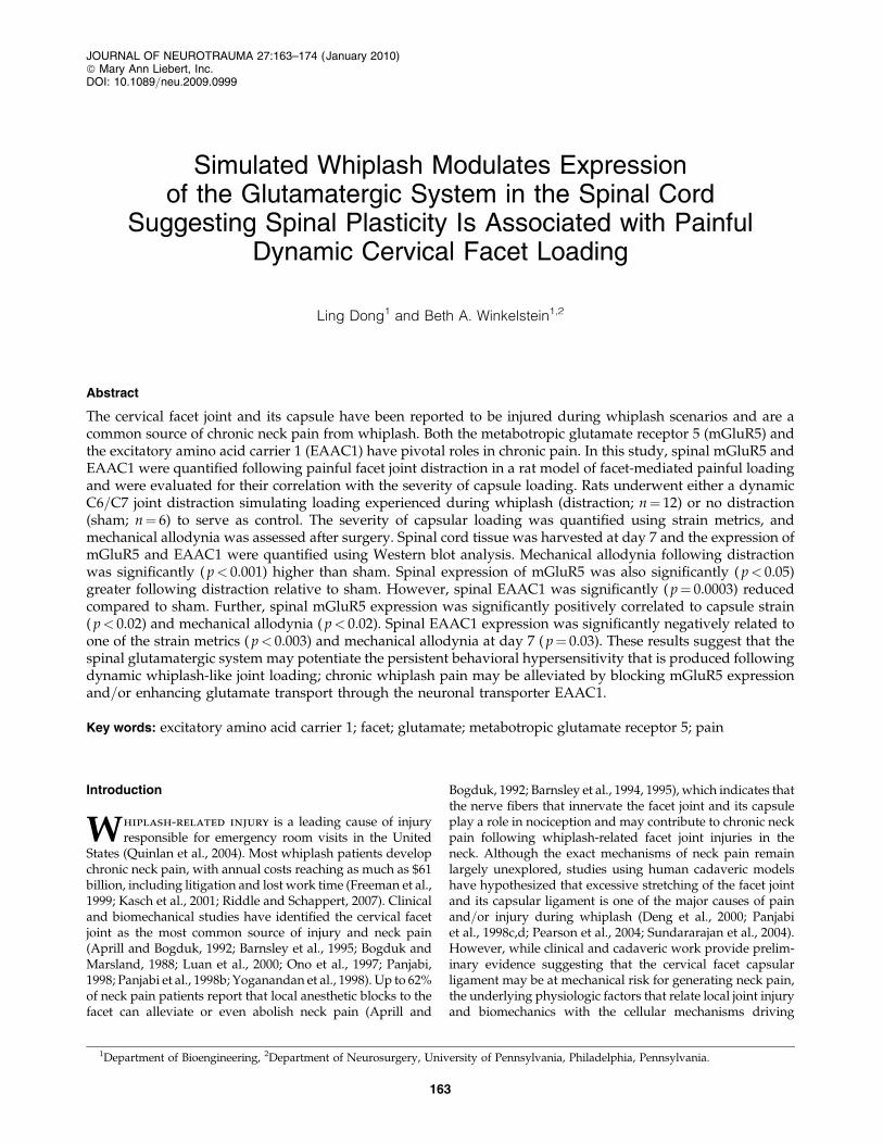
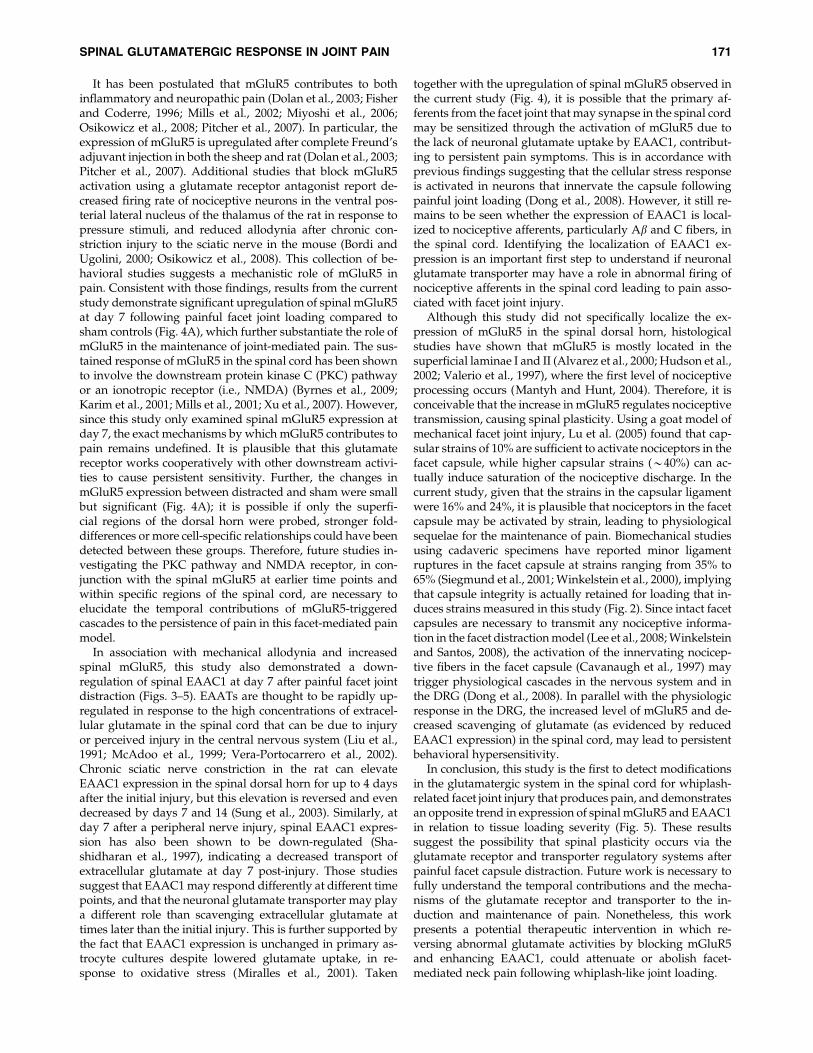
All procedures were performed under inhalation anesthe-sia (4% isoflurane for induction, 2.5% for maintenance) andwere modified from previously described procedures for afacet-mediated pain model in the rat (Dong et al., 2008; Leeet al., 2004a,b). Briefly, rats were placed in a prone positionand a skin incision was made to bilaterally isolate the C6=C7facet capsules. Each of the C6 and C7 vertebrae was attachedto microforceps coupled with a loading device which uses astepper motor (Danaher Precision Systems, Salem, NH) toapply controlled distraction across the C6=C7 facet capsulevia moving the C6 microforceps while C7 remained fixed(Dong et al., 2008). A linear variable differential transducer(LVDT) (MicroStrain Inc., Williston, VT) with a 24 mm strokeand 5.7 mm resolution was attached to the C7 microforceps tocontinuously record the microforceps displacement (Fig. 1).
For this study, C6=C7 facet joint distractions (distraction)were imposed to induce a painful capsule ligament loading.Sham procedures involved attachment of both the C6 and C7vertebrae to the loading device but had no distraction (0 mm)applied across the joint.
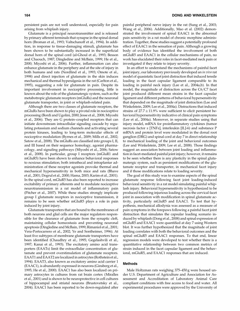
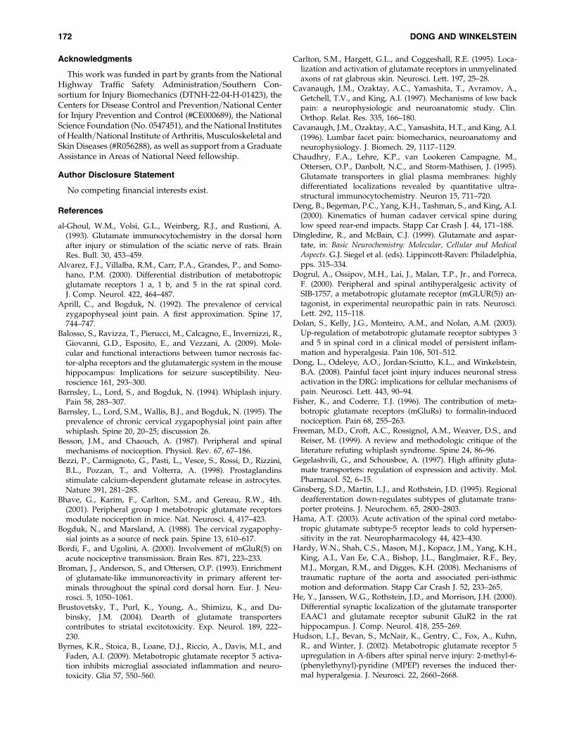
In order to quantify the facet joint and capsular ligamentloading mechanics in vivo and to quantify the severity ofcapsule loading that was applied for each distraction, imagingand standard measurements of joint mechanics were syn-chronized and acquired at 500 Hz during each applied jointdistraction. A load cell (Interface Inc., Scottsdale, AZ; 0.02 Nresolution) was attached to the C7 microforceps and moni-tored the tensile force generated across the C6=C7 joint duringdistraction. The motion of the C6 vertebra relative to C7 wastracked using polystyrene microspheres (Spherotech, Inc.,Libertyville, IL; diameter¼ 0.17� 0.01 mm) that were affixedto the lamina of each vertebra (Fig. 2). Additional trackingparticles were also placed in a grid covering the right C6=C7facet capsule that spanned from the origin to insertion of theligament (Fig. 2). These microspheres were placed directly onthe surface of lamina and capsule without slipping due totheir hydrophilic properties, and were monitored using high-speed imaging (Phantom v4.3 CCD camera; Vision Research,Inc., Wayne, NJ; 50 pixels=mm resolution). The severity ofloading applied across the facet joint capsule in each case wasmeasured by estimating the strain in the capsule; the maxi-mum tensile strain in the direction across the joint (in therostral–caudal direction) and the maximum principal strain inthe capsule were quantified to provide relevant mechanicalmetrics for loading to this joint. Both strain measurementswere measured for the C6=C7 right facet capsule joint, usingLS-DYNA software (Livermore Software Technology Corp.,Livermore, CA) according to methods previously describedfor in vivo studies of this joint and other biomechanical studiesof soft tissues (Cavanaugh et al., 1996; Dong et al., 2008;
FIG. 1. Customized facet joint loading device showing thedistraction device and set up including the microforceps,motor, LVDT, load cell, and nose cone holder for anesthesiadelivery. A surgical microscope was mounted above thedevice to acquire image data throughout loading while themicroforceps coupled to the C6 vertebra was displaced usingthe motor automated by a LABVIEW program; the micro-forceps coupled to the C7 vertebra remained stationary.
SPINAL GLUTAMATERGIC RESPONSE IN JOINT PAIN 165
Hardy et al., 2008; Lee et al., 2004b; Winkelstein and Santos,2000) (Fig. 2).
Behavioral assessments
Behavioral sensitivity was assessed in each rat after thesurgical procedures by measuring bilateral mechanical allo-dynia in the forepaws on post-operative days 1, 3, 5, and 7,using von Frey filaments of two strengths (2 and 4 g; StoeltingCo., Wood Dale, IL). Rats were also assessed prior to surgery
to provide baseline measurements and to serve as a controlunoperated response. Behavioral testing was performed witheach filament applied separately to each forepaw for threerounds of 10 stimulations each. Each round of stimulation wasseparated by at least 10 min to allow for an adequate restperiod. For each von Frey filament, the number of pawwithdrawals was taken as the total number of positive re-sponses counted for each rat for each forepaw. A paired t-testwas used to compare the responses between the left and rightpaws for each rat to test for symmetry in the behavioral sen-sitivity response following the bilateral loading. A repeatedmeasures ANOVA with post hoc Bonferonni correction wasused to statistically compare temporal allodynia between thedistraction and sham groups. In order to provide a quantita-tive measure of cumulative sensitivity for each rat followingjoint loading, total allodynia was also calculated as the sumof all paw withdrawals from all of the post-operative days 1,3, 5, and 7. This measure of cumulative sensitivity was em-ployed as the measure of behavioral response for the linearregression against maximum tensile and principal strains inthe capsule. All statistical tests were performed using SYSTAT(SYSTAT Software Inc., Richmond, CA), with significance atp< 0.05 for all tests.
Tissue harvest and Western blot analysis
Whole spinal cord tissue at the C6 cervical spinal level washarvested on day 7 to evaluate the relative expression ofmGluR5 and EAAC1. Normal rats (n¼ 2) were also used ascontrols for context of the normal protein response. Afterbehavioral testing on day 7, all rats were transcardially per-fused with 250 mL of phosphate-buffered saline (pH 7.4) andspinal cord tissue was rapidly removed. Following tissueharvest, spinal cord samples were homogenized using lysisbuffer containing 50 mM Tris HCl (pH 8.0), 1% Triton X-100,150 mM NaCl, 1 mM EDTA, and protease and phosphataseinhibitors (Sigma-Aldrich Corp., St. Louis, MO). Proteinsamples (50 mg) were heated at 95–1008C in preparation forelectrophoresis and were loaded on a Tris-HCl ready poly-acrylamide gel (BioRad Laboratories, Hercules, CA). Proteinwas transferred to a polyvinylidene difluoride (PVDF)membrane (Millipore, Billerica, MA) and was blocked for30 min with 5% dry-milk blocking reagent (Invitrogen Corp.,Carlsbad, CA) in 0.1% Tween-20 Tris-buffered saline. Themembrane was incubated overnight at 48C with the rabbitpolyclonal antibodies anti-mGluR5 (1:500; Chemicon Inter-national Inc., Billerica, MA) or anti-EAAC1 (1:200; AlphaDiagnostic International, San Antonio, TX). The membranewas washed in Tris-buffered saline with 0.1% Tween-20buffer three times for 10 min each followed by a 1 h incuba-tion at room temperature with horseradish peroxidase–conjugated anti-rabbit (1:10,000; Santa Cruz Biotechnology,Inc., Santa Cruz, CA). The membrane was exposed to HyBlotCL autoradiography film through a chemiluminescent reac-tion using the SuperSignal West Pico detection kit (PierceBiotechnology, Inc., Rockford, IL). The film was developedusing an X-OMAT processor (2000A; Eastman Kodak, Ro-chester, NY). To evaluate the amount of protein loaded for eachsample, the membrane was stripped with stripping buffer(Pierce Biotechnology Inc.) at room temperature for 15 minbefore reprobing for actin (1:200; Santa Cruz BiotechnologyInc.).
FIG. 2. Representative images showing the facet joint andcapsule as well as the associated strain field. (A) The C6 andC7 vertebrae were marked by vertebral markers and theirinitial separation length (xref) defined the joint referencecondition. (B) At the peak of capsule stretch (xpeak) C6 wasdistracted away from C7. Vertebral distraction was calcu-lated as the vector difference between these separations andthe maximum tensile strain (not shown) and maximumprincipal strain in the capsule was calculated using thecapsule markers. (C) The maximum principal strain fieldassociated with the images (A, B) is shown.
166 DONG AND WINKELSTEIN
Quantitative analysis was performed using Image J(National Institutes of Health, Bethesda, MD) to measure thedensity of immunoreactive bands for each lane. The amountof mGluR5 and EAAC1 was normalized separately againstthe relative amount of actin for each sample. Equal loading foreach sample was further confirmed by staining the PVDFmembranes with Coomassie Brilliant-Blue R-250 (BioRad)solution for 10 min followed by destaining with 25% aceticacid for three rounds of 10 min wash (Lindl et al., 2007). Themembrane was also scanned and analyzed by Image J toconfirm that the difference in protein expression was not dueto unequal protein loading between samples. There was nodifference detected in mGluR5 and EAAC1 expression be-tween normal and sham groups, so protein expression ofmGluR5 and EAAC1 was reported as a fold-increase oversham. A two-tailed t-test assuming equal variances was usedto compare spinal protein responses between groups (dis-traction vs. sham). Significance was set at p< 0.05.
Statistical analysis
Linear regression models were used to determine if thedegree of behavioral sensitivity (measured by cumulativemechanical allodynia) and spinal mGluR5 and EAAC1 ex-pression were sensitive to the severity of applied facet jointdistraction, and if behavioral hypersensitivity at day 7 wasrelated to the corresponding modifications of spinal mGluR5and EAAC1 expression. For these analyses, the tensile strainin the rostral–caudal direction and the maximum principalstrain in the facet capsule were individually used for eachdistraction (Fig. 2). As such, the maximum tensile strain andthe maximum principal strain in the capsule were separatelytaken as the independent variables for plots against totalmechanical allodynia using both of the von Frey filaments (2and 4 g, separately), and each of mGluR5 or EAAC1 proteinlevels, in separate analyses. For correlation analyses betweenmGluR5 and EAAC1 expression and mechanical allodynia,the number of paw withdrawals measured on day 7 was usedsince that time point corresponds to the day of tissue harvestand protein assessments of mGluR5 and EAAC1. The straincomponents for sham were taken as 0 since this group re-ceived 0 mm facet joint distraction. The significance of corre-lation for each regression was tested using an F-test anddefined at p< 0.05 for all regressions.
Results
No gross mechanical tissue rupture was observed in thecapsular ligament during loading for any joint distraction. Inaddition, careful examination of the facet capsule was alsoperformed under a surgical microscope at the time of tissueharvest and did not reveal any evidence of gross tissue rup-ture or ligament damage. After surgery, all rats showed nor-mal head mobility, consistent weight gain and normalgrooming, indicating that there were no adverse effects ofeither the surgical or joint loading procedures.
Mean applied vertebral distraction in the distraction groupwas 0.47� 0.17 mm, with an average force generated acrossthe joint of 4.04� 0.82 N. The average rate of applied dis-traction across the C6=C7 joint was measured to be14.5� 0.5 mm=s, with a corresponding applied strain rate of483� 16.7%=s. The corresponding average maximum tensilestrain in the rostral–caudal direction was 16� 7% and the
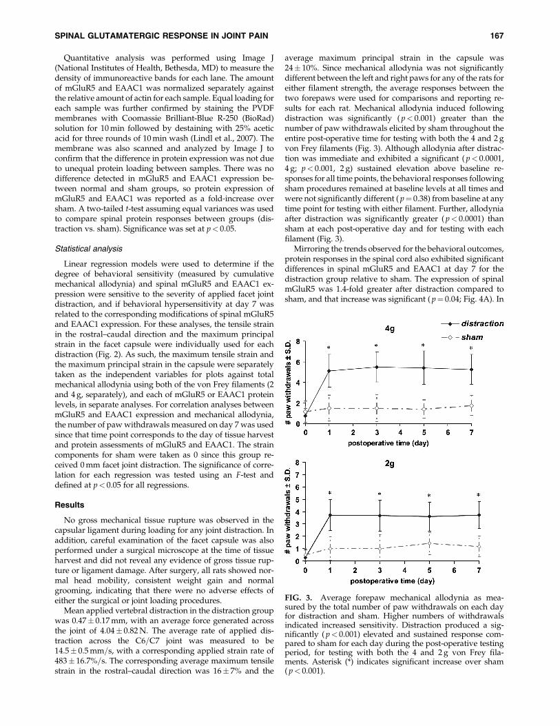
average maximum principal strain in the capsule was24� 10%. Since mechanical allodynia was not significantlydifferent between the left and right paws for any of the rats foreither filament strength, the average responses between thetwo forepaws were used for comparisons and reporting re-sults for each rat. Mechanical allodynia induced followingdistraction was significantly ( p< 0.001) greater than thenumber of paw withdrawals elicited by sham throughout theentire post-operative time for testing with both the 4 and 2 gvon Frey filaments (Fig. 3). Although allodynia after distrac-tion was immediate and exhibited a significant ( p< 0.0001,4 g; p< 0.001, 2 g) sustained elevation above baseline re-sponses for all time points, the behavioral responses followingsham procedures remained at baseline levels at all times andwere not significantly different ( p¼ 0.38) from baseline at anytime point for testing with either filament. Further, allodyniaafter distraction was significantly greater ( p< 0.0001) thansham at each post-operative day and for testing with eachfilament (Fig. 3).
Mirroring the trends observed for the behavioral outcomes,protein responses in the spinal cord also exhibited significantdifferences in spinal mGluR5 and EAAC1 at day 7 for thedistraction group relative to sham. The expression of spinalmGluR5 was 1.4-fold greater after distraction compared tosham, and that increase was significant ( p¼ 0.04; Fig. 4A). In
FIG. 3. Average forepaw mechanical allodynia as mea-sured by the total number of paw withdrawals on each dayfor distraction and sham. Higher numbers of withdrawalsindicated increased sensitivity. Distraction produced a sig-nificantly ( p< 0.001) elevated and sustained response com-pared to sham for each day during the post-operative testingperiod, for testing with both the 4 and 2 g von Frey fila-ments. Asterisk (*) indicates significant increase over sham( p< 0.001).
SPINAL GLUTAMATERGIC RESPONSE IN JOINT PAIN 167
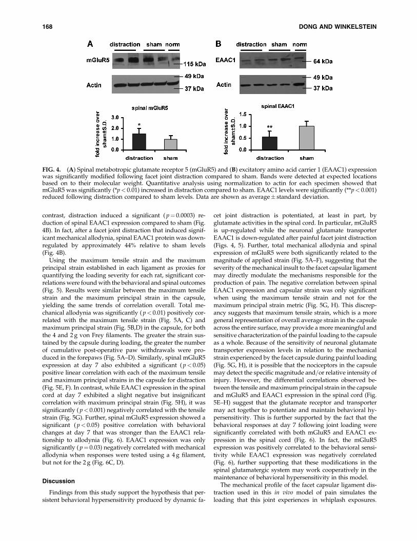
contrast, distraction induced a significant ( p¼ 0.0003) re-duction of spinal EAAC1 expression compared to sham (Fig.4B). In fact, after a facet joint distraction that induced signif-icant mechanical allodynia, spinal EAAC1 protein was down-regulated by approximately 44% relative to sham levels(Fig. 4B).
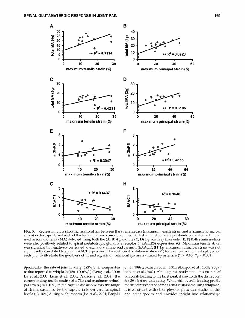
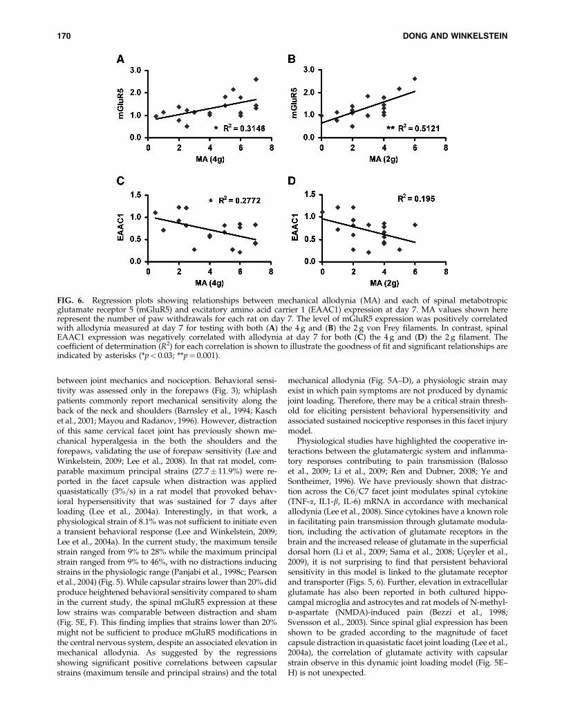
Using the maximum tensile strain and the maximumprincipal strain established in each ligament as proxies forquantifying the loading severity for each rat, significant cor-relations were found with the behavioral and spinal outcomes(Fig. 5). Results were similar between the maximum tensilestrain and the maximum principal strain in the capsule,yielding the same trends of correlation overall. Total me-chanical allodynia was significantly ( p< 0.01) positively cor-related with the maximum tensile strain (Fig. 5A, C) andmaximum principal strain (Fig. 5B,D) in the capsule, for boththe 4 and 2 g von Frey filaments. The greater the strain sus-tained by the capsule during loading, the greater the numberof cumulative post-operative paw withdrawals were pro-duced in the forepaws (Fig. 5A–D). Similarly, spinal mGluR5expression at day 7 also exhibited a significant ( p< 0.05)positive linear correlation with each of the maximum tensileand maximum principal strains in the capsule for distraction(Fig. 5E, F). In contrast, while EAAC1 expression in the spinalcord at day 7 exhibited a slight negative but insignificantcorrelation with maximum principal strain (Fig. 5H), it wassignificantly ( p< 0.001) negatively correlated with the tensilestrain (Fig. 5G). Further, spinal mGluR5 expression showed asignificant ( p< 0.05) positive correlation with behavioralchanges at day 7 that was stronger than the EAAC1 rela-tionship to allodynia (Fig. 6). EAAC1 expression was onlysignificantly ( p¼ 0.03) negatively correlated with mechanicalallodynia when responses were tested using a 4 g filament,but not for the 2 g (Fig. 6C, D).
Discussion
Findings from this study support the hypothesis that per-sistent behavioral hypersensitivity produced by dynamic fa-
cet joint distraction is potentiated, at least in part, byglutamate activities in the spinal cord. In particular, mGluR5is up-regulated while the neuronal glutamate transporterEAAC1 is down-regulated after painful facet joint distraction(Figs. 4, 5). Further, total mechanical allodynia and spinalexpression of mGluR5 were both significantly related to themagnitude of applied strain (Fig. 5A–F), suggesting that theseverity of the mechanical insult to the facet capsular ligamentmay directly modulate the mechanisms responsible for theproduction of pain. The negative correlation between spinalEAAC1 expression and capsular strain was only significantwhen using the maximum tensile strain and not for themaximum principal strain metric (Fig. 5G, H). This discrep-ancy suggests that maximum tensile strain, which is a moregeneral representation of overall average strain in the capsuleacross the entire surface, may provide a more meaningful andsensitive characterization of the painful loading to the capsuleas a whole. Because of the sensitivity of neuronal glutamatetransporter expression levels in relation to the mechanicalstrain experienced by the facet capsule during painful loading(Fig. 5G, H), it is possible that the nociceptors in the capsulemay detect the specific magnitude and=or relative intensity ofinjury. However, the differential correlations observed be-tween the tensile and maximum principal strain in the capsuleand mGluR5 and EAAC1 expression in the spinal cord (Fig.5E–H) suggest that the glutamate receptor and transportermay act together to potentiate and maintain behavioral hy-persensitivity. This is further supported by the fact that thebehavioral responses at day 7 following joint loading weresignificantly correlated with both mGluR5 and EAAC1 ex-pression in the spinal cord (Fig. 6). In fact, the mGluR5expression was positively correlated to the behavioral sensi-tivity while EAAC1 expression was negatively correlated(Fig. 6), further supporting that these modifications in thespinal glutamatergic system may work cooperatively in themaintenance of behavioral hypersensitivity in this model.
The mechanical profile of the facet capsular ligament dis-traction used in this in vivo model of pain simulates theloading that this joint experiences in whiplash exposures.
FIG. 4. (A) Spinal metabotropic glutamate receptor 5 (mGluR5) and (B) excitatory amino acid carrier 1 (EAAC1) expressionwas significantly modified following facet joint distraction compared to sham. Bands were detected at expected locationsbased on to their molecular weight. Quantitative analysis using normalization to actin for each specimen showed thatmGluR5 was significantly (*p< 0.01) increased in distraction compared to sham. EAAC1 levels were significantly (**p< 0.001)reduced following distraction compared to sham levels. Data are shown as average� standard deviation.
168 DONG AND WINKELSTEIN
Specifically, the rate of joint loading (483%=s) is comparableto that reported in whiplash (150–1000%=s) (Deng et al., 2000;Lu et al., 2005; Luan et al., 2000; Pearson et al., 2004); thecorresponding tensile strain (16� 7%) and maximum princi-pal strain (24� 10%) in the capsule are also within the rangeof strains sustained by the capsule in lower cervical spinallevels (13–40%) during such impacts (Ito et al., 2004; Panjabi
et al., 1998c; Pearson et al., 2004; Stemper et al., 2005; Yoga-nandan et al., 2002). Although this study simulates the rate ofwhiplash loading to the facet joint, it also holds the distractionfor 30 s before unloading. While this overall loading profilefor the joint is not the same as that sustained during whiplash,it is consistent with other physiologic in vivo studies in thisand other species and provides insight into relationships
FIG. 5. Regression plots showing relationships between the strain metrics (maximum tensile strain and maximum principalstrain) in the capsule and each of the behavioral and spinal outcomes. Both strain metrics were positively correlated with totalmechanical allodynia (MA) detected using both the (A, B) 4 g and the (C, D) 2 g von Frey filaments. (E, F) Both strain metricswere also positively related to spinal metabotropic glutamate receptor 5 (mGluR5) expression. (G) Maximum tensile strainwas significantly negatively correlated to excitatory amino acid carrier 1 (EAAC1), (H) but maximum principal strain was notsignificantly correlated to spinal EAAC1 expression. The coefficient of determination (R2) for each correlation is displayed oneach plot to illustrate the goodness of fit and significant relationships are indicated by asterisks (*p< 0.05; **p< 0.001).
SPINAL GLUTAMATERGIC RESPONSE IN JOINT PAIN 169
between joint mechanics and nociception. Behavioral sensi-tivity was assessed only in the forepaws (Fig. 3); whiplashpatients commonly report mechanical sensitivity along theback of the neck and shoulders (Barnsley et al., 1994; Kaschet al., 2001; Mayou and Radanov, 1996). However, distractionof this same cervical facet joint has previously shown me-chanical hyperalgesia in the both the shoulders and theforepaws, validating the use of forepaw sensitivity (Lee andWinkelstein, 2009; Lee et al., 2008). In that rat model, com-parable maximum principal strains (27.7� 11.9%) were re-ported in the facet capsule when distraction was appliedquasistatically (3%=s) in a rat model that provoked behav-ioral hypersensitivity that was sustained for 7 days afterloading (Lee et al., 2004a). Interestingly, in that work, aphysiological strain of 8.1% was not sufficient to initiate evena transient behavioral response (Lee and Winkelstein, 2009;Lee et al., 2004a). In the current study, the maximum tensilestrain ranged from 9% to 28% while the maximum principalstrain ranged from 9% to 46%, with no distractions inducingstrains in the physiologic range (Panjabi et al., 1998c; Pearsonet al., 2004) (Fig. 5). While capsular strains lower than 20% didproduce heightened behavioral sensitivity compared to shamin the current study, the spinal mGluR5 expression at theselow strains was comparable between distraction and sham(Fig. 5E, F). This finding implies that strains lower than 20%might not be sufficient to produce mGluR5 modifications inthe central nervous system, despite an associated elevation inmechanical allodynia. As suggested by the regressionsshowing significant positive correlations between capsularstrains (maximum tensile and principal strains) and the total
mechanical allodynia (Fig. 5A–D), a physiologic strain mayexist in which pain symptoms are not produced by dynamicjoint loading. Therefore, there may be a critical strain thresh-old for eliciting persistent behavioral hypersensitivity andassociated sustained nociceptive responses in this facet injurymodel.
Physiological studies have highlighted the cooperative in-teractions between the glutamatergic system and inflamma-tory responses contributing to pain transmission (Balossoet al., 2009; Li et al., 2009; Ren and Dubner, 2008; Ye andSontheimer, 1996). We have previously shown that distrac-tion across the C6=C7 facet joint modulates spinal cytokine(TNF-a, IL1-b, IL-6) mRNA in accordance with mechanicalallodynia (Lee et al., 2008). Since cytokines have a known rolein facilitating pain transmission through glutamate modula-tion, including the activation of glutamate receptors in thebrain and the increased release of glutamate in the superficialdorsal horn (Li et al., 2009; Sama et al., 2008; Uceyler et al.,2009), it is not surprising to find that persistent behavioralsensitivity in this model is linked to the glutamate receptorand transporter (Figs. 5, 6). Further, elevation in extracellularglutamate has also been reported in both cultured hippo-campal microglia and astrocytes and rat models of N-methyl-d-aspartate (NMDA)-induced pain (Bezzi et al., 1998;Svensson et al., 2003). Since spinal glial expression has beenshown to be graded according to the magnitude of facetcapsule distraction in quasistatic facet joint loading (Lee et al.,2004a), the correlation of glutamate activity with capsularstrain observe in this dynamic joint loading model (Fig. 5E–H) is not unexpected.
FIG. 6. Regression plots showing relationships between mechanical allodynia (MA) and each of spinal metabotropicglutamate receptor 5 (mGluR5) and excitatory amino acid carrier 1 (EAAC1) expression at day 7. MA values shown hererepresent the number of paw withdrawals for each rat on day 7. The level of mGluR5 expression was positively correlatedwith allodynia measured at day 7 for testing with both (A) the 4 g and (B) the 2 g von Frey filaments. In contrast, spinalEAAC1 expression was negatively correlated with allodynia at day 7 for both (C) the 4 g and (D) the 2 g filament. Thecoefficient of determination (R2) for each correlation is shown to illustrate the goodness of fit and significant relationships areindicated by asterisks (*p< 0.03; **p¼ 0.001).
170 DONG AND WINKELSTEIN
It has been postulated that mGluR5 contributes to bothinflammatory and neuropathic pain (Dolan et al., 2003; Fisherand Coderre, 1996; Mills et al., 2002; Miyoshi et al., 2006;Osikowicz et al., 2008; Pitcher et al., 2007). In particular, theexpression of mGluR5 is upregulated after complete Freund’sadjuvant injection in both the sheep and rat (Dolan et al., 2003;Pitcher et al., 2007). Additional studies that block mGluR5activation using a glutamate receptor antagonist report de-creased firing rate of nociceptive neurons in the ventral pos-terial lateral nucleus of the thalamus of the rat in response topressure stimuli, and reduced allodynia after chronic con-striction injury to the sciatic nerve in the mouse (Bordi andUgolini, 2000; Osikowicz et al., 2008). This collection of be-havioral studies suggests a mechanistic role of mGluR5 inpain. Consistent with those findings, results from the currentstudy demonstrate significant upregulation of spinal mGluR5at day 7 following painful facet joint loading compared tosham controls (Fig. 4A), which further substantiate the role ofmGluR5 in the maintenance of joint-mediated pain. The sus-tained response of mGluR5 in the spinal cord has been shownto involve the downstream protein kinase C (PKC) pathwayor an ionotropic receptor (i.e., NMDA) (Byrnes et al., 2009;Karim et al., 2001; Mills et al., 2001; Xu et al., 2007). However,since this study only examined spinal mGluR5 expression atday 7, the exact mechanisms by which mGluR5 contributes topain remains undefined. It is plausible that this glutamatereceptor works cooperatively with other downstream activi-ties to cause persistent sensitivity. Further, the changes inmGluR5 expression between distracted and sham were smallbut significant (Fig. 4A); it is possible if only the superfi-cial regions of the dorsal horn were probed, stronger fold-differences or more cell-specific relationships could have beendetected between these groups. Therefore, future studies in-vestigating the PKC pathway and NMDA receptor, in con-junction with the spinal mGluR5 at earlier time points andwithin specific regions of the spinal cord, are necessary toelucidate the temporal contributions of mGluR5-triggeredcascades to the persistence of pain in this facet-mediated painmodel.
In association with mechanical allodynia and increasedspinal mGluR5, this study also demonstrated a down-regulation of spinal EAAC1 at day 7 after painful facet jointdistraction (Figs. 3–5). EAATs are thought to be rapidly up-regulated in response to the high concentrations of extracel-lular glutamate in the spinal cord that can be due to injuryor perceived injury in the central nervous system (Liu et al.,1991; McAdoo et al., 1999; Vera-Portocarrero et al., 2002).Chronic sciatic nerve constriction in the rat can elevateEAAC1 expression in the spinal dorsal horn for up to 4 daysafter the initial injury, but this elevation is reversed and evendecreased by days 7 and 14 (Sung et al., 2003). Similarly, atday 7 after a peripheral nerve injury, spinal EAAC1 expres-sion has also been shown to be down-regulated (Sha-shidharan et al., 1997), indicating a decreased transport ofextracellular glutamate at day 7 post-injury. Those studiessuggest that EAAC1 may respond differently at different timepoints, and that the neuronal glutamate transporter may playa different role than scavenging extracellular glutamate attimes later than the initial injury. This is further supported bythe fact that EAAC1 expression is unchanged in primary as-trocyte cultures despite lowered glutamate uptake, in re-sponse to oxidative stress (Miralles et al., 2001). Taken
together with the upregulation of spinal mGluR5 observed inthe current study (Fig. 4), it is possible that the primary af-ferents from the facet joint that may synapse in the spinal cordmay be sensitized through the activation of mGluR5 due tothe lack of neuronal glutamate uptake by EAAC1, contribut-ing to persistent pain symptoms. This is in accordance withprevious findings suggesting that the cellular stress responseis activated in neurons that innervate the capsule followingpainful joint loading (Dong et al., 2008). However, it still re-mains to be seen whether the expression of EAAC1 is local-ized to nociceptive afferents, particularly Ab and C fibers, inthe spinal cord. Identifying the localization of EAAC1 ex-pression is an important first step to understand if neuronalglutamate transporter may have a role in abnormal firing ofnociceptive afferents in the spinal cord leading to pain asso-ciated with facet joint injury.
Although this study did not specifically localize the ex-pression of mGluR5 in the spinal dorsal horn, histologicalstudies have shown that mGluR5 is mostly located in thesuperficial laminae I and II (Alvarez et al., 2000; Hudson et al.,2002; Valerio et al., 1997), where the first level of nociceptiveprocessing occurs (Mantyh and Hunt, 2004). Therefore, it isconceivable that the increase in mGluR5 regulates nociceptivetransmission, causing spinal plasticity. Using a goat model ofmechanical facet joint injury, Lu et al. (2005) found that cap-sular strains of 10% are sufficient to activate nociceptors in thefacet capsule, while higher capsular strains (*40%) can ac-tually induce saturation of the nociceptive discharge. In thecurrent study, given that the strains in the capsular ligamentwere 16% and 24%, it is plausible that nociceptors in the facetcapsule may be activated by strain, leading to physiologicalsequelae for the maintenance of pain. Biomechanical studiesusing cadaveric specimens have reported minor ligamentruptures in the facet capsule at strains ranging from 35% to65% (Siegmund et al., 2001; Winkelstein et al., 2000), implyingthat capsule integrity is actually retained for loading that in-duces strains measured in this study (Fig. 2). Since intact facetcapsules are necessary to transmit any nociceptive informa-tion in the facet distraction model (Lee et al., 2008; Winkelsteinand Santos, 2008), the activation of the innervating nocicep-tive fibers in the facet capsule (Cavanaugh et al., 1997) maytrigger physiological cascades in the nervous system and inthe DRG (Dong et al., 2008). In parallel with the physiologicresponse in the DRG, the increased level of mGluR5 and de-creased scavenging of glutamate (as evidenced by reducedEAAC1 expression) in the spinal cord, may lead to persistentbehavioral hypersensitivity.
In conclusion, this study is the first to detect modificationsin the glutamatergic system in the spinal cord for whiplash-related facet joint injury that produces pain, and demonstratesan opposite trend in expression of spinal mGluR5 and EAAC1in relation to tissue loading severity (Fig. 5). These resultssuggest the possibility that spinal plasticity occurs via theglutamate receptor and transporter regulatory systems afterpainful facet capsule distraction. Future work is necessary tofully understand the temporal contributions and the mecha-nisms of the glutamate receptor and transporter to the in-duction and maintenance of pain. Nonetheless, this workpresents a potential therapeutic intervention in which re-versing abnormal glutamate activities by blocking mGluR5and enhancing EAAC1, could attenuate or abolish facet-mediated neck pain following whiplash-like joint loading.
SPINAL GLUTAMATERGIC RESPONSE IN JOINT PAIN 171
Acknowledgments
This work was funded in part by grants from the NationalHighway Traffic Safety Administration=Southern Con-sortium for Injury Biomechanics (DTNH-22-04-H-01423), theCenters for Disease Control and Prevention=National Centerfor Injury Prevention and Control (#CE000689), the NationalScience Foundation (No. 0547451), and the National Institutesof Health=National Institute of Arthritis, Musculoskeletal andSkin Diseases (#R056288), as well as support from a GraduateAssistance in Areas of National Need fellowship.
Author Disclosure Statement
No competing financial interests exist.
References
al-Ghoul, W.M., Volsi, G.L., Weinberg, R.J., and Rustioni, A.(1993). Glutamate immunocytochemistry in the dorsal hornafter injury or stimulation of the sciatic nerve of rats. BrainRes. Bull. 30, 453–459.
Alvarez, F.J., Villalba, R.M., Carr, P.A., Grandes, P., and Somo-hano, P.M. (2000). Differential distribution of metabotropicglutamate receptors 1 a, 1 b, and 5 in the rat spinal cord.J. Comp. Neurol. 422, 464–487.
Aprill, C., and Bogduk, N. (1992). The prevalence of cervicalzygapophyseal joint pain. A first approximation. Spine 17,744–747.
Balosso, S., Ravizza, T., Pierucci, M., Calcagno, E., Invernizzi, R.,Giovanni, G.D., Esposito, E., and Vezzani, A. (2009). Mole-cular and functional interactions between tumor necrosis fac-tor-alpha receptors and the glutamatergic system in the mousehippocampus: Implications for seizure susceptibility. Neu-roscience 161, 293–300.
Barnsley, L., Lord, S., and Bogduk, N. (1994). Whiplash injury.Pain 58, 283–307.
Barnsley, L., Lord, S.M., Wallis, B.J., and Bogduk, N. (1995). Theprevalence of chronic cervical zygapophysial joint pain afterwhiplash. Spine 20, 20–25; discussion 26.
Besson, J.M., and Chaouch, A. (1987). Peripheral and spinalmechanisms of nociception. Physiol. Rev. 67, 67–186.
Bezzi, P., Carmignoto, G., Pasti, L., Vesce, S., Rossi, D., Rizzini,B.L., Pozzan, T., and Volterra, A. (1998). Prostaglandinsstimulate calcium-dependent glutamate release in astrocytes.Nature 391, 281–285.
Bhave, G., Karim, F., Carlton, S.M., and Gereau, R.W., 4th.(2001). Peripheral group I metabotropic glutamate receptorsmodulate nociception in mice. Nat. Neurosci. 4, 417–423.
Bogduk, N., and Marsland, A. (1988). The cervical zygapophy-sial joints as a source of neck pain. Spine 13, 610–617.
Bordi, F., and Ugolini, A. (2000). Involvement of mGluR(5) onacute nociceptive transmission. Brain Res. 871, 223–233.
Broman, J., Anderson, S., and Ottersen, O.P. (1993). Enrichmentof glutamate-like immunoreactivity in primary afferent ter-minals throughout the spinal cord dorsal horn. Eur. J. Neu-rosci. 5, 1050–1061.
Brustovetsky, T., Purl, K., Young, A., Shimizu, K., and Du-binsky, J.M. (2004). Dearth of glutamate transporterscontributes to striatal excitotoxicity. Exp. Neurol. 189, 222–230.
Byrnes, K.R., Stoica, B., Loane, D.J., Riccio, A., Davis, M.I., andFaden, A.I. (2009). Metabotropic glutamate receptor 5 activa-tion inhibits microglial associated inflammation and neuro-toxicity. Glia 57, 550–560.
Carlton, S.M., Hargett, G.L., and Coggeshall, R.E. (1995). Loca-lization and activation of glutamate receptors in unmyelinatedaxons of rat glabrous skin. Neurosci. Lett. 197, 25–28.
Cavanaugh, J.M., Ozaktay, A.C., Yamashita, T., Avramov, A.,Getchell, T.V., and King, A.I. (1997). Mechanisms of low backpain: a neurophysiologic and neuroanatomic study. Clin.Orthop. Relat. Res. 335, 166–180.
Cavanaugh, J.M., Ozaktay, A.C., Yamashita, H.T., and King, A.I.(1996). Lumbar facet pain: biomechanics, neuroanatomy andneurophysiology. J. Biomech. 29, 1117–1129.
Chaudhry, F.A., Lehre, K.P., van Lookeren Campagne, M.,Ottersen, O.P., Danbolt, N.C., and Storm-Mathisen, J. (1995).Glutamate transporters in glial plasma membranes: highlydifferentiated localizations revealed by quantitative ultra-structural immunocytochemistry. Neuron 15, 711–720.
Deng, B., Begeman, P.C., Yang, K.H., Tashman, S., and King, A.I.(2000). Kinematics of human cadaver cervical spine duringlow speed rear-end impacts. Stapp Car Crash J. 44, 171–188.
Dingledine, R., and McBain, C.J. (1999). Glutamate and aspar-tate, in: Basic Neurochemistry: Molecular, Cellular and MedicalAspects. G.J. Siegel et al. (eds). Lippincott-Raven: Philadelphia,pps. 315–334.
Dogrul, A., Ossipov, M.H., Lai, J., Malan, T.P., Jr., and Porreca,F. (2000). Peripheral and spinal antihyperalgesic activity ofSIB-1757, a metabotropic glutamate receptor (mGLUR(5)) an-tagonist, in experimental neuropathic pain in rats. Neurosci.Lett. 292, 115–118.
Dolan, S., Kelly, J.G., Monteiro, A.M., and Nolan, A.M. (2003).Up-regulation of metabotropic glutamate receptor subtypes 3and 5 in spinal cord in a clinical model of persistent inflam-mation and hyperalgesia. Pain 106, 501–512.
Dong, L., Odeleye, A.O., Jordan-Sciutto, K.L., and Winkelstein,B.A. (2008). Painful facet joint injury induces neuronal stressactivation in the DRG: implications for cellular mechanisms ofpain. Neurosci. Lett. 443, 90–94.
Fisher, K., and Coderre, T.J. (1996). The contribution of meta-botropic glutamate receptors (mGluRs) to formalin-inducednociception. Pain 68, 255–263.
Freeman, M.D., Croft, A.C., Rossignol, A.M., Weaver, D.S., andReiser, M. (1999). A review and methodologic critique of theliterature refuting whiplash syndrome. Spine 24, 86–96.
Gegelashvili, G., and Schousboe, A. (1997). High affinity gluta-mate transporters: regulation of expression and activity. Mol.Pharmacol. 52, 6–15.
Ginsberg, S.D., Martin, L.J., and Rothstein, J.D. (1995). Regionaldeafferentation down-regulates subtypes of glutamate trans-porter proteins. J. Neurochem. 65, 2800–2803.
Hama, A.T. (2003). Acute activation of the spinal cord metabo-tropic glutamate subtype-5 receptor leads to cold hypersen-sitivity in the rat. Neuropharmacology 44, 423–430.
Hardy, W.N., Shah, C.S., Mason, M.J., Kopacz, J.M., Yang, K.H.,King, A.I., Van Ee, C.A., Bishop, J.L., Banglmaier, R.F., Bey,M.J., Morgan, R.M., and Digges, K.H. (2008). Mechanisms oftraumatic rupture of the aorta and associated peri-isthmicmotion and deformation. Stapp Car Crash J. 52, 233–265.
He, Y., Janssen, W.G., Rothstein, J.D., and Morrison, J.H. (2000).Differential synaptic localization of the glutamate transporterEAAC1 and glutamate receptor subunit GluR2 in the rathippocampus. J. Comp. Neurol. 418, 255–269.
Hudson, L.J., Bevan, S., McNair, K., Gentry, C., Fox, A., Kuhn,R., and Winter, J. (2002). Metabotropic glutamate receptor 5upregulation in A-fibers after spinal nerve injury: 2-methyl-6-(phenylethynyl)-pyridine (MPEP) reverses the induced ther-mal hyperalgesia. J. Neurosci. 22, 2660–2668.
172 DONG AND WINKELSTEIN
Ito, S., Ivancic, P.C., Panjabi, M.M., and Cunningham, B.W.(2004). Soft tissue injury threshold during simulated whiplash:a biomechanical investigation. Spine 29, 979–987.
Jesse, C.R., Savegnago, L., and Nogueira, C.W. (2008). Effect of ametabotropic glutamate receptor 5 antagonist, MPEP, on thenociceptive response induced by intrathecal injection of ex-citatory amino acids, substance P, bradykinin or cytokines inmice. Pharmacol. Biochem. Behav. 90, 608–613.
Kanai, Y., Smith, C.P., and Hediger, M.A. (1993). A new familyof neurotransmitter transporters: the high-affinity glutamatetransporters. FASEB J. 7, 1450–1459.
Karim, F., Wang, C.C., and Gereau, R.W., 4th. (2001). Metabo-tropic glutamate receptor subtypes 1 and 5 are activators ofextracellular signal-regulated kinase signaling required forinflammatory pain in mice. J. Neurosci. 21, 3771–3779.
Kasch, H., Stengaard-Pedersen, K., Arendt-Nielsen, L., andStaehelin Jensen, T. (2001). Headache, neck pain, and neckmobility after acute whiplash injury: a prospective study.Spine 26, 1246–1251.
Lee, K.E., Davis, M.B., Mejilla, R.M., and Winkelstein, B.A.(2004a). In vivo cervical facet capsule distraction: mechanicalimplications for whiplash and neck pain. Stapp Car Crash J.48, 373–395.
Lee, K.E., Davis, M.B., and Winkelstein, B.A. (2008). Capsularligament involvement in the development of mechanical hy-peralgesia after facet joint loading: behavioral and inflamma-tory outcomes in a rodent model of pain. J. Neurotrauma 25,1383–1393.
Lee, K.E., Thinnes, J.H., Gokhin, D.S., and Winkelstein, B.A.(2004b). A novel rodent neck pain model of facet-mediatedbehavioral hypersensitivity: implications for persistent painand whiplash injury. J. Neurosci. Methods 137, 151–159.
Lee, K.E., and Winkelstein, B.A. (2009). Joint distraction mag-nitude is associated with different behavioral outcomes andsubstance P levels for cervical facet joint loading in the rat.J. Pain 10, 436–445.
Li, J., Xie, W., Zhang, J.M., and Baccei, M.L. (2009). Peripheralnerve injury sensitizes neonatal dorsal horn neurons to tumornecrosis factor-alpha. Mol. Pain. 5, 10.
Lindl, K.A., Akay, C., Wang, Y., White, M.G., and Jordan-Sciutto, K.L. (2007). Expression of the endoplasmic reticulumstress response marker, BiP, in the central nervous system ofHIV-positive individuals. Neuropathol. Appl. Neurobiol. 33,658–669.
Liu, D., Thangnipon, W., and McAdoo, D.J. (1991). Excitatoryamino acids rise to toxic levels upon impact injury to the ratspinal cord. Brain Res. 547, 344–348.
Lu, Y., Chen, C., Kallakuri, S., Patwardhan, A., and Cavanaugh,J.M. (2005). Neural response of cervical facet joint capsule tostretch: a study of whiplash pain mechanism. Stapp Car CrashJ. 49, 49–65.
Luan, F., Yang, K.H., Deng, B., Begeman, P.C., Tashman, S., andKing, A.I. (2000). Qualitative analysis of neck kinematicsduring low-speed rear-end impact. Clin. Biomech. (Bristol,Avon) 15, 649–657.
Mantyh, P.W., and Hunt, S.P. (2004). Setting the tone: superficialdorsal horn projection neurons regulate pain sensitivity.Trends Neurosci. 27, 582–584.
Mao, J., Sung, B., Ji, R.R., and Lim, G. (2002). Chronic morphineinduces downregulation of spinal glutamate transporters:implications in morphine tolerance and abnormal pain sensi-tivity. J. Neurosci. 22, 8312–8323.
Mayou, R., and Radanov, B.P. (1996). Whiplash neck injury.J. Psychosom. Res. 40, 461–474.
McAdoo, D.J., Xu, G.Y., Robak, G., and Hughes, M.G. (1999).Changes in amino acid concentrations over time and spacearound an impact injury and their diffusion through the ratspinal cord. Exp. Neurol. 159, 538–544.
Mills, C.D., Johnson, K.M., and Hulsebosch, C.E. (2002). Group Imetabotropic glutamate receptors in spinal cord injury: rolesin neuroprotection and the development of chronic centralpain. J. Neurotrauma 19, 23–42.
Mills, C.D., Xu, G.Y., McAdoo, D.J., and Hulsebosch, C.E. (2001).Involvement of metabotropic glutamate receptors in excit-atory amino acid and GABA release following spinal cordinjury in rat. J. Neurochem. 79, 835–848.
Miralles, V.J., Martinez-Lopez, I., Zaragoza, R., Borras, E., Gar-cia, C., Pallardo, F.V., and Vina, J.R. (2001). Naþ dependentglutamate transporters (EAAT1, EAAT2, and EAAT3) in pri-mary astrocyte cultures: effect of oxidative stress. Brain Res.922, 21–29.
Miyoshi, K., Narita, M., Narita, M., and Suzuki, T. (2006).Involvement of mGluR5 in the ethanol-induced neuro-pathic pain-like state in the rat. Neurosci. Lett. 410,105–109.
Nordlind, K., Johansson, O., Liden, S., and Hokfelt, T. (1993).Glutamate- and aspartate-like immunoreactivities in humannormal and inflamed skin. Virchows Arch. B Cell Pathol. Incl.Mol. Pathol. 64, 75–82.
Omote, K., Kawamata, T., Kawamata, M., and Namiki, A.(1998). Formalin-induced release of excitatory amino acids inthe skin of the rat hindpaw. Brain Res. 787, 161–164.
Ono, K., Kaneoka, K., Wittek, A., and Kajzer, J. (1997). Cervicalinjury mechanism based on the analysis of human cervicalvertebral motion and head-neck-torso kinematics during lowspeed rear impacts. Proceedings of the 41st Stapp Car CrashConference, 339–356.
Osikowicz, M., Mika, J., Makuch, W., and Przewlocka, B. (2008).Glutamate receptor ligands attenuate allodynia and hyper-algesia and potentiate morphine effects in a mouse model ofneuropathic pain. Pain 139, 117–126.
Panjabi, M.M. (1998). Cervical spine models for biomechanicalresearch. Spine 23, 2684–2700.
Panjabi, M.M., Cholewicki, J., Nibu, K., Babat, L.B., and Dvorak,J. (1998a). Simulation of whiplash trauma using whole cervicalspine specimens. Spine 23, 17–24.
Panjabi, M.M., Cholewicki, J., Nibu, K., Grauer, J.N., Babat, L.B.,and Dvorak, J. (1998b). Mechanism of whiplash injury. Clin.Biomech. (Bristol, Avon) 13, 239–249.
Panjabi, M.M., Cholewicki, J., Nibu, K., Grauer, J., and Vahldiek,M. (1998c). Capsular ligament stretches during in vitrowhiplash simulations. J. Spinal Disord. 11, 227–232.
Panjabi, M.M., Nibu, K., and Cholewicki, J. (1998d). Whiplashinjuries and the potential for mechanical instability. Eur. SpineJ. 7, 484–492.
Pearson, A.M., Ivancic, P.C., Ito, S., and Panjabi, M.M. (2004).Facet joint kinematics and injury mechanisms during simu-lated whiplash. Spine 29, 390–397.
Pitcher, M.H., Ribeiro-da-Silva, A., and Coderre, T.J. (2007). Ef-fects of inflammation on the ultrastructural localization ofspinal cord dorsal horn group I metabotropic glutamate re-ceptors. J. Comp. Neurol. 505, 412–423.
Quinlan, K.P., Annest, J.L., Myers, B., Ryan, G., and Hill, H.(2004). Neck strains and sprains among motor vehicleoccupants—United States, 2000. Accid. Anal. Prev. 36, 21–27.
Ren, K., and Dubner, R. (2008). Neuron-glia crosstalk gets seri-ous: role in pain hypersensitivity. Curr. Opin. Anaesthesiol.21, 570–579.
SPINAL GLUTAMATERGIC RESPONSE IN JOINT PAIN 173
Riddle, D.L., and Schappert, S.M. (2007). Volume and charac-teristics of inpatient and ambulatory medical care for neckpain in the United States: data from three national surveys.Spine 32, 132–140; discussion 141.
Rimaniol, A.C., Mialocq, P., Clayette, P., Dormont, D., and Gras,G. (2001). Role of glutamate transporters in the regulation ofglutathione levels in human macrophages. Am. J. Physiol. CellPhysiol. 281, C1964–1970.
Rothstein, J.D., Martin, L., Levey, A.I., Dykes-Hoberg, M., Jin, L.,Wu, D., Nash, N., and Kuncl, R.W. (1994). Localization ofneuronal and glial glutamate transporters. Neuron 13, 713–725.
Sama, M.A., Mathis, D.M., Furman, J.L., Abdul, H.M., Artiushin,I.A., Kraner, S.D., and Norris, C.M. (2008). Interleukin-1 beta-dependent signaling between astrocytes and neurons dependscritically on astrocytic calcineurin=NFAT activity. J. Biol.Chem. 283, 21953–21964.
Satow, A., Maehara, S., Ise, S., Hikichi, H., Fukushima, M., Su-zuki, G., Kimura, T., Tanak, T., Ito, S., Kawamoto, H., andOhta, H. (2008). Pharmacological effects of the metabotropicglutamate receptor 1 antagonist compared with those of themetabotropic glutamate receptor 5 antagonist and metabo-tropic glutamate receptor 2=3 agonist in rodents: detailed in-vestigations with a selective allosteric metabotropic glutamatereceptor 1 antagonist, FTIDC [4-[1-(2-fluoropyridine-3-yl)-5-methyl-1H-1,2,3-triazol-4-yl]-N-isopropyl- N-methyl-3,6-dihydropyridine-1(2H)-carboxamide]. J. Pharmacol. Exp. Ther.326, 577–586.
Shashidharan, P., Huntley, G.W., Murray, J.M., Buku, A., Mor-an, T., Walsh, M.J., Morrison, J.H., and Plaitakis, A. (1997).Immunohistochemical localization of the neuron-specific glu-tamate transporter EAAC1 (EAAT3) in rat brain and spinalcord revealed by a novel monoclonal antibody. Brain Res. 773,139–148.
Siegmund, G.P., Myers, B.S., Davis, M.B., Bohnet, H.F., andWinkelstein, B.A. (2001). Mechanical evidence of cervical facetcapsule injury during whiplash: a cadaveric study usingcombined shear, compression, and extension loading. Spine26, 2095–2101.
Stemper, B.D., Yoganandan, N., Gennarelli, T.A., and Pintar,F.A. (2005). Localized cervical facet joint kinematics underphysiological and whiplash loading. J. Neurosurg. Spine 3,471–476.
Sundararajan, S., Prasad, P., Demetropoulos, C.K., Tashman, S.,Begeman, P.C., Yang, K.H., and King, A.I. (2004). Effect ofhead-neck position on cervical facet stretch of post mortemhuman subjects during low speed rear end impacts. Stapp CarCrash J. 48, 331–372.
Sung, B., Lim, G., and Mao, J. (2003). Altered expression anduptake activity of spinal glutamate transporters after nerveinjury contribute to the pathogenesis of neuropathic pain inrats. J. Neurosci. 23, 2899–2910.
Svensson, C.I., Hua, X.Y., Protter, A.A., Powell, H.C., and Yaksh,T.L. (2003). Spinal p38 MAP kinase is necessary for NMDA-
induced spinal PGE(2) release and thermal hyperalgesia.Neuroreport 14, 1153–1157.
Uceyler, N., Schafers, M., and Sommer, C. (2009). Mode of actionof cytokines on nociceptive neurons. Exp. Brain. Res. 196, 67–78.
Valerio, A., Paterlini, M., Boifava, M., Memo, M., and Spano, P.(1997). Metabotropic glutamate receptor mRNA expression inrat spinal cord. Neuroreport 8, 2695–2699.
Valtschanoff, J.G., Phend, K.D., Bernardi, P.S., Weinberg, R.J.,and Rustioni, A. (1994). Amino acid immunocytochemistry ofprimary afferent terminals in the rat dorsal horn. J. Comp.Neurol. 346, 237–252.
Vera-Portocarrero, L.P., Mills, C.D., Ye, Z., Fullwood, S.D.,McAdoo, D.J., Hulsebosch, C.E., and Westlund, K.N. (2002).Rapid changes in expression of glutamate transporters afterspinal cord injury. Brain Res. 927, 104–110.
Wang, S., Lim, G., Yang, L., Sung, B., and Mao, J. (2006).Downregulation of spinal glutamate transporter EAAC1 fol-lowing nerve injury is regulated by central glucocorticoid re-ceptors in rats. Pain 120, 78–85.
Winkelstein, B.A., Nightingale, R.W., Richardson, W.J., andMyers, B.S. (2000). The cervical facet capsule and its role inwhiplash injury: a biomechanical investigation. Spine 25,1238–1246.
Winkelstein, B.A., and Santos, D.G. (2008). An intact facet cap-sular ligament modulates behavioral sensitivity and spinalglial activation produced by cervical facet joint tension. Spine33, 856–862.
Xu, T., Jiang, W., Du, D., Xu, Y., Hu, Q., and Shen, Q. (2007).Role of spinal metabotropic glutamate receptor subtype 5in the development of tolerance to morphine-induced anti-nociception in rat. Neurosci. Lett. 420, 155–159.
Ye, Z.C., and Sontheimer, H. (1996). Cytokine modulation ofglial glutamate uptake: a possible involvement of nitric oxide.Neuroreport 7, 2181–2185.
Yoganandan, N., Pintar, F.A., and Cusick, J.F. (2002). Bio-mechanical analyses of whiplash injuries using an experi-mental model. Accid. Anal. Prev. 34, 663–671.
Yoganandan, N., Pintar, F.A., and Klienberger, M. (1998). Cer-vical spine vertebral and facet joint kinematics under whip-lash. J. Biomech. Eng. 120, 305–307.
Zimmermann, M. (1983). Ethical guidelines for investigations ofexperimental pain in conscious animals. Pain 16, 109–110.
Address correspondence to:Beth A. Winkelstein, Ph.D.
Department of BioengineeringUniversity of Pennsylvania
210 S. 33rd Street240 Skirkanich Hall
Philadelphia, PA 19104-6392
E-mail: [email protected]
174 DONG AND WINKELSTEIN