Embed Size (px)
Citation preview

Simpler, Faster and More
Reproducible Protein Digestion
Sample preparation challenges in the modern biopharmaceutical laboratory
thermofisher.com/smartdigest

Realize one resource that provides a portfolio of products delivering connected
chromatography solutions across key market workflows. A comprehensive catalog of
chromatography columns and consumables showcasing some of our new product
innovations including Thermo Scientific™ SMART Digest™ kits, Thermo Scientific™ Virtuoso™ Vial
Indentification System, Bio LC Columns, Thermo Scientific™ Accucore™ Vanquish™ Columns,
Thermo Scientific™ GC Septa and the Thermo Scientific™ LinerGOLD™ Range. These products
meet the world’s changing requirements and enable our customers to make the world
healthier, cleaner and safer.
• Visit: thermofisher.com/catalog
© 2
016
Ther
mo
Fish
er S
cien
tific In
c. A
ll rig
hts
rese
rved
. A
ll ot
her
trad
emar
ks a
re th
e pr
oper
ty o
f The
rmo
Fish
er S
cien
tific
Inc.
and
its
subs
idia
ries.
solutions
1.5
1.0
0.5
Connected chromatography
thermofisher.com/smartdigest
Realize one resource that provides a portfolio of products delivering connected
chromatography solutions across key market workflows. A comprehensive catalog of
chromatography columns and consumables showcasing some of our new product
innovations including Thermo Scientific™ SMART Digest™ kits, Thermo Scientific™ Virtuoso™ Vial
Indentification System, Bio LC Columns, Thermo Scientific™ Accucore™ Vanquish™ Columns,
Thermo Scientific™ GC Septa and the Thermo Scientific™ LinerGOLD™ Range. These products
meet the world’s changing requirements and enable our customers to make the world
healthier, cleaner and safer.
• Visit: thermofisher.com/catalog
© 2
016
Ther
mo
Fish
er S
cien
tific In
c. A
ll rig
hts
rese
rved
. A
ll ot
her
trad
emar
ks a
re th
e pr
oper
ty o
f The
rmo
Fish
er S
cien
tific
Inc.
and
its
subs
idia
ries.
solutions
1.5
1.0
0.5
Connected chromatography Contents
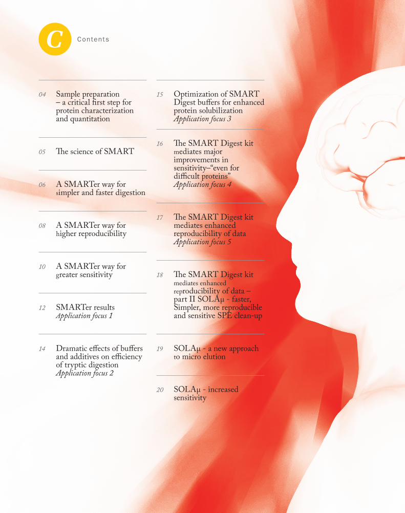
04 Sample preparation – a critical first step for protein characterization and quantitation
05 The science of SMART
06 A SMARTer way for simpler and faster digestion
08 A SMARTer way for higher reproducibility
10 A SMARTer way for greater sensitivity
12 SMARTer results Application focus 1
14 Dramatic effects of buffers and additives on efficiency of tryptic digestion Application focus 2
15 Optimization of SMART Digest buffers for enhanced protein solubilization Application focus 3
16 The SMART Digest kit mediates major improvements in sensitivity–“even for difficult proteins” Application focus 4
17 The SMART Digest kit mediates enhanced reproducibility of data Application focus 5
18 The SMART Digest kit mediates enhanced reproducibility of data – part II SOLAµ - faster, Simpler, more reproducible and sensitive SPE clean-up
19 SOLAµ - a new approach to micro elution
20 SOLAµ - increased sensitivity

Sponsored Feature4
Modern b iopharmaceu t ica l and proteomic laboratories perpetually seek to improve the speed, sensitivity and reproducibility of protein analysis. To this end, significant advances have been made in chromatography and mass spectrometry instrumentation. However, in virtually all cases, the protein sample must be treated in some way before it can be processed by these sophisticated machines. In fact, sample preparation is a critical first step in the analytical process, allowing sensitive hardware to accommodate diverse proteins presented in highly variable matrices – for example, therapeutic antibodies in commercial bioreactor medium, tumor antigens in a biopsy, and contaminants in processed food.
At the same time, sample preparation may be a costly pinch-point. In particular, the digestion stage of the sample preparation process - during which the protein analyte is enzymatically fragmented into its signature peptides, prior to purification and quantitative or qualitative analysis of the fragments -- can be both costly and problematic. The digestion step is usually undertaken with trypsin; issues associated with conventional trypsin digestions include the following:
• Sensitivity: Sensitivity is an important issue in modern proteomics. It is impacted by the trend of much lower sample volumes, and by the sample loss which may occur during
the evaporation / reconstitution steps used in conventional sample preparation protocols. Digestion efficiency also impacts sensitivity. If digestion of a small sample is incomplete, some fragments may be absent or difficult to detect.
• Variability: Data reproducibility is another key challenge for today’s proteomics lab. Avoiding repeats saves a huge amount of time, expense and effort - ‘right first time’ is the goal. Conventional digestions necessitate significant amounts of high quality trypsin to ensure consistent results - but this is costly.
• Labor intensity: Conventional trypsin-based sample preparation techniques are slow and resistant to automation. They therefore limit laboratory throughput and are associated with significant labor costs.
Improving reproducibility, sensitivity, and efficiency of analysis could have a significant impact - combining higher throughput and higher sensitivity with lower costs and fewer repeats can only improve the profitability and effectiveness of proteomics operations. How close are we to achieving this ideal? Here we discuss the impact of the Thermo ScientificTM SMART DigestTM Kit and Thermo ScientificTM SOLAµTM technology.
Video: Better Protein Digestion
Webinar: Peptide Maps
PDF: How Smart Digest
PDF: Smart Digest FAQs
Sample preparation - a critical first step for protein characterization and quantitation.
SMART Digest Kits
- facilitating perfect digestion
SMART Digest kits are designed for biopharmaceutical and proteomic applications which require highly reproducible digestion of proteins, combined with high data quality to enable robust high throughput workflows. SMART Digest Kits achieve this due to the highly stable immobilized trypsin design; which when combined with state of the art heating technology, provides fast, reproducible digestions ready for analysis by LC/MS.
Use the SMART DIGEST KIT tech guide to show how reproducible the digestion is with
SMART DIGEST.
www.thermoscientific.com/SMARTdigest
For internal use only
Thermo Scientific™ SMART Digest Kits 60109-101, 60109-102, 60109-103
• Highly reproducible digestion - enables fast generic methods and confidence in results
• Fast & simple digestion protocol - a few simple steps with samples typically ready for analysis in less than an hour
• Increase sensitivity and sequence coverage
Use the SMART DIGEST KIT tech guide to show how simple and fast the SMART DIGEST KIT
protocol is compared to in-solution digestion protocols.
Use the SMART DIGEST KIT tech guide to show how the SMART DIGEST Kit provides an increase in sensitivity compared to in-solution digests. This provides higher confidence in quantitation
and improved confidence in characterization.
Mor
e In
form
atio
n

Sponsored Feature 5
thermofisher.com/smartdigest
The SMART Digest kit was developed to address the need for faster, simpler, more sensitive and more reproducible methods in the field of sample preparation for protein analysis. In particular, the SMART Digest kit is designed to support the digestion stage of bottom-up protein analysis by mediating a step change in quality and efficiency. This advance is due to a unique combination of immobilized trypsin and optimized buffers.
Trypsin immobilisation In contrast to conventional procedures which digest proteins using free trypsin in solution, the SMART Digest kit is based on immobilized trypsin. This has a number of advantages:
• Tethering trypsin to a solid substrate allows it to be easily separated from the digestion products. This assists sample clean-up and results in more reproducible data.
• Immobilization stabilizes the trypsin, making it resistant to environmental changes, including high temperatures. Heat resistance in turn means that the protein sample can be denatured by heat without simultaneously denaturing the trypsin. Therefore, it is unnecessary to add chemicals to promote denaturation - a prerequisite for conventional digestions. In addition to saving time and materials costs, dispensing with chemical denaturation also eliminates the chemically-induced post-translational modifications associated with conventional digestion protocols. This removes another source of data variability.
• Finally, immobilized trypsin is less susceptible to autolysis. This helps make the digestion more efficient and complete, and therefore enhances the overall sensitivity of the process.
Immobilized enzymes therefore help to provide a simpler, more robust, more sensitive and more reproducible digestion method.
Buffer optimisationSecondly, rather than using standard buffering systems, the SMART Digest kit employs specific, optimized buffers. These enhance protein solubility and digestion speed and reproducibility, and facilitate downstream processing. The SMART Digest buffers therefore contribute to a simpler, faster and less variable method of sample preparation.
In combination, trypsin immobilization and optimized buffers result in faster and more complete digestion, simpler and easier to use systems, more reproducible and accurate data, and more sensitive assays. The net effect is a simpler, faster, better and less costly method of preparing protein samples for LC-MS analysis. Below, we examine the key features of the SMART Digest kit in more detail.
“Rather than using standard buffering
systems, the SMART Digest kit employs specific,
optimized buffers.”
Video: See how SMART Digest works
Mor
e In
form
atio
n
Sponsored Feature6
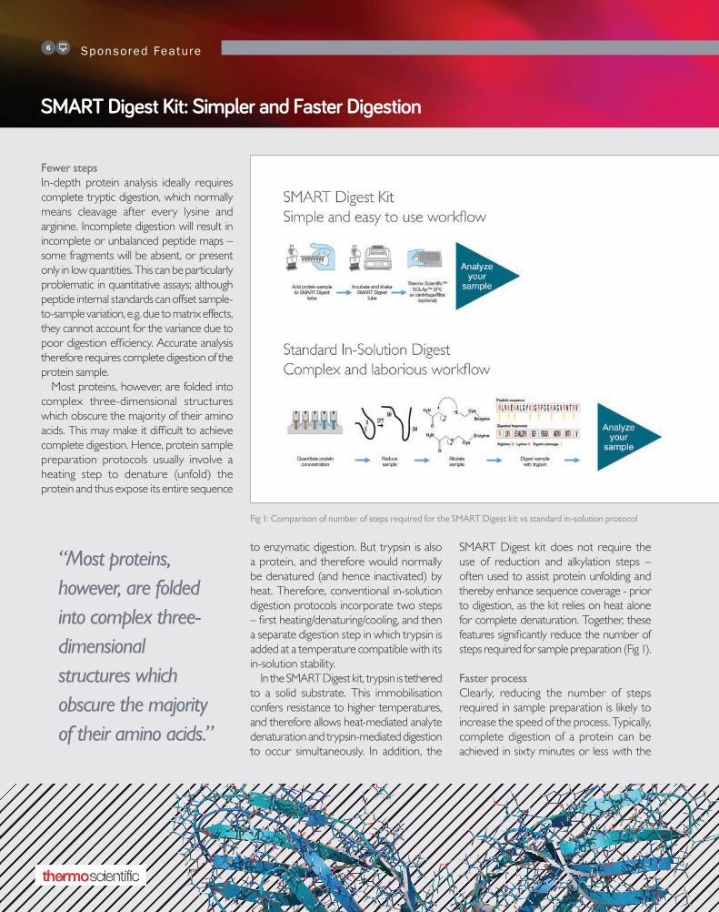
Fewer stepsIn-depth protein analysis ideally requires complete tryptic digestion, which normally means cleavage after every lysine and arginine. Incomplete digestion will result in incomplete or unbalanced peptide maps – some fragments will be absent, or present only in low quantities. This can be particularly problematic in quantitative assays; although peptide internal standards can offset sample-to-sample variation, e.g. due to matrix effects, they cannot account for the variance due to poor digestion efficiency. Accurate analysis therefore requires complete digestion of the protein sample.
Most proteins, however, are folded into complex three-dimensional structures which obscure the majority of their amino acids. This may make it difficult to achieve complete digestion. Hence, protein sample preparation protocols usually involve a heating step to denature (unfold) the protein and thus expose its entire sequence
to enzymatic digestion. But trypsin is also a protein, and therefore would normally be denatured (and hence inactivated) by heat. Therefore, conventional in-solution digestion protocols incorporate two steps – first heating/denaturing/cooling, and then a separate digestion step in which trypsin is added at a temperature compatible with its in-solution stability.
In the SMART Digest kit, trypsin is tethered to a solid substrate. This immobilisation confers resistance to higher temperatures, and therefore allows heat-mediated analyte denaturation and trypsin-mediated digestion to occur simultaneously. In addition, the
SMART Digest kit does not require the use of reduction and alkylation steps – often used to assist protein unfolding and thereby enhance sequence coverage - prior to digestion, as the kit relies on heat alone for complete denaturation. Together, these features significantly reduce the number of steps required for sample preparation (Fig 1).
Faster processClearly, reducing the number of steps required in sample preparation is likely to increase the speed of the process. Typically, complete digestion of a protein can be achieved in sixty minutes or less with the
SMART Digest Kit: Simpler and Faster Digestion
Fig 1: Comparison of number of steps required for the SMART Digest kit vs standard in-solution protocol
“Most proteins, however, are folded into complex three-dimensional structures which obscure the majority of their amino acids.”
SMART Digest kit. This compares with digestions requiring up to one and a half days or more with conventional protocols (Fig 2). The precise time will vary according to the nature of the sample, but for any
given protein using the SMART Digest kit is faster than conventional systems (Fig 3): for example, thyroglobulin, known to be ‘difficult,’ is digested to completion in 4 h with the SMART Digest kit as compared with 22 h for the solution-based protocol.
In addition, by providing a routine, standardized sample preparation protocol, the SMART Digest kit eliminates the need
for onerous and time-consuming method development. Aspects such as unfolding, cleavage, maintenance of tryptic activity and avoidance of auto-digestion no longer need to be optimized for each method, representing a significant time saving.
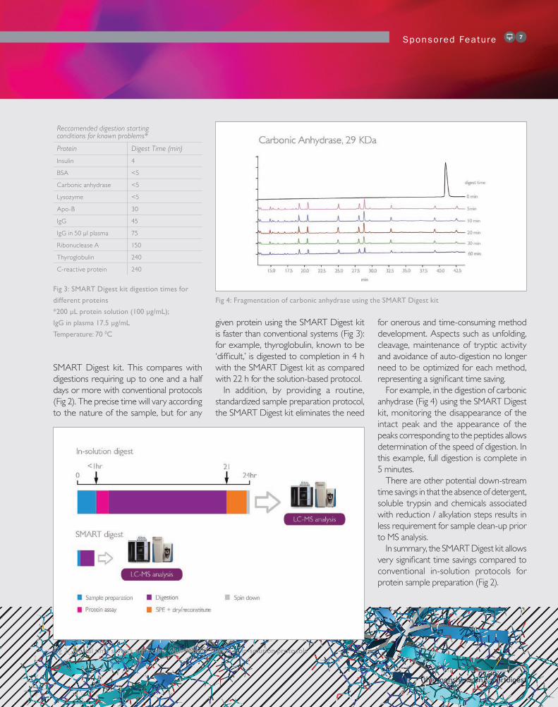
For example, in the digestion of carbonic anhydrase (Fig 4) using the SMART Digest kit, monitoring the disappearance of the intact peak and the appearance of the peaks corresponding to the peptides allows determination of the speed of digestion. In this example, full digestion is complete in 5 minutes.
There are other potential down-stream time savings in that the absence of detergent, soluble trypsin and chemicals associated with reduction / alkylation steps results in less requirement for sample clean-up prior to MS analysis.
In summary, the SMART Digest kit allows very significant time savings compared to conventional in-solution protocols for protein sample preparation (Fig 2).
Sponsored Feature 7
Fig 3: SMART Digest kit digestion times for
different proteins
*200 µL protein solution (100 µg/mL);
IgG in plasma 17.5 µg/mL
Temperature: 70 0C
Fig 4: Fragmentation of carbonic anhydrase using the SMART Digest kit
thermofisher.com/smartdigest
Reccomended digestion starting conditions for known problems*
Protein Digest Time (min)
Insulin 4
BSA <5
Carbonic anhydrase <5
Lysozyme <5
Apo-B 30
IgG 45
IgG in 50 µl plasma 75
Ribonuclease A 150
Thyroglobulin 240
C-reactive protein 240
Fig 2: Typical time savings achievable with SMART Digest vs in-solution protocols

Sponsored Feature8
A fundamental requirement of peptide mapping and quantitative analysis workf lows is reproducibility. This enables users to confidently assign data differences to the sample and not the methodological conditions used. Sources of variation in current approaches to peptide mapping and quantitative analysis include the number of different steps in the sample preparation process -- including protein assay, denaturation, alkylation, and reduction. These can differ between laboratories, making method transfer and data analysis between user groups problematic. Additionally, due to the number of steps required, in-solution digestion protocols are highly laborious, increasing the potential for user error. As a result, in-solution digestion may be associated with variable data, for example due to irreproducible protein cleavage which manifests in variation in the chromatographic profile.
Given that each manipulation is a potential source of variation, reducing the number of manipulations will reduce potential variation. Hence, the reduction in the number of steps in the SMART Digest kit sample preparation process (as described above) means that it is not only simpler and faster, but also has intrinsically less potential for user error and irreproducible data.
Other sources of variability include the presence of detergent, trypsin and reduction/alkylation-associated chemicals in sample preparation buffers. These process-associated contaminants can interfere with mass spectrometry, and therefore may hinder the collection and interpretation of data. The optimized
buffer systems of the SMART Digest kit, however, avoid the use of detergent and reduction/alkylation agents. Furthermore, there is no soluble trypsin in the SMART Digest kit – all trypsin is tethered to a solid substrate and therefore easily
separated from the analytes. The SMART Digest kits provide a highly stable environment for proteins and peptides, and support rapid, easy clean-up prior to MS. The system therefore generates highly reproducible data.
SMART Digest Kit: More Reproducible
Fig 5: Retention times and relative standard deviations for peptide fragments separated by the
Vanquish UHPLC system
Fig 6: Digestion reproducibility of a monoclonal antibody using the SMART Digest kit

Sponsored Feature 9
thermofisher.com/smartdigest
The controlled environment provided by the SMART Digest kit also avoids induction of chemically-induced post-translational modifications. For example, the pH 7.4 environment maintained by
the SMART Digest buffers prevents such modifications arising as a result of high pH. Since post-translational modifications are precisely the kind of variation many protein analysis protocols are intended to
identify, it is preferable to adopt a protocol that does not itself generate them.
Elimination of the above sources of variability in the SMART Digest kit results in cleaner, more reproducible data (Fig 5 and 6).
Furthermore, data indicate that the SMART Digest kit is associated with improved digestion efficiency, leading to more complete sequence coverage of the protein sample: for example, Digestion of the monoclonal antibody rituximab using the SMART Digestion kit indicates 100% sequence coverage, allowing post-translational modifications to be relatively quantitated with confidence (Fig 7 and 8).
Fig 8: Quantitation of post-translational modifications after treatment of rituximab with the SMART Digest kit
Protein Modification Recovery Abundance
Rituximab_LC Q1+NH3 loss Good 87.81%
Rituximab_LC W90+Oxidization Good 2.06%
Rituximab_HC ~Q1+NH3 loss Good 100.00%
Rituximab_HC W281+Oxidization Good 4.98%
Rituximab_HC N301+A1G0F Fair 2.87%
Rituximab_HC N301+A1G1F Fair 1.22%
Rituximab_HC N301+A2G0 Fair 1.30%
Rituximab_HC N301+A2G0F Fair 37.69%
Rituximab_HC N301+A2G1F Fair 44.86%
Rituximab_HC N301+A2G2F Fair 10.77%
Rituximab_HC N301+M5 Fair 1.07%
Rituximab_HC N365+Deamidation Good 2.72%
Rituximab_HC W385+Oxidation Good 5.37%
Rituximab_HC G450+Lys Good 3.2683%
PDF: Poster Note - Analysis of Monoclonal Antibodies
PDF: Application Note - Peptide Mapping Development
Poster Note - Speed & Reproducibility of Protein Digestion
Fig 7: Sequence coverage after treatment of rituximab with SMART Digest
Mor
e In
form
atio
n
Sponsored Feature10
The sensitivity of protein analysis methods refers to the extent to which they can detect low levels of analyte in a given sample. This in turn requires the sample preparation protocol to generate, retrieve and present uncontaminated analyte - often from very small quantities of sample material - to the detection instrumentation. In other words, to get the best out of highly sensitive LC-MS systems, you need an effective and efficient sample preparation method.
If sample quantity remains constant, method sensitivity usually depends significantly on the proportion of the total sample that is made available for LC-MS analysis. And to be available for LC-MS analysis the protein must first be solubilized, and then must remain in solution. Any reduction in the amount of sample available for analysis due to failure to dissolve – or subsequent precipitation – will impact the sensitivity of the process as a whole.
Furthermore, sensitivity is a function of the efficiency of the digestion step. This is because complete protein characterisation requires complete digestion, and complete detection of all peptides generated by digestion of that protein. If digestion is incomplete, some peptides will be absent, or present at undetectable levels. This may result in investigators erroneously concluding that mutations are present in the protein sequence.
The SMART Digest kit uses buffers that have been optimized for both solubility and digestion. For protein solubilisation, most sample preparation methods rely heavily on co-solvents, chaotropes or detergents. As discussed above, the addition of these chemicals can lead to downstream interference and clean-up issues, which themselves can impact sensitivity. However, the SMART Digest buffers permit an additive-free protocol which dissolves high proportions of the protein sample, and maintains them in solution. Also, the absence of the evaporation/reconstitution steps typically associated with sample preparation removes a potential source of sample loss. Users can therefore have confidence that a high proportion of the original sample will be delivered to detection and analysis instruments. Finally, the LC-MS signal is not suppressed by the presence of contaminants such as soluble trypsin or reduction-alkylation chemicals. All these factors contribute to the overall sensitivity of the system.
In addition, the buffers have been optimized for tryptic digestion. This feature of the system results in more complete digestion of the sample, and therefore generates higher levels of peptides, assisting the overall sensitivity of the protein analysis process. In some cases, the digestion may generate peptides that other sample preparation methods do not produce, permitting more complete analysis. The results of the improved sensitivity afforded
by the SMART Digest kit can be seen in Fig 9, which shows that use of the SMART Digest kit is associated with at least doubled sensitivity in the analysis of thyroglobulin (widely accepted as a ‘difficult’ protein). Comparison of the digestion of thyroglobulin using the SMART Digest kit with conventional in-solution digestion, with respect to three different peptides, shows that all peptides are present at far higher levels using the SMART Digest kit than with the in-solution protocol. Furthermore, the SMART Digest kit generated one peptide which was actually undetectable after conventional digestion.
SMART Digest Kit: More Sensitive
PDF: Application Note - Improved Proteome Coverage
PDF: Application Note - Improved Biomarker Identification
“Users can therefore have
confidence that a high proportion of
the original sample will be delivered to
detection and analysis
instruments.”
Mor
e In
form
atio
n

Sponsored Feature 11
Process-related costs arise not only from the direct use of materials and labor, but also from errors and inaccuracies which may require work to be repeated, and from inefficiencies which result in suboptimal outputs per unit time. In the highly regulated environment typical of many analysis laboratories, access to robust and controllable – preferably automatable – techniques is a great advantage, as it helps to eliminate the variability and human error which can force analyses to be repeated. Most protein sample preparation protocols, however, are labor-intensive, prone to human error, often give poorly reproducible results, and may be difficult to automate.
By contrast, the SMART Digest kit offers
significant savings in materials costs.These arise from, for example, the elimination of the chemicals associated with reduction and alkylation steps, and with sample evaporation and reconstitution. Energy savings also are associated with removal of these steps. Furthermore, the reduction in human error / process variability using the SMART Digest kit also saves the materials costs associated with repeated work. Finally, labor costs and opportunity costs are reduced by the massive time savings associated with the SMART Digest kit – in particular, the ability to complete protein digestion in an hour as opposed to a day (Fig 2), and even to digest a difficult protein such as thyroglobulin in a ~3.5 h process (Fig 3, 9).
Such savings could be further increased if the SMART Digest kit was applied in an automated system. The higher throughput achievable by automation would allow a commercial laboratory to increase its revenue earned per unit time, and would provide a higher return on investment .
SMART Digest Kit: Less Costly
Fig 9: Treatment of thyroglobulin with the SMART Digest kit
“The SMART Digest kit offers
significant savings in materials costs.”
thermofisher.com/smartdigest
Dramatic effects of buffers and additives on efficiency of tryptic digestionSummaryHere, we compared par tial trypsin digestion of insulin in various media, namely the SMART Digest buffer and a series of commonly used buffer species and additives. Insulin was chosen as the test protein because it creates a very simple peptide map – only two products, which are easily resolved from the intact protein using reversed phase chromatography. In all cases, digestion was effected using the SMART Digest immobilized trypsin (1 minute at 70 ˚C). A 1 minute time-point was chosen so as to produce only a partial digestion, and therefore to expose the efficiency of the process. Our resul t s show that diges t ion efficiency is highly dependent on the buffer and additives used, and that only one of the ~70 conditions tested was close to the SMART Digest buffer in terms of efficiency at 1 minute.
MethodBriefly, six sets of experiments were undertaken, respectively assessing the effects of the following buffer variables:
• Buffering ion (PBS, ABC, HEPES or Tris at concentrations 0.05 – 0.5 M)
• Electrolyte concentration (NaCl at concentrations 0.1 – 2 M)
• Chaotrope (guanidine HCl at concentrations 0.5 – 6 M; urea at concentrations 0.5 – 8 M; OGS at concentrations 0.05 – 2%)
• Detergents (Tween at 0.01 – 0.1% v/v; zwitterionic at 0.1 – 0.1%; sodium deoxycholate at 0.1 – 1%)
• Co-solvents (DMSO at 5-20% v/v; ACN at 5 – 20% v/v; MeOH at 5 – 20% v/v; IPA at 5 – 20% v/v; formamide at 5 – 20% v/v; TFE at 5 – 20% v/v)
• SMART Digest buffer.
100 µg/mL solutions of insulin were prepared in the various buffers, and 200 µL of each solution were added to a SMART Digest immobilized trypsin tube, and incubated for 1 minute at 70 ̊ C. After 1 minute, the digestion was quenched by acidification, and digestion products were analysed by LC/UV.
ResultsThe one minute time-point exposes dr amat ic ef fec t s of buf fer s and additives on the digestion efficiency of immobilized trypsin (Fig 10 A-F). The figures below compare the peak areas of the insulin C-terminal peptide generated by diges t ion in a SMART Diges t immobilized trypsin tube under various buffer conditions.
It is clear that some buffering ions can have a dramatic effect on digestion efficiency – ammonium bicarbonate and phosphate-buffered saline gave respectively a 90% and a 70% reduction in digestion product (Fig 10A).
The presence of salt may also be detrimental; the addition of sodium chloride reduces digestion product from immobilized trypsin by 20-30% over all the concentrations tested (Fig 10B).
Similarly, among the different pH conditions tested, all signif icantly reduced digest ion product from immobilized trypsin at one minute, with the exception of Tris buffer pH 8 (Fig 10C); Tris pH 8 was the only one of ~70 conditions which was equivalent to the SMART Digest buffer at a 1 minute time-point.
Chaotrope presence can have a highly significant effect on efficiency of digestion by immobilized trypsin. For guanidine HCl and urea the effect is clearly concentration dependent (Fig 10D), and these chaotropes virtually eliminate tryptic digestion at higher concentrations.
Detergents also have a negative effect on efficiency of digestion by immobilized trypsin, and in the case of zwitterionic detergent and sodium deoxycholate the presence even of low concentrations almost completely inhibited digestion (Fig 10E).
Finally, the presence of co-solvents had a significant and concentration-dependent inhibitory effect on digestion by immobilized trypsin. At higher concentrations, co-solvents almost completely inhibited digestion in the SMART Digest tube (Fig 10F).
ConclusionsIn the SMART Digest immobilized trypsin kit, the efficiency of digestion at one minute is significantly af fec ted by the buf fer species and additives used. Of the nearly 70 condit ions tested, only one approximated to the efficiency of the SMART Digest buffer kit, and this condition (Tris buffer pH 8) is known to be associated with chemically-induced post-translational modifications.
The SMART Digest buffer has been optimized for digestion efficiency, and the data shown above clearly show the enhanced efficiency at a one minute time point. In addition, the SMART Digest buffers have been designed for optimal protein solubility and stability – for example the pH is maintained at 7.4 to avoid unwanted protein modifications without losing digestion efficiency. Therefore the SMART Digest buffers would be expected to contribute to the enhanced reproducibility observed with the SMART Digest kit.
Applicat ion Note 12

thermofisher.com/smartdigest
Applicat ion Note 13
Fig 10. Peak areas of C-t peptide (GFFYTPKT) generated from the digestion of the protein using the SMART Digest kit as a function of a) variation of buffering
ion; b) salt concentration; c) pH; d) chaotropes; e) detergents; and f) organic co-solvents. The SMART Digest buffer result is shown as a red bar in each figure.
F) Effect of co-solvents

Optimization of the SMART Digest buffers for enhanced protein solubilizationSummaryBuffers for protein analysis protocols must accommodate conflicting demands. On the one hand, the necessity to dissolve and denature the protein analyte as completely as possible encourages the addition of co-solvents, detergents and chaotropes; conversely, the need to efficiently digest the dissolved protein, and to analyse the resulting peptides free of contaminants, requires that such additives should be kept to a minimum, and ideally dispensed with altogether. Here we show that the SMART Digest buffer can both maintain high levels of solubility and mediate efficient tryptic digestion with minimal additives.
MethodBriefly, the SMART Digest buffer was compared with other systems with respect to its ability to (i) solubilize bovine serum albumin (BSA) and (ii) to digest it. For the solubilisation experiments, the SMART Digest buffer was compared with Tris-buffered saline and a solution of trypsin in Tris-buffered saline; the ability of these buffers to support increasing concentrations of BSA up to 12.5 mg/ml was assessed. For the digestion experiments, the SMART Digest buffer alone was compared with the SMART Digest buffer containing varying concentrations of the following additives: acetonitrile; isopranol; dimethyl sulfoxide; methanol; trifluoroethanol; guanidine HCl; Tween 20; octylglucoside; deoxycholate. The ability of the buffer to maintain BSA solubility (12.5 mg/ml, post-
digestion) in the presence of these additives was examined.
ResultsIn the solubilisation experiments, significant protein precipitation was found to occur in both Tris and Tris-trypsin solutions (Fig 11, 12). The increase in solubility on addition of trypsin can largely be attributed to the effect of digestion of the protein to its constituent peptides, which are generally more soluble than the parent protein.
By contrast , the SMART Digest buffer was capable of solubilizing all concentrations of BSA up to as high as 12.5 mg/mL (Fig 13). This is five times higher than the other solvents.
In the post-digestion experiments, the SMART Digest buffer was found to support complete solubilisation of BSA and peptides. In all cases, the presence of additives resulted in increasing precipitation over time; the data for methanol (Fig 14) is typical.
ConclusionsStandard buffers and additives are associated with protein precipitation, both before and after digestion. SMART Digest buffers, by contrast, support protein and peptide solubility over a broad range of concentrations and time points. Therefore, sensitivity and accuracy of protein analysis protocols will be improved by using the SMART Digest buffers.
Fig 11: BSA solubility in Tris-buffered saline Fig 12: BSA solubility in Tris-buffered saline
with trypsin
Fig 13: BSA solubility in SMART Digest buffer Fig 14: Increased precipitation associated with
presence of methanol
Applicat ion Note 14

The SMART Digest Kit mediates major improvements in sensitivity – even for ‘difficult’ proteinsSummarySensitivity is an important parameter of protein analysis methods, particularly where the protein analyte is available in only small quantities and/or is difficult to digest, thereby liberating only low levels of peptide fragments. The overall sensitivity of a protein analysis method may be improved by ensuring that the protein is completely digested. Here we compare the digestion and consequent sens i t iv i t y mediated respectively by the SMART Digest kit and conventional solution-based tryptic digestion protocols. The test protein, thyroglobulin, is broadly recognized as difficult to digest, particularly in the context of a complex matrix (plasma). We show that the SMART Digest kit can mediate enhanced digestion of thyroglobulin, and results in major improvements in sensitivity with regard to particular thyroglobulin peptides, as compared with in-solution digestion.
MethodBriefly, for the SMART Digest kit study, 20 µg of thyroglobulin were added to 150 µL of SMART Digest buffer and 50 µL murine plasma, placed in a well containing the SMART Digest immobilized trypsin, and incubated at 70 ˚C. Samples were collected at 210 minutes (the optimum digestion time for thyroglobulin using
the SMART Digestion kit as determined by previous work). For the conventional ‘in-solution’ study, the method was as previously described (1). In both studies, three peptide sequences were monitored and compared. All samples were analysed by LC-MS.
ResultsComparison to previously published methods(1) using in-solution digestion showed less than half the signal response for each of the three peptides studied. One peptide that gave a very strong peak with the SMART Digest kit was entirely undetectable by the standard method (Fig 15). ConclusionsThe SMART Digest kit improves the sensitivity and efficiency of protein analysis
by providing a fast, simple, and clean tryptic digestion protocol. A comparison between the SMART Digest kit protocol and a published solution-based digestion protocol was run for the quantitation of thyroglobulin from whole murine plasma. Complete digestion was achieved in 3.5 hours with a SMART Digest kit, compared to 22 hours required for the solution-based protocol. Most significantly, the signal response of key SRM-peptides was found to be at least doubled when using the SMART Digest kit.
References
1. Clarke, N.J.; Zhang, Y.; Reitz, R.E. A novel mass
spectrometry-based assay for the accurate
measurement of thyroglobulin from patient
samples containing antithyroglobulin
autoantibodies, Journal of Investigative
Medicine, 2012, 60, 1157-1163
Fig 15: Comparison of peak area for three peptides generated from thyroglobulin by SMART Digest
and in-solution digestion respectively
thermofisher.com/smartdigest
Applicat ion Note 15

The SMART Digest kit mediates enhanced reproducibility of dataSummaryPeptide mapping of digested proteins is of high importance when characterizing biotherapeutics. Peptide maps are utilized to confirm the expression of the intended amino acid sequence, to confirm genetic stability or to identify pos t-t r ans lat iona l modi f icat ions , especially when interfaced with mass spectrometry. For highest confidence in the data, the retention time as well as the peak area has to be extremely stable. Here we demonstrate the unmatched retention time precision achieved with the Vanquish UHPLC system.
MethodBriefly, cetuximab monoclonal antibody (5 mg/ml) was di luted 1:4 with the SMART Diges t bu f fe r and incubated in a SMART Digest tube for 60 minutes at 70 ˚C. Digestion products were chromatographically analyzed with a Thermo Scientific™ Acclaim™ column.
ResultsUsing the SMART Digest kit, total preparation time of the monoclonal antibody digestion was less than 75 minutes. The results show excellent reproducibility across the whole chromatogram; Fig 16 shows an overlay of 13 repeated injections of the same sample of cetuximab tryptic digestion product. On average, standard deviation (SD) was of the order of 0.13 second(0.00214 minutes). SD for some peaks was as low as 0.065 seconds; and did
not exceed 0.3 seconds for any peptide. The relative standard deviation was consistently extremely low (Fig 17). Out of 110 peaks automatically integrated by Chromeleon CDS, 34 had RSD smaller than 0.01%, reaching the minimum value of 0.006% for the peak at retention time 23.057 minutes. The relative standard deviation of the peak areas was below 1.0% for all peptides. The average reproducibility was 0.4% highlighting the highly reliable sample injection and peak integration at challenging conditions.
ConclusionsThe Vanquish UHPLC system is extremely reproducible in both retention time and peak area reproducibility. The retention time precision provided by the system enables the analyst to be sure that any change in retention time relates to an actual change of the sample structure. The peak area reproducibility provided by Vanquish provides maximal confidence in quantitative work. Consequently, the Vanquish system meets the demanding requirements of peptide mapping analysis.
Fig 16: Overlay of 13 injections of the same sample of tryptic digest
Fig 17: Retention time relative standard deviation for 13 repeated injections of a tryptic digest
Applicat ion Note 16

The SMART Digest kit mediates enhanced reproducibility of data – Part IISummaryIn-solution trypsin digestion is associated with variable data quality, in part due to the multiple steps required and the conditions to which the analytes are exposed during the digestion process (for example, the presence of co-solvents, detergents, and chaotropes, which can interfere with MS analysis). Here, we tested the reproducibility of digestion that can be achieved when cytochrome C is digested with the SMART Digest immobilized trypsin. We found a relative standard deviation (RSD) of 6% for the peptides digested using the SMART Digest kit, compared with 5% for spiked peptide controls, indicating that the SMART Digest kit is highly reproducible.
MethodBriefly, 20 µg of cytochrome C was diluted to 200 µL in the SMART Digest buffer, and digested with the SMART immobilized trypsin for 10 minutes at 70 ̊ C, cleaned up and analysed by LC-MS. Four exogenous peptides were spiked in as controls to allow assessment of the reproducibility of the SMART Digest kit.
ResultsThe RSD of the cy tochrome C peptides indicates the level of precision of the entire process including the reproducibility of the digest. Comparing precision data between the exogenous and cytochrome C peptides allows assessment of the reproducibility of the digestion (Fig 18).
ConclusionsComparable levels of precision for both sets of peptides were observed. This indicates the following:
• The entire analytical workflow is precise (RSD of exogenous peptides = 5%)
• The reproducibility using the SMART Digest kit is precise (RSD of cytochrome C peptides = 6%).
Fig 18: Recovery of cytochrome C peptides after digestion using the SMART Digest kit
thermofisher.com/smartdigest
Applicat ion Note 17
SOLAµ: Faster, Simpler, More Reproducible and Sensitive SPE Sample Clean-up
Sponsored Feature18
Another critical step in the sample preparation process is solid phase extraction (SPE), in which the analyte of interest is separated from the matrix in which it is presented – for example, blood, water, ur ine or laboratory buffers – by being passed through a solid ‘stationary phase’. Typically, the stationary phase retains the analyte while contaminants are washed out. Subsequently, the analyte is recovered from the stationary phase by eluting with appropriate media.
Traditional SPE techniques, however, may not always be adequate for the demands of the modern proteomics laboratory. Larger molecules, such as proteins, can raise challenges for SPE, in
particular achieving the right selectivity. A particular challenge is the trend of much lower sample volumes, which means that laboratories must work in the micro-space to avoid dramatically diluting the sample; any sample loss - as is typically found at the evaporation-reconstitution stage – may be significant if the sample was very small to begin with. Micro-elution is a response to this, as it dispenses with the need to evaporate, but again standard techniques are not always ideal; for example, flow characteristics can become challenging when dealing with small bed volumes.
Therefore, although SPE is generally an effective technique for isolation of analytes from complex biological
matrices, conventional SPE is not always optimal; greater sensitivity and reproducibility would be welcome, including elimination of matrix effects.
A c co r d i n g l y , T h e r m o F i s h e r Scientific has developed SOLAµ, a low bed-weight Embedded Particle Technology (EPT) SPE device. It has been designed for increased sensitivity, enhanced reproducibility, speed and robustness. In addition, it is customisable according to the specific requirements of a given analysis. The flexibility of method development accorded by SOLAµ may be a key benefit in a regulated environment, as it enables a rapid accommodation of regulators’ requirements.
Video: Greater Reproducibility,
Increased SensitivityPDF: Benefits of
using SOLAµPDF: How
SOLAµ Helps PDF: Increasing
LC-MS Sensitivity
Sponsored Feature 19
thermofisher.com/smartdigest
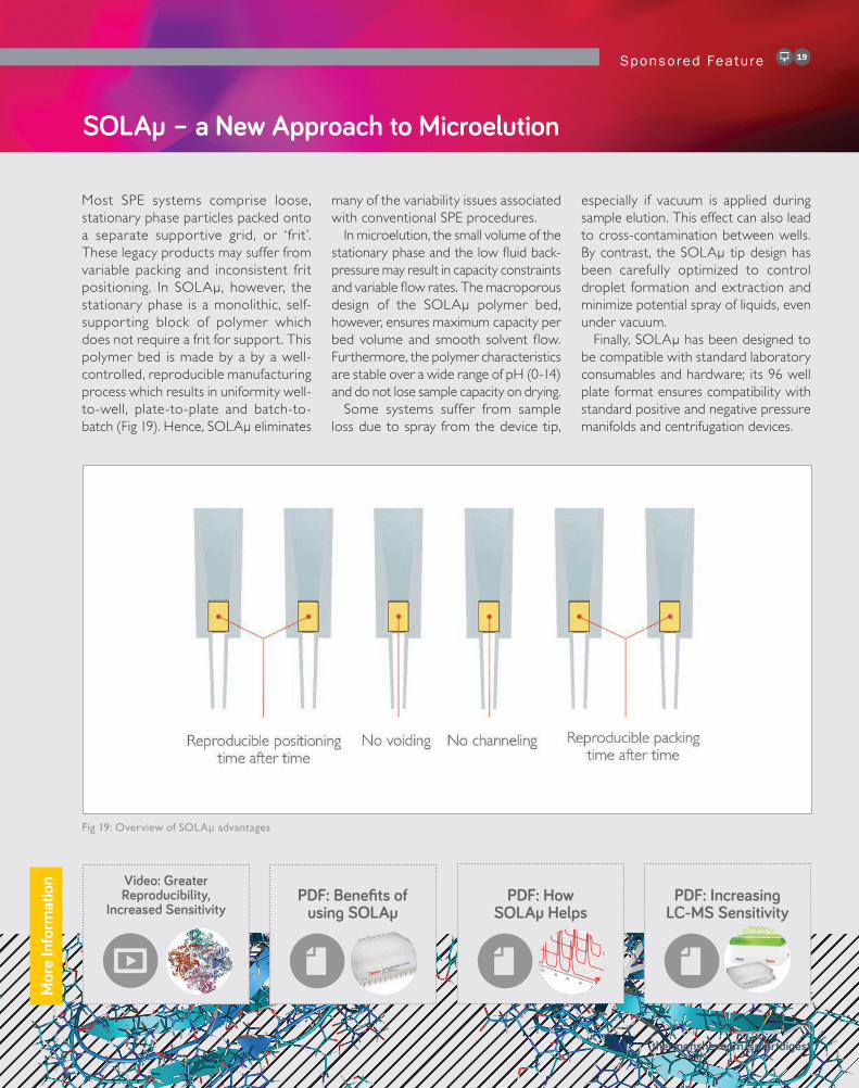
Most SPE systems comprise loose, stationary phase particles packed onto a separate supportive grid, or ‘frit’. These legacy products may suffer from variable packing and inconsistent frit positioning. In SOLAµ, however, the stationary phase is a monolithic, self-supporting block of polymer which does not require a frit for support. This polymer bed is made by a by a well-controlled, reproducible manufacturing process which results in uniformity well-to-well, plate-to-plate and batch-to-batch (Fig 19). Hence, SOLAµ eliminates
many of the variability issues associated with conventional SPE procedures.
In microelution, the small volume of the stationary phase and the low fluid back-pressure may result in capacity constraints and variable flow rates. The macroporous design of the SOLAµ polymer bed, however, ensures maximum capacity per bed volume and smooth solvent flow. Furthermore, the polymer characteristics are stable over a wide range of pH (0-14) and do not lose sample capacity on drying.
Some systems suffer from sample loss due to spray from the device tip,
especially if vacuum is applied during sample elution. This effect can also lead to cross-contamination between wells. By contrast, the SOLAµ tip design has been carefully optimized to control droplet formation and extraction and minimize potential spray of liquids, even under vacuum.
Finally, SOLAµ has been designed to be compatible with standard laboratory consumables and hardware; its 96 well plate format ensures compatibility with standard positive and negative pressure manifolds and centrifugation devices.
Fig 19: Overview of SOLAµ advantages
SOLAµ – a New Approach to Microelution
SOLAμ - no variability, just consistency
SOLAµ
mA
U
Time (min)
50.0
0
100
1015
2025
3035
4045
10
20
30
40
50
60
70
80
90
ANALYSIS 1
ANALYSIS 2
ANALYSIS 3
ANALYSIS n
Consistently Reproducible Results
• What do you need from your sample preparation?
• High levels of reproducibility
• Low failure rates
• High throughput
• Higher sensitivity
• Efficient process
• Ability to process samples with limited volume
• Ability to prevent loss of volatile samples
• Ability to prevent loss of samples susceptible to
solvation/non-specific binding issues
Mor
e In
form
atio
n

SOLAµ – Increased Sensitivity
Sponsored Feature20
The sensitivity of a bioanalysis system is partly a function of background noise – for example, due to contaminants interfering with the signal of interest – and partly a function of the amount of material presented to the detector. Overall sensitivity can be enhanced by reducing the former and increasing the latter.
Reduction of background noiseReduction of background contamination can be achieved by clean-up procedures in the sample preparation stage. Thus, a calibration exercise involving passing solutions of niflumic acid in human plasma through SOLAµ SPE provided a lower limit of quantitation of 40 pg/mL (see Fig 20 below), indicating very low signal interference. Similarly, matrix effects were very low, in the range 3.2 - 8.6% (Fig 21).
Increasing the available sampleIncreasing the amount of material available to the detector is achieved by optimizing recovery from the solid phase, minimizing loss in any subsequent steps, and – for a given volume – achieving higher concentrations.
Most SPE methods elute analytes from the solid phase in relatively large volumes, such that the cleaned-up analyte is relatively dilute. A given volume loaded onto the detector therefore will contain less analyte than may be optimal given the constraints of the detection technology. Accordingly, conventional SPE procedures usually include an evaporation step followed by reconstitution of the dried analyte in a smaller volume of buffer, to provide a more concentrated sample.
By contrast, the low elution volumes achievable with SOLAµ allow retrieval of samples which are already concentrated.
Fig 20: Calibration of niflumic acid in plasma, post SPE clean-up
Fig 21: Precision data for niflumic acid at low QC (0.4 ng/ml) and high QC (30 ng/mL)
Precision Data for Niflumic Acid Peak Area Ratio (%RSD) n = 18
Recovery of Niflumic Acid (%)
Matrix Effects (%)
QC Low (0.4ng/mL) 1.31 89.9 8.63
QC hIGH (30ng/mL) 1.06 94.0 3.21
Fig 22: SOLAµ decreases lower limit of quantitation by a factor of 20
PDF:Application Note - Speed & Reproducibility of
Protein Digestion
PDF:Application Note - Peptide Clean-up &
Concentration
Sponsored Feature 21
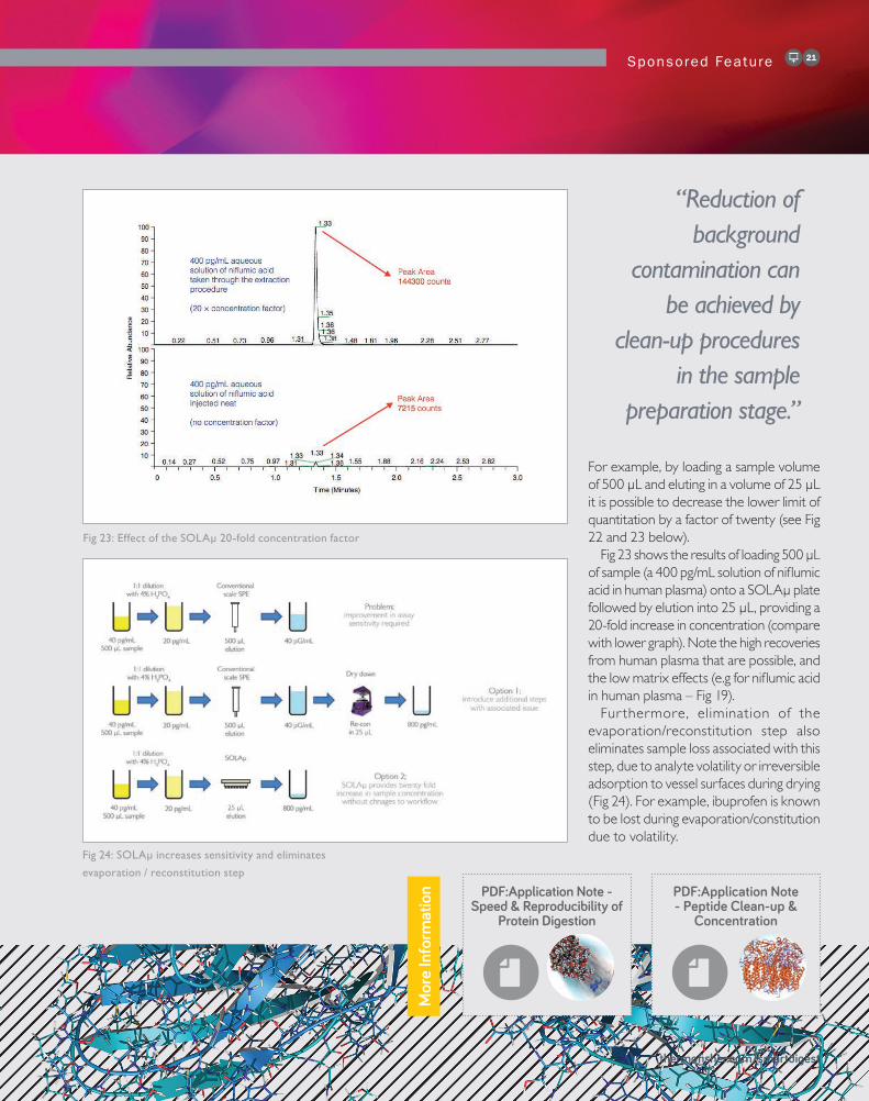
For example, by loading a sample volume of 500 µL and eluting in a volume of 25 µL it is possible to decrease the lower limit of quantitation by a factor of twenty (see Fig 22 and 23 below).
Fig 23 shows the results of loading 500 µL of sample (a 400 pg/mL solution of niflumic acid in human plasma) onto a SOLAµ plate followed by elution into 25 µL, providing a 20-fold increase in concentration (compare with lower graph). Note the high recoveries from human plasma that are possible, and the low matrix effects (e.g for niflumic acid in human plasma – Fig 19).
Fur thermore, elimination of the evaporation/reconstitution step also eliminates sample loss associated with this step, due to analyte volatility or irreversible adsorption to vessel surfaces during drying (Fig 24). For example, ibuprofen is known to be lost during evaporation/constitution due to volatility.
thermofisher.com/smartdigest
Fig 23: Effect of the SOLAµ 20-fold concentration factor
Fig 24: SOLAµ increases sensitivity and eliminates
evaporation / reconstitution step
“Reduction of background
contamination can be achieved by
clean-up procedures in the sample
preparation stage.”M
ore
Info
rmat
ion

www.theanalyticalscientist.com
A Sponsored Supplement From