Embed Size (px)
Citation preview

Page 1/24
Aging and MPTP-Sensitivity Depend on Molecular and UltrastructuralSignatures of Astroglia and Microglia in Mice Substantia NigraPL Abhilash
National Institute of Mental Health and Neuro SciencesUpasna Bharti
National Institute of Mental Health and Neuro SciencesMariamma Philip
National Institute of Mental Health and Neuro SciencesSanthosh Kumar Rashmi
National Institute of Mental Health and Neuro SciencesTrichur R Raju
National Institute of Mental Health and NeuroscienceBindu M Kutty
National Institute of Mental Health and Neuro SciencesBK Chandrasekhar Sagar
National Institute of Mental Health and Neuro SciencesPhalguni Anand Alladi ( [email protected] )
National Institute of Mental Health and Neuro Sciences https://orcid.org/0000-0002-2876-3478
Research
Keywords: Parkinson’s disease, 1-methyl-4-phenyl-1, 2, 3, 6-tetrahydropyridine (MPTP), Cytokine ELISA, Neuroin�ammation, Monoamine oxidases A&B,Unbiased stereology, C57BL/6J, CD-1 white mice, substantia nigra pars compacta, Fecal microbiome
Posted Date: February 12th, 2021
DOI: https://doi.org/10.21203/rs.3.rs-190412/v1
License: This work is licensed under a Creative Commons Attribution 4.0 International License. Read Full License
Loading [MathJax]/jax/output/CommonHTML/fonts/TeX/fontdata.js

Page 2/24
AbstractBackground
Both astroglia and microglia show region-speci�c distribution pattern in the central nervous system and often maladapt to age-associated alterations withintheir niche. Studies on autopsied substantia nigra of Parkinson’s disease (PD) patients and experimental models propose gliosis as a trigger for neuronal loss.Epidemiological studies propose an ethnic bias in PD prevalence, since Caucasians are more susceptible than non-whites living in Asia and Africa. Similarly,different mice strains are variably sensitive to the neurotoxin MPTP (1-methyl-4-phenyl-1,2,3,6-tetrahydropyridine). We had earlier likened divergent MPTP-sensitivity of C57BL/6J and CD-1 mice with differential susceptibility to PD, based on differences in neuronal numbers.
Methods
Here we examined whether the variable susceptibility was also incumbent to inter-strain differences in the glial features in the substantia nigra pars compacta(SNpc) of C57BL/6J and CD-1 mice. We performed unbiased stereology to quantify iba-1 immunoreactive microglia and s100β immunopositive astroglia onimmunohistochemically stained sections. Further, ELISA based estimation of pro- in�ammatory and anti-in�ammatory cytokines was supplemented withestimation of enzymes like fractalkine, hemeoxygenase, and monoamine oxidases A and B. Electron microscopy was performed to compare the effects on theorganelles.
Results
Stereological counts showed more microglia and fewer astrocytes in the substantia nigra of MPTP-susceptible normal C57BL/6J mice, which suggestspersistence of an immune-vigilant state. MPTP caused induction of microgliosis and astrogliosis in both strains, suggesting the involvement of these cells inpathogenesis. ELISA of pro-in�ammatory cytokines in the ventral-midbrain revealed augmentation of TNF-α and IL-6 at middle-age in both strains that reducedat old-age, suggesting middle-age as a critical, in�amm-aging associated time-point. TNF-α levels were persistently high in C57BL/6J, through aging and post-MPTP; while IL-6 and IL-1β were upregulated at old-age. CD-1 had higher levels of anti-in�ammatory cytokine TGF-β. MPTP-challenge caused upregulation ofenzymes MAO-A, MAO-B and iNOS in both strains. Post-MPTP enhancement in fractalkine and hemeoxygenase-1, may be neuronal compensatory signals.Lastly, ultrastructural observations of elongated mitochondria in astroglia and microglia vis-à-vis the shrunken ones in neurons, suggest upscaling of theirfunctions with neurotoxic consequences.
Conclusions
Thus, astroglia and microglia play a critical role in modulating aging and the susceptibility of an individual to PD.
HighlightsCD-1 substantia nigra has higher number of astroglia and fewer microglia than C57BL/6J
Both mice show age and MPTP-induced gliosis in the substantia nigra pars compacta
CD-1 nigra has lower levels of pro- and higher levels of anti-in�ammatory cytokines
Tilt of balance between pro- and anti-in�ammatory cytokines begins at middle age
Astrocytes and microglia show elongated mitochondria and intact ER upon MPTP-injection
IntroductionIn�amm-aging refers to the alterations in the neuron-glia communication during aging, that result from a persistent functional decline in the immune system,characterized by a generalized increase in the pro-in�ammatory markers (Franceschi et al., 2006). The system copes by releasing anti-in�ammatory cytokines;a process termed as “anti in�amm-aging”. The imbalance between in�amm-aging and anti in�amm-aging associated processes, supported by the geneticmake-up of the individual and environmental factors combine to trigger the onset or protect against age-related neurodegenerative diseases like Parkinson’sdisease (PD). PD is characterized by a selective loss of dopaminergic (DA) neurons, primarily in the substantia nigra pars compacta (SNpc), resulting instriatal dopamine depletion as well as dysfunction of the basal ganglia circuitry (Bernheimer et al., 1973; Damier et al., 1999). Non-motor symptoms likeconstipation, precede the motor symptoms by several years, thus suggesting a role for gut microbiota in the disease pathogenesis (Chaudhuri et al., 2006).
PD is characterized by T-cell in�ltration, microgliosis and astrogliosis (McGeer et al., 1988; Kohutnicka et al., 1998). Several preclinical and epidemiologicalstudies point at chronic neuroin�ammation as a prototypical event preceding and accompanying neuronal dysfunction. Neuroin�ammation marks thepresence of activated microglia and reactive astrocytes, direct participation of the adaptive immune system as well as increased synthesis of cytokines,chemokines, in�ammatory markers, reactive oxygen and nitrogen species (Boje and Arora, 1992; Boka et al., 1994; Kim et al., 2000). Midbrain DA neuronsexpress the receptors for cytokines such as tumor necrosis factor (TNF-α), interleukin-1β (IL-1β) and interferon-γ (IFN-γ), pointing at their sensitivity to thesecytokines (Boka et al., 1994; Mogi et al., 1994).
Epidemiological studies on PD suggest prevalence and incidence rates of approximately 108–257/100,000 and 11–19/100,000 person year, respectively (VanDen Eeden et al., 2003) in Europe. North America reported 329 − 107/100,000 in Nebraska-USA (Strickland and Bertoni, 2004) and 224 per 100,000 person-years in persons above 65 years (Wirdefeldt et al., 2011). The incidence among Indians, Chinese and Malays were less compared to the Westerners (Gourie-Devi M 2014; Abbas et al., 2017). Thus, the white populations have signi�cantly higher prevalence than non-whites; the mechanisms for which are unclear. Wedemonstrated the role of preservation of nigral neurons with age, maintenance of GNDF receptors and non-logarithmic increase in α-synuclein as few
Loading [MathJax]/jax/output/CommonHTML/fonts/TeX/fontdata.js

Page 3/24
neuroprotective factors in Asian Indians (Alladi et al., 2009; Alladi et al., 2010 a&b). We also found age-related morphological transformation of astrocytes andmicroglia (Jyothi et al., 2016). Direct comparisons on human tissues between populations were not possible.
A reliable recapitulation of the PD pathology in an animal model is observed following the injection of the neurotoxin 1-methyl-4-phenyl-1, 2, 3, 6-tetrahydropyridine (MPTP) (Jackson-Lewis and Przedborski, 2007). Different mice strains show varying responses to MPTP, for example, C57BL/6J mice ismore sensitive while CD-1 white, BALBc and Swiss Webster are resistant (Smeyne et al., 2001). C57BL/6J DA neurons exposed to MPP+ (1-methyl-4-phenylpyridinium) demonstrated a 39% loss when cultured on C57BL/6J glia compared to 17% neuron loss when cultured on SWR/J glia. Thus glia maymodulate the strain-dependent susceptibility of mice to MPTP and MPP+.
We have earlier reported that the CD-1 had more substantia nigra DA neurons than C57BL/6J and were better protected against MPTP (Vidyadhara et al.,2017). Soreq et al., (2017) reported that, astroglia show signi�cant senescence-associated changes in gene expression patterns. Yet, the role of glia in diseasemodulation as also in differential vulnerability is not completely understood. In the present study we systematically investigated the responses of astrogliaand microglia in the substantia nigra of the two different mice strains i.e. C57BL/6J and CD-1, in terms of aging and in�ammatory responses to MPTP.Studies in the last decade suggest a correlation between the gut microbiome and maturation as well as activation of the microglia (Heijtz et al., 2011; Erny etal., 2015). It is reported that in�amed gut releases pro-in�ammatory cytokines that cross the compromised BBB to trigger low grade in�ammation andin�amm-aging (Kelly et al., 2015). We therefore studied the microbiome composition of the two strains pre and post-MPTP. The outcome may be extrapolatedto understand the differential prevalence of PD between the Caucasians and Asian-Indians.
Materials And Methods
Experimental Animals and MPTP-HCl AdministrationWe used C57BL/6J (MPTP-susceptible) and CD-1 (MPTP-resistant) mice at 15–17 week (young adults), 10–12 months (middle-aged) and 18–20 months(old/aged). All the experiments were conducted on male mice as there is a male preponderance in prevalence and incidence of PD (reviewed by Georgiev et al.,2017). They were housed under standard laboratory conditions of temperature 25° ± 2°C, 12 h light:12 h dark cycle with ad libitum access to food and water.The mice received four intraperitoneal injections of MPTP-hydrochloride (15 mg/kg/dose) in saline, at 2-h intervals. The control ones received saline(Vidyadhara et al., 2017). The mice were sacri�ced at days 1, 4 & 7 after MPTP-injection and subjected to various analyses.
Tissue processing for immunohistochemistry (IHC):Male mice anaesthetized using iso�urane were intracardially perfused with 0.9% heparinized saline followed by 4% ice-cold buffered paraformaldehyde(0.1M). The brains were removed and post-�xed for 48 hours at 4°C; cryoprotected in buffered sucrose grades (10%, 20% & 30%). 40µm thick serial coronalsections of midbrains were collected on gelatin coated slides.
Immunostaining:Two different series were used for Iba-1 and s100-β labeling (n = 3–4/strain/age group/experimental condition); with a section periodicity of 1 in 6. Antigenwas unmasked using sodium citrate buffer (pH-6) at 80°C for 20 min. Endogenous peroxidase was quenched using 0.1% hydrogen peroxide (H2O2) in 70%methanol (20 min in dark). The non-speci�c binding was blocked with 3% buffered bovine serum albumin (BSA) for 4 hr at room temperature (RT). Theprimary antibody exposure (0.1M PBS-TX; dilution 1:500; Table 1) lasted for 72 hours at 4°C in a closed chamber. Biotinylated secondary labeling (8 hr;dilution 1:100, Vector labs, USA; Table 1) was followed by tertiary labeling with avidin-biotin complex (Vector labs, USA; 4 hr at RT). The staining wasvisualized with 0.05% DAB (3'-3'-diaminobenzidine) and 0.01% nickel sulphate in 0.1M acetate imidazole buffer (pH 7.4) containing 0.01% H2O2 resulting in ablack colored reaction. For the second antibody, similar procedure was followed (Table 1) except that the chromation was performed exclusively with DAB,resulting in brown colored pro�les [(TH (brown) and Iba-1/s100β black)]. Negative controls were processed without adding primary antibody. The sectionswere mounted with DPX following alcohol-based dehydration.
Loading [MathJax]/jax/output/CommonHTML/fonts/TeX/fontdata.js

Page 4/24
Table 1Details of Primary and Secondary antibodies used for immunohistochemistry
Staining modality, primary antibody and source Dilution and incubationtime
Secondary antibody and ABCkits
Dilution and incubationtime
Immuno-peroxidaseStaining
Anti-Goat Iba-1,
Abcam, UK
1:800
72 hr at 4°C
Goat Elite ABC kit,
Vector laboratories, USA
1:200, 4 hr at RT
Anti-Mouse S100β,
Sigma –Aldrich
1:800
72 hr at 4°C
MOM* ABC kit,
Vector laboratories,USA
1:500, 3 hr at RT
Anti-Rabbit TH,
Santa Cruz, USA
1:1000
72 hr at 4°C
Rabbit Elite ABC kit,
Vector laboratories,USA
1:200, 4 hr at RT
Immuno-�uorescence Anti-Goat Iba-1,
Abcam UK
1:800
72 hr at 4°C
FITC, Sigma Aldrich,USA 1; 200 6–8 hr at 4°C
Anti-Rabbit GFAP,
Abcam UK
1:500
24 hr at 4°C
Cy3, Sigma Aldrich,USA 1; 200 6–8 hr at 4°C
Anti-Rabbit Fractalkine, AbcamUK
1:500
72 hr at 4°C
Cy3, Sigma Aldrich, USA 1; 200 6–8 hr at 4°C
Anti Mouse CNPase, Abcam UK 1:1000
72 hr at 4°C
FITC, Sigma Aldrich, USA 1; 200 6–8 hr at 4°C
Anti Rabbit MFGE8,
Cloude Clone crop
1:800,
72 hr at 4°C
Cy3, Sigma Aldrich, USA 1; 200 6–8 hr at 4°C
MOM*: Mouse-on-mouse kits
For immuno�uorescence labeling, following the incubation with primary antibody, the sections were incubated in an appropriate �uorescently labeledsecondary antibodies (Sigma-Aldrich, USA). The sections were then mounted using Vectashield hard set mounting medium (Vector Laboratories, USA) andimages were captured using a laser scanning confocal microscope (DMIRE-TCS Leica Microsystems, Germany).
Stereological quanti�cation of cells:The immunoreactive (ir) cells in SNpc were quanti�ed using optical fractionator method with an Olympus BX61 Microscope (Olympus Microscopes, Japan)equipped with Stereoinvestigator software version 8.1 (Micro-bright�eld Inc., Colchester, USA). The SNpc in TH-ir midbrain sections was delineated on both thesides under 10X (Paxinos, 2013). Pilot studies determined the grid interval and counting frame size. Cells were counted in every sixth section (6–8sections/animal) under 100X, with a grid interval of 10000µm² (x = 100µm, y = 100µm) and counting frame of size 6400µm² (x = 80µm, y = 80µm) as per ourearlier report (Vidyadhara et al., 2017). The absolute numbers were derived and coe�cient of error was determined according to Gundersen et al., (1999).
Immunoblotting:The mice (n = 5/strain/age group/experimental condition) were sacri�ced by cervical dislocation and the midbrains were snap frozen in liquid nitrogen andstored at -80°C till further use. The brains were thawed to -20°C on a cryostat and 5µm thick sections of SN were solubilized in 100 ul of mammalian lysisbuffer 10% protease inhibitor cocktail (Sigma–Aldrich, USA). Following sonication (Q sonica, India) and centrifugation at 12000g (4°C) the proteinconcentration in the supernatant was assayed by Bradford methodand 60ug of protein/sample was electrophoresed (Bio-Rad, USA) on a 5%/10%(loading/separating) denaturing gel. The proteins were transferred onto a buffered poly-vinilidine di-�uoride (PVDF) membrane (Millipore, Germany). The non-speci�c staining was blocked by 5% skimmed milk protein (1X TBST; 4hr) followed by overnight incubation with primary antibody solution (Table 2.). Thiswas followed by incubation with appropriate HRP- conjugated secondary antibody for 2hr (Table 2.). The band was detected using chemi-luminescentsubstrate for HRP (Super Signal West Pico, USA) using a geldoc apparatus (Syngene International Ltd., India) and quanti�ed using Image J 1.48 v program(Vidyadhara et al., 2017).
Loading [MathJax]/jax/output/CommonHTML/fonts/TeX/fontdata.js
Page 5/24
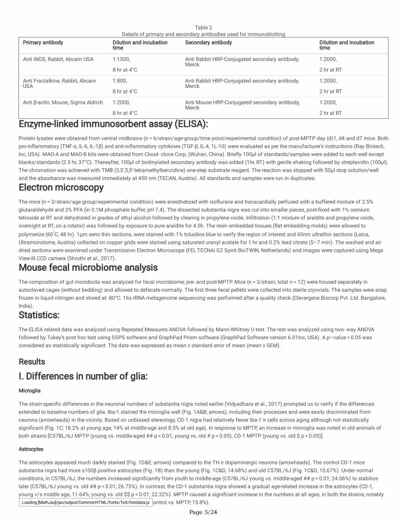
Table 2Details of primary and secondary antibodies used for immunoblotting
Primary antibody Dilution and incubationtime
Secondary antibody Dilution and incubationtime
Anti iNOS, Rabbit, Abcam USA 1:1500,
8 hr at 4°C
Anti Rabbit HRP-Conjugated secondary antibody,Merck
1:2000,
2 hr at RT
Anti Fractalkine, Rabbit, AbcamUSA
1:800,
8 hr at 4°C
Anti Rabbit HRP-Conjugated secondary antibody,Merck
1:2000,
2 hr at RT
Anti β-actin, Mouse, Sigma Aldrich 1:2000,
8 hr at 4°C
Anti Mouse HRP-Conjugated secondary antibody,Merck
1:2000,
2 hr at RT
Enzyme-linked immunosorbent assay (ELISA):Protein lysates were obtained from ventral midbrains (n = 6/strain/age-group/time point/experimental condition) of post-MPTP day (d)1, d4 and d7 mice. Bothpro-in�ammatory (TNF-α, IL-6, IL-1β) and anti-in�ammatory cytokines (TGF-β, IL-4, 1L-10) were evaluated as per the manufacturer’s instructions (Ray Biotech,Inc, USA). MAO-A and MAO-B kits were obtained from Cloud- clone Corp, (Wuhan, China). Brie�y 100µl of standards/samples were added to each well exceptblanks/standards (2.5 hr, 37°C). Thereafter, 100µl of biotinylated secondary antibody was added (1hr, RT) with gentle shaking followed by streptavidin (100µl).The chromation was achieved with TMB (3,3’,5,5’-tetramethylbenzidine) one-step substrate reagent. The reaction was stopped with 50µl stop solution/welland the absorbance was measured immediately at 450 nm (TECAN, Austria). All standards and samples were run in duplicates.
Electron microscopyThe mice (n = 3/strain/age group/experimental condition) were anesthetized with iso�urane and transcardially perfused with a buffered mixture of 2.5%glutaraldehyde and 2% PFA (in 0.1M phosphate buffer, pH 7.4). The dissected substantia nigra was cut into smaller pieces, post-�xed with 1% osmiumtetroxide at RT and dehydrated in grades of ethyl alcohol followed by clearing in propylene oxide. In�ltration (1:1 mixture of araldite and propylene oxide,overnight at RT, on a rotator) was followed by exposure to pure araldite for 4.5h. The resin embedded tissues (�at embedding molds) were allowed topolymerize (60°C, 48 hr). 1µm semi thin sections, were stained with 1% toluidine blue to verify the region of interest and 60nm ultrathin sections (Leica,Ultramicrotome, Austria) collected on copper grids were stained using saturated uranyl acetate for 1 hr and 0.2% lead citrate (5–7 min). The washed and airdried sections were examined under Transmission Electron Microscope (FEI, TECNAI G2 Spirit BioTWIN, Netherlands) and images were captured using MegaView-III CCD camera (Shruthi et al., 2017).
Mouse fecal microbiome analysisThe composition of gut microbiota was analyzed for fecal microbiome; pre- and post-MPTP. Mice (n = 3/strain; total n = 12) were housed separately inautoclaved cages (without bedding) and allowed to defecate normally. The �rst three fecal pellets were collected into sterile cryovials. The samples were snapfrozen in liquid nitrogen and stored at -80°C. 16s rRNA metagenome sequencing was performed after a quality check (Clevergene Biocorp Pvt. Ltd. Bangalore,India).
Statistics:The ELISA related data was analyzed using Repeated Measures ANOVA followed by Mann-Whitney U test. The rest was analyzed using two- way ANOVAfollowed by Tukey’s post hoc test using SSPS software and GraphPad Prism software (GraphPad Software version 6.01Inc, USA). A p–value < 0.05 wasconsidered as statistically signi�cant. The data was expressed as mean ± standard error of mean (mean ± SEM).
Results
I. Differences in number of glia:Microglia
The strain-speci�c differences in the neuronal numbers of substantia nigra noted earlier (Vidyadhara et al., 2017) prompted us to verify if the differencesextended to baseline numbers of glia. Iba-1 stained the microglia well (Fig. 1A&B; arrows), including their processes and were easily discriminated fromneurons (arrowheads) in the vicinity. Based on unbiased stereology, CD-1 nigra had relatively fewer Iba-1 ir cells across aging although not statisticallysigni�cant (Fig. 1C; 18.2% at young age; 14% at middle-age and 8.5% at old age). In response to MPTP, an increase in microglia was noted in old animals ofboth strains [C57BL/6J MPTP (young vs. middle-aged ## p < 0.01, young vs. old # p < 0.05), CD-1 MPTP (young vs. old $ p < 0.05)].
Astrocytes
The astrocytes appeared much darkly stained (Fig. 1D&E; arrows) compared to the TH ir dopaminergic neurons (arrowheads). The control CD-1 micesubstantia nigra had more s100β positive astrocytes (Fig. 1B) than the young (Fig. 1C&D, 14.68%) and old C57BL/6J (Fig. 1C&D, 10.67%). Under normalconditions, in C57BL/6J, the numbers increased signi�cantly from youth to middle-age (C57BL/6J young vs. middle-aged ## p < 0.01; 24.06%) to stabilizelater (C57BL/6J young vs. old ## p < 0.01; 26.75%). In contrast, the CD-1 substantia nigra showed a gradual age-related increase in the astrocytes (CD-1,young v/s middle age, 11.64%; young vs. old $$ p < 0.01; 22.32%). MPTP caused a signi�cant increase in the numbers at all ages, in both the strains; notablyhigher in young (C57BL/6J control v/s MPTP 20%; CD-1 control vs. MPTP, 15.8%).Loading [MathJax]/jax/output/CommonHTML/fonts/TeX/fontdata.js

Page 6/24
II. Cytokine expression in the ventral midbrain: Increase in the numbers of microglia and astrocytes with age and in response to MPTP led us to hypothesizethat gliosis may induce neuroin�ammation. We therefore estimated the levels of pro and anti-in�ammatory cytokines.
Pro-in�ammatory cytokine levels are relatively higher in C57BL/6J
The baseline TNF-α expression peaked at middle-age in the C57BL/6J (Fig. 2A; **** p < 0.0001). Between the two strains, the levels were signi�cantly higher inthe old C57BL/6J (C57BL/6J vs.CD-1 *p < 0.05). MPTP caused a sizeable upregulation in the C57BL/6J substantia nigra across ages and at all the timepoints, while CD-1 showed negligible changes e.g. young; (C57BL/6J vs. CD-1, post-MPTP d1,***p < 0.001; d7,*p < 0.05), middle-age (C57BL/6J vs. CD-1, post-MPTP d1, **** p < 0.0001; post-MPTP d7, *** p < 0.001), old (C57BL/6J vs. CD-1, post-MPTP d4, *p < 0.05).
Both the strains showed comparable patterns of IL-6 expression with age and following MPTP (Fig. 2B), with a signi�cantly higher expression at middle-age(C57BL/6J young vs. middle-age ####p < 0.0001); CD-1 young vs. middle-aged
p < 0.0001) and down-regulation at old-age [C57BL/6J (middle-aged vs. old ####p < 0.001); CD-1 (middle-aged vs. old aged
p < 0.0001)]. MPTP elicited similar responses in both strains at young as well as at middle-age when an upregulation was appreciated at 4 days post-MPTP (p < 0.05). At old-age the upregulation was better appreciated in C57BL/6J, that began at 4d and persisted till 7d (post-MPTP d4 C57BL/6J vs.CD-1 ****p < 0.0001, post-MPTP d7 C57BL/6J vs.CD-1 *p < 0.05).
IL-1β showed a signi�cant reduction with aging in both strains (Fig. 2C; C57BL/6J young vs. middle-aged ####p < 0.0001; CD-1 young vs. middle-aged
p < 0.0001; C57BL/6J middle-aged vs. old #### p < 0.001; CD-1 middle-aged vs. old
p < 0.001). MPTP-injected young C57BL/6J showed an acute increase at d1 (control vs. post-MPTP day1 #p < 0.05) followed by a reduction till d7 (post-MPTPd1 vs. post-MPTP d7 ###p < 0.001), whereas young CD-1 mice showed an increase at d4 (control vs. d4 $p < 0.05) followed by a reduction at d7 (d4 vs. d7 $$$p < 0.001). At middle-age there were no perceptible differences between the strains as both showed a signi�cant reduction at days 1–4 post MPTP followed by aresumption at d7 [C57BL/6J (control vs. post-MPTP day1 ####p < 0.0001, post-MPTP d4 vs.d7 ###p < 0.001); CD-1 (post-MPTP d4 vs.d7 $$$p < 0.001)] probablysuggesting overshadowing of this interleukin by TNF-α. The old C57BL/6J showed an augmented response at d7 (post-MPTP d7 C57BL/6J vs. CD-1 **** p < 0.0001) probably suggesting a late response by activated astrocytes.
B. Anti-in�ammatory cytokine levels are relatively lower in C57BL/6J:The basal level expression of anti-in�ammatory TGF-β was signi�cantly lower in C57BL/6J at all age points [(Fig. 3A C57BL/6J vs. CD-1; young *p < 0.05),middle-aged (****p < 0.0001), old (****p < 0.0001)]. In response to MPTP, CD-1 showed a profound increase in expression in the young [post-MPTP d7(Fig. 3A1, C57BL/6J vs. CD-1 #p < 0.05). Middle-aged CD-1 showed maximum strain-speci�c differences at d1 and d7 (post-MPTP d1 ****p < 0.0001; d7 ****p < 0.0001). At old-age the increase was less prominent at d1 (C57BL/6J vs. CD-1 *p < 0.05) but increased gradually till d7 (C57BL/6J vs. CD-1 **** p < 0.001).
Both the strains showed a signi�cant increase in IL-4 expression at middle-age, with relatively higher levels in C57BL/6J (Fig. 3B; C57BL/6J vs. CD-1 *p < 0.05). In the young CD-1, the expression decreased at d1 post-MPTP, yet causing a signi�cant upsurge at d4. C57BL/6J showed comparatively higher IL-4expression at old age in response to MPTP at d4 (C57BL/6J vs.CD-1 ** p < 0.01) and d7 (C57BL/6J vs.CD-1 * p < 0.05).
An upregulation in the basal level expression of IL-10 was observed in C57BL/6J at middle-age (Fig. 3C; C57BL/6J young v/s middle-aged ****p < 0.0001).MPTP caused a signi�cant upsurge in IL-10 expression in C57BL/6J at middle and old age (middle-aged post-MPTP d1&4 **p < 0.01; d7 *p < 0.05). At old age,MPTP elicited an increase only at d4 (C57BL/6J vs. CD-1 *** p < 0.001), and d7 (C57BL/6J vs. CD-1 **** p < 0.0001).
III: Differences in enzyme expression:
A: MAO-A and MAO-B levels:MAO-A positive punctae were localized to the cytoplasm (Fig. 4A1, red) of the DA neurons (Fig. 4A2) as also in the neuropil (Fig. 4A3 pink coloration inneurons and red in the neuropil). The basal levels were higher in the substantia nigra of young CD-1 (C57BL/6J vs. CD-1 *p < 0.05), however middle-age saw apeak in C57BL/6J (young vs. middle aged ###p < 0.001). In both strains, the levels reduced at old age (Fig. 4B, C57BL/6J middle aged vs old ### p < 0.001; CD-1 middle aged vs. old $$ p > 0.01). MPTP induced a notable increase in the young C57BL/6J; although both strains showed moderate enhancement at all ages(C57BL/6J control vs. MPTP ###p < 0.001).
The GFAP expressing glia (Fig. 4B1, green) showed MAO-B immunoreactivity (Fig. 4B2, red Fig. 4B3 merge, yellowish green). MAO-B levels were also higher inyoung CD-1 than C57BL/6J (Fig. 4 fD). An age-associated gradual down-regulation in CD-1 was contrasted by a gradual increase in C57BL/6J, althoughstatistically signi�cant.In summary, MPTP administration up regulated both MAO-A and MAO-B level in both strains
B: Inducible nitric oxide synthase (iNOS):Iba-1 immunopositive microglia (Fig. 5E1; green) expressed iNOS (Fig. 5E2; red) in addition to some non-microglial cells (Fig. 5E3 merge; red). The antibodyshowed a single band of 140 KDa (Fig. 5I). Young CD-1 had signi�cantly higher basal iNOS (Fig. 5F; C57BL/6J vs. CD-1 *p < 0.05). At middle-age both strainsshowed a moderate increase in expression. With aging, the CD-1 mice showed mild decrease in iNOS expression whereas C57BL/6J maintained the levelsattained at middle age. MPTP caused iNOS augmentation across ages in both strains (middle-aged C57BL/6J control vs. MPTP # p < 0.05, old C57BL/6Jcontrol vs. MPTP # p < 0.05, old CD-1 control vs. MPTP $ p < 0.05).
Loading [MathJax]/jax/output/CommonHTML/fonts/TeX/fontdata.js

Page 7/24
C: Fractalkine:Fractalkine was localized to the DA neuronal cytoplasm (Fig. 5G1-G3). The antibody showed a band at 140 KDa (Fig. 5J). The CD-1 substantia nigra hadmoderately high levels at all the ages, compared to C57BL/6J. A decrease was evident in both strains after middle age (Fig. 5H, C57BL/6J middle-age vs. old# p < 0.05, CD-1 middle-age vs. old $$ p < 0.01). MPTP elicited an increase in both the strains, but more appreciably in the young (C57BL/6J, young adults vs.middle-aged #### p < 0.0001, middle-aged vs. old #### p < 0.0001).
D: Hemeoxygenase-1 (HO-1):The young CD-1 midbrains showed mildly higher levels of HO-1 which reduced following MPTP. Both strains showed a signi�cant up-regulation at middle-agethat persisted till old age (Fig. 5k, C57BL/6J young vs. middle-aged #### p < 0.001, CD-1 young vs. middle-aged $$$ p < 0.001). Aged mice of both strainsshowed an increase in HO-1 expression in response to MPTP; moderate in C57BL/6J; signi�cant in CD-1 (CD-1 control vs. MPTP$$$ p < 0.001).
IV: Age-related and MPTP-induced ultrastructural changes:
A: Effects on Substantia Nigra NeuronsThe mitochondria of the young and middle aged C57BL/6J were relatively larger than those of the elderly (Fig. 5A,E&I). MPTP induced mitochondrialshrinkage at all ages. (Fig. 5 compare A&B and E&F, I&J; ‘M’ arrows). However in CD-1 they were well preserved with age (Fig. 5 compare C&D and G&H, K&L;‘M’ arrows) and even longer in response to MPTP (Fig. 5H, H1 and L, arrow). The endoplasmic reticular (ER) strands shortened with age and in response toMPTP in C57BL/6J (Fig. 5, compare A&B and E&F, I&J; ER).Whilst MPTP induced ER dilatation in the young (Fig. 5; D2) and middle-aged CD-1 (Fig. 5H; ER).The normal aged CD-1 showed presence of ER arrays (Fig. 5K1). Interestingly, the aged mice of both strains showed presence of several Golgi apparatus units(Fig. 5I ‘Go’). In the MPTP-administered aged C57BL/6J they appeared circular and possessed bloated saccules with “pearl necklace like appearance” (Fig. 5,F&H; J&L1; ‘Go’). In the older C57BL/6J apoptotic bodies were noted (Fig. 5, I1; ‘Ab’) while in CD-1, the neuronal nucleus was often crenellated. The neurons ofMPTP-injected young CD-1 harbored several lysosomes, which were conspicuously absent in the neurons of young C57BL/6J (Fig. 5B&D ‘Ly’).
B: AstrocytesThe astrocytic nuclei were larger but not uniformly ovoid/round like those of oligodendrocytes. Their nuclear chromatin was �ne and granular. The cytoplasmwas sparse and granular within the perinuclear zone (Luse SA, 1956), but more electron dense than the neuronal cytoplasm. The nucleus remainedeuchromatic through aging and in response to MPTP. The astrocytes of young and middle aged C57BL/6J had numerous long and tubular cytoplasmicmitochondria (Fig. 6, ‘M’, compare A&B and E&F) whereas those in the myelinated axons were spherical (‘Ma’). In CD-1, the cytoplasmic mitochondria wereoval (Fig. 6, compare C&D and G&H). The astrocytic mitochondria were relatively longer in the old CD-1 (Fig. 6, compare E&G). The ER was relatively wellmaintained through aging and in response to MPTP in both strains. Golgi saccules were semicircular and dilated in the middle aged MPTP-injected CD-1(Fig. 6, Go, compare G&H and K&L). The scale bar is 1µm for all micrographs except ‘K’.
C: MicrogliaMost microglia were present near the blood vessels and had electron dense cytoplasm with a bean shaped nucleus. Heterochromatin nets and electron densepockets were noted along the nuclear perimeter. In the young, the nuclei were euchromatic (Fig. 7, A-D; ‘M-nu’) while those of middle aged and old, wereelectron dense (Fig. 7, E-L; ‘M-nu’). In the cytoplasm and neighboring tracks of MPTP-injected young C57BL/6J, long tubular mitochondria were noted (Fig. 7B,‘M’). The young CD-1 showed phagocytotic microglia along with the engulfed cells (Fig. 7D; ‘EC’) as also the MPTP-injected old C57BL/6J (Fig. 7J).Interestingly amoeboid “dark cells” were seen around blood vessels in both strains after middle age (Fig. 7E, G and I; ‘DC’) and had thin rim of cytoplasmcontaining few organelles.
Thickening of the vascular basement membrane:Blood vessels showed comparable membrane architecture in the young mice of both strains in control conditions as well as upon MPTP challenge (Fig. 8A-D).At middle age, MPTP injected C57BL/6J showed a membrane discontinuity suggesting a possible breach (E vs F, arrowheads) and thickening of basementmembrane in C57BL/6J (arrows, E vs F) but not in CD-1 (F vs H). At old age, MPTP caused thickening of vascular basement membranes in both strains(arrows, I vs J; K vs L).Macrophage-like-cells (** I&J) were seen in the lumen in old C57BL/6J. Dark cells (DC) were noted too.
MPTP caused microbial dysbiosisIt has been observed that in�amed gut releases pro-in�ammatory cytokines which can cross the compromised BBB and thus trigger low grade in�ammationand in�amm-aging (Kelly et al., 2015). We therefore studied the microbiome composition of the two strains pre and post-MPTP. The metagenome dataanalysis showed differences in the relative abundance of fecal microbiota between C57BL/6J and CD-1 mice (Fig. 9). The heat map showed that thegenus/species belonging to Streptococcus, Lachnospiraceae, Bacteriodes, Candidatus, Ruminococcaceae were more abundant in the C57BL/6J whereas inCD-1 those belonging to Bacteriodales, Lactobacillus and Prevotellaceae were more and those of Prevotellawere less. MPTP injection increased thePrevotellaceae and Lachnospiraceaespp in CD-1 and Prevotella, Lachnospiraceae, Bacterodales, Candidatus-SaccharomonasandRuminococcus population inC57BL/6J.
DiscussionLoading [MathJax]/jax/output/CommonHTML/fonts/TeX/fontdata.js

Page 8/24
The baseline number of microglia and astrocytes vary between strains:Microglia are selectively more populous in the hippocampus and SNpc (Lawson et al., 1990), the target regions of the common age-associated diseases likeAlzheimer’s and Parkinson’s disease respectively; endorsing their role in neurodegeneration and susceptibility. In aging mice, microglial priming is equated toan immune vigilant state that results in their de-rami�cation, hypertrophy, increase in numbers and exaggerated response to sub-threshold challenges. Theysynthesize ROS and pro-in�ammatory cytokines like TNF-α, IL-6, IL-1β; while curtailing the synthesis of anti-in�ammatory cytokines (Damani et al., 2011;Grabert et al., 2016). The presence of more microglia at middle age corroborate with the possibility of a pre-in�ammed status and presence of damageassociated molecular patterns (DAMPs) in-situ, thereby in�ammasomes too may be vital in determining susceptibility. We found that normal C57BL/6J micehave fewer substantia nigra neurons (Vidyadhara et al., 2017) and more baseline apoptosis (Yarreiphang et al., 2020 unpublished data) vis-à-vis the CD-1mice. Neuronal apoptosis �ags the entry of microglial precursor cells into the zebra �sh brain (Casano et al., 2016) hence, the higher number of microglia inC57BL/6J substantia nigra complement higher apoptosis and suggest higher baseline susceptibility.
Aging selectively affects the already sparse astrocytes in the substantia nigra, as against the other mesencephalic niche (Damier et al., 1996). Underphysiological conditions, they secrete trophic factor GDNF (Grondin et al., 2003), therefore the presence of fewer astrocytes in young C57BL/6J and an acuteincrease in their numbers at middle-age that persisted till old age, suggest reduced neuroprotection and gliosis-assisted in�amm-aging linked with cytokines.Whereas, the sedate increase in astrocytic numbers in CD-1 implicate moderate age-related changes in the milieu. Excessive cytokine releasing glia recruit theirneighbors, to set off a vicious cycle, wherein the pro-in�ammatory environment becomes self-propagating (Noh et al., 2014).
Pro-in�ammatory cytokine levels are higher in the susceptible strain:DA neurons are sensitive to the pro-in�ammatory cytokine TNF–α (Sriram et al., 2006); which is primarily secreted by microglia and in synchrony with IFN-γ/IL-1β induces neurodegeneration (Chao et al., 1995; Jeohn et al., 1998). Thereby, the dying neurons hold microglia in their cytotoxic state, to escalate synthesis ofTNF-α leading to a self-perpetuating neuroin�ammation. The supernumerary microglia and the persistently higher baseline TNF-α level through aging inC57BL/6J, may be interlinked and their microglia may be consistently primed, leading to increased neuronal susceptibility. Elevated TNF-α levels are reportedin mouse models as well as in autopsied brain tissue/CSF of PD patients (Mogi et al., 1994). Low levels of TNF-α are protective (Chertoff et al., 2011). Thus,low levels in CD-1 in addition to being a mark of low susceptibility, may be potentially neuroprotective. Our auxiliary �nding of up-regulation of TNF-α atmiddle-age in C57BL/6J, indicates that imbalance in cytokine milieu precedes senescence.
IL-6, a pleiotropic cytokine, is up-regulated in SN, CSF and serum of PD patients (Blum-Degena et al., 1995; Hofmann et al., 2009). Conversely, Bolin et al.,(2002) reported ampli�ed MPTP-susceptibility in IL-6−/− mice. In our study, since IL-6 followed a similar age-related expression pattern in both strains, it maynot dictate baseline susceptibility. Yet, the differences following MPTP validate its role as a toxicity signal. The middle-age demarcates a period of enhancedsusceptibility. MPTP-induced IL-6 expression in old C57BL/6J at later stages of the challenge, suggests a link with astroglial responses or excessive neuronaldeath.
Reactive microglia in the degenerating SN overproduce IL-1β, to activate astrocytes and promote iNOS secretion (Chhor et al., 2013). Chronic IL-1β expressionin Wistar-rat substantia nigra induced progressive neurodegeneration, microgliosis and motor disabilities(Ferrari et al., 2006). The acute increase in IL-1β at d1post-MPTP that persists till d7 in the young C57BL/6J vis-à-vis CD-1endorses immediate microglial priming in the former. Thus, the mice differ in theimmediacy or respondence indices. Moreover noticeably higher basal TNF-α in C57BL/6J at middle and old age underlines its role in basal susceptibility,aging and neurodegeneration, whereas, IL-6 and IL-1β appear to be responders to MPTP and hence involved in pathogenesis.
The resistant strain has higher levels of anti-in�ammatory cytokines:TGFβ1 inhibits microglial activation and protects DA neurons against MPTP-toxicity (Arimoto et al., 2007; Pintado et al., 2011). The low baseline TGF-βexpression in C57BL/6J through aging and following MPTP, suggest that the sensitive strain is ill equipped against neuroin�ammation. MPTP-elicitedupregulation of TGF-β in middle-aged CD-1 at days 1 and 7 suggest a microglia mediated initiation that persists till the late activation stage of astrocytes.Since CD-1 have more substantia nigral DA neurons and a sizeable number resists MPTP (Vidyadhara et al., 2017), higher TGFβ1 levels allude toneuroprotection and DA neuronal survival.
IL-4 protects DA neurons against MPP+ toxicity by up-regulating CD200, a microglial resting signal (Lyons et al., 2009). The signi�cant upsurge at d4 in MPTP-injected young CD-1, suggests an auxiliary course by the microglia. Interestingly, the elevated response at late stages of MPTP exposure in old C57BL/6J, maybe a delayed attempt at neuroprotection (reviewed by Hirsch and Hunot, 2009). In view of the pronounced increase in MPTP-sensitive C57BL/6J at middle-age,it is likely that IL-4 may also have a pro-in�ammatory role. Bok et al., (2018) while showing the microglia-speci�c expression of IL-4 demonstrated an LPS-induced upregulation in expression and rescue of substantia nigra DA neuronal loss by antibodies against IL-4.
IL-10 stimulates CD200 expression in neurons and induces astrocytes to synthesize anti-in�ammatory TGF-β. It inhibits microglial synthesis of TNF-α, NO andROS, in-vitro, to neutralize oxidative stress (Balasingam and Yong, 1996; Ledeboer et al., 2002). The up-regulated baseline IL-10 expression in C57BL/6J andupon MPTP at middle/old-age may be a compensatory increase. However, these increases do not parallel a raise in TGF-β levels; hinting at a failed rescueattempt. Thus, the acute increase in basal levels of both TNF-α and IL-6 as well as the anti-in�ammatory IL-1β and IL-10 expound a hovering imbalance of pro-and anti-in�ammatory cytokines, at middle age; therefore tempting one to speculate that middle-age imitates the prodromal period in the susceptible strain.
Strain speci�c variability in enzyme responses:MAO-B inhibitors are promising candidates in the treatment of early-PD(Rabey et al., 2000). MAO-B level increases with age (Irwin et al., 1997)and is doubledin SN of PD patients (Damier et al., 1996). The age-associated up-regulation in MAO-A and MAO-B levels in C57BL/6J suggests a functional decline. Thegradual age-related reduction in CD-1 may be a natural phenomenon that assists in combating MPTP-related stress. Although the reasons for higher basalLoading [MathJax]/jax/output/CommonHTML/fonts/TeX/fontdata.js

Page 9/24
level of MAO-A and MAO-B in young CD-1 are unclear, it may be due to higher number of neurons or DA terminals and astrocytes in CD-1striatum or it may be abystander susceptibility marker. MPTP may cause a feed-forward effect on glia, setting off a toxicity cycle.
HO-1 a cytoprotective, anti-apoptotic, and anti-in�ammatory enzyme; down-regulates pro-in�ammatory cytokines like TNF-α1 and IL-1β and up-regulates anti-in�ammatory cytokine IL-10 in-vitro (Doré et al., 1999; Petrache et al., 2000). Predominantly expressed by astrocytes (Dwyer et al., 1995), it is positivelycorrelated with aging and PD (Schipper et al., 1998). It was projected as a potential biomarker due to high levels in the patient saliva(Song et al., 2018). HO-1overexpression tendered neuroprotection in MPP+ treated Parkinsonian rats via BDNF and GDNF(Hung et al., 2008). Thus, the gradual age-related increase inits levels in C57BL/6J and in response to MPTP may indicate a cellular offset response to oxidative stress. Induction of iNOS causes NO release, which whenprotracted triggers oxidative damage in DA neurons (Nathan and Xie, 1994). The MPTP-induced up-scaling of iNOS in both strains supports this hypothesis.
The higher baseline iNOS and MAO-B levels alongside lower HO-1 levels in CD-1 are presently un-explained; yet it is likely that higher number of astrocytes andneurons could be the reason. Alternatively, these may be markers of sub-threshold susceptibility.
Neurons secrete the chemokine fractalkine (CX3CL1), to maintain microglia in resting state (Cardona et al., 2006). The inherently higher fractalkine levels inCD-1 indicate their healthy status. Age-related decrease in C57BL/6J relays enhanced phagocytic signals, an indirect indicator of neuronal loss.Overexpression in response to MPTP in young mice implies the activation of compensatory responses during youth which decline with age.
Surviving neurons harbor strain-speci�c ultrastructural signatures:This is the �rst study on the alterations in the ultrastructure of mice substantia nigra with aging and in response to MPTP. The age-related and MPTP-inducedshrinkage of mitochondria in C57BL/6J validates the ensuing mitochondrial dysfunction and aging as a risk factor for PD. Upregulation of mitochondrial�ssion protein dynamin-like protein 1 (DLP1/DRP1) as well as downregulation of fusion proteins Mfn1 and Mfn2 were noted in the substantia nigra of PDpatients (Zhao et al., 2017). We earlier found higher DRP-1 levels in the lateral/ventral substantia nigra of C57BL/6J, earmarking the inherent susceptibility ofthe mitochondria. The well-preserved mitochondrial structure and size with age and upon-MPTP in CD-1 complements the higher HSD-10 (mitochondrialfusion-associated protein) expression (Seshadri and Alladi, 2019). Elongated mitochondria are deft in energy generation (reviewed by Galloway et al., 2010)and calcium uptake (Lewis et al., 2018). In Caenorhabditis elegans, modulation of mitochondrial proteases SPG-7 and PPGN-1 enhanced mitofusion(Chaudhari and Kipreos, 2017) to extend their overall survival. Thus enhanced HSD-10 expression (Seshadri and Alladi, 2019)and elongated mitochondria inCD-1 neurons may be the survival modalities.
Both fragmented and dilated ER are major pathological notations in A53TαS Tg mice substantia nigra (Colla et al., 2012) and rotenone model of sub-cutaneous administration (Zhang et al., 2017), suggesting ER dysfunction in PD. ER arrays in old CD-1 could be rejoinders of enhanced protein synthesis tocompensate for the concurrent protein loss or mis-folding. The MPTP-induced ER shortening in C57BL/6J and its dilation in CD-1 suggests existence of strain-speci�c differences as well as different aspects of ER dysfunction in PD, which needs to be studied in detail.
The presence of many intact Golgi units in the aged substantia nigra of both strains suggest a compensatory increase to circumvent age effects on proteinpackaging and post-translational processing. The “pearl necklace like globose” bloated saccules of Golgi units upon MPTP-injection in old C57BL/6J, hint atdisease-induced functional impairment. Knockdown of adhesion proteins like GRASP 55/65 cause Golgi cisternae swelling(Lee et al., 2014). Simulationstudies suggest that aberrations in biophysical properties like osmotic pressure, adsorption and adhesion energies, and precise vesicle addition frequency inaddition to biological properties like rim stabilizer proteins cause abnormal self-organization into circular/fused Golgi complexes (Tachikawa and Mochizuki,2017). The presence of lysosomes in neurons of MPTP-injected young CD-1 that were visibly fewer in C57BL/6J may suggest either the activation oflysosomal/UPR pathway or lysosomal accumulation due to impaired late endocytic pathway (Guerra et al., 2019). The apoptotic bodies in older C57BL/6J,signal the occurrence of age-associated apoptosis. The crenellated nuclei in MPTP-injected old CD-1, suggest necroptosis as seen in striatal cells of aHuntington’s mouse (Turmaine et al., 2000). Thus, majority of organelles were affected in C57BL/6J both with aging and upon MPTP indicating a�iction ofmany cellular processes. Contrarily, elongation of mitochondria, preservation of Golgi apparatus and presence of lysosomes may be pointers of resilience inCD-1. Amongst the organellar defects, ER dilation is a sure sign of susceptibility in CD-1.
Elongation of glial mitochondria, a distinctive feature of pathogenesis:In an interesting spin off, unlike the neurons, ultrastructure of glial organelles was preserved during aging and following MPTP. The glial mitochondria thatwere smaller and fewer in controls C57BL/6J, appeared enlarged/elongated in response to MPTP. Hoekstra et al., (2015) showed a reduction in �ssion proteinDLP1/DRP1 in both neurons and astrocytes in PD cortex along with fused elongated mitochondria in primary cortical astrocytes transfected with DLP1-siRNA.Co-culturing them with cortical neurons caused neuronal atrophy and excessive calcium release; suggesting neurotoxic effect of fused astrocyticmitochondria. In a stroke model, the penumbral astrocytes displayed hypertrophic and polarized processes; elongated mitochondria and lost theirneuroprotective ability (Fiebig et al., 2019). Overexpression of mutant ubiquitin (UBB + 1) protected astrocytes from oxidative stress and H2O2-induced celldeath by destabilizing mitochondrial �ssion-speci�c proteins, leading to mitochondrial fusion (Yim et al., 2014). Although the exact corollaries are not clear,the combination of astrogliosis and increased pro-in�ammatory cytokines; tempts one to speculate that elongated mitochondria assist astroglial propagationinto neurodegenerative sequels.
Mitochondrial elongation in microglia of MPTP-injected young C57BL/6J mice, may have similar neurodegenerative consequences. LPS-activated mousecerebral microglia, stimulate DRP-1 and ROS synthesis in-vitro, to elongate tract borne mitochondria (Katoh et al., 2017). The dilated Golgi apparatus inmicroglia indicate that while aggravating neuroin�ammation, the microglial protein packaging process is also affected. Under chronic stress, in aging and inAD; microglia had condensed, electron dense cytoplasm and nucleoplasm which imparted a striking “dark” appearance (Bisht et al., 2016). Dark cells noted inboth strains from middle age, may have similar pathological objective. In most cells, the cytoplasm appeared as a thin rim, reducing the scope to visualizeother organelles.
Loading [MathJax]/jax/output/CommonHTML/fonts/TeX/fontdata.js

Page 10/24
Our �ndings of mitochondrial elongation may have clinical implications. For instance, the antioxidant coenzyme Q10 (CoQ10) supports mitochondrialfunction while reducing the DA neuron loss in an animal model of PD (Spindler et al., 2009), yet it failed in the phase III clinical trials (The Parkinson StudyGroup QE3 Investigators 2014). Similarly, CoQ10 derivative MitoQ also failed (Snow et al., 2010). It is likely that these molecules stabilized the glialmitochondria too, thereby nullifying the neuronal outcome. It is vital to study the glial mitochondrial responses in isolation and on a temporal scale, to betterunderstand the phenomenon.
Earlier age at onset of MPTP-induced basement membrane features in C57BL/6J:MPTP caused basement membrane thickening similar to that reported in PD (Farkas et al., 2000). Presence of gaps in the middle aged C57BL/6J and old CD-1 implies that the blood-brain barrier (BBB) is vulnerable earlier in life in C57BL/6J, an additional signal of negative impact of aging or on the gut microbiota(Montagne et al., 2015) or of inherent susceptibility.
Gut microbiome composition is distinct in the two strains:The gut microbiome modulates the formation of BBB, neurogenesis, microglial maturation and also in�uences brain homeostasis and behavior (Heijtz et al.,2011; Erny et al., 2015). In�amed gut releases pro-in�ammatory cytokines that cross the compromised BBB to trigger low grade in�ammation and in�amm-aging(Kelly et al., 2015). PD patients show a reduction in microbes of Prevotellaceae, Lachnospiraceae and Ruminococceae family etc. alongside an increasein Enterobacteriaceae, Bi�dobacteriumetc. The abundance of Prevotella, Lachnospiraceae, Enterobacteriaceae and Bacterodales population in C57BL/6J vis-à-vis Prevotellaceae in CD-1 mice upon MPTP-injection was a serendipitous outcome that validates strain-typical differences in fecal microbiome responseswhile post-MPTP increase in Prevotellaceae portrays defense mechanisms in the latter. The abundance of Enterobacteriaceae correlated positively with theseverity of postural instability and gait di�culty in PD patients (Scheperjans et al., 2015). Post-MPTP worsening of motor de�cits is more prominent inC57BL/6J than CD-1 (Vidyadhara et al, 2019). Thus, fecal microbiome is a reliable non-invasive marker of susceptibility.
ConclusionIn summary, neuro-glial interactions, senescence related changes in glia, cytokine levels etc. are vital determinants of neuronal survival and differ greatly in theMPTP-resistant C57BL/6J and MPTP-susceptible CD-1 white mice strain. The intended use of male animals for the study may be a limiting factor, however inview of the male preponderance of the disease; our observations provide vital clues of disease pathogenesis. Besides, female mice are also known to showhigher fatality in response to MPTP, due to differences in peripheral metabolism of MPTP; independent of its effects on dopaminergic neurons. It is alsopertinent to compare these factors between male and female animals to understand the premise for neuroprotection in females.
Our �ndings in general, may be extrapolated to different human populations that are either vulnerable or resistant to PD. For instance, in our study, the CD-1represents Asian-Indians who have inherently lower prevalence rate than the Caucasians. The prominent differences in pro- and anti-in�ammatory cytokinelevels at middle-age suggests that by design, the middle-age milieu is acquiescent to neurodegeneration and may well be the critical soft period for the onsetof neurodegenerative diseases. It is likely that senescence may result from differences in the in-situ in�ammasomes, which merit detailed investigations. Thepathogenesis is effectively assisted by the diabolic differences between the neuronal and glial mitochondria. The differences in fecal microbiome highlight thepossibility of using it as a non-invasive marker of susceptibility. Thus glia are major players in aging and disease and may explain the ethnic bias inprevalence of PD.
AbbreviationsMPTP: 1-methyl-4-phenyl-1, 2, 3, 6-tetrahydropyridine
BBB: Blood brain barrier
BSA: Bovine serum albumin
Coenzyme Q10: CoQ10
D1: day 1 post-MPTP:
D4: day 4 post-MPTP
D7: day 7 post-MPTP
DAMP Damage associated molecular patterns
DLP1/DRP1: Dynamin-like protein 1
ELISA :Enzyme-linked immunosorbent assay
Endoplasmic reticular ER
GFAP: Glial Fibrillary Acidic Protein
H2O2: Hydrogen peroxide
Loading [MathJax]/jax/output/CommonHTML/fonts/TeX/fontdata.js

Page 11/24
HO-1:Hemeoxygenase-1
Iba-1: Ionized calcium-binding adaptor protein-1
IFN-γ: Interferon-γ
IL-1β: Interleukin-1β
iNOS: Inducible nitric oxide synthase
Ir: immunoreactive
MAO-A : Monoamine oxidase A
MAO-B : Monoamine oxidase B
MPP+ :1-methyl-4-phenylpyridinium
PD: Parkinson’s disease
PVDF: Poly-vinilidine di-�uoride
RT:.Room temperature
SNpc: Substantia nigra pars compacta
TMB: 3,3’,5,5’-tetramethylbenzidine
TNF-α: Tumor necrosis factor
DeclarationsEthics approval: All the experimental protocols on mice were approved by the Institutional Biosafety committee and Institutional (NIMHANS) Animal EthicsCommittee, in accordance with the guidelines of the CPCSEA, India, and NIH, USA.
Consent for publication: Not applicable
Availability of data and materials: The raw datasets used and/or analysed during the current study are available from the corresponding author on reasonablerequest.
Competing interests: None of the authors have any con�ict of interest.
Funding: The study was funded by DBT (No.BT/PR12518/MED/30/1462/2014) to PAA. APL was a UGC SRF.
Authors' contributions: APL contributed to study design, performed experiments and collected data and provided 1st draft; UB performed experiments andcollected data; MP performed statistical analysis of the data; RSK & BKCS performed electron microscopy and data analysis; TRR and BMK: contributed todata analysis and revision of MS; PAA conceptualized the study, performed data analysis, edited the manuscript and obtained funds,.
Acknowledgements: We thank Dr. M.M. Srinivas Bharath, Head, Department of Clinical Psychopharmacology and Neurotoxicology; Dr. Gayathri N, Departmentof Neuropathology; Dr. Monojit Debnath, Department of Human Genetics, NIMHANS for laboratory facilities.
ReferencesAbbas MM, Xu Z, Tan LCS.Epidemiology of Parkinson's Disease-East Versus West.Mov Disord Clin Pract. 2017 Dec 22;5(1):14-28. doi: 10.1002/mdc3.12568.eCollection 2018 Jan-Feb.
Alladi PA, Mahadevan A, Shankar SK, Raju TR, Muthane U. 2010a. Expression of GDNF receptors GFRalpha1 and RET is preserved in substantia nigra parscompacta of aging Asian Indians. J Chem Neuroanat. 2010 Sep;40(1):43-52. doi: 10.1016/j.jchemneu.2010.03.007. Epub 2010 Mar 27.
Alladi PA, Mahadevan A, Vijayalakshmi K, Muthane U, Shankar SK, Raju TR.2010b. Ageing enhances alpha-synuclein, ubiquitin and endoplasmic reticularstress protein expression in the nigral neurons of Asian Indians. Neurochem Int. 2010 Nov;57(5):530-9. doi: 10.1016/j.neuint.2010.06.018. Epub 2010 Jul 6.
Alladi PA, Mahadevan A, Yasha TC, Raju TR, Shankar SK, Muthane U. 2009. Absence of age-related changes in nigral dopaminergic neurons of Asian Indians:relevance to lower incidence of Parkinson's. Neuroscience. Mar 3;159(1):236-45. doi: 10.1016/j.neuroscience.2008.11.051.
Arimoto, T., Choi, D. Y., Lu, X., Liu, M., Nguyen, X. V., Zheng, N., … Bing, G. (2007). Interleukin-10 protects against in�ammation-mediated degeneration ofdopaminergic neurons in substantia nigra. Neurobiology of Aging. Jun; 28(6): 894-906. https://doi.org/10.1016/j.neurobiolaging.2006.04.011
Loading [MathJax]/jax/output/CommonHTML/fonts/TeX/fontdata.js

Page 12/24
Balasingam, V., & Yong, V. W. (1996). Attenuation of astroglial reactivity by interleukin-10. Journal of Neuroscience. May 1; 16(9): 2945-55.https://doi.org/10.1523/jneurosci.16-09-02945.1996
Bernheimer, H., Birkmayer, W., Hornykiewicz, O., Jellinger, K., & Seitelberger, F. (1973). Brain dopamine and the syndromes of Parkinson and Huntington Clinical,morphological and neurochemical correlations. Journal of the Neurological Sciences. Dec; 20(4): 415-55. https://doi.org/10.1016/0022-510X(73)90175-5
Bisht, K., Sharma, K. P., Lecours, C., Gabriela Sánchez, M., El Hajj, H., Milior, G., … Tremblay, M. È. (2016). Dark microglia: A new phenotype predominantlyassociated with pathological states. GLIA. May 1; 64(5): 826-839. https://doi.org/10.1002/glia.22966
Blum-Degena, D., Müller, T., Kuhn, W., Gerlach, M., Przuntek, H., & Riederer, P. (1995). Interleukin-1β and interleukin-6 are elevated in the cerebrospinal �uid ofAlzheimer’s and de novo Parkinson’s disease patients. Neuroscience Letters. Dec 29; 202(1-2): 17-20. https://doi.org/10.1016/0304-3940(95)12192-7
Boje, K. M., & Arora, P. K. (1992). Microglial-produced nitric oxide and reactive nitrogen oxides mediate neuronal cell death. Brain Research. Aug 7; 587(2): 250-6. https://doi.org/10.1016/0006-8993(92)91004-X
Bok, E., Cho, E. J., Chung, E. S., Shin, W. H., & Jin, B. K. (2018). Interleukin-4 contributes to degeneration of dopamine neurons in the lipopolysaccharidetreatedsubstantia nigra in vivo. Experimental Neurobiology. Aug; 27(4): 309-319. https://doi.org/10.5607/en.2018.27.4.309
Boka, G., Anglade, P., Wallach, D., Javoy-Agid, F., Agid, Y., & Hirsch, E. C. (1994). Immunocytochemical analysis of tumor necrosis factor and its receptors inParkinson’s disease. Neuroscience Letters. May 19; 172(1-2): 151-4. https://doi.org/10.1016/0304-3940(94)90684-X
Bolin, L. M., Strycharska-Orczyk, I., Murray, R., Langston, J. W., & Di Monte, D. (2002). Increased vulnerability of dopaminergic neurons in MPTP-lesionedinterleukin-6 de�cient mice. Journal of Neurochemistry. Oct; 83(1): 167-75. https://doi.org/10.1046/j.1471-4159.2002.01131.x
Cardona, A. E., Pioro, E. P., Sasse, M. E., Kostenko, V., Cardona, S. M., Dijkstra, I. M., … Ransohoff, R. M. (2006). Control of microglial neurotoxicity by thefractalkine receptor. Nature Neuroscience. Jul; 9(7): 917-24. https://doi.org/10.1038/nn1715
Casano, A. M., Albert, M., & Peri, F. (2016). Developmental Apoptosis Mediates Entry and Positioning of Microglia in the Zebra�sh Brain. Cell Reports. Jul 26;16(4): 897-906. https://doi.org/10.1016/j.celrep.2016.06.033
Chao, C. C., Hu, S. X., Ehrlich, L., & Peterson, P. K. (1995). Interleukin-1 and tumor necrosis factor-α synergistically mediate neurotoxicity: Involvement of nitricoxide and of n-methyl-d-aspartate receptors. Brain Behavior and Immunity. Dec; 9(4): 355-65. https://doi.org/10.1006/brbi.1995.1033
Chaudhari, S. N., & Kipreos, E. T. (2017). Increased mitochondrial fusion allows the survival of older animals in diverse C. Elegans longevity pathways. NatureCommunications. Aug 3; 8(1): 182. https://doi.org/10.1038/s41467-017-00274-4
Chaudhuri, K. R., Healy, D. G., & Schapira, A. H. V. (2006). Non-motor symptoms of Parkinson’s disease: Diagnosis and management. Lancet Neurology. Mar;5(3): 235-45. https://doi.org/10.1016/S1474-4422(06)70373-8
Chertoff, M., Di Paolo, N., Schoeneberg, A., Depino, A., Ferrari, C., Wurst, W., … Pitossi, F. (2011). Neuroprotective and neurodegenerative effects of the chronicexpression of tumor necrosis factor α in the nigrostriatal dopaminergic circuit of adult mice. Experimental Neurology. Feb; 227(2): 237-51.https://doi.org/10.1016/j.expneurol.2010.11.010
Chhor, V., Le Charpentier, T., Lebon, S., Oré, M. V., Celador, I. L., Josserand, J., … Fleiss, B. (2013). Characterization of phenotype markers and neuronotoxicpotential of polarised primary microglia In vitro. Brain, Behavior, and Immunity. Aug; 32: 75-80. https://doi.org/10.1016/j.bbi.2013.02.005
Colla, E., Coune, P., Liu, Y., Pletnikova, O., Troncoso, J. C., Iwatsubo, T., … Lee, M. K. (2012). Endoplasmic reticulum stress is important for the manifestations ofα-synucleinopathy in vivo. Journal of Neuroscience. Mar 7; 32(10): 3306-20. https://doi.org/10.1523/JNEUROSCI.5367-11.2012
Damani, M. R., Zhao, L., Fontainhas, A. M., Amaral, J., Fariss, R. N., & Wong, W. T. (2011). Age-related alterations in the dynamic behavior of microglia. AgingCell. Apr; 10(2): 263-76. https://doi.org/10.1111/j.1474-9726.2010.00660.x
Damier, P., Hirsch, E. C., Agid, Y., & Graybiel, A. M. (1999). The substantia nigra of the human brain: II. Patterns of loss of dopamine-containing neurons inParkinson’s disease. Brain. Aug; 122(Pt 8): 1437-48. https://doi.org/10.1093/brain/122.8.1437
Damier, Philippe, Kastner, A., Agid, Y., & Hirsch, E. C. (1996). Does monoamine oxidase type B play a role in dopaminergic nerve cell death in Parkinson’sdisease? Neurology. May; 46(5): 1262-9. https://doi.org/10.1212/wnl.46.5.1262
Doré, S., Takahashi, M., Ferris, C. D., Hester, L. D., Guastella, D., & Snyder, S. H. (1999). Bilirubin, formed by activation of heme oxygenase-2, protects neuronsagainst oxidative stress injury. Proceedings of the National Academy of Sciences of the United States of America. Mar 2; 96(5): 2445-50.https://doi.org/10.1073/pnas.96.5.2445
Dwyer, B. E., Nishimura, R. N., & Lu, S. Y. (1995). Differential expression of heme oxygenase-1 in cultured cortical neurons and astrocytes determined by the aidof a new heme oxygenase antibody. Response to oxidative stress. Molecular Brain Research. May; 30(1): 37-47. https://doi.org/10.1016/0169-328X(94)00273-H
Loading [MathJax]/jax/output/CommonHTML/fonts/TeX/fontdata.js

Page 13/24
Erny, D., De Angelis, A. L. H., Jaitin, D., Wieghofer, P., Staszewski, O., David, E., … Prinz, M. (2015). Host microbiota constantly control maturation and function ofmicroglia in the CNS. Nature Neuroscience. Jul; 18(7): 965-77. https://doi.org/10.1038/nn.4030
Farkas E, De Jong GI, de Vos RA, Jansen Steur EN, Luiten PG (2000) Pathological features of cerebral cortical capillaries are doubled in Alzheimer’s diseaseand Parkinson’s diseaseActa Neuropathol. Oct;100(4):395-402.
Ferrari, C. C., Pott Godoy, M. C., Tarelli, R., Chertoff, M., Depino, A. M., & Pitossi, F. J. (2006). Progressive neurodegeneration and motor disabilities induced bychronic expression of IL-1β in the substantia nigra. Neurobiology of Disease. Oct-24(1): 183-93. https://doi.org/10.1016/j.nbd.2006.06.013
Fiebig, C., Keiner, S., Ebert, B., Schäffner, I., Jagasia, R., Lie, D. C., & Beckervordersandforth, R. (2019). Mitochondrial dysfunction in astrocytes impairs thegeneration of reactive astrocytes and enhances neuronal cell death in the cortex upon photothrombotic lesion. Frontiers in Molecular Neuroscience. Feb 22;https://doi.org/10.3389/fnmol.2019.00040
Franceschi, C., Bonafè, M., Valensin, S., Olivieri, F., De Luca, M., Ottaviani, E., &De Benedictis, G. (2006). In�amm-aging: an evolutionary perspective onimmunosenescence. Annals of the new york academy of sciences. Jan 25. Https://doi.org/10.1111/j.1749-6632.2000.tb06651.x
Gourie-Devi M. 2014. Epidemiology of neurological disorders in India: review of background, prevalence and incidence of epilepsy, stroke, Parkinson's diseaseand tremors.Neurol India. 2014 Nov-Dec;62(6):588-98. doi: 10.4103/0028-3886.149365. Review. Erratum in: Neurol India. 2016 Sep-Oct;64(5):1110-1.
Grabert, K., Michoel, T., Karavolos, M. H., Clohisey, S., Kenneth Baillie, J., Stevens, M. P., … McColl, B. W. (2016). Microglial brain regionâ ’dependent diversity andselective regional sensitivities to aging. Nature Neuroscience. Mar; 19(3): 504-16. https://doi.org/10.1038/nn.4222
Grondin, R., Cass, W. A., Zhang, Z., Stanford, J. A., Gash, D. M., & Gerhardt, G. A. (2003). Glial cell line-derived neurotrophic factor increases stimulus-evokeddopamine release and motor speed in aged rhesus monkeys. Journal of Neuroscience. Mar 1; 23(5): 1974-80. https://doi.org/10.1523/jneurosci.23-05-01974.2003
Guerra, F., Girolimetti, G., Beli, R., Mitruccio, M., Pacelli, C., Ferretta, A., Gasparre G, Cocco T, Bucci, C. (2019). Synergistic Effect of Mitochondrial and LysosomalDysfunction in Parkinson’s Disease. Cells. May 14; 8(5). pii; E 452. https://doi.org/10.3390/cells8050452
Heijtz, R. D., Wang, S., Anuar, F., Qian, Y., Björkholm, B., Samuelsson, A., … Pettersson, S. (2011). Normal gut microbiota modulates brain development andbehavior. Proceedings of the National Academy of Sciences of the United States of America. Feb 15; 8(7): 3047-3052.https://doi.org/10.1073/pnas.1010529108
Hirsch, E. C., & Hunot, S. (2009, April). Neuroin�ammation in Parkinson’s disease: a target for neuroprotection? The Lancet Neurology, Apr; 8(4) 382–97.https://doi.org/10.1016/S1474-4422(09)70062-6
Hoekstra, J. G., Cook, T. J., Stewart, T., Mattison, H., Dreisbach, M. T., Hoffer, Z. S., & Zhang, J. (2015). Astrocytic dynamin-like protein 1 regulates neuronalprotection against excitotoxicity in Parkinson disease. American Journal of Pathology. Feb; 185(2): 536-49. https://doi.org/10.1016/j.ajpath.2014.10.022
Hofmann, K. W., Schuh, A. F. S., Saute, J., Townsend, R., Fricke, D., Leke, R., … Rieder, C. R. M. (2009). Interleukin-6 serum levels in patients with parkinson’sdisease. Neurochemical Research. Aug; 34(8): 1401-4. https://doi.org/10.1007/s11064-009-9921-z
Hung, S. Y., Liou, H. C., Kang, K. H., Wu, R. M., Wen, C. C., & Fu, W. M. (2008). Overexpression of heme oxygenase-1 protects dopaminergic neurons against 1-methyl-4-phenylpyridinium-induced neurotoxicity. Molecular Pharmacology. Dec; 74(6): 1564-75. https://doi.org/10.1124/mol.108.048611
Irwin, I., Delanney, L., Chan, P., Sandy, M. S., Monte, D. A. D., & Langston, J. W. (1997). Nigrostriatal monoamine oxidase A and B in aging squirrel monkeys andC57BL/6 mice. Neurobiology of Aging. Mar-Apr; 18(2): 235-41. https://doi.org/10.1016/S0197-4580(97)00003-1
Jackson-Lewis, V., & Przedborski, S. (2007). Protocol for the MPTP mouse model of Parkinson’s disease. Nature Protocols. 2(1): 141-51.https://doi.org/10.1038/nprot.2006.342
Jeohn, G. H., Kong, L. Y., Wilson, B., Hudson, P., & Hong, J. S. (1998). Synergistic neurotoxic effects of combined treatments with cytokines in murine primarymixed neuron/glia cultures. Journal of Neuroimmunology. May 1; 85(1): 1-10. https://doi.org/10.1016/S0165-5728(97)00204-X
Jyothi, H. J., Vidyadhara, D. J., Mahadevan, A., Philip, M., Parmar, S. K., Manohari, S. G., … Alladi, P. A. (2015). Aging causes morphological alterations inastrocytes and microglia in human substantia nigra pars compacta. Neurobiology of Aging, Dec; 36(12), 3321–3333.https://doi.org/10.1016/j.neurobiolaging.2015.08.024
Katoh, M., Wu, B., Nguyen, H. B., Thai, T. Q., Yamasaki, R., Lu, H., … Ohno, N. (2017). Polymorphic regulation of mitochondrial �ssion and fusion modi�esphenotypes of microglia in neuroin�ammation. Scienti�c Reports. Jul 10; 7(1): 4942. https://doi.org/10.1038/s41598-017-05232-0
Kelly, J. R., Kennedy, P. J., Cryan, J. F., Dinan, T. G., Clarke, G., & Hyland, N. P. (2015). Breaking down the barriers: The gut microbiome, intestinal permeabilityand stress-related psychiatric disorders. Frontiers in Cellular Neuroscience. Oct 14; 9(392). https://doi.org/10.3389/fncel.2015.00392
Kim, W. G., Mohney, R. P., Wilson, B., Jeohn, G. H., Liu, B., & Hong, J. S. (2000). Regional difference in susceptibility to lipopolysaccharide-induced neurotoxicityin the rat brain: Role of microglia. Journal of Neuroscience. Aug 15; 20(16): 6309-16. https://doi.org/10.1523/jneurosci.20-16-06309.2000
Loading [MathJax]/jax/output/CommonHTML/fonts/TeX/fontdata.js

Page 14/24
Kohutnicka, M., Lewandowska, E., Kurkowska-Jastrz bska, I., Członkowski, A., & Członkowska, A. (1998). Microglial and astrocytic involvement in a murinemodel of Parkinson’s disease induced by 1-methyl-4-phenyl-1,2,3,6-tetrahydropyridine (MPTP). Immunopharmacology. Jun; 39(3): 167-80.https://doi.org/10.1016/S0162-3109(98)00022-8
Lawson, L. J., Perry, V. H., Dri, P., & Gordon, S. (1990). Heterogeneity in the distribution and morphology of microglia in the normal adult mouse brain.Neuroscience. 39(1): 151-70. https://doi.org/10.1016/0306-4522(90)90229-W
Ledeboer, A., Brevé, J. J. P., Wierinckx, A., Van Der Jagt, S., Bristow, A. F., Leysen, J. E., … Van Dam, A. M. (2002). Expression and regulation of interleukin-10 andinterleukin-10 receptor in rat astroglial and microglial cells. European Journal of Neuroscience. Oct; 16(7): 1175-85. https://doi.org/10.1046/j.1460-9568.2002.02200.x
Lee, I., Tiwari, N., Dunlop, M. H., Graham, M., Liu, X., & Rothman, J. E. (2014). Membrane adhesion dictates Golgi stacking and cisternal morphology.Proceedings of the National Academy of Sciences of the United States of America. Feb 4; 111(5): 1849-54 . https://doi.org/10.1073/pnas.1323895111
Lewis, T. L., Kwon, S. K., Lee, A., Shaw, R., & Polleux, F. (2018). MFF-dependent mitochondrial �ssion regulates presynaptic release and axon branching bylimiting axonal mitochondria size. Nature Communications. https://doi.org/10.1038/s41467-018-07416-2
Lyons, A., McQuillan, K., Deighan, B. F., O’Reilly, J. A., Downer, E. J., Murphy, A. C., … Lynch, M. A. (2009). Decreased neuronal CD200 expression in IL-4-de�cientmice results in increased neuroin�ammation in response to lipopolysaccharide. Brain, Behavior, and Immunity. Oct; 23(7): 1020-7.https://doi.org/10.1016/j.bbi.2009.05.060
McGeer, P. L., Itagaki, S., Boyes, B. E., & McGeer, E. G. (1988). Reactive microglia are positive for HLA-DR in the: Substantia nigra of Parkinson’s and Alzheimer’sdisease brains. Neurology. Aug; 38(8): 1285-91. https://doi.org/10.1212/wnl.38.8.1285
Mogi, M., Harada, M., Riederer, P., Narabayashi, H., Fujita, K., & Nagatsu, T. (1994). Tumor necrosis factor-α (TNF-α) increases both in the brain and in thecerebrospinal �uid from parkinsonian patients. Neuroscience Letters. Jan 3; 165(1-2): 208-10. https://doi.org/10.1016/0304-3940(94)90746-3
Montagne, A., Barnes, S. R., Sweeney, M. D., Halliday, M. R., Sagare, A. P., Zhao, Z., … Zlokovic, B. V. (2015). Blood-Brain barrier breakdown in the aging humanhippocampus. Neuron. Jan 21; 85(2): 296-302 https://doi.org/10.1016/j.neuron.2014.12.032
Nathan, C., & Xie, Q. wen. (1994). Nitric oxide synthases: Roles, tolls, and controls. Cell. Sept 23; 78(6): 915-8. https://doi.org/10.1016/0092-8674(94)90266-6
Noh, H., Jeon, J., & Seo, H. (2014). Systemic injection of LPS induces region-speci�c neuroin�ammation and mitochondrial dysfunction in normal mousebrain. Neurochemistry International. Apr; 69: 35-40. https://doi.org/10.1016/j.neuint.2014.02.008
Petrache, I., Otterbein, L. E., Alam, J., Wiegand, G. W., & Choi, A. M. K. (2000). Heme oxygenase-1 inhibits TNF-α-induced apoptosis in cultured �broblasts.American Journal of Physiology - Lung Cellular and Molecular Physiology. Feb; 278(2): L312-9. https://doi.org/10.1152/ajplung.2000.278.2.l312
Pintado, C., Revilla, E., Vizuete, M. L., Jiménez, S., García-Cuervo, L., Vitorica, J., … Castaño, A. (2011). Regional difference in in�ammatory response to LPS-injection in the brain: Role of microglia cell density. Journal of Neuroimmunology. Sep 15; 238(1-2) : 44-51. https://doi.org/10.1016/j.jneuroim.2011.06.017
Rabey, J. M., Sagi, I., Huberman, M., Melamed, E., Korczyn, A., Giladi, N., … Berecz, G. (2000). Rasagiline mesylate, a new MAO-B inhibitor for the treatment ofParkinson’s disease: A double-blind study as adjunctive therapy to levodopa. Clinical Neuropharmacology. Nov-Dec; 23(6): 324-30.https://doi.org/10.1097/00002826-200011000-00005
Scheperjans, F., Aho, V., Pereira, P. A. B., Koskinen, K., Paulin, L., Pekkonen, E., … Auvinen, P. (2015). Gut microbiota are related to Parkinson’s disease andclinical phenotype. Movement Disorders. Mar;30(3):350-8. https://doi.org/10.1002/mds.26069
Schipper, H. M., Liberman, A., & Stopa, E. G. (1998). Neural heme oxygenase-1 expression in idiopathic Parkinson’s disease. ExperimentalNeurology.Mar;150(1):60-8. https://doi.org/10.1006/exnr.1997.6752
Seshadri, A., & Alladi, P. A. (2019). Divergent Expression Patterns of Drp1 and HSD10 in the Nigro-Striatum of Two Mice Strains Based on their MPTPSusceptibility. Neurotoxicity Research. Apr 16; 36(1):27-38. https://doi.org/10.1007/s12640-019-00036-8
Smeyne, M., Goloubeva, O., & Smeyne, R. J. (2001). Strain-dependent susceptibility to MPTP and MPP+-induced parkinsonism is determined by glia. GLIA. Apr15;34(2):73-80. https://doi.org/10.1002/glia.1042
Snow, B. J., Rolfe, F. L., Lockhart, M. M., Frampton, C. M., O’Sullivan, J. D., Fung, V., … Taylor, K. M. (2010). A double-blind, placebo-controlled study to assessthe mitochondria- targeted antioxidant MitoQ as a disease-modifying therapy in Parkinson’s disease. Movement Disorders. Aug 15;25(11):1670-4.https://doi.org/10.1002/mds.23148
Song, W., Kothari, V., Velly, A. M., Cressatti, M., Liberman, A., Gornitsky, M., & Schipper, H. M. (2018). Evaluation of salivary heme oxygenase-1 as a potentialbiomarker of early Parkinson’s disease. Movement Disorders. Apr;33(4):583-591. https://doi.org/10.1002/mds.27328
Soreq, L., Rose, J., Soreq, E., Hardy, J., Trabzuni, D., Cookson, M. R., … Ule, J. (2017). Major Shifts in Glial Regional Identity Are a Transcriptional Hallmark ofHuman Brain Aging. Cell Reports. Jan 10;18(2):557-570. https://doi.org/10.1016/j.celrep.2016.12.011
Loading [MathJax]/jax/output/CommonHTML/fonts/TeX/fontdata.js

Page 15/24
Spindler, M., Flint Beal, M., & Henchcliffe, C. (2009). Coenzyme Q10 effects in neurodegenerative disease. Neuropsychiatric Disease and Treatment. Nov 16; 5:597-610. https://doi.org/10.2147/ndt.s5212
Sriram, K., Miller, D. B., & O’Callaghan, J. P. (2006). Minocycline attenuates microglial activation but fails to mitigate striatal dopaminergic neurotoxicity: Roleof tumor necrosis factor-α. Journal of Neurochemistry. Jan 9; 96(3): 706-718. https://doi.org/10.1111/j.1471-4159.2005.03566.x
Strickland, D., & Bertoni, J. M. (2004). Parkinson’s prevalence estimated by a state registry. Movement Disorders. Mar;19(3):318-23.https://doi.org/10.1002/mds.10619
Tachikawa, M., & Mochizuki, A. (2017). Golgi apparatus self-organizes into the characteristic shape via postmitotic reassembly dynamics. Proceedings of theNational Academy of Sciences of the United States of America. May 16;114(20):5177-5182. https://doi.org/10.1073/pnas.1619264114
Turmaine, M., Raza, A., Mahal, A., Mangiarini, L., Bates, G. P., & Davies, S. W. (2000). Nonapoptotic neurodegeneration in a transgenic mouse model ofHuntington’s disease. Proceedings of the National Academy of Sciences of the United States of America. Jul 5;97(14):8093-7.https://doi.org/10.1073/pnas.110078997
Van Den Eeden SK, Tanner CM, Bernstein AL, Fross RD, Leimpeter A, Bloch DA, Nelson LM. 2003 Incidence of Parkinson's disease: variation by age, gender, andrace/ethnicity. Am J Epidemiol. Jun 1;157(11):1015-22.
Vidyadhara, D. J., Yarreiphang, H., Raju, T. R., & Alladi, P. A. (2017). Admixing of MPTP-Resistant and Susceptible Mice Strains Augments Nigrostriatal NeuronalCorrelates to Resist MPTP-Induced Neurodegeneration. Molecular Neurobiology. Oct;54(8):6148-6162. https://doi.org/10.1007/s12035-016-0158-y
Vidyadhara DJ, Sasidharan A, Kutty BM, Raju TR, Alladi PA. 2019Admixing MPTP-resistant and MPTP-vulnerable mice enhances striatal �eld potentials andcalbindin-D28K expression to avert motor behaviour de�cits. Behav Brain Res. Mar 15; 360:216-227. doi: 10.1016/j.bbr.2018.12.015. Epub 2018 Dec 7.
Wirdefeldt, K., Adami, H. O., Cole, P., Trichopoulos, D., & Mandel, J. (2011). Epidemiology and etiology of Parkinson’s disease: A review of the evidence.European Journal of Epidemiology.Jun; 26 Suppl 1:S1-58. https://doi.org/10.1007/s10654-011-9581-6
Yim, N., Ryu, S. W., Han, E. C., Yoon, J., Choi, K., & Choi, C. (2014). Mutant ubiquitin UBB+1 induces mitochondrial fusion by destabilizing mitochondrial �ssion-speci�c proteins and confers resistance to oxidative stress-induced cell death in astrocytic cells. PLoS ONE. Jun 18;9(6):e99937.https://doi.org/10.1371/journal.pone.0099937
Zhang, X., Du, L., Zhang, W., Yang, Y., Zhou, Q., & Du, G. (2017). Therapeutic effects of baicalein on rotenone-induced Parkinson’s disease through protectingmitochondrial function and biogenesis. Scienti�c Reports. Aug 30; 79968. https://doi.org/10.1038/s41598-017-07442-y
Zhao F, Wang W, Wang C, Siedlak SL, Fujioka H, Tang B, Zhu X. 2017 Mfn2 protects dopaminergic neurons exposed to paraquat both in vitro and in vivo:Implications for idiopathic Parkinson's disease.Biochim Biophys Acta Mol Basis Dis. Jun;1863(6):1359-1370. doi: 10.1016/j.bbadis.2017.02.016. Epub 2017Feb 16.
Figures
Loading [MathJax]/jax/output/CommonHTML/fonts/TeX/fontdata.js

Page 16/24
Figure 1
Baseline numbers of microglia and astrocytes differ in the two strains. More microglia in the susceptible strain: A&B) Representative DAB stainedphotomicrographs of Iba-1 ir microglia (black) and TH positive DA neurons (brown) in SNpc. C) The histogram shows C57BL/6J had more Iba-1 ir cells thanCD-1. Note the gradual increase in the microglial numbers during aging in both strains. Note the signi�cant increase in the number of microglia at all agesafter MPTP injection [Young (C57BL/6J normal vs. C57BL/6J MPTP ## p<0.01, CD-1 normal vs. CD-1 MPTP # p<0.05); middle-aged (C57BL/6J normal vs.C57BL/6J MPTP ## p<0.01, CD-1 normal vs. CD-1 MPTP
p<0.01); old (C57BL/6J normal vs. C57BL/6J MPTP ###p<0.001, CD-1 normal vs. CD-1 MPTP
$ p<0.001), (difference *between strains, #within C57BL/6J,$within CD-1). Scale bar: 50 μm. Fewer astrocytes in the susceptible strain: D&E) RepresentativeDAB stained photomicrographs of s100β-ir astrocytes (black) and TH immunopositive DA neurons (brown) in SNpc. F) Note the signi�cant increase in thenumbers of astrocytes during aging in both strains and in response to MPTP. C57BL/6J normal (young vs. middle-aged ## p<0.01, young vs. old ## p<0.01),C57BL/6J MPTP (young vs. middle-aged ## p<0.01, young vs. old # p<0.05), CD-1 normal (young vs. old $$ p<0.01) CD-1 MPTP (young vs. old $ p<0.05)(difference *between strains, #within C57BL/6J, $within CD-1). Scale bar: 50 μm.
Loading [MathJax]/jax/output/CommonHTML/fonts/TeX/fontdata.js

Page 17/24
Figure 2
Levels of Pro-in�ammatory cytokines A) Higher pro-in�ammatory cytokine levels (TNF-α) in C57BL/6J: Note the signi�cant increase in the basal levelexpression of TNF-α in the middle-age C57BL/6J (C57BL/6J vs. CD-1 **** p<0.0001). The level of pro-in�ammatory cytokine TNF-α was signi�cantly higher inold aged animals of C57BL/6J compared to CD-1, * p<0.05. Upon MPTP injection C57BL/6J showed an upregulation in TNF-α expression at all the time pointsand age groups where as in CD-1 the changes were negligible. Young (C57BL/6J vs. CD-1, post-MPTP d1, *** p<0.001),young (C57BL/6J vs. CD-1, post-MPTPd7, * p<0.05), middle-aged (C57BL/6J vs. CD-1, post-MPTP d1, **** p<0.0001), middle-aged (C57BL/6J vs. CD-1, post-MPTP d7, *** p<0.001), old (C57BL/6Jvs. CD-1, post-MPTP d4, * p<0.05). B) Middle-aged animals showed signi�cant increase in IL-6: Differences in the basal level expression of IL-6 in different agegroups were comparable between strains. Note the signi�cant increase in the basal level expression of IL-6 in the middle-aged group. C57BL/6J [(young vs.middle-aged #### p<0.0001), (middle-aged vs. old #### p<0.001)] CD-1 [(young vs. middle-aged
p<0.0001), (middle-aged vs. old
p<0.0001)] and following MPTP CD-1 young (post-MPTP d1 vs. d4 $ p<0.05), C57BL/6J middle-aged (control vs. post-MPTP day1 #### p<0.0001, post-MPTPd4 vs. d7 #### p<0.0001), CD-1 middle-aged (control vs. post-MPTP day1
p<0.001, post-MPTP d4 vs. d7
p<0.0001). MPTP administration signi�cantly up-regulated IL-6 expression in old age group of C57BL/6J. Old post-MPTP d4 (C57BL/6J vs. CD-1 ****p<0.0001), post-MPTP day7 (C57BL/6J vs. CD-1 * p<0.05). C) IL-1β is down regulated during normal aging: Note the signi�cant down-regulation in IL-1βexpression during aging in both strains. C57BL/6J [(young vs. middle-aged #### p<0.0001), (middle-aged vs. old #### p<0.001)] CD-1 (young vs. middle-aged
p<0.0001), (middle-aged vs. old
p<0.0001)]. In MPTP treated animals, young adults and middle-aged groups showed signi�cant difference at different time points. C57BL/6J young (controlvs. post-MPTP day1 # p<0.05, post-MPTP d1 vs. post-MPTP d7 ### p<0.001). CD-1 young (control vs. d4 $ p<0.05), CD-1 young (d4 vs. d7
$ p<0.001). C57BL/6J middle-aged (control vs. post-MPTP day1 #### p<0.0001, post-MPTP d4 vs. post-MPTP d7 ### p<0.001). CD-1 middle-age (control vLoading [MathJax]/jax/output/CommonHTML/fonts/TeX/fontdata.js

Page 18/24
$ p<0.001, post-MPTP d4 vs. d7 $$$ p<0.001). The basal level of IL-1β in old aged animals was higher in C57BL/6J (C57BL/6J vs. CD-1 ** p<0.01). MPTPtreatment upregulated IL-1β expression in old C57BL/6J (post-MPTP d7 C57BL/6J vs. CD-1 **** p<0.0001). (Difference *between strains, #withinC57BL/6J,$within CD-1)
Figure 3
Levels of Anti-in�ammatory cytokines A) Higher levels of anti-in�ammatory cytokine TGF-β in CD-1 mice: Note higher basal expression of anti-in�ammatoryTGF-β in CD-1 compared to C57BL/6J in all three age groups (young, middle and old aged), young control (C57BL/6J vs. CD-1 *p<0.05), middle-aged control(C57BL/6J vs. CD-1 **** p<0.0001), old (C57BL/6J vs. CD-1 **** p<0.0001). MPTP treatment upregulated TGF-β levels in CD-1 compared to C57BL/6J, middle-aged post-MPTP d7 (C57BL/6J vs. CD-1 **** p<0.0001), old post-MPTP d7 (C57BL/6J vs. CD-1 **** p<0.001). B)Increase in the basal level expression of IL-4in middle-aged animals: Middle-aged C57BL/6J showed a signi�cant increase compared to CD-1 (C57BL/6J vs. CD-1 * p<0.05). Upon MPTP administrationold animals showed a signi�cant increase in IL-4 expression of C57BL/6J, old post-MPTP d4 (C57BL/6J vs.CD-1 ** p<0.01), old post-MPTP d7 (C57BL/6Jvs.CD-1 * p<0.05). C) Compensatory response by IL-10 in C57BL/6J: Note a signi�cant increase in the basal expression of IL-10 in middle-age C57BL/6J,(C57BL/6J vs.CD-1 **** p<0.0001). Upon MPTP administration, middle-aged and old C57BL/6J showed signi�cant increase in IL-10 expression compared toCD-1. Middle-aged post-MPTP d1& 4 (C57BL/6J vs. CD-1 ** p<0.01), middle-aged post-MPTP day7 (C57BL/6J vs. CD-1 * p<0.05), old post-MPTP d4(C57BL/6J vs. CD-1 *** p<0.001), old post-MPTP day7 (C57BL/6J vs. CD-1 **** p<0.0001), (difference *between strains, #within C57BL/6J,$within CD-1).
Loading [MathJax]/jax/output/CommonHTML/fonts/TeX/fontdata.js

Page 19/24
Figure 4
Essential Enzymes are differentially modulated in a strain speci�c manner: A) Representative photomicrographs of DA neurons (A1, TH, blue; cytoplasmic) co-labeled with MAO-A (A2, red; punctate staining), and merge showing presence of MAO-A in dopaminergic neurons in SNpc (A3; pink). Note the punctatestaining of MAO-A vis-a-vis the pan cytoplasmic staining of TH. B) Representative immuno�uoresence photomicrographs of astrocytes (GFAP, green, B1) co-labeled with MAO-B (red, B2) in striatum and the merge showing glial localization of MAO-B (B3, yellowish green). B&D). The basal levels of MAO-A and MAO-B are higher in CD-1 (MAO-A C57BL/6J vs. CD-1 * p<0.05). Note the signi�cant upregulation of MAO-A level in middle-aged C57BL/6J (young vs. middle-aged### p<0.001). Aging increased the MAO-B levels in C57BL/6J whereas CD-1 showed a down regulation. MPTP enhanced MAO-A and MAO-B levels in bothstrains. (Difference *between strains, #within C57BL/6J,$within CD-1).E) Representative immuno�uorescence photomicrographs of microglia (E1, Iba-1,green) co-labeled with iNOS (E2, red) and their co-labeling (E3, merge, yellowish orange) in SNpc. Note the presence of some INOS ir cells that were not Iba-1 ir.(E3, red). F).Histograms show that basal iNOS level was signi�cantly higher in CD-1 mice in young adults (C57BL/6J vs. CD-1 * p<0.05; I) I) Western blotshowing a single band of iNOS at 140kDa. Note the gradual increase at middle-age in both strains. MPTP caused a strain independent augmentation in allage groups (middle-aged C57BL/6J control vs. MPTP# p<0.05, old C57BL/6J control vs. MPTP# p<0.05, old CD-1 control vs. MPTP$ p<0.05. G)Representative immuno�uorescence photomicrographs of DA neurons (G1, TH, green) co-labelled with fractalkine (G2, red) and their co-labeling (G3, merge,yellow-green) in SNpc. H) histograms show higher levels in CD-1 at all ages. Both strains show a gradual decrease with age (C57BL/6J middle-age vs. old#p<0.05, CD-1 middle-age vs. old
p<0.01). In C57BL/6J, the response to MPTP at middle and old age was lesser (young vs. middle-aged####p<0.0001, middle-age vs. old ####p<0.0001).CD
$ p<0.001). Both strains show increased expression after MPTP (C57BL/6J vs CD-1, young *p<0.05; CD-1 old control vs. MPTP$$$ p<0.001), (Difference*between strains, #within C57BL/6J, $ within CD-1).
Loading [MathJax]/jax/output/CommonHTML/fonts/TeX/fontdata.js

Page 20/24
Figure 5
Age-related and MPTP-induced ultrastructural changes in substantia nigra neurons Representative electron micrographs of substantia nigra neurons atdifferent ages and in response to MPTP. Note that the mitochondria of the elderly C57BL/6J were relatively shrunken (compare A,E&I). MPTP inducedmitochondrial shrinkage at all ages (compare A&B and E&F, I&J; ‘M’ arrows). In CD-1 the mitochondrial size was fairly preserved with age (compare C&D, D1and G&H, K&L; ‘M’ arrows) or longer in response to MPTP (H, H1 and L, arrow). Note shortening of ER strands with age and in response to MPTP in C57BL/6J(compare A&B and E&F, I&J; ER). Note the dilated ER in CD-1 neurons upon MPTP in young (D2) and at middle-age (ER, H & H1). Note the ER arrays in agedCD-1 (K1) and presence of many Golgi units in middle aged and old mice of both strains (‘Go’ in I&K). MPTP-administered aged C57BL/6J have circular Golgiwith bloated saccules (J&L1; ‘Go’). Older C57BL/6J have apoptotic bodies (I1; ‘Ab’) and CD-1 has crenellated neuronal nucleus. Note several lysosomes inyoung MPTP injected CD-1 against few in C57BL/6J (B&D ‘Ly’). The scale bar for A=B, and others are speci�ed for each photomicrograph.
Loading [MathJax]/jax/output/CommonHTML/fonts/TeX/fontdata.js

Page 21/24
Figure 6
Astrocytic organelles mostly resist age and MPTP induced alterations Representative electron micrographs of astrocytes at different ages and in response toMPTP.Astrocytes had large and euchromatic nucleus. Note the numerous long and tubular cytoplasmic mitochondria in young and middle aged C57BL/6J(‘M’, compare A&B and E&F) and spherical ones (‘Ma’) in the myelinated axons; oval mitochondria in CD-1 cytoplasm (compare C&D and G&H). In oldC57BL/6J, the mitochondria are smaller (compare I&K). MPTP caused qualitative increase in mitochondrial numbers and their elongation (Compare I&J).Golgi saccules were semicircular and dilated in the middle-aged CD-1 injected with MPTP (Go, compare G&H and K&L). The scale bar is 1μm for allmicrographs except ‘K’.
Loading [MathJax]/jax/output/CommonHTML/fonts/TeX/fontdata.js

Page 22/24
Figure 7
Microglia and Dark cells Representative electron micrographs of microglia at different ages and in response to MPTP. Most microglia are seen near the bloodvessels, have bean-shaped nucleus, electron dense cytoplasmand electron dense pockets along the nuclear perimeter. Note the euchromatic nuclei in young(A-D; ‘M-nu’) and electron dense ones in middle aged and old animals (E-L; ‘M-nu’). In the cytoplasm and neighboring tracks of MPTP-injected youngC57BL/6J, mitochondria are long and tubular (B, ‘M’). Phagocytotic microglia along with the engulfed cells are seen in young CD-1 (D, ‘EC’) and MPTP-injectedold C57BL/6J (J). Amoeboid “dark cells” are seen around the blood vessels in both strains (E, G and I; DC), they have thin rim of cytoplasm containing fewerorganelles. The scale bars are speci�ed for each micrograph.
Loading [MathJax]/jax/output/CommonHTML/fonts/TeX/fontdata.js

Page 23/24
Figure 8
MPTP affects vasculature at middle age in C57BL/6J Representative electron micrographs of blood vessels from different age groups and in response toMPTP. Note the thin basement membrane of the blood vessels in the young (A-D). MPTP injected middle-aged C57BL/6J showa discontinuous membranesuggesting a possible breach, (‘F’, arrowheads) and thickening of basement membrane in C57BL/6J (arrows, E vs F) but not in CD-1 (F vs H). Note that at oldage, in response to MPTP, the basement membranes of the vessels appear thicker in both strains (arrows, I vs J; K vs L).
Loading [MathJax]/jax/output/CommonHTML/fonts/TeX/fontdata.js

Page 24/24
Figure 9
MPTP caused dysbiosis of the fecal microbiome: The heat-map of metagenome-based data analysis, showing differences in the relative abundance of fecalmicrobiome between C57BL/6J and CD-1 mice under control and MPTP injected situations. Note that the microbes belonging to Streptococcus,Lachnospiraceae, Bacteriodes, Candidatus, Ruminococcaceaegenus/species are more abundant in C57BL/6J; Bacteriodales, Lactobacillus and Prevotellaceaeare more and Prevotellais less in CD-1. Note that MPTP injection increased the Prevotellaceae and Lachnospiraceaespp in CD-1 and Prevotella,Lachnospiraceae, Bacterodales, and Ruminococcus in C57BL/6J.
Loading [MathJax]/jax/output/CommonHTML/fonts/TeX/fontdata.js