Embed Size (px)
Citation preview
RESEARCHARTICLE
SignalingModification by GPCR Heteromerand Its Implication on X-Linked NephrogenicDiabetes InsipidusHans K. H. Ng1, Kaleeckal G. Harikumar2, Laurence J. Miller2, Billy K. C. Chow1*
1 School of Biological Sciences, The University of Hong Kong, Hong Kong, China, 2 DepartmentofMolecular Pharmacology and Experimental Therapeutics, Mayo Clinic, Scottsdale, Arizona, 85259, UnitedStates of America
AbstractThe involvement of secretin (SCT) and secretin receptor (SCTR) in regulating body water
homeostasis is well established. Identified as one of the vasopressin (Vp)-independent
mechanisms in fluid balance, SCT regulates aquaporin 2 (AQP2) in the kidney distal collect-
ing duct cells through activating intracellular cAMP production. This ability to bypass Vp-
mediatedwater reabsorption in kidney implicates SCT’s potential to treat nephrogenic dia-
betes insipidus (NDI). Research on NDI in the past has largely been focused on the search-
ing for mutations in vasopressin receptor 2 (AVPR2), while the functional relationship
between SCTR, AVPR2 and NDI remains unclear. Here, we demonstrate the interaction
between SCTR and AVPR2 to modulate cellular signaling in vitro. Interestingly, we show inthis report that upon heteromer formationwith SCTR, R137H, a NDI-causing AVPR2 mutant
that is defective in trafficking to cell surface, can functionally be rescued. Our data may pro-
vide an explanation for this clinically mild case of NDI, and insights into the pathological
development of NDI in the future.
IntroductionWater homeostasis is one of the most tightly regulated physiological events in the human body[1]. In addition to the well-recognized Vp axis, the existence of Vp-independent mechanismsin regulating water reabsorption is confirmed [2–12]. Among these, SCT was discovered to bea neurohypophysial factor secreted alongside Vp in the posterior pituitary to control fluid bal-ance by stimulating Vp expression and release from the hypothalamic paraventricular nucleus[11]. SCT also stimulates water reabsorption in the kidney via activating the cAMP signalingpathway and subsequently AQP2 trafficking in the kidney distal collecting duct cells [12]. X-linked NDI is a form of NDI caused by AVPR2 gene mutation on the X chromosome, and thecondition is characterized by very low urine osmolality plus marked increase in urine output[13]. Over 170 different mutations were discovered leading to various degree of impairment inkidney’s responsiveness to Vp stimulation [14]. There is no known cure for the disease; NDI
PLOSONE | DOI:10.1371/journal.pone.0163086 September 20, 2016 1 / 14
a11111
OPENACCESS
Citation:Ng HKH, Harikumar KG, Miller LJ, ChowBKC (2016) SignalingModificationby GPCRHeteromer and Its Implication on X-LinkedNephrogenicDiabetes Insipidus. PLoS ONE 11(9):e0163086. doi:10.1371/journal.pone.0163086
Editor:Giovanna Valenti, Universita degli Studi diBari Aldo Moro, ITALY
Received:April 16, 2016
Accepted:September 4, 2016
Published:September 20, 2016
Copyright:© 2016 Ng et al. This is an open accessarticle distributed under the terms of the CreativeCommons Attribution License, which permitsunrestricteduse, distribution, and reproduction in anymedium, provided the original author and source arecredited.
Data Availability Statement:All relevant data arewithin the paper.
Funding: This project was supported by the HongKong GovernmentResearchGrants Council grants(http://www.ugc.edu.hk/eng/rgc/) HKU 765011M,764812M, 765113M and HKU6/CRF/11G to BKCC.The funder had no role in study design, datacollection and analysis, decision to publish, orpreparationof the manuscript.
Competing Interests: The authors have declaredthat no competing interests exist.
patient management relies primarily on diuretics to reduce glomerular filtration rate, and sup-plemented by tightly controlled intake of sodium and water [13]. A handful of novel treatmentstrategies for NDI are currently under investigation. Notably, the vasopressin 1a receptorantagonist SR 49059 was reported as being effective [15, 16]. In the past, elucidation of themolecular mechanism of the disease focused heavily on AVPR2 [17, 18]. Most studies werebased on clinical reports of AVPR2 mutations, followed by cloning and functional characteri-zation of the mutants [19–21]. However, these studies were conducted in heterologous systemexpressing only the mutants [22–24]. There are recent evidences showing G protein-coupledreceptors (GPCRs) function as monomer and oligomers, with oligomerization of GPCRs mod-ulating a number of receptor physiologies from cellular signaling cascade to receptor trafficking[25, 26]. In light of SCT’s role in regulating body fluid, SCT was suggested as a potential treat-ment option [27]. As SCTR and AVPR2 are co-localized in the kidney distal tubules [10], inthis report, we studied potential heteromer formation between SCTR with Vp receptors. Wefound that SCTR specifically hetero-oligomerizes with AVPR2, but not with AVPR1b. BothSCTR and AVPR2 primarily utilize the cAMP signaling pathway, but SCTR is also known tosignaling through the calcium-IP3 pathway [28, 29]. We therefore investigated the functionalconsequences of receptor oligomer formation, and found that the interaction between SCTRand AVPR2 elicits differential receptor functions in vitro. Interestingly, we show here thatupon heteromer formation with SCTR, R137H, a NDI-causing AVPR2 mutant that is defectivein trafficking to cell surface, can functionally be rescued. Our data may provide an explanationfor this clinically mild case of NDI, and insights into the pathological development of NDI inthe future.
Results
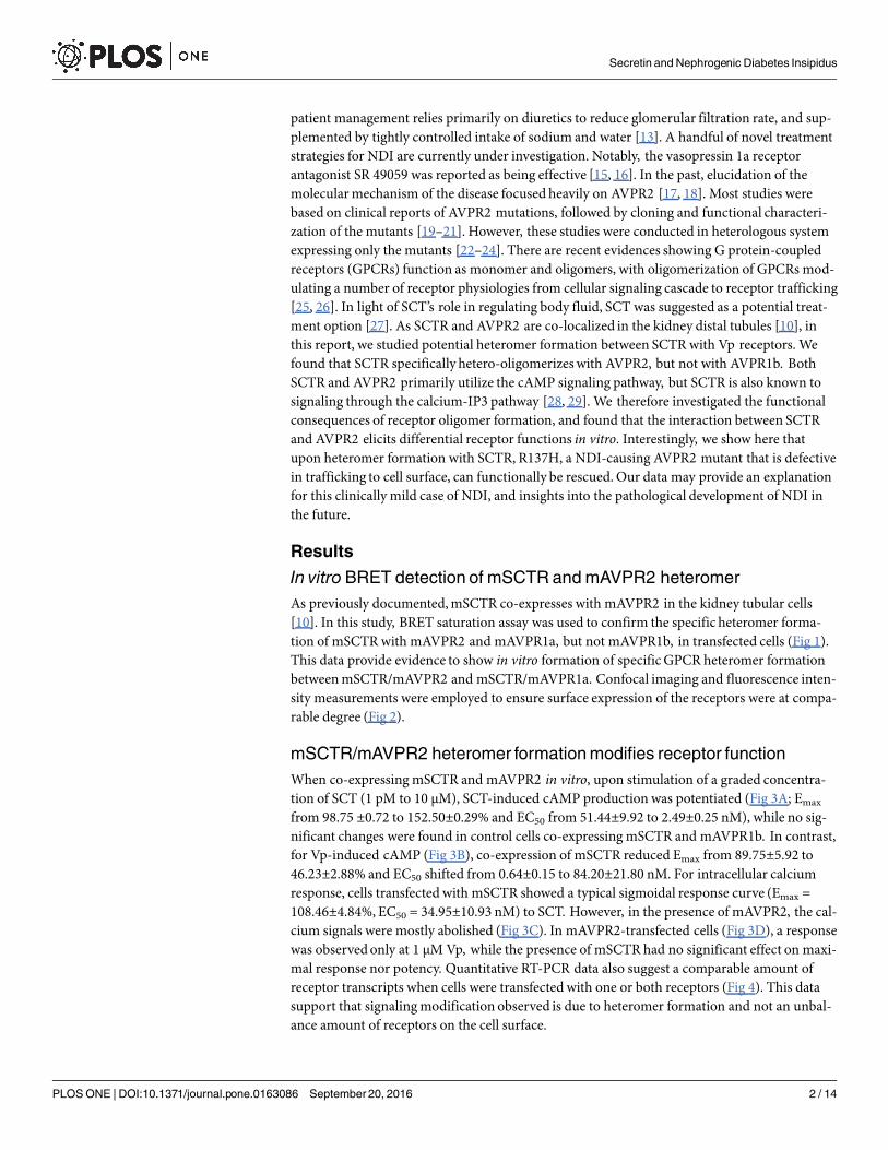
In vitroBRET detection of mSCTR andmAVPR2 heteromerAs previously documented, mSCTR co-expresses with mAVPR2 in the kidney tubular cells[10]. In this study, BRET saturation assay was used to confirm the specific heteromer forma-tion of mSCTR with mAVPR2 and mAVPR1a, but not mAVPR1b, in transfected cells (Fig 1).This data provide evidence to show in vitro formation of specific GPCR heteromer formationbetween mSCTR/mAVPR2 and mSCTR/mAVPR1a. Confocal imaging and fluorescence inten-sity measurements were employed to ensure surface expression of the receptors were at compa-rable degree (Fig 2).
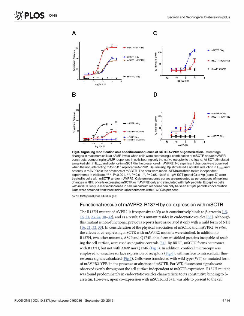
mSCTR/mAVPR2 heteromer formationmodifies receptor functionWhen co-expressing mSCTR and mAVPR2 in vitro, upon stimulation of a graded concentra-tion of SCT (1 pM to 10 μM), SCT-induced cAMP production was potentiated (Fig 3A; Emax
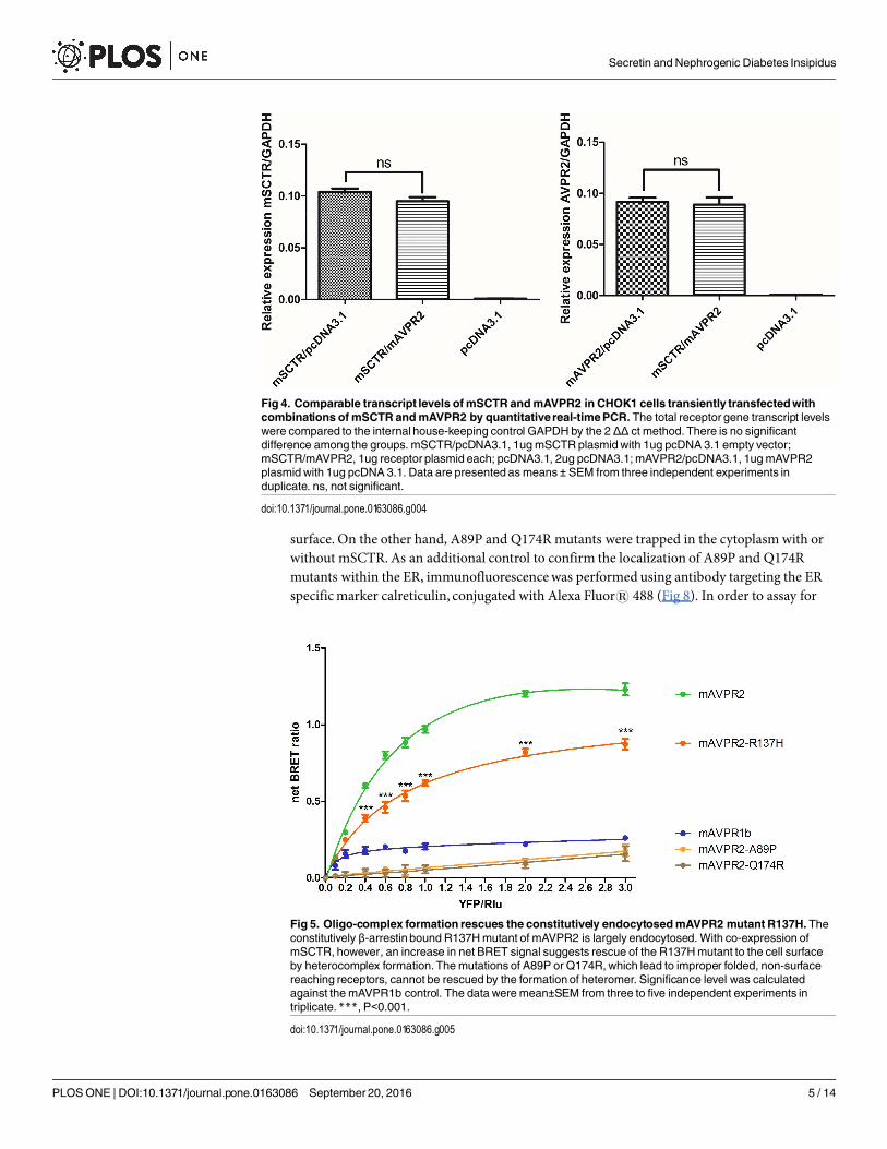
from 98.75 ±0.72 to 152.50±0.29% and EC50 from 51.44±9.92 to 2.49±0.25 nM), while no sig-nificant changes were found in control cells co-expressing mSCTR and mAVPR1b. In contrast,for Vp-induced cAMP (Fig 3B), co-expression of mSCTR reduced Emax from 89.75±5.92 to46.23±2.88% and EC50 shifted from 0.64±0.15 to 84.20±21.80 nM. For intracellular calciumresponse, cells transfected with mSCTR showed a typical sigmoidal response curve (Emax =108.46±4.84%, EC50 = 34.95±10.93 nM) to SCT. However, in the presence of mAVPR2, the cal-cium signals were mostly abolished (Fig 3C). In mAVPR2-transfected cells (Fig 3D), a responsewas observed only at 1 μM Vp, while the presence of mSCTR had no significant effect on maxi-mal response nor potency. Quantitative RT-PCR data also suggest a comparable amount ofreceptor transcripts when cells were transfected with one or both receptors (Fig 4). This datasupport that signaling modification observed is due to heteromer formation and not an unbal-ance amount of receptors on the cell surface.
Secretin and Nephrogenic Diabetes Insipidus
PLOSONE | DOI:10.1371/journal.pone.0163086 September 20, 2016 2 / 14
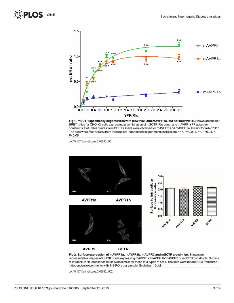
Fig 1. mSCTR specifically oligomerizeswithmAVPR2, andmAVPR1a, but notmAVPR1b. Shown are the netBRET ratios for CHO-K1 cells expressing a combination of mSCTR-Rlu donor andmAVPR-YFP acceptorconstructs. Saturable curves fromBRET assays were obtained for mAVPR2 and mAVPR1a, but not for mAVPR1b.The data were mean±SEM from three to five independent experiments in triplicate.***, P<0.001. **, P<0.01. *,P<0.05.
doi:10.1371/journal.pone.0163086.g001
Fig 2. Surface expressionofmAVPR1a, mAVPR1b, mAVPR2 andmSCTR are similar. Shown arerepresentative images of CHOK1 cells expressingmAVPR1a/mAVPR1b/mAVPR2 or mSCTR constructs. Surfaceto intracellular fluorescence ratios were similar for these four types of cells. The data were mean±SEM from threeindependent experiments with 5–6 ROIs per sample. Scale bar, 10μM.
doi:10.1371/journal.pone.0163086.g002
Secretin and Nephrogenic Diabetes Insipidus
PLOSONE | DOI:10.1371/journal.pone.0163086 September 20, 2016 3 / 14
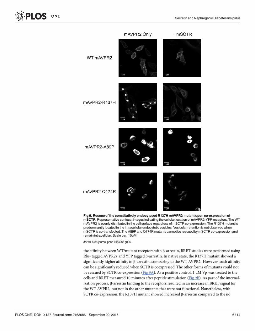
Functional rescue of mAVPR2-R137H by co-expression with mSCTRThe R137H mutant of AVPR2 is irresponsive to Vp as it constitutively binds to β-arrestin [15,16, 21, 23, 24, 30–32], and as a result, this mutant resides in endocytotic vesicles [33]. Althoughthis mutant is non-functional, previous reports have associated it only with a mild form of NDI[16, 21, 32, 33]. In consideration of the physical association of mSCTR and mAVPR2 in vitro,the effects of co-expressing mSCTR with mAVPR2 mutants were studied. In addition toR137H, two other mutants, A89P and Q174R, that form misfolded proteins incapable of reach-ing the cell surface, were used as negative controls [34]. By BRET, mSCTR forms heteromerwith R137H, but not with A89P nor Q174R (Fig 5). In addition, confocal microscopy wasemployed to visualize surface expression of receptors (Fig 6), with surface to intracellular fluo-rescence signals calculated (Fig 7). Cells were transfected with wild type (WT) or mutated formof mAVPR2-YFP, in the presence or absence of mSCTR. For WT, fluorescent signals wereobserved evenly throughout the cell surface independent to mSCTR expression. R137H mutantwas found predominately in endocytotic vesicles characteristic to its constitutive binding to β-arrestin. However, upon co-expression with mSCTR, R137H was able to present to the cell
Fig 3. Signalingmodification as a specific consequence of SCTR-AVPR2 oligomerization.Percentagechanges in maximumcellular cAMP levels when cells were expressing a combination of mSCTR and/ormAVPRconstructs, comparing to cAMP responses in cells bearing only the native receptor to the ligand. A) SCT stimulateda marked shift in Emax and potency in mSCTR in the presence of mAVPR2. No significant changes were observedwhen the non-interactingmAVPR1b replacedmAVPR2. B) Similarly, Vp stimulated a notable reduction in Emax andpotency in mAVPR2 in the presence of mSCTR. The data were mean±SEM from three to five independentexperiments in triplicate.***, P<0.001. **, P<0.01. *, P<0.05. 10pM to 1μMSCT (panel C) or Vp (panel D) weretreated to cells with mSCTRand/ormAVPR2. Calcium response curves are presented as percentages of maximalchanges in RFU of cells expressing mSCTR or mAVPR2 only and stimulatedwith 1μMpeptide. Except for cellswith mSCTRonly, a marked increase in cellular calcium response can only be seen at 1μMpeptide concentration.Data were obtained from three individual experiments with 5–6 ROIs per dose.
doi:10.1371/journal.pone.0163086.g003
Secretin and Nephrogenic Diabetes Insipidus
PLOSONE | DOI:10.1371/journal.pone.0163086 September 20, 2016 4 / 14
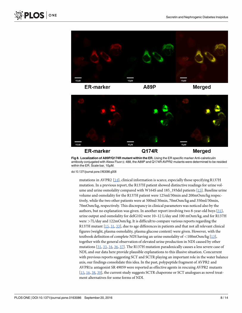
surface. On the other hand, A89P and Q174R mutants were trapped in the cytoplasm with orwithout mSCTR. As an additional control to confirm the localization of A89P and Q174Rmutants within the ER, immunofluorescence was performed using antibody targeting the ERspecific marker calreticulin, conjugated with Alexa Fluor1 488 (Fig 8). In order to assay for
Fig 4. Comparable transcript levels ofmSCTR andmAVPR2 in CHOK1 cells transiently transfectedwithcombinations of mSCTR andmAVPR2 by quantitativereal-timePCR. The total receptor gene transcript levelswere compared to the internal house-keeping control GAPDH by the 2 ΔΔ ct method. There is no significantdifference among the groups. mSCTR/pcDNA3.1, 1ugmSCTR plasmidwith 1ug pcDNA 3.1 empty vector;mSCTR/mAVPR2, 1ug receptor plasmid each; pcDNA3.1, 2ug pcDNA3.1;mAVPR2/pcDNA3.1, 1ugmAVPR2plasmid with 1ug pcDNA 3.1. Data are presented as means ± SEM from three independent experiments induplicate. ns, not significant.
doi:10.1371/journal.pone.0163086.g004
Fig 5. Oligo-complex formation rescues the constitutively endocytosedmAVPR2 mutant R137H.Theconstitutively β-arrestin bound R137Hmutant of mAVPR2 is largely endocytosed. With co-expression ofmSCTR, however, an increase in net BRET signal suggests rescue of the R137Hmutant to the cell surfaceby heterocomplex formation. Themutations of A89P or Q174R, which lead to improper folded, non-surfacereaching receptors, cannot be rescued by the formation of heteromer. Significance level was calculatedagainst themAVPR1b control. The data were mean±SEM from three to five independent experiments intriplicate.***, P<0.001.
doi:10.1371/journal.pone.0163086.g005
Secretin and Nephrogenic Diabetes Insipidus
PLOSONE | DOI:10.1371/journal.pone.0163086 September 20, 2016 5 / 14
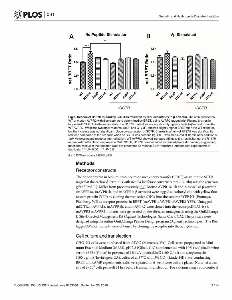
the affinity between WT/mutant receptors with β-arrestin, BRET studies were performed usingRlu- tagged AVPR2s and YFP tagged β-arrestin. In native state, the R137H mutant showed asignificantly higher affinity to β-arrestin, comparing to the WT AVPR2. However, such affinitycan be significantly reduced when SCTR is coexpressed. The other forms of mutants could notbe rescued by SCTR co-expression (Fig 9A). As a positive control, 1 μM Vp was treated to thecells and BRET measured 10 minutes after peptide stimulation (Fig 9B). As part of the internal-ization process, β-arrestin binding to the receptors resulted in an increase in BRET signal forthe WT AVPR2, but not in the other mutants that were not functional. Nonetheless, withSCTR co-expression, the R137H mutant showed increased β-arrestin compared to the no
Fig 6. Rescue of the constitutively endocytosedR137HmAVPR2 mutant upon co-expression ofmSCTR.Representative confocal images indicating the cellular location of mAVPR2-YFP receptors. TheWTmAVPR2 is evenly distributed in the cell surface regardless of mSCTR co-expression. The R137Hmutant ispredominantly located in the intracellular endocytotic vesicles. Vesicular retention is not observedwhenmSCTR is co-transfected. The A89P and Q174Rmutants cannot be rescued by mSCTR co-expression andremain intracellular. Scale bar, 10μM.
doi:10.1371/journal.pone.0163086.g006
Secretin and Nephrogenic Diabetes Insipidus
PLOSONE | DOI:10.1371/journal.pone.0163086 September 20, 2016 6 / 14
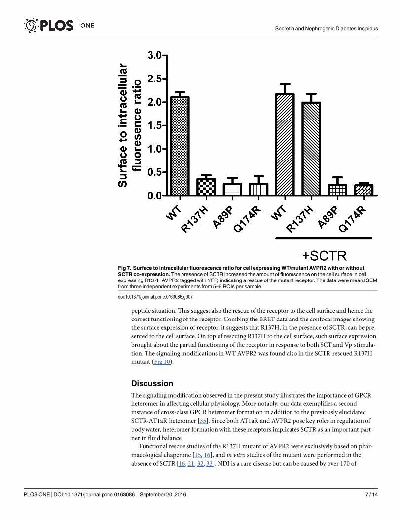
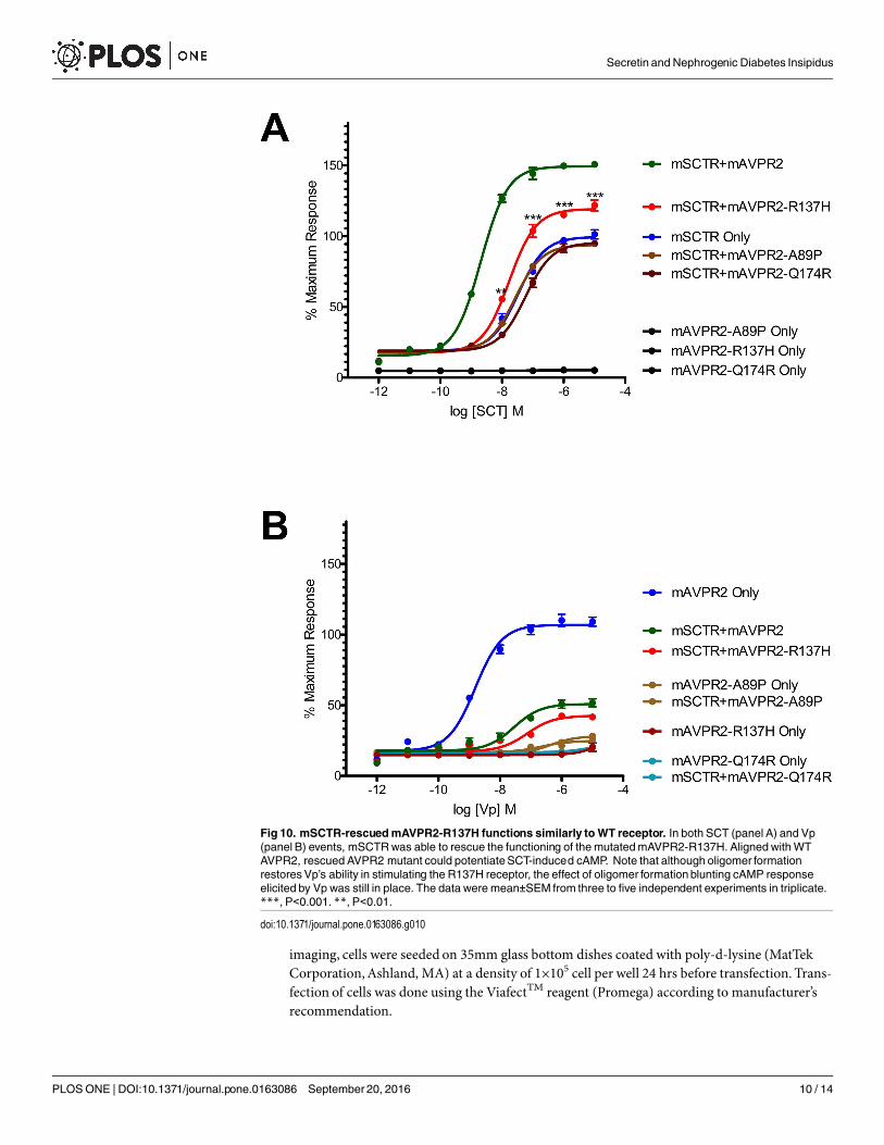
peptide situation. This suggest also the rescue of the receptor to the cell surface and hence thecorrect functioning of the receptor. Combing the BRET data and the confocal images showingthe surface expression of receptor, it suggests that R137H, in the presence of SCTR, can be pre-sented to the cell surface. On top of rescuing R137H to the cell surface, such surface expressionbrought about the partial functioning of the receptor in response to both SCT and Vp stimula-tion. The signaling modifications in WT AVPR2 was found also in the SCTR-rescued R137Hmutant (Fig 10).
DiscussionThe signaling modification observed in the present study illustrates the importance of GPCRheteromer in affecting cellular physiology. More notably, our data exemplifies a secondinstance of cross-class GPCR heteromer formation in addition to the previously elucidatedSCTR-AT1aR heteromer [35]. Since both AT1aR and AVPR2 pose key roles in regulation ofbody water, heteromer formation with these receptors implicates SCTR as an important part-ner in fluid balance.
Functional rescue studies of the R137H mutant of AVPR2 were exclusively based on phar-macological chaperone [15, 16], and in vitro studies of the mutant were performed in theabsence of SCTR [16, 21, 32, 33]. NDI is a rare disease but can be caused by over 170 of
Fig 7. Surface to intracellular fluorescence ratio for cell expressingWT/mutantAVPR2 with or withoutSCTR co-expression.The presence of SCTR increased the amount of fluorescenceon the cell surface in cellexpressing R137HAVPR2 taggedwith YFP, indicating a rescue of the mutant receptor. The data were mean±SEMfrom three independent experiments from 5–6 ROIs per sample.
doi:10.1371/journal.pone.0163086.g007
Secretin and Nephrogenic Diabetes Insipidus
PLOSONE | DOI:10.1371/journal.pone.0163086 September 20, 2016 7 / 14
mutations in AVPR2 [14], clinical information is scarce, especially those specifying R137Hmutation. In a previous report, the R137H patient showed distinctive readings for urine vol-ume and urine osmolality compared with W164S and 185_193del patients [15]. Baseline urinevolume and osmolality for the R137H patient were 125ml/30min and 200mOsm/kg respec-tively, while the two other patients were at 500ml/30min, 70mOsm/kg and 350ml/30min,70mOsm/kg, respectively. This discrepancy in clinical parameters was noticed also by theauthors, but no explanation was given. In another report involving two 8-year-old boys [31],urine output and osmolality for delG102 were 10–12 L/day and 100 mOsm/kg, and for R137Hwere>7L/day and 122mOsm/kg. It is difficult to compare various reports regarding theR137H mutant [15, 31, 33], due to age differences in patients and that not all relevant clinicalfigures (weight, plasma osmolality, plasma glucose content) were given. However, with thetextbook definition of complete NDI having an urine osmolality of <100mOsm/kg [13],together with the general observation of elevated urine production in NDI caused by othermutations [31, 33, 34, 36, 37]. The R137H mutation paradoxically causes a less severe case ofNDI, and our data here provide plausible explanations to this illusive situation. Concurrentwith previous reports suggesting SCT and SCTR playing an important role in the water balanceaxis, our findings consolidate this idea. In the past, polypeptide fragment of AVPR2 andAVPR1a antagonist SR 49059 were reported as effective agents in rescuing AVPR2 mutants[15, 16, 38, 39], the current study suggests SCTR chaperone or SCT analogues as novel treat-ment alternatives for some forms of NDI.
Fig 8. Localization of A89P/Q174Rmutant within the ER.Using the ER specific marker Anti-calreticulinantibody conjugated with Alexa Fluor1 488, the A89P and Q174RAVPR2 mutants were determined to be residedwithin the ER. Scale bar, 10μM.
doi:10.1371/journal.pone.0163086.g008
Secretin and Nephrogenic Diabetes Insipidus
PLOSONE | DOI:10.1371/journal.pone.0163086 September 20, 2016 8 / 14
Methods
Receptor constructsThe donor protein in bioluminescence resonance energy transfer (BRET) assay, mouse SCTRtagged at the carboxyl-terminus with Renilla luciferase construct (mSCTR-Rlu) was the generousgift of Prof. L.J. Miller from previous study [35]. Mouse AVPR 1a, 1b and 2, as well as β-arrestin(mAVPR1a, mAVPR1b, and mAVPR2; β-arrestin) were tagged at carboxyl end with yellow fluo-rescent protein (YFP) by cloning the respective cDNA into the vector pEYFP-N1 (Promega,Fitchburg, WI) as acceptor proteins in BRET (mAVPR1a/AVPR1b/AVPR2-YFP). UntaggedmSCTR, mAVPR1a, mAVPR1b, and mAVPR2 were cloned into the vector pcDNA3.1(+).mAVPR2 mAVPR2 mutants were generated by site-directed mutagenesis using the QuikChangeII Site-Directed Mutagenesis Kit (Agilent Technologies, Santa Clara, CA). The primers weredesigned using the online QuikChange Primer Design program (Agilent Technologies). The Rlu-tagged AVPR2 mutants were obtained by cloning the receptor into the Rlu-plasmid.
Cell culture and transfectionCHO–K1 cells were purchased from ATCC (Manassas, VA). Cells were propagated in Mini-mum Essential Medium (MEM), pH 7.3 (Gibco, CA) supplemented with 10% (v/v) fetal bovineserum (FBS) (Gibco) in presence of 1% (v/v) penicillin G (100 U/ml) and streptomycin(100 μg/ml) (Invitrogen, CA), cultured at 37°C with 5% CO2 (Linde, HK). For conductingBRET and cAMP experiments, cells were plated on 6-well tissue culture plates (Nunc) at a den-sity of 3×10⁵ cells per well 24 hrs before transient transfection. For calcium assays and confocal
Fig 9. Rescue of R137Hmutant by SCTR as reflectedby reducedaffinity to β-arrestin.The affinity betweenWT or mutant AVPR2 with β-arrestin were determined by BRET, using AVRP2 taggedwith Rlu and β-arrestintaggedwith YFP. A) In the native state, the R137Hmutant shows significantly higher affinity to β-arrestin than theWT AVPR2. While the two othermutants, A89P and Q174R, showed slightly higher BRET than theWT receptor,but the increasewas not significant.Upon co-expression of SCTR, β-arrestin affinity of R137Hwas significantlyreduced compared to the scenariowhen no SCTRwas present. B) BRET was measured at 10 min after addition of1μMVp to stimulate receptor internalization. WT AVPR2 showed increase affinity to β-arrestin, but not the R137Hmutant without SCTR co-expression. With SCTR, R137H demonstrated increasedβ-arrestin binding, suggestingfunctional rescue of the receptor. Data are presented as means±SEM from three independent experiments induplicate. ***, P<0.001, **, P<0.01.
doi:10.1371/journal.pone.0163086.g009
Secretin and Nephrogenic Diabetes Insipidus
PLOSONE | DOI:10.1371/journal.pone.0163086 September 20, 2016 9 / 14
imaging, cells were seeded on 35mm glass bottom dishes coated with poly-d-lysine (MatTekCorporation, Ashland, MA) at a density of 1×10⁵ cell per well 24 hrs before transfection. Trans-fection of cells was done using the ViafectTM reagent (Promega) according to manufacturer’srecommendation.
Fig 10. mSCTR-rescuedmAVPR2-R137H functions similarly toWT receptor. In both SCT (panel A) and Vp(panel B) events, mSCTRwas able to rescue the functioning of themutatedmAVPR2-R137H. AlignedwithWTAVPR2, rescued AVPR2 mutant could potentiateSCT-induced cAMP. Note that although oligomer formationrestoresVp’s ability in stimulating the R137H receptor, the effect of oligomer formation blunting cAMP responseelicited by Vp was still in place. The data were mean±SEM from three to five independent experiments in triplicate.***, P<0.001. **, P<0.01.
doi:10.1371/journal.pone.0163086.g010
Secretin and Nephrogenic Diabetes Insipidus
PLOSONE | DOI:10.1371/journal.pone.0163086 September 20, 2016 10 / 14
Quantitative Real Time PCRQuantitative real time PCR experiments were performed using Taqman reagents according tomanufacturer’s protocol (Invitrogen). Gene transcript levels were compared to the internalhouse-keeping control GAPDH by the 2 ΔΔ ct method. The probes were as follows, SCTR:Mm1290794_m1; AVPR2: Mm00517071_m1.
BRET assaysFor saturation BRET assays, 1μg of the donor mSCTR-Rlu construct was transfected withgraded amount (0.0–3.0μg) of acceptor constructs. An appropriate amount of pcDNA3.1empty vector was added to maintain the total amount of DNA transfected be 4μg in all assays.BRET assays were performed 48 hrs after transfection. Cells were lifted using the non-enzy-matic cell dissociation reagent Versene (Invitrogen) and washed in Hanks’ Buffered SalineSolution (HBSS, Invitrogen). After counting using an automated cell counter (LUNA; LogosBiosystems, Inc., S. Korea), 100,000 cells were added to each well of a black 96-well test plate(SPL life sciences, S. Korea). Renilla luciferase substrate Coelenterazine-h (Promega) wasadded to each well to a final concentration of 5μM. Bioluminescence emission was immediatelymeasured at 440–500 nm (luciferin) and 510–590 nm (YFP) using a VICTOR X4 MultilabelPlate Reader (PerkinElmer, Inc., Waltham, MA). BRET ratios were calculated as long (510–590) / short (440–500) emission signals. Net BRET ratio was the BRET ratio of experimentalgroup minus the BRET ratio of the negative control which expressed donor molecule only.
cAMP assaysCells were transfected with a combination of non-tagged receptors, at 1μg each. cAMP assay wasperformed 48 hrs after transfection using the LANCE cAMP kit (PerkinElmer) according tomanufacturer’s protocol. Dose-dependent cAMP responses were assayed by treatment of mouseSCT (GenScript) or mouse Vp (Phoenix Pharmaceuticals, Inc., Burlingame, CA) at concentra-tions from 1 pM to 10 μM for 30 mins. Basal cellular cAMP was measured without peptide treat-ment. The Time-Resolved Fluorescence signal was detected in Victor X4 (PerkinElmer).
Calcium assaysTransfected cells having 1μg each of non-tagged receptors were rinsed twice in solution α (HBSSwith 2.5mM probenecid, 250mM NaOH, adjusted to pH 7.4 by HCl) 24 hrs after transfection.Loading of cells with cell permeant Fluo-4, AM (Invitrogen) was done at 1 μM Fluo-4, AM dilutedin solution α containing 0.003% Pluronic1 F-127 (Invitrogen) for 30 mins at room temperature.The cells were rinsed twice in solution α and allowed to incubate at room temperature for 30 minsbefore fluorescence signal was monitored. Fluorescence signal was measured on an LSM 710 NLOConfocal Laser Scanning Microscope (Carl Zeiss Microscopy GmbH, Jena, Germany) using aPlan-Neofluar 20x/0.50 Ph2 objective. The machine was set to excite the samples through anargon laser (LASOS Lasertechnik GmbH, Jena, Germany) at 488 nm and record 493–622 nmemission in a time series manner. The pinhole was set at 44μm and pixel dwell at 1.58 μs. An 8-bitframe was captured every two seconds for 150 seconds. Cell viability was assayed by stimulatingthe cells with 60 mM KCl at the end of the experiment. The changes in relative fluorescence unit(RFU) were calculated by selecting at least five regions of interest (ROI) from each experiment.
Fluorescence confocal imagingCells were transfected with 1μg YFP tagged WT mAVPR2 receptors or mutant receptors, plusor minus 1μg mSCTR. 24 hrs post transfection, cells were rinsed with HBSS and fixed in
Secretin and Nephrogenic Diabetes Insipidus
PLOSONE | DOI:10.1371/journal.pone.0163086 September 20, 2016 11 / 14
paraformaldehyde at room temperature for 20 mins. They were then mounted using theFluoro-Gel mounting medium (Electron Microscopy Sciences, Hatfield, PA). Fluorescence sig-nal was measured on the same confocal microscope and objective. The machine was set toexcite the samples at 514 nm and record 519–621 nm emission. The pinhole was set at 44μmand pixel dwell at 12.6 μs. Signals were recorded as 12-bit images. Surface to intracellular fluo-rescence ratio was calculated using the software ImageJ (NIH, US).
Immunofluorescence stainingSamples preparation were the same as fluorescence confocal imaging until the mounting step.After fixation, samples were blocked with 1% BSA in PBST (PBS +0.1% Tween 20) for 30 minat room temperature. Samples were then incubated overnight at 4°C with 1:100 Anti-Calreticu-lin antibody [EPR3924]—ER Marker (Alexa Fluor1 488) (Abcam, Cambridge, MA). Afterthree wash of PBS, Fluoro-Gel mounting were done before image acquisition.
Statistical analysisStatistical analysis and graph plotting were done by the computer software PRISM (version5.03; GraphPad, San Diego, CA). All data were presented as means ± SEM from at least threeindependent experiments, each in duplicate or triplicate. Data were analyzed based on theassumption that the sample data followed a normal distribution. One-way ANOVA followedby a Dunnett’s test was used to compare experimental means against the control means for sig-nificance levels. Saturation BRET curves were fitted using the one-site total binding model.Dose response curves were fitted using the agonist stimulation model (three parameters) andvalues for maximal response (Emax) and the half maximal effective concentration (EC50) wereobtained from the curves.
Author Contributions
Conceived and designed the experiments: HKHN BKCC.
Performed the experiments: HKHN.
Analyzed the data: HKHN KGH LJM BKCC.
Contributed reagents/materials/analysis tools: LJM BKCC.
Wrote the paper: HKHN KGH LJM BKCC.
References1. Verbalis JG. Disorders of body water homeostasis. Best Pract Res Clin Endocrinol Metab. 2003; 17
(4):471–503. PMID: 14687585.
2. Jeon US, Joo KW, Na KY, Kim YS, Lee JS, Kim J, et al. Oxytocin induces apical and basolateral redis-tribution of aquaporin-2 in rat kidney. NephronExp Nephrol. 2003; 93(1):e36–45. PMID: 12411748.
3. Li C, Wang W, Summer SN, CadnapaphornchaiMA, Falk S, Umenishi F, et al. Hyperosmolality in vivoupregulates aquaporin 2 water channel and Na-K-2Cl co-transporter in Brattleboro rats. J Am SocNephrol. 2006; 17(6):1657–64. doi: 10.1681/ASN.2005121381 PMID: 16672318.
4. LorenzD, Krylov A, HahmD, Hagen V, Rosenthal W, Pohl P, et al. Cyclic AMP is sufficient for triggeringthe exocytic recruitmentof aquaporin-2 in renal epithelial cells. EMBORep. 2003; 4(1):88–93. doi: 10.1038/sj.embor.embor711 PMID: 12524527; PubMedCentral PMCID: PMCPMC1315811.
5. Kim S, Choi HJ, Jo CH, Park JS, Kwon TH, KimGH. Cyclophosphamide-induced vasopressin-indepen-dent activation of aquaporin-2 in the rat kidney. Am J Physiol Renal Physiol. 2015; 309(5):F474–83.doi: 10.1152/ajprenal.00477.2014 PMID: 26109089.
6. Olesen ET, RutzlerMR,Moeller HB, PraetoriusHA, Fenton RA. Vasopressin-independent targeting ofaquaporin-2 by selective E-prostanoid receptor agonists alleviates nephrogenic diabetes insipidus.
Secretin and Nephrogenic Diabetes Insipidus
PLOSONE | DOI:10.1371/journal.pone.0163086 September 20, 2016 12 / 14
Proc Natl Acad Sci U S A. 2011; 108(31):12949–54. doi: 10.1073/pnas.1104691108 PMID: 21768374;PubMedCentral PMCID: PMCPMC3150913.
7. MichimataM,Mizukami K, Suzuki M, Kazama I, NakamuraY, Suzuki K, et al. Vasopressin-independentrenal urinaryconcentration: increased rBSC1 and enhanced countercurrent multiplication. Kidney Int.2003; 64(3):933–8. doi: 10.1046/j.1523-1755.2003.00182.x PMID: 12911543.
8. Wilke C, Sheriff S, SoleimaniM, Amlal H. Vasopressin-independent regulation of collecting duct aqua-porin-2 in food deprivation. Kidney Int. 2005; 67(1):201–16. doi: 10.1111/j.1523-1755.2005.00071.xPMID: 15610244.
9. Cheng CY, Chu JY, Chow BK. Vasopressin-independent mechanisms in controllingwater homeosta-sis. J Mol Endocrinol. 2009; 43(3):81–92. doi: 10.1677/JME-08-0123PMID: 19318428.
10. Chu JY, Chung SC, LamAK, Tam S, Chung SK, Chow BK. Phenotypes developed in secretin recep-tor-null mice indicated a role for secretin in regulating renal water reabsorption. Mol Cell Biol. 2007; 27(7):2499–511. doi: 10.1128/MCB.01088-06PMID: 17283064; PubMedCentral PMCID:PMCPMC1899889.
11. Chu JY, Lee LT, Lai CH, Vaudry H, Chan YS, Yung WH, et al. Secretin as a neurohypophysial factorregulating body water homeostasis. Proc Natl Acad Sci U S A. 2009; 106(37):15961–6. doi: 10.1073/pnas.0903695106 PMID: 19805236; PubMed Central PMCID: PMCPMC2747226.
12. Chu JY, Cheng CY, Lee VH, Chan YS, Chow BK. Secretin and body fluid homeostasis. Kidney Int.2011; 79(3):280–7. doi: 10.1038/ki.2010.397PMID: 20944548.
13. Skorecki K, ChertowGM,Marsden PA, Taal MW, Yu ASL. Brenner & Rector's the kidney. 10th edition.ed. Philadelphia, PA: Elsevier; 2016. p. p.
14. Tajima A, Miyata I, Katayama A, Toyoda S, Eto Y. A novel mutation of the arginine vasopressin receptor2 gene in a patient with congenital nephrogenic diabetes insipidus. Clin Pediatr Endocrinol. 2005; 14(1):27–33. doi: 10.1297/cpe.14.27 PMID: 24790307; PubMedCentral PMCID: PMCPMC4004929.
15. BernierV, Morello JP, ZarrukA, DebrandN, Salahpour A, LonerganM, et al. Pharmacologic chaper-ones as a potential treatment for X-linked nephrogenic diabetes insipidus. J Am Soc Nephrol. 2006; 17(1):232–43.doi: 10.1681/ASN.2005080854 PMID: 16319185.
16. BernierV, Lagace M, LonerganM, ArthusMF, Bichet DG, BouvierM. Functional rescue of the constitu-tively internalizedV2 vasopressin receptormutant R137H by the pharmacological chaperone action ofSR49059. Mol Endocrinol. 2004; 18(8):2074–84. doi: 10.1210/me.2004-0080PMID: 15166253.
17. Spanakis E, Milord E, Gragnoli C. AVPR2 variants andmutations in nephrogenic diabetes insipidus:review andmissensemutation significance. J Cell Physiol. 2008; 217(3):605–17. doi: 10.1002/jcp.21552 PMID: 18726898.
18. Neocleous V, Skordis N, Shammas C, EfstathiouE, MastroyiannopoulosNP, Phylactou LA. Identifica-tion and characterization of a novel X-linked AVPR2 mutation causing partial nephrogenic diabetesinsipidus: a case reportand review of the literature. Metabolism. 2012; 61(7):922–30. doi: 10.1016/j.metabol.2012.01.005PMID: 22386940.
19. Bockenhauer D, CarpentierE, Rochdi D, van't HoffW, Breton B, BernierV, et al. Vasopressin type 2receptor V88Mmutation: molecular basis of partial and complete nephrogenic diabetes insipidus.NephronPhysiol. 2010; 114(1):p1–10. doi: 10.1159/000245059PMID: 19816050.
20. LemaireM, Chitayat D, GearyDF, Bichet DG, Licht C. A novel disease-causingmutation in AVPR2:Q96H. NDT Plus. 2009; 2(1):20–2. doi: 10.1093/ndtplus/sfn163PMID: 25949277; PubMed CentralPMCID: PMCPMC4421472.
21. Rochdi MD, Vargas GA, CarpentierE, Oligny-Longpre G, Chen S, Kovoor A, et al. Functional charac-terizationof vasopressin type 2 receptor substitutions (R137H/C/L) leading to nephrogenic diabetesinsipidus and nephrogenic syndromeof inappropriate antidiuresis: implications for treatments.MolPharmacol. 2010; 77(5):836–45. doi: 10.1124/mol.109.061804PMID: 20159941; PubMedCentralPMCID: PMCPMC2872969.
22. ArmstrongSP, Seeber RM, Ayoub MA, Feldman BJ, Pfleger KD. Characterization of three vasopressinreceptor 2 variants: an apparent polymorphism (V266A) and two loss-of-functionmutations (R181CandM311V). PLoSOne. 2013; 8(6):e65885. doi: 10.1371/journal.pone.0065885 PMID: 23762448;PubMedCentral PMCID: PMCPMC3675069.
23. Barak LS, Oakley RH, LaporteSA, CaronMG. Constitutive arrestin-mediated desensitization of ahuman vasopressin receptor mutant associatedwith nephrogenic diabetes insipidus. Proc Natl AcadSci U S A. 2001; 98(1):93–8. doi: 10.1073/pnas.011303698 PMID: 11134505; PubMed CentralPMCID: PMCPMC14550.
24. Takahashi K, Makita N, Manaka K, HisanoM, Akioka Y, Miura K, et al. V2 vasopressin receptor (V2R)mutations in partial nephrogenic diabetes insipidus highlight protean agonism of V2R antagonists. JBiol Chem. 2012; 287(3):2099–106. doi: 10.1074/jbc.M111.268797 PMID: 22144672; PubMed CentralPMCID: PMCPMC3265889.
Secretin and Nephrogenic Diabetes Insipidus
PLOSONE | DOI:10.1371/journal.pone.0163086 September 20, 2016 13 / 14
25. MilliganG. G protein-coupled receptor dimerization: function and ligand pharmacology. Mol Pharmacol.2004; 66(1):1–7. doi: 10.1124/mol.104.000497PMID: 15213289.
26. Terrillon S, Bouvier M. Roles of G-protein-coupled receptor dimerization.EMBORep. 2004; 5(1):30–4.doi: 10.1038/sj.embor.7400052 PMID: 14710183; PubMedCentral PMCID: PMCPMC1298963.
27. ProcinoG, Milano S, CarmosinoM, BarbieriC, Nicoletti MC, Li JH, et al. Combination of secretin andfluvastatin ameliorates the polyuria associatedwith X-linked nephrogenic diabetes insipidus in mice.Kidney Int. 2014; 86(1):127–38. doi: 10.1038/ki.2014.10PMID: 24522493; PubMedCentral PMCID:PMCPMC4080339.
28. Siu FK, Lam IP, Chu JY, Chow BK. Signalingmechanisms of secretin receptor. Regul Pept. 2006; 137(1–2):95–104. doi: 10.1016/j.regpep.2006.02.011 PMID: 16930743.
29. Birnbaumer M. Vasopressin receptors. Trends in Endocrinology &Metabolism. 2000; 11(10):406–10.
30. Hamdan FF, Rochdi MD, Breton B, Fessart D, MichaudDE, Charest PG, et al. Unraveling G protein-coupled receptor endocytosis pathways using real-time monitoringof agonist-promoted interactionbetween beta-arrestins and AP-2. J Biol Chem. 2007; 282(40):29089–100. doi: 10.1074/jbc.M700577200 PMID: 17675294.
31. Schoneberg T, Schulz A, Biebermann H, GrutersA, GrimmT, Hubschmann K, et al. V2 vasopressinreceptor dysfunction in nephrogenic diabetes insipidus caused by different molecular mechanisms.HumMutat. 1998; 12(3):196–205. doi: 10.1002/(SICI)1098-1004(1998)12:3<196::AID-HUMU7>3.0.CO;2-FPMID: 9711877.
32. Kocan M, See HB, SampaioNG, Eidne KA, Feldman BJ, Pfleger KD. Agonist-independent interactionsbetween beta-arrestins andmutant vasopressin type II receptors associated with nephrogenic syn-drome of inappropriate antidiuresis. Mol Endocrinol. 2009; 23(4):559–71. doi: 10.1210/me.2008-0321PMID: 19179480; PubMedCentral PMCID: PMCPMC2667710.
33. Shoji Y, Takahashi T, Suzuki Y, Suzuki T, Komatsu K, HironoH, et al. Mutational analyses of AVPR2gene in three Japanese families with X-linked nephrogenic diabetes insipidus: two recurrentmutations,R137H and deltaV278, caused by the hypermutability at CpG dinucleotides. HumMutat. 1998;Suppl 1::S278–83. PMID: 9452109.
34. Boselt I, Tramma D, KalamitsouS, Niemeyer T, Nykanen P, Graf KJ, et al. Functional characterizationof novel loss-of-functionmutations in the vasopressin type 2 receptor gene causing nephrogenic diabe-tes insipidus. Nephrol Dial Transplant. 2012; 27(4):1521–8. doi: 10.1093/ndt/gfr487PMID: 21917732.
35. Lee LT, Ng SY, Chu JY, Sekar R, HarikumarKG, Miller LJ, et al. Transmembrane peptides as uniquetools to demonstrate the in vivo action of a cross-class GPCR heterocomplex. FASEB J. 2014; 28(6):2632–44. doi: 10.1096/fj.13-246868 PMID: 24599969; PubMedCentral PMCID:PMCPMC4021437.
36. Vargas-Poussou R, Forestier L, Dautzenberg MD, Niaudet P, DechauxM, AntignacC. Mutations in thevasopressin V2 receptor and aquaporin-2 genes in 12 families with congenital nephrogenic diabetesinsipidus. J Am Soc Nephrol. 1997; 8(12):1855–62. PMID: 9402087.
37. Okamoto T, Kobayashi N, Naito H, Tajima T. A novel v2 vasopressin receptormutationwith x-linkednephrogenic diabetes insipidus. Clin Pediatr Endocrinol. 2006; 15(1):41–3.doi: 10.1297/cpe.15.41PMID: 24790319; PubMedCentral PMCID: PMCPMC4004903.
38. Schoneberg T, Yun J, Wenkert D, Wess J. Functional rescue of mutant V2 vasopressin receptors caus-ing nephrogenic diabetes insipidus by a co-expressed receptor polypeptide. EMBO J. 1996; 15(6):1283–91. PMID: 8635461; PubMedCentral PMCID: PMCPMC450031.
39. Schoneberg T, Sandig V, Wess J, GudermannT, Schultz G. Reconstitution of mutant V2 vasopressinreceptors by adenovirus-mediated gene transfer. Molecular basis and clinical implication. J Clin Invest.1997; 100(6):1547–56. doi: 10.1172/JCI119678PMID: 9294123; PubMedCentral PMCID:PMCPMC508336.
Secretin and Nephrogenic Diabetes Insipidus
PLOSONE | DOI:10.1371/journal.pone.0163086 September 20, 2016 14 / 14