Embed Size (px)
Citation preview

YALE JOURNAL OF BIOLOGY AND MEDICINE 72 (1999), pp. 153-168.Copyright © 2000. All rights reserved.
Signal Transduction in Esophageal and LESCircular Muscle Contraction
Karen M. Harnett, Weibiao Cao, Nayoung Kim, Uy Dong Sohn,Harlan Rich, Jose Behar, and Piero BiancaniaDepartment of Medicine, Rhode Island Hospital and Brown University, Providence,Rhode Island
Contraction of normal esophageal circular muscle (ESO) in response to acetylcholine (ACh) islinked to M2 muscarinic receptors activating at least three intracellular phospholipases, i.e., phos-phatidylcholine-specific phospholipase C (PC-PLC), phospholipase D (PLD), and the high molec-ular weight (85 kDa) cytosolic phospholipase A2 (cPL42) to induce phosphatidylcholine (PC)metabolism, production ofdiacylglycerol (DAG) and arachidonic acid (AA), resulting in activationofa protein kinase C (PKC)-dependent pathway.
In contrast, lower esophageal sphincter (LES) contraction induced by maximally effective doses ofACh is mediated by muscarinic M3 receptors, linked to pertussis toxin-insensitive GTP-binding pro-teins of the G /,, type. They activate phospholipase C, which hydrolyzes phosphatidylinositol bis-phosphate (PIP2), producing inositol 1, 4, 5-trisphosphate (IP3) and DAG. IP3 causes release ofintracellular Ca++ andformation ofa Ca++-calmodulin complex, resulting in activation of myosinlight chain kinase and contraction through a calmodulin-dependent pathway.
Signal transduction pathways responsible for maintenance ofLES tone are quite distinctfrom thoseactivated during contraction in response to maximally effective doses of agonists (e.g., ACh).Resting LES tone is associated with activity of a low molecular weight (-14 kDa) pancreatic-like(group I) secreted phospholipase A2 (sPLA2) and production of arachidonic acid (AA), which ismetabolized to prostaglandins and thromboxanes. These AA metabolites act on receptors linked toG-proteins to induce activation of Pl- and PC-specific phospholipases, and production of secondmessengers. Resting LES tone is associated with submaximal PI hydrolysis resulting in submaximallevels of inositol trisphosphate (IP3)-induced Ca++ release, and interaction with DAG to activatePKC.
In an animal model ofacute esophagitis, acid-induced inflammation alters the contractile pathwayofESO and LES. In LES circular muscle, after induction of experimental esophagitis, basal levelsof P1 hydrolysis are substantially reduced and intracellular Ca++ stores are functionally damaged,
aTo whom all correspondence should be addressed: Piero Biancani, Ph.D., Rhode IslandHospital, 593 Eddy Street, 5SWP, Providence, RI 02903. Tel.:401-444-5629; Fax: 401-444-5890; E-mail: [email protected]: AA, arachidonic acid; AE, acute experimental esophagitis; ACh, acetyl-choline; CE, chronic experimental esophagitis; cPLA2, cytosolic phospholipase A2; DAG,diacylglycerol; ESO, esophagus/esophageal; GERD, gastro-esophageal reflux disease; IL,interleukin; IP3, inositol 1-4-5 triphosphate; LES, lower esophageal sphincter; LT,leukotriene; NANC, non-adrenergic non-cholinergic; NDGA, nordihydro-guaiaretic acid; PA,phosphatidic acid; PC, phosphatidylcholine; PC-PLC, phosphatidylcholine-specific phos-pholipase C; PG, prostaglandin; PI, phosphatidylinositol; PI-PLC, phosphatidylinositol-spe-cific phospholipase C; PIP2, phosphatidylinositol bisphosphate; PKC, protein kinase C;PLD, phospholipase D; sPLA2, secreted phospholipase A2; TX, thromboxane.
153

154 Harnett et al.: Signal transduction in LES and esophagus
resulting in a reduction ofresting tone. The reduction in intracellular Ca++ release causes a switchin the signal transduction pathway mediating contraction in response to ACh. In the normal LES,ACh causes release of Ca++ from intracellular stores and activation of a calmodulin-dependentpathway. After esophagitis, ACh-induced contraction depends on influx of extracellular Ca++,which is insufficient to activate calmodulin, and contraction is mediated by a PKC-dependent path-way. These changes are reproduced in normal LES cells by thapsigargin-induced depletion of Ca++stores, suggesting that the amount ofCa++ availablefor releasefrom intracellular stores defines thesignal transduction pathway activated by a maximally effective dose ofACh.
INTRODUCTION
Disorders of esophageal motor func-tion and lower esophageal sphincter(LES)b competence affect more than onein ten adults over 40 years of age and onein four adults over 60. Knowledge of themechanisms responsible for esophagealcontraction and LES tone may be useful tounderstand normal function and some ofthe changes associated with esophagealdisease.
The esophagus is relaxed at rest andcontracts with a peristaltic contractionupon swallowing, propelling the foodbolus from the pharynx into the stomach,whereas the LES is spontaneously con-tracted, and relaxes in a timely way whenthe esophagus contracts, to allow passageof the bolus. The swallow-induced con-traction of the esophagus is mediated bythe neurotransmitter acetylcholine (ACh).The tonic contraction of the LES is due tospecialized myogenic mechanisms, whichmay be modulated by inhibitory non-adrenergic-non-cholinergic (NANC) andby excitatory cholinergic neural pathways.The present review describes the cellularbasis for ACh-induced ESO and LES con-traction and spontaneous LES tone andhow inflammation induced by acid or byreflux of gastric contents affects the signaltransduction mechanisms mediating con-traction of these smooth muscles.
A smooth muscle esophagus is pre-sent in marsupials, felines, and primates.We have used the cat to study esophagealand lower esophageal sphincter functionand have occasionally obtained normal
esophageal/LES muscle specimens fromhuman organ donors. We observed thatbecause of similarities of signal transduc-tion mechanisms with the human esopha-gus, th;e cat is a reasonable model for thestudy of signal transduction in esophagealand LES circular muscle.
We have examined normal animalsand two experimental models of esophagi-tis. An acute model of experimentalesophagitis (AE) was obtained byesophageal perfusion with 0.1 N HCl for45 min on three successive days, withexperiments carried out on the fourth day[1-5].
More chronic models of experimentalesophagitis (CE) were obtained by per-forming a myotomy of the LES circularmuscle and allowing esophageal inflam-mation to develop over the course of sixmonths (six-month CE) or 12 to 14months (one-year CE) [6-9]. In the 6-month CE model we find that inflamma-tion-induced changes in mucosal histol-ogy and in contractile mechanisms aresimilar to the acute model and reversibleby treatment with acid suppressants [8].Histologic and mechanical changes weremore pronounced in the acute model, sup-porting this as a reasonable model for thestudy of inflammation-induced distur-bances of esophageal and LES motorfunction.
ACh-induced contraction of ESO andLES circular muscle depends on distinctintracellular pathways beginning with themuscarinic receptors acted upon by theneurotransmitter, extending to the G-pro-teins, phospholipases, second messen-

Harnett et al.: Signal transduction in LES and esophagus 155
gers, and effector mechanisms mediatingmuscle contraction. We will review sepa-rately the signal transduction pathwaysresponsible for contraction of ESO andLES and for maintenance of LES tone innormal animals and in the two experimen-tal models of esophagitis.
CONTRACTION OF NORMALESOPHAGEAL CIRCULARMUSCLE
Esophageal contraction in response toACh is mediated by M2 muscarinic recep-tors since ACh-induced contraction isselectively inhibited by the M2 muscarinicantagonist methoctramine. We have exam-ined the G-proteins linked to muscarinicand other receptors in the contractile path-way of ESO [10-12] by examining theeffect of selective G-protein antibodies oncontraction. Our data suggest that M2muscarinic receptors are linked mostly toGi3 because ACh-induced contraction ofpermeable cells is inhibited by antibodiesagainst the a-subunit of GO3, but not byantibodies against the a-subunit of Gq, Goor Gi1/2 (Figure 1).
G-proteins are linked to phospholi-pases, which generate intracellular secondmessengers from membrane phospho-lipids. ACh-induced contraction of ESO isinhibited by selective inhibitors or anti-bodies of phosphatidylcholine-specificphospholipase C (PC-PLC), PLD, andcPLA2 suggesting, a link between GO3 andthese phospholipases [10, 11].
PLA2 preferentially hydrolyzes phos-pholipids containing arachidonic acid(AA) in the sn-2 position (most oftenphosphatidylcholine), producing AA andlysophospholipid [13, 14]. PC-PLChydrolyzes phosphatidylcholine at the sn3position, producing diacylglycerol (DAG)and phosphocholine [15-17]. PLD alsohydrolyzes phosphatidylcholine at the sn3position, producing choline and phospha-tidic acid (PA). PA may act as a second
messenger or may be dephosphorylated toDAG by a phosphatidic acid phosphohy-drolase [16-20].
Activation of phospholipases PC-PLCand PLD results in production of DAG[21, 22], and activation of cPLA2 producesAA [23]. In the normal esophagus AAcauses little contraction by itself butpotentiates contraction induced by thePKC agonist DAG. DAG and AA act syn-ergistically to activate protein kinase C[23]. ACh-induced contraction and activa-tion of phospholipase requires influx ofextr4cellular Ca++ because ACh-inducedcontraction and DAG production decreasewith decreasing extracellular Ca++ levels.However, activation of PKC by DAG isCa++-independent because DAG-inducedcontraction does not significantly changewhen extracellular or cytosolic Ca++ isreduced to zero [24]. These data suggestthat the influx of extracellular Ca++ isrequired only to activate the phospholipas-es, and once the second messengers areproduced contraction proceeds by the acti-vation of a Ca++-independent PKC [22].
We have examined the PKC isozymespresent in ESO and found fll, y, and £PKC isozymes [25]. However, when ESOis stimulated by ACh only the Ca++-inde-pendent £ isozyme translocates from thecytosol to the membrane, suggesting thatPKCe is involved in agonist-induced con-traction of ESO. This view is supported bythe findings that PKCe antibodies andisozyme-selective pseudosubstrates inhibitACh-induced contraction of ESO [25].
To conclude, in normal ESO ACh-induced contraction is mediated by activa-tion of M2 muscarinic receptors coupled toGi3 type G-proteins. G-protein activationof PC-PLC, PLD and cPLA2 phospholi-pases results in phosphatidylcholinehydrolysis and production of the secondmessengers DAG and AA. DAG and AAsynergistically activate a Ca++-indepen-dent PKCe and produce a PKC-dependentcontraction.

156 Harnett et al.: Signal transduction in LES and esophagus
ESOPHAGEAL CONTRACTION INMODELS OF ESOPHAGITIS
Acute esophagitis.In vivo esophageal contraction in
response to swallowing and in vitroresponse to electrical stimulation areantagonized by atropine, suggesting theinvolvement of cholinergic excitatory neu-rons. After induction of acute experimen-tal esophagitis by repeated acid perfusion,contraction in response to KCI and to ACh,which act directly on the muscle, is notaffected [ 1, 2]. However, the in vivoresponse to swallowing and the in vitroresponse to electrical, i.e., neural stimula-tion, are significantly reduced. These datasuggest that after acid perfusion
esophageal muscle is still capable of con-traction, but that the cholinergic neuralmechanisms responsible for release ofexcitatory neurotransmitters may beaffected.
Intestinal inflammation in man [26]and animals [27] has been reported toresult in changes in motility caused byalterations in enteric nerve and muscle.Pro-inflammatory cytokines present ininflammatory sites, such as interleukin-l1(ILl,B), have been shown to alter musclecontractility by suppressing the release ofneurotransmitters ACh and norepinephrine[28, 29].
We examined the effect of pro-inflam-matory cytokines on normal esophagealsmooth muscle function. We find that in
Normal Esophagus Esophagitis
I~~~~~~~~~~~1T11
(( Myosin Light Chain
Figure 1. In the normal esophagus, ACh-induced contraction is mediated by M2 mus-carinic receptors linked to a pertussis toxin-sensitive GTP-binding protein of the G13type and results in activation of at least three phospholipases acting on membranephospholipids [10]. Phosphatidylcholine-specific phospholipase C (PC-PLC) and phos-pholipase D (PLD) hydrolyze phosphatidylcholine (PC) to produce diacylglycerol (DAG).Cytosolic phospholipase A2 (cPLA2) is also activated, resulting in production of arachidon-ic acid (AA) [23]. Activation of these phospholipases requires the presence of Ca++ [22, 24],which is provided by the influx of extracellular Ca++ through voltage-dependent channels[50], possibly augmented by release of Ca++ from stores. Once DAG is produced, the Ca++-independent protein kinase Ce (PKCe) is activated, and contraction proceeds through acalmodulin-independent pathway [24]. AA, produced by PLA2, which is also Ca++-depen-dent, potentiates the DAG-induced activation of PKC [23]. Acute esophagitis (AE) modifiesACh signaling in the esophageal circular smooth muscle: a group I secreted PLA (sPLA2)produces AA, which is metabolized by lipoxygenase (lipoxy) to leukotrienes (LT), whichcontribute to activation of PKC [3].

Harnett et al.: Signal transduction in LES and esophagus 157
Normal LES - Tone
Ca++Ca
!~~~~~~~~~~~~|| ~~~(Myosin Light Chain)
Figure 2. Resting LES tone is associated with spontaneous activity of phos-phatidylinositol-specific phospholipase C (P1-PLC) [53] and phosphatidyicholine-specific phospholipase C (PC-PLC) [21] resulting in production of inositol 1,4,5-trisphosphate (IP3) and diacyiglycerol (DAG) [21] and activation of a Ca ~-sensitiveprotein kinase Cj3S (PKC,B) [25]. A low molecular weight (14 kDA) secreted, pancreatic-like (group 1) phospholipase A2 (sPLA2) may play a role resting LES tone by producingarachidonic acid (AA), which is then metabolized to prostaglandins (PG) and thrombox-anes (TX). PG and TX are membrane permeable and bind to specific G-protein-linkedreceptors, to cause activation of Pl-PLC and PC-PLC, producing second messengers andcontraction [57].
normal esophageal muscle IL-iIp and IL-6cause a significant reduction in contractionin response to electrical stimulation buthas no effect on ACh-induced contraction.In contrast, IL-8 and tissue necrosis factora (TNFa) do not effect either the responseto ACh or the response to electrical stimu-lation, but inhibit both responses at higherconcentrations [30]. These results suggestthat IL-1, and IL-6, but not IL-8 or TNFainhibit the release of excitatory neuro-transmitter in response to electrical stimu-lation without affecting the ability of themuscle to contract in response to ACh,and, thus, mimic the changes observed inour model of acute experimental esophagi-tis. The data also suggest that ACh releasefrom the in vitro esophageal strips inresponse to electrical stimulation may be
reduced after induction of acute experi-mental esophagitis and that this reductionmay be caused by inflammatory cytokinessuch as IL-13 and IL-6.
In addition, induction ofAE modifiesACh-induced signaling (Figure 1). In nor-mal esophageal muscle a high molecularweight (85 kDa) (group IV) cytosolicphospholipase A2 (cPLA2) participates inacetylcholine-induced contraction ofesophageal circular smooth muscle [23].Since PLA2, arachidonic acid, and itsmetabolites are involved in inflammatoryresponses, we examined their role inesophageal smooth muscle cells (ESO)isolated by enzymatic digestion from thecircular layer of normal and esophagitisanimals.
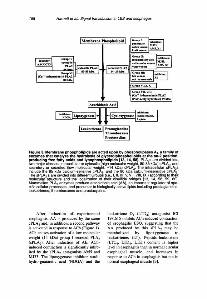
158 Harnett et al.: Signal transduction in LES and esophagus
| (Ca independnt) i rLA2 J 114 -29 kDa gcobravenom AM,X
88-8kDa 14-29 iDamal
sr ~~~(PAF-acetylhydrolase) 29 kDa
| rahdonic Aid|
inhibit~~~~Goup : inbtos
AACOCF cytosLipoxgeasttlCeIxyense~nakethvenoAM, X
| ektienes | PosaglninsImromboxaneslPrstcyclins
Figure 3. Membrane phospholipids are acted upon by phospholipases A2, a family ofenzymes that catalyze the hydrolysis of glycerolphospholipids at the sn-2 position,producing free fatty acids and lysophospholipids [13, 14, 58]. PLA2s are divided intotwo major classes, intracellular or cytosolic (high molecular weight, 80-85 kDa) cPLA2, andsecretory or secreted (low molecular weight, 14 kDa) cPLA2. The intracellular cPLA2sinclude the 85 kDa calcium-sensitive cPLA2, and the 80 kDa calcium-insensitive cPLA2.The sPLA2 s are divided into different Groups (i.e., 1,11,111, V, VII, VII, IX) according to theirmolecular structure and the localization of their disulfide bridges [13, 14, 58, 59, 80].Mammalian PLA2 enzymes produce arachidonic acid (AA), an important regulator of spe-cific cellular processes, and precursor to biologically active lipids including prostaglandins,leukotrienes, thromboxanes and prostacyclins.
After induction of experimentalesophagitis, AA is produced by the samecPLA2 and, in addition, a second pathwayis activated in response to ACh (Figure 1).ACh causes activation of a low molecularweight (14 kDa) group I-secreted PLA2(sPLA2). After induction of AE, ACh-induced contraction is significantly inhib-ited by the sPLA2 antagonists AM5 andMJ33. The lipoxygenase inhibitor nordi-hydro-guaiaretic acid (NDGA) and the
leukotriene D4 (LTD4) antagonist ICI198,615 inhibits ACh-induced contractionof esophagitis ESO, suggesting that theAA produced by this sPLA2 may bemetabolized by lipoxygenase toleukotrienes (LT). Peptido-leukotriene(LTC4, LTD4, LTE4) content is higherlevel in esophagitis than in normal circularesophageal muscle, and increases inresponse to ACh in esophagitis but not innormal esophageal muscle [3].

Harnett et al.: Signal transduction in LES and esophagus 159
The same changes are observed in theone-year CE model [6]. These data suggestthat in inflammation-free controls AA isproduced by cPLA2 and not by sPLA2 andis metabolized by cycloxygenase, and notby lipoxygenase. In AE and CE, activationof sPLA2 causes additional production ofAA that is metabolized by lipoxygenase toproduce leukotrienes, which contribute toACh-induced contraction.
Production of IL-lp by inflammatoryor target cells may explain some of thesechanges in esophageal circular muscle. IL-1B has been shown to cause activation ofsecreted PLA2 and of 5-lipoxygenase inseveral experimental preparations [31-39],and subsequent production if IL-6 [40-46].
LES TONE
The LES circular muscle is a majordeterminant of LES tone. Although the rel-ative neurogenic contribution may varywith the animal species, a significant com-ponent of tone is thought to be myogenic,as it is not affected by neural antagonists,including tetrodotoxin. Functionally, thismuscle is specialized, with muscle stripsfrom this region developing higher totaland active forces than esophageal strips[47-49]. This distinctive contractility maybe, at least in part, related to the ability ofthe LES muscle to handle Ca++ differentlythan esophageal circular muscle [50, 51].LES muscle maintains tension in a Ca++-free environment for some time afteresophageal strips are no longer capable ofcontraction in response to field stimulationor high concentrations of acetylcholine.
These findings suggest that LES mus-cle can use Ca++, released from intracellu-lar storage sites, to maintain tonic contrac-tion, and they are consistent with the his-tology and biochemistry of these muscles.The LES circular muscle has more abun-dant endoplasmic reticulum than theesophageal circular muscle [52].
We have reported that LES tone isassociated with spontaneous, low-level
activity of phospholipase C, resulting inproduction of submaximal concentrationsof DAG and inositol trisphosphate (1P3),which causes release of Ca++ from stores(Figure 2). The elevated concentrations of1P3 and DAG, present in LES smooth mus-cle in the absence of stimulation, decreasewhen the LES relaxes in response to VIP[2, 21]. Increased 'P3 turnover, resulting inspontaneously elevated 'P3 levels andsteady Ca++ release from storage sites,may be responsible for LES tonic contrac-tion. In addition, concurrent activity of aPC-PLC in the LES contributes to the pro-duction of additional DAG [21, 53]. 'P3and DAG, in turn, activate PKC [25]. IP3and DAG, produced at submaximal levels,act synergistically; their interactiondepends on Ca++ release and is mediatedthrough the Ca++-sensitive PKC,B isozyme[25, 53].
Since G-proteins are linked to phos-pholipases, we examined the G-proteinspresent in the LES. We find, by WesternBlot analysis, that Gq, GD3, and Gil12 arepresent in LES circular muscle [11] andthat these G-proteins are spontaneouslyactive, i.e., bound to GTP, in the absenceof exogenously added excitatory neuro-transmitters. In unstimulated LES smoothmuscle, [35S]GTPyS binding to GC3, Gil/2,and Gq antibodies is higher than in ESOsmooth muscle, suggesting that these G-protein may be activated. Spontaneousactivation of G-proteins may provide thespontaneous activation of the phospholi-pases required to maintain threshold levelsof IP3 and DAG, which, in turn, activate aPKC-dependent tone.
Evidence from our laboratory sug-gests that production ofAA by a low mol-ecular weight (14 kDa) group I-secretedPLA2 [54-55] may contribute to mainte-nance of LES tone by producing AAmetabolites, such as prostaglandin-F2x orthromboxanes, which maintain activationof G-proteins [56-57] (Figure 2).
Phospholipases A2 are a growingfamily of enzymes that catalyze the
160 Harnett et al.: Signal transduction in LES and esophagus
hydrolysis of glycerolphospholipids at thesn-2 position, producing free fatty acidsand lysophospholipids (Figure 3) [13, 14,58]. PLA2s are divided into two majorclasses, intracellular or cytosolic (highmolecular weight, 80-85 kDa) group IVcPLA2, and secretory or secreted (lowmolecular weight, -14 kDa) sPLA2.
The sPLA2s are divided into differentgroups (i.e., I, II, III, V, VII, VIII, IX)according to their molecular structure andthe localization of their disulfide bridges[59]. Many sPLA2 s function extracellular-ly, but some have also been localized with-in mitochondria [60, 61].
LES tone may be mediated by theactivity of a group I (secreted) sPLA2because: 1) unstimulated LES circularsmooth muscle has higher AA levels thanESO and spontaneously releases more AAthan ESO smooth muscle; 2) MJ33, aselective inhibitor of group I sPLA2, sig-nificantly reduces AA content and sponta-neous tone of LES circular muscle strips,whereas the group II sPLA2 antagonistMJ45 and the cPLA2 inhibitor AACOCF3has no effect on LES tone; 3) cobra venom(group I) sPLA2, but not rattlesnake(group II) or bee venom (group III) sPLA2,causes dose-dependent contraction of LESstrips [56].
These data suggest that AA produc-tion, through group I sPLA2, participatesin maintenance of LES tone. It is possiblethat the selectivity of the group I sPLA2 inLES muscle may be conferred by the spe-cific interaction of sPLA2 with cell surfacereceptors. Specific membrane receptorsfor neuronal (N)-type and muscle (M)-type sPLA2s, have been identified withsnake venom sPLA2 [62-67]. One of thesesPLA2 receptors, the 180 kDa muscle M-type, has been cloned in rabbit [66] andman [68] and has been shown to have veryhigh affinity for mammalian sPLA2.Receptor binding of sPLA2 is thought tomediate some of the physiological effectsof mammalian sPLA2, including vascularsmooth muscle contraction, cell prolifera-
tion, and internalization of sPLA2 [69-71].For example, antigen stimulation results inthe selective binding of group I sPLA2 andrelease of AA from bone marrow mastcells, which have been shown to containthe mRNA for the group I PLA2 receptor[72].
The AA produced by sPLA2 in theLES is metabolized to prostaglandins,such as PGF2a and thromboxanes which,in turn contribute to maintaining tonebecause the cycloxygenase inhibitorsindomethacin and aspirin, and not thelipoxygenase inhibitor NDGA, dose-dependently reduce LES tone. We find thatPGF2a content is significantly higher inLES than in ESO and that PGF2ax dose-dependently contracts LES strips and sin-gle cells. Thromboxanes A2 and B2 mayalso be involved in LES tone, since throm-boxane B2 dose-dependently contractsLES strips and the thromboxane A2 antag-onist SQ29548 dose-dependently reducesLES tone [56, 57]. Whether other productsof AA metabolism are present and play arole in maintenance of LES tone remainsto be determined.
The AA metabolites PGF2a andthromboxanes A2/B2 may maintain toneby binding to their respective receptorswhich are coupled to G-proteins. PGF2aand the thromboxane A2 analog U46619significantly increases the [35S]GTPySbinding of GO3, and Gq in solubilized LEScircular muscle membranes. In addition,[35S]GTPyS binding in LES circularsmooth muscle is significantly reduced byindomethacin, suggesting that G-proteinsare activated by cycloxygenase-dependentproduction of AA metabolites [57]. Thesedata suggest the following hypothesis:
Spontaneous activation of a group IsPLA2 causes production of AA, and AAmetabolites such as PGF2a and thrombox-anes, which maintain activation of G-pro-teins such as GD3, Gi1l2, and Gq. These G-proteins activate phospholipases such asphosphatidylinositol-specific phospholi-pase C (PI-PLC) and PC-PLC, which, in

Harnett et al.: Signal transduction in LES and esophagus 161
Normal LES - AChCa'+X
CE]~ACE ~E
inhbition
Figure 4. Contraction of LES cells by a maximally effective dose of ACh is mediatedby activation of phosphatidylinositol-specific phospholipase C (PI-PLC), and pro-duction of inositol 1,4,5-trisphosphate (IP3) [10] and diacylglycerol (DAG). IP3 caus-es release of Ca++ from stores at a concentration sufficient to cause activation of calmod-ulin (CAM) [53]. Ca++-CAM causes activation of myosin light chain kinase (MLC kinase)and inhibition of protein kinase C (PKC), inducing a contraction that is entirely calmodulin-dependent [81]. Ca++-CAM-induced inhibition of PKC masks the presence of other factorsthat would otherwise contribute to activation of PKC.
turn, produce DAG and 'P3. DAG and 1P3synergistically activate PKC. The origin ofthe sPLA2 remains to be found, howeverpreliminary Western Blot studies usingmonoclonal antibodies against pancreatic(i.e., group I) human sPLA2 indicate that apancreatic-like sPLA2 is present in humanLES circular smooth muscle.
Since LES tone may be maintained bythe activity of a secreted PLA2, we haveused sPLA2-induced contraction as a pos-sible model of tone. We find that contrac-tion induced by sPLA2 is mediated by the
same signal transduction pathway that isactive in maintenance of LES tone. In con-trol LES, sPLA2-induced contraction isreduced by the same inhibitors that affectLES tone of in vitro circular muscle strips.D609 (PC-PLC inhibitor), U73122 (Pl-PLC inhibitor), and chelerythrine (PKCinhibitor) reduces both LES tone andsPLA2-induced contraction [21, 53], sup-porting the view that sPLA2-induced con-traction, like "spontaneous" LES tone,depends on the activity of PI-PLC, PC-

162 Harnett et al.: Signal transduction in LES and esophagus
PLC, resulting in contraction through aPKC-dependent pathway.
ACH-INDUCED LES CONTRACTIONIN NORMAL ANIMALS
In contrast to spontaneous tone, con-traction induced by maximally effectivedoses of the cholinergic neurotransmitteracetylcholine is mediated through mus-carinic M3 receptors, linked to pertussistoxin-insensitive GTP-binding proteins ofthe Gq/i1 type. They activate phospholi-pase C, which hydrolyzes PIP2, producingIP3 and DAG. 1P3 causes release of intra-cellular Ca++ and formation of a Ca"-calmodulin complex, resulting in activa-tion of myosin light chain kinase and con-traction through a calmodulin-dependentpathway (Figure 4) [53].
Thus, unlike LES tone, which is asso-ciated with spontaneous, submaximalphospholipase C activity and activation ofa PKC,-dependent pathway, maximalcholinergic stimulation activates a calmod-ulin-dependent pathway. The mechanismsresponsible for the switch from a PKC-dependent to a calmodulin-dependentpathway are not entirely clear. They mayresult from the different Ca++ requirementsof calmodulin and PKC. Lower Ca++ levelsare required for PKC activation than forcalmodulin activation [4, 5, 53]. Forinstance, relatively low (180 nmolIL)cytosolic Ca++ levels can support contrac-tion induced by the PKC agonist DAG, butcontraction induced by calmodulinrequires Ca++ levels approaching 1 gmol/L[73-78]. In addition, when Ca++ levels aresufficiently elevated to activate calmod-ulin, calmodulin may inhibit PKC. Themechanism of calmodulin-induced inhibi-tion of PKC activity has not been exten-sively investigated. Kruger et al. [76]examined tryptic fragments of calmodulinand found that two PKC inhibitorysequences were localized to the first andthird Ca++ binding domains of calmodulin,and that calmodulin-induced PKC inhibi-tion was not affected by calmodulin antag-
onists. Thus it is possible that, at Ca++ lev-els insufficient to activate calmodulin, con-traction will be PKC-dependent. In con-trast, at Ca++ levels sufficient to fully acti-vate calmodulin, the contraction will becalmodulin-dependent, and PKC activitywill be inhibited [79].
The inhibitory role of calmodulin onPKC-induced contraction is relevant inorder to understand the switch in signaltransduction pathways that occurs in anexperimental model of acute esophagitis(AE) [5].
ACH-INDUCED LES CONTRACTIONIN ACUTE AND CHRONIC MODELSOF ESOPHAGITIS
Acute esophagitis
Repeated perfusion of the esophageallumen with 0.1 N hydrochloric acid forthree to four days causes a reduction inresting in vivo LES pressure, in sponta-neous in vitro tone, in levels of IP3, and inreleasable intracellular Ca++ stores [1, 2,4].
We have discussed how contraction ofnormal LES smooth muscle in response toa maximally effective dose of ACh acti-vates M3 muscarinic receptors, which arecoupled to Gq/1 1 type G-proteins, linkedto PI-PLC. Activation of PI-PLC producesDAG and IP3, which causes release ofCa++ from intracellular stores, activationof calmodulin and contraction by acalmodulin-dependent pathway. AE caus-es a shift in the intracellular pathwaymediating the response to a maximallyeffective dose of ACh from a calmodulin-dependent to a PKC-dependent pathway(Figure 5) [5]. After AE, contractioninduced by a maximally effective dose ofACh is mediated through M2 muscarinicreceptors, linked to Gi3-type G proteins,which activate phosphatidylcholine-dependent phospholipase C and phospho-lipase D to produce DAG. This ACh-induced contraction depends on influx ofextracellular Ca" which is insufficient to

Harnett et al.: Signal transduction in LES and esophagus 163
Esophagitis LES - ACh
/ntermediate
Ca11 C
ML3><iaSe(Myosin Light Chain
Figure 5. Acute esophagitis (AE) modifies ACh signaling in LES circular smoothmuscle. After induction of AE, intracellular Ca++ stores are functionally damaged or deplet-ed [4], basal and ACh-induced phosphatidylinositol bisphosphate (PIP2) hydrolysis aresubstantially reduced [2, 5], and the resulting reduction in IP3 and intracellular Ca++ releaseis insufficient to activate calmodulin (CAM), and inhibit PKC. The signal transduction path-way mediating contraction in response to ACh, thus switches to a PKC-dependent path-way, activated by M2 muscarinic receptors linked to G3 PC-PLC and PLD resulting inhydrolysis of phosphatidylcholine (PC), production of 6AG and activation of PKCi [5].Influx of extracellular Ca++ is required to activate PC-PLC and PLD. These changes arereproduced in normal cells by thapsigargin-induced depletion of Ca++ stores.
activate calmodulin, resulting in a PKC-dependent contraction [2, 4, 8].
These changes in the functional signaltransduction pathway are mimicked innormal LES muscle by acute depletion ofintracellular Ca++ stores by thapsigargin[5]. They are, therefore, related toimpaired release of Ca++ from intracellularstores, which arises both from impairedproduction of 1P3 [2] and from depletionof releasable Ca++ stores [4] subsequent toinduction of AE. Because release of Ca++from intracellular stores is reduced in AE,the available Ca++, which arises mostlyfrom Ca++ influx, may be insufficient toactivate calmodulin, and, thus, a PKC-dependent pathway is "unmasked" thatwould otherwise be suppressed by
calmodulin activation. A reduction in Ca++release by inflammation, secondary to AE,may be the central event, from which allother observed changes follow, and mayalso explain the reduction in resting toneassociated with AE.
Chronic esophagitisThe significance of the subchronic
changes in cat acute esophagitis to theunderstanding of esophagitis in humans,where gastroesophageal reflux disease(GERD) is likely to develop over a longertime period, remains to be established.However, in CE, we find similar but lessaccentuated, endoscopic, histologic andfunctional changes, up to four to sixmonths after surgery [8]. The magnitude

164 Harnett et al.: Signal transduction in LES and esophagus
of the changes may be related to thedegree of damage caused by repeated acidperfusion, as the degree of damage in theacute model (45-min acid perfusion x 3days) is greater than the damage inducedby spontaneous reflux in the chronicmodel. In addition, in the chronic model,the suppression of HCI secretion, eitherafter the onset of mild chronic esophagitis,or at the time of myotomy, reverses or pre-vents the changes in smooth muscle signaltransduction, presumably by inhibiting orpreventing the injury caused by reflux [8].
LES TONE IN ACUTE ESOPHAGITISAcute esophagitis causes a decrease
of in vivo and in vitro resting LES tone.The decrease in tone may be explained bythe same mechanisms that affect the signaltransduction pathway activated by ACh.
In normal LES, resting tone dependson activity of PI-PLC, resulting in equimo-lar formation of 1P3 and DAG. AdditionalDAG is produced by activity of PC PLC.IP3-induced Ca++ release potentiatesDAG-induced activation of the Ca++ sensi-tive PKC,B, responsible for maintenance oftone. Since AE reduces IP3 formation byPI-PLC the associated DAG production isalso decreased, resulting in a decrease oftotal DAG. In addition, AE causes deple-tion of Ca++ stores, thus Ca++ release andDAG formation are substantiallydecreased, resulting in reduced activationof PKCi and reduced LES tone.
CONCLUSIONWe conclude that in esophageal circu-
lar muscle ACh-induced contraction, ismediated by M2 muscarinic receptors,linked to GO3, PC-PLC, PLD, and cPLA2,resulting in production of DAG and AAand activation of the Ca++-insensitivePKCe. In this contractile pathway Ca++ isrequired for activation of the phospholi-pases and production of the second mes-sengers DAG and AA, as PKCE is Ca++-
independent and DAG causes contractionof esophageal muscle cells even in theabsence of Ca++.
Inflammation causes activation of asecond PLA2, which is a group I sPLA2.This PLA2 causes additional production ofAA, which is metabolized to leukotrienes,resulting in increased levels of LTs in thebasal state and in response to ACh. LT for-mation in response to ACh contributes tocontraction of esophageal muscle, whichremains of normal amplitude, when direct-ly exposed to ACh. Inflammation causes areduction in contraction in response toneural stimulation, most likely due toreduced neurotransmitter release sec-ondary to inflammation. These results aremimicked by exposing normal esophagealmuscle strips to the pro-inflammatorycytokines IL- I , and IL-6.
In normal LES smooth muscle cells,ACh-induced contraction is mediated byM3 receptors linked to Gq/1 1 and PI-PLC,causing formation of IP3, release of Ca++from stores and activation of calmodulin.This results in inhibition of PKC and acti-vation of a calmodulin-dependent contrac-tile pathway. In AE, releasable Ca++ storesand IP3 formation are reduced, resulting inreduced Ca++ release in response to AChand dependence on Ca++ influx for con-traction. The reduced Ca++ release pre-vents activation of calmodulin and pre-vents the calmodulin-induced inhibition ofa PKC-dependent pathway, which is notactivated in the normal LES. Thus AEcauses a switch in contractile pathways,from a calmodulin-dependent to a PKC-dependent contraction. This switch ismimicked by thapsigargin-induced deple-tion of Ca++ stores in normal LES muscle.
The reduction in Ca++ release mayalso account for the reduction of in vivoand in vitro LES resting tone associatedwith AE [1, 2, 49].
Acknowledgements: Supported by NIHDK-28614, Glaxo Research Institute,and Astra Hassle AB.
Harnett et aL: Signal transduction in LES and esophagus 165
REFERENCES:1. Biancani, P., Barwick, K., Selling, J., andMcCallum, R. Effects of acute experimen-tal esophagitis in mechanical properties ofthe lower esophageal sphincter. Gastro-enterology 87:8-16, 1984.
2. Biancani, P., Billett, G., Hillemeier, C.,Nissenshon, M., Rhim, B.Y., Sweczack, S.,and Behar, J. Acute experimental esophagi-tis impairs signal transduction in cat LEScircular muscle. Gastroenterology103:1199-1206, 1992.
3. Kim, N.Y., Sohn, U.D., Mangannan, V.,Rich, H., Behar, J., and Biancani, P.Leukotrienes in ACh-induced contractionof esophageal circular smooth muscle inexperimental esophagitis. Gastroenterol-ogy. 112:1548-1558, 1997.
4. Rich, H., Sohn, U. D., Behar, J., andBiancani, P. Experimental esophagitisaffects intracellular calcium stores in thecat lower esophageal sphincter. Am. J.Physiol. 272:G1523-G1529, 1997.
5. Sohn, U.D., Harnett, K.M., Cao, W., Rich,H., Kim, N., Behar, J., and Biancani, P.Acute experimental esophagitis activates asecond signal transduction pathway in catsmooth muscle from the lower esophagealsphincter. J. Pharmacol. Exp. Ter.283:1293-1304, 1997.
6. Rich, H., Cao, W., Harnett, K. M., Migliori,S., Amaral, J., Chrostek, C., Behar, J., andBiancani, P. PLA2 and arachidonic acid-(AA) induced contraction of loweresophageal sphincter smooth muscle cells(LES) in chronic esophagitis. Gastro-enterology 116:A1070, 1999.
7. Rich, H.G., Cao, W., Harnett, K.M.,Migliori, S.J., Amaral, J.F., Chrostek, C.A.,Behar, J., and Biancani, P. PLA2 andarachidonic acid in contraction ofesophageal smooth muscle in chronicesophagitis. Gastroenterology 116:A1070,1999.
8. Rich, H., Sohn, U.D., Harnett, K.M., Cao,W. B., Chrostek, C., Amaral, J., Migliori,S., Behar, J., and Biancani, P. Ranitidineprevents/reverses reflux-induced changesin signal transduction in cat LES in achronic model of experimental esopahgitis.Gastroenterology 112:A813, 1997.
9. Rich, H., Sohn, U.D., Harnett, K.M.,Behar, J., and Biancani, P. Signal transduc-tion pathways in a chronic cat model ofexperimental esophagitis. Gastroenterology106:A559, 1994.
10. Sohn, U.D., Harnett, K.M., De Petris, G.,Behar, J., and Biancani, P. Distinct mus-carinic receptors, G-proteins, and phospho-
lipases in esophageal and lower esophagealsphincter circular muscle. J. Pharmacol.Exp. Ther. 267:1205-1214, 1993.
11. Sohn, U.D., Han, B., Tashjian, A.H., Jr.,Behar, J., and Biancani, P. Agonist inde-pendent, muscle type specific signal trans-duction pathways In cat esophageal andlower esophageal sphincter (LES) circularsmooth muscle. J. Pharmacol. Exp. Ther.273:482-491, 1995.
12. Kim, N., Song, I.S., Kim, C.Y, Cao, W.,and Biancani, P. Leukotriene-induced con-traction of cat esophageal and loweresophageal sphincter circular smooth mus-cle. Gastroenterology 112:A760, 1997.
13. Dennis, E.A. Diversity of group types, reg-ulation, and function of phospholipase A2.J Biol. Chem. 269:13057-13060, 1994.
14. Dennis, E.A. The growing phospholipaseA2 superfamily of signal transductionenzymes. Trends Biol. Sci. 22:1-2, 1997.
15. Billah, M.M., Eckel, S., Mullmann, T.J.,Egan, R.W., and Siegel, M.I. Phospha-tidylcholine hydrolysis by phospholipase Ddetermines phosphatidate and diacylglyc-eride levels in chemotactic peptide-stimu-lated human neutrophils. J. Biol. Chem.264:17069-17077, 1989.
16. Qian, Z. and Drewes, L.R. A novel mecha-nism for acetylcholine to generate diacyl-glycerol in brain. J. Biol. Chem. 265:3607-3610, 1990.
17. Qian, Z. and Drewes, L.R. Cross-talkbetween receptor-regulated phospholipaseD and phospholipase C in brain. FASEB J.5:315-319, 1991.
18. Billah, M.M. and Anthes, J.C. The regula-tion and cellular functions of phosphatidyl-choline hydrolysis. Biochem. J. 269:281-291, 1990.
19. Exton, J.H. Signaling through phos-phatidylcholine breakdown. J. Biol. Chem.265:1-4, 1990.
20. Dennis, E.A., Rhee, S.G., Billah, M.M.,and Hannun, Y A. Role of phospholipasesin generating lipid second messengers insignal transduction. FASEB J. 5:2068-2077, 1991.
21. Hillemeier, A.C., Bitar, K.B., Sohn, U.D.,and Biancani, P. Protein kinase C mediatesspontaneous tone in the cat loweresophageal sphincter. J. Pharmacol. Exp.Ther. 277:144-149, 1996.
22. Cao, W., Chen, Q., Sohn, U.D., Kim, N.Y,Kirber, M.T., Harnett, K.M., Behar, J., andBiancani, P. Calcium induced contractionof cat esophageal circular smooth musclecells [Submitted for publication]. Am. J.Physiol. 1998.

166 Harnett et al.: Signal transduction in LES and esophagus
23. Sohn, U.D., Kim, D.K., Bonventre, J.V.,Behar, J., and Biancani, P. Role of 100 kDacytosolic PLA2 in ACh-induced contrac-tion of esophageal circular muscle. Am. J.Physiol. 267:G433-G441, 1994.
24. Sohn, U.D., Chiu, T.T., Bitar, K.N., andHillemeier, C. Calcium requirements forACh induced contraction of cat esophagealcircular muscle cells. Am. J. Physiol.266:G330-G338, 1994.
25. Sohn, U.D., Zoukhri, D., Dartt, D.,Sergheraert, C., Harnett, K. M., Behar, J.,and Biancani, P. Different PKC isozymesmediate lower esophageal sphincter (LES)tone and phasic contraction of esophageal(ESO) circular smooth muscle in the cat.Mol. Pharmacol. 51:462-470, 1997.
26. Kern, F.J., Almy, T.P., Abbot, F.K., andBogdonoff, M.D. Motility of the distalcolon in nonspecific ulcerative colitis.Gastroenterology 19:492-503, 1951.
27. Palmer, J.M., Weisbrodt, N.M., and Castro,G.A. Trichinella spiralis: intestinal myo-electrical activity during enteric infusion inthe rat. Exp. Parasitol. 57:132-141, 1984.
28. Stanley, E., Stead, R., and Collins, S.M.E.coli endotoxin exerts a biphasic effect onacetylcholine release from rat myentericnerves. Gastroenterology 102:A700, 1992.
29. Ruhl, A., Berezin, I., and Collins, S.M.Involvement of eicosanoids andmacrophage-like cells in cytokine-mediat-ed changes in rat myenteric nerves.Gastroenterology 109:1852-1862, 1995.
30. Cao, W., Rich, H., Fiocchi, C., Behar, J.,and Biancani, P. The inflammatorycytokines IL-10 and IL-6 inhibit neurally-mediated but not myogenic contraction ofcat esophagus. Gastroenterology114:A730, 1998.
31. Conti, R, Panara, M.R., Barbacane, R.C.,Placido, F.C., Bongrazio, M., Reale, M.,Dempsey, R.A., and Fiore, S. Blocking theinterleukin-1 receptor inhibits leukotrieneB4 and prostaglandin E2 generation inhuman monocyte cultures. Cell Immunol.145:199-209, 1992.
32. Homaidan, F.R., Zhao, L., and Burakoff, R.IL-I beta induces synthesis of phospholi-pase A2-activating protein in rabbit distalcolon. Am. J. Physiol. 272:G1338-G1346,1997.
33. Kuwata, H., Nakatani, Y., Murakami, M.,and Kudo, I. Cytosolic phospholipase A2 isrequired for cytokine-induced expressionof type HA secretory phospholipase A2 thatmediates optimal cyclooxygenase-2-depen-dent delayed prostaglandin E2 generation
in rat 3Y1 fibroblasts. J. Biol. Chem.273:1733-1740, 1998.
34. Jacques, C., Bereziat, G., Humbert, L.,Olivier, J.L., Corvol, M.T., Masliah, J., andBerenbaum, F. Posttranscriptional effect ofinsulin-like growth factor-I on interleukin-lbeta-induced type II-secreted phospholi-pase A2 gene expression in rabbit articularchondrocytes. J. Clin. Invest. 99:1864-1872, 1997.
35. Ma, Z., Ramanadham, S., Corbett, J. A.,Bohrer, A., Gross, R.W., McDaniel, M.L.,and Turk, J. Interleukin-1 enhances pancre-atic islet arachidonic acid 12-lipoxygenaseproduct generation by increasing substrateavailablility through a nitric oxide-depen-dent mechanism. J. Biol. Chem. 271:1029-1042, 1996.
36. Murakami, M., Austen, K.F., and Arm, J.P.The immediate phase of c-kit ligand stimu-lation of mouse bone marrow-derived mastcells elicits rapid leukotriene C4 generationthrough posttranslational activation ofcytosolic phospholipase A2 and 5-lipoxy-genase. J. Exp. Med. 182:197-206, 1995.
37. Nassar, G.M., Montero, A., Fukunaga, M.,and Badr, K. F. Contrasting effects of pro-inflammatory and T-helper lymphocytesubset-2 cytokines on the 5-lipoxygenasepathway in monocytes. Kidney Int.51:1520-1528, 1997.
38. Murakami, M., Kuwata, H., Amakasu, Y.,Shimbara, S., Nakatani, Y., Atsumi, G., andKudo, I. Prostaglandin E2 amplifiescytosolic phospholipase A2- and cyclooxy-genase-2-dependent delayed prostaglandinE2 generation in mouse osteoblastic cells.Enhancement by secretory phospholipaseA2. J. Biol. Chem. 272:19891-19897,1997.
39. Pruzanski, W., Stefanski, E., Vadas, P.,Kennedy, B.P., and van den Bosch, H.Regulation of the cellular expression ofsecretory and cytosolic phospholipases A2,and cyclooxygenase-2 by peptide growthfactors. Biochim. Biophys. Acta 1403:47-56, 1998.
40. Hinson, R.M., Williams, J.A., and Shacter,E. Elevated interleukin 6 is induced byprostaglandin E2 in a murine model ofinflammation: possible role of cyclooxyge-nase-2. Proc. Natl. Acad. Sci. U.S.A.93:4885-4890, 1996.
41. Komatsu, H., Yaju, H., Chiba, K., andOkumoto, T. Inhibition of cyclo-oxygenaseinhibitors of interleukin-6 production byhuman peripheral blood mononuclear cells.Int. J. Immunopharmacol. 13:1137-1146,1991.

Harnett et al.: Signal transduction in LES and esophagus 167
42. Anderson, G.D., Hauser, S.D., McGarity,K.L., Bremer, M.E., Isakson, P.C., andGregory, S.A. Selective inhibition ofcyclooxygenase (COX)-2 reverses inflam-mation and expression of COX-2 and inter-leukin 6 in rat adjuvant arthritis. J. Clin.Invest. 97:2672-2679, 1996.
43. Leisten, J.C., Gaarde, W.A., and Scholz, W.Interleukin-6 serum levels correlate withfootpad swelling in adjuvant-inducedarthritic Lewis rats treated with cyclosporinA or indomethacin. Clin Immonol.Immunopathol. 56:108-115, 1990.
44. Ogle, C.K., Guo, X., Szczur, K., Hartmann,S., and Ogle, J.D. Production of tumornecrosis factor, interleukin-6 andprostaglandin E2 by LPS-stimulated ratbone marrow macrophages after thermalinjury: effect of indomethacin.Inflammation 18: 175-185, 1994.
45. Portanova, J.P., Zhang, Y., Anderson, G.D.,Hauser, S.D., Masferrer, J.L., Seibert, K.,Gregory, S.A., and Isakson, P.C. Selectiveneutralization of prostaglandin E2 blocksinflammation, hyperalgesia, and inter-leukin 6 production in vivo. J. Exp. Med.184:883-891, 1996.
46. Williams, J.A., and Shacter, E. Regulationof macrophage cytokine production byprostaglandin E2. Distinct roles ofcyclooxygenase-I and -2. J. Biol. Chem.272:25693-25699, 1997.
47. Biancani, P., Zabinski, M., Kerstein, M.,and Behar, J. Lower esophageal sphinctermechanics: anatomic and physiologic rela-tionships of the esophagogastric junction ofthe cat. Gastroenterology 82:468-475,1982.
48. Christensen, J., Conklin, J.L., andFreeman, B.W. Physiologic specializationat the esophagogastric junction in threespecies. Am. J. Physiol. 225:1265, 1973.
49. Christensen, J., Freeman, B.Q., and Miller,J.K. Some physiological characteristics ofthe esophagogastric junction in the opos-sum. Gastroenterology 64:1119, 1973.
50. Biancani, P., Hillemeier, C., Bitar, K.N.,and Makhlou, G. M. Contraction mediatedby Ca++ influx in the esophagus and byCa++ release in the LES. Am. J. Physiol.253:G760-G766, 1987.
51. DeCarle, D.J., Christensen, J., and Szabo,A.C. Calcium dependence of neuromuscu-lar events in esophageal smooth muscle ofthe opossum. Am J Physiol 232:E547,1977.
52. Christensen, J., and Roberts, R.L.Differences between esophageal body andlower esophageal sphincter in mitochondria
of smooth muscle in opossum.Gastroenterology 85:650, 1983.
53. Biancani, P., Harnett, K.M., Sohn, U.D.,Rhim, B.Y, Behar, J., Hillemeier, C., andBitar, K.N. Differential signal transductionpathways in LES tone and response to ACh.Am. J. Physiol. 266:G767-G774, 1994.
54. Glaser, K., Mobilio, D., Chang, J., andSenko, N. Phospholipase A2 enzymes: reg-ulation and inhibition. Trend in Pharmacol.Sci. 14:92-98, 1993.
55. Gelb, M.H., Jain, M.K., and Berg, O.G.Inhibition of phospholipase A2. FASEB J.8:916-924, 1994.
56. Cao, W.B., Chen, Q., Jain, M.K., Behar, J.,and Biancani, P. Arachidonic acid metabo-lites contribute to maintenance of cat LEStone. Gastroenterology 112:A708, 1997.
57. Cao, W.B., Harnett, K.M., Chen, Q., Jain,M.K., Behar, J., and Biancani, P. Group Isecreted PLA2 (sPLA2) and arachidonicacid metabolites in the maintenance of catLES tone. Am. J. Physiol. (submitted forpublication), 1999.
58. Kudo, I., Murakami, M., Hara, S., andInoue, K. Mammalian non-pancreaticphospholipases A2. Biochim. Biophys.Acta 1170:217-231, 1993.
59. Heinrikson, R.L., Krueger, E.T., and Keim,P.S. Amino acid sequence of phospholipaseA2-alpha from venom of Crotalus adaman-teus. A new classification of phospholipaseA2 based upon structural determinations. J.Biol. Chem. 252:4913-4921, 1977.
60. Van den Bosch, H., Aarsman, A.J., de Jong,J.G.N., Arnoldussen, E., Neys, F.W., andWasenaar, P.D. Immunoaffinity purifica-tion, partial sequence, and subcellularlocalization of rat liver phospholipase A2.J. Biol. Chem. 264:10008-10014, 1989.
61. Tischfield, J.A. A reassessment of the lowmolecular weight phospholipase A2 genefamily in mammals. J. Biol. Chem.272:17247-17250, 1997.
62. Lambeau, G., Barhanian, B., Schweitz, H.,Qar, J., and Lazdunski, M. Identificationand properties of very high affinity brainmembrane-binding sites for a neurotoxicphospholipase from taipan venom. J. Biol.Chem. 264:11503-11510, 1989.
63. Lambeau, G., Schmid-Alliana, A.,Lazdunaki, M., and Barhanin, J.Identification and purification of a veryhigh affinity binding protein for toxic phos-pholipases A2 in skeletal muscle. J. Biol.Chem. 265:9526-9532, 1990.
64. Lambeau, G., Lazdunski, M., andBarhanin, J. Properties of receptors for theneurotoxic phospholipases A2 in different
168 Harnett et al.: Signal transduction in LES and esophagus
tissues. Neurochem. Res. 16:651-658,1991.
65. Lambeau, G., Barhanin, J., and Lazdunski,M. Identification of different receptor typesfor toxic phospholipases A2 in rabbit skele-tal muscle. FEBS Lett. 293:29-33, 1991.
66. Lambeau, G., Ancian, P., Barhanin, J., andLazdunski, M. Cloning and expression of amembrane receptor for secretory phospho-lipase A2. J. Biol. Chem. 269:1575-1578,1994.
67. Lambeau, G., Ancian, P., Nicholas, J. P.,Beiboer, S.H., Moinier, D., Verheij, H., andLazdunski, M. Structural elements of secre-tory phospholipases A2 involved in thebinding to M-type receptors. J. Biol. Chem.270:5534-5540, 1995.
68. Ancian, P., Lambeau, G., Mattei, M.G., andLazdunski, M. The human 180-kDa recep-tor for secretory phospholipases A2.Molecular cloning, identification of asecreted soluble form, expression, andchromosomal localization. J. Biol. Chem.270:8963-8970, 1995.
69. Arita, H., Hanasaki, K., Nakano, T., Oka,S., Teraoka, H., and Matsumoto, K. Novelproliferative effect of phospholipase A2 inSwiss 3T3 cells via specific binding site. J.Biol. Chem. 266:19139-19141, 1991.
70. Nakajima, M., Hanasaki, K., Ueda, M., andArita, H. Effect of pancreatic type phos-pholipase A2 on isolated porcine cerebralarteries via its specific binding sites. FEBSLett. 309:261-264, 1992.
71. Sommers, C.D., Bobbitt, J.L., Bemis, K.G., and Snyder, D. W. Porcine pancreaticphospholipase A2-induced contractions ofguinea pig lung pleural strips. Eur. J.Pharmacol. 216:87-96, 1992.
72. Fonteh, A.N., Samet, J.M., Surette, M.,Reed, W., and Chilton, F.H. Lipid media-tors: recent advances in molecular biology,understanding of regulation and pharma-cology. In: Murphy, R.C. and Prescott,S.M., eds. Keystone Symposia.; 1997, p. 21
73. Chakravarthy, B.R., Isaacs, R.J., Morley, P.,and Whitfield, J.F. Ca2+ x calmodulin pre-vents myristoylated alanine-rich kinase Csubstrate protein phosphorylation by pro-tein kinase Cs in C6 rat glioma cells. J.Biol. Chem. 270:24911-6, 1995.
74. Chakravarthy, B.R., Isaacs, R.J., Morley, P.,Durkin, J.P., and Whitfield, J.F. Stimulationof protein kinase C during Ca(2+)-inducedkeratinocyte differentiation. Selectiveblockade of MARCKS phosphorylation bycalmodulin. J Biol Chem 270:1362-8,1995.
75. Kraft, A.S., and Anderson, W.B. Phorbolesters increase the amount of Ca2+, phos-pholipid-dependent protein kinase associat-ed with the plasma membrane. Nature301:621-623, 1983.
76. Kruger, H., Schroder, W., Buchner, K., andHucho, F. Protein kinase C inhibition bycalmodulin and its fragments. J. ProteinChem. 9:467-473, 1990.
77. Yu, P., Harnett, K.M., Biancani, P.,DePetris, G., and Behar, J. Interactionbetween signal transduction pathways con-tributing to gallbladder tonic contraction.Am. J. Physiol. 265:G1082-G1089, 1993.
78. Zhao, D., Hollenberg, M.D., and Severson,D.L. Calmodulin inhibits the protein kinaseC-catalyzed phosphorylation of an endoge-nous protein in A10 smooth-muscle cells.Biochem J. 277:445-450, 1991.
79. Sohn, U.D., Choi, C.H., and P., B. DifferentCa2+ levels activate calmodulin- or PKC-dependent contractile pathways in cat LEScircular smooth muscle. Gastroenterology112:A829, 1997.
80. Davidson, F.F., and Dennis, E.A.Evolutionary relationships and implica-tions for the regulation of phospholipaseA2 from snake venom to human secretedforms. J. Mol. Evol. 31:228-238, 1990.
81. Sohn, U.D., Tang, D.C., Stull, J.T.,Haeberle, J.R., Wang, C.-L.A., Harnett, K.M., and Biancani, P. Myosin light chainkinase dependent and PKC dependent con-traction of LES and esophageal smoothmuscle. J. Pharmacol. Exp. Ther. :(submit-ted for publication), 1998.