Embed Size (px)
Citation preview

Reconstructive Urology
Short-term Outcomes of Chait Trapdoor forAntegrade Continence Enema in AdultsJeremy B. Myers, Eric M. Hu, Sean P. Elliott, Andrew Nguyen, Patrick Hovert,William O. Brant, Blake D. Hamilton, M. Chad Wallis, and Jeffrey D. Redshaw
OBJECTIVE To examine our short-term experience of antegrade continence enema (ACE) delivered via a
Financial Disclosure: The authoFinancial Support: This projecreconstructive urology from AmerFrom the Department of Surg
Men’s Health, University of UUrology, University of MinnesotaReprint requests: Jeremy B. My
Center for Reconstructive UrologCity, UT 84132. E-mail: jeremySubmitted: October 25, 2013,
ª 2014 Elsevier Inc.All Rights Reserved
Chait Trapdoor (Cook Medical, Bloomington, IN) in adults with intractable neurogenic bowel.
METHODS We performed a retrospective review at the Universities of Utah and Minnesota of 15 patientswith Chait Trapdoor placed for the purpose of ACE from 2011 to 2013. Our primary outcome wascontinued utilization of the Chait Trapdoor. Secondary outcomes included volume of ACE usedand time to produce a bowel movement.
RESULTS All patients had neurogenic bowel refractory to conventional bowel regimen. Mean follow-up was
6 months (range, 1-17 months). Thirteen patients had the Chait Trapdoor placed in the splenicflexure and 2 had it placed in the cecum. Of the 15 patients, 12 (80%) were still using the ChaitTrapdoor at last follow-up. A median of 425 mL (range, 120-1000 mL) of fluid was used toproduce a bowel movement in 5-120 minutes. Two patients developed postoperative woundinfections, requiring return to the operating room (Clavien IIIb). Long-term complicationsincluded 5 patients with a dislodged tube requiring replacement by interventional radiology and 2patients with local cellulitis. Two patients had the Chait Trapdoor moved to a new location toimprove efficacy.CONCLUSION Although the revision, removal, and complication rates were high, 80% of the patients were
satisfied with the function and continued to use the Chait Trapdoor. The volume of irrigationrequired for ACE and the time it takes to produce a bowel movement vary significantly betweenpatients. UROLOGY -: -e-, 2014. � 2014 Elsevier Inc.n 1990, Malone et al1 originally described theMalone antegrade continence enema (ACE) for the
Ipurpose of treating children with intractable neuro-genic fecal incontinence. The procedure involves thecreation of a catheterizable channel that connects to thecolon, typically with the use of the appendix. Over theyears, there have been multiple variations of themethod.2-11 For example, when the appendix is notavailable, other portions of the gastrointestinal tract havebeen tubularized as a channel such as a Monti-Yang tubeof the small bowel. Antegrade enema irrigation is thenadministered with a temporary catheter, which is inserteddaily. An alternative to creation of a catheterizablechannel for ACE is a permanent cecostomy tube. Onedesign of a permanent cecostomy tube is the ChaitTrapdoor (Cook Medical, Bloomington, IN). The Chait
rs declare that they have no relevant financial interests.t was supported by a generous fellowship grant inican Medical Systems Inc, Minnetonka, MN.ery and the Center for Reconstructive Urology andtah, Salt Lake City, UT; and the Department of, Minneapolis, MNers, M.D., F.A.C.S., Department of Surgery and they and Men’s Health, University of Utah, Salt [email protected] (with revisions): January 22, 2014
h
Trapdoor (henceforth, Chait) is opened once daily andthe irrigation is given via the permanent tube. Someadvantages of using a Chait are that it can potentially beplaced percutaneously12,13 and it can be used in theabsence of an appendix or in more distal locations in thecolon, which may be desirable for some patients withsevere neurogenic bowel.
In children with neurogenic fecal incontinence, theutility and outcomes of creation of a catheterizablechannel for the purpose of ACE have been welldescribed.14,15 Likewise, in children, use of the Chait hasalso been shown to have similar results to the creation ofa catheterizable ACE.16 However, little is known aboutthe use of ACE in adults, especially using a Chait tube.17
We hypothesized that adult patients with neurogenicfecal retention and incontinence would benefit fromACE delivered via a Chait tube. Our study set out tocharacterize the short-term results of ACE flushes deliv-ered from a Chait tube in adults with a variety of causes ofneurogenic bowel.
MATERIAL AND METHODS
After approval from the institutional review boards at theUniversities of Utah and Minnesota, a retrospective review wasperformed and 15 patients were identified who had a Chait tube
0090-4295/14/$36.00 1ttp://dx.doi.org/10.1016/j.urology.2014.01.023
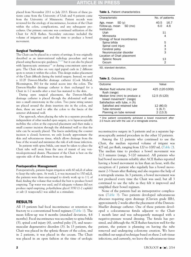
Table 1. Patient characteristics
Characteristic No. of patients
Age, mean � SD (y) 40.5 � 16.7Follow-up, mean � SD (mo) 6.0 � 4.6InstitutionUtah 11Minnesota 4
Etiology of fecal incontinenceSpina bifida 5
placed from November 2011 to July 2013. Eleven of these pa-tients came from the University of Utah and 4 patients camefrom the University of Minnesota. Patient records werereviewed for the etiology of incontinence, location of the Chaitwithin the colon, complications, and any subsequent pro-cedures. Our primary outcome was continued utilization of theChait for ACE flushes. Secondary outcomes included thevolume of irrigation used and the time to produce a bowelmovement.
Spinal cord injury 4Cerebral palsy 3Neuromuscular disorder 3
Location of Chait placementSplenic flexure 13Cecum 2
SD, standard deviation.
Table 2. Outcomes
Outcome Value
Median fluid volume (mL) perflush (range)
425 (120-1000)
Median time (min) to produce abowel movement* (range)
12.5 (5-120)
Satisfaction with tube, n (%)Satisfied and retained tube 12 (80.0)Tube removed 1 (6.7)Planning on tube removal 2 (13.3)
* One patient consistently achieved a bowel movement after2-3 hours and with the use of a retrograde enema.
Surgical TechniqueThe Chait can be placed in a variety of settings. It was originallydescribed as an interventional radiology procedure and wasplaced using fluoroscopic guidance,12,13 but it can also be placedwith laparoscopic assistance18 or during concomitant open sur-gery. The Chait relies on very rigid pigtail curls in 2 differentspots to retain it within the colon. This design makes placementof the Chait difficult during the initial surgery. Instead, we useda 10.2F Dawson-Mueller drainage catheter (Cook Medical,Bloomington, IN) for the initial access into the colon. TheDawson-Mueller drainage catheter is then exchanged for aChait in 1-2 months after a tract has matured to the skin.
During open surgical placement, the Dawson-Muellerdrainage catheter is placed through the abdominal wall andinto a small enterotomy in the colon. Two purse string suturesare placed around the drain insertion site in the colon, andthen, these are used to affix the colon to the fascia of theanterior abdominal wall.
Our approach, when placing the tube in a separate procedureindependent of other needed open surgery, is to laparoscopicallymobilize the colon at the expected placement and then make asmall counter incision through the abdominal wall so that thetube can be securely placed. The fascia underlying the counterincision is closed; however, we only loosely approximate theskin and subcutaneous tissue, which allows drainage from thebase of the wound and minimizes the risk of soft-tissue infection.
In patients with spina bifida, care must be taken to place theChait tube well away from the area of transit of any ven-triculoperitoneal shunts. Placement of the Chait is best on theopposite side of the abdomen from any shunt.
Postoperative ManagementPostoperatively, patients began irrigation with 60 mL/d of salineto keep the tube open. At week 2, it was increased to 150 mL/d,the patients were then encouraged to slowly work up to 1 L offluid, finding the volume that worked the best to produce bowelemptying. Tap water was used, and if adequate volumes did notproduce rapid emptying, polyethylene glycol 3350 (1-2 capfuls)or salt (1 teaspoon/L) was added as a stimulant.
RESULTSAll 15 patients had fecal incontinence or retention re-fractory to a conventional bowel regimen (Table 1). Themean follow-up was 6 months (standard deviation, 4.6months). Fecal incontinence was secondary to spina bifida(5), spinal cord injury (4), cerebral palsy (3), and neuro-muscular degenerative disorders (3). In 13 patients, theChait was placed in the splenic flexure of the colon, andin 2 patients, it was placed in the cecum. The Chaitwas placed in an open fashion at the time of urologic
2
reconstructive surgery in 5 patients and as a separate lap-aroscopically assisted procedure in the other 10 patients.
Among the 12 patients who continued to use theChait, the median reported volume of irrigant was425 mL per flush, ranging from 120 to 1000 mL (Table 2).The median time to produce a bowel movement was12.5 minutes (range, 5-120 minutes). All patients whohad bowel movements reliably after ACE flushes reportedhaving a bowel movement in less than an hour, with theexception of 1 patient who regularly has a bowel move-ment 2-3 hours after flushing and also requires the help ofa retrograde enema. In 3 patients, a bowel movement wasnot produced every time the Chait was used, but all 3continued to use the tube as they felt it improved andsimplified their bowel regimen.
None of the patients had an intraoperative complica-tion (Table 3). Two patients developed subcutaneousabscesses requiring open drainage (Clavien grade IIIb),approximately 2 weeks after the placement of the Dawson-Mueller drainage catheter. One of these patients devel-oped a colocutaneous fistula adjacent to the Chait1 month later and was subsequently managed with anegative-pressure wound dressing. The fistula has per-sisted, and although the ACE flushes function well for thepatient, the patient is planning on having the tuberemoved and undergoing colostomy creation. We havemodified our surgical technique based on these 2 soft-tissueinfections, and currently, we leave the subcutaneous tissue
UROLOGY - (-), 2014

Table 3. Complications
Complication No. of patients
Intraoperative complications 0Local cellulitis treated with antibiotics 2Subcutaneous abscess requiringdrainage (Clavien IIIb)
2
Chait moved to new location 2Chait dislodged requiring IR replacement 5
IR, interventional radiology.
and skin loosely approximated after placement of the tube,to allow drainage of the wound.
In the 5 patients who had spina bifida, ventriculoper-itoneal shunts were present in 3 patients, and there wereno shunt-related complications. In 2 patients, the shuntwas on the opposite side of the abdominal wall from theChait. The other patient had a shunt running on bothsides of the abdomen. She had the original Chait placedat the cecum and moved to the splenic flexure. Shuntinfection did not occur with either placement.
Other complications included dislodgment of the Chaitin 5 patients, necessitating replacement by interventionalradiology. Two patients developed a localized cellulitissurrounding the Chait site and were treated with an oralcourse of antibiotics. In 2 patients, the Chait was relocatedin the colon in an attempt to improve its function. In onepatient, it was moved from the splenic flexure to the he-patic flexure in the hope of more thorough evacuation,and in another patient, it was moved from the cecum tothe splenic flexure because of slow transit.
At the last follow-up, 12 of 15 patients (80%) were stillusing the Chait. Among the three who have discontinuedits use, 1 patient had it removed because of lack of efficacyand the other 2 are planning on having the Chaitremoved. The 2 patients planning on removal were thepatient who developed the colocutaneous fistula and thepatient who had the tube moved from the splenic to thehepatic flexure in hopes of getting better evacuation of thecolon, and both these patients are planning on colostomy.
COMMENTThe use of ACE flushes acts to prevent fecal incontinenceby emptying the bowel with mechanical irrigation of thecolon. Fecal incontinence that would be expected torespond to this type of therapy arises from severe con-stipation or fecal retention. Patients who have neuro-genic fecal retention can either have small frequentincontinence episodes as additional fecal material isadded to the colon, which is already at its capacity, orpatients can have large volume incontinence when thefull colon completely reflexively empties. With regularACE flushes, patients avoid incontinent episodes byemptying the colon with a predictable schedule, whichgives them a day or more to reaccumulate stool beforeneeding to empty the colon again. Retrograde enemas canalso be used effectively in some patients with neurogenicbowel but are limited by the fluid not reaching proximally
UROLOGY - (-), 2014
enough in the colon to accomplish effective emptyingand the physical limitations in giving enemas in patientswith neurogenic disease.
Dr. Peter Chait, a pediatric interventional radiologist,developed the Chait Trapdoor in 1994 as a minimallyinvasive percutaneous alternative to formal catheterizableACE surgery. The tube he designed is a 10F polyurethanecatheter that is held in place internally with helical coilsand has a flattened button-like trapdoor lying against theskin. He reported outcomes in 1996 in a small pilot studyin 15 children.12 A subsequent follow-up study in 1997expanded the original cohort to 42 patients with a meanage of 11.5 years and a mean follow-up of 265 days.13
Tube placement and ACE was successful in all patientsand resulted in near-complete resolution of fecal incon-tinence. Studies have also demonstrated similar resultsindependent of the type of method used for ACE delivery(catheterizable ACE or permanent cecostomy tubes).19
Other options for an indwelling cecostomy tube includean MIC-KEY gastrostomy button (Kimberly-Clark,Roswell, GA) or a Foley catheter.
Overall, the use of ACE in children has high successrates. In 2008, a meta-analysis of 676 patients with amean age of 10.7 years found that 93% of the patientsachieved fecal continence after ACE.15 In this pediatricpopulation, the leading causes of neurogenic bowel werespina bifida (67%), idiopathic constipation (17%), andanorectal malformation (10%). In the vast majority ofcases (76%), the appendix was used to create a cathe-terizable channel for ACE at the cecum.
Similar to this large meta-analysis, the leading etiologyin our study was also spina bifida; however, the locationof the Chait was most often the splenic flexure. This wasdone at first because many of the early patients hadconcomitant urologic reconstruction using the cecum andascending colon (right colon pouch or continent cuta-neous ileal cecocystoplasty). We found that patients hadrapid transit at this location, and indeed in one patient,moving the Chait from the cecum to the splenic flexureresulted in much shorter times for evacuation with ACEflushes. If ACE flushes take too long to evacuate thecolon, they lose their advantage in simplifying a bowelregimen and increase the likelihood of patients reportingfailure of the therapy or having complications such aspressure ulcers from remaining on the toilet too long(a very real concern in this population).
In the limited literature on ACE use in adults, successrates have varied from 50% to 78%20,21 and have notcompared favorably to pediatric outcomes. Gerharz et al22
prospectively followed a cohort of 16 adult patients with amean age of 29.9 years who had a catheterizable channelcreated for ACE. In this study, the underlying causes ofincontinence included spina bifida (44%), spinal cordlesion (25%), anorectal anomaly (19%), and neuropathicdisease of unclear cause (13%), which was very similar toour patient cohort. ACE was associated with a high failurerate of 50%, with a mean follow-up of 41.7months. Reasonsfor ACE failure included nausea, leakage, peristomal
3

infection, cramping/pain, and noncompliance due to psy-chosocial issues. The complication rate was also very high,with 11 complications seen in 7 patients (44%).22
We observed a higher success in our cohort of patients(80%) compared with what was reported in the Gerharzstudy; however, it must be acknowledged that ourmean follow-up was also considerably shorter (6 vs41.7 months). One possible explanation for the higherobserved success in our study was the more distal place-ment of the Chait. At least in children, ACE flushesdelivered to more distal colon produce faster transit timesthan at the cecum23, and the higher success rates weobserved may be due to the faster transit.
Further supporting this hypothesis is our observationthat in many patients with congenital causes of neuro-genic incontinence, irrigations via a cecal ACE takelonger to evacuate as they age into adulthood. We suspectthat this is due to decades of constipation leading todilation and elongation of the distal colon and sigmoidcolon in particular. The volume of ACE needed to pro-duce a bowel movement in our patients (median of425 mL) was similar to that in children with an ACE atthe cecum15 (approximately 500 mL) and less than whathas been reported in adults with an ACE at the cecum.22
We did not prescribe the volume used for ACE, allowingeach patient to titrate up or down to the desired effect. Asa result, the volume of ACE used by our patients washighly variable (120 to 1000 mL).
We observed a high rate of complications, with 4 of thepatients experiencing local infection and 2 of these pa-tients requiring operative drainage. Additionally 5 pa-tients had the tube dislodged (mostly while they werebecoming familiar with the use of the Chait tube). Thesetypes of complications compare with the report byDr. Chait in children, in which dislodgement was themost common complication followed by local cellulitis.13
In contrast to the complications in our patients, however,no patient in this original series had serious infection suchas the 2 cases we observed. It may be that the seriousinfections we observed reflect a higher risk in placementof the tubes using a counter incision, which opens apotential space for infection to occur.
LimitationsThere are major limitations to our study; they include thesmall number of patients, short follow-up, and the retro-spective nature of our data collection. We also used thebroadest definition of success (whether patients continued touse the Chait tube). A future long-term report of our series,with an expanded analysis of patient factors influencingoutcomes and patient measure of success will be needed todetermine if this therapy is durable and worthwhile.
CONCLUSIONWe observed a high revision and complication rate inadults with Chait Trapdoor placement; however, 80% ofthe patients continued to use ACE flushes via the Chait.The amount of fluid required for ACE and the subsequent
4
time it takes to produce a bowel movement variedsignificantly depending on the patient.
References
1. Malone PS, Ransley PG, Kiely EM. Preliminary report: the ante-grade continence enema. Lancet. 1990;336:1217-1218.
2. Heshmat S, DeFoor W, Minevich E, et al. Use of customizedMIC-KEY gastrostomy button for management of MACE stomalcomplications. Urology. 2008;72:1026-1029.
3. Tackett LD, Minevich E, Benedict JF, et al. Appendiceal versus ilealsegment for antegrade continence enema. J Urol. 2002;167(2 Pt 1):683-686.
4. Diamond DA, Pohl HG. Use of a colon based tubularized flap for anantegrade continence enema. J Urol. 2003;169:324-326.
5. Bruce RG, el-Galley RE, Wells J, et al. Antegrade continenceenema for the treatment of fecal incontinence in adults: use ofgastric tube for catheterizable access to the descending colon. J Urol.1999;161:1813-1816.
6. Yerkes EB, Rink RC, Cain MP, et al. Use of a Monti channel foradministration of antegrade continence enemas. J Urol. 2002;168(4 Pt 2):1883-1885.
7. Fukunaga K, Kimura K, Lawrence JP, et al. Button device forantegrade enema in the treatment of incontinence and con-stipation. J Pediatr Surg. 1996;31:1038-1039.
8. Levitt MA, Soffer SZ, Peña A. Continent appendicostomy in thebowel management of fecally incontinent children. J Pediatr Surg.1997;32:1630-1633.
9. Webb HW, Barraza MA, Crump JM. Laparoscopic appendicostomyfor management of fecal incontinence. J Pediatr Surg. 1997;32:457-458.
10. Fonkalsrud EW, Dunn JC, Kawaguchi AI. Simplified technique forantegrade continence enemas for fecal retention and incontinence.J Am Coll Surg. 1998;187:457-460.
11. Calado AA, Macedo A, Barroso U, et al. The Macedo-Maloneantegrade continence enema procedure: early experience. J Urol.2005;173:1340-1344.
12. Shandling B, Chait PG, Richards HF. Percutaneous cecostomy: anew technique in the management of fecal incontinence. J PediatrSurg. 1996;31:534-537.
13. Chait PG, Shandling B, Richards HM, et al. Fecal incontinence inchildren: treatment with percutaneous cecostomy tube placement—a prospective study. Radiology. 1997;203:621-624.
14. Yerkes EB, Cain MP, King S, et al. The Malone antegrade conti-nence enema procedure: quality of life and family perspective.J Urol. 2003;169:320-323.
15. Sinha CK, Grewal A, Ward HC. Antegrade continence enema(ACE): current practice. Pediatr Surg Int. 2008;24:685-688.
16. Chait PG, Shlomovitz E, Connolly BL, et al. Percutaneous cecostomy:updates in technique and patient care. Radiology. 2003;227:246-250.
17. Hoekstra LT, Kuijper CF, Bakx R, et al. The Malone antegradecontinence enema procedure: the Amsterdam experience. J PediatrSurg. 2011;46:1603-1608.
18. Lorenzo AJ, Chait PG, Wallis MC, et al. Minimally invasiveapproach for treatment of urinary and fecal incontinence in selectedpatients with spina bifida. Urology. 2007;70:568-571.
19. Hoy NY, Metcalfe P, Kiddoo DA. Outcomes following fecalcontinence procedures in patients with neurogenic bowel dysfunc-tion. J Urol. 2013;189:2293-2297.
20. Poirier M, Abcarian H, Nelson R. Malone antegrade continentenema: an alternative to resection in severe defecation disorders. DisColon Rectum. 2007;50:22-28.
21. Hirst GR, Arumugam PJ, Watkins AJ, et al. Antegrade continenceenema in the treatment of obstructed defaecation with or withoutfaecal incontinence. Tech Coloproctol. 2005;9:217-221.
22. Gerharz EW, Vik V, Webb G, et al. The value of the MACE(Malone antegrade colonic enema) procedure in adult patients.J Am Coll Surg. 1997;185:544-547.
23. Ellison JS, Haraway AN, Park JM. The distal left Malone antegradecontinence enema—is it better? J Urol. 2013;190:1529-1533.
UROLOGY - (-), 2014