Embed Size (px)
Citation preview
R E S EARCH ART I C L E
B IOPHYS IC S
1Department of Physics, Technische Universität München, 85748 Garching, Germany.2Max Planck Institute for the Physics of Complex Systems, 01187 Dresden, Germany.3Université de Montpellier, Laboratoire Charles Coulomb, UMR 5221, CNRS, F-34095Montpellier, France. 4The Francis Crick Institute, Lincoln’s Inn Fields Laboratories, LondonWC2A 3LY, UK.*Corresponding author. E-mail: [email protected]
Loiseau et al. Sci. Adv. 2016; 2 : e1500465 15 April 2016
2016 © The Authors, some rights reserved;
exclusive licensee American Association for
the Advancement of Science. Distributed
under a Creative Commons Attribution
NonCommercial License 4.0 (CC BY-NC).
10.1126/sciadv.1500465
Shape remodeling and blebbing of activecytoskeletal vesicles
Etienne Loiseau,1 Jochen A. M. Schneider,2 Felix C. Keber,1 Carina Pelzl,1 Gladys Massiera,3Guillaume Salbreux,2,4 Andreas R. Bausch1*
hD
ownloaded from
Morphological transformations of living cells, such as shape adaptation to external stimuli, blebbing, invagination,or tethering, result from an intricate interplay between the plasma membrane and its underlying cytoskeleton,where molecular motors generate forces. Cellular complexity defies a clear identification of the competing pro-cesses that lead to such a rich phenomenology. In a synthetic biology approach, designing a cell-like modelassembled from a minimal set of purified building blocks would allow the control of all relevant parameters. Wereconstruct actomyosin vesicles in which the coupling of the cytoskeleton to the membrane, the topology of thecytoskeletal network, and the contractile activity can all be precisely controlled and tuned. We demonstrate thattension generation of an encapsulated active actomyosin network suffices for global shape transformation of cell-sized lipid vesicles, which are reminiscent of morphological adaptations in living cells. The observed polymorphismof our cell-like model, such as blebbing, tether extrusion, or faceted shapes, can be qualitatively explained by theprotein concentration dependencies and a force balance, taking into account the membrane tension, the density ofanchoring points between the membrane and the actin network, and the forces exerted by molecular motors in theactin network. The identification of the physical mechanisms for shape transformations of active cytoskeletal vesi-cles sets a conceptual and quantitative benchmark for the further exploration of the adaptation mechanisms of cells.
ttp://a
INTRODUCTIONon Novem
ber 5, 2017dvances.sciencem
ag.org/
Cells need to continuously regulate their shapes to achieve vital pro-cesses such as division, motility, or intracellular transport (1–3). At themicroscopic scale, the identification of the mechanisms by which cellsachieve such shape changes is hampered by cellular complexity, whereahugenumber of components interact simultaneously.Aminimalmodelsystem limited to only a fewbuilding blockswould be useful in identifyingthe physical mechanisms leading to these observedmorphologies. Such asynthetic biology approach inspired numerous in vitro studies, rangingfrom the elasticity of pure lipid vesicles (4–6), to the dynamics and me-chanics of the cytoskeleton (7–9), to the encapsulation of cytoskeletalcomponents inside vesicles (10–15). From amechanistic point of view,all cellular shape changes rely on the composite nature of the involvedsystem: a fluid and easily deformable, but inextensible membrane istightly coupled to a relatively rigid cytoskeleton that not only providesmechanostability against shear forces to the cell but also actively de-forms because of the presence of active motors (16–22).
Membrane deformations beyond the well-studied equilibriumshapes observed in passive vesicles (4–6) are only possible in compositesystems, where membrane tension and local force production, or reor-ganization of the cytoskeleton is directly coupled to the membrane.Experiments linking the lipid membrane to a cytoskeletal network lo-cated outside the vesicle lead either to a rupture of the cortex, followedby shrinkage around the vesicle, or to a complete crushing of the vesicle(15). In this configuration, contractility of the actin network results in acompressive force on the membrane.
Having a tension-generating network inside the vesicle should fun-damentally change the situation. Stresses generated in the network re-
sult in inward pulling forces acting on the membrane, leading to anincrease inmembrane surface tension and, because of the law of Laplace,to an internal hydrostatic excess pressure. Such an internal overpressuretends to push water outside the vesicle; however, osmotic pressurealso constrains the volume of the vesicle, such that the vesicle doesnot collapse, and the difference in osmotic and hydrostatic pressureacross the membrane balances each other at equilibrium. The tensiongenerated under this set of conditions should now be able to remodelthe shape of the vesicles. Such an internal excess pressure is thought tounderlie the phenomenon of blebbing (23) in living cells, yet a directevidence for themechanism can only be achieved with a bottom-up ap-proach using purified components (24).
RESULTS
Control of actin network formation in passivecytoskeletal vesiclesHere, we present a minimal model system of a cytoskeletal vesicle byencapsulating actin filaments and the cross-linking protein anillin intovesicles using amodified emulsion transfer technique, continuous drop-let interface crossing encapsulation (cDICE) (25, 26). To obtain stableand spherical vesicles, we adjusted the inner and outer osmotic pres-sures, such that the reduced volume, defined by the ratio between thevolume of liquid present in the vesicle and the volume enclosed by asphere of the same surface area as the vesicle, is only slightly lower than1. Attachment of the actin network (10 mM) to the membrane is con-trolled by varying the concentrations of anillin, which has a His tag, aswell as of the Ni-NTA (nitrilotriacetic acid) lipids in the lipid membrane(Fig. 1; control experiments in the absence of Ni-NTA are shown in fig.S1). In cells, anillin plays a major role during cytokinesis by maintainingthe stability of the cell. Thereby, anillin cross-links the actin while linkingthe actomyosin network to the cell membrane via a phosphatidylinositol
1 of 8
R E S EARCH ART I C L E
on Novem
ber 5, 2017http://advances.sciencem
ag.org/D
ownloaded from
His-anillin
Actin Ni-NTAlipid
B
A
Ni-
NTA
D
F
AnillinE
The model system
Myosin
Self-assembly
f
Bleb
Tube
G H
I J
P
C
Fig. 1. Reconstructionof a cytoskeleton inside vesicles. (A) The lipidmembrane contains a fraction of functionalized lipidwith aNi-NTA group. Elementarybuildingblocks encapsulated in the vesicle consist of actin, a polyhistidine-taggedanillin cross-linker, andmyosin IImolecularmotors. The self-organization ofthese bricks results in the formation of an active cytoskeletal network coupled to the lipidmembrane. In the network, themyosin produces a stress s, which istransduced to the membrane via the Ni-NTA lipid/His-anillin links. Pulling on the membrane leads to the generation of an internal overpressure, Dp,and, hence, an increase of themembrane tension. (B) Three-dimensional network coupled to themembrane (10 mMactin, 0.5 mManillin, and 0.1%Ni-NTA lipid).(C) Intensity profile of the actin network along one diameter of the vesicle [dashed line in (B)]. a.u., arbitrary units. (D) Increasing the Ni-NTA from 0.1% to suc-cessively 1 and 10% favors the recruitment of anillin at the membrane. The bulk is depleted in anillin, which leads to thinner bundles. (E) At 10% Ni-NTA lipids,increasing the anillin concentration from0.1 to 0.5 and 1 mM leads to a transition froma 3Dnetwork (0.1 and 0.5 mM) to a 2D cortex-like cytoskeleton (1 mM). (F) A2D cortex-like structure is obtained for 10 mMactin, 1 mManillin, and 10%Ni-NTA lipid. A confocal picture of the equatorial plane is shown. (G) Size distribution ofthe areaswith a lowermembrane/cortex anchoring. The size of the areaswith a fluorescence intensity lower than the average intensity is extracted from the actinintensity profile plotted along the circumference of the cortex. An example of such a profile is given in the inset. (H) The intensity profile of the actin along onediameter [dashed line in (F)] of the vesicle. The two peaks show that actin is recruited at the membrane to form a 2D cortex, but some cytoskeletal materialremains in the bulk of the vesicle. (I) Vesicle with a 2D cortex, 10 mMactin, 1.5 mManillin, and 10%Ni-NTA lipid. (J) Heterogeneities in the cortex are characterizedthe sameway as in (G). The higher anillin concentration favors the recruitment of actin at themembrane, and the heterogeneities are smaller. Scale bars, 20 mm.
Loiseau et al. Sci. Adv. 2016; 2 : e1500465 15 April 2016 2 of 8
R E S EARCH ART I C L E
on Novem
ber 5, 2017http://advances.sciencem
ag.org/D
ownloaded from
4,5-bisphosphate lipid interaction (27). Using the cDICEmethod to pro-duce these biomimetic vesicles allows an efficient encapsulation of thecytoskeletal proteins and a robust reproducibility (25). In a typical exper-iment, we consider about 75% of the produced vesicles for the analysis.The remaining 25% of the vesicles result from the characteristic poly-dispersity of the method. The analyzed vesicles remain stable for over aday, at least. For a given set of experimental conditions, at least 40 ve-sicles with homogeneous morphology were analyzed.
At a concentration of 0.1% Ni-NTA lipids, we observe a volume-spanning actin network inside the vesicles anchored to the membranevia the Ni-NTA–anillin (0.5 mM) interaction (Fig. 1, B and C).Increasing the Ni-NTA lipid concentration to 1% results in a signifi-cantly less-bundled network that still spans the entire volume of the ves-icle (Fig. 1D). A further increase of the Ni-NTA lipid concentration upto 10% results in even thinner filaments in the volume of the vesicle, yeta cortex remains to be formed. This shows that a higher ratio ofNi-NTAlipids significantly favors the recruitment of the anillin to themembranebut is not sufficient for cortex formation. This suggests that the concen-tration of cross-linkingmolecules is the limiting factor for cortex forma-tion. At 0.5 mManillin, a large fraction of anillin is already bound to themembrane when polymerization and cross-linking of the actin networkoccur, which depletes anillin from the volume and thus reduces thebundling effect in the vesicle.
Indeed, by increasing the anillin concentration by a factor of 2, wecan produce a cortex network tightly coupled to the membrane when10% Ni-NTA lipids are present (Fig. 1E). Both a high ratio of Ni-NTAlipid (10%) and a high concentration of anillin (1 mM) are required to
Loiseau et al. Sci. Adv. 2016; 2 : e1500465 15 April 2016
obtain a two-dimensional (2D) cortex-like structure. Cortex formationwas not achieved at a low Ni-NTA concentration of 0.1%, even after a10-fold higher concentration of anillin was added. Actin is recruited atthe membrane, as can be seen on the actin intensity profile along thediameter of a vesicle (Fig. 1H). The fraction of cortical actin dependson the anillin concentration and can be determined by computing thecortex/bulk ratio of actin fluorescence intensity. At 1 mManillin, corticalactin accounts for 40 to 50% of the total actin. An increase of anillinconcentration to 1.5 mM favors the recruitment of cortical actin up to75%. The cortex has intrinsic heterogeneities consisting of areas of lowercortical actin, as can be observed in Fig. 1 (F and I). We characterizedthese heterogeneities in the case of two different anillin concentra-tions by measuring the size of the areas that have an actin fluorescenceintensity below the mean intensity. The size distributions are reportedin Fig. 1 (G and J). At 1 mManillin, the low-intensity cortical actin areasrange from 2 to 6 mm, whereas at 1.5 mM, the size of the heterogeneitiesdrops to ∼0.5 mm. This is consistent with the increase of the fraction ofcortical actin previously calculated.
Switching the network geometry from a 3D network (Fig. 1B) to a2D cortex-like structure (Fig. 1F) does not result in change of the overallvesicle shape; in all cases, the vesicles remain spherical. The fraction ofbound anillin can be determined by labeling anillin with Alexa Fluor488. By computing the ratio of intensities in the volume of the vesiclewith the circumference, the absolute bound anillin concentration wasdetermined (see figs. S2 to S4 and the Supplementary Materials fordetailed calculations). By this means, the distances between the attach-ment points are determined and reported in the final phase diagram.
C
D
E
d Ro
A
B
C
D
E
Anillin 0.1 µM0.2 µM
10%
Ni-
NTA
Fig. 2. Stability of single-bleb vesicles containing 1 mMmyosin. (A) At an anillin/myosin concentration of 0.2:1, we observe the formation of a single blebthat remains stable overtime. The internal overpressure is not strong enough to globally disrupt the coupling to the membrane. (B to E) Upon decreasing thenumber of membrane/cytoskeleton links (0.1:1, anillin/myosin), the contraction of the actomyosin network decreases the neck of the bleb (B) and themembrane/cytoskeleton links rupture until the vesicle recovers a spherical shape (C to E). Scale bars, 20 mm. d, diameter of the bleb; R0, radius of the spherical vesicle.
3 of 8
R E S EARCH ART I C L E
on Novem
ber 5, 2017http://advances.sciencem
ag.org/D
ownloaded from
Blebbing of active cytoskeletal vesiclesUpon addition of myosin II filaments to the system, a rich morphologyis observed. Blebs appear (Figs. 2 and 3) and the shapes of the vesicleschange markedly (Fig. 4). Because of the coupling between the networkand the membrane, the network cannot contract freely, but instead isconstrained by the membrane being pulled against the liquid volumeof the vesicle (Fig. 1A). This process results in the establishment of anexcess internal pressure pwithin the fluid. The internal pressure exerts apushing force on themembrane that is balanced by a force acting on thelinks joining the actin network to the membrane. This force f is relatedto the density of the membrane linker r and to internal pressurethrough the relationship f ∼ p/r, because the resistance to the intra-cellular pressure is shared between linkers. The typical force necessaryto rupture an individual Ni-NTA binding link is on the order of a few
Loiseau et al. Sci. Adv. 2016; 2 : e1500465 15 April 2016
piconewtons, as determined by single-molecule experiments (28, 29):Above this critical force f*, the molecular bonds attaching the actinnetwork to the vesicle membrane will break. When a region of themembrane detaches from the actin network, intracellular pressurecan then drive the formation of a bleb by exerting a pushing force onthe detached membrane region (Fig. 1A).
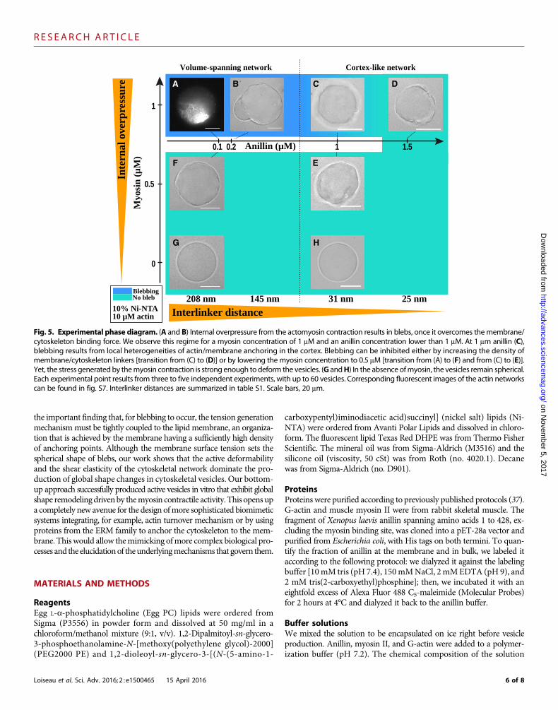
At high myosin and intermediate cross-linking protein concentra-tions (0.2:1, anillin/myosin in micromolar), the actin network spans thewhole vesicle, and a big stable bleb appears (Fig. 2A). The large bleb has aradius of curvature of about 15 mm (see fig. S5), and its volume accountsfor 10 to 17% of the total vesicle volume. The stable bleb is under tension,as seen by the absence of any visible thermal fluctuations of the mem-brane (Fig. 2A). Because of the vesicle productionmethod,we are not ableto image the dynamics of formation of these big blebs that form within afewminutes. Nevertheless, because blebs result from membrane detach-ment, the growth phase should depend on the total density of linkers; atlower linker densities, bleb growth would be favored, resulting in largerfinal bleb sizes. Indeed, slightly decreasing the number of membrane at-tachment points (0.1:1, anillin/myosin inmicromolar) results in the ap-pearance of a bleb that takes up the entire vesicle, a process that lasts formore than 15min (Fig. 2B). First, global contraction of the actin networkresults in membrane detachment, producing a single bleb that is undertension (Fig. 2C). Subsequently, the cytoskeletal tension becomes so suf-ficiently high that it ruptures most membrane attachments, allowingalmost complete contraction of the network. Alternatively, localized slid-ing of the anchoringpoints could also lead to the observed tension release.While the network collapses, the vesicle recovers its spherical shape, andthe membrane starts to fluctuate freely, indicating the release of thecytoskeletal-induced membrane tension (Fig. 2, C to E, and movie S1).
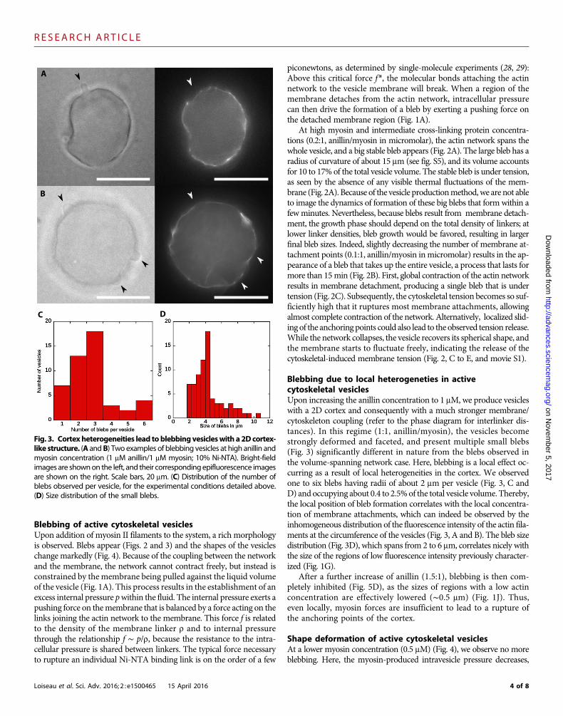
Blebbing due to local heterogeneties in activecytoskeletal vesiclesUpon increasing the anillin concentration to 1 mM, we produce vesicleswith a 2D cortex and consequently with a much stronger membrane/cytoskeleton coupling (refer to the phase diagram for interlinker dis-tances). In this regime (1:1, anillin/myosin), the vesicles becomestrongly deformed and faceted, and present multiple small blebs(Fig. 3) significantly different in nature from the blebs observed inthe volume-spanning network case. Here, blebbing is a local effect oc-curring as a result of local heterogeneities in the cortex. We observedone to six blebs having radii of about 2 mm per vesicle (Fig. 3, C andD) and occupying about 0.4 to 2.5%of the total vesicle volume. Thereby,the local position of bleb formation correlates with the local concentra-tion of membrane attachments, which can indeed be observed by theinhomogeneous distribution of the fluorescence intensity of the actin fila-ments at the circumference of the vesicles (Fig. 3, A and B). The bleb sizedistribution (Fig. 3D), which spans from 2 to 6 mm, correlates nicely withthe size of the regions of low fluorescence intensity previously character-ized (Fig. 1G).
After a further increase of anillin (1.5:1), blebbing is then com-pletely inhibited (Fig. 5D), as the sizes of regions with a low actinconcentration are effectively lowered (∼0.5 mm) (Fig. 1J). Thus,even locally, myosin forces are insufficient to lead to a rupture ofthe anchoring points of the cortex.
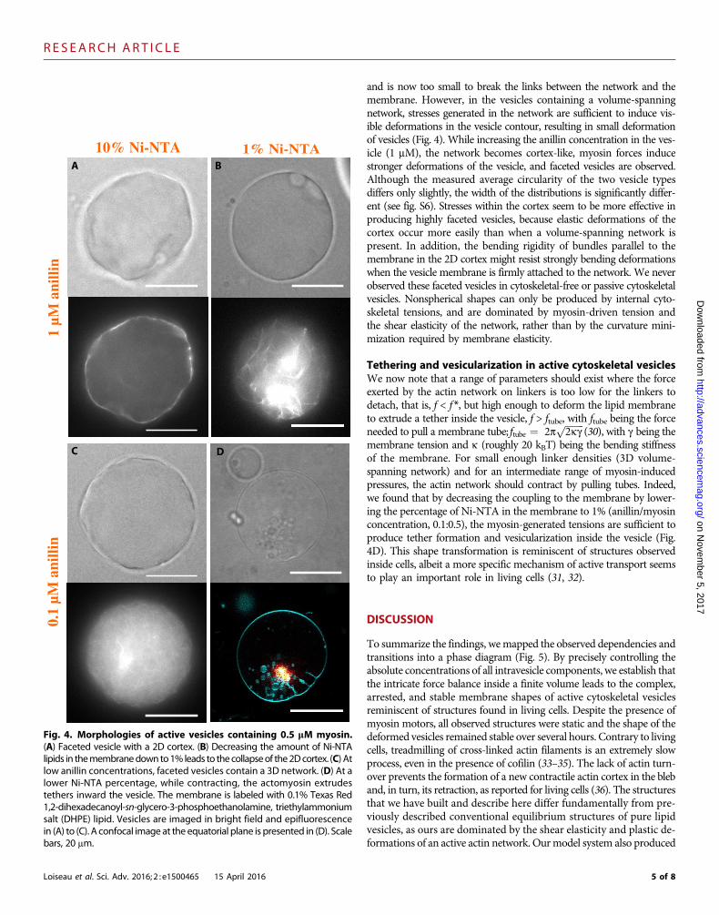
Shape deformation of active cytoskeletal vesiclesAt a lower myosin concentration (0.5 mM) (Fig. 4), we observe no moreblebbing. Here, the myosin-produced intravesicle pressure decreases,
A
B
C D
Fig. 3. Cortex heterogeneities lead toblebbingvesicleswith a 2D cortex-like structure. (A andB) Two examples of blebbing vesicles at high anillin and
myosin concentration (1 mM anillin/1 mM myosin; 10% Ni-NTA). Bright-fieldimages are shownon the left, and their correspondingepifluorescence imagesare shown on the right. Scale bars, 20 mm. (C) Distribution of the number ofblebs observed per vesicle, for the experimental conditions detailed above.(D) Size distribution of the small blebs.4 of 8
R E S EARCH ART I C L E
Loiseau et al. Sci. Adv. 2016; 2 : e1500465 15 April 2016
on Novem
ber 5http://advances.sciencem
ag.org/D
ownloaded from
and is now too small to break the links between the network and themembrane. However, in the vesicles containing a volume-spanningnetwork, stresses generated in the network are sufficient to induce vis-ible deformations in the vesicle contour, resulting in small deformationof vesicles (Fig. 4). While increasing the anillin concentration in the ves-icle (1 mM), the network becomes cortex-like, myosin forces inducestronger deformations of the vesicle, and faceted vesicles are observed.Although the measured average circularity of the two vesicle typesdiffers only slightly, the width of the distributions is significantly differ-ent (see fig. S6). Stresses within the cortex seem to be more effective inproducing highly faceted vesicles, because elastic deformations of thecortex occur more easily than when a volume-spanning network ispresent. In addition, the bending rigidity of bundles parallel to themembrane in the 2D cortex might resist strongly bending deformationswhen the vesicle membrane is firmly attached to the network. We neverobserved these faceted vesicles in cytoskeletal-free or passive cytoskeletalvesicles. Nonspherical shapes can only be produced by internal cyto-skeletal tensions, and are dominated by myosin-driven tension andthe shear elasticity of the network, rather than by the curvature mini-mization required by membrane elasticity.
Tethering and vesicularization in active cytoskeletal vesiclesWe now note that a range of parameters should exist where the forceexerted by the actin network on linkers is too low for the linkers todetach, that is, f < f *, but high enough to deform the lipid membraneto extrude a tether inside the vesicle, f > ftube, with ftube being the forceneeded to pull a membrane tube; ftube ¼ 2p
ffiffiffiffiffiffiffiffi
2kgp
(30), with g being themembrane tension and k (roughly 20 kBT) being the bending stiffnessof the membrane. For small enough linker densities (3D volume-spanning network) and for an intermediate range of myosin-inducedpressures, the actin network should contract by pulling tubes. Indeed,we found that by decreasing the coupling to the membrane by lower-ing the percentage of Ni-NTA in the membrane to 1% (anillin/myosinconcentration, 0.1:0.5), the myosin-generated tensions are sufficient toproduce tether formation and vesicularization inside the vesicle (Fig.4D). This shape transformation is reminiscent of structures observedinside cells, albeit a more specific mechanism of active transport seemsto play an important role in living cells (31, 32).
, 2017
DISCUSSION
To summarize the findings, wemapped the observed dependencies andtransitions into a phase diagram (Fig. 5). By precisely controlling theabsolute concentrations of all intravesicle components, we establish thatthe intricate force balance inside a finite volume leads to the complex,arrested, and stable membrane shapes of active cytoskeletal vesiclesreminiscent of structures found in living cells. Despite the presence ofmyosin motors, all observed structures were static and the shape of thedeformed vesicles remained stable over several hours. Contrary to livingcells, treadmilling of cross-linked actin filaments is an extremely slowprocess, even in the presence of cofilin (33–35). The lack of actin turn-over prevents the formation of a new contractile actin cortex in the bleband, in turn, its retraction, as reported for living cells (36). The structuresthat we have built and describe here differ fundamentally from pre-viously described conventional equilibrium structures of pure lipidvesicles, as ours are dominated by the shear elasticity and plastic de-formations of an active actin network. Ourmodel system also produced
BA
C D
10% Ni-NTA 1% Ni-NTA
1 µM
ani
llin
0.1
µM a
nilli
n
Fig. 4. Morphologies of active vesicles containing 0.5 mM myosin.(A) Faceted vesicle with a 2D cortex. (B) Decreasing the amount of Ni-NTAlipids in themembranedownto1% leads to thecollapseof the2Dcortex. (C) Atlow anillin concentrations, faceted vesicles contain a 3D network. (D) At alower Ni-NTA percentage, while contracting, the actomyosin extrudestethers inward the vesicle. The membrane is labeled with 0.1% Texas Red1,2-dihexadecanoyl-sn-glycero-3-phosphoethanolamine, triethylammoniumsalt (DHPE) lipid. Vesicles are imaged in bright field and epifluorescencein (A) to (C). A confocal image at the equatorial plane is presented in (D). Scalebars, 20 mm.
5 of 8
R E S EARCH ART I C L E
on Novem
ber 5, 2017http://advances.sciencem
ag.org/D
ownloaded from
the important finding that, for blebbing to occur, the tension generationmechanismmust be tightly coupled to the lipid membrane, an organiza-tion that is achieved by the membrane having a sufficiently high densityof anchoring points. Although the membrane surface tension sets thespherical shape of blebs, our work shows that the active deformabilityand the shear elasticity of the cytoskeletal network dominate the pro-duction of global shape changes in cytoskeletal vesicles. Our bottom-up approach successfully produced active vesicles in vitro that exhibit globalshape remodelingdrivenby themyosin contractile activity. This opens upa completely new avenue for the design ofmore sophisticated biomimeticsystems integrating, for example, actin turnover mechanism or by usingproteins from the ERM family to anchor the cytoskeleton to the mem-brane. This would allow themimicking ofmore complex biological pro-cesses and the elucidationof theunderlyingmechanisms that govern them.
MATERIALS AND METHODS
ReagentsEgg L-a-phosphatidylcholine (Egg PC) lipids were ordered fromSigma (P3556) in powder form and dissolved at 50 mg/ml in achloroform/methanol mixture (9:1, v/v). 1,2-Dipalmitoyl-sn-glycero-3-phosphoethanolamine-N-[methoxy(polyethylene glycol)-2000](PEG2000 PE) and 1,2-dioleoyl-sn-glycero-3-[(N-(5-amino-1-
Loiseau et al. Sci. Adv. 2016; 2 : e1500465 15 April 2016
carboxypentyl)iminodiacetic acid)succinyl] (nickel salt) lipids (Ni-NTA) were ordered from Avanti Polar Lipids and dissolved in chloro-form. The fluorescent lipid Texas Red DHPE was from Thermo FisherScientific. The mineral oil was from Sigma-Aldrich (M3516) and thesilicone oil (viscosity, 50 cSt) was from Roth (no. 4020.1). Decanewas from Sigma-Aldrich (no. D901).
ProteinsProteins were purified according to previously published protocols (37).G-actin and muscle myosin II were from rabbit skeletal muscle. Thefragment of Xenopus laevis anillin spanning amino acids 1 to 428, ex-cluding the myosin binding site, was cloned into a pET-28a vector andpurified from Escherichia coli, with His tags on both termini. To quan-tify the fraction of anillin at the membrane and in bulk, we labeled itaccording to the following protocol: we dialyzed it against the labelingbuffer [10mM tris (pH 7.4), 150mMNaCl, 2mMEDTA (pH 9), and2 mM tris(2-carboxyethyl)phosphine]; then, we incubated it with aneightfold excess of Alexa Fluor 488 C5-maleimide (Molecular Probes)for 2 hours at 4°C and dialyzed it back to the anillin buffer.
Buffer solutionsWe mixed the solution to be encapsulated on ice right before vesicleproduction. Anillin, myosin II, and G-actin were added to a polymer-ization buffer (pH 7.2). The chemical composition of the solution
A B
F
G H
E
C D
Fig. 5. Experimental phase diagram. (A and B) Internal overpressure from the actomyosin contraction results in blebs, once it overcomes themembrane/cytoskeleton binding force. We observe this regime for a myosin concentration of 1 mM and an anillin concentration lower than 1 mM. At 1 mm anillin (C),blebbing results from local heterogeneities of actin/membrane anchoring in the cortex. Blebbing can be inhibited either by increasing the density ofmembrane/cytoskeleton linkers [transition from (C) to (D)] or by lowering the myosin concentration to 0.5 mM [transition from (A) to (F) and from (C) to (E)].Yet, the stress generatedby themyosin contraction is strong enough to deform the vesicles. (G andH) In the absenceofmyosin, the vesicles remain spherical.Each experimental point results from three to five independent experiments, with up to 60 vesicles. Corresponding fluorescent images of the actin networkscan be found in fig. S7. Interlinker distances are summarized in table S1. Scale bars, 20 mm.
6 of 8
R E S EARCH ART I C L E
on Novem
ber 5, 2017http://advances.sciencem
ag.org/D
ownloaded from
(including salts from protein buffers) consisted of 10 mM imidazole,1 mM MgCl2, 1 mM adenosine triphosphate (ATP), 1 mM EGTA,30 mM KCl, 2 mM dithiothreitol, 300 mM sucrose, 0.5 mM AlexaFluor 488 phalloidin, and ATP regenerating system [20 mM creatinephosphate and creatine phosphokinase (0.1 mg/ml)]. The outside so-lution was made of glucose, whose osmotic pressure was adjusted 10to 15 mosmol higher than the inside solution.
Preparation of the lipid-in-oil solutionLipids were dissolved according to a previously published protocol (26)that we modified to be able to encapsulate proteins. Lipids dissolved inchloroformandEggPC lipids dissolved in chloroform/methanol (9:1, v/v)were dispersed into a mineral oil/silicone oil mixture according to thefollowing protocol. In a 20-ml glass vial, lipids were added to 600 ml ofdecane. Then, 9.4ml of themineral oil/silicone oilmixture was added tothe lipids/decane solution while gently vortexing. The resulting lipidconcentration was 0.5 mM and the oil composition consisted of 80%silicone oil and 20% (mineral oil + initial decane).
Vesicle productionVesicleswere producedusing the cDICEmethoddescribed byAbkarianet al. (25). Briefly, it consisted of a cylindrical rotating chamber, succes-sively filled with a glucose solution to collect the vesicles, a lipid-in-oilsolution to saturate the oil/water (O/W) interfaces, and decane as thecontinuous phase in which droplets were produced. The solution con-taining the cytoskeletal elements was injected from a glass capillary byinserting the capillary’s tip in the decane. Because of the centrifugalforce, droplets detached from the tip. The droplets thenmoved throughthe lipid-in-oil solution where they were coated by a first lipid mono-layer and then by a second lipid monolayer while crossing the O/Winterface. The twomonolayers zipped together to form a bilayer. Vesicleswere collected in the glucose solution, which was sucked with a micro-pipette once the chamber was stopped. For the process to succeed, theosmolarity of the encapsulated solution has to match that of the glucosesolution. Themembranewas dopedwith 2.5%of PEG2000PE to preventnonspecific protein adsorption. Thewhole process was achieved in a coldroom maintained at 5°C to prevent fast polymerization of the cyto-skeleton. We produced vesicles in a span of 2 min, which allowed us tohave the sample on themicroscope 5 to 7min after proteinmixing.Duringthis time, the actin already polymerized and the final state of the vesicleswas reached, which prevented us from imaging the initiation of bleb for-mation or shape changes. Although cDICE is a high-yieldmethod, result-ing in hundreds of vesicles undermost conditions, encapsulating proteinsat high concentrations (10 mm of actin and up to 1 mm of anillin andmyosin) resulted in a decrease of the yield. At the highest protein concen-trations we reported here, a 100-ml sample contained about 50 vesicles.
MicroscopyVesicles were imaged with a Leica Microscope DMI3000 B and a 63×numerical aperture (N.A.) 1.3 oil immersion objective for bright-fieldmicroscopy and epifluorescence, in combination with a HamamatsuORCA-ER camera. Confocal pictures were acquired with a Leica TSCSP5 and a 63× N.A. 1.4 oil immersion objective.
SUPPLEMENTARY MATERIALSSupplementary material for this article is available at http://advances.sciencemag.org/cgi/content/full/2/4/e1500465/DC1Shape analysis of faceted vesicles
Loiseau et al. Sci. Adv. 2016; 2 : e1500465 15 April 2016
His-anillin binding to the actin network and to the membraneEstimates of binding affinitiesMembrane/network bound anillintable S1. Calculated bound anilin concentration under different conditions.fig. S1. Actin network in vesicles in the absence of Ni-NTA lipids.fig. S2. Different contributions of anillin.fig. S3. Controls of membrane functionalization and encapsulation.fig. S4. Determination of the ratio of cortical anillin.fig. S5. Size distribution of the big stable blebs.fig. S6. Circularity of faceted vesicles containing 0.5 mM myosin.fig. S7. Phase diagram with fluorescent images.movie S1. Fluctuations of the lipid membrane before and after complete actomyosin contraction.References (38–40)
REFERENCES AND NOTES1. C. Le Clainche, M.-F. Carlier, Regulation of actin assembly associated with protrusion and
adhesion in cell migration. Physiol. Rev. 88, 489–513 (2008).2. T. Lecuit, P.-F. Lenne, E. Munro, Force generation, transmission, and integration during cell
and tissue morphogenesis. Annu. Rev. Cell Dev. Biol. 27, 157–184 (2011).3. G. Charras, E. Paluch, Blebs lead the way: How to migrate without lamellipodia. Nat. Rev.
Mol. Cell Biol. 9, 730–736 (2008).4. H.-G. Döbereiner, E. Evans, M. Kraus, U. Seifert, M. Wortis, Mapping vesicle shapes into
the phase diagram: A comparison of experiment and theory. Phys. Rev. E 55, 4458–4474(1997).
5. K. Berndl, J. Käs, R. Lipowsky, E. Sackmann, U. Seifert, Shape transformations of giant ves-icles: Extreme sensitivity to bilayer asymmetry. Europhys. Lett. 13, 659 (1990).
6. Y. Li, H. Kusumaatmaja, R. Lipowsky, R. Dimova, Wetting-induced budding of vesicles incontact with several aqueous phases. J. Phys. Chem. B 116, 1819–1823 (2012).
7. S. Köhler, V. Schaller, A. R. Bausch, Structure formation in active networks. Nat. Mater. 10,462–468 (2011).
8. M. P. Murrell, M. L. Gardel, F-actin buckling coordinates contractility and severing in abiomimetic actomyosin cortex. Proc. Natl. Acad. Sci. U.S.A. 109, 20820–20825 (2012).
9. A.-C. Reymann, R. Boujemaa-Paterski, J.-L. Martiel, C. Guérin, W. Cao, H. F. Chin,E. M. De La Cruz, M. Théry, L. Blanchoin, Actin network architecture can determine myosinmotor activity. Science 336, 1310–1314 (2012).
10. F. C. Keber, E. Loiseau, T. Sanchez, S. J. DeCamp, L. Giomi, M. J. Bowick, M. C. Marchetti,Z. Dogic, A. R. Bausch, Topology and dynamics of active nematic vesicles. Science 345,1135–1139 (2014).
11. D. Merkle, N. Kahya, P. Schwille, Reconstitution and anchoring of cytoskeleton inside giantunilamellar vesicles. ChemBioChem 9, 2673–2681 (2008).
12. M. Murrell, L.-L. Pontani, K. Guevorkian, D. Cuvelier, P. Nassoy, C. Sykes, Spreading dynam-ics of biomimetic actin cortices. Curr. Biol. 100, 1400–1409 (2011).
13. F.-C. Tsai, B. Stuhrmann, G. H. Koenderink, Encapsulation of active cytoskeletal proteinnetworks in cell-sized liposomes. Langmuir 27, 10061–10071 (2011).
14. L.-L. Pontani, J. van der Gucht, G. Salbreux, J. Heuvingh, J.-F. Joanny, C. Sykes, Reconstitu-tion of an actin cortex inside a liposome. Biophys. J. 96, 192–198 (2009).
15. K. Carvalho, F.-C. Tsai, E. Lees, R. Voituriez, G. H. Koenderink, C. Sykes, Cell-sized liposomesreveal how actomyosin cortical tension drives shape change. Proc. Natl. Acad. Sci. U.S.A.110, 16456–16461 (2013).
16. A. D. Lieber, S. Yehudai-Resheff, E. L. Barnhart, J. A. Theriot, K. Keren, Membrane tension inrapidly moving cells is determined by cytoskeletal forces. Curr. Biol. 23, 1409–1417 (2013).
17. M. Dogterom, G. Koenderink, Cell-membrane mechanics: Vesicles in and tubes out. Nat.Mater. 10, 561–562 (2011).
18. A. Clark, O. Wartlick, G. Salbreux, E. K. Paluch, Stresses at the cell surface during animal cellmorphogenesis. Curr. Biol. 24, R484–R494 (2014).
19. E. Paluch, C.-P. Heisenberg, Biology and physics of cell shape changes in development.Curr. Biol. 19, R790–R799 (2009).
20. V. Ruprecht, S. Wieser, A. Callan-Jones, M. Smutny, H. Morita, K. Sako, V. Barone,M. Ritsch-Marte, M. Sixt, R. Voituriez, C.-P. Heisenberg, Cortical contractility triggers astochastic switch to fast amoeboid cell motility. Cell 160, 673–685 (2015).
21. Y.-J. Liu, M. Le Berre, F. Lautenschlaeger, P. Maiuri, A. Callan-Jones, M. Heuzé, T. Takaki,R. Voituriez, M. Piel, Confinement and low adhesion induce fast amoeboid migration ofslow mesenchymal cells. Cell 160, 659–672 (2015).
22. G. Salbreux, G. Charras, E. Paluch, Actin cortex mechanics and cellular morphogenesis.Trends Cell Biol. 22, 536–545 (2012).
23. J.-Y. Tinevez, U. Schulze, G. Salbreux, J. Roensch, J.-F. Joanny, E. Paluch, Role of corticaltension in bleb growth. Proc. Natl. Acad. Sci. U.S.A. 106, 18581–18586 (2009).
24. A. R. Bausch, K. Kroy, A bottom-up approach to cell mechanics. Nat. Phys. 2, 231–238 (2006).
7 of 8
R E S EARCH ART I C L E
http:/D
ownloaded from
25. M. Abkarian, E. Loiseau, G. Massiera, Continuous droplet interface crossing encapsulation(cDICE) for high throughput monodisperse vesicle design. Soft Matter 7, 4610–4614(2011).
26. C. Claudet, M. In, G. Massiera, Method to disperse lipids as aggregates in oil for bilayersproduction. Eur. Phys. J. E Soft Matter Biol. Phys. 39, 9 (2016).
27. J. Liu, G. D. Fairn, D. F. Ceccarelli, F. Sicheri, A. Wilde, Cleavage furrow organization requiresPIP2-mediated recruitment of anillin. Curr. Biol. 22, 64–69 (2012).
28. R. Merkel, P. Nassoy, A. Leung, K. Ritchie, E. Evans, Energy landscapes of receptor ligandbonds explored with dynamic force spectroscopy. Nature 397, 50–53 (1999).
29. C. Verbelen, H. J. Gruber, Y. F. Dufrłne, The NTA–His6 bond is strong enough for AFMsingle-molecular recognition studies. J. Mol. Recognit. 20, 490–494 (2007).
30. I. Derényi, F. Jülicher, J. Prost, Formation and interaction of membrane tubes. Phys. Rev.Lett. 88, 238101 (2002).
31. C. G. Almeida, A. Yamada, D. Tenza, D. Louvard, G. Raposo, E. Coudrier, Myosin 1b pro-motes the formation of post-Golgi carriers by regulating actin assembly and membraneremodelling at the trans-golgi network. Nat. Cell Biol. 13, 779–789 (2011).
32. S. Miserey-Lenkei, G. Chalancon, S. Bardin, E. Formstecher, B. Goud, A. Echard, Rab andactomyosin-dependent fission of transport vesicles at the Golgi complex. Nat. Cell Biol.12, 645–654 (2010).
33. K. M. Schmoller, C. Semmrich, A. R. Bausch, Slow down of actin depolymerization by cross-linking molecules. J. Struct. Biol. 173, 350–357 (2011).
34. D. Breitsprecher, S. A. Koestler, I. Chizhov, M. Nemethova, J. Mueller, B. L. Goode, J. V. Small,K. Rottner, J. Faix, Cofilin cooperates with fascin to disassemble filopodial actin filaments.J. Cell Sci. 124, 3305–3318 (2011).
35. E. De La Cruz, How cofilin severs an actin filament. Biophys. Rev. 1, 51–59 (2009).36. G. T. Charras, C.-K. Hu, M. Coughlin, T. J. Mitchison, Reassembly of contractile actin cortex
in cell blebs. J. Cell Biol. 175, 477–490 (2006).37. S. Köhler, K. M. Schmoller, A. H. Crevenna, A. R. Bausch, Regulating contractility of the
actomyosin cytoskeleton by pH. Cell Rep. 2, 433–439 (2012).
Loiseau et al. Sci. Adv. 2016; 2 : e1500465 15 April 2016
38. S. Suzuki, K. Abe, Topological structural analysis of digitized binary images by borderfollowing. Comput. Vision Graphics Image Process. 30, 32–46 (1985).
39. A. Weins, J. S. Schlondorff, F. Nakamura, B. M. Denker, J. H. Hartwig, T. P. Stossel,M. R. Pollak, Disease-associated mutant a-actinin-4 reveals a mechanism for regulatingits F-actin-binding affinity. Proc. Natl. Acad. Sci. U.S.A. 104, 16080–16085 (2007).
40. O. Lieleg, M. M. A. E. Claessens, A. R. Bausch, Structure and dynamics of cross-linked actinnetworks. Soft Matter 6, 218–225 (2010).
Acknowledgments:Funding: Research was supported by ERC-SelfOrg (European Research Council–Self Organizationin Cytoskeletal Systems) (E.L., F.C.K., and A.R.B.) and partly by the SFB863 and the NanosystemsInitiative Munich (E.L., F.C.K., and A.R.B.). A.R.B. acknowledges the hospitality of the Miller Institutefor Basic Research in Science at the University of Berkeley. Author contributions: E.L., G.S., andA.R.B. planned the experiment. J.A.M.S. and G.S. developed the theoretical model; E.L., F.C.K., andC.P. performed the experiments; E.L. and A.R.B. performed data analysis; G.M. provided impor-tant insights into the vesicle formation process; and E.L., J.A.M.S., G.S., and A.R.B. wrote thepaper. Competing interests: The authors declare that they have no competing interests. Dataand materials availability: All data needed to evaluate the conclusions in the paper are presentin the paper and/or the Supplementary Materials. Additional data related to this paper may berequested from the authors.
Submitted 13 April 2015Accepted 22 March 2016Published 15 April 201610.1126/sciadv.1500465
Citation: E. Loiseau, J. A. M. Schneider, F. C. Keber, C. Pelzl, G. Massiera, G. Salbreux,A. R. Bausch, Shape remodeling and blebbing of active cytoskeletal vesicles. Sci. Adv. 2,e1500465 (2016).
/ad
8 of 8
on Novem
ber 5, 2017vances.sciencem
ag.org/

Shape remodeling and blebbing of active cytoskeletal vesicles
BauschEtienne Loiseau, Jochen A. M. Schneider, Felix C. Keber, Carina Pelzl, Gladys Massiera, Guillaume Salbreux and Andreas R.
DOI: 10.1126/sciadv.1500465 (4), e1500465.2Sci Adv
ARTICLE TOOLS http://advances.sciencemag.org/content/2/4/e1500465
MATERIALSSUPPLEMENTARY http://advances.sciencemag.org/content/suppl/2016/04/11/2.4.e1500465.DC1
REFERENCES
http://advances.sciencemag.org/content/2/4/e1500465#BIBLThis article cites 40 articles, 9 of which you can access for free
PERMISSIONS http://www.sciencemag.org/help/reprints-and-permissions
Terms of ServiceUse of this article is subject to the
registered trademark of AAAS.is aScience Advances Association for the Advancement of Science. No claim to original U.S. Government Works. The title
York Avenue NW, Washington, DC 20005. 2017 © The Authors, some rights reserved; exclusive licensee American (ISSN 2375-2548) is published by the American Association for the Advancement of Science, 1200 NewScience Advances
on Novem
ber 5, 2017http://advances.sciencem
ag.org/D
ownloaded from